Tinea Incognito in Korea and Its Risk Factors: Nine-Year ... · Only the cases confined to dorsum...
7
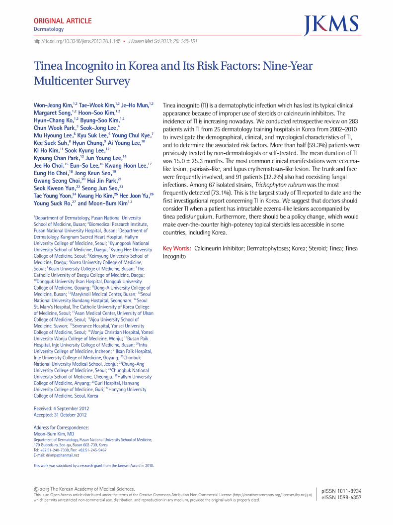
© 2013 The Korean Academy of Medical Sciences. This is an Open Access article distributed under the terms of the Creative Commons Attribution Non-Commercial License (http://creativecommons.org/licenses/by-nc/3.0) which permits unrestricted non-commercial use, distribution, and reproduction in any medium, provided the original work is properly cited. pISSN 1011-8934 eISSN 1598-6357 Tinea Incognito in Korea and Its Risk Factors: Nine-Year Multicenter Survey Tinea incognito (TI) is a dermatophytic infection which has lost its typical clinical appearance because of improper use of steroids or calcineurin inhibitors. The incidence of TI is increasing nowadays. We conducted retrospective review on 283 patients with TI from 25 dermatology training hospitals in Korea from 2002–2010 to investigate the demographical, clinical, and mycological characteristics of TI, and to determine the associated risk factors. More than half (59.3%) patients were previously treated by non-dermatologists or self-treated. The mean duration of TI was 15.0 ± 25.3 months. The most common clinical manifestations were eczema- like lesion, psoriasis-like, and lupus erythematosus-like lesion. The trunk and face were frequently involved, and 91 patients (32.2%) also had coexisting fungal infections. Among 67 isolated strains, Trichophyton rubrum was the most frequently detected (73.1%). This is the largest study of TI reported to date and the first investigational report concerning TI in Korea. We suggest that doctors should consider TI when a patient has intractable eczema-like lesions accompanied by tinea pedis/unguium. Furthermore, there should be a policy change, which would make over-the-counter high-potency topical steroids less accessible in some countries, including Korea. Key Words: Calcineurin Inhibitor; Dermatophytoses; Korea; Steroid; Tinea; Tinea Incognito Won-Jeong Kim, 1,2 Tae-Wook Kim, 1,2 Je-Ho Mun, 1,2 Margaret Song, 1,2 Hoon-Soo Kim, 1,2 Hyun-Chang Ko, 1,2 Byung-Soo Kim, 1,2 Chun Wook Park, 3 Seok-Jong Lee, 4 Mu Hyoung Lee, 5 Kyu Suk Lee, 6 Young Chul Kye, 7 Kee Suck Suh, 8 Hyun Chung, 9 Ai Young Lee, 10 Ki Ho Kim, 11 Sook Kyung Lee, 12 Kyoung Chan Park, 13 Jun Young Lee, 14 Jee Ho Choi, 15 Eun-So Lee, 16 Kwang Hoon Lee, 17 Eung Ho Choi, 18 Jong Keun Seo, 19 Gwang Seong Choi, 20 Hai Jin Park, 21 Seok Kweon Yun, 22 Seong Jun Seo, 23 Tae Young Yoon, 24 Kwang Ho Kim, 25 Hee Joon Yu, 26 Young Suck Ro, 27 and Moon-Bum Kim 1,2 1 Department of Dermatology, Pusan National University School of Medicine, Busan; 2 Biomedical Research Institute, Pusan National University Hospital, Busan; 3 Department of Dermatology, Kangnam Sacred Heart Hospital, Hallym University College of Medicine, Seoul; 4 Kyungpook National University School of Medicine, Daegu; 5 Kyung Hee University College of Medicine, Seoul; 6 Keimyung University School of Medicine, Daegu; 7 Korea University College of Medicine, Seoul; 8 Kosin University College of Medicine, Busan; 9 The Catholic University of Daegu College of Medicine, Daegu; 10 Dongguk University Ilsan Hospital, Dongguk University College of Medicine, Goyang; 11 Dong-A University College of Medicine, Busan; 12 Maryknoll Medical Center, Busan; 13 Seoul National University Bundang Hostpital, Seongnam; 14 Seoul St. Mary’s Hospital, The Catholic University of Korea College of Medicine, Seoul; 15 Asan Medical Center, University of Ulsan College of Medicine, Seoul; 16 Ajou University School of Medicine, Suwon; 17 Severance Hospital, Yonsei University College of Medicine, Seoul; 18 Wonju Christian Hospital, Yonsei University Wonju College of Medicine, Wonju; 19 Busan Paik Hospital, Inje University College of Medicine, Busan; 20 Inha University College of Medicine, Incheon; 21 Ilsan Paik Hospital, Inje University College of Medicine, Goyang; 22 Chonbuk National University Medical School, Jeonju; 23 Chung-Ang University College of Medicine, Seoul; 24 Chungbuk National University School of Medicine, Cheongju; 25 Hallym University College of Medicine, Anyang; 26 Guri Hospital, Hanyang University College of Medicine, Guri; 27 Hanyang University College of Medicine, Seoul, Korea Received: 4 September 2012 Accepted: 31 October 2012 Address for Correspondence: Moon-Bum Kim, MD Department of Dermatology, Pusan National University School of Medicine, 179 Gudeok-ro, Seo-gu, Busan 602-739, Korea Tel: +82.51-240-7338, Fax: +82.51-245-9467 E-mail: [email protected] This work was subsidized by a research grant from the Janssen Award in 2010. http://dx.doi.org/10.3346/jkms.2013.28.1.145 • J Korean Med Sci 2013; 28: 145-151 ORIGINAL ARTICLE Dermatology
-
Upload
dangkhuong -
Category
Documents
-
view
214 -
download
0
Transcript of Tinea Incognito in Korea and Its Risk Factors: Nine-Year ... · Only the cases confined to dorsum...
© 2013 The Korean Academy of Medical Sciences.This is an Open Access article distributed under the terms of the Creative Commons Attribution Non-Commercial License (http://creativecommons.org/licenses/by-nc/3.0) which permits unrestricted non-commercial use, distribution, and reproduction in any medium, provided the original work is properly cited.
pISSN 1011-8934eISSN 1598-6357
Tinea Incognito in Korea and Its Risk Factors: Nine-Year Multicenter Survey
Tinea incognito (TI) is a dermatophytic infection which has lost its typical clinical appearance because of improper use of steroids or calcineurin inhibitors. The incidence of TI is increasing nowadays. We conducted retrospective review on 283 patients with TI from 25 dermatology training hospitals in Korea from 2002–2010 to investigate the demographical, clinical, and mycological characteristics of TI, and to determine the associated risk factors. More than half (59.3%) patients were previously treated by non-dermatologists or self-treated. The mean duration of TI was 15.0 ± 25.3 months. The most common clinical manifestations were eczema-like lesion, psoriasis-like, and lupus erythematosus-like lesion. The trunk and face were frequently involved, and 91 patients (32.2%) also had coexisting fungal infections. Among 67 isolated strains, Trichophyton rubrum was the most frequently detected (73.1%). This is the largest study of TI reported to date and the first investigational report concerning TI in Korea. We suggest that doctors should consider TI when a patient has intractable eczema-like lesions accompanied by tinea pedis/unguium. Furthermore, there should be a policy change, which would make over-the-counter high-potency topical steroids less accessible in some countries, including Korea.
Key Words: Calcineurin Inhibitor; Dermatophytoses; Korea; Steroid; Tinea; Tinea Incognito
Won-Jeong Kim,1,2 Tae-Wook Kim,1,2 Je-Ho Mun,1,2 Margaret Song,1,2 Hoon-Soo Kim,1,2 Hyun-Chang Ko,1,2 Byung-Soo Kim,1,2 Chun Wook Park,3 Seok-Jong Lee,4 Mu Hyoung Lee,5 Kyu Suk Lee,6 Young Chul Kye,7 Kee Suck Suh,8 Hyun Chung,9 Ai Young Lee,10 Ki Ho Kim,11 Sook Kyung Lee,12 Kyoung Chan Park,13 Jun Young Lee,14 Jee Ho Choi,15 Eun-So Lee,16 Kwang Hoon Lee,17 Eung Ho Choi,18 Jong Keun Seo,19 Gwang Seong Choi,20 Hai Jin Park,21 Seok Kweon Yun,22 Seong Jun Seo,23 Tae Young Yoon,24 Kwang Ho Kim,25 Hee Joon Yu,26 Young Suck Ro,27 and Moon-Bum Kim1,2
1Department of Dermatology, Pusan National University School of Medicine, Busan; 2Biomedical Research Institute, Pusan National University Hospital, Busan; 3Department of Dermatology, Kangnam Sacred Heart Hospital, Hallym University College of Medicine, Seoul; 4Kyungpook National University School of Medicine, Daegu; 5Kyung Hee University College of Medicine, Seoul; 6Keimyung University School of Medicine, Daegu; 7Korea University College of Medicine, Seoul; 8Kosin University College of Medicine, Busan; 9The Catholic University of Daegu College of Medicine, Daegu; 10Dongguk University Ilsan Hospital, Dongguk University College of Medicine, Goyang; 11Dong-A University College of Medicine, Busan; 12Maryknoll Medical Center, Busan; 13Seoul National University Bundang Hostpital, Seongnam; 14Seoul St. Mary’s Hospital, The Catholic University of Korea College of Medicine, Seoul; 15Asan Medical Center, University of Ulsan College of Medicine, Seoul; 16Ajou University School of Medicine, Suwon; 17Severance Hospital, Yonsei University College of Medicine, Seoul; 18Wonju Christian Hospital, Yonsei University Wonju College of Medicine, Wonju; 19Busan Paik Hospital, Inje University College of Medicine, Busan; 20Inha University College of Medicine, Incheon; 21Ilsan Paik Hospital, Inje University College of Medicine, Goyang; 22Chonbuk National University Medical School, Jeonju; 23Chung-Ang University College of Medicine, Seoul; 24Chungbuk National University School of Medicine, Cheongju; 25Hallym University College of Medicine, Anyang; 26Guri Hospital, Hanyang University College of Medicine, Guri; 27Hanyang University College of Medicine, Seoul, Korea
Received: 4 September 2012Accepted: 31 October 2012
Address for Correspondence:Moon-Bum Kim, MDDepartment of Dermatology, Pusan National University School of Medicine, 179 Gudeok-ro, Seo-gu, Busan 602-739, Korea Tel: +82.51-240-7338, Fax: +82.51-245-9467 E-mail: [email protected]
This work was subsidized by a research grant from the Janssen Award in 2010.
http://dx.doi.org/10.3346/jkms.2013.28.1.145 • J Korean Med Sci 2013; 28: 145-151
ORIGINAL ARTICLE Dermatology
Kim W-J, et al. • Tinea Incognito in Korea and Its Risk Factors
146 http://jkms.org http://dx.doi.org/10.3346/jkms.2013.28.1.145
INTRODUCTION
Tinea incognito (TI) is the term given to a dermatophyte infec-tion, which has been modified in appearance by improper use of steroids or calcineurin inhibitors (1, 2). Since it was first de-scribed by Ive and Marks in 1968 (3), a few case reports and a number of review articles have been published on TI in English journals (1, 2, 4-6). Though typical dermatophytic infection on most skin surfaces except scalp, volar areas and nails usually present as annular lesions with erythematous scaly border and central clearing. Therefore, dermatophytic infection may be confused with other skin disorders such as granuloma annu-lare, discoid lupus erythematosus, pityriasis rosea, erythema annulare centrifugum, erythema migrans, or other dermato-logical lesions (7). Most textbooks or review articles state that mycological confirmation with laboratory testing before the start of antifungal therapy is recommended because the clinical diagnosis of fungal presence could be inaccurate (8). However, this is not easy to do in practice because of time pressure, inad-equate access to equipment, lack of experience, low reproduc-ibility, and so on (8, 9). Misdiagnosed dermatophytic disorders could be treated improperly with steroids. As some high-poten-cy topical steroids are easily accessible as over-the-counter (OTC) products and non-dermatologists can also prescribe topical steroids freely without any fungal examination, the inci-dence of TI seems to be gradually increasing in Korea (10).
However, there has been no published large-scale study on TI in Korea, as yet. For this reason, we investigated the demo-graphics and past medical histories of TI patients, and clinical and mycological characteristics of TI in Korea.
MATERIALS AND METHODS
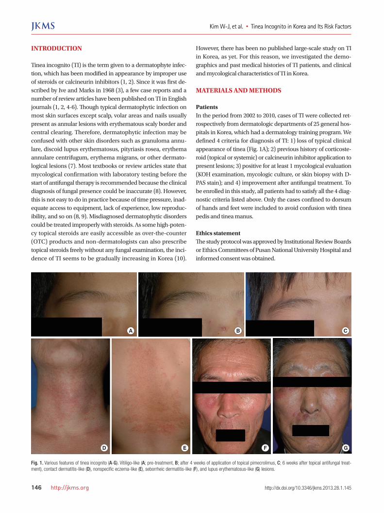
Patients In the period from 2002 to 2010, cases of TI were collected ret-rospectively from dermatologic departments of 25 general hos-pitals in Korea, which had a dermatology training program. We defined 4 criteria for diagnosis of TI: 1) loss of typical clinical appearance of tinea (Fig. 1A); 2) previous history of corticoste-roid (topical or systemic) or calcineurin inhibitor application to present lesions; 3) positive for at least 1 mycological evaluation (KOH examination, mycologic culture, or skin biopsy with D-PAS stain); and 4) improvement after antifungal treatment. To be enrolled in this study, all patients had to satisfy all the 4 diag-nostic criteria listed above. Only the cases confined to dorsum of hands and feet were included to avoid confusion with tinea pedis and tinea manus.
Ethics statementThe study protocol was approved by Institutional Review Boards or Ethics Committees of Pusan National University Hospital and informed consent was obtained.
Fig. 1. Various features of tinea incognito (A-G). Vitiligo-like (A; pre-treatment, B; after 4 weeks of application of topical pimecrolimus, C; 6 weeks after topical antifungal treat-ment), contact dermatitis-like (D), nonspecific eczema-like (E), seborrheic dermatitis-like (F), and lupus erythematosus-like (G) lesions.
A CB
D E F G
Kim W-J, et al. • Tinea Incognito in Korea and Its Risk Factors
http://jkms.org 147http://dx.doi.org/10.3346/jkms.2013.28.1.145
Demographics, past histories, and clinical characteristics Clinical data including charts and clinical photos from 25 hos-pitals were systematically and retrospectively reviewed. Demo-graphic information included age, gender, coexisting diseases, and other dermatologic diseases. Past medical histories includ-ed the duration of TI, how the patients obtained the topical ste-roid or calcineurin inhibitor, and treatment modality. After divid-ing the patients into 3 groups (dermatologist-treated, non-der-matologist-treated, and self-treated TI groups), the duration of TI and treatment modality were compared among the 3 groups. Regarding the clinical characteristics of TI, the distribution, the most likely clinical feature, and coexisting fungal infections were investigated.
Mycological dataThe KOH examination (20% potassium hydroxide) was per-formed to check for the presence of fungi. Mycological culture was performed on Sabouraud dextrose agar with chloramphe- nicol and cycloheximide. After incubation at 25°C for at least 3 weeks, dermatophytes were identified by means of gross mor-phology, light microscopy, and/or biopsy with PAS stain (11).
Statistical analysis Pearson’s chi-square test was used to compare the frequency of treatment modalities and one-way ANOVA was used to com-pare the duration of TI among dermatologist-treated, non-der-matologist-treated, and self-treated TI patients group. A P value of less than 0.05 was considered statistically significant.
RESULTS
Demographics After thorough review, 283 patients fulfilled the diagnostic cri-teria of TI in this study. The mean age was 44.0 ± 22.5 yr (range 3-94) and 125 patients (44.3%) were female. Table 1 shows the age distribution of TI patients with a slightly lower frequency of
patients with TI under 10 and over 80 yr old. Sixty-five patients (23.0%) had coexisting diseases at first clinic visit such as hyper-tension in 37 (13.1%), diabetes in 23 (8.1%), and hepatitis in 7 (2.5%). Five patients had underlying malignancy (1.8%), 2 patients suffered from angina, and 2 patients had asthma. In addition, 1 patient had adrenal insufficiency, 1 patient had myasthenia gravis, 1 had depression, and 1 had epilepsy. Sixteen patients (5.7%) had coexisting dermatologic diseases including 5 pa-tients with atopic dermatitis (1.8%), 4 patients with psoriasis (1.4%), 3 with systemic lupus erythematosus (1.1%), and 2 with
Table 1. Demographics and past histories of 283 cases of tinea incognito in Korea during 2002-2010
Parameters No. (%)
No. of patients 283 (100.0)Male: Female, No. (%) 158:125 (55.7:44.3)Mean age (yr) 44.6 ± 22.3 (range 3-94)Age distribution, No. (%) 0-10 11-20 21-30 31-40 41-50 51-60 61-70 71-80 81-90 > 90
24 (8.5)25 (8.8)
31 (11.0) 38 (13.4) 40 (14.1) 41 (14.5) 44 (15.5) 33 (11.7) 6 (2.1) 1 (0.4)
Coexisting disease, No. (%) Hypertension Diabetes Hepatitis Malignancy Angina Asthma Others
65 (23.0) 37 (13.1)23 (8.1) 7 (2.5) 5 (1.8) 2 (0.7) 2 (0.7) 4 (1.4)
Coexisting dermatologic diseases, No. (%) Atopic dermatitis Psoriasis Systemic lupus erythematosus Seborrheic dermatitis Others (rosacea, bullous pemphigoid)
16 (5.7) 5 (1.8) 4 (1.4) 3 (1.1) 2 (0.7) 2 (0.7)
Table 2. Mean duration of the disease and previous treatment modalities according to past physician’s specialty
Disease status Dermatologists Non-dermatologists Self-treatment † Total P value*
Mean duration of the disease (mo± SD) 16.4 ± 25.8 15.7 ± 28.1 9.0 ± 11.1 15.0 ± 25.3 0.234Treatment modalities, No. (%) Topical steroid only Systemic steroid only Topical and systemic steroid Topical steroid and topical calcineurin inhibitor Topical steroid and systemic antibiotics Steroid intralesional injection Topical steroid and steroid intralesional injection Systemic steroid and topical calcineurin inhibitor Systemic steroid and topical antibiotics Topical calcineurin inhibitor only
90 (31.8)
- 9 (3.2) 8 (2.8) 2 (0.7) 1 (0.4) 1 (0.4) 1 (0.4) 1 (0.4) 2 (0.7)
112 (39.6)
- 9 (3.2) 1 (0.4) 2 (0.7)
-----
44 (15.5)
---------
246 (86.9)
-18 (6.4) 9 (3.2) 4 (1.4) 1 (0.4) 1 (0.4) 1 (0.4) 1 (0.4) 2 (0.7)
< 0.001
-0.1750.0100.6920.2240.4740.4740.4740.224
Total, N (%) 115 (40.6) 124 (43.8) 44 (15.5) 283 (100.0)
*P value < 0.05 considered statistically significant; using one-way ANOVA test in mean duration and Pearson’s chi-square test in treatment modalities; †For children younger than 12, self-treatment group also include treatment by parents or others.
Kim W-J, et al. • Tinea Incognito in Korea and Its Risk Factors
148 http://jkms.org http://dx.doi.org/10.3346/jkms.2013.28.1.145
seborrheic dermatitis (0.7%). There was 1 patient with rosacea, and 1 patient with bullous pemphigoid.
Past medical historiesThe mean duration of TI in the study patients was 15.0 ± 25.3 months. While mean duration of self-treated TI patients was 9.0 ± 11.1 months, that of TI patients treated by dermatologists and non-dermatologists was 16.4 ± 25.8 and 15.7 ± 28.1 months, respectively. There was no statistical significance among the 3 groups (P = 0.234) (Table 2). Before coming to the teaching hospital, 40.6% of TI patients received treatment from a dermatologist, 43.8% from non-der-matologists, and another 15.5% were self-treated. While all of self-treated patients used topical steroids only, people treated by dermatologists or non-dermatologists used various treatment modalities such as topical/systemic steroids, topical/systemic antibiotics, topical calcineurin inhibitor, steroid intralesional
injection, or a combination of aforementioned agents. Overall, most of TI patients were treated with topical steroids only (86.9%), and other treatment modalities included topical and systemic steroids (6.4%), topical steroid and topical calcineurin inhibitor (1.4%), and topical calcineurin inhibitor (0.7%), etc. There were no significant differences in treatment modalities according to past physician’s specialty (P > 0.05).
Clinical characteristics Overall, the trunk (30.4%) is the most commonly affected area of TI followed by the face (24.4%), foot (13.8%), multiple involve-ments (13.8%), the groin (9.9%), and hand (7.8%) (Table 3). The clinical features were variable, but regardless of distribution, over more than three-quarters of all study patients showed ec-zema-like (82.0%) lesions which included nonspecific eczema, contact dermatitis, seborrheic dermatitis, and atopic dermatitis. Less often, TI mimicked psoriasis (6.0%), lupus erythematosus
Table 3. Clinical and mycological characteristics of 283 cases of tinea incognito in Korea during 2002-2010
TI distribution Face Trunk Groin Hand* Foot* Multiple Total
Clinical manifestation, No. (%) Eczema-like Nonspecific eczema Contact dermatitis Atopic dermatitis Seborrheic dermatitis Diaper dermatitis Intertrigo Nummular dermatitis Stasis dermatitis Psoriasis-like Lupus erythematosus-like Impetigo-like Urticaria-like Folliculitis-like Lichen simplex chronicus-like Vitiligo-like Xerosis-like Others
69 (24.4)53 (76.8)30 (43.5)16 (23.2)3 (4.3)3 (4.3)
--
1 (1.4)-
1 (1.4)6 (8.7)2 (2.9)1 (1.4)1 (1.4)
-2 (2.9)
-3 (4.3)
86 (30.4)68 (79.1)54 (62.8)8 (9.3)1 (1.2)1 (1.2)2 (2.3)1 (1.2)
-1 (1.2)9 (10.5)
-2 (2.3)1 (1.2)
-1 (1.2)
--
5 (5.8)
28 (9.9)25 (89.3)25 (89.3)
-------
1 (3.6)--
1 (3.6)----
1 (3.6)
22 (7.8)22 (100.0)19 (86.4)3 (13.6)
---------------
39 (13.8)37 (94.9)35 (89.7)
1 (2.6)1 (2.6)
------------
2 (5.1)-
39 (13.8)27 (69.2)23 (59.0)4 (10.3)
------
6 (15.4)1 (2.6)--
1 (2.6)1 (2.6)
--
3 (7.7)
283 (100.0)232 (82.0)186 (65.7)32 (11.3)5 (1.8)4 (1.4)2 (0.7)1 (0.4)1 (0.4)1 (0.4)
17 (6.0)7 (2.5)4 (1.4)3 (1.1)2 (0.7)2 (0.7)2 (0.7)2 (0.7)
12 (4.2)The percentage of children (age < 12, N = 28)
8 (28.6) 10 (35.7) 1 (3.6) 2 (7.1) 4 (14.3) 3 (10.7) 28 (100.0)
Combined fungal disease‡
T. pedis T. unguium T. pedis et unguium
12 (4.2)4 (33.3)1 (8.3)7 (58.3)
24 (8.5)16 (66.7)5 (20.8)3 (12.5)
11 (3.9)9 (81.8)
-2 (18.2)
6 (2.1)3 (50.0)3 (50.0)
-
19 (6.7)-
12 (63.2)7 (36.8)§
19 (6.7)7 (36.8)8 (42.1)4 (21.1)
91 (32.2)39 (42.9)29 (31.9)23 (25.3)
KOH†
Positive Negative
69 (24.4)59 (85.5)10 (14.5)
86 (30.4)80 (93.0)6 (7.0)
28 (9.9)28 (100.0)
-
22 (7.8)20 (90.9)2 (9.1)
39 (13.8)38 (97.4)
1 (2.6)
39 (13.8)35 (89.7)4 (10.3)
283 (100.0)260 (91.9)23 (8.1)
Biopsy with D-PAS stain Positive (fungal hyphae or spores) Negative
21 (7.4)19 (90.5)2 (9.5)
15 (5.3)12 (80.0)3 (20.0)
1 (0.4)1 (100.0)
-
3 (1.1)2 (66.7)1 (33.3)
1 (0.4)1 (100.0)
-
8 (2.8)7 (87.5)1 (12.5)
49 (17.3)42 (85.7)7 (14.3)
Culture Trichophyton rubrum Trichophyton mentagrophytes Trichophyton tonsurans Trichophyton verrucosum Microsporum canis Microsporum gypseum
19 (6.7)10 (52.6)3 (15.8)1 (5.3)
-3 (15.8)2 (10.5)
15 (5.3)11 (73.3)
-1 (6.7)1 (6.7)2 (13.3)
-
5 (1.8)4 (80.0)
--
1 (20.0)--
3 (1.1)3 (100.0)
-----
6 (2.1)5 (83.3)1 (16.7)
----
19 (6.7)16 (84.2)2 (10.5)
--
1 (5.3)-
67 (23.7)49 (73.1)6 (9.0)2 (3.0)2 (3.0)6 (9.0)2 (3.0)
*TI involves hand and foot but confined to dorsal aspects; †KOH, potassium hydroxide examination; ‡Combined fungal disease: fungal disease which involves distant areas with present TI; §Tinea unguium with tinea corporis.
Kim W-J, et al. • Tinea Incognito in Korea and Its Risk Factors
http://jkms.org 149http://dx.doi.org/10.3346/jkms.2013.28.1.145
(2.5%), impetigo (1.4%), urticaria (1.2%), folliculitis (0.7%), and other dermatological lesions (Table 3). According to the anatom-ical distribution, facial TI presented as eczema-like (76.8%), lu-pus erythematosus-like (8.7%), impetigo-like (2.9%), and vitili-go-like (2.9%) lesions. Trunk TI presented as eczema-like (79.1%) and psoriasis-like (10.5%) lesions, and almost all of groin, hand, and foot TI resembled eczema. When TI involved multiple sites, it appeared similar to eczema (69.2%), psoriasis (15.4%), follic-ulitis (2.6%), and other dermatological lesions (Table 3). In chil-dren, TI was most likely to be found in the facial area (11.6%), and the trunk (11.6%), and least likely to be found in the groin (3.6%). In 91 cases (32.2%), other fungal diseases such as tinea pedis (42.9%), tinea unguium (31.9%), tinea pedis et unguium, or tin-ea unguium/tinea corporis (25.3%) were diagnosed apart from TI sites (Table 3). According to anatomical distribution, TI of the trunk, groin, or hand was commonly seen with tinea pedis (> 50.0%), TI involving foot or multiple areas usually accompa-nied tinea unguium, and facial TI was strongly associated with tinea pedis et unguium.
Mycological dataDirect microscopic examination was performed in all cases and 260 cases (91.9%) were positive. Of 49 biopsied specimens, 42 (85.7%) showed fungal hyphae and/or spores by D-PAS stain. Sixty-seven cases (23.7%) were cultured in our study and Tricho-phyton rubrum was the most frequently detected dermatophyte (49/67, 73.%), regardless of TI distribution. Trichophyton men-tagrophytes (6/67, 9.0%) and Microsporum canis (6/67, 9.0%) were the second-most frequently detected causative agents, and T. tonsurans, T. verrucosum and M. gypseum were also isolated in a few cases. While only 1 or 2 species of dermatophytes were found in groin, hand, and foot TI, various kinds of fungi were identified in face or trunk TI (Table 3).
DISCUSSION
Tinea incognito had been defined as tinea modified by the im-proper use of systemic or topical corticosteroids. However, as
the use of topical calcineurin inhibitors has been increasing gradually in many dermatologic diseases such as atopic derma-titis, seborrheic dermatitis, intertriginous psoriasis, contact der-matitis and other dermatological lesions (12), the number of cases of modified tinea has also increased (7, 13, 14). Thus, we propose that TI be defined as certain dermatophytoses which have lost their usual clinical manifestation because of erroneous use of systemic/topical corticosteroids or topical calcineurin inhibitor, as in 1 recent article (2). In addition, we think that TI, which involves the hand or the foot, should be confined to the dorsal surface, because tinea pedis and tinea manus cannot be definitively differentiated from TI involving the palm or the sole. It has been suggested that the use of immunosuppressants de-creases the fungus-induced local inflammation, and this may al-low the fungus to grow slowly with less erythema or scaling caus-ing a “modification” of the typical manifestation of tinea (7). While TI seems to be common in dermatology practices cur-rently, only a few numbers of large scale studies have been re-ported (1, 2, 4). These studies were done in Italy, Spain, and Iran. Our study in Korea was designed to be the largest scale study on TI. While previous case reports of TI in Korean literature (10, 15-21) (Table 4) showed female predominance, this study showed relatively equal gender distribution and relatively uniform age distribution (mean: 44.0 ± 22.5 yr) were found except for patients over 80 yr. A recent article regarding TI in Italy (1) also reported equal gender distribution and similar mean age (42 yr), and an-other article in Iran (4) also revealed equal gender distribution with slightly younger mean age (32.6 yr). Based on these data, we can postulate that TI is common in middle-aged persons with little difference in gender. Moreover, 65 (23.0%) patients in our study had coexisting non-dermatologic diseases such as hypertension, diabetes, hepatitis, malignancy, and so on, and 16 (5.7%) patients had coexisting dermatologic disorders requir-ing systemic steroids or other immunosuppressants. This was lower than the previous Italian report in that 40% of patients with TI had non-dermatological pathologies which required treatment with systemic steroids (1). Though the percentage of the patients in this study who received immunosuppressive therapy was lower than in the Italian report, the possibility of TI
Table 4. Previous reports of tinea incognito in Korean literatures
Case Report S/A Clinical manifestation Site Previous treatment Dermatophyte isolated
1 Yang et al. (15) F/47 Eczema-like Face, trunk, extremities Topical steroids T. mentagrophytes2 Kang et al. (16) F/64 Eczema-like Face Topical steroids T. mentagrophytes3 Kim et al. (17) F/70 Contact dermatitis-like Face, scalp Topical steroids T. rubrum4 Choi et al. (10) M/8 Atopic dermatitis-like Face Topical calcineurin inhibitor T. mentagrophytes5 Han et al. (18) M/49 Atopic dermatitis-like Face Topical and systemic steroid T. rubrum6 Park et al. (19) F/40 Eczema-like Face Topical steroid and
systemic antibioticsT. mentagrophytes
7 Hwang et al. (20) F/15 Eczema-like Leg Topical steroid and topical calcineurin inhibitor
T. rubrum
8 Lee et al. (21) F/23 Insect bite-like Arm Topical steroids T. mentagrophytes
S/A, sex and age (years).
Kim W-J, et al. • Tinea Incognito in Korea and Its Risk Factors
150 http://jkms.org http://dx.doi.org/10.3346/jkms.2013.28.1.145
should be kept in mind whenever the patient with skin lesions is on immunosuppressant medications. There have been no published data regarding TI according to past treating physician’s specialty or treatment modalities, as yet. Based on our study, over half of the patients were either treated by non-dermatologists (124/283, 43.8%) or self-treated (44/283, 15.5%). TI was thought to be associated with easy ac-cess to high-potency OTC topical steroids such as betametha-sone valerate by patients and with lack of understanding of tin-ea by non-dermatologists. Therefore, there should be swift poli-cy changes to limit OTC access of high-potency steroids to pa-tients in Korea. This would limit inappropriate tinea treatment by patients. Furthermore, to reduce the number of cases of TI caused by non-dermatologists, education regarding skin dis-eases including fungal infections could be provided by Korean Dermatologic Associations. Surprisingly, about 40% of the patients were treated by der-matologists in this study. Even though tinea can mimic many other skin disorders and there could be selection bias, this ratio seems to be too high. This may mean a lack of mycological eval-uation and carelessness of dermatologists when diagnosing tin-ea infection. It is important for dermatologists to consider fun-gal infection in the differential diagnosis of skin disorders, and increase the use of laboratory tests for mycological evaluation. In practice, the medical cost for mycological examinations is very low in Korea. However, the patient load is high and doctors are pressed for time. This could be the prime reason for misdi-agnosis of fungal infections (22). Therefore, we think that if phy-sicians were better paid for mycological evaluations there might be more active mycological examinations and fewer misdiag-noses of fungal infections. On the aspect of distribution, the trunk was the most com-monly involved site of TI and the face was another commonly involved area, as reported in other original articles (1, 4) and previous Korean literatures. Another recent study regarding 54 childhood TI cases also reported similar results with highest incidence in the trunk and face (2), and our study backed it up with the same results. From these findings, we can postulate that the most common sites of TI are trunk and face regardless of age. The clinical features of TI were reported to be variable, and the most prevalent features seen are eczema-like disorders such as nonspecific eczema, contact dermatitis, and atopic dermati-tis (1, 4, 7), and previous Korean reports about TI also in accor-dance with it. Similarly, 232 TI patients in this study showed quite various clinical features such as eczema-like, psoriasis-like, lupus erythematosus-like, and etc. Specifically, nearly all cases of hand and foot TI showed eczema-like features. There-fore, when dealing with recalcitrant eczematous lesions on the hand or foot, mycological examination should always be con-sidered. Compared to TI of groin, and hand and foot, where the
eczema-like features were quite high, TI of the face, trunk, or multiple areas showed more variable features. Therefore, not only eczema-like lesions but also other recalcitrant skin mani-festations resembling psoriasis, lupus erythematosus, impetigo, urticaria, etc., should also be carefully evaluated to rule out TI especially when skin diseases involve the face, the trunk, or mul-tiple areas (Table 5). Moreover, one-third of our study population (32.2%) had combined fungal diseases, which involved distant areas from present TI, and most of them had dermatophytic infection on their feet regardless of affected areas, including tinea pedis and tinea unguium. Therefore, in patients with refractory skin dis-ease especially resembling eczema and also with concomitant tinea pedis or tinea unguium, TI should be ruled out because these coexisting fungal infections could be an autoinoculation source of superficial dermatophytic infection in another body part at any time. As many previous studies confirmed (1, 23-25), Trichophyton rubrum (T. rubrum) was also the most frequently identified der-matophyte. T. rubrum is one of the anthropophilic dermato-phytes and the most common pathogen in tinea corporis, tinea cruris, tinea manus, tinea pedis, and tinea unguium (26). Since TI affecting the trunk, groin, hands, and feet accounted for al-most 60% of T1 in our study, it would not be surprising that T. rubrum was the most commonly isolated dermatophyte. More-over, the high prevalence of combined tinea pedis and tinea un-guium might also have contributed to the high isolation rate of T. rubrum. Anthropophilic dermatophytes have adapted to hu-mans and elicit a mild to non-inflammatory host response unlike zoophilic and geophilic infections (26). This mild inflammatory response might be the cause of the long duration of TI because topical corticosteroids or topical calcineurin inhibitor could al-leviate the inflammation, which could be the main mechanism of disguising the typical manifestation of tinea. In summary, our research was the largest study of TI in Korea to date. We investigated the characteristics of TI according to the primary physician’s specialty though the clinical and myco-logical results were similar to previous studies. From this study, we can suggest that long-lasting erythematous scaly skin lesions unresponsive to steroids or calcineurin inhibitor as the most important risk factors of TI. Not only truncal or facial involve-ment, but also combined tinea pedis/unguium or the history of immunosuppressant treatment could also be a good clue in di-agnostic approach of TI. Moreover, we can suggest several things
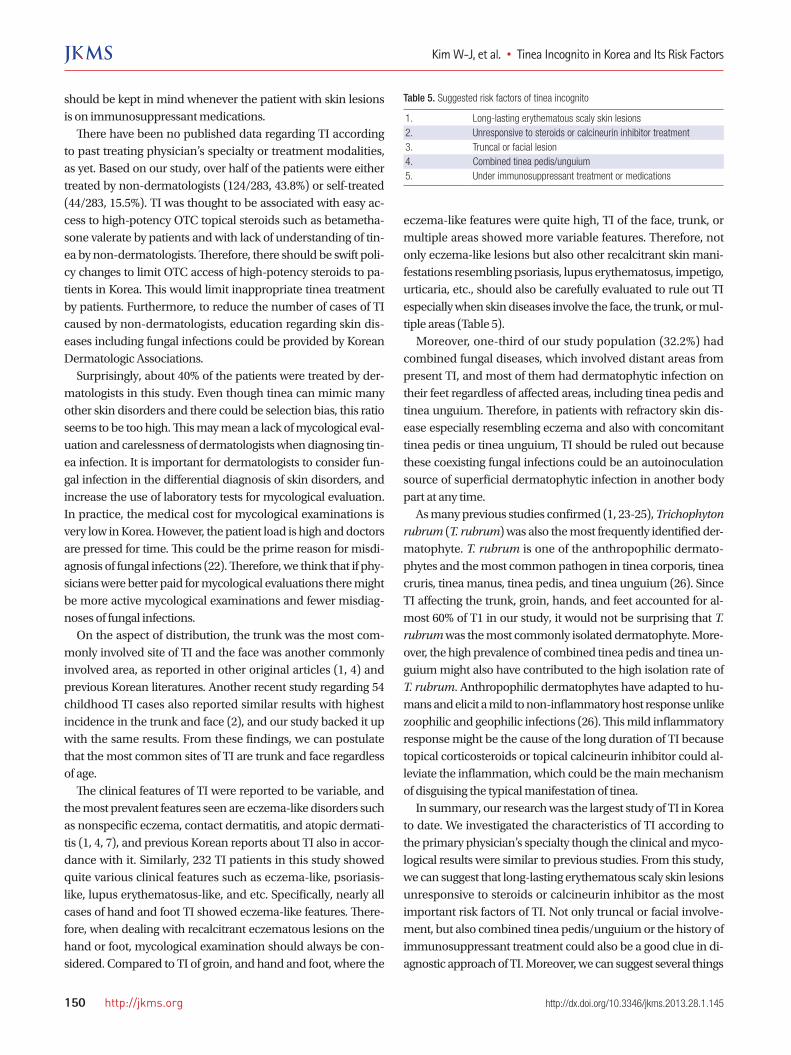
Table 5. Suggested risk factors of tinea incognito
1. Long-lasting erythematous scaly skin lesions2. Unresponsive to steroids or calcineurin inhibitor treatment3. Truncal or facial lesion4. Combined tinea pedis/unguium5. Under immunosuppressant treatment or medications
Kim W-J, et al. • Tinea Incognito in Korea and Its Risk Factors
http://jkms.org 151http://dx.doi.org/10.3346/jkms.2013.28.1.145
to reduce the incidence of TI based on our results. First, reform of OTC sales system of high-potency topical steroids is needed so that they are not as easily available to the public in some coun-tries including Korea. Second, non-dermatologists need to be informed and educated that superficial dermatophytic infection could appear in a variety of forms. Care by experienced derma-tologists could also be needed especially when dealing with long-lasting erythematous scaly skin lesions, which have prov-en to be unresponsive to steroids or calcineurin inhibitor treat-ment. Finally, we recommend dermatologists not to neglect TI as a possibility in cases of recalcitrant variable skin lesions, not hesitating to do active mycological examinations, which would give them some critical clues in diagnosis of TI, and doing care-ful clinical examinations when finding combined tinea pedis or tinea unguium.
ACKNOWLEDGMENTS
The authors have no conflicts of interest to disclose.
REFERENCES
1. Romano C, Maritati E, Gianni C. Tinea incognito in Italy: a 15-year sur-
vey. Mycoses 2006; 49: 383-7.
2. del Boz J, Crespo V, Rivas-Ruiz F, de Troya M. Tinea incognito in chil-
dren: 54 cases. Mycoses 2011; 54: 254-8.
3. Ive FA, Marks R. Tinea incognito. Br Med J 1968; 3: 149-52.
4. Ansar A, Farshchian M, Nazeri H, Ghiasian SA. Clinico-epidemiological
and mycological aspects of tinea incognito in Iran: a 16-year study. Med
Mycol J 2011; 52: 25-32.
5. Arenas R, Moreno-Coutino G, Vera L, Welsh O. Tinea incognito. Clin
Dermatol 2010; 28: 137-9.
6. Lange M, Jasiel-Walikowska E, Nowicki R, Bykowska B. Tinea incognito
due to Trichophyton mentagrophytes. Mycoses 2009; 53: 455-7.
7. Rallis E, Koumantaki-Mathioudaki E. Pimecrolimus induced tinea incog-
nito masquerading as intertriginous psoriasis. Mycoses 2008; 51: 71-3.
8. Garcia-Doval I, Cabo F, Monteagudo B, Alvarez J, Ginarte M, Rodriguez-
Alvarez MX, Abalde MT, Fernandez ML, Allegue F, Perez-Perez L, Flo-
rez A, Cabanillas M, Peon G, Zulaica A, Del Pozo J, Gomez-Centeno P.
Clinical diagnosis of toenail onychomycosis is possible in some patients:
cross-sectional diagnostic study and development of a diagnostic rule. Br
J Dermatol 2010; 163: 743-51.
9. Rajpar SF, Abdullah A. Management of onychomycosis and awareness
of guidelines among dermatologists. Br J Dermatol 2006; 155: 1080-2.
10. Choi YL, Kim JA, Rho NK, Lee DY, Lee JH, Yang JM, Lee ES, Kim WS. A
case of tinea incognito induced by 1% pimecrolimus (Elidel) cream. Ko-
rean J Dermatol 2006; 44: 731-3.
11. Hsiao YP, Lin HS, Wu TW, Shih HC, Wei SJ, Wang YL, Lin KL, Chiou HL,
Yang JH. A comparative study of KOH test, PAS staining and fungal cul-
ture in diagnosis of onychomycosis in Taiwan. J Dermatol Sci 2007; 45:
138-40.
12. Wollina U, Hansel G, Koch A, Abdel-Naser MB. Topical pimecrolimus
for skin disease other than atopic dermatitis. Expert Opin Pharmacother
2006; 7: 1967-75.
13. Crawford KM, Bostrom P, Russ B, Boyd J. Pimecrolimus-induced tinea
incognito. Skinmed 2004; 3: 352-3.
14. Siddaiah N, Erickson Q, Miller G, Elston DM. Tacrolimus-induced tinea
incognito. Cutis 2004; 73: 237-8.
15. Yang CW, Lee BG, Lee MH, Kim NI. A case of tinea incognito. Korean J
Dermatol 1989; 27: 79-82.
16. Kang HY, Son HC, Lim YS, Cho YW, Han JY. A case of tinea incognito on
the face due to Trichophyton mentagrophytes. Korean J Dermatol 2000;
38: 1124-6.
17. Kim KJ, Jee MS, Choi JH, Sung KJ, Moon KC, Koh JK. A case of tinea in-
cognito presented as folliculitis. Korean J Dermatol 2001; 39: 1328-30.
18. Han TY, Rho YK, Seo SJ, Hong CK, Song KY. A case of tinea incognito
presented like furunculosis. Korean J Med Mycol 2008; 13: 138-41.
19. Park SB, Lee YW, Park EJ, Kwon IH, Kim KH, Kim KJ. A case of tinea fa-
ciei caused by Trichophyton mentagrophytes with atypical presentation.
Korean J Med Mycol 2010; 15: 170-4.
20. Hwang SM, Kim DM, Suh MK, Ha GY, Kim JR. Eczema-like tinea incog-
nito occurring leg. Korean J Med Mycol 2011; 16: 51-5.
21. Lee JS, Cho YS, Song KH, Hwang SR, Park J, Yun SK, Kim HU. Tinea in-
cognito with changes in clinical feature related to antifungal treatment.
Korean J Med Mycol 2011; 16: 118-23.
22. Kim WJ, Song M, Kim HS, Kim SH, Ko HC, Kim BS, Kim MB. Various
nail disorders misdiagnosed and treated as onychomycosis. Korean J
Dermatol 2011; 49: 408-14.
23. Szepietowski JC, Matusiak L. Trichophyton rubrum autoinoculation
from infected nails is not such a rare phenomenon. Mycoses 2008; 51:
345-6.
24. Nenoff P, Mugge C, Herrmann J, Keller U. Tinea faciei incognito due to
Trichophyton rubrum as a result of autoinoculation from onychomyco-
sis. Mycoses 2007; 50 Suppl 2: 20-5.
25. Serarslan G. Pustular psoriasis-like tinea incognito due to Trichophyton
rubrum. Mycoses 2007; 50: 523-4.
26. Schieke SM, Garg A. Fungal disease. In: Fitzpatrick’s dermatology in
general medicine, 8th ed. NewYork:McGraw-Hill 2012: 2277-97.