Timing of pentoxifylline treatment determines its protective effect on diabetes development in the...
8
Timing of pentoxifylline treatment determines its protective effect on diabetes development in the Bio Breeding rat Jeroen Visser, Herman Groen, Flip Klatter, Jan Rozing * Department of Cell Biology, Immunology Section,Faculty Medical Sciences, Groningen University, A. Deusinglaan 1, 9713 AV Groningen, The Netherlands Received 5 November 2001; received in revised form 10 April 2002; accepted 16 April 2002 Abstract Diabetes-prone Bio Breeding (DP-BB) rats spontaneously develop diabetes between 60 and 120 days of age. Diabetes-resistant (DR)-BB rats can be induced to develop diabetes by poly(I:C) and anti-RT6. Here, we studied the effect of pentoxifylline, a potent anti-inflammatory agent, on diabetes development in both BB rat models of insulin-dependent diabetes mellitus and investigated whether these effects were related to differential modulation of tumour necrosis factor (TNF)-a and interleukin-10. When DP-BB rats received pentoxifylline from day 60 onwards, diabetes development was delayed and reduced. The other treatment protocols had no effect. In DR-BB rats, pentoxifylline treatment resulted only in a delay of diabetes development. In both BB rat models, in vivo pentoxifylline treatment potently suppressed TNF- a, but only moderately affected interleukin-10 production in vitro. These results show that timing of pentoxifylline treatment determines its protective effect on diabetes development in DP-BB rats. The observed pentoxifylline-induced increase of the interleukin-10/TNF-a ratio might be a mechanism for protection or delay of the diabetes development. D 2002 Elsevier Science B.V. All rights reserved. Keywords: Diabetes; Pentoxifylline; Interleukin-10; TNF-a; BB, rat 1. Introduction Diabetes-prone (DP)-BB rats spontaneously develop diabetes between 60 and 120 days of age (Mordes et al., 2001). Diabetes-resistant (DR)-BB rats can be induced to develop diabetes by depleting them of their regulatory RT6 + T cells and injection of low dose poly(I:C) (Greiner et al., 1987a,b). Likewise, protection of DP-BB rats against the onset of diabetes can be achieved by injecting them with these regulatory RT6 + T cells (Burstein et al., 1989). In the DP-BB rat model of diabetes, an early time interval has been identified in which diabetes can be prevented. For example, thymectomy of DP-BB rats around 30 days of age prevents insulin-dependent diabetes mellitus (Mordes et al., 2001), whereas thymectomy at day 60 has no effect (Mordes et al., 2001; Like et al., 1982). Moreover, injection with RT6 + T cells from 30 to 60 days of age prevents diabetes in DP-BB rats, while injection from 60 to 100 days does not influence the disease (Burstein et al., 1989). These findings indicate that the period between 30 and 60 days of age is critical for the development of diabetes in DP-BB rats. DP-BB rats develop diabetes in an age range of 60– 120 days. This stage of disease development is charac- terised by severe insulitis (Mordes et al., 2001). The islets of Langerhans are infiltrated by macrophages, Natural Killer (NK) cells and cytotoxic T cells leading to destruc- tion of the h-cells (Mordes et al., 2001). Cytokines, particularly the proinflammatory ones such as tumour necrosis factor-alpha (TNF-a) and interleukin-1, are cyto- toxic for the h-cells and induce the inflammatory cascade leading to h-cell destruction (Rabinovitch, 1998). Several reports show that increased expression of proinflammatory cytokines such as interleukin-12, TNF-a, interleukin-1 and interferon-g is associated with h-cell destructive insulitis, whereas non-destructive insulitis is more associ- ated with increased expression of anti-inflammatory type 2 cytokines such as interleukin-10 and interleukin-4 and the type 3 cytokine transforming growth factor (TGF)-h (Rabinovitch, 1998; Rabinovitch et al., 1996; Kolb et al., 1996). 0014-2999/02/$ - see front matter D 2002 Elsevier Science B.V. All rights reserved. PII:S0014-2999(02)01625-4 * Corresponding author. Tel.: +31-50-3632530 (2522); fax: +31-50- 3632512. E-mail address: [email protected] (J. Rozing). www.elsevier.com/locate/ejphar European Journal of Pharmacology 445 (2002) 133 – 140
-
Upload
jeroen-visser -
Category
Documents
-
view
213 -
download
1
Transcript of Timing of pentoxifylline treatment determines its protective effect on diabetes development in the...

Timing of pentoxifylline treatment determines its protective effect on
diabetes development in the Bio Breeding rat
Jeroen Visser, Herman Groen, Flip Klatter, Jan Rozing *
Department of Cell Biology, Immunology Section, Faculty Medical Sciences, Groningen University, A. Deusinglaan 1, 9713 AV Groningen, The Netherlands
Received 5 November 2001; received in revised form 10 April 2002; accepted 16 April 2002
Abstract
Diabetes-prone Bio Breeding (DP-BB) rats spontaneously develop diabetes between 60 and 120 days of age. Diabetes-resistant (DR)-BB
rats can be induced to develop diabetes by poly(I:C) and anti-RT6. Here, we studied the effect of pentoxifylline, a potent anti-inflammatory
agent, on diabetes development in both BB rat models of insulin-dependent diabetes mellitus and investigated whether these effects were
related to differential modulation of tumour necrosis factor (TNF)-a and interleukin-10. When DP-BB rats received pentoxifylline from day
60 onwards, diabetes development was delayed and reduced. The other treatment protocols had no effect. In DR-BB rats, pentoxifylline
treatment resulted only in a delay of diabetes development. In both BB rat models, in vivo pentoxifylline treatment potently suppressed TNF-
a, but only moderately affected interleukin-10 production in vitro. These results show that timing of pentoxifylline treatment determines its
protective effect on diabetes development in DP-BB rats. The observed pentoxifylline-induced increase of the interleukin-10/TNF-a ratio
might be a mechanism for protection or delay of the diabetes development. D 2002 Elsevier Science B.V. All rights reserved.
Keywords: Diabetes; Pentoxifylline; Interleukin-10; TNF-a; BB, rat
1. Introduction
Diabetes-prone (DP)-BB rats spontaneously develop
diabetes between 60 and 120 days of age (Mordes et
al., 2001). Diabetes-resistant (DR)-BB rats can be induced
to develop diabetes by depleting them of their regulatory
RT6 + T cells and injection of low dose poly(I:C) (Greiner
et al., 1987a,b). Likewise, protection of DP-BB rats
against the onset of diabetes can be achieved by injecting
them with these regulatory RT6 + T cells (Burstein et al.,
1989).
In the DP-BB rat model of diabetes, an early time
interval has been identified in which diabetes can be
prevented. For example, thymectomy of DP-BB rats around
30 days of age prevents insulin-dependent diabetes mellitus
(Mordes et al., 2001), whereas thymectomy at day 60 has no
effect (Mordes et al., 2001; Like et al., 1982). Moreover,
injection with RT6 + T cells from 30 to 60 days of age
prevents diabetes in DP-BB rats, while injection from 60 to
100 days does not influence the disease (Burstein et al.,
1989). These findings indicate that the period between 30
and 60 days of age is critical for the development of diabetes
in DP-BB rats.
DP-BB rats develop diabetes in an age range of 60–
120 days. This stage of disease development is charac-
terised by severe insulitis (Mordes et al., 2001). The islets
of Langerhans are infiltrated by macrophages, Natural
Killer (NK) cells and cytotoxic T cells leading to destruc-
tion of the h-cells (Mordes et al., 2001). Cytokines,
particularly the proinflammatory ones such as tumour
necrosis factor-alpha (TNF-a) and interleukin-1, are cyto-
toxic for the h-cells and induce the inflammatory cascade
leading to h-cell destruction (Rabinovitch, 1998). Several
reports show that increased expression of proinflammatory
cytokines such as interleukin-12, TNF-a, interleukin-1
and interferon-g is associated with h-cell destructive
insulitis, whereas non-destructive insulitis is more associ-
ated with increased expression of anti-inflammatory type
2 cytokines such as interleukin-10 and interleukin-4 and
the type 3 cytokine transforming growth factor (TGF)-h(Rabinovitch, 1998; Rabinovitch et al., 1996; Kolb et al.,
1996).
0014-2999/02/$ - see front matter D 2002 Elsevier Science B.V. All rights reserved.
PII: S0014 -2999 (02 )01625 -4
* Corresponding author. Tel.: +31-50-3632530 (2522); fax: +31-50-
3632512.
E-mail address: [email protected] (J. Rozing).
www.elsevier.com/locate/ejphar
European Journal of Pharmacology 445 (2002) 133–140
Immunosuppressive drugs like corticosteroids, cyclo-
sporin and phosphodiesterase inhibitors are used for the
treatment of various autoimmune diseases like rheumatoid
arthritis, inflammatory bowel disease and multiple sclerosis
(Bach 1993). Moreover, these drugs are also capable of
preventing the development of experimental autoimmune
diseases such as experimental autoimmune encephalomye-
litis (EAE) (MacPhee et al., 1989), streptococcal cell wall-
induced arthritis (Sternberg et al., 1989) and bacteria-
induced intestinal inflammation (Herfarth et al., 1998).
Suppression of inflammatory cytokines such as TNF-a,
interferon-g and interleukin-12 is one of the main mecha-
nisms for the protective effect of these drugs in autoim-
munity (Bach, 1993).
The phosphodiesterase inhibitor pentoxifylline is a potent
suppressor of TNF-a and other proinflammatory cytokines
both in vivo and in vitro (Tilg et al., 1993; Neuner et al.,
1994; Bernard et al., 1995; Staak et al., 1997; Voisin et al.,
1998). On the other hand, pentoxifylline is able to enhance
the production of the anti-inflammatory cytokine interleu-
kin-10 in vitro (Platzer et al., 1995). Therefore, selective
modulation of these cytokines by pentoxifylline might be
one of the mechanisms in the protective effects of pentox-
ifylline in the induction of experimental autoimmune dis-
eases like EAE (Rott et al., 1993).
In this study, we investigated the effect of pentoxifylline
on diabetes development in the BB rat. In this study, we
performed tests to determine in which phase of disease
development treatment with pentoxifylline can prevent or
delay the development of diabetes and whether this
involves a selective regulation of TNF-a and interleukin-
10.
2. Materials and methods
2.1. Animals
DP-BB and DR-BB rats were kept under viral antibody-
free conditions in the Central Animal Facility of the Uni-
versity of Groningen and fed standard chow (Hope Farms,
rodent diet no. Rmh-B2181, Woerden, the Netherlands) and
acidified water ad libitum. In this study, rats of both sexes
were used. In our colony, over 90% of DP-BB rats sponta-
neously developed diabetes before 130 days of age, regard-
less of gender.
Diabetes was induced in DR-BB rats by intraperitoneal
(i.p.) injection three times a week with anti-RT6 (0.4 mg
purified DS4.23/100 g rat) and 0.125 mg/100 g rat poly(I:C)
(batch no.: 038H4146, Sigma, St. Louis, MO, USA). In this
study, all rats became diabetic within 8 weeks of the start of
the anti-RT6 and poly(I:C) treatment. The treatment started
at 30 days of age.
Rats were weighed three times a week and screened for
hyperglycemia using blood glucose test strips (Roche Diag-
nostics, Almere, The Netherlands). Rats were diagnosed
diabetic on the basis of a plasma glucose concentration of
> 11 mM on two consecutive occasions. Diagnosis was
confirmed by histological inspection of pancreatic tissue
obtained at autopsy.
All animals received humane care in compliance with the
principles of laboratory animal care (NIH publication no.
85-23, revised 1985) and the Dutch law on experimental
animal care.
2.2. Experimental design
2.2.1. DR-BB rat studies
DR-BB rats were treated with 80 mg/kg body weight
(BW) pentoxifylline (Sigma). Pentoxifylline was adminis-
tered i.p. simultaneously with poly(I:C) and anti-RT6. This
concentration of pentoxifylline has been demonstrated to
protect Lewis rats against the induction of EAE (Rott et al.,
1993).
At 95 days after the start of pentoxifylline treatment,
all animals were sacrificed and blood or tissue samples
were taken for further analysis. By that time, all DR-BB
rats were diabetic. Rats that had become diabetic earlier
during the treatment period, were kept normoglycemic
with exogenous insulin (subcutaneous pellets, release
1.5–2 IU/24 h; Linshin Canada, Scarborough, Ontario,
Canada) while the treatment was continued until day 95.
To acquire blood and tissue samples, rats were anesthe-
tized using halothane (Zeneca, Ridderkerk, The Nether-
lands) and killed after the procedure with euthanasate
(Zeneca).
In order to investigate the effect of in vivo pentoxifylline
treatment in healthy DR-BB rats on the induction of inter-
leukin-10 and TNF-a in vitro, four healthy DR-BB rats
received i.p. 80 mg/kg BW pentoxifylline dissolved in
PBS, or only PBS for 7 days. At the end of the treatment
period, blood samples were collected and used for cytokine
analysis.
2.2.2. DP-BB rat studies
DP-BB rats received pentoxifylline mixed with regular
food used at the animal facility (Hope Farms, Woerden,
The Netherlands) at a concentration of 1 mg/g. Normal ad
libitum food intake of the rats was, on average, approx-
imately 20–30 g/day. This food intake resulted in an
uptake of pentoxifylline of approximately 80 mg/kg BW.
DP-BB rats received pentoxifylline from weaning until day
130 (continuous treatment), from weaning until day 60
(short treatment) and from day 60 to day 130 (effector-
phase treatment). At the end of the experimental period
(day 130), blood was collected from rats and used for
cytokine analysis. All remaining rats at that time were not
diabetic.
In order to investigate the effect of in vivo pentoxifylline
treatment in healthy DP-BB rats on the induction of inter-
leukin-10 and TNF-a in vitro, four healthy 50-day-old DP-
BB rats received i.p. 80 mg/kg BW pentoxifylline dissolved
J. Visser et al. / European Journal of Pharmacology 445 (2002) 133–140134
in PBS, or only PBS for 7 days. At the end of the treatment
period, blood samples were collected and used for cytokine
analysis.
2.3. Pancreas histology
Upon autopsy, the pancreas was removed and cleaned of
fat and lymph nodes, fixed in Bouin’s solution and pro-
cessed for histological analysis. Sections (7 Am) were
stained with hematoxyllin and eosin for evaluation of
macrophage/mononuclear cell infiltration (insulitis) and
degree of islet damage using a Zeiss Microscope. The
degree of insulitis was rated on a scale of 1 to 4 as follows:
1, normal islet appearance and no infiltration; 2, mild
insulitis where macrophages/mononuclear cells are around,
but not in the islets; 3, severe insulitis, where macrophages/
mononuclear cells completely penetrate and infiltrate the
islets; 4, end stage islets. Per pancreas section an average
histological insulitis score was calculated by adding up the
histological insulitis score of each islet and dividing it by
the total number of islets counted. Per section, a minimum
of five islets were counted. Depending on the diabetes
status of the animal, the number of islets per section ranged
from 5 to 20. Analysis was performed independently by two
persons.
2.4. Whole blood cultures
Heparinized whole blood was collected via cardiac punc-
ture, five times diluted in RPMI (Gibco) supplemented with
60 Ag/ml gentamycin, 2mM L-glutamine and 50 AM h-mercaptoethanol, and stimulated with 100, 500 and 1000
ng/ml lipopolysaccharide (Escherichia coli, serotype
0127:B8; Sigma) for 24 h to induce cytokine production.
The number of leukocytes in the whole blood was determined
using a Coulter counter (Coulter). After 24 h of culture,
supernatant was collected and stored at � 20j until cytokineassay.
2.5. Cytokine analysis
Interleukin-10 and TNF-a levels in the supernatants were
measured by enzyme-linked immunosorbent assay (ELISA),
using commercially available ELISA kits (OPTEIAI ELISA
kits, Pharmingen, Becton Dickenson, The Netherlands)
according to the manufacturer’s instructions.
2.6. Statistical analysis
The product limit method of Kaplan and Meier was used
to estimate diabetes incidence. Test groups were compared
using the log rank test. The mean cytokine production
between the groups was compared using the Mann–Whit-
ney U-test. We considered P values of less than 0.05 to be
significant. For statistical analysis, SPSS 8.0 software pack-
age for Windows was used.
3. Results
3.1. Pentoxifylline postpones the onset of diabetes in DR-BB
rats
To investigate whether pentoxifylline can protect DR-BB
rats from the induction of diabetes by anti-RT6 and low-
dose poly(I:C), DR-BB rats were treated with 80 mg/kg BW
pentoxifylline.
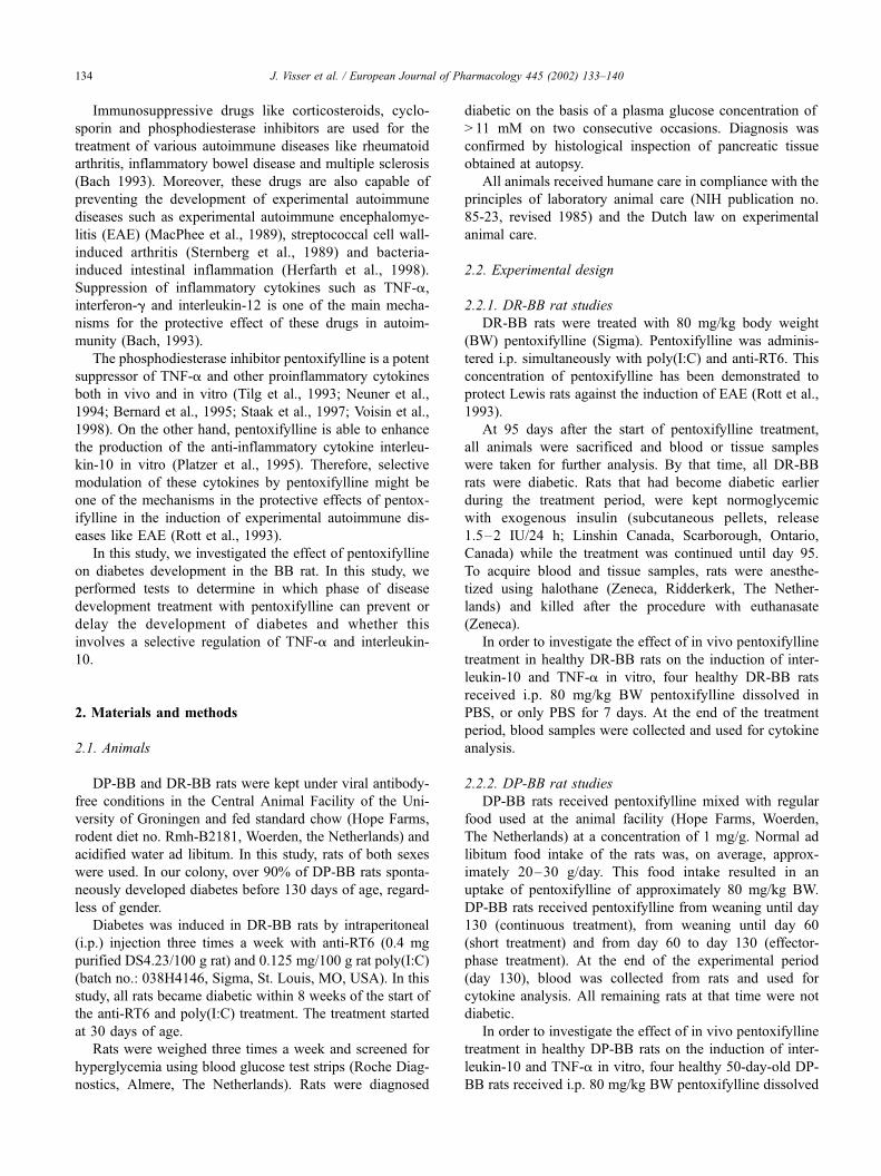
Fig. 1 shows that pentoxifylline significantly delayed the
onset of diabetes development in the DR-BB rat (P < 0.02,
log rank test, Kaplan Meier survival curves). Treatment with
pentoxifylline increased the mean day of onset from
32F 3.3 days in the poly(I:C) + anti-RT6-treated group to
61F12.8 days in the pentoxifylline + poly(I:C) + anti-RT6-
treated group (P < 0.05, Mann–Whitney U-test). However,
pentoxifylline did not prevent the induction of diabetes,
because at 95 days after the start of the treatment all
pentoxifylline-treated DR-BB rats had become diabetic.
3.2. Oral treatment of DP-BB rats with pentoxifylline
reduces and delays diabetes development, but only when it
is applied in the effector phase of disease development
We subsequently investigated the effect of pentoxifylline
on the spontaneous development of diabetes in the DP-BB
rat model. For this purpose, DP-BB rats received pentoxifyl-
line mixed with their food in a concentration of 80 mg/kg
BW as described in Materials and methods. The growth
curves of rats that were given pentoxifylline were not differ-
ent from the rats on regular chow (data not shown). The
animals received pentoxifylline via their food in order to
prevent effects of stress (caused by injections) on diabetes
Fig. 1. Pentoxifylline postpones the onset of anti-RT6 and poly(I:C)
induced diabetes in DR-BB rats. Diabetes was induced in DR-BB rats by
i.p. injection of anti-RT6 (0.4 mg purified DS4.23/100 g rat and poly(I:C)
(0.125 mg/100 g rat). Thirteen rats received anti-RT6 and poly(I:C) (o)
only and five rats were treated with pentoxifylline (8 mg/100 g rat,
injected simultaneously with anti-RT6 and poly(I:C) (n). Statistical
significance was calculated using the log rank test (Kaplan Meier survival
curves).
J. Visser et al. / European Journal of Pharmacology 445 (2002) 133–140 135
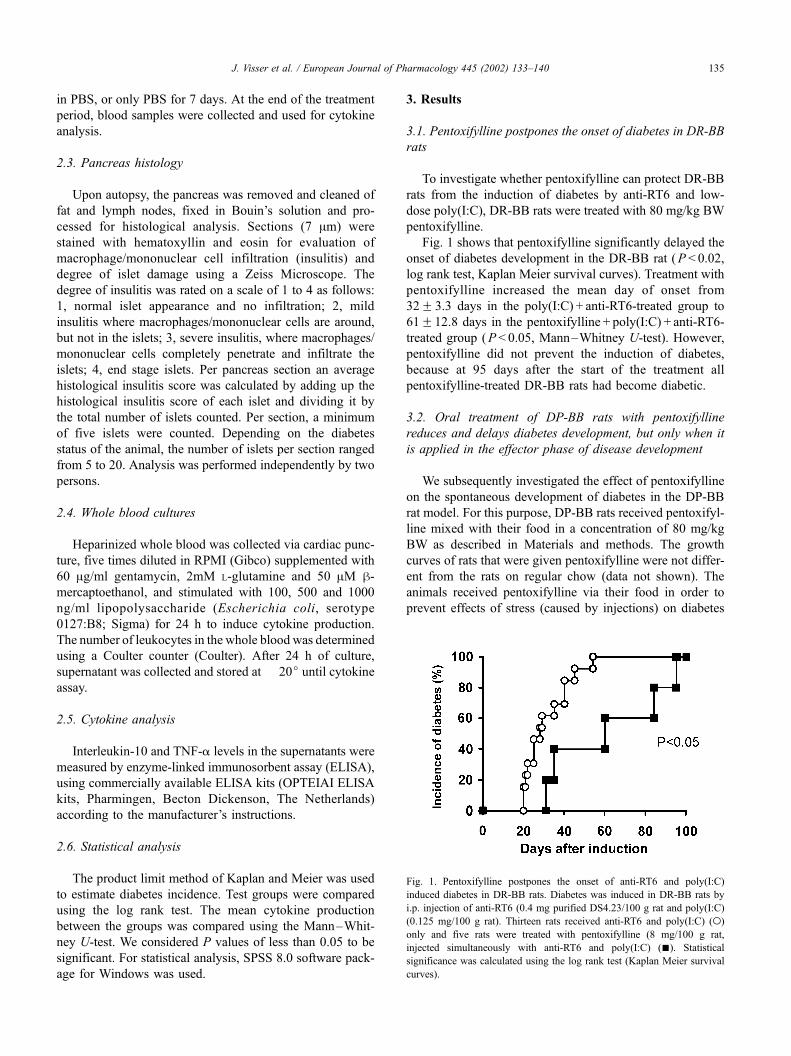
development in DP-BB rats. Fig. 2 shows that oral treatment
of DP-BB rats with pentoxifylline significantly delayed and
reduced diabetes development only when it was applied in
the effector phase (from day 60 to day 130) of disease
development (P= 0.02, log rank test, Kaplan Meier survival
curves). This pentoxifylline treatment increased the mean
day of onset from 81.7F 4.7 days in the control group to
97.8F 6.7 days in the effector phase-treated group (P <
0.05, Mann–Whitney U-test).
Treatment with pentoxifylline from weaning to day 60 or
even throughout the experimental period (weaning to day
130) had no effect on the development of diabetes.
3.3. Non diabetic DP-BB rats treated with pentoxifylline in
the effector phase of disease development have no insulitis,
whereas the non-diabetic rats of the other groups show
signs of insulitis
Among untreated DP-BB rats, all animals became dia-
betic before 130 days of age. Only 1 out 13 in the
continuous treatment group did not develop diabetes, com-
pared to 2 out of 14 in the short treatment group and 4 out of
12 in the effector-phase treatment group. At the moment of
diagnosis of diabetes or at the end of the experimental
period of 130 days, the pancreas was removed and pro-
cessed for histological analysis.
As demonstrated in Table 1 no difference in the histo-
logical score of insulitis between the diabetic animals of
various treatment groups was observed.
However, the non-diabetic animals in the effector-phase-
treated group showed no histological signs of insulitis,
whereas the non-diabetic animals of other groups showed
histological signs of insulitis. These results suggest that the
non-diabetic animals of other groups might have developed
diabetes later, whereas the absence of insulitis in the non-
diabetic rats of the effector-phase-treated group indicates
complete protection against diabetes development.
3.4. Differential effects of pentoxifylline treatment in vivo,
on the induction of TNF-a and interleukin-10 in vitro
3.4.1. DR-BB rat studies
At 95 days after the induction of diabetes by anti-RT6
and Poly(I:C) treatment, all animals were sacrificed and
blood samples were taken for further analysis. By that time,
all DR-BB rats were diabetic. Rats that had become diabetic
earlier during the treatment period were kept normoglyce-
mic with exogenous insulin until day 95 while they still
received anti-RT6 and Poly(I:C). Blood was obtained from
all five pentoxifylline + poly(I:C) + anti-RT6-treated animals
and of three poly(I:C) + anti-RT6-treated animals. Blood
was placed in culture and stimulated for 24 h with lip-
opolysaccharide to establish TNF-a and interleukin-10
production. Three untreated 125-day-old DR-BB rats served
as a control.
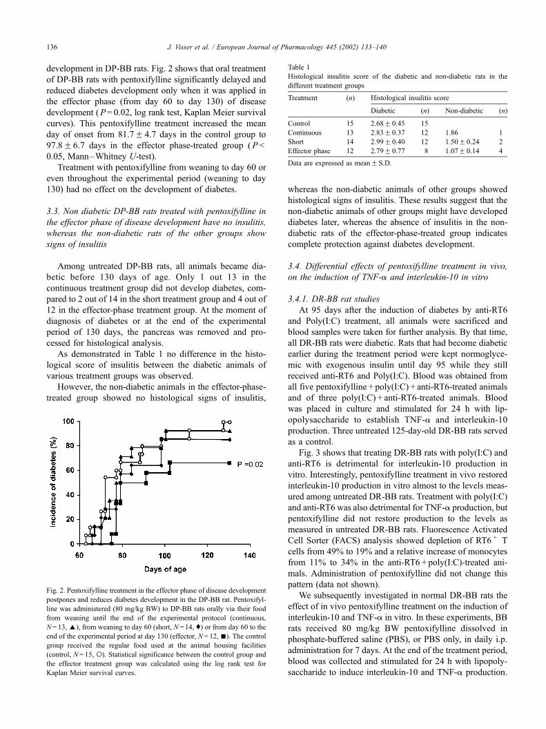
Fig. 3 shows that treating DR-BB rats with poly(I:C) and
anti-RT6 is detrimental for interleukin-10 production in
vitro. Interestingly, pentoxifylline treatment in vivo restored
interleukin-10 production in vitro almost to the levels meas-
ured among untreated DR-BB rats. Treatment with poly(I:C)
and anti-RT6 was also detrimental for TNF-a production, but
pentoxifylline did not restore production to the levels as
measured in untreated DR-BB rats. Fluorescence Activated
Cell Sorter (FACS) analysis showed depletion of RT6 + T
cells from 49% to 19% and a relative increase of monocytes
from 11% to 34% in the anti-RT6 + poly(I:C)-treated ani-
mals. Administration of pentoxifylline did not change this
pattern (data not shown).
We subsequently investigated in normal DR-BB rats the
effect of in vivo pentoxifylline treatment on the induction of
interleukin-10 and TNF-a in vitro. In these experiments, BB
rats received 80 mg/kg BW pentoxifylline dissolved in
phosphate-buffered saline (PBS), or PBS only, in daily i.p.
administration for 7 days. At the end of the treatment period,
blood was collected and stimulated for 24 h with lipopoly-
saccharide to induce interleukin-10 and TNF-a production.
Table 1
Histological insulitis score of the diabetic and non-diabetic rats in the
different treatment groups
Treatment (n) Histological insulitis score
Diabetic (n) Non-diabetic (n)
Control 15 2.68F 0.45 15
Continuous 13 2.83F 0.37 12 1.86 1
Short 14 2.99F 0.40 12 1.50F 0.24 2
Effector phase 12 2.79F 0.77 8 1.07F 0.14 4
Data are expressed as meanF S.D.
Fig. 2. Pentoxifylline treatment in the effector phase of disease development
postpones and reduces diabetes development in the DP-BB rat. Pentoxifyl-
line was administered (80 mg/kg BW) to DP-BB rats orally via their food
from weaning until the end of the experimental protocol (continuous,
N = 13,E), from weaning to day 60 (short, N = 14, y) or from day 60 to the
end of the experimental period at day 130 (effector, N= 12, n). The control
group received the regular food used at the animal housing facilities
(control, N= 15, o). Statistical significance between the control group and
the effector treatment group was calculated using the log rank test for
Kaplan Meier survival curves.
J. Visser et al. / European Journal of Pharmacology 445 (2002) 133–140136
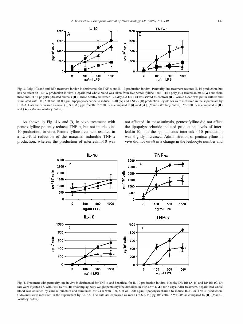
As shown in Fig. 4A and B, in vivo treatment with
pentoxifylline potently reduces TNF-a, but not interleukin-
10 production, in vitro. Pentoxifylline treatment resulted in
a two-fold reduction of the maximal inducible TNF-a
production, whereas the production of interleukin-10 was
not affected. In these animals, pentoxifylline did not affect
the lipopolysaccharide-induced production levels of inter-
leukin-10, but the spontaneous interleukin-10 production
was slightly increased. Administration of pentoxifylline in
vivo did not result in a change in the leukocyte number and
Fig. 4. Treatment with pentoxifylline in vivo is detrimental for TNF-a and beneficial for IL-10 production in vitro. Healthy DR-BB (A, B) and DP-BB (C, D)
rats were injected i.p. with PBS (N = 4, .) or 80 mg/kg body weight pentoxifylline dissolved in PBS (N= 4, E) for 7 days. After treatment, heparinized whole
blood was obtained by cardiac puncture and stimulated for 24 h with 100, 500 or 1000 ng/ml lipopolysaccharide to induce IL-10 or TNF-a production.
Cytokines were measured in the supernatant by ELISA. The data are expressed as mean (F S.E.M.) pg/106 cells. *P < 0.05 as compared to (.) (Mann–
Whitney U-test).
Fig. 3. Poly(I:C) and anti-RT6 treatment in vivo is detrimental for TNF-a and IL-10 production in vitro. Pentoxifylline treatment restores IL-10 production, but
has no effect on TNF-a production in vitro. Heparinized whole blood was taken from five pentoxifylline + anti-RT6+ poly(I:C)-treated animals (E) and from
three anti-RT6 + poly(I:C)-treated animals (n). Three healthy untreated 125-day-old DR-BB rats served as controls (.). Whole blood was put in culture and
stimulated with 100, 500 and 1000 ng/ml lipopolysaccharide to induce IL-10 (A) and TNF-a (B) production. Cytokines were measured in the supernatant by
ELISA. Data are expressed as mean (F S.E.M.) pg/106 cells. *P< 0.05 as compared to (.) and (E), (Mann–Whitney U-test). **P < 0.05 as compared to (n)
and (E), (Mann–Whitney U-test).
J. Visser et al. / European Journal of Pharmacology 445 (2002) 133–140 137

the levels of monocytes, T cells, h-cells and CD4/CD8 ratio
in whole blood as compared to PBS-treated animals (data
not shown).
3.4.2. DP-BB rat studies
As in the DR-BB rat studies, we also investigated the
effect of 7-day in vivo pentoxifylline treatment on the
induction of interleukin-10 and TNF-a in vitro. In DP-BB
rats (Fig. 4C,D), pentoxifylline treatment resulted in a
reduced induction of both TNF-a and interleukin-10 in
vitro. However, the effect on TNF-a was much more evi-
dent as compared to interleukin-10.
As can be seen in Fig. 4, diabetes-prone BB rats have a
three-fold reduced cytokine production when compared to
diabetes-resistant BB rats. This is probably due to the fact
that diabetes-prone BB rats are naturally deficient of regu-
latory RT6 + T cells, since the same phenomenon was also
observed in diabetes-resistant BB rats after depletion of their
RT6 + T cells with anti RT6 (Fig. 3).
As in the DR-BB rat studies, administration of pentox-
ifylline in vivo to DP-BB rats did not result in a change in
the leukocyte number and the levels of monocytes, T cells,
h-cells and CD4/CD8 ratio in whole blood as compared to
PBS-treated animals (data not shown).
4. Discussion
In this study, we have demonstrated that pentoxifylline
reduces diabetes development in the BB rat, but only
provides protection against spontaneous diabetes develop-
ment when applied in the effector phase of disease develop-
ment. Treatment of rats with pentoxifylline in vivo
dramatically reduces the induction of TNF-a by lipopoly-
saccharide in vitro, but the induction of interleukin-10 re-
mains mostly unaffected.
The effector phase of diabetes development after 60
days of age in the DP-BB rat is characterized by severe
insulitis and destruction of the h-cells by cytotoxic T cells,
macrophages and NK cells (Mordes et al., 2001). Further-
more, proinflammatory cytokines like TNF-a and interleu-
kin-1, which are produced in high amount by these
infiltrating cells, are cytotoxic for the h-cells (Rabinovitch,1998; Saldeen, 2000). It is therefore likely that blocking
these inflammatory responses will delay or prevent the
total destruction of the h-cell and subsequently the onset
of diabetes. Indeed, when we treat DP-BB rats in the
effector phase of disease development with pentoxifylline,
a potent suppressor of inflammation, the onset of diabetes
is delayed and even prevented. However, continuous treat-
ment of the DP BB rats with pentoxifylline or a short
treatment during the insulin-dependent diabetes mellitus
sensitive window did not influence the disease process.
The absence of insulitis in the non-diabetic rats of the
effector-phase-treated group indicates complete protection
against diabetes development. Moreover, induction of
diabetes in the DR-BB rat was also delayed by pentoxifyl-
line.
In the induced model of diabetes development, the DR-
BB rats are depleted of their regulatory RT6 + T cells and
poly(I:C) is added for activation of the immune system
(Mordes et al., 2001; Greiner et al., 1987a,b; Sobel et al.,
1992). Poly(I:C) is a synthetic double-stranded RNA, which
mimics a viral infection. For instance, poly(I:C) has been
demonstrated to be a potent inducer of TNF-a and inter-
leukin-12 production and an activator of dendritic cells
(Verdijk et al., 1999). Interestingly, we observed that treat-
ment with poly(I:C) and aRT6 in vivo not only reduced the
induction of interleukin-10, but also of TNF-a by lipopo-
lysaccharide in vitro. Pentoxifylline was able to restore
interleukin-10 production to the levels found in untreated
healthy DR-BB rats, while TNF-a production remained low.
This differential effect of pentoxifylline on interleukin-10
and TNF-a production was also demonstrated in whole
blood cultures of DR-BB rats that were treated with pentox-
ifylline only. Because interleukin-10 can be protective and
TNF-a detrimental in the effector phase of diabetes devel-
opment (Rabinovitch, 1998), the enhancement of interleu-
kin-10 and reduction of TNF-a will reduce the inflam-
matory cascade leading to the destruction of the h-cell.Therefore, the fact that pentoxifylline could only postpone
diabetes development in DR-BB rats can be explained by
the fact that pentoxifylline reduced the adjuvant effect of
poly(I:C) but did not counteract the depletion of the regu-
latory RT6 + T cells.
As shown in Fig. 4, suppression of TNF-a and the
subsequent increase of the IL-10/TNF-a ratio might be the
main reason why pentoxifylline prevents the induction of
diabetes in the effector phase of disease development in the
DP-BB rat. Accordingly, the protection of non-obese dia-
betic (NOD) mice against diabetes by pentoxifylline was
related to the suppression of TNF-a and other proinflam-
matory cytokines (Liang et al., 1998). However, TNF-a
exhibits paradoxical effects on diabetes development in both
the NOD mouse and DP-BB rat model of diabetes. Accel-
erating or protective effects of TNF-a on diabetes develop-
ment depends on dose and timing (Rabinovitch, 1998). For
example, Satoh et al. (1990) demonstrated that administra-
tion of TNF-a from weaning onwards could prevent the
onset of diabetes in the DP-BB rat. Intriguingly, the admin-
istration of other proinflammatory cytokines like interferon-
g also protects DP-BB rats from diabetes development
(Nicoletti et al., 1998; Sobel and Newsome, 1997). Interest-
ingly, when these proinflammatory cytokines are given in
the effector phase of disease development, an acceleration of
the disease can occur (Rabinovitch, 1998). For instance, in
NOD mice, a very narrow time window is related to
protection or acceleration of the disease by TNF-a (Yang
et al., 1994; Green and Flavell, 2000; Green et al., 2000).
The anti-inflammatory cytokine interleukin-10, on the
other hand, can provide protection against diabetes develop-
ment (Pennline et al., 1994). Like TNF-a, interleukin-10 has
J. Visser et al. / European Journal of Pharmacology 445 (2002) 133–140138

also been shown to exhibit paradoxical effects on the
development of autoimmune diabetes (Rabinovitch, 1998).
Timing and localisation of interleukin-10 distribution are of
critical importance. For instance, local tissue expression of
interleukin-10 in the pancreas accelerates diabetes develop-
ment in NOD mice (Moritani et al., 1994), whereas systemic
administration of interleukin-10 more in the effector phase
of disease development rescues the NOD mice from dia-
betes development (Pennline et al., 1994).
Sobel et al. (2000) demonstrated that an induction of
regulatory T cells by cyclophosphamide in the period
between 30 and 60 days can prevent diabetes development
in the DP-BB rat. Such an induction of a protective mecha-
nism by TNF-a or interferon-g when these cytokines are
applied from weaning is proposed to be responsible for the
prevention of diabetes in the DP-BB rat (Rabinovitch, 1998).
Interestingly, we observed in 130-day-old non-diabetic
DP-BB rats much higher cytokine levels compared to non-
diabetic 60-day-old DP-BB rats. As was described by
Rabinovitch (1998) and Satoh et al. (1990), diabetes devel-
opment in the DP-BB rat can be prevented by immunosti-
mulation. Therefore, high cytokine levels in old non-
diabetic DP-BB rats might represent a more active immune
system and subsequently protect these DP-BB rats for
diabetes development.
When pentoxifylline is applied from weaning, the pro-
duction of TNF-a and interferon-g is suppressed and sub-
sequently, the induction of a protective mechanism. This
suppression of a protective mechanism might be responsible
for the paradoxical result that continuous treatment with
pentoxifylline does not prevent diabetes in the DP-BB rat.
Accordingly, it also suggests that prophylactic pentoxifyl-
line treatment of children at risk for developing diabetes
might not be beneficial as opposed to the observed preser-
vation of h-cell function by pentoxifylline in newly diag-
nosed diabetes patients (MacDonald et al., 1994).
In conclusion, our results show that prevention of dia-
betes development by pentoxifylline depends on the timing
of treatment. Obviously, the observed pentoxifylline-in-
duced suppression of TNF-a and relative enhancement of
interleukin-10 might be the underlying mechanisms for
prevention or delay of diabetes.
Acknowledgements
This work was supported by research grants DFN96.606
and DFN98.148 from the Dutch Diabetes Foundation. We
would like to thank Ar Jansen, Lucas Vijfschaft and Hilda
de Vries for technical assistance.
References
Bach, J.F., 1993. Immunosuppressive therapy of autoimmune diseases.
Immunol. Today 14, 322–326.
Bernard, C., Barnier, P., Merval, R., Esposito, B., Tedgui, A., 1995. Pen-
toxifylline selectivity inhibits tumor necrosis factor synthesis in the
arterial wall. J. Cardiovasc. Pharmacol. 25 (Suppl. 2), S30–S33.
Burstein, D., Mordes, J.P., Greiner, D.L., Stein, D., Nakamura, N., Handler,
E.S., Rossini, A.A., 1989. Prevention of diabetes in BB/Wor rat by
single transfusion of spleen cells. Parameters that affect degree of pro-
tection. Diabetes 38, 24–30.
Green, E.A., Flavell, R.A., 2000. The temporal importance of TNFalpha
expression in the development of diabetes. Immunity 12, 459–469.
Green, E.A., Wong, F.S., Eshima, K., Mora, C., Flavell, R.A., 2000. Neo-
natal tumor necrosis factor alpha promotes diabetes in nonobese dia-
betic mice by CD154-independent antigen presentation to CD8(+) T
cells. J. Exp. Med. 191, 225–238.
Greiner, D.L., Mordes, J.P., Handler, E.S., Angelillo, M., Nakamura, N.,
Rossini, A.A., 1987a. Depletion of RT6 + T lymphocytes induces dia-
betes in resistant BioBreeding (BB/W) rats. J. Exp. Med. 166, 461–475.
Greiner, D.L., Mordes, J.P., Handler, E.S., Nakamura, N., Angelillo, M.,
Rossini, A.A., 1987b. Prothymocyte development in diabetes-prone BB
rats: description of a defect that predisposes to immune abnormalities.
Transplant. Proc. Feb. 19 (1 Pt. 2), 976–978.
Herfarth, H.H., Bocker, U., Janardhanam, R., Sartor, R.B., 1998. Subther-
apeutic corticosteroids potentiate the ability of interleukin 10 to prevent
chronic inflammation in rats. Gastroenterology 115, 856–865.
Kolb, H., Worz-Pagenstert, U., Kleemann, R., Rothe, H., Rowsell, P., Scott,
F.W., 1996. Cytokine gene expression in the BB rat pancreas: natural
course and impact of bacterial vaccines. Diabetologia 39, 1448–1454.
Liang, L., Beshay, E., Prud’homme, G.J., 1998. The phosphodiesterase
inhibitors pentoxifylline and rolipram prevent diabetes in NOD mice.
Diabetes 47, 570–575.
Like, A.A., Kislauskis, E., Williams, R.M., Rossini, A.A., 1982. Neonatal
thymectomy prevents spontaneous diabetes mellitus in the BB/W rat.
Science 216, 644–646.
MacDonald, M.J., Shahidi, N.T., Allen, D.B., Lustig, R.H., Mitchell, T.L.,
Cornwell, S.T., 1994. Pentoxifylline in the treatment of children with
new-onset type I diabetes mellitus [letter]. J. Am. Med. Assoc. 271,
27–28.
MacPhee, I.A., Antoni, F.A., Mason, D.W., 1989. Spontaneous recovery of
rats from experimental allergic encephalomyelitis is dependent on reg-
ulation of the immune system by endogenous adrenal corticosteroids. J.
Exp. Med. 169, 431–445.
Mordes, J.P., Bortell, R., Groen, H., Guberski, D.L., Rossini, A.A., Greiner,
D., 2001. In: Sima, A.A.F., Shafir, E. (Eds.), Autoimmune Diabetes
Mellitus in the BB rat. Harwood Academic Publishers, Amsterdam,
The Netherlands, pp. 1–41.
Moritani, M., Yoshimoto, K., Tashiro, F., Hashimoto, C., Miyazaki, J., Ii, S.,
Kudo, E., Iwahana, H., Hayashi, Y., Sano, T., 1994. Transgenic expres-
sion of IL-10 in pancreatic islet A cells accelerates autoimmune insulitis
and diabetes in non-obese diabetic mice. Int. Immunol. 6, 1927–1936.
Neuner, P., Klosner, G., Schauer, E., Pourmojib, M., Macheiner, W., Grun-
wald, C., Knobler, R., Schwarz, A., Luger, T.A., Schwarz, T., 1994.
Pentoxifylline in vivo down-regulates the release of IL-1 beta, IL-6, IL-
8 and tumour necrosis factor-alpha by human peripheral blood mono-
nuclear cells. Immunology 83, 262–267.
Nicoletti, F., Zaccone, P., Di Marco, R., Magro, G., Grasso, S., Stivala, F.,
Calori, G., Mughini, L., Meroni, P.L., Garotta, G., 1998. Paradoxical
antidiabetogenic effect of gamma-interferon in DP-BB rats. Diabetes
47, 32–38.
Pennline, K.J., Roque-Gaffney, E., Monahan, M., 1994. Recombinant hu-
man IL-10 prevents the onset of diabetes in the nonobese diabetic
mouse. Clin. Immunol. Immunopathol. 71, 169–175.
Platzer, C., Meisel, C., Vogt, K., Platzer, M., Volk, H.D., 1995. Up-regu-
lation of monocytic IL-10 by tumor necrosis factor-alpha and cAMP
elevating drugs. Int. Immunol. 7, 517–523.
Rabinovitch, A., 1998. An update on cytokines in the pathogenesis of
insulin-dependent diabetes mellitus. Diabetes Metab. Rev. 14, 129–
151.
Rabinovitch, A., Suarez-Pinzon, W., El Sheikh, A., Sorensen, O., Power,
R.F., 1996. Cytokine gene expression in pancreatic islet-infiltrating
J. Visser et al. / European Journal of Pharmacology 445 (2002) 133–140 139

leukocytes of BB rats: expression of Th1 cytokines correlates with
beta-cell destructive insulitis and IDDM. Diabetes 45, 749–754.
Rott, O., Cash, E., Fleisher, B., 1993. Phosphodiesterase inhibitor pentox-
ifylline, a selective suppressor of T helper type 1-but not type 2-asso-
ciated lymphokine production, prevents induction of experimental
autoimmune encephalomyelitis in Lewis rats. Eur. J. Immunol. 23,
1745–1751.
Saldeen, J., 2000. Cytokines induce both necrosis and apoptosis via a
common Bcl-2-inhibitable pathway in rat insulin-producing cells. En-
docrinology 141, 2003–2010.
Satoh, J., Seino, H., Shintani, S., Tanaka, S., Ohteki, T., Masuda, T., No-
bunaga, T., Toyota, T., 1990. Inhibition of type I diabetes in BB rats
with recombinant human tumor necrosis factor-a. J. Immunol. 145,
1395–1399.
Sobel, D.O., Newsome, J., 1997. Gamma interferon prevents diabetes in the
BB rat. Clin. Diagn. Lab. Immunol. 4, 764–768.
Sobel, D.O., Newsome, J., Ewel, C.H., Bellanti, J.A., Abbassi, V., Cres-
well, K., Blair, O., 1992. Poly I:C induces development of diabetes
mellitus in BB rat. Diabetes 41, 515–520.
Sobel, D.O., Ahvazi, B., Jun, H.S., Chung, Y.H., Yoon, J.W., 2000. Cyclo-
phosphamide inhibits the development of diabetes in the diabetes-prone
BB rat. Diabetologia 43, 986–994.
Staak, K., Prosch, S., Stein, J., Priemer, C., Ewert, R., Docke, W.D.,
Kruger, D.H., Volk, H.D., Reinke, P., 1997. Pentoxifylline promotes
replication of human cytomegalovirus in vivo and in vitro. Blood 89,
3682–3690.
Sternberg, E.M., Young, W.S., Bernardini, R., Calogero, A.E., Chrousos,
G.P., Gold, P.W., Wilder, R.L., 1989. A central nervous system defect in
biosynthesis of corticotropin-releasing hormone is associated with sus-
ceptibility to streptococcal cell wall-induced arthritis in Lewis rats.
Proc. Natl. Acad. Sci. U. S. A. 86, 4771–4775.
Tilg, H., Eibl, B., Pichl, M., Gachter, A., Herold, M., Brankova, J., Huber,
C., Niederwieser, D., 1993. Immune response modulation by pentox-
ifylline in vitro. Transplantation 56, 196–201.
Verdijk, R.M., Mutis, T., Esendam, B., Kamp, J., Melief, C.J., Brand, A.,
Goulmy, E., 1999. Polyriboinosinic polyribocytidylic acid (poly(I:C))
induces stable maturation of functionally active human dendritic cells. J.
Immunol. 163, 57–61.
Voisin, L., Breuille, D., Ruot, B., Ralliere, C., Rambourdin, F., Dalle, M.,
Obled, C., 1998. Cytokine modulation by PX differently affects specific
acute phase proteins during sepsis in rats. Am. J. Physiol. 275, R1412–
R1419.
Yang, X.-D., Tisch, R., Singer, S.M., Cao, Z.A., Liblau, R.S., Schreiber,
R.D., McDevitt, H.O., 1994. Effect of tumor necrosis factor a on In-
sulin Dependent Diabetes Mellitus in NOD mice: I. The early develop-
ment of autoimmunity and the diabetogenic process. J. Exp. Med. 180
(3), 995–1004.
J. Visser et al. / European Journal of Pharmacology 445 (2002) 133–140140