![[STAY TUNED- Estigues connectat!]](https://static.fdocuments.in/doc/165x107/58eed2df1a28abee7b8b45d3/stay-tuned-estigues-connectat.jpg)
Timing is everything Fine-tuned molecular machines ... · Timing is everything: Fine-tuned...
14
Review Timing is everything: Fine-tuned molecular machines orchestrate paramyxovirus entry Sayantan Bose a,n,1 , Theodore S. Jardetzky c , Robert A. Lamb a,b,nn a Department of Molecular Biosciences, Northwestern University, Evanston, IL 60208-3500, United States b Howard Hughes Medical Institute, Northwestern University, Evanston, IL 60208-3500, United States c Department of Structural Biology and Program in Immunology, Stanford University School of Medicine, Stanford, CA 94305, United States article info Article history: Received 23 December 2014 Returned to author for revisions 21 January 2015 Accepted 18 February 2015 Available online 12 March 2015 Keywords: Membrane fusion Membrane glycoproteins Viral envelope proteins Paramyxovirus entry Atomic structure of viral glycoproteins Viral receptors Fusion protein abstract The Paramyxoviridae include some of the great and ubiquitous disease-causing viruses of humans and animals. In most paramyxoviruses, two viral membrane glycoproteins, fusion protein (F) and receptor binding protein (HN, H or G) mediate a concerted process of recognition of host cell surface molecules followed by fusion of viral and cellular membranes, resulting in viral nucleocapsid entry into the cytoplasm. The interactions between the F and HN, H or G viral glycoproteins and host molecules are critical in determining host range, virulence and spread of these viruses. Recently, atomic structures, together with biochemical and biophysical studies, have provided major insights into how these two viral glycoproteins successfully interact with host receptors on cellular membranes and initiate the membrane fusion process to gain entry into cells. These studies highlight the conserved core mechanisms of paramyxovirus entry that provide the fundamental basis for rational anti-viral drug design and vaccine development. & 2015 Elsevier Inc. All rights reserved. Contents Introduction............................................................................................................ 518 Gaining access to the cytoplasm: viral membrane fusion proteins................................................................. 519 Cleavage by cellular and tissue proteases converts F into an active, pre-triggered form................................................ 520 Atomic structures and biophysical studies of paramyxovirus F proteins provide insights into the refolding process that lead to membrane fusion ..... 521 Multifunctional attachment proteins recognize and bind to host cell molecules as receptors ........................................... 522 Receptor binding by the attachment proteins initiates activation of the fusion protein ................................................ 523 The fusion protein and the attachment protein physically interact to mediate membrane fusion ........................................ 523 Two distinct molecular hypotheses attempt to explain the interaction between the fusion protein and the attachment protein ............... 524 Regions of the fusion protein involved in attachment protein interactions offer insights into the F-triggering process ....................... 524 Molecular cooperation between the different domains of the attachment protein ensures timing of F-activation and membrane fusion ........ 525 Diverse inhibitory mechanisms found in different paramyxoviruses appear to regulate access of the fusion protein to the attachment protein stalk prior to ‘stalk exposure’ .................................................................................................. 527 Acknowledgments ....................................................................................................... 527 References ............................................................................................................. 528 Introduction Paramyxoviruses are a diverse family of viruses, which includes many human and animal pathogens that are of global importance to public health and economy. Highly infectious pathogens like respiratory syncytial virus (RSV), measles virus (MeV), mumps virus (MuV), parainfluenza viruses 1–5 (PIV1–5) and human metapneumovirus (hMPV) contribute significantly to the annual Contents lists available at ScienceDirect journal homepage: www.elsevier.com/locate/yviro Virology http://dx.doi.org/10.1016/j.virol.2015.02.037 0042-6822/& 2015 Elsevier Inc. All rights reserved. n Corresponding author. Tel: þ1 617-432-1927 Fax: þ1 617 738 7664. nn Corresponding author at: Department of Molecular Biosciences, Northwestern University, 2205 Tech Drive, Evanston, IL 60208-3500, United States. Tel: þ1 847- 491-5433 Fax: þ1 847 491 2467. E-mail addresses: [email protected] (S. Bose), [email protected] (R.A. Lamb). 1 Current address: Department of Microbiology and Immunobiology, Harvard Medical School, Room 940, 77 Avenue Louis Pasteur, Boston, MA 02115, United States. Virology 479-480 (2015) 518–531
-
Upload
nguyenquynh -
Category
Documents
-
view
218 -
download
0
Transcript of Timing is everything Fine-tuned molecular machines ... · Timing is everything: Fine-tuned...

Review
Timing is everything: Fine-tuned molecular machines orchestrateparamyxovirus entry
Sayantan Bose a,n,1, Theodore S. Jardetzky c, Robert A. Lamb a,b,nn
a Department of Molecular Biosciences, Northwestern University, Evanston, IL 60208-3500, United Statesb Howard Hughes Medical Institute, Northwestern University, Evanston, IL 60208-3500, United Statesc Department of Structural Biology and Program in Immunology, Stanford University School of Medicine, Stanford, CA 94305, United States
a r t i c l e i n f o
Article history:Received 23 December 2014Returned to author for revisions21 January 2015Accepted 18 February 2015Available online 12 March 2015
Keywords:Membrane fusionMembrane glycoproteinsViral envelope proteinsParamyxovirus entryAtomic structure of viral glycoproteinsViral receptorsFusion protein
a b s t r a c t
The Paramyxoviridae include some of the great and ubiquitous disease-causing viruses of humans andanimals. In most paramyxoviruses, two viral membrane glycoproteins, fusion protein (F) and receptorbinding protein (HN, H or G) mediate a concerted process of recognition of host cell surface moleculesfollowed by fusion of viral and cellular membranes, resulting in viral nucleocapsid entry into the cytoplasm.The interactions between the F and HN, H or G viral glycoproteins and host molecules are critical indetermining host range, virulence and spread of these viruses. Recently, atomic structures, together withbiochemical and biophysical studies, have provided major insights into how these two viral glycoproteinssuccessfully interact with host receptors on cellular membranes and initiate the membrane fusion process togain entry into cells. These studies highlight the conserved core mechanisms of paramyxovirus entry thatprovide the fundamental basis for rational anti-viral drug design and vaccine development.
& 2015 Elsevier Inc. All rights reserved.
Contents
Introduction. . . . . . . . . . . . . . . . . . . . . . . . . . . . . . . . . . . . . . . . . . . . . . . . . . . . . . . . . . . . . . . . . . . . . . . . . . . . . . . . . . . . . . . . . . . . . . . . . . . . . . . . . . . . 518Gaining access to the cytoplasm: viral membrane fusion proteins. . . . . . . . . . . . . . . . . . . . . . . . . . . . . . . . . . . . . . . . . . . . . . . . . . . . . . . . . . . . . . . . . 519Cleavage by cellular and tissue proteases converts F into an active, pre-triggered form. . . . . . . . . . . . . . . . . . . . . . . . . . . . . . . . . . . . . . . . . . . . . . . . 520Atomic structures and biophysical studies of paramyxovirus F proteins provide insights into the refolding process that lead to membrane fusion . . . . . 521Multifunctional attachment proteins recognize and bind to host cell molecules as receptors . . . . . . . . . . . . . . . . . . . . . . . . . . . . . . . . . . . . . . . . . . . 522Receptor binding by the attachment proteins initiates activation of the fusion protein . . . . . . . . . . . . . . . . . . . . . . . . . . . . . . . . . . . . . . . . . . . . . . . . 523The fusion protein and the attachment protein physically interact to mediate membrane fusion . . . . . . . . . . . . . . . . . . . . . . . . . . . . . . . . . . . . . . . . 523Two distinct molecular hypotheses attempt to explain the interaction between the fusion protein and the attachment protein . . . . . . . . . . . . . . . 524Regions of the fusion protein involved in attachment protein interactions offer insights into the F-triggering process . . . . . . . . . . . . . . . . . . . . . . . 524Molecular cooperation between the different domains of the attachment protein ensures timing of F-activation and membrane fusion . . . . . . . . 525Diverse inhibitory mechanisms found in different paramyxoviruses appear to regulate access of the fusion protein to the attachment protein stalkprior to ‘stalk exposure’ . . . . . . . . . . . . . . . . . . . . . . . . . . . . . . . . . . . . . . . . . . . . . . . . . . . . . . . . . . . . . . . . . . . . . . . . . . . . . . . . . . . . . . . . . . . . . . . . . . 527Acknowledgments . . . . . . . . . . . . . . . . . . . . . . . . . . . . . . . . . . . . . . . . . . . . . . . . . . . . . . . . . . . . . . . . . . . . . . . . . . . . . . . . . . . . . . . . . . . . . . . . . . . . . . . 527References . . . . . . . . . . . . . . . . . . . . . . . . . . . . . . . . . . . . . . . . . . . . . . . . . . . . . . . . . . . . . . . . . . . . . . . . . . . . . . . . . . . . . . . . . . . . . . . . . . . . . . . . . . . . . 528
Introduction
Paramyxoviruses are a diverse family of viruses, which includesmany human and animal pathogens that are of global importanceto public health and economy. Highly infectious pathogens likerespiratory syncytial virus (RSV), measles virus (MeV), mumpsvirus (MuV), parainfluenza viruses 1–5 (PIV1–5) and humanmetapneumovirus (hMPV) contribute significantly to the annual
Contents lists available at ScienceDirect
journal homepage: www.elsevier.com/locate/yviro
Virology
http://dx.doi.org/10.1016/j.virol.2015.02.0370042-6822/& 2015 Elsevier Inc. All rights reserved.
n Corresponding author. Tel: þ1 617-432-1927 Fax: þ1 617 738 7664.nn Corresponding author at: Department of Molecular Biosciences, Northwestern
University, 2205 Tech Drive, Evanston, IL 60208-3500, United States. Tel: þ1 847-491-5433 Fax: þ1 847 491 2467.
E-mail addresses: [email protected] (S. Bose),[email protected] (R.A. Lamb).
1 Current address: Department of Microbiology and Immunobiology, HarvardMedical School, Room 940, 77 Avenue Louis Pasteur, Boston, MA 02115,United States.
Virology 479-480 (2015) 518–531

global disease burden in humans, infecting millions of individualsworldwide and leading to a large number of deaths in areas havinginadequate health care resources. Many of these viruses are alsore-emerging in previously immune populations due to a decreasein vaccination and corresponding breakdown of herd immunity(Gahr et al., 2014; Munoz-Alia et al., 2014; Rubin et al., 2012; Yanget al., 2014). Other paramyxoviruses are more sporadic in theiroutbreaks and viruses like the zoonotic Nipah virus (NiV) andHendra virus (HeV) cause deadly localized outbreaks, resulting inhigh morbidity and mortality in human populations around theworld. NiV and HeV are classified as Biosafety Level 4 (BSL-4)select agents. Cases of human-to-human transmission of NiV havebecome more prevalent in recent outbreaks in Bangladesh gen-erating significant concern in terms of the epidemiology andtransmission of these diseases (Daszak et al., 2012; Luby et al.,2009, 2006; Mahalingam et al., 2012). Animal viruses like theNewcastle disease virus (NDV) cause severe and sometimes fatalepidemics in poultry populations, leading to extensive economiclosses. Canine distemper virus (CDV) is a fatal, highly contagiousdisease affecting canines. Many recent host reservoir samplingstudies have indicated that a large number of paramyxovirusesremain undiscovered and uncharacterized, with no existingknowledge of the zoonotic potential, spread or host range ofthese viruses (Drexler et al., 2012; Lamb and Parks, 2013; Marshet al., 2012). Both well-characterized and yet undiscoveredparamyxoviruses highlight the considerable hazard posed bysuch emerging pathogens in an era of increasing global popula-tion, human-wildlife territorial conflicts and internationaltravel.
Paramyxoviruses are enveloped viruses harboring a negative-sense RNA genome. Based on sequence homology and proteinfunctions, paramyxoviruses are classified into two sub-families –
Pneumovirinae and Paramyxovirinae, with the two sub-familiesfurther divided into multiple genera (Fig. 1). Like most viruses,paramyxoviruses utilize molecules present on cellular mem-branes, to identify host cells. Attachment via these viral ‘recep-tors’ leads to fusion of viral and cellular membranes and entry ofthe viral genome in the form of a nucleocapsid, into the host cellcytoplasm (Lamb and Parks, 2013). To infect host cells, mostparamyxoviruses depend on the concerted actions of two majorglycoproteins present on the viral membrane, namely the attach-ment protein (HN, H or G), and the fusion (F) protein (Heminwayet al., 1994a; Horvath et al., 1992; Hu et al., 1992; Morrison andPortner, 1991; Yao et al., 1997). The membrane fusion event thatmediates viral entry appears to occur at neutral pH on the plasmamembrane for most paramyxoviruses. Unlike viruses of thesubfamily Paramyxovirinae, in members of the subfamily Pneu-movirinae, the F protein was found to be sufficient for viralpropagation in cell culture (Biacchesi et al., 2005, 2004; Karronet al., 1997) and the cellular pathway of entry for this subfamily ofviruses is yet unclear with membrane fusion at the cell mem-brane (Srinivasakumar et al., 1991), clathrin-mediated endocyto-sis (Kolokoltsov et al., 2007; Schowalter et al., 2009, 2006) ormacropinocytosis (Krzyzaniak et al., 2013), suggested as entryroutes for various members of this subfamily. Clathrin-mediatedendocytosis (CME) was proposed as an entry pathway for RSVbased on interactions with clathrin light chain proteins(Kolokoltsov et al., 2007) and association with cholesterol micro-domains and membrane Rho-GTPases, (San-Juan-Vergara et al.,2012). Recently, Krzyzaniak and colleagues suggested macropi-nocytosis as the initial uptake step of RSV, based on thedependence of RSV infection on Rab5 and othermacropinocytosis-associated proteins (Krzyzaniak et al., 2013).Thus Pneumovirinae appear to utilize one or more of thesepathways to gain access to the host cell cytoplasm, whileParamyxovirinae primarily utilize the cellular surface entry route.
Gaining access to the cytoplasm: viral membrane fusionproteins
Paramyxovirus glycoproteins F and HN, H or G are important forthe initial infection step, as well as subsequent cell–cell spread. Thelatter mode of transmission has being suggested as the majorclinical route of spread within tissues of a living host (Duprex et al.,1999; Ehrengruber et al., 2002; Sattentau, 2008). F and HN, H or Gtransiently expressed in cells are able to cause cell–cell fusion,potentially creating a transmission route for the viral nucleocapsidbetween adjacent cells (McChesney et al., 1997). Additionally, arecent report shows a secondary route for cell–cell spread of PIV5using actin-associated intercellular connections that may bypassmembrane fusion requirements between some cells of a tissue(Roberts et al., 2015).
Paramyxovirus F proteins are Class I viral membrane fusionproteins which are structurally and functionally similar to other ClassI viral membrane fusion proteins from viruses that include Ebolavirus, human immunodeficiency virus (HIV), influenza virus andsevere acute respiratory virus-coronavirus SARS-CoV among manyothers (Bartesaghi et al., 2013; Caffrey et al., 1999; Chan et al., 1997;Julien et al., 2013; Lee et al., 2008a; Li et al., 2005; Malashkevich et al.,1999; McLellan et al., 2013, 2011; Pancera et al., 2014; Swanson et al.,2010; Varghese and Colman, 1991; Weissenhorn et al., 1998; Wiley
Fig. 1. Family Paramyxoviridae. Classification of viruses in the family Paramyxovir-idae, showing subfamilies – Paramyxovirinae and Pneumovirinae, along with thevarious genera and representative examples of each genus.
S. Bose et al. / Virology 479-480 (2015) 518–531 519

and Skehel, 1977, 1987; Wilson et al., 1981; Yin et al., 2005, 2006;Zhao et al., 2000), reviewed in (Lamb and Jardetzky, 2007). F proteinson synthesis fold into a metastable, prefusion trimer conformation(Fig. 2A–B). The transition of these metastable, higher energy prefu-sion trimers to stable, low energy post-fusion trimers drives theprocess of viral and cellular membrane merger down an energygradient without requiring ATP hydrolysis, making this transitionirreversible in nature (Lamb et al., 2006) (Fig. 2C). Ultimately Fproteins are converted to their stable post-fusion trimeric form oncompletion of membrane merger (Fig. 2D–F). For the Paramyxovirinaesubfamily, the attachment proteins are believed to provide the triggerfor this refolding process by overcoming an activation energy barrierwhen they bind a cellular receptor (Heminway et al., 1994a; Horvathet al., 1992; Hu et al., 1992; Morrison and Portner, 1991; Yao et al.,1997). Heat acting as a surrogate can also be used to artificiallyovercome this thermodynamic barrier and convert prefusion F to its
post-fusion form (Ader et al., 2013; Bose et al., 2012; Chan et al., 2012;Connolly et al., 2006).
Cleavage by cellular and tissue proteases converts F into anactive, pre-triggered form
Paramyxovirus F proteins, like the other Class I fusion proteins aresynthesized as a biologically inactive precursor (F0) that has to becleaved to the biologically active form, F1 and F2, which are linkedtogether by a disulfide bond. Cleavage releases a hydrophobic fusionpeptide at the N-terminus of the membrane anchored F1 fragment.The cleaved F1 protein on activation by the attachment protein orheat undergoes a refolding process that results in the fusion peptidebeing inserted into the target membrane. Subsequent refolding,through a ‘hairpin-like’ intermediate brings together the viral and
Fig. 2. The fusion proteins of paramyxoviruses mediate merger of viral and cellular envelopes through molecular refolding. (A–B) Atomic resolution structures of paramyxovirusF proteins in their prefusion forms, (A) RSV F (PDB ID: 4JHW), B) PIV5 F (PDB ID: 2B9B). The fusion protein domains are colored as follows: domain I, yellow; domain II, red;domain III, purple; fusion peptide, pink; HRB domain, blue. C) Schematic model depicting proposed rearrangements of the activated prefusion F proteins leading to fusionpeptide insertion in the target membrane and refolding into a post-fusion form through a series of intermediates, eventually causingmembrane merger. (D–F) Atomic resolutionstructures of paramyxovirus F proteins in their post-fusion forms, (D) RSV F (PDB ID: 3RRT), (E) hPIV3 F (PDB ID: 1ZTM) and (F) NDV F (PDB ID: 3MAW). In addition to the color-coding scheme described above, the HRA domain is colored green for (C–F). (G) Surface representation of the PIV5 F prefusion trimer (PDB ID: 2B9B) showing the potential areasof attachment protein interaction. Positions of mutations in the Ig-like domain and the adjoining hydrophobic cavity and the bordering flexible strap are shown. Various colorsmark the residues that are important for interaction of PIV5 F with PIV5 HN (cyan) or MeV F and MeV H (black) (based on sequence alignment) or residues that affect both PIV5F/HN and CDV F/H interactions (based on sequence alignment) (green) or residues that affect both MeV F/H and PIV5 F/HN interactions (slate) or those that align for all the threeF proteins above and disrupt all three pairs of F–HN or F–H interactions (silver). (H) Cartoon depiction of the PIV5 F structure showing the ‘strap’ region composed of beta sheets.The protomers of the F are colored variously. The most dynamic peptides identified by FPOP labeling during the process of F-refolding are marked in red. A region of the strapresponsible for transfer of HN specificity between closely related paramyxoviruses is shown in blue. Point mutations that destabilize PIV5 F (green) or MeV F (pink) or CDV F(yellow) are located on this ‘strap’ region or within the adjoining hydrophobic cavity at the junction of two protomers of F.
S. Bose et al. / Virology 479-480 (2015) 518–531520

cellular membranes for merger (Jardetzky and Lamb, 2014; Lamb andJardetzky, 2007; Lamb et al., 2006) (Fig. 2C). For most paramyxo-viruses, the cleavage activation event is believed to occur in the transGolgi network, through the action of cellular furin-like proteasesduring F protein transport to the cellular surface (Homma, 1971;Homma and Ohuchi, 1973; Muramatsu and Homma, 1980; Scheidand Choppin, 1974). For Henipaviruses, the F0 protein is recycled fromthe cell surface by endocytosis into endosomes, where it is cleaved bycathepsin L (Diederich et al., 2005; Pager et al., 2006; Pager andDutch, 2005). Most paramyxovirus F0 proteins have a single cleavagesite, but the RSV F protein is cleaved at two sites, releasing a short,soluble peptide fragment (Gonzâlez-Reyes et al., 2001). Recently,Krzyzaniak and colleagues demonstrated a sequential cleavage ofRSV-F with the first cleavage occurring during its transport throughthe exocytic pathway to the cell surface and a second cleavage, by afurin-like protease, occurring after the virus particle is internalizedinto endosomes by macropinocytosis (Krzyzaniak et al., 2013). Thissecond cleavage has been implicated to destabilize F and initiaterefolding leading to membrane fusion (Gonzâlez-Reyes et al., 2001).Interestingly, a similar hypothesis has very recently been suggestedfor another Class I fusion protein – the Middle Eastern RespiratorySyndrome Coronavirus (MERS-CoV) S (spike) protein, perhaps sug-gesting a convergence of molecular mechanisms of fusion (Burkardet al., 2014; Millet and Whittaker, 2014). Other examples of Class Ifusion proteins like SARS-CoV S and Ebola virus GP, are likewiseproteolytically activated in the endosomal compartment (Chandranet al., 2005; Simmons et al., 2004). Interestingly, transplanting thetwo RSV F cleavage sites into Sendai virus (a member of theParamyxovirinae subfamily) F protein caused the Sendai F proteinto lose its dependence on the Sendai HN protein for activation(Rawling et al., 2011, 2008) presumably because the sequentialcleavage of the RSV F cleavage sites destabilized the chimeric Fprotein. Though the mechanism for F-activation is yet unclear forRSV, taken together, these data suggest that perhaps F proteins fromsome viruses of the Pneumovirinae subfamily, with their uniquesequential cleavage and the minimal requirement for an attachmentprotein for fusion, might share the molecular mechanisms of Factivation more closely with viruses that utilize a single Class I fusionprotein for receptor binding and fusion.
Unlike the Pneumovirinae however, for the Paramyxovirinae sub-family of viruses, the single cleavage event of F is not sufficient totrigger F to refold from its prefusion to post-fusion form. A solubleform of the PIV5 F protein when cleaved did not show significantchanges in the conformation of its metastable, prefusion form (Welchet al., 2012). In addition, biochemical data shows that cleaved F can bedetected on the surface of cells using prefusion F antibodies (Connollyet al., 2009, 2006). Thus for Paramyxovirinae, the timing and location ofmembrane fusion is determined by interaction with the attachmentprotein, when the latter binds to receptors on the host cell.
Atomic structures and biophysical studies of paramyxovirus Fproteins provide insights into the refolding process that lead tomembrane fusion
The exact steps involved in the refolding event converting Ffrom a prefusion form to a post-fusion form are not yet completelyunderstood, but biochemical and biophysical evidence, togetherwith X-ray crystal structures have started to provide a clearerpicture of how cellular and viral membranes are fused by Fproteins. Atomic structures of F proteins from various paramyx-oviruses – hPIV3, RSV and NDV, have been obtained in their post-fusion forms (McLellan et al., 2011; Swanson et al., 2010; Yin et al.,2005; Zhao et al., 2000) (Fig. 2D–F). In addition, atomic structuresof stabilized prefusion forms of PIV5 F and more recently,RSV F have been obtained (Fig. 2A–B) (McLellan et al., 2013; Yin
et al., 2006). The human metapneumovirus F (hMPV-F) wasco-crystallized with an anti-hMPV-F antibody, and it was foundthat the hMPV-F structure partially resembled the prefusion form(Wen et al., 2012). All the paramyxovirus F proteins known so farare trimeric in nature. There was a strong structural conservationacross F proteins obtained from the various paramyxoviruses.Comparison between prefusion and post-fusion atomic structuresof RSV F and comparison between the prefusion structure of PIV5 Fand the post-fusion structure of the closely related hPIV3 F showthat the prefusion F proteins assume a more rounded shape of theglobular heads, while the post-fusion F proteins' globular headsare angular in shape. This difference in shape of the F proteinheads can also be observed through electron microscopy ofpurified soluble forms of prefusion and post-fusion F (Connollyet al., 2006). In the F protein structures the head domains arecomposed of domains I, II and III. Heptad repeat regions HRA andHRB flank these domains. In the prefusion forms of F, HRAdomains remain globular with a series of short connected helicesand HRB domains form the C-terminal stalk of the trimer, whichleads into the transmembrane domain followed by a short cyto-plasmic tail (Fig. 2A–B). On cleavage and activation, the HRAregions convert from a set of compact helical structures into anextended 107 Å long helical trimeric coiled-coil domain withhydrophobic fusion peptides at their end. Short peptides targetedto trap F proteins during refolding, suggested that paramyxovirus Fproteins at this stage attain an extended intermediate conforma-tion followed by a hairpin structure, during its transition along theenergy landscape towards a final stable post-fusion form (Chanet al., 2012; Russell et al., 2001, 2003). The extended intermediatestage is probably where the liberated fusion peptides of the Ftrimer insert into the host cell membrane (Fig. 2C), and whenvisualized by electron microscopy, show the F protein bridgingtwo cellular membranes at an average distance of 210 Å (Kim et al.,2011). The HRB domains on the other hand come apart from thecentral core of the globular head and flip around 1961 and ‘zipperup’ with the HRA trimeric coiled-coil to form a stable 6-helixbundle (6HB), which is characteristic of Class I fusion proteinstructures obtained so far from a variety of viruses (Lamb andParks, 2007). However, a recent study has suggested that the finalstep of full zippering up of the heptad repeat domains to form acomplete 6HB is not an absolute requirement for membranefusion. A partially completed 6HB is able to bring the membranesclose enough for merger (Brindley et al., 2014). A more detailedinsight into the refolding process of F has recently been obtained(Poor et al., 2014). This study utilized oxidative footprinting,followed by high performance liquid chromatography and tandemmass spectrometry (HPLC–MS–MS) to observe the transitions ofthe refolding process during F-activation by incubation of purifiedPIV5 F protein at various temperatures. In this study, it was foundthat the 'strap' peptide was the first region of F to be released(Poor et al., 2014).
Once F is triggered, successful membrane merger requires thatthe target membrane must be within the range of insertion of thefusion peptide, estimated at a distance of �210 Å (Kim et al.,2011). The viral membrane attachment proteins HN, H or G, boundto receptors on the host membrane presumably brings cellular andviral membranes within this range such that productive insertionof the F fusion peptide into the target membrane can occur toinitiate the fusion process. However, due to the irreversible natureof the F refolding process, this event must be triggered only at thecorrect time and location when the anchoring and target mem-branes are physically within the above range. Recent structuraland functional studies of various paramyxovirus HN, H and Gproteins have yielded significant insights into this preciselychoreographed process between the fusion protein and the attach-ment protein that results in viral entry.
S. Bose et al. / Virology 479-480 (2015) 518–531 521
Multifunctional attachment proteins recognize and bind tohost cell molecules as receptors
To detect and bind the host cell, most paramyxoviruses haveevolved to recognize a variety of cellular surface molecules as viralreceptors through their attachment proteins. The Paramyxovirinaesubfamily viruses that bear HN as the attachment protein (e.g.parainfluenza viruses 1–5, mumps virus and NDV) bind to sialic acidas receptor. In addition, these HN proteins cleave sialic acid fromcomplex carbohydrate chains on glycoproteins and glycolipids (neur-aminidase activity). This enzymatic activity occurs during egressfrom the cell to prevent the progeny virus from re-associating withthe same cell or themselves. The affinities of different HN proteins totheir receptors vary according to the virus type (Villar and Barroso,2006) and receptor binding preferences to different sialic acid endglycans have been extensively studied in paramyxoviruses hPIV1–3(Song et al., 2011). On the other hand, those viruses bearing H or G asthe attachment protein bind protein receptors. MeV H binds to cellsurface molecules CD46, CD150/SLAM or Nectin-4, depending uponvirus strain and tissue type (Dorig et al., 1993; Manchester et al.,2000; Muhlebach et al., 2011; Naniche et al., 1993; Noyce et al., 2011;Tatsuo et al., 2000, 2001). Receptors bound by the NiV and HeV Gproteins include Ephrin B2 and Ephrin B3 (Bonaparte et al., 2005;
Negrete et al., 2005, 2006). Viruses classified under the Pneumovir-inae subfamily (e.g. RSV, hMPV) incorporate a G protein differentfrom those found in the Paramyxovirinae subfamily, in terms of size,sequence and domain composition (Doreleijers et al., 1996; Langedijket al., 1996). The available data suggests that RSV or hMPV G proteinsmay be dispensable in tissue culture (Biacchesi et al., 2005, 2004;Karron et al., 1997), but the presence of RSV G appears to somewhatenhance cell–cell fusion activity of RSV F in transfected tissue culturemonolayers (Heminway et al., 1994b). These results suggest that RSVor hMPV F could itself bind host cell receptors. Molecules includingintercellular adhesion molecule-1 (ICAM-1), heparin, annexin II,integrins and heparan have been proposed as receptors for RSV For hMPV F (Behera et al., 2001; Chang et al., 2012; Krusat andStreckert, 1997; Malhotra et al., 2003). However, the specific role ofthose molecules in RSV or hMPV entry is not clear as multiple virusesare known to bind to host cells initially through weak interactionswith these ubiquitously expressed cell surface molecules. Recently,nucleolin was reported as a receptor for RSV F based on theobservation that expression of human nucleolin renders non-permissive Spodoptera frugiperda (Sf9) cells susceptible to infection(Tayyari et al., 2011). However, based on tissue and cell surfacedistributions, the presence of nucleolin in a large 500 kD multi-protein complex, the lack of membrane anchoring domains of
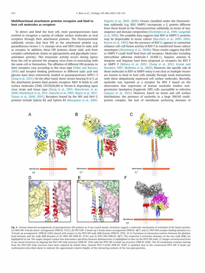
Fig. 3. Various observed arrangements of paramyxovirus HN proteins in X-ray crystal atomic structures suggest a molecular mechanism of activation of the fusion protein.(A) NDV HN ‘4 heads down’ arrangement (PDB ID: 3T1E), (B) PIV5 HN ‘2 heads up-2 heads down arrangement (PDB ID: 4JF7) and (C) PIV5 HN receptor binding domains in a‘4 heads up arrangement’ (PDB ID 1Z4X) placed with respect to the PIV5 HN stalk 4HB domain (PDB ID: 3TSI). (D–E) Variations in interaction surfaces between the globularhead domains and the stalk 4HB domains of (D) NDV HN (PDB ID: 3T1E) and (E) PIV5 HN (PDB ID: 4JF7). The respective F-activation domains on the two stalk 4HBs arehighlighted in red. The single charged residue that determines specificity between Rubulaviruses is highlighted in blue on the PIV5 HN stalk. (F) Images reconstructed fromX-ray crystal structures by aligning the PIV5 HN stalk structure (PDB ID: 3TSI) with the PIV5 HN 4-heads up structure (PDB ID 1Z4X). The 20 ectodomain residues missingfrom the PIV5-HN stalk structure have been replaced by dotted lines. Cleaved PIV5 F-GCNt (PDB ID: 4GIP) is modeled next to the constructed PIV5 HN ‘4 heads up’conformation described above to indicate the approximate relative heights of the interacting surfaces of the two glycoproteins.
S. Bose et al. / Virology 479-480 (2015) 518–531522

nucleolin, and the fact that nucleolin-RSV F interactions are abolishedin the presence of heparin (Holguera et al., 2014; Srivastava andPollard, 1999), the specific role of nucleolin as a receptor in RSV entryis as yet unclear.
Receptor binding by the attachment proteins initiatesactivation of the fusion protein
Paramyxovirus attachment proteins are single-pass, type IImembrane proteins. X-ray crystal structures and electron micro-graphs of soluble attachment protein ectodomains from a varietyof different paramyxoviruses have indicated that HN/H or Gectodomains consist of a large globular head connected to a stalk.The atomic structures of the HN, H or G globular head domainshave been determined for PIV5, NDV, NiV, HeV, MeV and hPIV3(Bowden et al., 2008a, 2010; Colf et al., 2007; Crennell et al., 2000;Hashiguchi et al., 2007, 2011; Lawrence et al., 2004; Santiago et al.,2010; Xu et al., 2008; Yuan et al., 2012, 2011, 2005), and werefound to be highly conserved in terms of protein secondarystructure folds and overall shape. The globular head domain ofHN, H or G binds receptor through a typical six-bladed β-propellerfold common to sialidases. In HN proteins, sialic acid is boundthrough an active site in the center of the β-propeller fold; NiV andHeV G proteins bind Ephrin B2 or B3 through residues located onthe top of the G globular head (Bowden et al., 2008a; Xu et al.,2008) and various binding sites were identified on the sides of theMeV H heads that bind CD46, CD150/SLAM or Nectin-4 (Colf et al.,2007; Hashiguchi et al., 2007, 2011; Mateo et al., 2013, 2014;Santiago et al., 2010). In NDV HN and hPIV3 HN, evidence has beenpresented of a second sialic acid binding site, which lacksneuraminidase activity (Bousse et al., 2004; Mahon et al., 2011;Porotto et al., 2012b; Zaitsev et al., 2004). An atomic structure ofthe globular head domain of NDV HN from a low virulence(lentogenic) strain (Ulster) (Yuan et al., 2012) showed a longerC-terminal extension (Gorman et al., 1990; Nagai et al., 1976;Sakaguchi et al., 1989) that was found to be involved in auto-inhibition of receptor binding by obscuring both the primary andsecondary sialic acid receptor binding sites of NDV HN (Yuan et al.,2012). Proteolytic cleavage of this C-terminal extension is requiredfor receptor binding and fusion in these NDV strains (Yuan et al.,2012). However for hPIV3 HN, the second receptor binding site ismasked by an N-linked glycan at residue 523, and removal of thisN-glycan by mutating the residue at position 523 restores activityof this second site (Mishin et al., 2010) making it difficult to drawconclusive interpretations from results obtained in a study thatinvestigated the biological significance of the second site in hPIV3HN (Porotto et al., 2006, 2007).
The atomic structures of the attachment proteins together withnumerous biochemical and biophysical studies (Bossart et al., 2005;Bowden et al., 2008b; Brindley and Plemper, 2010; Ng et al., 1990;Yuan et al., 2008, 2005) indicate that all of the HN, H or G proteinsshow a tetrameric or more precisely, a dimer-of-dimer arrangementof the globular heads, with the stalk domains playing a majorstabilizing role in the oligomerization process through covalent ornon-covalent associations (Ng et al., 1990, 1989; Parks and Lamb,1990; Yuan et al., 2008). The two X-ray crystal structures of the stalkdomains of paramyxovirus attachment proteins obtained thus farindicate that the stalk domains of NDV HN and PIV5 HN proteins are4-helix bundles (4HBs), with strong central hydrophobic cores (Boseet al., 2011; Yuan et al., 2011) (Fig. 3A–C). Based on these structures,subsequent studies have shown that sequences of other paramyx-ovirus receptor-binding protein stalks can also be modeled as 4HBs(Ader et al., 2012; Maar et al., 2012; Porotto et al., 2012b). Thoughreceptor engagement occurs through the globular head domains ofthe attachment proteins, an overwhelming amount of biochemical
and biophysical evidence suggests that HN, H or G proteins physicallyinteract with the F protein through these stalk domains. Thisinteraction presumably triggers the F protein into carrying out itsrearrangements leading to membrane fusion (Bishop et al., 2007;Bose et al., 2014, 2011; Bousse et al., 1994; Corey and Iorio, 2007;Deng et al., 1999, 1995; Ennis et al., 2010; Melanson and Iorio, 2004,2006; Paal et al., 2009; Porotto et al., 2003; Stone-Hulslander andMorrison, 1999; Tanabayashi and Compans, 1996).
Interestingly, atomic structures of NDV HN (Fig. 3A) and PIV5 HN(Fig. 3B) (Welch et al., 2013; Yuan et al., 2011) full-length proteinsdisplayed two drastically different arrangements of the globular headdomains with respect to the stalk, when compared to the previouslyobserved PIV5 HN dimer-of-dimer arrangement (Fig. 3C) (Yuan et al.,2005). In all these arrangements, the individual monomers and thedimer interface remain constant, while the dimer-of-dimer interfaceis drastically altered. In a recent study by Hashiguchi and colleagues,the atomic structure of MeV H protein bound to its cellular receptorSLAM, suggested that the measles H dimer-of-dimers could poten-tially be arranged into two different tetrameric conformations(Hashiguchi et al., 2011). Thus, taking together these data, alongwith electron micrographs of purified HN proteins (Bose et al., 2012;Yuan et al., 2008), it is evident that paramyxovirus attachmentproteins globular head domains are connected to the stalk domainsthrough flexible, unstructured linkers as a result of which, theglobular head domains can attain various different positions withrespect to the stalk domains.
Unlike the HN, H and G proteins of the Paramyxovirinae subfamilyviruses, atomic structures of G proteins of pneumoviruses like RSV orhMPV have not been obtained to date. Biochemical studies of hMPVG and bovine RSV G predict these to be made up of a hydrophobiccenter between two large mucin-rich domains (Doreleijers et al.,1996; Langedijk et al., 1996), which makes the protein appearphysically larger than F and possibly aids in the immune evasionstrategies of the virus (Leyrat et al., 2014). Though hMPV G has beenassociated with binding to cellular glycosaminoglycans (Thammawatet al., 2008), this was found to be a strain specific effect (Adamsonet al., 2012), suggesting that Pneumovirinae G proteins are notintimately associated with membrane fusion like the HN, H and Gproteins from members of the Paramyxovirinae subfamily.
The fusion protein and the attachment protein physicallyinteract to mediate membrane fusion
For Paramyxovirinae subfamily viruses, the attachment proteinand the fusion protein are believed to directly interact with eachother, while resident on the same membrane and generally thisinteraction requires homotypic pairs of fusion and attachmentproteins derived from the same virus. A few exceptions have beenidentified where heterotypic paramyxovirus F–HN interactions arefunctional in in-vitro assays. For example, hPIV2 HN is able tosubstitute for hPIV4a HN, and SV41 HN or MuV HN are able tosubstitute for hPIV2 HN or PIV5 HN in transfected tissue culturecells (Bose et al., 2014; Tsurudome et al., 1998, 1995), suggestingthat the F–HN interactions may be conserved among closely relatedviruses. The exact nature of the F-G/H/HN interaction and how thisinteraction spatially and temporally couples receptor bindingmediated by HN, H or G, and membrane fusion mediated by F hasbeen a topic of considerable debate. Some HN, H or G stalk mutants,that are deficient for fusion, block the attachment protein-fusionprotein interaction directly (as assessed by co-immunoprecipita-tion) (Melanson and Iorio, 2006; Paal et al., 2009; Stone-Hulslanderand Morrison, 1999). However, the cellular location, duration andstrength of the F–HN, H or G association have been found to differwidely between viruses. Studies of MeV H and CDV H as well as NiVG and HeV G proteins indicate that the strength of the F–G or F–H
S. Bose et al. / Virology 479-480 (2015) 518–531 523

interaction is inversely related to their fusion activity, suggestinga more intimate association between the two proteins prior to Ftriggering (Aguilar et al., 2006; Bishop et al., 2008; Plemper et al.,2001, 2002). On the contrary, certain mutations in the NDV HN stalkshowed a direct relationship between fusion activity and F–HNbinding, while other mutations in the NDV HN and MeV H stalkdomains decrease fusion, but retain F-association (Brindley et al.,2012; Corey and Iorio, 2007; Melanson and Iorio, 2004; Mirza andIorio, 2013). In Henipaviruses, F–G complexes are also believed toremain associated on cell surfaces and F is released to be endocy-tosed, cleavage-activated and re-transported to the plasma mem-brane (Diederich et al., 2005; Pager et al., 2006; Pager and Dutch,2005). For morbilliviruses, F and H remain associated together infusion complexes during transport to the cell surface (Ader et al.,2012; Lee et al., 2008b; Paal et al., 2009; Plemper et al., 2001, 2002),unlike PIV5 and hPIV3 F and HN (Paterson et al., 1997).
Two distinct molecular hypotheses attempt to explain theinteraction between the fusion protein and the attachmentprotein
Based on the above biochemical data, two distinct models haveemerged that could explain F-activation at the right place and theright time. The ‘dissociation’ or ‘clamp’ model (Bossart et al., 2013;Plemper et al., 2001; Sergel et al., 1993) proposes that theattachment protein associates with F intracellularly and ‘clamps’the F protein during transport to the cell surface, preventing Ffrom prematurely converting into the post-fusion form. On bind-ing receptor, the attachment protein releases F allowing themetastable F protein to be destabilized and undergo the refoldingprocess leading to membrane fusion. In the second model, knownas the ‘association’ or ‘provocateur’ model (Connolly et al., 2009),the attachment protein, on binding receptor, actively triggers themetastable F protein through destabilization. This destabilizationand F-activation can also be brought about by heat acting as asurrogate for HN, H or G, which indicates that the attachmentprotein interaction with F overcomes an energy barrier ofF-activation (Ader et al., 2013; Bose et al., 2012; Chan et al.,2012; Connolly et al., 2009). Based on the differences in thebiochemical data of F interactions with HN, H and G protein stalks,it was suggested that for viruses binding protein receptors (with Gor H as attachment proteins), the closer F–G or F–H associationsindicate an F-stabilization through a ‘clamp’ hypothesis, while forthose that bound sialic acid (possessing HN as attachment pro-tein), the more transient F–HN interaction destabilizes F andactivates it for fusion (provocateur hypothesis). However, recentdata for MeV, CDV, NiV, HeV, as well as PIV5 and hPIV3 are notcompatible with the ‘clamp’ hypothesis. Many of the F proteinsfrom the above viruses can be expressed in their prefusion formwithout the attachment protein needing to stabilize F, suggestingthat prefusion F proteins that are involved in F–H, F–G or F–HNinteractions do not require the attachment protein ‘clamp’ forstabilization (Ader et al., 2013; Bose et al., 2012; Brindley et al.,2012; Chan et al., 2012; Connolly et al., 2006). Also many of theseprefusion F proteins could be converted to the postfusion form byelevated temperature, consistent with the notion that the energybarrier of metastable F proteins could be overcome by externalheat, according to the ‘provocateur’ hypothesis. However, the twomodels can be reconciled if not all F–H or F–G interactions areproductive (Jardetzky and Lamb, 2014). Non-productive F–G orF–H associations may occur through alternative interaction regions ofF and H/G (Liu et al., 2015, 2013) or through a conformation of theattachment protein that does not allow productive triggering of F(Avila et al., 2014; Brindley et al., 2015). Such interactions betweenthe attachment and fusion proteins in the ‘pre-receptor binding’
stage may perhaps be necessary for proper folding, cellulartransport or increased F–G or F–H concentrations in fusion com-plexes that can be rapidly triggered (Avila et al., 2014; Brindley etal., 2015). However, based on the above evidence, such ‘pre-receptor binding’ F–G/H or HN interactions do not appear to beessential to stabilize prefusion F, as proposed by the ‘clamp’hypothesis.
In recent years, X-ray crystal structures of the fusion andattachment proteins from various paramyxoviruses have signifi-cantly aided in identifying the productive interactions betweenF–HN, F–H or F–G that lead to membrane fusion. In particular,atomic structures of HN stalk domains have helped to provideresidue-level information of the potential region of contactbetween F and the HN protein stalks. The atomic structures ofthe PIV5 HN and NDV HN stalk domains indicate that the stalk 4HBarrangements have a strong hydrophobic core, with a kink in thecentral portion of the stalks (Bose et al., 2011; Yuan et al., 2011).Mutagenesis studies along the length of the HN, H or G stalks havesuggested that HN, H or G proteins productively interact with the Fprotein broadly through this central region of the stalk domain(Ader et al., 2012; Apte-Sengupta et al., 2013; Bishop et al., 2007;Bose et al., 2014, 2011; Corey and Iorio, 2007; Deng et al., 1999,1995; Liu et al., 2013; Melanson and Iorio, 2004; Navaratnarajahet al., 2012; Stone-Hulslander and Morrison, 1999; Wang and Iorio,1999; Yuan et al., 2011). A large proportion of these residues in thecentral part of attachment protein stalks are hydrophobic innature. In closely related HN proteins, the F-interaction regionwas narrowed down to a highly conserved stretch of 7–8 residuesflanking the kink region of the 4HB (Bose et al., 2014) (Fig. 3D–E).Interestingly, a single acidic amino acid immediately adjacent tothis region determines the homotypic specificity of F–HN interac-tion in the rubulavirus subfamily (Bose et al., 2014). A syntheticantibody bound to this ‘F-activation’ region of the PIV5 HN stalkdomain, was found to strongly neutralize the fusion promotionactivity of PIV5 HN, without affecting the neuraminidase andreceptor-binding activities resident in the globular head domain(Welch et al., 2014). Escape mutants generated in residues of theF-activation domain abrogated the neutralizing activity of the aboveantibody (Welch et al., 2014). In more distantly related paramyx-oviruses, the F-activating region, though located close to the centralpart of the stalk, appears to vary somewhat in its location or is moreextensive in nature (Aguilar et al., 2009; Lee et al., 2008b; Liu et al.,2015; Maar et al., 2012; Paal et al., 2009). Insertion of structurallyrigid α-helical segments within the MeV H stalk to increase thedistance of the F-activating region of the stalk from the anchoringmembrane abrogated fusion, while insertion of these α-helicalsegments above this region did not (Paal et al., 2009), suggestingthat this F-interacting region must be present at an optimal heightmatching with the corresponding H-interaction region of MeV F.
Regions of the fusion protein involved in attachment proteininteractions offer insights into the F-triggering process
A number of regions on various paramyxovirus F proteins havebeen found to potentially interact with the HN, H or G proteins.Many of these regions harbor hydrophobic residues and aresurface exposed, suggesting a protein–protein interaction interfacewith the hydrophobic F-interacting region on the central part ofthe attachment protein stalks, discussed above (Apte-Senguptaet al., 2012; Avila et al., 2014; Bose et al., 2013; Lee et al., 2007)(Fig. 3F). A number of such residues identified on the PIV5 Fprotein and the MeV F protein appear to be localized near animmunoglobulin-like (Ig-like domain) fold of domain II and alsowithin an adjacent hydrophobic cavity (Apte-Sengupta et al., 2012;Bose et al., 2013) (Fig. 2G). This cavity is created by residues from
S. Bose et al. / Virology 479-480 (2015) 518–531524

two adjacent protomers of F (Apte-Sengupta et al., 2012; Avilaet al., 2014; Bose et al., 2013), that juxtapose next to two longβ-strands forming a ‘strap’ that connects the HRA domains of the Fprotein head (Fig. 2H). Mutations of residues in these threeadjacent regions – the Ig-like domain, the hydrophobic cavityand the ‘strap’ regions of different paramyxovirus F proteins havemany effects including, transfer of HN specificity among different Fproteins (Tsurudome et al., 2011, 2013), complete abrogation ofF-activation or destabilization of the F protein, making F lessreliant on the attachment protein trigger (Avila et al., 2014; Bose etal., 2013; Plattet et al., 2009). Through a more recent dynamic view ofPIV5 F refolding it was observed that only the outer loop of the Ig likedomain shows conformational mobility, while the rest of the Ig-likedomain remains unchanged. In contrast, the ‘strap’ regions under-went some of the largest conformational changes as F proteinconverted from its prefusion to its post-fusion form (Poor et al.,2014). Based on these data an attractive hypothesis for F triggeringcould be that HN, H or G interactions through the structurally welldefined Ig-like domain could dock the attachment protein stalk intothe adjacent hydrophobic cavity, which in turn destabilizes the Fprotein by initiating structural rearrangements in the adjoining‘strap’ regions and ultimately culminating in the sequential cascadeof refolding events that convert F into its postfusion form. Furtherstudies are required to tease out more of the details as to how HNinteraction with F leads to F destabilization and eventual membranemerger (Fig. 2G–H).
Molecular cooperation between the different domains of theattachment protein ensures timing of F-activation andmembrane fusion
In silico models of MeV F and H proteins (Lee et al., 2008b; Paalet al., 2009) suggested that prior to fusion activation, the MeVprefusion F and H protein heads are positioned at different levelsto each other (‘staggered heads’ arrangement), where the Hprotein head rises above that of the F protein. However, conflictingresults from electron micrograph studies of virions suggested thatthe glycoprotein spikes appear to be of the same height (“parallel-head” model) (Jain et al., 2008). Recently obtained atomic structuresof HN proteins of PIV5 (Welch et al., 2013; Yuan et al., 2005) andNDV (Yuan et al., 2011) and electron microscopy data of purifiedproteins (Bose et al., 2012; Yuan et al., 2008) have suggested thatthe attachment protein globular heads can attain various differentconformational arrangements. Of these, a ‘four heads down’ structureobserved for NDV HN has the globular head dimers folded back andmaking contacts with the 4HB stalk domains (Yuan et al., 2011) thuslowering the height of HN. This, or a similar arrangement couldrepresent the “parallel head” model observed by EM on virions. Onthe other hand, the ‘four heads up’ arrangement of PIV5 HN globularheads forming a dimer-of-dimer interface (Yuan et al., 2005) repre-sents a taller form of the HN protein as proposed by the ‘staggeredhead’ model. Interestingly, PIV5 HN was also crystallized in a thirdarrangement, which showed a shifted dimer-of dimer interface andappeared to represent a hybrid between the two HN arrange-ments described above (2 heads up-2 heads down arrangement)(Welch et al., 2013), with one dimer making contacts with the 4HBstalk domain and the other maintaining the arrangement seen inthe 4-heads up structure. As there was very little change inconformation of HN, H and G monomers when they bind receptor(Bowden et al., 2008a; Colf et al., 2007; Lawrence et al., 2004;Santiago et al., 2010; Xu et al., 2008; Yuan et al., 2005), these datasuggested that the rearrangements of the attachment proteinglobular head domains might have a role in translating the receptorbinding event into F-activation through the stalk domains. Notablyhowever, except for PIV5 HN (Yuan et al., 2005), all the other
receptor-ligand complexes studies were carried out with solubleattachment protein globular heads.
A possible model of how the F-activation signal could be linkedto receptor binding proposed the globular heads of HN, H or G onbinding their specific receptors, transmit a series of specificconformational changes through the linkers down into the stalkdomain. The stalk domain in turn undergoes a series of conforma-tional changes that mediate a productive F-interaction leading toF-activation (Plemper et al., 2011; Porotto et al., 2011, 2012a,2012c). However, this model has been difficult to rationalize inthe light of some recent results. Firstly, measles H can be re-targeted to bind alternative molecules on cells by inserting 6-Histags or other protein-binding domains into the globular head(Allen et al., 2006; Navaratnarajah et al., 2011; Paraskevakou et al.,2007), suggesting that the specificity of the receptor-bindinginteraction, though critical for determining the virus host range,is not critical in transmitting specific conformational changes tothe stalk. Also measles H binds multiple receptors (e.g. SLAM,Nectin 4 or CD46) through partially overlapping but very differentbinding surfaces in the globular head (Colf et al., 2007; Hashiguchiet al., 2007, 2011; Mateo et al., 2013, 2014; Santiago et al., 2010),but they eventually channel all these inputs into a single output –F-activation and membrane fusion. Secondly, many differentfunctionally active chimeric proteins have been generated byappending the globular head of one attachment protein tothe stalk of another (Bose et al., 2012; Deng et al., 1995; Farzanet al., 2011; Porotto et al., 2011, 2012b; Talekar et al., 2013; Wanget al., 2004). Importantly, in all of these chimeric proteins, theF proteins that can be successfully activated are always homotypicto the stalk domain of the chimeric attachment protein. Thus,if a specific receptor-binding signal propagated down the flexiblelinkers into the stalk these chimeric proteins should have failedto maintain the conformational crosstalk between the head andthe stalk. Thirdly, as there is very little change in the globularheads on receptor binding, the rearrangement generated wouldnot be substantial enough to propagate down into the 4HBstalk through the highly flexible linker domains connecting thehead and the stalk domains. Fourthly, it was observed that aheadless stalk of PIV5 HN protein could successfully activate PIV5 F(Bose et al., 2012), suggesting that productive F-activation ispossible even in complete absence of the head domains. Inaddition, this PIV5 HN headless stalk domain could activate Fmore efficiently at sub-optimal temperatures when compared tothe full-length HN protein and also activate a hypofusogenicmutant of PIV5 F (P22L) at significantly higher levels comparedto full length HN (Bose et al., 2012), suggesting that the HNglobular heads are associated with an energy requirement for Factivation. Subsequently through the work of different groups,fusion activation by headless stalks has now been extended toother paramyxoviruses of different genera – MeV H, NiV G, MuVHN and NDV HN (Bose et al., 2014; Brindley et al., 2013; Liu et al.,2013) – demonstrating that the receptor binding event itself is notspecific in the process of F protein activation. Nonetheless, ifreceptor-binding to the HN, H or G globular head domains arenot directly responsible for productive F-triggering, then what roledo they play in mediating the membrane fusion process and entryof paramyxoviruses?
Taking together the above structural and functional data, a newmodel for F activation has recently been proposed (Bose et al.,2012) (Fig. 4). The ‘stalk exposure’ model suggests that prior tobinding receptor, the globular heads of HN, H or G proteinsphysically restrict the access of the F protein to the F-activatingregion residing on the HN, H or G stalk domains. This could bepossible if the attachment protein heads attain an arrangementwith respect to the stalk 4HB, similar to the ‘4 heads down’arrangement observed for NDV HN (Yuan et al., 2011). Binding of
S. Bose et al. / Virology 479-480 (2015) 518–531 525

receptor results in a molecular rearrangement of the dimer-of-dimer globular heads of attachment proteins, causing them toattain a conformation close to that observed in the PIV5 HN4-heads up atomic structure (Yuan et al., 2005) or the PIV5 HN2 heads up -2 heads down atomic structure (Welch et al., 2013).These conformations expose the F-activating regions of the HN, Hor G stalk domains, allowing the F protein to interact with thisregion of the HN, H or G stalk and be activated to undergorefolding and subsequent membrane fusion (Bose et al., 2014,2012). Interestingly, it was observed that the F-activating functionof the HN protein could be maintained even after the PIV5HN molecule was locked into its 2 heads up- 2 heads down
conformation using engineered disulfide bonds (Welch et al.,2013) and that binding of only a single dimer of measles H to itsreceptor was sufficient to trigger F (Brindley et al., 2012). The ‘stalkexposure’model suggests that the globular heads of HN, H or G areinvolved in the critical role of ‘regulatory domains’, restrictingaccess of F to interact with the ‘activating domains’ present in thestalk. The restriction is only lifted when the head domains bindreceptor. The ‘stalk exposure’ model accounts for the fact that adiverse variety of natural host cell receptors or artificial moleculescan be recognized through different interfaces and multiple activesites of the HN, H or G proteins as inputs, all of which can then beconverted into a common output that is, F-activation.
Fig. 4. (A–C) Models of receptor-dependent fusion activation for paramyxoviruses of the Paramyxovirinae subfamily. Structural and functional data indicate that the HN, H orG attachment proteins (yellow) of this subfamily are structurally related and have a globular, C-terminal receptor-binding domain (head) that can interact with various typesof host receptors, through receptor binding-sites in the globular heads (blue rings). Putative regions of F-interaction on HN, H or G proteins are indicated on the stalkdomains in red. The F proteins (purple) fold into a functional, prefusion state in the presence or absence of the attachment proteins, but without requiring the attachmentprotein as a stabilizing ‘clamp’. The F proteins are cleaved by proteases to release a hydrophobic fusion peptide (light blue). The HRA domain of F, which refolds intoelongated helices post triggering is shown in dark green. (A) F and HN proteins are transported individually to the cell surface and biochemical data suggests that the F–HNinteraction is transient. Receptor binding by HN heads results in stalk exposure and possibly a ‘induced fit’ mechanism between the F protein head and the exposedF-activating region on the HN stalk that triggers F to undergo refolding. (B) In morbilliviruses, where F and H are intimately associated in fusion complexes during cellulartransport, it has been proposed that the H globular head domains maintain the stalk domains in a ‘pre-triggering’ conformation, possibly through H head-stalk contacts. Stalkexposure, coupled with release of the stalk domains from the globular heads, allows the F protein to mediate productive interaction with the F-activating regions of the Hstalk. (C) In Henipaviruses, the initial F–G interaction may be mediated through F interacting with the globular head domains of G or the C-terminal upper portion of the Gstalk, which prevents premature F activation during cellular transport within F–G complexes. Stalk exposure and a switch of binding interfaces, allows F to undergoproductive interaction with the exposed F-activating domains in the G stalk and be triggered to undergo refolding. (D) For Pneumovirinae, the mechanism of F triggering isyet unclear and it is likely that the distinct G protein does not play a role in this process. The F protein is believed to bind specifically to cellular receptors through bindingsites in the F globular head (white ring) and the timing of F-cleavage by both cellular and extracellular proteases is believed to play a role in the triggering process for some ofthe Pneumovirinae.
S. Bose et al. / Virology 479-480 (2015) 518–531526

Diverse inhibitory mechanisms found in differentparamyxoviruses appear to regulate access of the fusionprotein to the attachment protein stalk prior to ‘stalk exposure’
In Henipavirus and morbilliviruses, preformed F–G or F–H com-plexes are transported to the cell surface respectively before incor-poration into viral particles, while for those paramyxoviruses withHN as the receptor binding protein F–HN interactions occur primarilyat the cell surface. Preformed F–G or F–H complexes likely increasethe efficiency of productive F interactions but increase the risk ofpremature F-triggering within these complexes. Separate transport ofF and HN protein possibly keeps these two glycoproteins frominteracting prior to HN binding receptor. Recent studies in Henipa-viruses and morbilliviruses have suggested that in addition to stalkexposure, mechanisms involving the globular heads or the upperparts of the stalk may prevent F from interacting prematurely withthe F-activating domain of the H and G stalks, (Avila et al., 2014;Brindley et al., 2015; Liu et al., 2015, 2013).
In many mutagenesis studies involving HN proteins, the globularhead-proximal upper part of the stalks of these HN proteins wereshown to tolerate mutations and addition of carbohydrate moietieswithin the 4HB, without affecting the F-activation process (Bishop et al.,2007; Bose et al., 2011; Corey and Iorio, 2007; Melanson and Iorio,2004; Stone-Hulslander and Morrison, 1999; Wang and Iorio, 1999). Onthe other hand, many lines of evidence have shown that the centralF-activating region of almost all the stalk domains of HN, H or G requiresome structural rearrangements to interact with and activate F (Aderet al., 2012; Apte-Sengupta et al., 2013; Bose et al., 2014; Brindley et al.,2012; Navaratnarajah et al., 2012). This requirement of a ‘primed’ stalkdomain conformation that can successfully interact with and activate Fis also evident in the headless stalks. However, while MeV headlessstalks required an active stabilization through tags to keep themfunctional (Brindley et al., 2013), the NDV HN headless stalk domainswere inhibited in their ability to activate F by engineering disulfidebonds within the 4 HB, similar to that observed in full length NDV HN(Bose et al., 2014). Thus for F–HN protein interactions it has beenproposed that the F proteins might interact with the central part of theHN stalks through an ‘induced fit’ mechanism, where the conforma-tional changes in the stalk are passive and do not directly depend onthe presence of the globular head domains (Bose et al., 2014) (Fig. 4A).
For morbilliviruses, the F-interaction region on the H stalk domainappears to be more extensive compared to those of HN proteins(Apte-Sengupta et al., 2013; Lee et al., 2008b; Navaratnarajah et al.,2014; Paal et al., 2009), but overlaps with residues co-linear to those ofthe HN stalk implicated for F triggering. The MeV H stalk also containsa structurally stabilized region in the C-terminal part of the stalk thatextends beyond that of HN stalks (Navaratnarajah et al., 2014, 2012;Paal et al., 2009). This extensive contact region may provide clues tohow F-H interactions are mediated within the fusion complexes andhow F is prevented from triggering prematurely. A recent study hassuggested that the MeV H globular heads actively inhibit the structuralrearrangements required by the H stalk to trigger F (Brindley et al.,2015). On receptor binding, the H globular heads undergo a con-formational change, exposing the F-activating domain and releasingthe inhibition on the stalk to allow conformational changes in the stalkthat trigger F (Avila et al., 2014; Brindley et al., 2015). These studiesalso demonstrated that during cellular transport of the associated F–Hglycoprotein complexes, F cleavage was found to weaken the F–Hinteraction, following which F is primed for activation and dissociatesfrom the F–H complex. However, it is yet unclear whether the F–Hstalk contacts remain constant during this process or switch from lessproductive to more productive interactions within the extensiveF-interacting domain of H. It may be possible for MeV F to form aninitial association with the H stalk while the globular heads of H arefolded down in a ‘4-heads down-like’ conformation. On F cleavage,this interaction is weakened and as soon as at least one H dimer binds
receptor (Brindley et al., 2012) and the stalk is exposed, F could nowswitch to a more productive interaction with the stalk. However,importantly this switch should not be able to occur by stalk exposurealone, as cleavage of F as well as rearrangements of the stalk appear tobe important for productive F interaction. An initial MeV F interactionwith the MeV H stalk could be possible considering that 'heads down'orientations of the globular head monomers with respect to the 4HBstalks of closely related NDV HN and PIV5 HN expose variable regionsof the stalk domain (compare Fig. 3D and E). This, and the variousobserved MeV globular head arrangements in the atomic structure(Hashiguchi et al., 2011) suggest that the extent of F accessibility to thestalk in the ‘4 heads down’-like conformation may vary significantlyfor morbillivirus H proteins. Though the exact details of how morbilli-virus F–H complexes initially prevent premature F activation require tobe worked out in detail, biochemical and modeling studies ofmorbilliviruses (Brindley et al., 2015; Navaratnarajah et al., 2014,2012) suggest that the core paramyxovirus fusion mechanism isconserved according to the ‘stalk exposure’ model, where the HN, Hor G head domains act as regulators and the stalks act as activatingdomains (Bose et al., 2012).
Liu and colleagues recently proposed a 3-step process ofHenipavirus F activation, which suggests that, a bidentate G–Finteraction of the F protein head with the G protein upper stalk orhead prevents F from interacting with the G stalk F-activatingregion prematurely (Liu et al., 2013). On G binding receptor,globular head rearrangement and stalk exposure allows the Finteraction to switch to a more productive association with thecentral part of the stalk, leading to F-triggering and membranefusion. Though atomic structures of full-length Henipavirus Gproteins have not yet been obtained, given that Henipavirus Gproteins possess a unique, highly structured, disulfide-stabilizedregion in the upper stalk (Aguilar et al., 2009; Maar et al., 2012),which was found to be important for fusion promotion; the initialpre-receptor-bound G-F complex interaction could possibly occurthrough this region (Fig. 4C) (Liu et al., 2013). A very recent studyby this group has identified a stretch of amino acids at themembrane-distal portion of the NiV G stalk that are importantfor G–F interactions, receptor induced conformational changes andF-triggering (Liu et al., 2015). Similar to the F–H interactions, it ispossible for F–G interactions to occur through the upperC-terminal part of the attachment protein stalk even when allfour heads are in a ‘down’ conformation as the shape of the Gheads and angle of contact with the stalk most likely varies amongparamyxoviruses (compare Fig. 3D and E).
Thus for paramyxoviruses that utilize two glycoproteins tomediate cell entry, it is becoming increasingly clear that the coremolecular mechanism of membrane fusion is highly conserved. Theattachment protein globular heads act as regulatory domains, whichon the binding of host receptors determine the correct spatio-temporal juncture at which the fusion protein must refold in orderto productively carry out membrane fusion. To prevent prematureF-activation, paramyxoviruses have evolved various differentmechanisms that either separate the glycoprotein pairs physicallyduring transport through the exocytic pathway to prevent F–HNinteraction or abrogate F-triggering through various intramolecularand intermolecular exchanges between these glycoproteins. Forsome Pneumovirinae, the spatio-temporal nature of F protein clea-vage appears to play a role in activation of F, but significant details inthe mechanism of activation for membrane fusion in this subfamilyof paramyxoviruses remain unknown (Fig. 4D).
Acknowledgments
Research in the authors' laboratories was supported in part byNational Institutes of Health Research Grants R01-AI-23173 (to R.A.L.),
S. Bose et al. / Virology 479-480 (2015) 518–531 527
and R01-GM-61050 (to T.S.J.). R.A.L. is an Investigator of the HowardHughes Medical Institute
References
Adamson, P., Thammawat, S., Muchondo, G., Sadlon, T., Gordon, D., 2012. Diversityin glycosaminoglycan binding amongst hMPV G protein lineages. Viruses 4,3785–3803.
Ader, N., Brindley, M., Avila, M., Orvell, C., Horvat, B., Hiltensperger, G., Schneider-Schaulies, J., Vandevelde, M., Zurbriggen, A., Plemper, R.K., Plattet, P., 2013.Mechanism for active membrane fusion triggering by morbillivirus attachmentprotein. J. Virol. 87, 314–326.
Ader, N., Brindley, M.A., Avila, M., Origgi, F.C., Langedijk, J., Orvell, C., Vandevelde, M.,Zurbriggen, A., Plemper, R.K., Plattet, P., 2012. Structural rearrangements of thecentral region of the morbillivirus attachment protein stalk domain trigger Fprotein refolding for membrane fusion. J. Biol. Chem. 287, 16324–16334.
Aguilar, H.C., Ataman, Z.A., Aspericueta, V., Fang, A.Q., Stroud, M., Negrete, O.A.,Kammerer, R.A., Lee, B., 2009. A novel receptor-induced activation site in theNipah virus attachment glycoprotein (G) involved in triggering the fusionglycoprotein (F). J. Biol. Chem. 284, 1628–1635.
Aguilar, H.C., Matreyek, K.A., Filone, C.M., Hashimi, S.T., Levroney, E.L., Negrete, O.A.,Betrolotti-Ciarlet, A., Choi, D.Y., McHardy, I., Fulcher, J.A., Su, S.V., Wolf, M.C.,Kohatsu, L., Baum, L.G., Lee, B., 2006. N-glycans on Nipah virus fusion proteinprotect against neutralization but reduce membrane fusion and viral entry.J. Virol. 80, 4878–4889.
Allen, C., Vongpunsawad, S., Nakamura, T., James, C.D., Schroeder, M., Cattaneo, R.,Giannini, C., Krempski, J., Peng, K.W., Goble, J.M., Uhm, J.H., Russell, S.J., Galanis, E.,2006. Retargeted oncolytic measles strains entering via the EGFRvIII receptormaintain significant antitumor activity against gliomas with increased tumorspecificity. Cancer Res. 66, 11840–11850.
Apte-Sengupta, S., Navaratnarajah, C.K., Cattaneo, R., 2013. Hydrophobic andcharged residues in the central segment of the measles virus hemagglutininstalk mediate transmission of the fusion-triggering signal. J. Virol. 87,10401–10404.
Apte-Sengupta, S., Negi, S., Leonard, V.H., Oezguen, N., Navaratnarajah, C.K., Braun,W., Cattaneo, R., 2012. Base of the measles virus fusion trimer head receives thesignal that triggers membrane fusion. J. Biol. Chem. 287, 33026–33035.
Avila, M., Alves, L., Khosravi, M., Ader-Ebert, N., Origgi, F., Schneider-Schaulies, J.,Zurbriggen, A., Plemper, R.K., Plattet, P., 2014. Molecular determinants definingthe triggering range of prefusion F complexes of canine distemper virus. J. Virol.88, 2951–2966.
Bartesaghi, A., Merk, A., Borgnia, M.J., Milne, J.L., Subramaniam, S., 2013. Prefusionstructure of trimeric HIV-1 envelope glycoprotein determined by cryo-electronmicroscopy. Nat. Struct. Mol. Biol. 20, 1352–1357.
Behera, A.K., Matsuse, H., Kumar, M., Kong, X., Lockey, R.F., Mohapatra, S.S., 2001.Blocking intercellular adhesion molecule-1 on human epithelial cells decreasesrespiratory syncytial virus infection. Biochem. Biophys. Res. Commun. 280, 188–195.
Biacchesi, S., Pham, Q.N., Skiadopoulos, M.H., Murphy, B.R., Collins, P.L., Buchholz, U.J.,2005. Infection of nonhuman primates with recombinant human metapneumo-virus lacking the SH, G, or M2-2 protein categorizes each as a nonessentialaccessory protein and identifies vaccine candidates. J. Virol. 79, 12608–12613.
Biacchesi, S., Skiadopoulos, M.H., Yang, L., Lamirande, E.W., Tran, K.C., Murphy, B.R.,Collins, P.L., Buchholz, U.J., 2004. Recombinant human metapneumoviruslacking the small hydrophobic SH and/or attachment G glycoprotein: deletionof G yields a promising vaccine candidate. J. Virol. 78, 12877–12887.
Bishop, K.A., Hickey, A.C., Khetawat, D., Patch, J.R., Bossart, K.N., Zhu, Z., Wang, L.F.,Dimitrov, D.S., Broder, C.C., 2008. Residues in the stalk domain of the Hendravirus G glycoprotein modulate conformational changes associated with recep-tor binding. J. Virol. 82, 11398–11409.
Bishop, K.A., Stantchev, T.S., Hickey, A.C., Khetawat, D., Bossart, K.N., Krasnoperov,V., Gill, P., Feng, Y.R., Wang, L., Eaton, B.T., Wang, L.F., Broder, C.C., 2007.Identification of Hendra virus G glycoprotein residues that are critical forreceptor binding. J. Virol. 81, 5893–5901.
Bonaparte, M.I., Dimitrov, A.S., Bossart, K.N., Crameri, G., Mungall, B.A., Bishop, K.A.,Choudhry, V., Dimitrov, D.S., Wang, L.F., Eaton, B.T., Broder, C.C., 2005. Ephrin-B2 ligand is a functional receptor for Hendra virus and Nipah virus. Proc. Natl.Acad. Sci. USA 102, 10652–10657.
Bose, S., Heath, C.M., Shah, P.A., M., A., Jardetzky, T.S., Lamb, R.A., 2013. Mutations inthe paramyxovirus 5 fusion protein reveal domains importnant for fusiontriggering and metastability. J. Virol. 87, 13520–13531.
Bose, S., Song, A.S., Jardetzky, T.S., Lamb, R.A., 2014. Fusion activation throughattachment protein stalk domains indicates a conserved core mechamism ofparamyxovirus entry into cells. J. Virol. 88, 3925–3941.
Bose, S., Welch, B.D., Kors, C.A., Yuan, P., Jardetzky, T.S., Lamb, R.A., 2011. Structureand mutagenesis of the parainfluenza virus 5 hemagglutinin-neuraminidasestalk domain reveals a four-helix bundle and the role of the stalk in fusionpromotion. J. Virol. 85, 12855–12866.
Bose, S., Zokarkar, A., Welch, B.D., Leser, G.P., Jardetzky, T.S., Lamb, R.A., 2012. Fusionactivation by a headless parainfluenza virus 5 hemagglutinin-neuraminidasestalk suggests a modular mechanism for triggering. Proc. Natl. Acad. Sci. USA109, E2625–E2634.
Bossart, K.N., Crameri, G., Dimitrov, A.S., Mungall, B.A., Feng, Y.R., Patch, J.R.,Choudhary, A., Wang, L.F., Eaton, B.T., Broder, C.C., 2005. Receptor binding,
fusion inhibition, and induction of cross-reactive neutralizing antibodies by asoluble G glycoprotein of Hendra virus. J. Virol. 79, 6690–6702.
Bossart, K.N., Fusco, D.L., Broder, C.C., 2013. Paramyxovirus entry. Adv. Exp. Med.Biol. 790, 95–127.
Bousse, T., Takimoto, T., Gorman, W.L., Takahashi, T., Portner, A., 1994. Regions onthe hemagglutinin-neuraminidase proteins of human parainfluenza virus type-1and Sendai virus important for membrane fusion. Virology 204, 506–514.
Bousse, T.L., Taylor, G., Krishnamurthy, S., Portner, A., Samal, S.K., Takimoto, T., 2004.Biological significance of the second receptor binding site of Newcastle diseasevirus hemagglutinin-neuraminidase protein. J. Virol. 78, 13351–13355.
Bowden, T.A., Aricescu, A.R., Gilbert, R.J., Grimes, J.M., Jones, E.Y., Stuart, D.I., 2008a.Structural basis of Nipah and Hendra virus attachment to their cell-surfacereceptor ephrin-B2. Nat. Struct. Biol. 15, 567–572.
Bowden, T.A., Crispin, M., Harvey, D.J., Aricescu, A.R., Grimes, J.M., Jones, E.Y., Stuart, D.I.,2008b. Crystal structure and carbohydrate analysis of Nipah virus attachmentglycoprotein: a template for antiviral and vaccine design. J. Virol. 82, 11628–11636.
Bowden, T.A., Crispin, M., Harvey, D.J., Jones, E.Y., Stuart, D.I., 2010. Dimericarchitecture of the Hendra virus attachment glycoprotein: evidence for aconserved mode of assembly. J. Virol. 84, 6208–6217.
Brindley, M.A., Chaudhury, S., Plemper, R.K., 2015a. Measles virus glycoproteincomplexes preassemble intracellularly and relax during transport to the cellsurface in preparation for fusion. J. Virol. 89, 1230–1241.
Brindley, M.A., Plattet, P., Plemper, R.K., 2014. Efficient replication of a paramyx-ovirus independent of full zippering of the fusion protein six-helix bundledomain. Proc. Natl. Acad. Sci. USA 111, E3795–E3804.
Brindley, M.A., Plemper, R.K., 2013. Blue native PAGE and biomolecular comple-mentation reveal a tetrameric or higher-order oligomer organization of thephysiological measles virus attachment protein H. J. Virol. 84, 12174–12184.
Brindley, M.A., Suter, R., Schestak, I., Kiss, G., Wright, E.R., Plemper, R.K., 2013. Astabilized headless measles virus attachment protein stalk efficiently triggersmembrane fusion. J. Virol. 87, 11693–11703.
Brindley, M.A., Takeda, M., Plattet, P., Plemper, R.K., 2012. Triggering the measlesvirus membrane fusion machinery. Proc. Natl. Acad. Sci. USA 109, E3018–E3027.
Burkard, C., Verheije, M.H., Wicht, O., van Kasteren, S.I., van Kuppeveld, F.J.,Haagmans, B.L., Pelkmans, L., Rottier, P.J., Bosch, B.J., de Haan, C.A., 2014.Coronavirus cell entry occurs through the endo-/lysosomal pathway in aproteolysis-dependent manner. PLoS Pathog. 10, e1004502.
Caffrey, M., Kaufman, J., Stahl, S., Wingfield, P., Gronenborn, A.M., Clore, G.M., 1999.Monomer-trimer equilibrium of the ectodomain of SIV gp41: insight into themechanism of peptide inhibition of HIV infection. Protein Sci. 8, 1904–1907.
Chan, D.C., Fass, D., Berger, J.M., Kim, P.S., 1997. Core structure of gp41 from the HIVenvelope glycoprotein. Cell 89, 263–273.
Chan, Y.P., Lu, M., Dutta, S., Yan, L., Barr, J., Flora, M., Feng, Y.R., Xu, K., Nikolov, D.B.,Wang, L.F., Skiniotis, G., Broder, C.C., 2012. Biochemical, conformational andimmunogenic analysis of soluble trimeric forms of Henipavirus fusion glyco-proteins. J. Virol. 86, 11457–11471.
Chandran, K., Sullivan, N.J., Felbor, U., Whelan, S.P., Cunningham, J.M., 2005.Endosomal proteolysis of the Ebola virus glycoprotein is necessary for infection.Science 308, 1643–1645.
Chang, A., Masante, C., Buchholz, U.J., Dutch, R.E., 2012. Human metapneumovirus(HMPV) binding and infection are mediated by interactions between the HMPVfusion protein and heparan sulfate. J. Virol. 86, 3230–3243.
Colf, L.A., Juo, Z.S., Garcia, K.C., 2007. Structure of the measles virus hemagglutinin.Nat. Struct. Mol. Biol. 14, 1227–1228.
Connolly, S.A., Leser, G.P., Jardetzky, T.S., Lamb, R.A., 2009. Bimolecular comple-mentation of paramyxovirus fusion and hemagglutinin-neuraminidase proteinsenhances fusion: implications for the mechanism of fusion triggering. J. Virol.83, 10857–10868.
Connolly, S.A., Leser, G.P., Yin, H.S., Jardetzky, T.S., Lamb, R.A., 2006. Refolding of aparamyxovirus F protein from prefusion to postfusion conformations observedby liposome binding and electron microscopy. Proc. Natl. Acad. Sci. USA 103,17903–17908.
Corey, E.A., Iorio, R.M., 2007. Mutations in the stalk of the measles virushemagglutinin protein decrease fusion but do not interfere with virus-specific interaction with the homologous fusion protein. J. Virol. 81, 9900–9910.
Crennell, S., Takimoto, T., Portner, A., Taylor, G., 2000. Crystal structure of themultifunctional paramyxovirus hemagglutinin-neuraminidase. Nat. Struct. Biol.7, 1068–1074.
Daszak, P., Zambrana-Torrelio, C., Bogich, T.L., Fernandez, M., Epstein, J.H., Murray,K.A., Hamilton, H., 2012. Interdisciplinary approaches to understanding diseaseemergence: the past, present, and future drivers of Nipah virus emergence.Proc. Natl. Acad. Sci. USA 110, 3681–3688.
Deng, R., Wang, Z., Mahon, P.J., Marinello, M., Mirza, A., Iorio, R.M., 1999. Mutationsin the Newcastle disease virus hemagglutinin-neuraminidase protein thatinterfere with its ability to interact with the homologous F protein in thepromotion of fusion. Virology 253, 43–54.
Deng, R., Wang, Z., Mirza, A.M., Iorio, R.M., 1995. Localization of a domain on theparamyxovirus attachment protein required for the promotion of cellular fusionby its homologous fusion protein spike. Virology 209, 457–469.
Diederich, S., Moll, M., Klenk, H.D., Maisner, A., 2005. The Nipah virus fusion proteinis cleaved within the endosomal compartment. J. Biol. Chem. 280,29899–29903.
Doreleijers, J.F., Langedijk, J.P., Hard, K., Boelens, R., Rullmann, J.A., Schaaper, W.M.,van Oirschot, J.T., Kaptein, R., 1996. Solution structure of the immunodominantregion of protein G of bovine respiratory syncytial virus. Biochemistry 35,14684–14688.
S. Bose et al. / Virology 479-480 (2015) 518–531528
Dorig, R.E., Marcil, A., Chopra, A., Richardson, C.D., 1993. The human CD46 moleculeis a receptor for measles virus (Edmonston strain). Cell 75, 295–305.
Drexler, J.F., Corman, V.M., Muller, M.A., Maganga, G.D., Vallo, P., Binger, T., Gloza-Rausch, F.,Rasche, A., Yordanov, S., Seebens, A., Oppong, S., Adu Sarkodie, Y., Pongombo, C.,Lukashev, A.N., Schmidt-Chanasit, J., Stocker, A., Carneiro, A.J., Erbar, S., Maisner, A.,Fronhoffs, F., Buettner, R., Kalko, E.K., Kruppa, T., Franke, C.R., Kallies, R., Yandoko, E.R.,Herrler, G., Reusken, C., Hassanin, A., Kruger, D.H., Matthee, S., Ulrich, R.G., Leroy, E.M.,Drosten, C., 2012. Bats host major mammalian paramyxoviruses. Nat. Commun.3, 796.
Duprex, W.P., McQuaid, S., Hangartner, L., Billeter, M.A., Rima, B.K., 1999. Observa-tion of measles virus cell-to-cell spread in astrocytoma cells by using a greenfluorescent protein-expressing recombinant virus. J. Virol. 73, 9568–9575.
Ehrengruber, M.U., Ehler, E., Billeter, M.A., Naim, H.Y., 2002. Measles virus spreadsin rat hippocampal neurons by cell-to-cell contact and in a polarized fashion.J. Virol. 76, 5720–5728.
Ennis, M.K., Hu, C., Naik, S.K., Hallak, L.K., Peng, K.W., Russell, S.J., Dingli, D., 2010.Mutations in the stalk region of the measles virus hemagglutinin inhibitsyncytium formation but not virus entry. J. Virol. 84, 10913–10917.
Farzan, S.F., Palermo, L.M., Yokoyama, C.C., Orefice, G., Fornabaio, M., Sarkar, A.,Kellogg, G.E., Greengard, O., Porotto, M., Moscona, A., 2011. Premature activa-tion of the paramyxovirus fusion protein before target cell attachment withcorruption of the viral fusion machinery. J. Biol. Chem. 286, 37945–37954.
Gahr, P., DeVries, A.S., Wallace, G., Miller, C., Kenyon, C., Sweet, K., Martin, K., White, K.,Bagstad, E., Hooker, C., Krawczynski, G., Boxrud, D., Liu, G., Stinchfield, P., LeBlanc, J.,Hickman, C., Bahta, L., Barskey, A., Lynfield, R., 2014. An outbreak of measles in anundervaccinated community. Pediatrics 134, e220–e228.
Gonzâlez-Reyes, L., Ruiz-Argèuello, M.B., Garcâia-Barreno, B., Calder, L., Lâopez, J.A.,Albar, J.P., Skehel, J.J., Wiley, D.C., Melero, J.A., 2001. Cleavage of the humanrespiratory syncytial virus fusion protein at two distinct sites is required foractivation of membrane fusion. Proc. Natl. Acad. Sci. USA 98, 9859–9864.
Gorman, W.L., Gill, D.S., Scroggs, R.A., Portner, A., 1990. The hemagglutinin-neuraminidase glycoproteins of human parainfluenza virus type 1 and Sendaivirus have high structure-function similarity with limited antigenic cross-reactivity. Virology 175, 211–221.
Hashiguchi, T., Kajikawa, M., Maita, N., Takeda, M., Kuroki, K., Sasaki, K., Kohda, D.,Yanagi, Y., Maenaka, K., 2007. Crystal structure of measles virus hemagglutininprovides insight into effective vaccines. Proc. Natl. Acad. Sci. USA 104,19535–19540.
Hashiguchi, T., Ose, T., Kubota, M., Maita, N., Kamishikiryo, J., Maenaka, K., Yanagi, Y.,2011. Structure of the measles virus hemagglutinin bound to its cellularreceptor SLAM. Nat. Struct. Biol. 18, 135–141.
Heminway, B.R., Yu, Y., Galinski, M.S., 1994a. Paramyxovirus mediated cell fusionrequires co-expression of both the fusion and hemagglutinin-neuraminidaseglycoproteins. Virus Res. 31, 1–16.
Heminway, B.R., Yu, Y., Tanaka, Y., Perrine, K.G., Gustafson, E., Bernstein, J.M.,Galinski, M.S., 1994b. Analysis of respiratory syncytial virus F, G, and SHproteins in cell fusion. Virology 200, 801–805.
Holguera, J., Villar, E., Munoz-Barroso, I., 2014. Identification of cellular proteinsthat interact with Newcastle disease virus and human respiratory syncytialvirus by a two-dimensional virus overlay protein binding assay (VOPBA). VirusRes. 191, 138–142.
Homma, M., 1971. Trypsin action on the growth of Sendai virus in tissue culturecells. I. Restoration of the infectivity for L cells by direct action of trypsin on Lcell-borne Sendai virus. J. Virol. 8, 619–629.
Homma, M., Ohuchi, M., 1973. Trypsin action on the growth of Sendai virus in tissueculture cells. III. Structural differences of Sendai viruses grown in eggs andtissue culture cells. J. Virol. 12, 1457–1465.
Horvath, C.M., Paterson, R.G., Shaughnessy, M.A., Wood, R., Lamb, R.A., 1992.Biological activity of paramyxovirus fusion proteins: factors influencing forma-tion of syncytia. J. Virol. 66, 4564–4569.
Hu, X., Ray, R., Compans, R.W., 1992. Functional interactions between the fusionprotein and hemagglutinin-neuraminidase of human parainfluenza viruses.J. Virol. 66, 1528–1534.
Jain, S., McGinnes, L.W., Morrison, T.G., 2008. Overexpression of thiol/disulfideisomerases enhances membrane fusion directed by the Newcastle disease virusfusion protein. J. Virol. 82, 12039–12048.
Jardetzky, T.S., Lamb, R.A., 2014. Activation of paramyxovirus membrane fusion andvirus entry. Curr. Opin. Virol. 5, 24–33.
Julien, J.P., Cupo, A., Sok, D., Stanfield, R.L., Lyumkis, D., Deller, M.C., Klasse, P.J.,Burton, D.R., Sanders, R.W., Moore, J.P., Ward, A.B., Wilson, I.A., 2013. Crystalstructure of a soluble cleaved HIV-1 envelope trimer. Science 342, 1477–1483.
Karron, R.A., Buonagurio, D.A., Georgiu, A.F., Whitehead, S.S., Adamus, J.E.,Clements-Mann, M.L., Harris, D.O., Randolph, V.B., Udem, S.A., Murphy, B.R.,Sidhu, M.S., 1997. Respiratory syncytial virus (RSV) SH and G proteins are notessential for viral replication in vitro: clinical evaluation and molecularcharacterization of a cold-passaged, attenuated RSV subgroup B mutant. Proc.Natl. Acad. Sci. USA 94, 13961–13966.
Kim, Y.H., Donald, J.E., Grigoryan, G., Leser, G.P., Fadeev, A.Y., Lamb, R.A., DeGrado,W.F., 2011. Capture and imaging of a prehairpin fusion intermediate of theparamyxovirus PIV5. Proc. Natl. Acad. Sci. USA 108, 20992–20997.
Kolokoltsov, A.A., Deniger, D., Fleming, E.H., Roberts Jr., N.J., Karpilow, J.M., Davey, R.A.,2007. Small interfering RNA profiling reveals key role of clathrin-mediatedendocytosis and early endosome formation for infection by respiratory syncytialvirus. J. Virol. 81, 7786–7800.
Krusat, T., Streckert, H.J., 1997. Heparin-dependent attachment of respiratorysyncytial virus (RSV) to host cells. Arch. Virol. 142, 1247–1254.
Krzyzaniak, M.A., Zumstein, M.T., Gerez, J.A., Picotti, P., Helenius, A., 2013. Host cellentry of respiratory syncytial virus involves macropinocytosis followed byproteolytic activation of the F protein. PLoS Pathog. 9, e1003309.
Lamb, R.A., Jardetzky, T.S., 2007. Structural basis of viral invasion: lessons fromparamyxovirus F. Curr. Opin. Struct. Biol. 17, 427–436.
Lamb, R.A., Parks, G.D., 2007. Paramyxoviridae: the viruses and their replication. In:Knipe, D.M., Howley, P.M. (Eds.), Fields Virology, fifth edition LippincottWilliams & Wilkins, Philadelphia, pp. 1449–1496.
Lamb, R.A., Parks, G.D., 2013. Paramyxoviridae: The viruses and their replication. In:Knipe, D.M., Howley, P.M. (Eds.), Fields Virology, sixth edition Wolters Kluwer/Lippincott, Williams and Wilkins, Philadelphia, pp. 957–995.
Lamb, R.A., Paterson, R.G., Jardetzky, T.S., 2006. Paramyxovirus membrane fusion:lessons from the F and HN atomic structures. Virology 344, 30–37.
Langedijk, J.P., Schaaper, W.M., Meloen, R.H., van Oirschot, J.T., 1996. Proposedthree-dimensional model for the attachment protein G of respiratory syncytialvirus. J. Gen. Virol. 77 (Pt 6), 1249–1257.
Lawrence, M.C., Borg, N.A., Streltsov, V.A., Pilling, P.A., Epa, V.C., Varghese, J.N.,McKimm-Breschkin, J.L., Colman, P.M., 2004. Structure of the haemagglutinin-neuraminidase from human parainfluenza virus type III. J. Mol. Biol. 335,1343–1357.
Lee, J.E., Fusco, M.L., Hessell, A.J., Oswald, W.B., Burton, D.R., Saphire, E.O., 2008a.Structure of the Ebola virus glycoprotein bound to an antibody from a humansurvivor. Nature 454, 177–182.
Lee, J.K., Prussia, A., Paal, T., White, L.K., Snyder, J.P., Plemper, R.K., 2008b. Functionalinteraction between paramyxovirus fusion and attachment proteins. J. Biol.Chem. 283, 16561–16572.
Lee, J.K., Prussia, A., Snyder, J.P., Plemper, R.K., 2007. Reversible inhibition of thefusion activity of measles virus F protein by an engineered intersubunitdisulfide bridge. J. Virol. 81, 8821–8826.
Leyrat, C., Paesen, G.C., Charleston, J., Renner, M., Grimes, J.M., 2014. Structuralinsights into the human metapneumovirus glycoprotein ectodomain. J. Virol.88, 11611–11616.
Li, F., Li, W., Farzan, M., Harrison, S.C., 2005. Structure of SARS coronavirus spikereceptor-binding domain complexed with receptor. Science 309, 1864–1868.
Liu, Q., Bradel-Tretheway, B., Monreal, A.I., Saludes, J.P., Lu, X., Nicola, A.V., Aguilar,H.C., 2015. Nipah virus attachment glycoprotein stalk C-terminal region linksreceptor binding to fusion triggering. J. Virol. 89, 1838–1850.
Liu, Q., Stone, J.A., Bradel-Tretheway, B., Dabundo, J., Benavides Montano, J.A.,Santos-Montanez, J., Biering, S.B., Nicola, A.V., Iorio, R.M., Lu, X., Aguilar, H.C.,2013. Unraveling a three-step spatiotemporal mechanism of triggering ofreceptor-induced Nipah virus fusion and cell entry. PLoS Pathog. 9, e1003770.
Luby, S.P., Gurley, E.S., Hossain, M.J., 2009. Transmission of human infection withNipah virus. Clin. Infect. Dis. 49, 1743–1748.
Luby, S.P., Rahman, M., Hossain, M.J., Blum, L.S., Husain, M.M., Gurley, E., Khan, R.,Ahmed, B.N., Rahman, S., Nahar, N., Kenah, E., Comer, J.A., Ksiazek, T.G., 2006.Foodborne transmission of Nipah virus, Bangladesh. Emerg. Infect. Dis. 12,1888–1894.
Maar, D., Harmon, B., Chu, D., Schulz, B., Aguilar, H.C., Lee, B., Negrete, O.A., 2012.Cysteines in the stalk of the Nipah virus G glycoprotein are located in a distinctsubdomain critical for fusion activation. J. Virol. 86, 6632–6642.
Mahalingam, S., Herrero, L.J., Playford, E.G., Spann, K., Herring, B., Rolph, M.S.,Middleton, D., McCall, B., Field, H., Wang, L.F., 2012. Hendra virus: an emergingparamyxovirus in Australia. Lancet Infect. Dis. 12, 799–807.
Mahon, P.J., Mirza, A.M., Iorio, R.M., 2011. Role of the two sialic acid binding sites onthe newcastle disease virus HN protein in triggering the interaction with the Fprotein required for the promotion of fusion. J. Virol. 85, 12079–12082.
Malashkevich, V.N., Schneider, B.J., McNally, M.L., Milhollen, M.A., Pang, J.X., Kim, P.S.,1999. Core structure of the envelope glycoprotein GP2 from Ebola virus at 1.9-Aresolution. Proc. Natl. Acad. Sci. USA 96, 2662–2667.
Malhotra, R., Ward, M., Bright, H., Priest, R., Foster, M.R., Hurle, M., Blair, E., Bird, M.,2003. Isolation and characterisation of potential respiratory syncytial virusreceptor(s) on epithelial cells. Microbes Infect. 5, 123–133.
Manchester, M., Eto, D.S., Valsamakis, A., Liton, P.B., Fernandez-Munoz, R., Rota, P.A.,Bellini, W.J., Forthal, D.N., Oldstone, M.B., 2000. Clinical isolates of measles virususe CD46 as a cellular receptor. J. Virol. 74, 3967–3974.
Marsh, G.A., de Jong, C., Barr, J.A., Tachedjian, M., Smith, C., Middleton, D., Yu, M.,Todd, S., Foord, A.J., Haring, V., Payne, J., Robinson, R., Broz, I., Crameri, G., Field,H.E., Wang, L.F., 2012. Cedar virus: a novel henipavirus isolated from Australianbats. PLoS Pathog. 8, e1002836.
Mateo, M., Navaratnarajah, C.K., Syed, S., Cattaneo, R., 2013. The measles virushemagglutinin beta-propeller head beta4-beta5 hydrophobic groove governsfunctional interactions with nectin-4 and CD46 but not those with the signalinglymphocytic activation molecule. J. Virol. 87, 9208–9216.
Mateo, M., Navaratnarajah, C.K., Willenbring, R.C., Maroun, J., Iankov, I., Lopez, M.,Sinn, P.L., Cattaneo, R., 2014. Different roles of the three loops forming theadhesive interface of nectin-4 in measles virus binding and cell entry, nectin-4homodimerization, and heterodimerization with nectin-1. J. Virol. 88,14161–14171.
McChesney, M.B., Miller, C.J., Rota, P.A., Zhu, Y.D., Antipa, L., Lerche, N.W., Ahmed, R.,Bellini, W.J., 1997. Experimental measles. I. Pathogenesis in the normal and theimmunized host. Virology 233, 74–84.
McLellan, J.S., Chen, M., Leung, S., Graepel, K.W., Du, X., Yang, Y., Zhou, T., Baxa, U.,Yasuda, E., Beaumont, T., Kumar, A., Modjarrad, K., Zheng, Z., Zhao, M., Xia, N.,Kwong, P.D., Graham, B.S., 2013. Structure of RSV fusion glycoprotein trimerbound to a prefusion-specific neutralizing antibody. Science 340, 1113–1117.
S. Bose et al. / Virology 479-480 (2015) 518–531 529

McLellan, J.S., Yang, Y., Graham, B.S., Kwong, P.D., 2011. Structure of respiratorysyncytial virus fusion glycoprotein in the postfusion conformation revealspreservation of neutralizing epitopes. J. Virol. 85, 7788–7796.
Melanson, V.R., Iorio, R.M., 2004. Amino acid substitutions in the F-specific domainin the stalk of the Newcastle disease virus HN protein modulate fusion andinterfere with its interaction with the F protein. J. Virol. 78, 13053–13061.
Melanson, V.R., Iorio, R.M., 2006. Addition of N-glycans in the stalk of the Newcastledisease virus HN protein blocks its interaction with the F protein and preventsfusion. J. Virol. 80, 623–633.
Millet, J.K., Whittaker, G.R., 2014. Host cell entry of Middle East respiratorysyndrome coronavirus after two-step, furin-mediated activation of the spikeprotein. Proc. Natl. Acad. Sci. USA 111, 15214–15219.
Mirza, A.M., Iorio, R.M., 2013. A mutation in the stalk of the NDV HN proteinprevents triggering of the F protein despite allowing efficient HN-F complexformation. J. Virol. 87, 8813–8815.
Mishin, V.P., Watanabe, M., Taylor, G., Devincenzo, J., Bose, M., Portner, A., Alymova, I.V.,2010. N-linked glycan at residue 523 of human parainfluenza virus type 3hemagglutinin-neuraminidase masks a second receptor-binding site. J. Virol. 84,3094–3100.
Morrison, T., Portner, A., 1991. Structure, function, and intracellular processing ofthe glycoproteins of Paramyxoviridae. In: Kingsbury, D.W. (Ed.), The Paramyx-oviruses. Plenum Press, New York, pp. 347–382.
Muhlebach, M.D., Mateo, M., Sinn, P.L., Prufer, S., Uhlig, K.M., Leonard, V.H.,Navaratnarajah, C.K., Frenzke, M., Wong, X.X., Sawatsky, B., Ramachandran, S.,McCray Jr., P.B., Cichutek, K., von Messling, V., Lopez, M., Cattaneo, R., 2011.Adherens junction protein nectin-4 is the epithelial receptor for measles virus.Nature 480, 530–533.
Munoz-Alia, M.A., Fernandez-Munoz, R., Casasnovas, J.M., Porras-Mansilla, R.,Serrano-Pardo, A., Pagan, I., Ordobas, M., Ramirez, R., Celma, M.L., 2014. Measlesvirus genetic evolution throughout an imported epidemic outbreak in a highlyvaccinated population. Virus Res. 196C, 122–127.
Muramatsu, M., Homma, M., 1980. Trypsin action on the growth of Sendai virus intissue culture cells. V. An activating enzyme for Sendai virus in the chorioal-lantoic fluid of the embryonated chicken egg. Microbiol. Immunol. 24, 113–122.
Nagai, Y., Klenk, H.-D., Rott, R., 1976. Proteolytic cleavage of the viral glycoproteinsand its significance for the virulence of Newcastle disease virus. Virology 72,494–508.
Naniche, D., Varior-Krishnan, G., Cervoni, F., Wild, T.F., Rossi, B., Rabourdidn-Combe,C., Gerlier, D., 1993. Human membrane cofactor protein (CD46) acts as a cellularreceptor for measles virus. J. Virol. 67, 6025–6032.
Navaratnarajah, C.K., Kumar, S., Generous, A., Apte-Sengupta, S., Mateo, M.,Cattaneo, R., 2014. The measles virus hemagglutinin stalk: structures andfunctions of the central fusion activation and membrane-proximal segments.J. Virol. 88, 6158–6167.
Navaratnarajah, C.K., Negi, S., Braun, W., Cattaneo, R., 2012. Membrane fusiontriggering: three modules with different structure and function in the upperhalf of the measles virus attachment protein stalk. J. Biol. Chem. 287,38543–38551.
Navaratnarajah, C.K., Oezguen, N., Rupp, L., Kay, L., Leonard, V.H., Braun, W.,Cattaneo, R., 2011. The heads of the measles virus attachment protein moveto transmit the fusion-triggering signal. Nat. Struct. Mol. Biol. 18, 128–134.
Negrete, O.A., Levroney, E.L., Aguilar, H.C., Bertolotti-Ciarlet, A., Nazarian, R., Tajyar, S.,Lee, B., 2005. EphrinB2 is the entry receptor for Nipah virus, an emergent deadlyparamyxovirus. Nature 436, 401–405.
Negrete, O.A., Wolf, M.C., Aguilar, H.C., Enterlein, S., Wang, W., Muhlberger, E., Su, S.V.,Bertolotti-Ciarlet, A., Flick, R., Lee, B., 2006. Two key residues in ephrinB3 arecritical for its use as an alternative receptor for Nipah virus. PLoS Pathog. 2, e7.
Ng, D.T., Hiebert, S.W., Lamb, R.A., 1990. Different roles of individual N-linkedoligosaccharide chains in folding, assembly, and transport of the simian virus 5hemagglutinin-neuraminidase. Mol. Cell. Biol. 10, 1989–2001.
Ng, D.T., Randall, R.E., Lamb, R.A., 1989. Intracellular maturation and transport ofthe SV5 type II glycoprotein hemagglutinin-neuraminidase: specific andtransient association with GRP78-BiP in the endoplasmic reticulum andextensive internalization from the cell surface. J. Cell Biol. 109, 3273–3289.
Noyce, R.S., Bondre, D.G., Ha, M.N., Lin, L.T., Sisson, G., Tsao, M.S., Richardson, C.D.,2011. Tumor cell marker PVRL4 (nectin 4) is an epithelial cell receptor formeasles virus. PLoS Pathog. 7, e1002240.
Paal, T., Brindley St, M.A., Clair, C., Prussia, A., Gaus, D., Krumm, S.A., Snyder, J.P.,Plemper, R.K., 2009. Probing the spatial organization of measles virus fusioncomplexes. J. Virol. 83, 10480–10493.
Pager, C.T., Craft Jr., W.W., Patch, J., Dutch, R.E., 2006. A mature and fusogenic formof the Nipah virus fusion protein requires proteolytic processing by cathepsin L.Virology 346, 251–257.
Pager, C.T., Dutch, R.E., 2005. Cathepsin L is involved in proteolytic processing of theHendra virus fusion protein. J. Virol. 79, 12714–12720.
Pancera, M., Zhou, T., Druz, A., Georgiev, I.S., Soto, C., Gorman, J., Huang, J., Acharya, P.,Chuang, G.Y., Ofek, G., Stewart-Jones, G.B., Stuckey, J., Bailer, R.T., Joyce, M.G.,Louder, M.K., Tumba, N., Yang, Y., Zhang, B., Cohen, M.S., Haynes, B.F., Mascola, J.R.,Morris, L., Munro, J.B., Blanchard, S.C., Mothes, W., Connors, M., Kwong, P.D., 2014.Structure and immune recognition of trimeric pre-fusion HIV-1 Env. Nature 514,455–461.
Paraskevakou, G., Allen, C., Nakamura, T., Zollman, P., James, C.D., Peng, K.W.,Schroeder, M., Russell, S.J., Galanis, E., 2007. Epidermal growth factor receptor(EGFR)-retargeted measles virus strains effectively target EGFR- or EGFRvIIIexpressing gliomas. Mol. Ther. 15, 677–686.
Parks, G.D., Lamb, R.A., 1990. Folding and oligomerization properties of a solubleand secreted form of the paramyxovirus hemagglutinin-neuraminidase glyco-protein. Virology 178, 498–508.
Paterson, R.G., Johnson, M.L., Lamb, R.A., 1997. Paramyxovirus fusion (F) protein andhemagglutinin-neuraminidase (HN) protein interactions: intracellular reten-tion of F and HN does not affect transport of the homotypic HN or F protein.Virology 237, 1–9.
Plattet, P., Langedijk, J.P., Zipperle, L., Vandevelde, M., Orvell, C., Zurbriggen, A.,2009. Conserved leucine residue in the head region of morbillivirus fusionprotein regulates the large conformational change during fusion activity.Biochemistry 48, 9112–9121.
Plemper, R.K., Brindley, M.A., Iorio, R.M., 2011. Structural and mechanistic studies ofmeasles virus illuminate paramyxovirus entry. PLoS Pathog. 7, e1002058.
Plemper, R.K., Hammond, A.L., Cattaneo, R., 2001. Measles virus envelope glyco-proteins hetero-oligomerize in the endoplasmic reticulum. J. Biol. Chem. 276,44239–44246.
Plemper, R.K., Hammond, A.L., Gerlier, D., Fielding, A.K., Cattaneo, R., 2002. Strengthof envelope protein interaction modulates cytopathicity of measles virus.J. Virol. 76, 5051–5061.
Poor, T.A., Jones, L.M., Sood, A., Leser, G.P., Plasencia, M.D., Rempel, D.L., Jardetzky, T.S.,Woods, R.J., Gross, M.L., Lamb, R.A., 2014. Probing the paramyxovirus fusion (F)protein-refolding event from pre- to postfusion by oxidative footprinting. Proc.Natl. Acad. Sci. USA 111, E2596–E2605.
Porotto, M., Devito, I., Palmer, S.G., Jurgens, E.M., Yee, J.L., Yokoyama, C.C., Pessi, A.,Moscona, A., 2011. Spring-loaded model revisited: paramyxovirus fusionrequires engagement of a receptor binding protein beyond initial triggeringof the fusion protein. J. Virol. 85, 12867–12880.
Porotto, M., Fornabaio, M., Greengard, O., Murrell, M.T., Kellogg, G.E., Moscona, A.,2006. Paramyxovirus receptor-binding molecules: engagement of one site onthe hemagglutinin-neuraminidase protein modulates activity at the secondsite. J. Virol. 80, 1204–1213.
Porotto, M., Fornabaio, M., Kellogg, G.E., Moscona, A., 2007. A second receptorbinding site on human parainfluenza virus type 3 hemagglutinin-neuraminidase contributes to activation of the fusion mechanism. J. Virol. 81,3216–3228.
Porotto, M., Murrell, M., Greengard, O., Moscona, A., 2003. Triggering of humanparainfluenza virus 3 fusion protein (F) by the hemagglutinin-neuraminidase(HN) protein: an HN mutation diminishes the rate of F activation and fusion.J. Virol. 77, 3647–3654.
Porotto, M., Palmer, S.G., Palermo, L.M., Moscona, A., 2012a. Mechanism of fusiontriggering by human parainfluenza virus type III: communication between viralglycoproteins during entry. J. Biol. Chem. 287, 778–793.
Porotto, M., Salah, Z., DeVito, I., Talekar, A., Palmer, S.G., Xu, R., Wilson, I.A.,Moscona, A., 2012b. The second receptor binding site of the globular head ofthe Newcastle disease virus hemagglutinin-neuraminidase activates the stalk ofmultiple paramyxovirus receptor binding proteins to trigger fusion. J. Virol. 86,5730–5741.
Porotto, M., Salah, Z.W., Gui, L., Devito, I., Jurgens, E.M., Lu, H., Yokoyama, C.C.,Palermo, L.M., Lee, K., Moscona, A., 2012c. Regulation of paramyxovirus fusionactivation: the hemagglutinin-neuraminidase protein stabilizes the fusionprotein in a pre-triggered state. J. Virol. 86, 12838–12848.
Rawling, J., Cano, O., Garcin, D., Kolakofsky, D., Melero, J.A., 2011. RecombinantSendai viruses expressing fusion proteins with two furin cleavage sites mimicthe syncytial and receptor-independent infection properties of respiratorysyncytial virus. J. Virol. 85, 2771–2780.
Rawling, J., Garcia-Barreno, B., Melero, J.A., 2008. Insertion of the two cleavage sitesof the respiratory syncytial virus fusion protein in Sendai virus fusion proteinleads to enhanced cell-cell fusion and a decreased dependency on the HNattachment protein for activity. J. Virol. 82, 5986–5998.
Roberts, K.L., Manicassamy, B., Lamb, R.A., 2015. Influenza A virus uses intercellularconnections to spread to neighboring cells. J. Virol 89, 1537–1549.
Rubin, S.A., Link, M.A., Sauder, C.J., Zhang, C., Ngo, L., Rima, B.K., Duprex, W.P., 2012.Recent mumps outbreaks in vaccinated populations: no evidence of immuneescape. J. Virol. 86, 615–620.
Russell, C.J., Jardetzky, T.S., Lamb, R.A., 2001. Membrane fusion machines ofparamyxoviruses: capture of intermediates of fusion. EMBO J. 20, 4024–4034.
Russell, C.J., Kantor, K.L., Jardetzky, T.S., Lamb, R.A., 2003. A dual-functionalparamyxovirus F protein regulatory switch segment: activation and membranefusion. J. Cell Biol. 163, 363–374.
Sakaguchi, T., Toyoda, T., Gotoh, B., Inocencio, N.M., Kuma, K., Miyata, T., Nagai, Y.,1989. Newcastle disease virus evolution. I. Multiple lineages defined bysequence variability of the hemagglutinin-neuraminidase gene. Virology 169,260–272.
San-Juan-Vergara, H., Sampayo-Escobar, V., Reyes, N., Cha, B., Pacheco-Lugo, L.,Wong, T., Peeples, M.E., Collins, P.L., Castano, M.E., Mohapatra, S.S., 2012.Cholesterol-rich microdomains as docking platforms for respiratory syncytialvirus in normal human bronchial epithelial cells. J. Virol. 86, 1832–1843.
Santiago, C., Celma, M.L., Stehle, T., Casasnovas, J.M., 2010. Structure of the measlesvirus hemagglutinin bound to the CD46 receptor. Nat. Struct. Mol. Biol. 17,124–129.
Sattentau, Q., 2008. Avoiding the void: cell-to-cell spread of human viruses. Nat.Rev. Microbiol. 6, 815–826.
Scheid, A., Choppin, P.W., 1974. Identification of biological activities of paramyx-ovirus glycoproteins. Activation of cell fusion, hemolysis, and infectivity ofproteolytic cleavage of an inactive precursor protein of Sendai virus. Virology57, 475–490.
S. Bose et al. / Virology 479-480 (2015) 518–531530

Schowalter, R.M., Chang, A., Robach, J.G., Buchholz, U.J., Dutch, R.E., 2009. Low-pHtriggering of human metapneumovirus fusion: essential residues and impor-tance in entry. J. Virol. 83, 1511–1522.
Schowalter, R.M., Smith, S.E., Dutch, R.E., 2006. Characterization of human metap-neumovirus F protein-promoted membrane fusion: critical roles for proteolyticprocessing and low pH. J. Virol. 80, 10931–10941.
Sergel, T., McGinnes, L.W., Peeples, M.E., Morrison, T.G., 1993. The attachmentfunction of the Newcastle disease virus hemagglutinin-neuraminidase proteincan be separated from fusion promotion by mutation. Virology 193, 717–726.
Simmons, G., Reeves, J.D., Rennekamp, A.J., Amberg, S.M., Piefer, A.J., Bates, P., 2004.Characterization of severe acute respiratory syndrome-associated coronavirus(SARS-CoV) spike glycoprotein-mediated viral entry. Proc. Natl. Acad. Sci. USA101, 4240–4245.
Song, X., Yu, H., Chen, X., Lasanajak, Y., Tappert, M.M., Air, G.M., Tiwari, V.K., Cao, H.,Chokhawala, H.A., Zheng, H., Cummings, R.D., Smith, D.F., 2011. A sialylatedglycan microarray reveals novel interactions of modified sialic acids withproteins and viruses. J. Biol. Chem. 286, 31610–31622.
Srinivasakumar, N., Ogra, P.L., Flanagan, T.D., 1991. Characteristics of fusion ofrespiratory syncytial virus with HEp-2 cells as measured by R18 fluorescencedequenching assay. J. Virol. 65, 4063–4069.
Srivastava, M., Pollard, H.B., 1999. Molecular dissection of nucleolin's role in growthand cell proliferation: new insights. FASEB J. 13, 1911–1922.
Stone-Hulslander, J., Morrison, T.G., 1999. Mutational analysis of heptad repeats inthe membrane-proximal region of Newcastle disease virus HN protein. J. Virol.73, 3630–3637.
Swanson, K., Wen, X., Leser, G.P., Paterson, R.G., Lamb, R.A., Jardetzky, T.S., 2010.Structure of the Newcastle disease virus F protein in the post-fusion conforma-tion. Virology 402, 372–379.
Talekar, A., Moscona, A., Porotto, M., 2013. Measles virus fusion machinery activatedby sialic acid binding globular domain. J. Virol. 87, 13619–13627.
Tanabayashi, K., Compans, R.W., 1996. Functional interaction of paramyxovirusglycoproteins: identification of a domain in Sendai virus HN which promotescell fusion. J. Virol. 70, 6112–6118.
Tatsuo, H., Ono, N., Tanaka, K., Yanagi, Y., 2000. SLAM (CDw150) is a cellularreceptor for measles virus. Nature 406, 893–897.
Tatsuo, H., Ono, N., Yanagi, Y., 2001. Morbilliviruses use signaling lymphocyteactivation molecules (CD150) as cellular receptors. J. Virol. 75, 5842–5850.
Tayyari, F., Marchant, D., Moraes, T.J., Duan, W., Mastrangelo, P., Hegele, R.G., 2011.Identification of nucleolin as a cellular receptor for human respiratory syncytialvirus. Nat. Med. 17, 1132–1135.
Thammawat, S., Sadlon, T.A., Hallsworth, P.G., Gordon, D.L., 2008. Role of cellularglycosaminoglycans and charged regions of viral G protein in human metap-neumovirus infection. J. Virol. 82, 11767–11774.
Tsurudome, M., Ito, M., Nishio, M., Kawano, M., Okamoto, K., Kusagawa, S., Komada, H.,Ito, Y., 1998. Identification of regions on the fusion protein of human parainfluenzavirus type 2 which are required for haemagglutinin-neuraminidase proteins topromote cell fusion. J. Gen. Virol. 79, 279–289.
Tsurudome, M., Ito, M., Nishio, M., Nakahashi, M., Kawano, M., Komada, H., Nosaka, T.,Ito, Y., 2011. Identification of domains on the fusion (F) protein trimer thatinfluence the hemagglutinin-neuraminidase specificity of the f protein in mediat-ing cell-cell fusion. J. Virol. 85, 3153–3161.
Tsurudome, M., Kawano, M., Yuasa, T., Tabata, N., Nishimo, M., Komada, H., Ito, Y.,1995. Identification of regions on the hemagglutinin-neuraminidase protein ofhuman parainfluenza virus type 2 important for promoting cell fusion. Virology213, 190–203.
Tsurudome, M., Nakahashi, M., Matsushima, Y., Ito, M., Nishio, M., Kawano, M.,Komada, H., Nosaka, T., 2013. Full conversion of the hemagglutinin-neuraminidase specificity of the parainfluenza virus 5 fusion protein byreplacement of 21 amino acids in its head region with those of the simianvirus 41 fusion protein. J. Virol. 87, 8342–8350.
Varghese, J.N., Colman, P.M., 1991. Three-dimensional structure of the neuramini-dase of influenza virus A/Tokyo/3/67 at 2.2 Å resolution. J. Mol. Biol. 221,473–486.
Villar, E., Barroso, I.M., 2006. Role of sialic acid-containing molecules in paramyx-ovirus entry into the host cell: a minireview. Glycoconj. J. 23, 5–17.
Wang, Z., Iorio, R.M., 1999. Amino acid substitutions in a conserved region in thestalk of the Newcastle disease virus HN glycoprotein spike impair its neur-aminidase activity in the globular domain. J. Gen. Virol. 80, 749–753.
Wang, Z., Mirza, A.M., Li, J., Mahon, P.J., Iorio, R.M., 2004. An oligosaccharide at theC-terminus of the F-specific domain in the stalk of the human parainfluenzavirus 3 hemagglutinin-neuraminidase modulates fusion. Virus Res. 99,177–185.
Weissenhorn, W., Calder, L.J., Wharton, S.A., Skehel, J.J., Wiley, D.C., 1998. Thecentral structural feature of the membrane fusion protein subunit from theEbola virus glycoprotein is a long triple-stranded coiled coil. Proc. Natl. Acad.Sci. USA 95, 6032–6036.
Welch, B.D., Liu, Y., Kors, C.A., Leser, G.P., Jardetzky, T.S., Lamb, R.A., 2012. Structureof the cleavage-activated prefusion form of the parainfluenza virus 5 fusionprotein. Proc. Nat. Acad. Sci. USA 109, 16672–16677.
Welch, B.D., Paduch, M., Leser, G.P., Bergman, Z., Kors, C.A., Paterson, R.G., Jardetzky,T.S., Kossiakoff, A.A., Lamb, R.A., 2014. Probing the functions of the paramyx-ovirus glycoproteins F and HN with a panel of synthetic antibodies. J. Virol. 88,11713–11725.
Welch, B.D., Yuan, P., Bose, S., Kors, C.A., Lamb, R.A., Jardetzky, T.S., 2013. Structureof the parainfluenza virus 5 (PIV5) hemagglutinin-neuraminidase (HN) ecto-domain. PLoS Pathog. 9, e1003534.
Wen, X., Krause, J.C., Leser, G.P., Cox, R.G., Lamb, R.A., Williams, J.V., Crowe Jr., J.E.,Jardetzky, T.S., 2012. Structure of the human metapneumovirus fusion proteinwith neutralizing antibody identifies a pneumovirus antigenic site. Nat. Struct.Mol. Biol. 19, 461–463.
Wiley, D.C., Skehel, J.J., 1977. Crystallization and x-ray diffraction studies on thehaemagglutinin glycoprotein from the membrane of influenza virus. J. Mol. Biol.112, 343–347.
Wiley, D.C., Skehel, J.J., 1987. The structure and function of the hemagglutininmembrane glycoprotein of influenza virus. Annu. Rev. Biochem. 56, 365–394.
Wilson, I.A., Skehel, J.J., Wiley, D.C., 1981. Structure of the haemagglutininmembrane glycoprotein of influenza virus at 3 Å resolution. Nature 289,366–375.
Xu, K., Rajashankar, K.R., Chan, Y.P., Himanen, J.P., Broder, C.C., Nikolov, D.B., 2008.Host cell recognition by the Henipaviruses: crystal structures of the Nipah Gattachment glycoprotein and its complex with ephrin-B3. Proc. Natl. Acad. Sci.USA 105, 9953–9958.
Yang, T.U., Kim, J.W., Eom, H.E., Oh, H.K., Kim, E.S., Kang, H.J., Nam, J.G., Kim, K.S.,Kim, S.S., Lee, C.K., Park, Y.J., Park, O., 2014. Resurgence of measles in a countryof elimination: interim assessment and current control measures in theRepublic of Korea in early 2014. Int. J. Infect. Dis. 33C, 1–14.
Yao, Q., Hu, X., Compans, R.W., 1997. Association of the parainfluenza virus fusionand hemagglutinin-neuraminidase glycoproteins on cell surfaces. J. Virol. 71,650–656.
Yin, H.-S., Paterson, R.G., Wen, X., Lamb, R.A., Jardetzky, T.S., 2005. Structure of theuncleaved ectodomain of the paramyxovirus (hPIV3) fusion protein. Proc. Natl.Acad. Sci. USA 102, 9288–9293.
Yin, H.S., Wen, X., Paterson, R.G., Lamb, R.A., Jardetzky, T.S., 2006. Structure of theparainfluenza virus 5 F protein in its metastable, prefusion conformation.Nature 439, 38–44.
Yuan, P., Leser, G.P., Demeler, B., Lamb, R.A., Jardetzky, T.S., 2008. Domainarchitecture and oligomerization properties of the paramyxovirus PIV5hemagglutinin-neuraminidase (HN) protein. Virology 378, 282–291.
Yuan, P., Paterson, R.G., Leser, G.P., Lamb, R.A., Jardetzky, T.S., 2012. Structure of theUlster strain Newcastle disease virus hemagglutinin-neuraminidase revealsauto-inhibitory interactions associated with low virulence. PLoS Pathog. 8,e1002855.
Yuan, P., Swanson, K.A., Leser, G.P., Paterson, R.G., Lamb, R.A., Jardetzky, T.S., 2011.Structure of the Newcastle disease virus hemagglutinin-neuraminidase (HN)ectodomain reveals a four-helix bundle stalk. Proc. Natl. Acad. Sci. USA 108,14920–14925.
Yuan, P., Thompson, T., Wurzburg, B.A., Paterson, R.G., Lamb, R.A., Jardetzky, T.S.,2005. Structural studies of the parainfluenza virus 5 hemagglutinin-neuraminidase tetramer in complex with its receptor, sialyllactose. Structure13, 1–13.
Zaitsev, V., Von Itzsteine, M., Groves, D., Kiefel, M., Takimoto, T., Portner, A., Taylor,G., 2004. Second sialic acid binding site in Newcastle disease virus hemagglu-tinin-neuraminidase: implications in fusion. J. Virol. 78, 3733–3741.
Zhao, X., Singh, M., Malashkevich, V.N., Kim, P.S., 2000. Structural characterizationof the human respiratory syncytial virus fusion protein core. Proc. Natl. Acad.Sci. USA 97, 14172–14177.
S. Bose et al. / Virology 479-480 (2015) 518–531 531