Time and dose-dependent effects of Labisia pumila on the ... · Labisia pumila var. alata (LP) is a...
11
RESEARCH ARTICLE Open Access Time and dose-dependent effects of Labisia pumila on the bone strength of postmenopausal osteoporosis rat model Nadia Mohd Effendy 1 , Shahrum Abdullah 2 , Mohd Faridz Mod Yunoh 2 and Ahmad Nazrun Shuid 1* Abstract Background: Post-menopausal osteoporosis has long been treated and prevented by estrogen replacement therapy (ERT). Despite its effectiveness, ERT is associated with serious adverse effects. Labisia pumila var. alata (LP) is a herb with potential as an alternative agent to ERT due to its phytoestrogenic, antioxidative and anti-inflammatory effects on bone. This study aimed to determine the effects of LP supplementation on bone biomechanical strength of postmenopausal osteoporosis rat model. Methods: Ninety-six female Sprague–Dawley rats aged 4 to 5 months old were randomly divided into six groups; six rats in the baseline group (BL) and eighteen rats in each group of; Sham- operated (Sham), ovariectomised control (OVXC) and ovariectomised with daily oral gavages of Premarin at 64.5 μg/kg (ERT), LP at 20 mg/kg (LP20) and LP at 100 mg/kg (LP100) respectively. These groups were subdivided into three, six and nine weeks of treatment periods. Rats in BL group were euthanized before the start of the study, while other rats were euthanized after completion of their treatments. Femora were dissected out for biomechanical strength analysis using Instron Universal Model 5848 Micro Tester. Results: OVXC group showed deterioration in the bone biomechanical strength with time. Both ERT and LP supplemented rats showed improvements in bone strength parameters such as maximum load, displacement, stiffness, stress, and Young Modulus. The most improved bone strength was seen in rats given LP at the dose of 100 mg/kg for nine weeks. Conclusion: LP supplementation at 100 mg/kg was more effective than ERT in reversing ovariectomy-induced bone biomechanical changes. Keywords: Osteoporosis, Postmenopausal, Estrogen, Bone strength, Biomechanical Background Osteoporosis is considered as a serious public health concern due to its increasing prevalence worldwide. It is a skeletal disease characterized by low bone mass and microarchitectural deterioration with a consequent in- crease in bone fragility and susceptibility to fracture [1]. The main types of osteoporotic fractures are hip, wrist and vertebral fracture. Hip fracture is the most common form of osteoporotic fractures and is the most serious complication of osteoporosis. The hip fracture incidence has been reported to increase worldwide during the last five decades [2,3]. Women are more susceptible to hip fractures due to their lower bone density compared to men. The lifetime risk of hip fracture was 17.5% in women and 6% in men [4]. The current total number of women sustaining a hip fracture is estimated to be one million annually [5] and the total number of hip frac- tures are expected to surpass 6 million by the year 2050 [6]. Women during the senescence years are mostly affected due to estrogen deficiency due to menopause or bilateral ovariectomy. This condition leads to accelerated bone loss and promotes the development of postmeno- pausal osteoporosis [7,8]. * Correspondence: [email protected] 1 Department of Pharmacology, Faculty of Medicine, Universiti Kebangsaan Malaysia, Jalan Raja Muda Abd Aziz, 50300 KL Kuala Lumpur, Malaysia Full list of author information is available at the end of the article © 2015 Mohd Effendy et al.; licensee BioMed Central. This is an Open Access article distributed under the terms of the Creative Commons Attribution License (http://creativecommons.org/licenses/by/2.0), which permits unrestricted use, distribution, and reproduction in any medium, provided the original work is properly credited. The Creative Commons Public Domain Dedication waiver (http://creativecommons.org/publicdomain/zero/1.0/) applies to the data made available in this article, unless otherwise stated. Mohd Effendy et al. BMC Complementary and Alternative Medicine (2015) 15:58 DOI 10.1186/s12906-015-0567-x
-
Upload
trankhuong -
Category
Documents
-
view
215 -
download
0
Transcript of Time and dose-dependent effects of Labisia pumila on the ... · Labisia pumila var. alata (LP) is a...
Mohd Effendy et al. BMC Complementary and Alternative Medicine (2015) 15:58 DOI 10.1186/s12906-015-0567-x
RESEARCH ARTICLE Open Access
Time and dose-dependent effects of Labisiapumila on the bone strength of postmenopausalosteoporosis rat modelNadia Mohd Effendy1, Shahrum Abdullah2, Mohd Faridz Mod Yunoh2 and Ahmad Nazrun Shuid1*
Abstract
Background: Post-menopausal osteoporosis has long been treated and prevented by estrogen replacementtherapy (ERT). Despite its effectiveness, ERT is associated with serious adverse effects. Labisia pumila var. alata (LP) isa herb with potential as an alternative agent to ERT due to its phytoestrogenic, antioxidative and anti-inflammatoryeffects on bone. This study aimed to determine the effects of LP supplementation on bone biomechanical strengthof postmenopausal osteoporosis rat model.
Methods: Ninety-six female Sprague–Dawley rats aged 4 to 5 months old were randomly divided into six groups;six rats in the baseline group (BL) and eighteen rats in each group of; Sham- operated (Sham), ovariectomisedcontrol (OVXC) and ovariectomised with daily oral gavages of Premarin at 64.5 μg/kg (ERT), LP at 20 mg/kg (LP20) andLP at 100 mg/kg (LP100) respectively. These groups were subdivided into three, six and nine weeks of treatmentperiods. Rats in BL group were euthanized before the start of the study, while other rats were euthanized aftercompletion of their treatments. Femora were dissected out for biomechanical strength analysis using InstronUniversal Model 5848 Micro Tester.
Results: OVXC group showed deterioration in the bone biomechanical strength with time. Both ERT and LPsupplemented rats showed improvements in bone strength parameters such as maximum load, displacement,stiffness, stress, and Young Modulus. The most improved bone strength was seen in rats given LP at the dose of100 mg/kg for nine weeks.
Conclusion: LP supplementation at 100 mg/kg was more effective than ERT in reversing ovariectomy-inducedbone biomechanical changes.
Keywords: Osteoporosis, Postmenopausal, Estrogen, Bone strength, Biomechanical
BackgroundOsteoporosis is considered as a serious public healthconcern due to its increasing prevalence worldwide. It isa skeletal disease characterized by low bone mass andmicroarchitectural deterioration with a consequent in-crease in bone fragility and susceptibility to fracture [1].The main types of osteoporotic fractures are hip, wristand vertebral fracture. Hip fracture is the most commonform of osteoporotic fractures and is the most seriouscomplication of osteoporosis. The hip fracture incidence
* Correspondence: [email protected] of Pharmacology, Faculty of Medicine, Universiti KebangsaanMalaysia, Jalan Raja Muda Abd Aziz, 50300 KL Kuala Lumpur, MalaysiaFull list of author information is available at the end of the article
© 2015 Mohd Effendy et al.; licensee BioMedCreative Commons Attribution License (http:/distribution, and reproduction in any mediumDomain Dedication waiver (http://creativecomarticle, unless otherwise stated.
has been reported to increase worldwide during the lastfive decades [2,3]. Women are more susceptible to hipfractures due to their lower bone density compared tomen. The lifetime risk of hip fracture was 17.5% inwomen and 6% in men [4]. The current total number ofwomen sustaining a hip fracture is estimated to be onemillion annually [5] and the total number of hip frac-tures are expected to surpass 6 million by the year 2050[6]. Women during the senescence years are mostlyaffected due to estrogen deficiency due to menopause orbilateral ovariectomy. This condition leads to acceleratedbone loss and promotes the development of postmeno-pausal osteoporosis [7,8].
Central. This is an Open Access article distributed under the terms of the/creativecommons.org/licenses/by/2.0), which permits unrestricted use,, provided the original work is properly credited. The Creative Commons Publicmons.org/publicdomain/zero/1.0/) applies to the data made available in this

Mohd Effendy et al. BMC Complementary and Alternative Medicine (2015) 15:58 Page 2 of 11
Estrogen plays an important role in bone remodellingvia direct effects on estrogen receptors (ERα and ERβ)located on bone cells. Activation of estrogen-receptorcomplex will stimulate osteoblast differentiation andsimultaneously suppress osteoclast activity [9]. The acti-vation of estrogen-receptor complex will downregulateReceptor activator of nuclear factor kappa-B ligand(RANKL), which is crucial for osteoclast formation.Estrogen will also stimulate the production of an osteo-clastogenesis inhibitory factor known as osteoprotegrin(OPG), which will suppress osteoclastic activity. Oppos-itely, estrogen deficiency will lead to the upregulation ofcytokines such as interleukin-1 (IL-1), IL-6, macrophagecolony-stimulating factor (M-CSF) and tumor necrosisfactor (TNF-α) [10]. These inflammatory cytokines willinduce osteoclast differentiation and inhibit its apop-tosis. Based on the aforementioned bone protectiveeffects of estrogen, it is obvious that estrogen deficiencyin postmenopausal women will result in bone loss par-ticularly cancellous bone on the endosteal surface, whichis an active site for bone growth and remodeling [11,12].Bone formation activity is unable to keep pace with boneresorption, resulting in bone loss and fracture risk.Since estrogen protects against bone loss, its deficiency
in postmenopausal women could be replaced in the formof estrogen replacement therapy (ERT). In a longitudinalstudy of ERT in older postmenopausal women, ERT wasfound to increase the bone mineral density at both lum-bar spine and proximal femur. Histological analysis alsoshowed an increase in cancellous bone volume and wallthickness [13]. Estrogen, which can be given alone or incombination with progesterone exerted antiresorptiveeffects on bone cells, affecting the osteoclast activity andlifespan [14,15]. According to Komm et al. [16], one yeartreatment with estrogen in ovariectomised rats weresufficient to improve the biomechanical properties oftrabecular bone. In another study, administration ofestrogen was able to increase trabecular thickness andmaintain its plate-like trabecular structures, which werecorrelated with improved bone strength of ovariecto-mised mice [17]. However, in human, prolonged use ofERT may result in long term risks which may outweighthe benefits. In a large cohort of postmenopausal womenin their 60s, the use of ERT was associated with anincreased risk of breast cancer, coronary heart disease,stroke and dementia [18-20].Another form of treatment for postmenopausal osteo-
porosis is selective estrogen-receptor modulators (SERMs)such as raloxifene and tamoxifen. They are non-steroidalagents that bind to estrogen receptors, acting as an agoniston bone but as antagonist to other organs [21]. SERMscould positively influence the bone mineral density andstrength of the lumbar spine of estrogen-deficient rats[22]. Raloxifene is the most widely used SERMs for
prevention and treatment of osteoporosis. It was re-ported to prevent bone loss and reduce fracture risk inpostmenopausal women with low bone mass [23,24]. Ithas been shown to improve the trabecular bone densityof women with postmenopausal osteoporosis [25]. Itcould also improve the structural component of bonestrength [26,27]. Although SERMs are known to reducethe risks of breast cancer [28], their prolonged use mayresult in adverse effects such as thromboembolism,uterine cancer and cataract [29-31]. Other commonanti-osteoporotic agent is biphosphonates such as alen-dronate, risedronate and zoledronate. They are potentinhibitors of bone resorption and effective in the treat-ment of osteoporosis [32,33]. Their adverse effectincludes abdominal pain, constipation, esophagitis andosteonecrosis of the mandible [34].Findings linking estrogen use to several serious dis-
eases have led to many postmenopausal women lookingfor other treatment options to prevent and treat post-menopausal osteoporosis. These alternative treatmentsshould be effective and with minimal side effects. Todate, some of the natural remedies used for the treat-ment of osteoporosis includes soy, tocotrienols, Nigellasativa (black seed), Piper sarmentosum and Labisiapumila. Labisia pumila (LP) is a traditional herb usedwidely by Asian women to treat menstrual irregularities,painful menstruation, facilitate labour, promote sexualhealth function and for post-partum medicine [35,36].Besides improving women’s health, LP was also reportedto be effective in treating rheumatism and sickness inthe bones [37,38]. There are three known varieties of LPwhich are var. pumila, var. alata, and var. lanceolata[39,40]. In Malaysia, LP is also known by the locals asKacip Fatimah, Akar Fatimah, Pokok Pinggang andBelangkas Hutan [41,42]. It has long been speculatedthat LP possesses phytoestrogenic properties, which mayexplain its therapeutic values in women’s health. Phy-toestrogens are naturally-occurring plant compoundsthat are structurally and functionally similar to mamma-lian estrogens and their metabolites [43]. Most phytoes-trogens such as triterpene and saponins, which arefound in LP, bind to both estrogen receptors ERα andERβ, exerting a weaker estrogenic effect compared tothe natural estrogen [44]. Once bound, phytoestrogensdo not act like typical estrogen agonists but rather morelike selective estrogen receptor modulators (SERMs)[45]. Phytoestrogens in LP could also manipulate steroidbiosynthesis by stimulating sex hormone-binding globu-lin (SHBG) and displacing estradiol or testosterone [46].Phytoestrogens are often potent antioxidants and anti-
inflammatory agents which may explain the effects of LPon women’s health. In a previous study by Nazrun et al.[47], it was reported that supplementation of LP at thedose of 17.5 mg/kg was able to increase bone formation

Mohd Effendy et al. BMC Complementary and Alternative Medicine (2015) 15:58 Page 3 of 11
marker and reduce bone resorption marker in ovariecto-mized rats. Another study reported an improvement inthe bone structural parameters of LP-supplemented ratsand was able to reverse the osteoporotic changes inovariectomized rats [48]. Although LP was proven toexert an effective effect on bone biochemical and struc-tural parameters, these results did not truly reflect bonestrength which is an important determinant in the diag-nosis of osteoporosis and assessment of fracture risk.The gold standard of assessing risk of osteoporosis has
long been the bone mineral density (BMD) assessmentby dual X-ray absorptiometry (DEXA) [49-51]. Despitethe sensitivity and effectiveness in assessing the risk ofosteoporosis, DEXA does not take into account of bonestrength, the best indicator of fracture risk. Biomechan-ical test is the gold standard for assessment of bonestrength, which is performed by exerting a load to thebone until it fractures [52]. In animal models, bone canbe dissected out and its strength tested biomechanically.However, the human bone strength could only beassessed indirectly using computer software such as fi-nite element analysis via micro-computed tomography(Micro-CT) [53,54].LP, being a good candidate for ERT alternative, must be
able to exert and sustain its bone strengthening effects withtime. To the best of our knowledge, there is no report ofsuch parameters on LP. Therefore, this study aimed to de-termine the time-dependent effects of two LP doses on thebone strength of postmenopausal osteoporosis rat model.
MethodsAnimals and treatment96 female Sprague–Dawley rats aged 4–5 months weigh-ing between 200-250 g were obtained from the Univer-siti Kebangsaan Malaysia Laboratory Animal ResearchUnit. The rats were housed in plastic cages at atemperature of 29 ± 3°C under natural day/night cycle.They were allowed to adjust to the new environmentfor a week before the study was started. They were fedwith commercial food pellets (Gold Coin, Port Klang,Malaysia) and deionised water ad libitum. They werethen randomly divided into six main groups with sixrats in the baseline group (Baseline) and eighteen ratsin each group of Sham-operated (Sham), ovariecto-mized control (OVXC), ovariectomized with estrogenPremarin at 64.5 μg/kg (ERT), ovariectomized withLabisia pumila at 20 mg/kg (LP20) and ovariectomizedwith Labisia pumila at 100 mg/kg (LP100) respectively.All the treatments were given daily via oral gavages.These groups were subdivided into three, six and nineweeks of treatment periods. Body weights were measuredbefore the start of treatment and weekly until the end ofthe study. The study was performed according to the ex-perimental protocol approved by Universiti Kebangsaan
Malaysia Animal Ethics Committee (Ethical approvalnumber: FP/FAR/2011/NAZRUN/30-NOVEMBER/415-NOVEMBER-2011-MAY-2012).
Labisia pumila var. alata (LP) and Estrogen (ERT)A raw powdered form of the LP was supplied by DelimaJelita Herbs (Alor Setar, Kedah). The Labisia pumila var.alata whole plant was ground and freeze dried intopowdered form. The dried powdered LP extract was sentto Forest Research Institute Malaysia (FRIM) for phyto-chemical screening to detect the phytochemical constit-uents. Based on the phytochemical screening, LP extractused in this study was found to contain flavonoids, sapo-nins, tannins, triterpenes and steroids. LP was dissolved indeionised water and given to the respective groups via oralgavages at doses of 20 mg/kg and 100 mg/kg rat weightdaily at 9 am for 3, 6 and 9 weeks. Estrogen Premarin®(Wyeth-Ayerst, Canada) tablet containing 0.625 mg ofconjugated estrogen was crushed, dissolved in deionisedwater and given to the respective groups via oral gavagesat the dose of 64.5 μg/kg rat weight daily at 9 am for 3, 6and 9 weeks.
Bone samplingRats in the BL group were euthanized before the start ofthe study while the other rats were euthanized upon com-pletion of their treatments. Femora were dissected andcleaned from all soft tissues. They were then wrapped inphosphate-buffered saline-soaked gauze and rewrappedwith aluminium foil prior to storage in −70°C freezer.These bone samples were allowed to thaw at roomtemperature before biomechanically tested. They were alsokept moist at all time during the testing procedure.
Bone biomechanical testBiomechanical properties of the femoral bones wereassessed using three-point bending test method. Thistest was performed using Instron Universal Testing ma-chine (Model 5848 Microtester, Canton, USA) (Figure 1).Diameter of each femur was measured and the averagevalue recorded prior to analysis. The femur was placedon two holders, one at each end with 5 mm apart. Theholders are perpendicular to the horizontal axis and theforce was applied downward at the specimen midpoint(Figure 2). Force at a rate of 10 mm/sec was applied tothe midpoint of the femur diaphysis such that the anter-ior surface was in compression and the posterior surfacein tension until it fractures. The load was increased untilthe bone breaks.Cross-sectional area and shape of the femur were
assumed constant throughout the test. Force and dis-placement data recorded during the tests were used incombination with bone geometry parameters to computemechanical properties which comprises of both extrinsic

Figure 1 Instron Universal Microtester. Figure 2 Force applied to the midpoint of the femur.
Mohd Effendy et al. BMC Complementary and Alternative Medicine (2015) 15:58 Page 4 of 11
and intrinsic properties [55]. Graph of stress againststrain were plotted. The slope of the stress–strain curvein the elastic region represents Young modulus ormodulus of elasticity. The extrinsic parameters (load,displacement and stiffness) measure the whole boneproperties, while the intrinsic parameters (stress, strainand Young modulus) measure the bone material.
Statistical analysisThe data were analysed using Statistical Package forSocial Sciences software (SPSS 19.0, Chicago, USA).Firstly, the data was tested for normality using theKolmogorov-Smirnov test (n = <100). For normally dis-tributed data, the statistical tests used were the analysisof variance (ANOVA), followed by Tukey’s HSD test. Fordata that was not normally distributed, Kruskal-Wallis andMann–Whitney tests were used. All the results wereexpressed as mean ± standard error of the mean (SEM).
ResultsMaximum load (Max load)There were gradual increments in the Max load parameterfor the Sham, ERT, LP20 and LP100 groups until theywere significantly higher than the Baseline group at
9 weeks. At the same period, the Max load parameter ofthe OVX group were significantly lower than the Shamgroup. All the treatment groups also showed significantlyhigher Max load compared to the OVX group (Figure 3).
DisplacementThere was no significant difference in the displacementvalue between the Sham and OVX groups. At 6 weeks oftreatment, all the treatment groups showed significantlyhigher displacement value than the OVX group at thecorresponding treatment duration. At 9 weeks of treat-ment, the LP100 group showed a significantly higherdisplacement than the ERT group (Figure 4).
StiffnessAn increasing trend in the stiffness value with time canbe seen in all the groups except for the OVX group. At9 weeks of treatment, the Sham, LP20 and LP100 groupsshowed significantly higher stiffness value than the OVXgroup (Figure 5).
StressAn increasing trend in the stress value with time can beseen in all the groups except for the OVX group. At

Figure 3 Maximum load value for all the groups after 3, 6 and 9 weeks of treatment. Data presented as mean ± SEM (p < 0.05). Sham:sham-operated, OVX: ovariectomized control, ERT: ovariectomized and estrogen supplementation, LP20: ovariectomized with LP supplementation(20 mg/kg), LP100: ovariectomized with LP supplementation (100 mg/kg). aP < 0.05 vs Baseline, bP < 0.05 vs Sham at 9 weeks, cP < 0.05 vs OVX ofthe corresponding week.
Mohd Effendy et al. BMC Complementary and Alternative Medicine (2015) 15:58 Page 5 of 11
9 weeks of treatment, the ERT and LP-treatment groupsshowed significantly higher stress value than the Baselinegroup. At 6 and 9 weeks of treatments, the Sham and allthe treatment groups showed significantly higher stressvalue than the OVX group of the corresponding treat-ment durations. The stress value of the LP100 groupwas significantly higher than that of the ERT group(Figure 6).
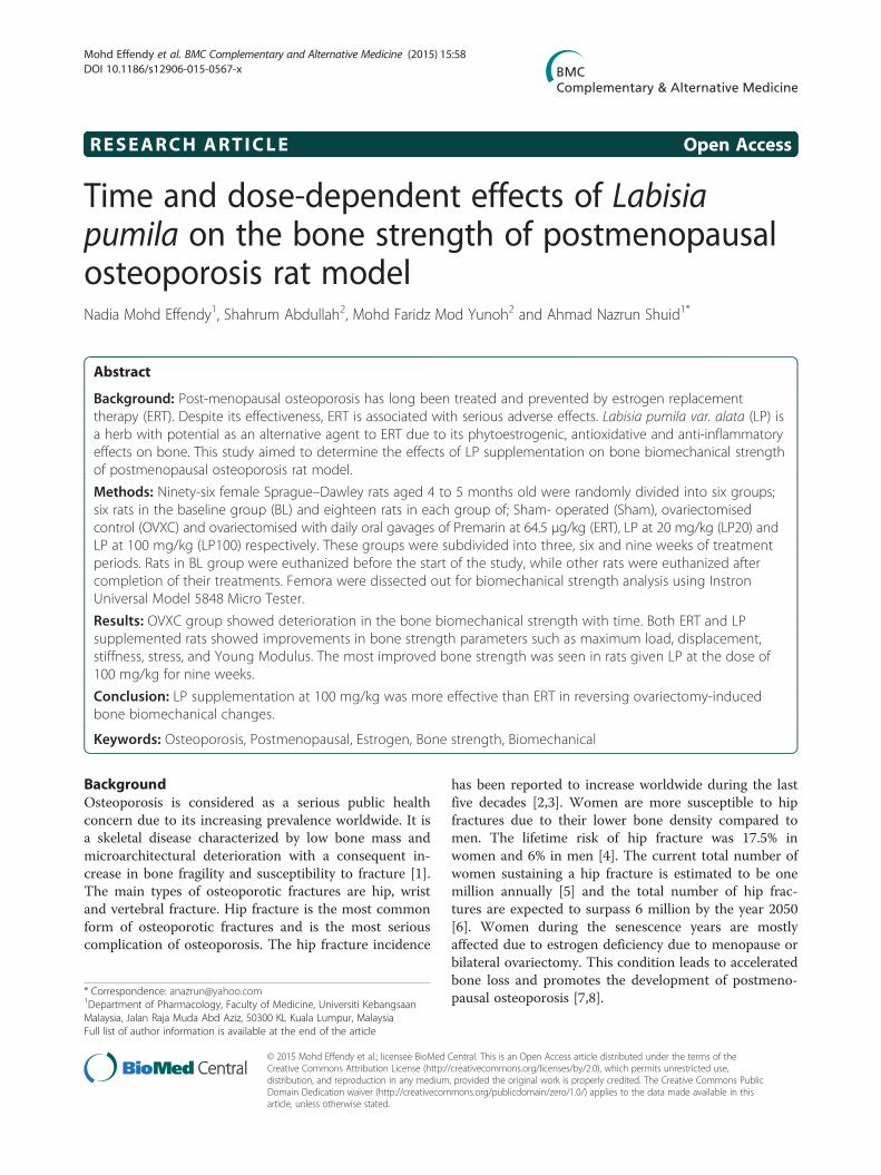
StrainThere was an increasing trend in the strain value for allthe treatment groups but no significant difference wasreported (Figure 7).
Figure 4 Displacement value for all the groups after 3, 6 and 9 weekssham-operated, OVX: ovariectomized control, ERT: ovariectomized and estro(20 mg/kg), LP100: ovariectomized with LP supplementation (100 mg/kg). a
of treatment.
Young modulusThe Young Modulus of the Sham group was significantlyhigher than the OVX group at 9 weeks of treatment.There was a time-dependent increment in the Youngmodulus value for the Sham and LP-treated groups. At9 weeks of treatment, both the LP20 and LP100 groupsshowed significantly higher Young modulus than theBaseline and OVX group (Figure 8).
DiscussionOsteoporosis is one of the most common diseases affecting1 in 3 women and 1 in 12 men [56]. Worldwide, approxi-mately 200 million women suffered from osteoporosis and
of treatment. Data presented as mean ± SEM (p < 0.05). Sham:gen supplementation, LP20: ovariectomized with LP supplementationP < 0.05 vs OVX at 6 weeks of treatment, bP < 0.05 vs ERT at 9 weeks

Figure 5 Stiffness value for all the groups after 3, 6 and 9 weeks of treatment. Data presented as mean ± SEM (p < 0.05). Sham: sham-operated,OVX: ovariectomized control, ERT: ovariectomized and estrogen supplementation, LP20: ovariectomized with LP supplementation (20 mg/kg), LP100:ovariectomized with LP supplementation (100 mg/kg). aP < 0.05 vs OVX.
Mohd Effendy et al. BMC Complementary and Alternative Medicine (2015) 15:58 Page 6 of 11
by the age of 50 the lifetime risk of osteoporotic fracture isnearly 40% [57]. Although effective treatments are availablefor osteoporosis, their prolonged use was associated withadverse effects such as breast cancer, thromboembolic andcoronary heart disease. This has led to an increase in thedemand for alternative medicine to treat and preventosteoporosis. It has also attracted studies to search for po-tential agents to replace ERT. In recent years, a medicinalplant known as Labisia pumila (LP) was reported to pos-sess anti-osteoporosis activities. It was reported that LPwas able to increase bone formation and reduce the boneresorption markers in ovariectomized rats [47]. LP supple-mentation was also shown to protect the bone structurechanges of estrogen-deficient rats [48].In a previous study, the authors have investigated the
time-dependent effects of two doses of LP (20 mg a/kg
Figure 6 Stress value for all groups at 3, 6 and 9 weeks of treatment.ovariectomized control, ERT: ovariectomized and estrogen supplementationovariectomized with LP supplementation (100 mg/kg). aP < 0.05 vs Baseline
and 100 mg/kg) using in vitro micro-CT. Supplementa-tion of LP 100 mg/kg for 9 weeks showed the best effectin reversing the ovariectomy-induced bone changes.Bone structural parameters such as bone volume frac-tion, connectivity density and trabecular number wereincreased and trabecular separation decreased signifi-cantly in OVX rats treated with LP100 for 9 weeks.Three dimensional (3D) analysis also showed that thetrabecular microarchitecture of LP treated rats improvedsignificantly compared to other groups [58]. Followingthese positive results of LP on bone microarchitecture,our present study was performed to evaluate further theeffects of LP on bone strength.The diagnosis of osteoporosis is often based on bone
density. However, a denser bone does not always meanstronger bone as in the case of fluoride treatment of
Data presented as mean ± SEM (p < 0.05). Sham: sham-operated, OVX:, LP20: ovariectomized with LP supplementation (20 mg/kg), LP100:, bP < 0.05 vs OVX, cP < 0.05 vs ERT.
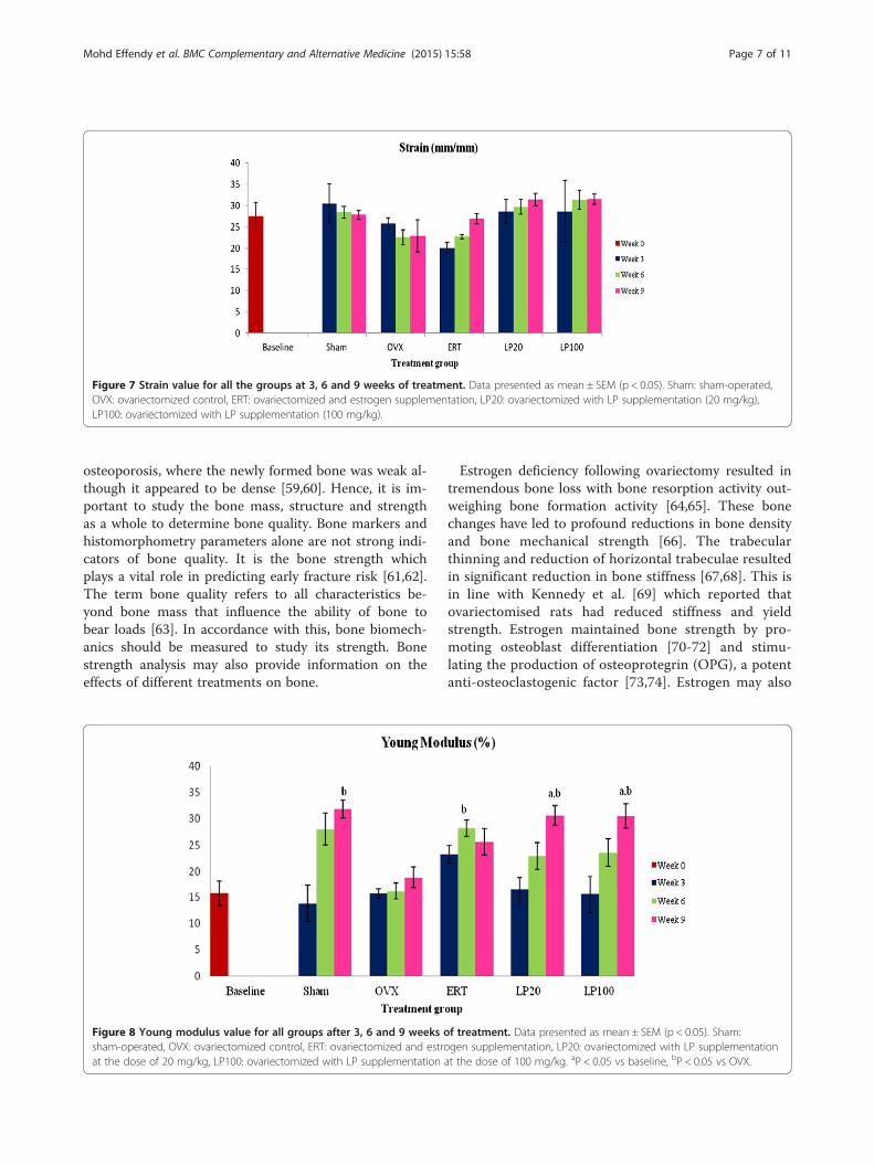
Figure 7 Strain value for all the groups at 3, 6 and 9 weeks of treatment. Data presented as mean ± SEM (p < 0.05). Sham: sham-operated,OVX: ovariectomized control, ERT: ovariectomized and estrogen supplementation, LP20: ovariectomized with LP supplementation (20 mg/kg),LP100: ovariectomized with LP supplementation (100 mg/kg).
Mohd Effendy et al. BMC Complementary and Alternative Medicine (2015) 15:58 Page 7 of 11
osteoporosis, where the newly formed bone was weak al-though it appeared to be dense [59,60]. Hence, it is im-portant to study the bone mass, structure and strengthas a whole to determine bone quality. Bone markers andhistomorphometry parameters alone are not strong indi-cators of bone quality. It is the bone strength whichplays a vital role in predicting early fracture risk [61,62].The term bone quality refers to all characteristics be-yond bone mass that influence the ability of bone tobear loads [63]. In accordance with this, bone biomech-anics should be measured to study its strength. Bonestrength analysis may also provide information on theeffects of different treatments on bone.
Figure 8 Young modulus value for all groups after 3, 6 and 9 weeks osham-operated, OVX: ovariectomized control, ERT: ovariectomized and estroat the dose of 20 mg/kg, LP100: ovariectomized with LP supplementation
Estrogen deficiency following ovariectomy resulted intremendous bone loss with bone resorption activity out-weighing bone formation activity [64,65]. These bonechanges have led to profound reductions in bone densityand bone mechanical strength [66]. The trabecularthinning and reduction of horizontal trabeculae resultedin significant reduction in bone stiffness [67,68]. This isin line with Kennedy et al. [69] which reported thatovariectomised rats had reduced stiffness and yieldstrength. Estrogen maintained bone strength by pro-moting osteoblast differentiation [70-72] and stimu-lating the production of osteoprotegrin (OPG), a potentanti-osteoclastogenic factor [73,74]. Estrogen may also
f treatment. Data presented as mean ± SEM (p < 0.05). Sham:gen supplementation, LP20: ovariectomized with LP supplementationat the dose of 100 mg/kg. aP < 0.05 vs baseline, bP < 0.05 vs OVX.

Mohd Effendy et al. BMC Complementary and Alternative Medicine (2015) 15:58 Page 8 of 11
increase the anti-oxidative enzyme levels, thus alleviat-ing the oxidative stress-induced bone loss [75].In this study, two doses of LP were given to ovariecto-
mized rats for three different time intervals; 3, 6 and9 weeks to evaluate the dose and time-dependent effectsof LP on bone biomechanical strength. The 20 mg/kgdose was a round up value of the 17.5 mg/kg standarddose used in a previous study [47,48]. While, the higherdose was set at five times higher to 100 mg/kg. Ideally,several doses of LP should have been tested, but due toethical reasons, this was not possible as we are required toreduce the number of animals used in the study. Accord-ing to previous toxicity studies, LP extract was shown tobe safe with the lethal dose 50 (LD50) of more than5.0 g/kg [76]. In other studies, LP extract was shown toexhibit no-adverse-effect-level (NOAEL) at the dose of50 mg/kg in a sub-acute study [77], 1000 mg/kg in asub-chronic study [78] and 800 mg/kg in reproductivetoxicity testing. Therefore, the doses of 20 mg/kg and100 mg/kg used in the study were safe. In humans, LP isnormally taken by women in the dose of 500 to1000 mg/daily.Whole bone biomechanical tests can be performed in
bending, tension, compression and torsional loading set-tings [79]. Bending is most commonly applied to ro-dents’ long bones due to difficulties of machining tensileor compressive specimens from small bones. There aretwo types of parameters which can be derived from abiomechanical test; the extrinsic and intrinsic parame-ters. Extrinsic parameters reflect the properties of wholebone which are affected by various external factors. Theycomprise of load, displacement and stiffness. Intrinsicparameters on the other hand reflect the inner materialof bones such as its geometric distribution and cellularmetabolic activity affecting the bone’s ability to bearloads. Intrinsic parameters comprise of stress, strain andmodulus of elasticity.In this study, the bone of rats supplemented with LP
showed significant improvement in both the extrinsicand intrinsic parameters. As expected, the OVX groupshowed deterioration in the biomechanical strength pa-rameters and by the ninth-week of treatment most ofthese parameters were significantly lower than the Shamgroup. This finding was supported by previous studieswhich reported that ovariectomized rats showed signifi-cant deteriorations in the bone structure in the firstthree months [80]. Another study reported that therewas no sign of bone changes in less than one monthafter ovariectomy. This is consistent with our findingsthat there was no significant change in bone strengthparameters at 3 weeks post-ovariectomy.Maximum or ultimate load indicates the whole bone
strength at the point where the femur started to changefrom elastic to plastic phase. Maximum load can be
defined as the maximum amount of force needed tobreak the bone. It reflects the general integrity of thebone structure. The bones of all the treatment groupsshowed gradual increments in the maximum load untilthey were significantly higher compared to the Baselineand OVX groups at nine weeks post-ovariectomy. Bothdoses of LP supplementations improved bone strengthand were comparable to ERT in withstanding the givenload. Displacement is another extrinsic parameter whichis defined as the length of deformation that the bonecan sustain before failing [81]. It can be used to measurebone ductility and is inversely related to the brittlenessof the bone [82]. Rats supplemented with 100 mg/kg doseof LP for the duration of nine weeks had the most ductilebones compared to others. Their bones were also havingsignificantly higher displacement values compared to theERT group. These results showed that although both theERT and LP100 groups had strong bones in terms of sus-taining high amount of load, the bones of LP100 weremore ductile and hence harder to break.The load - displacement curve may give a clearer un-
derstanding of bone strength. The linear region of thiscurve represents the elastic property of a bone, wherethe deformation upon loading is reversible [83]. The gra-dient under this elastic region depicts the extrinsic stiff-ness or rigidity. It represents bone mineralization of therelative hydroxyapatite and collagen fibers proportion[84,85]. Beyond the point of yielding is the plastic region,where permanent deformation occurs upon compressiveforce. In this study, the Sham, LP20 and LP100 groupsshowed significantly higher bone stiffness at 9 weekspost-treatment compared to the OVX group. The infer-ior bone stiffness of the OVX group was expected asmany studies have reported that ovariectomy affectednot only the bone mass but the bone quality as well[86,87]. Surprisingly, LP supplementation was able toimprove the bone stiffness of ovariectomised rats whileERT failed to do so.Bone biomechanical testing is not only focused on the
mechanical behaviour of the whole bone but also themechanical properties at tissue level or intrinsic proper-ties. Intrinsic parameters measured were stress, strainand Young’s modulus. Stress is the strength of the bonetissue under a given loading condition. In this study, theSham and all the treatment groups were able to receivehigher stress compared to the OVX group after 6 and9 weeks of treatment. The LP100 group was able toreceive the greatest stress at 9 weeks, which was signifi-cantly higher than the ERT group. This indicated thatthe bone tissue of the LP100 group was able to absorbhigher energy before failure than the ERT group. As forthe Strain parameter, it represents ductility of the bone[88,89]. However, there were no significant changes inthis parameter for all the groups.

Mohd Effendy et al. BMC Complementary and Alternative Medicine (2015) 15:58 Page 9 of 11
The slope from stress - strain curve represented themodulus of elasticity which is also known as Young’smodulus. Young’s modulus is influenced by the amountof collagen and calcification process in the bone. After9 weeks of treatment, the Sham, LP20 and LP100 groupshad significantly higher bone elasticity than the OVXgroup. This indicated that the bones of the Sham and LPgroups were much more elastic than the OVX groupand less likely to fracture. Based on all the results, sup-plementation of LP at 100 mg/kg were able to preservebone strength from the deleterious effects of ovariec-tomy. At this dose, LP was found to be better than ERTin maintaining bone strength and ductility.There are several possible mechanisms behind the
ability of LP to retain bone strength during estrogen defi-ciency state. LP extract contains triterpenes and saponins,which are known phytoestrogens [90]. Phytoestrogen canmimic or modulate the action of endogenous estrogens bybinding to estrogen receptors [91,92]. It was reported thatphytoestrogen exerted bone sparing effects in a rat model[93,94]. Similar to estrogen, LP through its phytoestogenicactivity may induce and inhibit osteoclasts and osteoblastapoptosis respectively. Therefore, LP may maintain bonestrength by reducing bone resorption and increasing boneformation activities [95].Besides the phytoestrogenic property, LP may exert
anti-oxidative effects on the bone. According to Norhaizaet al. [96], the anti-oxidative property of LP was con-tributed by its content of flavonoids, ascorbic acids, beta-carotene, anthocyanin and phenolic compounds. Estrogendeficiency leads to deterioration in anti-oxidant defensesystem and upregulation of reactive oxygen species (ROS).These imbalances resulted in lipid peroxidation and boneloss [97]. Hence, supplementation of LP may abate oxida-tive stress, thus preventing bone loss and maintainingbone strength.There are some raised concerns on the risk of endo-
metrial hyperplasia and excessive cell growth in theuterus, secondary to the use of phytoestrogens. However,many previous studies had shown that LP exhibits anti-proliferative effects [98]. A polar solvent extract such asLP water extract had been shown to reduce risks of cellproliferation. The bioactive compounds that are respon-sible for estrogenic activity are more polar in nature,hence they are highly expressed in water extract. This isin contrast to a less polar solvent extracts such as ethanoland dichloromethane LP extracts which may induce an in-crease in cell proliferation [99]. Hence, LP water extractused in this study may exhibit an estrogenic effect withoutthe risk of excessive cell proliferation. This is also sup-ported by previous toxicity studies which reported that LPextract did not alter the rats’ general health and no grossvisceral changes of the ovaries or uterus were found at thedose up to 800 mg/kg/day [76,77]. Although LP is safe
and effective, further studies are warranted to document aconclusive mechanisms of its therapeutic action.
ConclusionsAs a conclusion, LP supplementation at the dose of100 mg/kg for 9 weeks duration of treatment was foundto be more effective than ERT in maintaining the bonestrength of a postmenopausal osteoporosis rat model.Based on its safety profile and ability to preserve bonestrength, LP has potential as an alternative treatment forpostmenopausal osteoporosis.
Competing interestsThe authors declare that they have no competing interests.
Authors’ contributionsNME carried out the biomechanical bone tests, performed the statisticalanalysis and wrote the manuscript. SA and MFMY carried out thebiomechanical bone tests and helped with the analysis. ANS conceived ofthe study, participated in its design and coordination and helped to draftthe manuscript. All authors read and approved the final manuscript.
AcknowledgementThis study was made possible through the grant provided by the Faculty ofMedicine, UKM (UKM-DLP-2011-044). The authors would like to thank MrMuhamad Arizi Aziz, Ms Juliana Abdul Hamid, Ms Nurul Hafizah Abas, MrFadhlullah Zuhair and Mrs Farhana Mohd Fozi from the Department ofPharmacology for their technical assistance.
Author details1Department of Pharmacology, Faculty of Medicine, Universiti KebangsaanMalaysia, Jalan Raja Muda Abd Aziz, 50300 KL Kuala Lumpur, Malaysia.2Department of Mechanical and Materials Engineering, Faculty ofEngineering, Universiti Kebangsaan Malaysia, Kuala Lumpur, Malaysia.
Received: 2 August 2013 Accepted: 19 February 2015
References1. Christiansen C. Consensus Development Conference: prophylaxis and
treatment of osteoporosis. Am J Med. 1991;90(1):107–10.2. Koh LK, Saw SM, Lee JJ, Leong KH, Lee J. Hip fractures incidence rates in
Singapore 1991–1998. Osteoporos Int. 2001;12(4):311–8.3. Cummings SR, Melton LJ. Epidemiology and outcomes of osteoporotic
fractures. Lancet. 2002;359:1761–7.4. Melton 3rd LJ. Who has osteoporosis? A conflict between clinical and public
health perspectives. J Bone Miner Res. 2000;15:2309.5. Henrik GA, Bjӧrn ER, Teppo LNJ, Cecilia R, Jan-Ake N, Ingemar S, et al.
Prevalence of osteoporosis and incidence of hip fracture in women-seculartrends over 30 years. BMC Musculoskelet Disord. 2010;11(48):1–7.
6. Osteoporosis Australia. 2004. http://www.osteoporosis.org.au7. Rohr UD. The impact of testosterone imbalance on depression and
women’s health. Maturitas. 2002;41 Suppl 1:S25–46.8. Parves T. Postmenopausal osteoporosis. JK-Practitioner. 2004;11(4):281–3.9. Jilka RL. Cytokines, bone remodelling and estrogen deficiency. Bone.
1998;23(2):75–81.10. Pfeilschifter J, Koditz R, Pfohl M, Schatz H. Changes in proinflammatory
cytokine activity after menopause. Endocrinol Rev. 2002;23:90–119.11. Bart C. Normal bone anatomy & physiology. Clin J Am Soc Nephrol.
2008;3:S131–9.12. Iain HK. Principles of bone healing. Neurosurg Focus. 2001;10(4):1–8.13. Khastgir G, Studd J, Holland N. Anabolic effect of estrogen replacement on
bone in postmenopausal women with osteoporosis: Histomorphometricevidence in a longitudinal study. J Clin Endocrinol Metabol. 2001;86:289–95.
14. Shumaker SA, Legault C, Rapp R, Thal L, Wallace RB, Ockene JK, et al.Estrogen plus progestin and the incidence of dementia and mild cognitiveimpairment in postmenopausal women: the Women’s Health InitiativeMemory: a randomized controlled trial. JAMA. 2003;289(20):2651–62.

Mohd Effendy et al. BMC Complementary and Alternative Medicine (2015) 15:58 Page 10 of 11
15. Al-Azzawi F. Prevention of postmenopausal osteoporosis and associatedfractures: Clinical evaluation of the choice between estrogen andbisphosphonates. Gynecol Endocrinol. 2008;24(11):601–9.
16. Komm BS, Chines AA. An update on selective estrogen receptor modulatorsfor the prevention and treatment of osteoporosis. Maturitas. 2010;71(3):221–6.
17. Cano A, Dapia S, Noguela J, Pineda B, Hermenegildo C, del Val R, et al.Comparative effects of 17β-estradiol, raloxifene and genistein on bone 3Dmircroarchitecture and volumetric bone mineral density in ovariectomizedmice. Osteoporos Int. 2008;19:793–800.
18. Roussow JE, Anderson GL, Prentice RL, LaCroix A, Kooperberg C, StefanickML. Risks and benefits of estrogen plus progestin in healthypostmenopausal women: principal results from the Women’s HealthInitiative randomized controlled trial. JAMA. 2002;288:321–33.
19. Wassertheil-Smoller S, Hendrix SL, Limacher M. Effect of estrogen plusprogestin on stroke in postmenopausal women: the Women’s HealthInitiative: a randomized trial. JAMA. 2003;289:2673–84.
20. Chlebowski RT, Hendrix SL, Langer RD. Influence of estrogen plus progestinon breast cancer and mammography in healthy postmenopausal women:the Women’s Health Initiative Randomized Trial. JAMA. 2003;289:3243–53.
21. Henry GB. Selective oestrogen receptor modulators. Horm Res. 2000;53:25–9.22. FDA Approves New Uses for Evista (Press release). U.S. Food and Drug
Administration. 2007-09-14.23. Delmas PD, Genant HK, Crans GG, Stock JL, Wong M, Siris E, et al. Severity of
prevalent vertebral fractures and the risk of subsequent vertebral andnonvertebral fractures: results from the MORE trial. Bone. 2003;33:522–32.
24. Kanis JA, Johnell O, Black DM, Downs Jr RW, Sarkar S, Fuerst T, et al. Effect ofraloxifene on the risk of new vertebral fracture in postmenopausal womenwith osteopenia or osteoporosis: a reanalysis of the Multiple Outcomes ofRaloxifene Evaluation trial. Bone. 2003;33:293–300.
25. Johnell O, Scheele WH, Lu Y, Reginster J-Y, Need AG, Seeman E. Additiveeffects of raloxifene and alendronate on bone density and biochemicalmarkers of bone remodeling in postmenopausal women with osteoporosis.J Clin Endocrinol Metab. 2002;87:985–92.
26. Turner RT, Rickard DJ, Iwaniec UT, Spelsberg TC. Estrogens and progestins.In: Bilezikian JP, Raisz LG, Martin TJ, editors. Principles of bone biology.3rd ed. USA: Academic Pres; 2008.
27. Yan MZ, Xu Y, Gong YX, Liu JM, Lu SY, Huang L, et al. Raloxifene inhibitsbone loss and improves bone strength through an OPg-independentmechanism. Endocrine. 2010;37(1):55–61.
28. Delmas PD, Ensrud KE, Adachi JD. Efficacy of raloxifene on vertebral fracturerisk reduction in postmenopausal women with osteoporosis: Four-year resultsfrom a randomized clinical trial. J Clin Endocrinol Metabol. 2002;87:3609–17.
29. Grady D, Ettinger B, Moscarelli E. Safety and adverse effects associated withraloxifene: multiple outcomes of raloxifene evaluation. Obstet Gynecol.2004;104:837–44.
30. Vogel V, Joseph C, Lawrence W. Effects of Tamoxifen vs. Raloxifene on theRisk of Developing Invasive Breast Cancer and Other Disease Outcomes.J Am Med Assoc. 2006;295(23):2727–41.
31. Reginster J, Minne HW, Sorensen OH. Randomized trial of the effects ofrisedronate on vertebral fractures in women with establishedpostmenopausal osteoporosis. Vertebral Efficacy with Risedronate Therapy(VERT) Study Group. Osteoporos Int. 2000;11:83–91.
32. Stevenson M, Jones ML, De Nigris E, Brewer N, Davis S, Oakley J. Asystematic review and economic evaluation of alendronate, etidronate,risedronate, raloxifene and teriparatide for the prevention and treatment ofpostmenopausal osteoporosis. Health Technol Assess. 2005;9:1–160.
33. Reiner B, Bertha F, Emmo VT, Christoph B. Biphosphonates in MedicalPractice. New York: Springer; 2007.
34. Khosla S, Burr D, Cauley J. Bisphosphonate-associated osteonecrosis of thejaw: report of a Task Force of the American Society for Bone and MineralResearch. J Bone Miner Res. 2007;22(10):1479–91.
35. Zakaria M, Mohd MA. Traditional Malay Medicinal Plants. Kuala Lumpur,Malaysia: Penerbit Fajar Bakti; 1994.
36. Bodeker G. Health and Beauty from the Rainforest: Malaysian Traditions ofRamuan. Kuala Lumpur, Malaysia: Editions Didier Millet Pty; 1999.
37. Fasihuddin A, Rahman AH, Hasmah R. Medicinal plants used by bajaucommunity in sabah. In: Chan KL, editor. Trends in Traditional MedicineResearch. Penang, Malaysia: The School of Pharmaceutical Sciences,University of Science Malaysia; 1995. p. 493–504.
38. Jamal JA, Houghton PJ, Milligan SR. Testing of labisia pumila for oestrogenicactivity using a recombinant yeast screen. J Pharm Pharmacol. 1998;50:79.
39. Karimi E, Jaafar HZ, Ahmad S. Antifungal, anti-inflammatory and cytotoxicityactivities of three varieties of labisia pumila benth: from microwave obtainedextracts. BMC Complement Altern Med. 2013;13(20):1–10.
40. Burkill IH. Dictionary of the Economic Products of the Malay Peninsula.London: Publisher Crown Agents for the Colonies; 1935.
41. Rasadah MA, Zainon AS. Database on ASEAN Herbal and Medicinal Plants,ASEAN Publication. 2003.
42. Whitten PL, Kudo S, Okubo KK. Isoflavonoids. In: D’ Mello JPF, editor.Handbook of Plant and FUngal Toxicants. Boca Raton: CRC Press; 1997.
43. Pfitscher A, Reiter E, Jungbauer A. Receptor binding and transactivationactivities of red clover isoflavones and their metabolites. J Steroid BiochemMol Biol. 2008;112:87–94.
44. Oseni T, Patel R, Pyle J, Jordan VC. Selective estrogen receptor modulatorsand phytoestrogens. Planta Med. 2008;74:1656–65.
45. Degen GH, Janning P, Diel P, Bolt HM. Estrogenic isoflavones in rodentdiets. Toxicol Lett. 2002;128:145–57.
46. Karkola S, Lilienkampf A, Wähälä K. Phytoestrogens in drug discovery forcontrolling steroid biosynthesis. In: Daayf F, Lattanzio V, editors. RecentAdvances in Polyphenol Research. Oxford, UK: Wiley-Blackwell; 2009.
47. Nazrun AS, Lee PL, Norliza M, Norazlina M, Ima Nirwana S. The effects ofLabisia pumila var. alata on bone markers and bone calcium in a ratmodel of post-menopausal osteoporosis. J Ethnopharmacol.2011;133(2):538–42.
48. Fathilah SN, Nazrun AS, Norazlina M, Norliza M, Ima Nirwana S. Labisia pumilaprotects the bone of estrogen-deficient rat model: A histomorphometric study.J Ethnopharmacol. 2012;142:294–9.
49. Binkley N, Bilezikian JP, Kendler DL, Leib ES, Lewiecki EM, Petak SM.International Society for Clinical Densitometry. Official positions of theInternational Society for Clinical Densitometry and executive summary ofthe 2005 Position Development Conference. J Clin Densitom. 2006;9:4–14.
50. Kanis JA, Glüer CC, for the Committee of Scientific Advisors. InternationalOsteoporosis Foundation. An update on the diagnosis and assessment ofosteoporosis with densitometry. Osteoporos Int. 2000;11:192–202.
51. National Osteoporosis Foundation (NOF). Physician’s Guide to preventionand treatment of osteoporosis. Washington, DC: National OsteoporosisFoundation; 2003.
52. Voide R, Van Lenthe H, Schneider P, Thurner P, Wyss P, Sennhauser U, et al.Functional Microimaging: an integrated approach for advanced bonebiomechanics and failure analysis. In: Armando M, Amir AA, editors. MedicalImaging: Physiology, Function and Structure from Medical Images. SanDiego: Proc SPIE 6143; 2006.
53. Grant B, Tony MK. Trabecular bone strength predictions using finite elementanalysis of micro-scale images at limited spatial resolution. Bone.2009;44:579–84.
54. Joshua AM, Steven KB. Bone strength at the distal radius can be estimatedfrom high resolution peripheral quantitative computed tomography andthe finite element method. Bone. 2008;42:1203–13.
55. Helene B, Eric L, Claude-Laurent B. Evaluation of macrostructural bonebiomechanics. Joint Bone Spine. 2007;74:233–9.
56. Vijender A, Dharmendra G. Recent update on osteoporosis. Int J Med SciPublic Health. 2013;2(2):164–8.
57. Melton LJ, Lane AW, Cooper C, Eastell R, O’Fallon WM, Riggs BL. Prevalenceand incidence of vertebral deformities. Osteoporos Int. 1993;3:113–9.
58. Nadia ME, Fadhli MK, Ima Nirwana S, Nazrun AS. The effects of Labisiapumila on postmenopausal osteoporotic rat model: Dose and time-dependent micro-CT analysis. J X-ray Sci Technol. 2014;22(4):503–18.
59. Bohatyrewicz A. Effects of fluoride on mechanical properties of femoralbone in growing rats. Fluoride. 1999;32:47–54.
60. Chacha D, Turner CH, Dunipace AJ, Grynpas MD. The effect of fluoridetreatment on bone mineral in rabbits. Calcif Tissue Int. 1999;64:345–51.
61. Ammann P, Rizzoli R. Bone strength and its determinants. Osteoporos Int.2003;14(3):13–8.
62. Sambrook PN, Cameron ID, Chen JS, Cumming RG, Lord SR, March LM, et al.Influence of fall related fractures and bone strength on fracture risk in thefrail elderly. Osteoporos Int. 2007;18:603–10.
63. Jacqueline HC, Marjolein CH van der M. Whole bone mechanics and bonequality. Clin Orthop Relat Res. 2011;469:2139–49.
64. Riggs BL. The mechanisms of estrogen regulation of bone resorption. J ClinInvestig. 2000;106(10):1203–4.
65. Falahati-Nini A, Riggs BL, Atkinson EJ, O’Fallon WM, Eastell R, Khosla S.Relative contributions of testosterone and estrogen in regulating bone

Mohd Effendy et al. BMC Complementary and Alternative Medicine (2015) 15:58 Page 11 of 11
resorption and formation in normal elderly men. J Clin Investig.2000;106(12):1553–60.
66. Zi-xiang W, Wei L, Yun-yu H, Hai-qiang W, Shi-yong W, Zhen-sheng M, et al.Effect of ovariectomy on BMD, micro-architecture and biomechanics of corticaland cancellous bones in a sheep model. Med Eng Phys. 2008;30:1112–8.
67. Parfitt AM. Age-related structural changes in trabecular and corticalbone:cellular mechanisms and biomechanical consequences. Calcif Tissue Int.1984;36 Suppl 1:123–8.
68. Augat P, Link T, Lang TF, Lin JC, Majumdar S, Genant HK. Anisotropy of theelastic modulus of trabecular bone specimens from different anatomicallocations. Med Eng Phys. 1998;20:124–31.
69. Kennedy OD, Brennan O, Rackard SM, Staines A, O’Brien FJ, Taylor D, et al.Effects of ovariectomy on bone turnover, porosity and biomechanicalproperties in ovine compact bone 12 months postsurgery. J Orthop Res.2009;27(3):303–9.
70. Grumbach MM. Estrogen, bone, growth and sex: a sea change inconventional wisdom. J Pediatr Endocrinol Metab. 2000;13 Suppl 6:1439–55.
71. Gautam K, Joh S, Nigel H, Jamshid A-Z, Simon F, Jade C. Anabolic effect ofestrogen replacement on bone in postmenopausal women with osteoporosis:Histomorphometric evidence in a longitudinal study. J Clin Endocrinol Metabol.2001;86(1):289–95.
72. Okazaki R, Inoue D, Shibata M, Saika M, Kido S, Ooka H, et al. Estrogenpromotes early osteoblast differentiation and inhibits adipocytedifferentiation in mouse bone marrow stromal cell lines that expressestrogen receptor (ER) alpha or beta. Endocrinology. 2002;143(6):2349–56.
73. Bord S, Irelan DC, Beaven SR, Compston JE. The effects of estrogen onosteoprotegerin, RANKL and estrogen receptor expression in humanosteoblasts. Bone. 2003;32(2):136–41.
74. Mitani M, Miura Y, Saura R, Kitagawa A, Fukuyama T, Hashiramoto A, et al.Estrogen specifically stimulates expression and production of osteoprotegrinfrom rheumatoid synovial fibroblasts. Int J Mol Med. 2005;15(5):827–32.
75. Badeau M, Adlercreutz H, Kaihovaara P, Tikkanen MJ. Estrogen A-ringstructure and antioxidative effect on lipoproteins. J Steroid Biochem MolBiol. 2005;96(3–4):271–8.
76. Wan Ezumi MF, Siti Amrah S, Suhaimi AW, Mohsin SSJ. Evaluation of thefemale reproductive toxicity of aqueous extract of Labisia pumila var. alatain rats. Indian J Pharm. 2007;39(1):30–2.
77. Singh GD, Ganjoo M, Youssouf MS, Koul A, Sharma R, Singh S, et al. Sub-acutetoxicity evaluation of an aqueous extract of Labisia pumila, a Malaysian herb.Food Chem Toxicol. 2009;47(10):2661–5.
78. Taneja SC. Sub-Chronic (90 days) Oral Toxicity Studies of Aqueous Extract ofLabisia pumila in Wistar Rats (250, 500&1000 mg/kg b. wt. only). India: IndianInstitute of Integrative Medicine Jammu; 2004.
79. Richard MA. Mechanical testing of bone ex vivo. In: Helfrich MH, Ralston SH,editors. Methods in Molecular Medicine. Bone Research Protocols. Totowa,New Jersey: Humana Press; 2003. p. 369–77.
80. Waarsing JH, Day JS, Linden van der, Ederveen AG, Spanjers C, Clerck ND,et al. Detecting and tracking local changes in the tibiae of individual rats:a novel method to analyse longitudinal in vivo micro-CT data. Bone.2004;34(1):163–9.
81. Hernandez CH, Keavenly TM. A biomechanical perspective on bone quality.Bone. 2006;39(6):1173–81.
82. Charles HT. Bone strength: current concepts. Ann N Y Acad Sci.2006;1068:429–46.
83. Wendlova J. Bone quality, elasticity and strength. Bratisl Lek Listy.2008;109(9):383–6.
84. Robert OR, Markus JB, Paul H. Plasticity and toughness in bone. Phys Today.2009;62(6):4.
85. Gleeson JP, Plunkett NA, O’Brien FJ. Addition of hydroxyapatite improvesstiffness, interconnectivity and osteogenic potential of a highly porouscollagen-based scaffold for bone tissue regeneration. Eur Cell Mater.2010;20:218–30.
86. Mosekilde L, Danielsen CC, Knudsen UB. The effect of aging andovariectomy on the vertebral bone mass and biomechanical properties ofmature rats. Bone. 1993;14(1):1–6.
87. Kennedy OD, Brennan O, Rackard SM, Staines A, O’Brien FJ, Taylor D, et al. Effectsof ovariectomy on bone turnover, porosity and biomechanical properties inovine compact bone 12 months post surgery. J Orthop Res. 2008;27(3):303–9.
88. Keavenly TM, Wachtel EF, Kopperdahl DL. Mechanical behaviour ofdamaged trabecular bone. J Biomech. 1999;27(11):1309–18.
89. Tony MK, Elise FM, Oscar CY. Bone Mechanics in Standard Handbook ofBiomedical Engineering and Design. New York: McGraw Hill; 2004.
90. Hertrampf T, Schleipen B, Offermanns C, Velders M, Laudenbach U, Diel P.Comparison of the bone protective effects of an isoflavone-rich diet withdietary and subcutaneous administrations of genistein in ovariectomizedrats. Toxicol Lett. 2009;184(3):198–203.
91. Avula B, Wang YH, Ali Z, Smillie TJ, Khan IA. Quantitative determination oftriperpene saponins and alkenated-phenolics from Labisia pumila by LCUV/ELSD method and confirmation by LC-ESI-TOF. Planta Med. 2010;76:25.
92. Ososki AL, Kenelly EJ. Phytoestrogens: a review of the present state ofresearch. Phytother Res. 2003;17:845–69.
93. Cos P, De Bruyne T, Apers S, Vanden BD, Pieters L, Vlietinck AJ.Phytoestrogens recent developments. Planta Med. 2003;69:589–99.
94. Cassidy A, Albertazzi P, Lise Nielsen I, Hall W, Williamson G, Tetens I, et al.Critical review of health effects of soyabean phyto-oestrogens inpost-menopausal women. Proc Nutr Soc. 2006;65(1):76–92.
95. Bahram HA. The role of phytoestrogens in the prevention and treatmentof osteoporosis in ovarian hormone deficiency. J Am Coll Nutr.2001;20 Suppl 5:3985–4025.
96. Norhaiza M, Maziah M, Hakiman M. Antioxidative properties of leaf extractsof a popular Malaysian herb, Labisia pumila. J Med Plant Res. 2009;3(4):217–23.
97. Halliwell B, Gutteridge JMC. Free radicals in biology and medicine. NewYork: Oxford University Press; 2007.
98. Jamal J, Houghton P, Milligan S, Ibrahim J. The estrogenic and cytotoxiceffects of the extracts of Labisia pimula var. alata and Labisia pumila var.pumila in-vitro. Malays J Health Sci. 2003;1:53–60.
99. Melissa PSW, Navaratnam V, Yin CY. Estrogenic assessment of Labisia pumilaextracts using a human endometrial cell line. Int J Pharm Pharmaceut Sci.2013;5(2):448–52.
Submit your next manuscript to BioMed Centraland take full advantage of:
• Convenient online submission
• Thorough peer review
• No space constraints or color figure charges
• Immediate publication on acceptance
• Inclusion in PubMed, CAS, Scopus and Google Scholar
• Research which is freely available for redistribution
Submit your manuscript at www.biomedcentral.com/submit