TiMC Cancer, ten Holte
39
Top Med Chem (2007) 1: 293–331 DOI 10.1007/7355_2006_007 © Springer-Verlag Berlin Heidelberg 2007 Published online: 13 January 2007 HDAC Inhibition in Cancer Therapy: An Increasingly Intriguing Tale of Chemistry, Biology and Clinical Benefit P. ten Holte 1 (✉) · K. Van Emelen 1 · M. Janicot 2 · P. C. Fong 3 · J. S. de Bono 3 · J. Arts 2 1 Dept. of Medicinal Chemistry, Johnson & Johnson Pharmaceutical Research & Development, Division of Janssen Pharmaceutica NV, 2340 Beerse, Belgium [email protected] 2 Dept. of Oncology, Johnson & Johnson Pharmaceutical Research & Development, Division of Janssen Pharmaceutica NV, 2340 Beerse, Belgium 3 Drug Development Unit, Royal Marsden Hospital, Sutton UK 1 Introduction ................................... 295 2 Biochemistry of the Histone Deacetylases and Histone Acetyl Transferases 296 2.1 HDACs and their Link to Cancer ........................ 296 2.2 The HDAC Family of Enzymes ......................... 297 3 HDAC Inhibitors ................................ 300 3.1 Historic Overview ................................ 300 3.2 Recent Medicinal Chemistry Efforts—An Overview ............. 303 3.2.1 The Cyclic Peptides ............................... 303 3.2.2 Hydroxamic Acid Replacements—the Holy Grail? .............. 305 3.2.3 Is the Type of Spacer Really all that Important? ................ 309 3.2.4 The Capping Group under Scrutiny ...................... 311 3.2.5 Connecting the Spacer with the Capping Group ............... 313 3.2.6 The Quest for Selective HDAC Inhibitors ................... 314 4 Clinical Experience with HDAC Inhibitors .................. 317 4.1 Hydroxamates .................................. 318 4.1.1 Vorinostat (SAHA) ............................... 319 4.2 NonHydroxamates ................................ 320 4.2.1 Depsipeptide (FR901228 or FK-228) ...................... 322 4.2.2 MS-275 ...................................... 322 4.2.3 CI-994 (Tacedinaline) .............................. 323 4.3 Summary and Future Development ...................... 323 5 Perspectives and Conclusion .......................... 324 References ....................................... 326 Abstract This review presents a wide-ranging selection of key literature examples in the histone deacetylase (HDAC) field. The review starts off with the biological background
-
Upload
peter-ten-holte -
Category
Documents
-
view
35 -
download
0
Transcript of TiMC Cancer, ten Holte

Top Med Chem (2007) 1: 293–331DOI 10.1007/7355_2006_007© Springer-Verlag Berlin Heidelberg 2007Published online: 13 January 2007
HDAC Inhibition in Cancer Therapy:An Increasingly Intriguing Tale of Chemistry, Biologyand Clinical Benefit
P. ten Holte1 (�) · K. Van Emelen1 · M. Janicot2 · P. C. Fong3 ·J. S. de Bono3 · J. Arts2
1Dept. of Medicinal Chemistry,Johnson & Johnson Pharmaceutical Research & Development,Division of Janssen Pharmaceutica NV, 2340 Beerse, [email protected]
2Dept. of Oncology,Johnson & Johnson Pharmaceutical Research & Development,Division of Janssen Pharmaceutica NV, 2340 Beerse, Belgium
3Drug Development Unit, Royal Marsden Hospital, Sutton UK
1 Introduction . . . . . . . . . . . . . . . . . . . . . . . . . . . . . . . . . . . 295
2 Biochemistry of the Histone Deacetylases and Histone Acetyl Transferases 2962.1 HDACs and their Link to Cancer . . . . . . . . . . . . . . . . . . . . . . . . 2962.2 The HDAC Family of Enzymes . . . . . . . . . . . . . . . . . . . . . . . . . 297
3 HDAC Inhibitors . . . . . . . . . . . . . . . . . . . . . . . . . . . . . . . . 3003.1 Historic Overview . . . . . . . . . . . . . . . . . . . . . . . . . . . . . . . . 3003.2 Recent Medicinal Chemistry Efforts—An Overview . . . . . . . . . . . . . 3033.2.1 The Cyclic Peptides . . . . . . . . . . . . . . . . . . . . . . . . . . . . . . . 3033.2.2 Hydroxamic Acid Replacements—the Holy Grail? . . . . . . . . . . . . . . 3053.2.3 Is the Type of Spacer Really all that Important? . . . . . . . . . . . . . . . . 3093.2.4 The Capping Group under Scrutiny . . . . . . . . . . . . . . . . . . . . . . 3113.2.5 Connecting the Spacer with the Capping Group . . . . . . . . . . . . . . . 3133.2.6 The Quest for Selective HDAC Inhibitors . . . . . . . . . . . . . . . . . . . 314
4 Clinical Experience with HDAC Inhibitors . . . . . . . . . . . . . . . . . . 3174.1 Hydroxamates . . . . . . . . . . . . . . . . . . . . . . . . . . . . . . . . . . 3184.1.1 Vorinostat (SAHA) . . . . . . . . . . . . . . . . . . . . . . . . . . . . . . . 3194.2 NonHydroxamates . . . . . . . . . . . . . . . . . . . . . . . . . . . . . . . . 3204.2.1 Depsipeptide (FR901228 or FK-228) . . . . . . . . . . . . . . . . . . . . . . 3224.2.2 MS-275 . . . . . . . . . . . . . . . . . . . . . . . . . . . . . . . . . . . . . . 3224.2.3 CI-994 (Tacedinaline) . . . . . . . . . . . . . . . . . . . . . . . . . . . . . . 3234.3 Summary and Future Development . . . . . . . . . . . . . . . . . . . . . . 323
5 Perspectives and Conclusion . . . . . . . . . . . . . . . . . . . . . . . . . . 324
References . . . . . . . . . . . . . . . . . . . . . . . . . . . . . . . . . . . . . . . 326
Abstract This review presents a wide-ranging selection of key literature examples in thehistone deacetylase (HDAC) field. The review starts off with the biological background

294 P. ten Holte et al.
of HDACs and their link to cancer and cancer treatment. The body of the work consistsof a categorized and chronological medicinal chemistry overview. This part describes keymedicinal chemistry contributions ranging from the very early HDAC inhibitors to com-pounds currently in the clinic. The result of all these medicinal chemistry and biologyefforts have been captured in the last section that gives an overview of the current statusof HDAC inhibitors in the clinic.
Keywords Histone deacetylase · HDAC inhibitor · Hydroxamic acid ·Clinical development · Isoform selective
AbbreviationsAML acute myeloid leukemiaAPC adenomatosis polyposis coliAPHA aroyl pyrrolyl hydroxamic acidsAPL acute promyelocytic leukemiaCBHA m-carboxycinnamic acid bishydroxamideCDK cyclin dependent kinaseCHAP cyclic hydroxamic acid-containing peptideCR complete responseCTCL cutaneous T cell lymphomaDLT dose limiting toxicityDTT dithiothreitolEKG electrocardiographHAT histone acetyl transferaseHDAC histone deacetylaseHDACi HDAC inhibitorHDLP histone deacetylase-like proteinhERG human ether-a-go-go related geneHIF hypoxia-inducible factorMEF myocyte enhancer factorMEL murine erythroleukemiaMITR MEF2-interacting transcription repressorMTD maximum tolerated dosen.d. not determinedPBMNCperipheral blood mononuclear cellsPD pharmacodynamicPK pharmacokineticPR partial responsePTCL peripheral T-cell lymphomaRAR retinoic acid receptorRR response rateSAHA suberoylanilide hydroxamic acidSCC squamous cell carcinomaSCOP sulfur-containing cyclic peptidesTPX trapoxinTRAIL tumor necrosis factor related apoptosis inducing ligandTSA trichostatin AuPR unconfirmed PRVEGF vascular endothelial growth factor

HDAC Inhibition in Cancer Therapy 295
1Introduction
During the past decade, epigenetic phenomena have been proven to be in-volved in the onset and promotion of carcinogenesis. Aberrations in thecomplex chromatin control of gene expression result in silencing of tumor-suppressor genes, decreased DNA repair and inactivation of apoptotic path-ways. Chromatin plays a central role in these processes, since it representsa key component in the compact structure of the mammalian genome to al-low for the nucleus to accommodate the full DNA sequence. The fundamentalrepeating unit of chromatin, the nucleosome, consists of an octamer of corehistones, which have long been shown to play an essential role in the assemblyof chromatin into higher-order structures of the DNA, required for effi-cient condensation. Furthermore, it has become clear that post-translationalmodifications of the histones by various chromatin-associated proteins reg-ulate the gene-expression profile. These modifications target mainly theN-terminal tails of highly conserved lysine residues within the histonesand include acetylation, methylation, phosphorylation, ubiquitinylation andglycosylation.
Historically, histone deacetylases (HDACs) were considered as promisingdrug targets in anticancer therapy due to their regulating role in the histoneacetylation status implicated in the epigenetic chromatin control. As a con-sequence, the potential of HDAC inhibitors was initially attributed to theircapacity as chromatin-modulating drugs.
An increasing amount of data has recently led to better insight into thepleiotropic effects of HDAC inhibitors, demonstrating that HDACs also act asregulators of cellular processes such as proliferation, apoptosis and angiogen-esis through deacetylation of other protein substrates.
The identification of the first small molecule HDAC inhibitors in the lateseventies triggered an exponential growth in medicinal chemistry activity.Three decades and many thousand compounds later, the availability of di-verse HDAC inhibitors such as short-chain fatty acids, hydroxamic acids,benzamides and tetracyclic peptides, has not only enabled the elucidationof the catalytic mechanism underlying the deacetylating capacity of HDACs,but has also assisted in the investigation of the biological role of the variousHDAC subtypes. Furthermore, HDAC inhibitors are currently being evaluatedin the clinic and have shown therapeutic potential in the treatment of cancer.
In this work, we will discuss in detail the role of HDACs as regulators ofcritical cellular processes, the implication of disregulated HDAC activity incarcinogenesis, as well as a retrospective account of the continued medicinalchemistry efforts in the field and an overview of HDAC inhibitors currentlyundergoing clinical evaluation.

296 P. ten Holte et al.
2Biochemistry of the Histone Deacetylases and Histone Acetyl Transferases
2.1HDACs and their Link to Cancer
The family of HDAC enzymes has been named after their first substrate identi-fied, i.e., the nuclear histone proteins. Histone proteins (H2A, H2B, H3 and H4)form an octamer complex, around which the DNA helix is wrapped in order toestablish a condensed chromatin structure. The acetylation status of histonesis in a dynamic equilibrium governed by histone acetyl transferases (HATs),which acetylate and HDACs which are responsible for the deacetylation of his-tone tails (Fig. 1). Inhibition of the HDAC enzyme promotes the acetylation ofnucleosome histone tails, favoring a more transcriptionally competent chro-matin structure, which in turn leads to altered expression of genes involvedin cellular processes such as cell proliferation, apoptosis and differentiation.Inhibition of HDAC activity results in the activation of only a limited set ofpre-programmed genes; microarray experiments have shown that ∼ 2% of allgenes are activated by structurally different HDAC inhibitors [1–5]. In recentyears, a growing number of additional nonhistone HDAC substrates have beenidentified, which will be discussed in more detail below.
Fig. 1 The dynamic equilibrium between acetylation and deacetylation of lysine residuesof the histones is controlled by the opposing enzymatic activities of HATs and HDACs.The acetylation status determines whether a lysine residue is either neutral (acetylated)or positively charged (deacetylated). The consequent changes in the internucleosomal in-teractions and condensation status of chromosomal domains govern the transcriptionalcompetence of DNA (© Diane Bruyninckx)
HDAC Inhibition in Cancer Therapy 297
Disruption of HAT or histone deacetylase (HDAC) activity is associatedwith the development of cancer [6]. HAT mutations or translocations are fre-quently observed in tumors from both hematological and epithelial origin,e.g., acute myeloid leukemia (AML), colorectal, breast and gastric tumors,and glioblastomas. A HAT mutation also lies at the root of the Rubinstein–Taybi syndrome, a developmental disorder associated with an increased riskof cancer. Disregulated and constant HDAC recruitment in conjunction withoncogenic transcription factors to the chromatin is observed in specificforms of leukemia and lymphoma, such as acute promyelocytic leukemia(APL), non-Hodgkin’s lymphoma and AML M2 subtype 2,3 [6, 7]. Upregu-lation of HDAC1 at the protein level was observed in prostate cancer cells,as the disease progresses from pre-malignant lesions and well-differentiatedandrogen-responsive prostate adenocarcinoma towards the phenotypicallyde-differentiated androgen-insensitive prostate cancer [8]. In addition, in-creased HDAC2 expression is found in the majority of human colon cancerexplants which is triggered by the loss of the tumor suppressor adenomatosispolyposis coli (APC) [9].
In agreement with the aberrant HDAC/HAT activity equilibrium in cancer,HDAC inhibitors have been shown to induce cell-cycle arrest, terminal differ-entiation and/or apoptosis in a broad spectrum of human tumor cell lines invitro, to inhibit angiogenesis and to exhibit in vivo antitumor activity in hu-man xenograft models in nude mice [10–12]. Several HDAC inhibitors are inadvanced stages of development and antitumor activity has been observed inhematological malignancies at doses that were well tolerated (Sect. 3).
2.2The HDAC Family of Enzymes
The HDAC family of enzymes are commonly divided into three classes: i.e.,classes I, II and III [13]. In this review, the focus will be on classes I and IIonly, since these have been predominantly implied to mediate the effects ofHDAC inhibitors currently in clinical development.
The class-I group HDACs, which consists of HDAC family members 1–3and 8 have been shown to be crucial for tumor cell proliferation. Knock-downof HDAC1 and HDAC3 using siRNA techniques caused inhibition of prolif-eration and changed the cell’s structure into a more flattened morphologywith extensive focal contacts [14]. Lagger et al. [15] showed that disruptionof HDAC1 in mouse embryonic stem cells resulted in an increase in H3 andH4 acetylation and gene induction, thereby linking histone deacetylation andthe subsequent transcriptional modulation to the enzymatic activity of theclass-I HDACs. Recently Ropero et al. also reported a truncating mutation inHDAC2 found in human cancers that renders them less sensitive to the HDACinhibitor trichostatin A (TSA), further emphasizing the key role of class-IHDACs [16].
298 P. ten Holte et al.
Among the wide variety of transcription factors that utilize class-I HDACsto silence specific promoters, the best known example is the class of nuclearhormone receptors, which only bind HDAC3 in absence of their ligand, andthus maintain a state of transcriptional silencing. This complex is dissoci-ated in a ligand-dependent manner, e.g. by retinoids, estrogens or andro-gens, resulting in gene expression and differentiation. Another key exampleis the HDAC1-dependent silencing of the cyclin-dependent kinase inhibitorp21waf1,cip1. The crucial role of p21waf1,cip1 induction in the antiproliferativeeffects of HDAC inhibitors was demonstrated by studies showing a 6-fold in-crease in resistance to the HDAC inhibitor TSA in p21waf1,cip1 deficient cells ascompared to the parental HCT-116 cells [17, 18]. In addition, unlike genuinetumor suppressor genes, p21waf1,cip1 is ubiquitously present in tumor cells,and induced by HDAC inhibitors.
It should be noted that histones are not the only substrates of the class-IHDACs. For example, HDACs 1–3 deacetylate the tumor suppressor p53,which as a consequence gets ubiquitinated and degraded. Since p53 is a po-tent tumor suppressor, inducing cell cycle arrest and apoptosis, maintaininglow levels of this protein is key for allowing survival and uncontrolled prolif-eration of tumor cells [19]. A concise overview of the acetylome has recentlybeen published by Minucci and Pelicci [20].
The class-II HDACs can be divided into two subclasses: class-IIa con-taining HDACs 4, 5, 7, 9 and the HDAC9 splice variant MEF2-interactingtranscription repressor (MITR). Class IIb comprises HDAC6 and HDAC10,which both have duplicated HDAC domains. Class-IIa HDACs do not possessintrinsic histone deacetylase enzymatic activity [21] but regulate gene expres-sion by functioning as bridging factors since they associate both with class-IHDAC complexes and with transcription factor/DNA complexes.
So far, inhibition of class-IIa HDAC isotypes has not been shown to af-fect tumor cell proliferation directly, since inhibition of expression of class-IIHDACs 4 and 7 in HeLa cells using siRNA technology did not result in de-creased proliferation [14]. Although HDAC4 is not directly involved in cellcycle progression, HDAC4 does interact with p53BP1 to mediate the DNAdamage response to agents causing double strand breaks. Silencing of HDAC4abrogates DNA-damage induced G2 arrest in HeLa cells [22]. Concerningthe other class-IIa family members, HDAC5 over-expression was found toinduce tumor cell apoptosis, but a role for the endogenous level of this pro-tein in cell cycle progression has not been shown [23]. Attar et al. [24]reported the identification of a novel class-II HDAC9 isoform which is overexpressed in breast and prostate tumor tissue and promotes anchorage in-dependent growth, oncogenic transformation and proliferation in NIH3T3cells.
HDAC6, a member of class IIb, has received attention due to its identifi-cation as a Hsp90 deacetylase. This results in degradation of Hsp-90 associ-ated pro-survival and pro-proliferative client proteins. Key examples include

HDAC Inhibition in Cancer Therapy 299
Fig. 2 HDACs deacetylate a panel of protein substrates, resulting in the regulation ofseveral signaling pathways that are key in tumorigenesis. Class-I HDACs, which havebeen shown to be crucial for tumor cell proliferation, are recruited to the chromatin bytranscription factors, and locally deacetylate histone proteins, thereby regulating geneexpression. Class-I HDACs also deacetylate the tumor suppressor p53, resulting in itsdegradation. HDAC6, a member of class IIb, is a Hsp-90 deacetylase, and inhibition ofthis protein results in degradation of Hsp-90 associated pro-survival and pro-proliferativeclient proteins. Key examples include Her-2, Bcr-Abl, glucocorticoid receptor, mutantFLT-3, c-Raf and Akt. Hsp90 has also been demonstrated to be key for the stabilization ofconstitutively activated oncogenic kinases, such as for EGFR (L858R) and B-raf (V600E).In addition to Hsp90, HDAC6 also mediates tubulin deacetylation, which results in mi-crotubule destabilization under stressed conditions, which is key for cell motility. HDAC7has been shown to activate Hypoxia-inducible factor (HIF)1α, which is also a client pro-tein of Hsp90. HIF1α is activated in tumor cells, and induces the transcription of vascularendothelial growth factor (VEGF), which is a key regulator of angiogenesis (© DianeBruyninckx)

300 P. ten Holte et al.
Her-2, Bcr-Abl, glucocorticoid receptor, mutant FLT-3, c-Raf and Akt [25, 26].In addition to Hsp90, HDAC6 also mediates tubulin deacetylation, whichresults in microtubule destabilization under stressed conditions [27]. Thebiological role of HDAC6 was further confirmed by a recent report show-ing that a specific small molecule inhibitor of HDAC6, tubacin, caused α-tubulin hyperacetylation and decreased cell motility without affecting cellcycle progression [28]. In agreement, HDAC6 was found to be key for theestradiol-stimulated cell migration of MCF-7 breast carcinoma cells [28]. Fi-nally, HDAC6 plays a crucial role in the cellular management of misfoldedprotein-induced stress by binding poly-ubiquitinated misfolded proteins andclearing these from the cytoplasm [29].
In summary, due to the large panel of cell cycle regulatory proteins regu-lated by HDACs at the level of either their expression or activity, the antipro-liferative effect of HDAC inhibitors cannot be linked to a single mechanism ofaction. The relative importance of the different proteins affected by HDACsvaries between tumors. In Fig. 2, a visual overview of the role of HDACs invarious hallmark processes in the development of cancer is shown.
3HDAC Inhibitors
In the past decade, the scientific interest in HDAC inhibitors has increasedenormously. This growing interest was accompanied by a sudden increaseof the number of publications on the subject. The extensive publishing andpatenting in the field of HDAC inhibition does not allow us to even considera full coverage of the literature here. Instead, in this review we will focuson the evolution of HDAC inhibitors, significant medicinal chemistry studiesfrom the literature that have contributed to the understanding of HDAC inhi-bition and a number of examples from the patent literature. This review is byno means an attempt to cover all the literature on this subject.
3.1Historic Overview
The impact of small molecules on the acetylation status of histones hasattracted the interest of the medicinal chemistry community for almosta decade now. Nevertheless, the fast and reversible increase in cellular histoneacetylation in the presence of n-butyrate was already recognized in 1977 byRiggs et al. (Fig. 3) [30]. Two years later, it was proven that n-butyrate, amongsome related and less active small linear aliphatic carboxylates, was a non-competitive inhibitor of histone deacetylating enzymes [31–34]. More thanten years after the initial interest in n-butyrate, Yoshida et al. showed thattrichostatin A (TSA, Fig. 3), originally reported as an antifungal agent [35],
HDAC Inhibition in Cancer Therapy 301
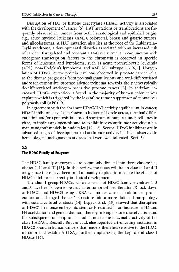
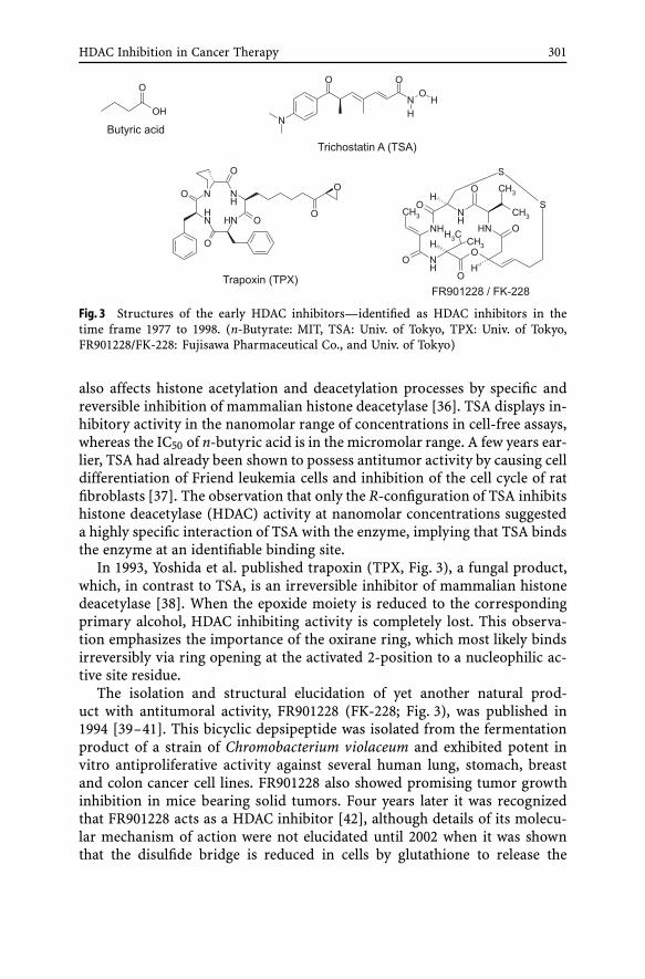
Fig. 3 Structures of the early HDAC inhibitors—identified as HDAC inhibitors in thetime frame 1977 to 1998. (n-Butyrate: MIT, TSA: Univ. of Tokyo, TPX: Univ. of Tokyo,FR901228/FK-228: Fujisawa Pharmaceutical Co., and Univ. of Tokyo)
also affects histone acetylation and deacetylation processes by specific andreversible inhibition of mammalian histone deacetylase [36]. TSA displays in-hibitory activity in the nanomolar range of concentrations in cell-free assays,whereas the IC50 of n-butyric acid is in the micromolar range. A few years ear-lier, TSA had already been shown to possess antitumor activity by causing celldifferentiation of Friend leukemia cells and inhibition of the cell cycle of ratfibroblasts [37]. The observation that only the R-configuration of TSA inhibitshistone deacetylase (HDAC) activity at nanomolar concentrations suggesteda highly specific interaction of TSA with the enzyme, implying that TSA bindsthe enzyme at an identifiable binding site.
In 1993, Yoshida et al. published trapoxin (TPX, Fig. 3), a fungal product,which, in contrast to TSA, is an irreversible inhibitor of mammalian histonedeacetylase [38]. When the epoxide moiety is reduced to the correspondingprimary alcohol, HDAC inhibiting activity is completely lost. This observa-tion emphasizes the importance of the oxirane ring, which most likely bindsirreversibly via ring opening at the activated 2-position to a nucleophilic ac-tive site residue.
The isolation and structural elucidation of yet another natural prod-uct with antitumoral activity, FR901228 (FK-228; Fig. 3), was published in1994 [39–41]. This bicyclic depsipeptide was isolated from the fermentationproduct of a strain of Chromobacterium violaceum and exhibited potent invitro antiproliferative activity against several human lung, stomach, breastand colon cancer cell lines. FR901228 also showed promising tumor growthinhibition in mice bearing solid tumors. Four years later it was recognizedthat FR901228 acts as a HDAC inhibitor [42], although details of its molecu-lar mechanism of action were not elucidated until 2002 when it was shownthat the disulfide bridge is reduced in cells by glutathione to release the
302 P. ten Holte et al.
thiol that subsequently interacts with the active-site zinc of primarily class-IHDACs [43]. FR901228/FK-228 is currently in phase II clinical trials.
As outlined before, it is believed that the TPX epoxyketone chain acts as anisosteric substrate mimic for the natural N-acetyl lysine. In 1996, Schreiberet al. exploited the irreversible binding nature of TPX in an affinity matrixby immobilizing modified TPX onto an activated agarose support [44]. Inthis way a mammalian histone deacetylase protein (HDAC1) was isolated andcharacterized for the first time.
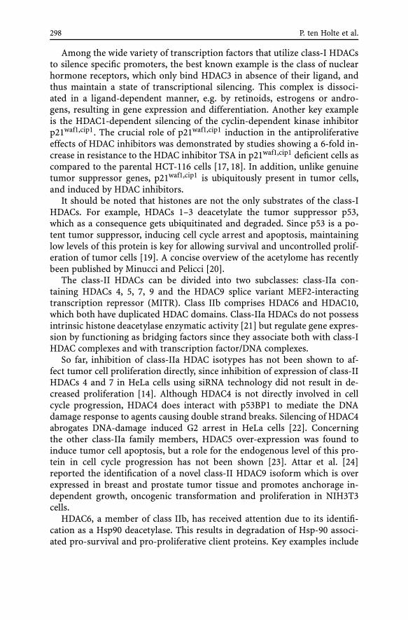
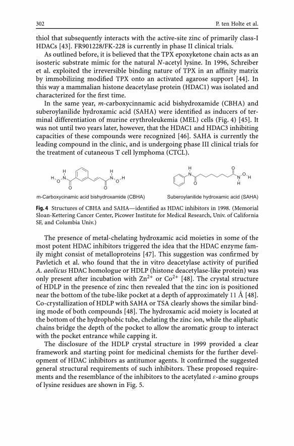
In the same year, m-carboxycinnamic acid bishydroxamide (CBHA) andsuberoylanilide hydroxamic acid (SAHA) were identified as inducers of ter-minal differentiation of murine erythroleukemia (MEL) cells (Fig. 4) [45]. Itwas not until two years later, however, that the HDAC1 and HDAC3 inhibitingcapacities of these compounds were recognized [46]. SAHA is currently theleading compound in the clinic, and is undergoing phase III clinical trials forthe treatment of cutaneous T cell lymphoma (CTCL).
Fig. 4 Structures of CBHA and SAHA—identified as HDAC inhibitors in 1998. (MemorialSloan-Kettering Cancer Center, Picower Institute for Medical Research, Univ. of CaliforniaSF, and Columbia Univ.)
The presence of metal-chelating hydroxamic acid moieties in some of themost potent HDAC inhibitors triggered the idea that the HDAC enzyme fam-ily might consist of metalloproteins [47]. This suggestion was confirmed byPavletich et al. who found that the in vitro deacetylase activity of purifiedA. aeolicus HDAC homologue or HDLP (histone deacetylase-like protein) wasonly present after incubation with Zn2+ or Co2+ [48]. The crystal structureof HDLP in the presence of zinc then revealed that the zinc ion is positionednear the bottom of the tube-like pocket at a depth of approximately 11 A [48].Co-crystallization of HDLP with SAHA or TSA clearly shows the similar bind-ing mode of both compounds [48]. The hydroxamic acid moiety is located atthe bottom of the hydrophobic tube, chelating the zinc ion, while the aliphaticchains bridge the depth of the pocket to allow the aromatic group to interactwith the pocket entrance while capping it.
The disclosure of the HDLP crystal structure in 1999 provided a clearframework and starting point for medicinal chemists for the further devel-opment of HDAC inhibitors as antitumor agents. It confirmed the suggestedgeneral structural requirements of such inhibitors. These proposed require-ments and the resemblance of the inhibitors to the acetylated ε-amino groupsof lysine residues are shown in Fig. 5.

HDAC Inhibition in Cancer Therapy 303
Fig. 5 Generally supposed structural requirements of HDAC inhibitors and some actualexamples
3.2Recent Medicinal Chemistry Efforts—An Overview
3.2.1The Cyclic Peptides
Since the discovery of trapoxin (TPX), a number of related cyclic peptideshave been found to also demonstrate HDAC inhibitory activity, explaining inpart the phenotypic effects previously described for these compounds. One

304 P. ten Holte et al.
example, closely related to TPX, is chlamydocin, differing from TPX in onlyone amino acid residue (Fig. 6) [49, 50]. More specific, in chlamydocin one ofthe two phenylalanine residues of TPX is replaced with a 2-aminoisobutyricacid. The mode of action of both molecules is believed to be identical, andto proceed via covalent and thus irreversible binding to the HDAC enzymethrough reaction of an active site nucleophile with the electrophilic oxiranering of chlamydocin or TPX.
Fig. 6 Cyclic peptides as reversible and irreversible HDAC inhibitors. (Chlamydocin: San-doz AG; CHAP 1: Univ. of Tokyo; Apidicin: Merck & Co; reversed hydroxamic acid analogsof Cyl-1: Kyushu Institute of Technology and Japan Science and Technology Agency)
Replacing the electrophilic epoxy ketone moiety in TPX by a reversiblezinc chelator such as a hydroxamic acid was carried out by Yoshida et al.(Fig. 6) [51]. This modification led to a low nanomolar reversible inhibitorof the HDAC1 enzyme. Several other cyclic tetrapeptides containing theepoxyketone feature, such as chlamydocin, were converted into their hydrox-amic acid counterparts as well [52]. Additionally, the introduction of reversedhydroxamic acids (– N(OH)COR, with R=H or Me) onto the structure ofCyl-1 was reported to give potent HDAC inhibitors as illustrated in Fig. 6 [53].Generally, the most potent inhibitors were the examples with R=H andm = 2. Apicidin, a cyclic peptide more remotely related to TPX, exhibits po-tent antiprotozoal activity via HDAC inhibition in parasites [54].
The prodrug concept of FK-228, outlined in Sect. 3.1, was exploited byNishino et al. in the development of sulfur-containing cyclic peptide-(SCOP)-based prodrugs [55]. A set of SCOP prodrugs, based on CHAP31, was syn-thesized and their in vitro HDAC inhibitory activity was evaluated (Fig. 7and Table 1). The dimer was 4-fold less potent (IC50 = 0.142 µM) on HDAC1than reference FK228, but on HDAC4 it was almost 4-fold more active (IC500.145 µM) than FK228. The dimer did not show any activity on HDACs 6 and
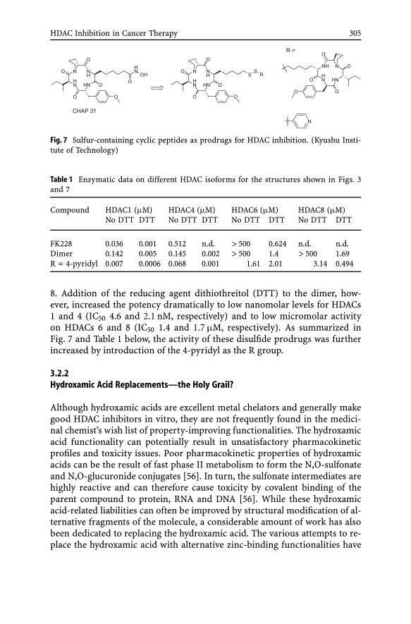
HDAC Inhibition in Cancer Therapy 305
Fig. 7 Sulfur-containing cyclic peptides as prodrugs for HDAC inhibition. (Kyushu Insti-tute of Technology)
Table 1 Enzymatic data on different HDAC isoforms for the structures shown in Figs. 3and 7
Compound HDAC1 (µM) HDAC4 (µM) HDAC6 (µM) HDAC8 (µM)No DTT DTT No DTT DTT No DTT DTT No DTT DTT
FK228 0.036 0.001 0.512 n.d. > 500 0.624 n.d. n.d.Dimer 0.142 0.005 0.145 0.002 > 500 1.4 > 500 1.69R = 4-pyridyl 0.007 0.0006 0.068 0.001 1.61 2.01 3.14 0.494
8. Addition of the reducing agent dithiothreitol (DTT) to the dimer, how-ever, increased the potency dramatically to low nanomolar levels for HDACs1 and 4 (IC50 4.6 and 2.1 nM, respectively) and to low micromolar activityon HDACs 6 and 8 (IC50 1.4 and 1.7 µM, respectively). As summarized inFig. 7 and Table 1 below, the activity of these disulfide prodrugs was furtherincreased by introduction of the 4-pyridyl as the R group.
3.2.2Hydroxamic Acid Replacements—the Holy Grail?
Although hydroxamic acids are excellent metal chelators and generally makegood HDAC inhibitors in vitro, they are not frequently found in the medici-nal chemist’s wish list of property-improving functionalities. The hydroxamicacid functionality can potentially result in unsatisfactory pharmacokineticprofiles and toxicity issues. Poor pharmacokinetic properties of hydroxamicacids can be the result of fast phase II metabolism to form the N,O-sulfonateand N,O-glucuronide conjugates [56]. In turn, the sulfonate intermediates arehighly reactive and can therefore cause toxicity by covalent binding of theparent compound to protein, RNA and DNA [56]. While these hydroxamicacid-related liabilities can often be improved by structural modification of al-ternative fragments of the molecule, a considerable amount of work has alsobeen dedicated to replacing the hydroxamic acid. The various attempts to re-place the hydroxamic acid with alternative zinc-binding functionalities have
306 P. ten Holte et al.
seen different degrees of success. A brief overview with a number of examplesof some of the approaches pursued is given below.
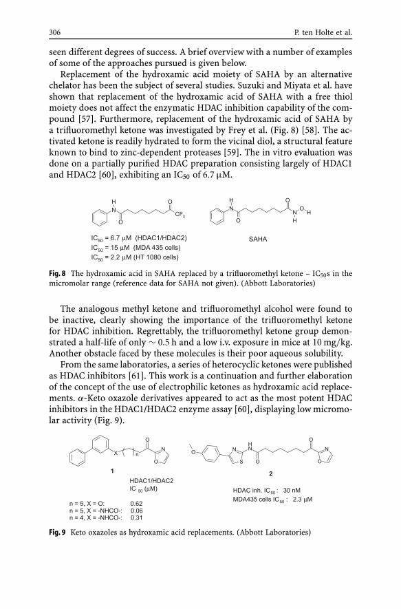
Replacement of the hydroxamic acid moiety of SAHA by an alternativechelator has been the subject of several studies. Suzuki and Miyata et al. haveshown that replacement of the hydroxamic acid of SAHA with a free thiolmoiety does not affect the enzymatic HDAC inhibition capability of the com-pound [57]. Furthermore, replacement of the hydroxamic acid of SAHA bya trifluoromethyl ketone was investigated by Frey et al. (Fig. 8) [58]. The ac-tivated ketone is readily hydrated to form the vicinal diol, a structural featureknown to bind to zinc-dependent proteases [59]. The in vitro evaluation wasdone on a partially purified HDAC preparation consisting largely of HDAC1and HDAC2 [60], exhibiting an IC50 of 6.7 µM.
Fig. 8 The hydroxamic acid in SAHA replaced by a trifluoromethyl ketone – IC50s in themicromolar range (reference data for SAHA not given). (Abbott Laboratories)
The analogous methyl ketone and trifluoromethyl alcohol were found tobe inactive, clearly showing the importance of the trifluoromethyl ketonefor HDAC inhibition. Regrettably, the trifluoromethyl ketone group demon-strated a half-life of only ∼ 0.5 h and a low i.v. exposure in mice at 10 mg/kg.Another obstacle faced by these molecules is their poor aqueous solubility.
From the same laboratories, a series of heterocyclic ketones were publishedas HDAC inhibitors [61]. This work is a continuation and further elaborationof the concept of the use of electrophilic ketones as hydroxamic acid replace-ments. α-Keto oxazole derivatives appeared to act as the most potent HDACinhibitors in the HDAC1/HDAC2 enzyme assay [60], displaying low micromo-lar activity (Fig. 9).
Fig. 9 Keto oxazoles as hydroxamic acid replacements. (Abbott Laboratories)

HDAC Inhibition in Cancer Therapy 307
The potency of these α-keto oxazole derivatives was influenced by both thelength of the spacer as well as the mode of connection to the capping group.From all the mono- and bisaromatic moieties tested, the meta-substituted bis-phenyl α-keto oxazoles 1 (Fig. 9) displayed the most potent inhibition andwere thus used to perform the initial comparison studies. A spacer length ofn = 5, in combination with the presence of an amide connector to the cappingregion (X = – NHCO –) proved to be the most active combination, displayingan IC50 of 60 nM for the HDAC enzyme assay. After further variation of thecapping moiety, while keeping the optimal spacer length and amide connec-tor, a para-methoxyphenyl substituted thiazole capping group 2 was found togive the most potent HDAC inhibitor (IC50 30 nM) that, in addition, showedantiproliferative activity in MDA435 cells (IC50 2.3 µM, Fig. 9). The authorssuggest that the cellular activity, however, was compromised by the instabil-ity of these compounds due to rapid reduction of the keto functionality to theinactive alcohol.
A series of benzamides as replacement for the zinc-binding hydrox-amic acid was also synthesized and investigated for HDAC inhibitory ac-tivity [62, 63]. A clear SAR could be derived from the examples prepared(Fig. 10).
Fig. 10 Benzamides as hydroxamic acid substitutes—discovery of MS-275. (Mitsui Phar-maceuticals)
Thus, the 2′-amino benzamide (entry 1, MS-275) showed low micromolarinhibition of HDAC enzyme (IC50 = 4.8 µM), whereas the activity was com-pletely abolished by shifting the amino group to the 3′ or 4′ position (entries 2and 3). Remarkably, when a hydroxyl group was introduced in the 2′ pos-ition (entry 4), HDAC inhibitory activity was fully restored (IC50 = 2.2 µM)suggesting that the hydrogen-bonding capability of the 2′ group is an im-portant requisite for interaction with the enzyme. Taking MS-275 (entry 1)as a starting point, methyl groups were introduced at the 3′, 4′ and 5′ pos-itions (entries 5, 6 and 7). Only the product with the methyl at the 5′ positionshowed HDAC inhibition (entry 7, IC50 = 2.8 µM). Steric hindrance seemsto be the most plausible reason for the inactivity of compounds with methylsubstitution at the 3′ and 4′ positions (entries 5 and 6).

308 P. ten Holte et al.
An additional example of a benzamide showing HDAC inhibitory activityis N-acetyl dinaline, also known as CI-994 or tacedinaline (Fig. 11) [64–66],which is the acetylated derivative of the earlier identified dinaline (GOE 1734,PD 104 208) [67]. Both CI-994 and MS-275 have been in clinical develop-ment [68, 69].
Fig. 11 Other HDAC inhibiting benzamides. (dinaline and N-acetyl dinaline (CI-994 ortacedinaline): Erasmus Univ.; MGCD0103: MethylGene Inc.)
Although both MS-275 and CI-994 elicit the classical hallmarks of HDACinhibitors in tumor cell-based assays—accumulation of histone H3 acetyla-tion, increased expression of cyclin-dependent kinase inhibitor p21WAF1/Cip1
and accumulation in the G1 phase—their molecular mechanism of actionis still poorly understood and remains somewhat controversial. As com-pared to the hydroxamic acid-containing HDAC inhibitors (e.g., TSA, SAHA,PXD-101, LBH-589, R306465), benzamide derivatives have been shown in nu-merous independent studies to be relatively weak inhibitors (10- to 1000-foldlower inhibition) of HDAC activity in classical HeLa cell nuclear extract, andimmunoprecipitated or recombinant HDAC enzymes [70–73]. Nevertheless,extensive SAR analyses on benzamide derivatives have been carried out tosupport HDAC inhibition, coupled to the claim of a high degree of flexibilityin the active site-pocket for accommodating groups with different stereoelec-tronic properties. As outlined in detail before, a structural analogue of MS-275—possessing a 3′-aminophenyl instead of a 2′-aminophenyl group—didnot display any inhibition of HDAC activity in biochemical cell-free assays,and binding of the 2′-aminophenyl group to an unidentified but specific siteon HDAC enzymes has been hypothesized. The 2′-substituent of benzanilidemight act as a hydrogen-bonding site or other electrostatic interaction siteand be indispensable to the specific interaction with the enzymes. In addition,steric hindrance may play an important role. Over the years, these consistentobservations on weak in vitro potency and the lack of a molecular mechanismof action have supported the question of the identification of these benzamidederivatives as “genuine” HDAC inhibitors. Furthermore, recent studies com-paring gene expression profiling of multiple HDAC inhibitors have indicatedsubstantial differences in up- or down-regulation of sets of genes induced byTSA or SAHA, as compared to MS-275 [74]. One may speculate that thesedistinctly different expression profiles could be related to their differencesin potency against the HDAC enzymes, but these observations do not ex-

HDAC Inhibition in Cancer Therapy 309
clude the possibility that benzamide derivatives, or at least MS-275, may acton HDAC-dependent downstream signalling pathways by indirect mechan-ism(s).
MGCD0103, a compound that is structurally closely related to MS-275,Fig. 11, is currently in clinical trials (Sect. 4.2).
3.2.3Is the Type of Spacer Really all that Important?
The spacer region of HDAC inhibitors has been the subject of optimizationin several medicinal chemistry reports. Examples of a class of compoundshaving an aromatic moiety present in the spacer region are depicted inFig. 12 [75–77].
Fig. 12 Hydroxamic acid and benzamide containing HDAC inhibitors with aromatic spac-ers. (4 and 5: MethylGene Inc.; 3: MethylGene Inc. and TopoTarget UK Ltd.)
Vinyl benzene hydroxamic acid 3 with R = Ph, (IC50 = 10 nM for HDAC1)was found after variation of the spacer length as well as modification of thesubstituent on the capping sulfonyl aryl moiety. Electron-rich groups at thepara position of the arylsulfonyl provided the most active inhibitors. For theanalogous alkyl benzene hydroxamic acids 4 it was also found that the activ-ity for HDAC could be tuned by changing the length of the spacer. Additionor removal of only one methylene group in the alkyl benzene hydroxamic acidseries resulted in a ten-fold decrease in activity in both instances (Fig. 12). In-terestingly, the vinyl benzene hydroxamic acids were also compared to a set ofanalogs containing the 2-amino benzamide group (5) instead of the hydrox-amic acid, also depicted in Fig. 12, which appeared to be a factor 10–100 lesspotent on HDAC1 than their hydroxamate counterparts (Fig. 12).
An extensive study focusing on sulfonamide containing hydroxamic acidderivatives as HDAC inhibitors led to the discovery of PXD101 (Fig. 13) [78–80], which is currently in clinical trials. It was shown that for the meta sub-stituted sulfonamides, the so-called “reverse” sulfonamides were consistently(2- to 7-fold) more potent HDAC inhibitors than the “forward” sulfonamides.

310 P. ten Holte et al.
This trend was not observed for para substituted compounds. An inter-esting comparison in this respect is that of vinyl benzene hydroxamic acid3 (R = Ph) shown earlier in Fig. 12 with its “reverse” analog 6 (Fig. 13) [78].These two compounds, having reversed sulfonamide bonds, show similar ac-tivities.
Fig. 13 PXD101 and its reversed analog possess unsaturated spacers. (TopoTarget UKLtd.)
The role of aromaticity in the spacer region on the potency of hydrox-amic acid containing inhibitors of HDAC was further investigated by Uesatoet al. [81]. The data in Fig. 14 represent HDAC inhibitory activities againstpartially purified HDACs from human T cell leukemia Jurkat cells [82]. Thesedata suggest that aromatic hydroxamic acids exhibit a much greater affinityfor HDACs than their aliphatic counterparts. It is speculated in the paper thatthe 1,4-phenylene moiety may interact with aromatic and hydrophobic aminoacid residues in the pocket, while the 2-naphthyl group would be engaged insimilar interactions with aromatic amino acid residues at the pocket entrance,thereby capping the pocket.
Fig. 14 Saturation of the aromatic spacer deteriorates the potency of a hydroxamic acid-based HDAC inhibitor. (Kansai Univ.)
Introduction of a heteroaromatic group in the spacer proved beneficial inthe work by Johnson & Johnson researchers Arts et al. [83] and Angibaudet al. [84]. Optimization of the initial lead, having a 1,4-substituted phenylpresent in the spacer region (Fig. 15, entry 1), was done by replacing this R

HDAC Inhibition in Cancer Therapy 311
Fig. 15 Fine tuning of the (hetero)aromatic spacer of a hydroxamic acid-based HDACinhibitor—discovery of R306465. (Johnson & Johnson Pharmaceutical R&D)
group with a series of six-membered heteroaryls. It was found that pyrim-idyl (entry 3) was the most efficient R group of the series, demonstratingan IC50 of 6 nM for the enzyme assay consisting of a mixture of HDAC iso-forms from a HeLa cell nuclear extract1. This increase of activity is likelyto be the result of both steric and electronic factors. Finally, introduction ofa functional group (such as dimethylamino or ethoxy) at the 4-position ofthe pyridyl causes complete loss of activity. These optimization studies of thelinker led to the discovery of R306465, which is currently in Phase I clinicaltrials.
3.2.4The Capping Group under Scrutiny
The importance of the structure of the capping group of HDAC inhibitors wasunderlined by a study in which Dai et al. carried out systematic modificationsin this capping region [85]. SAHA analogues with reversed amides were func-tionalized with heteroaromatic residues as in the general structure (7) shownin Fig. 16. The indole capping group (e.g., see compound 8) proved the mosteffective, demonstrating nanomolar inhibitory activity on HDAC1/2 enzyme.
1 HeLa cell nuclear extracts were incubated at different concentrations with a radiolabeled acety-lated Histone 4 peptide fragment as substrate. HDAC activity was assessed measuring release of freeacetyl groups.

312 P. ten Holte et al.
Fig. 16 Introduction of heteroaromatic groups in the capping region of hydroxamic acidcontaining HDAC inhibitors. (Abbott Laboratories)
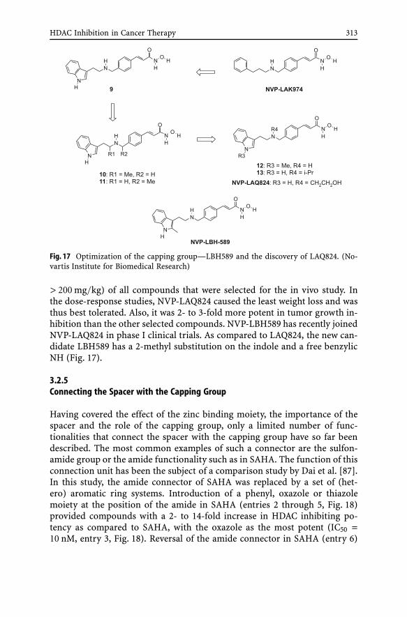
The indole functionality was also used as a capping group by researchersfrom Novartis [86]. The initial hit from high-throughput screening (NVP-LAK974, Table 2 and Fig. 17), bearing a phenyl propylamine capping group,demonstrated acceptable in vitro activity but had poor efficacy in the HCT116human colon tumor xenograft model. Replacement of this phenyl propy-lamine moiety with a tryptamine (9) triggered an overall increase of in vitropotency up to a factor 2 compared to the original hit. Introduction of a methylgroup at either the chain carbon α to the benzylamine (10) or the benzyliccarbon (11) provided a 2-fold increase in enzymatic HDAC inhibitory activ-ity and an approximate 2-fold increase in cellular potency on the HCT116 cellline while keeping the same antiproliferative activity on H1299 cells. Cellularpotency was further improved by a factor 2 via methylation of the indole N(12). Finally, nonpolar aliphatic substituents on the benzylic amine generallyimproved cellular potency as is illustrated by the introduction of an isopropylgroup (13), giving a HDAC inhibitor with IC50s of 6 and 30 nM on HCT116and H1299 cells, respectively.
The N-hydroxyethyl analogue NVP-LAQ824 (Table 2, Fig. 17) also showedgood overall potency in vitro, but excelled in the succeeding in vivo ex-periments. It demonstrated the highest maximum tolerated dose (MTD;
Table 2 Published enzymatic and cellular data for the structures shown in Fig. 17 [86]
Compound HDAC enzyme ∗ H1299 cells HCT116 cells(IC50, nM) (IC50, nM) (IC50, nM)
NVP-LAK974 150 (±94) 800 509 63 (±10) 400 30
10 24 (±5) 410 2011 23 (±12) 480 1512 23 (±12) 170 1013 23 (±6) 30 6NVP-LAQ824 32 (±18) 150 10
∗ Mixture of HDAC isoforms from H1299 whole cell extracts
HDAC Inhibition in Cancer Therapy 313
Fig. 17 Optimization of the capping group—LBH589 and the discovery of LAQ824. (No-vartis Institute for Biomedical Research)
> 200 mg/kg) of all compounds that were selected for the in vivo study. Inthe dose-response studies, NVP-LAQ824 caused the least weight loss and wasthus best tolerated. Also, it was 2- to 3-fold more potent in tumor growth in-hibition than the other selected compounds. NVP-LBH589 has recently joinedNVP-LAQ824 in phase I clinical trials. As compared to LAQ824, the new can-didate LBH589 has a 2-methyl substitution on the indole and a free benzylicNH (Fig. 17).
3.2.5Connecting the Spacer with the Capping Group
Having covered the effect of the zinc binding moiety, the importance of thespacer and the role of the capping group, only a limited number of func-tionalities that connect the spacer with the capping group have so far beendescribed. The most common examples of such a connector are the sulfon-amide group or the amide functionality such as in SAHA. The function of thisconnection unit has been the subject of a comparison study by Dai et al. [87].In this study, the amide connector of SAHA was replaced by a set of (het-ero) aromatic ring systems. Introduction of a phenyl, oxazole or thiazolemoiety at the position of the amide in SAHA (entries 2 through 5, Fig. 18)provided compounds with a 2- to 14-fold increase in HDAC inhibiting po-tency as compared to SAHA, with the oxazole as the most potent (IC50 =10 nM, entry 3, Fig. 18). Reversal of the amide connector in SAHA (entry 6)

314 P. ten Holte et al.
Fig. 18 Extensive variation of the amide connection unit of SAHA leads to a single digitnanomolar HDAC inhibitor. (Abbott Laboratories)
resulted in a much weaker inhibitor that showed only micromolar HDAC in-hibition. Replacement with an ether or methylene linkage (entries 7 and 8,respectively) also led to deterioration of HDAC inhibition.
3.2.6The Quest for Selective HDAC Inhibitors
Over the years, it has become evident that HDACs not only play a key rolein carcinogenesis but also in a number of nonmalignant differentiation pro-cesses. This is most apparent for the class-IIa HDACs 4, 5, 7, 9. For example,HDAC7 has been suggested to play a critical role in the thymic maturation ofT-cells [88], while HDAC4 has been implicated in the regulation of chondro-cyte hypertrophy and endochondral bone formation by inhibiting the activityof the Runx2 transcription factor [89]. Most concerns, however, have focusedaround the role of the class-IIa HDACs in muscle differentiation. HDACs 4, 5,7 and 9 all suppress the differentiation of myocytes (muscle cells) as a con-sequence of being transcriptional co-repressors of myocyte enhancer factor 2(MEF2) [90]. Deletion of the MEF2 binding domain of the most abundant my-ocyte class-II HDAC, HDAC9, results in development of cardiac hypertrophyin 9-months old mice due to hypersensitivity to hypertrophic signaling [91].
The observations above have led to speculation that HDAC inhibitors maycause cardiac hypertrophy. Surprisingly however, it was observed that HDACinhibitors may actually be beneficial in treating cardiac hypertrophy. TSA has
HDAC Inhibition in Cancer Therapy 315
been shown to block the fetal gene program associated with cardiomyocytehypertrophy in response to hypertrophic agents. It was therefore proposedthat inhibition of other HDACs (e.g. class I) may counteract the expression ofthe hypertrophic genes associated to the class-II HDACs [92].
Even though it is currently unclear whether any of the side effects observedin the clinic with the current pan-HDAC inhibitors are linked to inhibition ofthe class-II HDACs, these observations, nevertheless, triggered the quest forHDAC isotype specific inhibitors, which will be further discussed below.
The HDAC inhibitors TSA and TPX (Fig. 2) have been utilized as struc-tural leads in the early stages of the quest for new and more selective smallmolecule inhibitors of the HDAC enzyme family. In order to investigate thefunction of the individual HDAC members, Schreiber et al. synthesized a li-brary of 7200 potential HDAC inhibitors based on the structural featuresof TSA and TPX [93]. The members of this library were prepared on solidsupport by means of split pool methods. The key characteristics of these com-pounds consist of a dioxane-containing capping region and a zinc bindingmotive, connected via an aliphatic chain. Three different zinc binders, i.e.,carboxylic acid, o-aminoanilide and hydroxamic acid were used.
Assuming that equal purities were obtained for the different classes ofmetal chelators, it was found in both the AcTubulin and the AcLysine cy-toblot assays that the hydroxamic acids were the most active inhibitorsof both α-tubulin and histone deacetylation [94]. On the other hand, theo-aminoanilides demonstrated the weakest inhibition of HDAC enzyme. An-alysis of the compounds using principal component analysis followed byresynthesis disclosed the structures of two examples of selective inhibitors.The first, tubacin, is a selective inhibitor of α-tubulin deacetylation withno effect on the histone acetylation status. The function of HDAC6 as anα-tubulin deacetylase enzyme [95] and its role in mediating cell cycle pro-gression, microtubule stability, and cell motility has been studied usingtubacin as the selective inhibitor [28]. The second, histacin, is a selective in-hibitor of histone deacetylation (Fig. 19).
Mai et al. have carried out a comprehensive study of the aroyl pyrrolylhydroxamic acids (APHAs) as HDAC inhibitors [96–98] and succeeded in ob-taining class-II selectivity (Fig. 20) [99].
The APHA with a fluorine atom at the 3-position of the aryl exhibitsa class-II/class-I HDAC selectivity of 176, whereas substitution at the 2-or 4-position gives respectively much lower selectivity ranging from 34 toa value less than 2. It is interesting to note that this “meta-effect” is much lesspronounced when the substituent is a chloro atom and the effect is completelylost when a bromo-atom is introduced (data not shown).
A rational approach toward the design of class-I isoform selective HDACinhibitors was reported recently by Wiest et al. [100]. In order to understandthe difference between these class-I isoforms, three-dimensional models ofHDAC1, HDAC2, HDAC3, and HDAC8 were built using homology modeling.

316 P. ten Holte et al.
Fig. 19 Discovery of tubacin and histacin—two selective deacetylation inhibitors. (Har-vard University)
Fig. 20 Class-II selectivity has been accomplished for some APHAs. (Università degliStudi di Roma “La Sapienza”)
The high homology of the active site region of the different class-I HDACs aswell as the considerable similarity of their 11 A channel do not leave enoughroom for a selectivity prediction based on these parts of the enzyme. Themodels show, however, that electronic and steric dissimilarity around theopening of the active sites holds potential for differentiating between HDAC1,HDAC3, and HDAC8. Differentiation between HDAC1 and HDAC2, though,is predicted to be more difficult. The design of novel HDAC inhibitors usingthese models is currently in progress.
It is evident that the quest for selective HDAC inhibitors has just be-gun and that the optimal HDAC subtype selectivity profile for an anticancerdrug based on HDAC inhibition is still far from being established. Never-

HDAC Inhibition in Cancer Therapy 317
theless, the first important steps toward the rational design and synthesis ofisoform selective HDAC inhibitors have been taken. The first clinical trialswith MGCD0103 (Fig. 11), an isotype selective inhibitor of human HDACsare ongoing [101]. Moreover, the understanding of the biological and clini-cal consequences of different HDAC inhibitory profiles is increasing steadilyas more and more of the biology becomes known while a growing number ofcompounds are being evaluated in the clinic.
4Clinical Experience with HDAC Inhibitors
Histone deacetylases are linked to the pathogenesis of malignancy froma mechanistic perspective. The capacity of HDAC inhibitors (HDACi) to inter-fere with the enzyme function has led to the observed preclinical and clinicalactivity in cancer therapy. Although the exact mechanism of anti-tumor ac-tivity is not fully elucidated, various cellular pathways have been shown tobe involved. From the first clinical trials involving HDACi with short chainfatty acids to the newer generation hydroxamic acid derivatives and cyclictetrapeptides, a number of structurally diverse compounds have made thetransition from the laboratory to the clinical arena. For purposes of this partof the discussion, HDACi are arbitrarily divided into the hydroxamates andnonhydroxamates.
Most of the studies reported are in early phase (Phase I and II) with the ex-ception of Vorinostat (suberoylanilide hydroxamic acid [SAHA]), which hasentered Phase III. Some of these studies have only been published in abstractform. Encouragingly, activity has been seen especially in lymphoproliferativediseases, leukemia and some solid tumors, including prostate cancer.
Generally, the impression so far is that HDACi display a somewhat lowertoxicity profile compared to conventional cytotoxics. The most common tox-icity seen is nausea/vomiting and fatigue, mild myelosuppression and diar-rhea and these feature as adverse effects in many clinical trials. The toxicitiesobserved may be due to the individual drug or the consequence of inhibit-ing HDAC itself (class effect). It is postulated that interference with additionalcellular pathways, not just histone acetylation, may be responsible for the dif-ferential toxicity seen clinically, especially if these different compounds areused at higher doses.
The relationship between toxicity and pharmacokinetic (PK) and phar-macodynamic (PD) parameters is a difficult one and somewhat poorly char-acterized. The key targets of HDACi are unknown and predicting whichpatients will respond to HDACi therapy is difficult. Correlation between sur-rogate markers (for example, levels of acetylated histones in peripheral bloodmononuclear cells [PBMNC] pre- and post-dosing) is not always in keepingwith measured PK profiles.

318 P. ten Holte et al.
4.1Hydroxamates
Various agents in this class have gone on to phase I evaluation and beyond.These include: vorinostat (SAHA), LAQ824, LBH589, PXD101 and R306465,some of which are discussed in greater detail below and listed in Table 3.
Table 3 Selected hydroxamates continuing in clinical development in solid tumors. CR:complete response, PR: partial response, uPR: unconfirmed PR, SCC: squamous cell car-cinoma
Drug/Phase Schedule Tumor type Response/ Mainno. of toxicitiespatients
2 hours i.v. Solid tumors n = 37 Fatigue,days 1–5, 8–12, dehydration,15–19 every anorexia
SAHA 4 weeksPhase I [102]
See above for structure Various oral, Solid tumors 1CR, 3 PR, Fatigue,SAHA 200–600 mg and hemato- 2 uPR, gastro-Phase I [103] qd to bd logical n = 73 intestinal,
hematological(thrombo-cytopenia)
See above for structure Oral, bid or tid Leukemia, 3 CR, 1 PR Nausea,SAHA d1–14 myelodys- n = 41 vomiting,Phase I [114] every 21 days plastic syn- diarrhea,
drome fatigue
See above for structure Oral, Head & neck 1 minor Anorexia,SAHA 400 mg qd SCC response anemia,Phase II [115] n = 13 fatigue,
hematological
See above for structure Various oral, Cutaneous 10 PR Fatigue, rash,SAHA 400 mg qd, T-cell n = 37 hematologicalPhase II [116] 300 mg bid, lymphoma (thrombo-
200 mg bid cytopenia)
30 minutes i.v. Solid tumors n = 42 Fatigue,days 1–15 every Accrual nausea,21 days continues vomiting,
PXD101 diarrhea,Phase I [117] phlebitis
HDAC Inhibition in Cancer Therapy 319
4.1.1Vorinostat (SAHA)
SAHA is the HDACi that has advanced farthest in clinical trials. Both in-travenous and oral Phase I trials involving 110 patients have been re-ported [102, 103]. Briefly from these studies, the mean intravenous half life(t1/2) is between 92 to 127 minutes, whereas the oral half life is longer. There isdemonstration of linear pharmacokinetics, oral bioavailability of more than40%, and increased duration of acetylated histone H3 (AcH3) was seen withincreasing dose and prolonged dosing. However, acetylation effects, althoughrapid, are transient and return to near baseline levels by 8 hours except athigher dose cohorts. The maximum tolerated dose (MTD) was 200 mg twicedaily or 400 mg daily continuously or 600 mg twice daily 3 times per week.
The most common drug related Grade 3/4 toxicities are fatigue, nausea,vomiting, diarrhea, anorexia, anemia, thrombocytopenia, hyperglycemia andhypocalcemia. No clinically significant electrocardiograph (EKG) changes orcardiac toxicities, including arrhythmias attributable to the drug, were seen.In fact, SAHA is probably the only HDACi that has not resulted in EKGchanges. Toxicities including myelosuppression were rapidly reversible upondiscontinuation of drug. It has been postulated that the thrombocytopenia isdue to impairment of megakaryocyte differentiation [104].
A significant proportion (30%) of patients on SAHA remained on the drugbetween 4 to > 37 months, with chronic dosing demonstrating prolongeddisease stabilization, maintained biological effect and drug tolerability. Re-sponses were seen in lymphoma, laryngeal carcinoma, thyroid cancer andmesothelioma.
Phase I studies have also been conducted in various hematological malig-nancies including myeloma [105]. Other Phase I studies looking at SAHA incombination with retinoic acid and gemcitabine, Phase II studies in tumorspecific areas of head and neck squamous cell carcinoma, T-cell lymphoma,melanoma and glioma, and a Phase III study in mesothelioma have beencompleted or are in progress.
Phase I studies of intravenous LAQ824 and LBH589 [106–109], novel cin-namyl hydroxamates, have also been completed, with 112 patients dosed intrials of both agents. LBH589 has also been administered orally. LAQ824is a potent HDACi given intravenously and has been shown to also inhibitHsp90 [110]. In these trials cardiac toxicity, including prolonged QT inter-val (QTc) effects, nonspecific ST segment and T wave changes on EKG andarrhythmia were reported at high doses when administered intravenously.Overall, both LAQ824 and LBH-589 were found to induce dose-related in-creases in QTcF (Fridericia correction) of 20 msec or less at doses up to200 mg/m2 and 20 mg/m2, respectively [111, 112]. Cardiac repolarizationchanges were often delayed until day 3, and may not be due to a direct effectof the agents on the hERG (human ether-a-go-go related gene) channel [113].

320 P. ten Holte et al.
Currently, orally administered LBH589 is in Phase II clinical trials and atlower doses the electrocardiographic change can be abrogated. A Phase IIstudy for LBH-589 in solid and liquid tumors is ongoing.
Enrolment is ongoing for PXD101, for which another Phase I study inhematological malignancies is in progress, exploring the possibility for oraldosing. A Phase II study in multiple myeloma is also currently ongoing.
4.2NonHydroxamates
Various classes of short-chain fatty acids, cyclic tetrapeptides and benza-mides have also been in clinical trials (Table 4).
The short chain fatty acids include butyrate derivatives like phenylbu-tyrate, AN-9 (pivaloyloxymethyl butyrate) and valproate. Unfortunately, thesecompounds have poor potency and pharmacokinetic properties, includingshort half-life. Numerous Phase I studies with phenylbutyrate, in various oraland intravenous schedules [118–120] have been performed, with neurolog-ical toxicity at higher doses being reported. AN-9 showed initial promisein a Phase I study, where the MTD was not reached [121]. The subsequentPhase II study in nonsmall cell lung cancer in 47 patients resulted in fatigue,nausea and dysgeusia as common toxicities. Three partial responses (PR)
Table 4 Selected nonhydroxamates continuing in clinical development. MTD: maximumtolerated dose, PR: partial response
Drug/Phase Schedule Tumor Response/ Main Commenttype n = no. of toxicities
patients
4 hr i.v. Solid 1 PR Nausea, MTDdays 1 and 5 tumors n = 37 vomiting, 17.8 mg/m2
every 21 days thrombo-cytopenia,atrialfibrillation
Depsipeptide (FK-228)Phase I [124]
See above for structure 4 hr i.v. Solid n = 33 Fatigue, MTDDepsipeptide (FK-228) days 1, 8 and 15 tumors thrombo- 13.3 mg/m2
Phase I [125] every 28 days cytopenia
See above for structure 4 hr i.v. Hema- n = 20 Nausea, dosed atDepsipeptide (FK-228) days 1, 8 and 15 tological fatigue, 13 mg/m2
Phase I [137] every 28 days anorexia
HDAC Inhibition in Cancer Therapy 321
Table 4 (continued)
Drug/Phase Schedule Tumor Response/ Main Commenttype n = no. of toxicities
patients
See above for structure 13 mg/m2 Castration 2 PR Fatigue,Depsipeptide (FK-228) 4 hr i.v. refractory n = 16 nausea,Phase II [129] days 1, 8 and 15 prostrate anorexia
every 28 days cancer
See above for structure 13 mg/m2 Renal 1 PR Fatigue,Depsipeptide (FK-228) 4 hr i.v. cancer n = 30 nausea,Phase II [128] days 1, 8 and 15 vomiting,
every 28 days anemia,anorexia
Oral Solid n = 30 Anorexia, MTDfortnightly tumors nausea, 10 mg/m2
vomiting,fatigue
MS-275Phase I [133]
See above for structure Oral Solid 1 PR Fatigue,MS-275 various tumors n = 24 neutropenia,Phase I [135] hypophos-
photaemia
See above for structure Oral weekly Solid n = 13 Neutropenia, AccrualMS-275 X4 every tumors, nausea/ continuesPhase I [134] 6 weeks lymphoma vomiting
3 times/wk Solid n = 24 Fatigue, Accrualfor 2 wk tumors nausea, continuesevery 3 wk vomiting,
anorexiaMGCD0103Phase I [123]
were reported [122]. However, safety concerns regarding its combination withcytotoxics has led to interruption of its development.
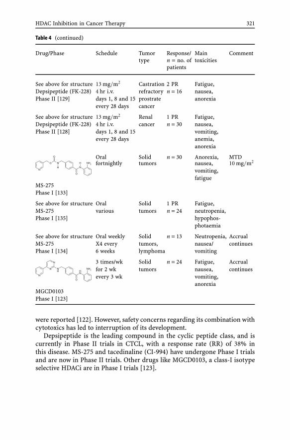
Depsipeptide is the leading compound in the cyclic peptide class, and iscurrently in Phase II trials in CTCL, with a response rate (RR) of 38% inthis disease. MS-275 and tacedinaline (CI-994) have undergone Phase I trialsand are now in Phase II trials. Other drugs like MGCD0103, a class-I isotypeselective HDACi are in Phase I trials [123].
322 P. ten Holte et al.
4.2.1Depsipeptide (FR901228 or FK-228)
Preclinical studies have shown improved tolerability and antitumor activity,with an intermittent dosing schedule as the result of the ability to administerhigher doses, with shorter infusions found to induce less toxicity [124]. In theinitial study, dose limiting toxicities (DLTs) included nausea and vomiting,thrombocytopenia and cardiac arrhythmia with atrial fibrillation. Becausecardiac toxicity had been predicted from preclinical studies (myocardial hem-orrhage and ischemia) patients were treated under continuous cardiac moni-toring. This and a further study [125] showed no clinically significant cardiacadverse effects were observed, although subtle EKG changes were reported(QTc interval prolongation, ST segment and T wave changes). Toxicities ob-served included nausea/vomiting, thrombocytopenia, fatigue and hypophos-photemia.
Studies in patients with T-cell lymphoma have used a schedule of dep-sipeptide administered on days 1, 8, and 15 of a 28-day cycle at a dose of14 mg/m2 [126]. This study involved intensive cardiac monitoring, cardiacbiochemistry markers and functional imaging monitoring. No definitive orclinically significant changes have been seen so far. In the updated multiplecohort Phase II study of cutaneous T-cell lymphoma (CTCL) and periph-eral T-cell lymphoma (PTCL), 66 patients have been treated with responsesin 10 CTCL and 6 PTCL patients [127], which is very encouraging in thisheavily pre-treated group. A Phase II study in renal cancer [128] showed 1 re-sponse in 30 patients. An ongoing Phase II study in castration refractoryprostate cancer showed 2 partial responses in 16 evaluable patients [129]. Fur-ther Phase II studies involving tumor types such as myeloma, acute myeloidleukemia (AML) and colorectal cancer have been conducted or are ongo-ing [130–132].
4.2.2MS-275
This synthetic benzamide was studied in two different schedules, with thedaily schedule exceeding MTD at first dose-level of 2 mg/m2, unpredicted,possibly due to long t1/2 from possible enterohepatic recirculation. The fort-nightly schedule was found to be feasible, and an MTD of 10 mg/m2 has beenestablished from 28 patients [133]. There were no clinically significant cardiactoxicities either from a rhythm perspective or from assessment of left ven-tricular ejection fraction. Toxicities seen include anorexia, nausea, vomiting,diarrhea, fatigue, myelosuppression, hypoalbuminemia and hypophospho-temia.
Clinical studies with a weekly dosing schedule are reported to be on-going [134]. Another study exploring three different schedules of biweekly,
HDAC Inhibition in Cancer Therapy 323
twice weekly and weekly for 3 out of 4 weeks has evaluated 24 patients [135].Fatigue, hypophosphotemia and neutropenia were some of the significanttoxicities.
4.2.3CI-994 (Tacedinaline)
The mechanism of action of this benzamide compound is not entirely un-derstood but it has been shown to inhibit HDAC and cellular proliferation.It displays linear kinetics and is rapidly absorbed after oral administration.The main dose limiting toxicity (DLT) reported was thrombocytopenia withthe MTD at 8 mg/m2/day, although other toxicities like nausea, vomiting, di-arrhea and fatigue were seen [136]. One partial response was seen in the 53patients evaluated.
4.3Summary and Future Development
The early and prolonged responses reported in clinical trials with HDACiinvolving patients with cutaneous T-cell lymphoma (CTCL), acute myeloidleukemia and other solid tumors have been encouraging. A submission tothe FDA of vorinostat (SAHA) for CTCL was filed in the second quarter of2006. The potential for HDACi therapy however probably goes beyond singleagent use. The wide ranging molecular pathways affected by HDACi’s makeit a promising candidate for the exploration of combinatorial studies in theclinical setting.
In vitro studies have evaluated the additive and synergistic antitumor ac-tivity of HDACi with many agents including cytotoxics, targeted molecules,and radiation. Considerable interest has been focused on combinations withDNA methyltransferase inhibitors like 5 aza-2′deoxycytidine (decitabine)and retinoic acid receptor (RAR)-targeted drugs. Furthermore, enhancementof apoptosis has been shown with traditional cytotoxics like the topoiso-merase II agents and taxanes, TRAIL (tumor necrosis factor related apoptosisinducing ligand), CDK (cyclin dependent kinase) inhibitors, Hsp-90 antag-onists like 17-AAG (17-allylamino-17-demethoxygeldanamycin), proteosomeinhibitors and enhanced radio sensitivity to ionizing radiation [11, 138].Combination clinical studies of HDACi with retinoic acid [139] and con-ventional cytotoxics like carboplatin, paclitaxel, capecitabine and gemc-itabine [140–143] have already been shown to be feasible in the clinic. Al-though improved response rates have yet to be demonstrated, trial character-istics make it difficult to draw definitive conclusions at such an early stage.
Experience so far from clinical trials has shown these agents can be welltolerated at biologically active doses. However, cardiac toxicity, mainly QTCprolongation, and cardiac arrhythmias, including atrial fibrillation and tor-
324 P. ten Holte et al.
sades de pointes, appears to be a recurrent theme with both the hydroxamatesand nonhydroxamates. The clinical significance of these findings, if at all, willbecome more apparent with later phase studies. Dose-limiting toxicity fromthe various agents generally involves constitutional symptoms, in particularfatigue and nausea.
The improved PD effect seen with more frequent dosing in the SAHA studyfavors the development of oral agents in the effort to sustain HDAC inhibitionvia more continuous exposure [103]. The next generation hydroxamate agentswith prolonged PD responses have entered clinical trials.
With novel and newer generation HDACi emerging, the importance of val-idating drug effect lies mainly in determining acetylation of histones (H3 andH4), from surrogate tissue/cells such as from PBMNC. Validating drug ef-fect in tumor tissue, although not always practical, is critical in establishing“proof of concept” of biological modulation. However, robust data of correla-tion of degree of acetylation with tumor response is not available at present.Furthermore, it is unknown if inhibition of histone deacetylation, acetylationof nonhistone proteins or effects on other cellular pathways is responsible forthe clinical benefits seen. As the knowledge expands rapidly on nonhistonesubstrates of HDACs, development of new biomarkers, as well as quick, sim-ple and easily reproducible methods of quantifying the degree of acetylationof HDACi, will be crucial to the future of these drugs.
5Perspectives and Conclusion
Chromatin has evolved into an established therapeutic target. Accumulatingevidence suggests that chromatin-modulating drugs are on the verge of be-coming a new drug class on their own with significant medical potential.
HDAC inhibition holds particular promise in anticancer therapy, where theconcerted effects on multiple pathways involved in growth inhibition, differ-entiation and apoptosis may prove to be advantageous in the treatment ofa heterogeneous pathology such as tumor formation and growth.
It remains to be seen whether pan-HDAC inhibition is a prerequisite forclinical efficacy, or whether more subtype-specific HDAC inhibition offersclinical advantages in relation to efficacy and/or toxicity. It should be un-derstood however that the current state of the art suggests that chromatinremodeling is not the only way in which HDACi exert their antitumor effects.As more and more evidence indicates that HDACs not only play a central rolein the epigenetic status of chromatin, but are also involved in other levels ofenzymatic control, their ability to act on multiple molecular pathways onlyadds to their multi-targeting properties.
From a chemogenomics perspective, the past and present generations ofHDAC inhibitors, while providing much insight into the molecular mechan-

HDAC Inhibition in Cancer Therapy 325
isms and resulting biology of HDAC inhibition, remain to some extent limitedin diversity.
With the exception of the benzamide class, represented by MS-275, and toa lesser degree natural products such as depsipeptide, the majority of HDACinhibitors feature the hydroxamic acid functionality, attached to a cap groupvia a spacer. The fact that this privileged structure appears in many of the re-cently disclosed HDAC inhibitors may be a consequence of the very specifictopology and the resulting restrictive molecular recognition at the catalyticsite of the HDAC metallo-enzyme family. The catalytic site has all the appear-ances of a pocket located inwards of the enzyme and containing the catalyticZn-cation, which is only moderately accessible to ion chelating functional-ities. This restricted access, attributed to the presence of two hydrophobicphenylalanine residues constituting a narrow tube-like bottleneck towardsthe catalytic site, is reflected in the nature of the spacer moiety, which isinvariably of (hetero)aromatic or aliphatic nature, and not amenable to exten-sive variation. As a result, many of the recently disclosed HDAC inhibitors canbe considered as variations on one and the same theme.
Accordingly, pharmaceutical companies are rapidly covering the intellec-tual property space with generous patent scopes, leaving increasingly lessroom for maneuvering when in search of novel enzymatic HDAC inhibitors.In addition, all efforts to replace the hydroxamic acid moiety in an estab-lished HDAC inhibitor bearing that same moiety have not led to a majorbreakthrough. Although modifications such as electrophilic ketones, thiols,mercaptoamides and N-formyl hydroxylamines have been reported, some ofthem showing significant antiproliferative activity and HDAC isoenzyme se-lectivity, none have improved the often poor pharmacokinetic properties ofthe hydroxamic acid counterparts.
Many questions and opportunities remain to be investigated before HDACinhibitors can take the center stage as chromatin-modulating drugs.
Further development of this emerging class of drugs demands greater un-derstanding of the molecular events mediating the observed biological effectsand their selectivity for cancer cells in order to design compounds with im-proved efficacy while minimizing toxicity. Newer HDAC inhibitors are beingdeveloped with higher specificity for different classes of HDAC, hopefullyenabling correlation of anti-tumor effects with particular patterns of HDACexpression.
Much remains to be done to improve the physicochemical properties andthe pharmacokinetic characteristics of the established compound classes.A critical observer cannot help but wonder about the PK/PD profiles ofmany of the compounds currently undergoing clinical development: withlimited oral bioavailability, often necessitating intravenous administration,and rather short half lives in combination with often transient acetylation ef-fects, the need for HDAC inhibitors with a more beneficial pharmacokineticprofile seems key.
326 P. ten Holte et al.
This leaves the medicinal chemistry community with the challenge of hav-ing to operate in a relatively small chemistry space, while targeting not onlypharmacologically relevant HDAC inhibition but also, and perhaps more im-portantly, improved pharmokinetic properties.
Acknowledgements The authors thank Dr. Karen Vermuyten and Dr. Patrick Angibaud forcritically proof-reading the manuscript.
References
1. Suzuki H, Gabrielson E, Chen W, Anbazhagan R, van Engeland M, Weijenberg MP,Herman JG, Baylin SB (2002) Nat Genet 31:141
2. Van Lint L, Emiliani S, Verdin E (1996) Gen Express 5:2453. Mitsiades CS, Mitsiades NS, McMullan CJ, Poulaki V, Shringarpure R, Hideshima T,
Akiyama M, Chauhan D, Munshi N, Gu X, Bailey C, Joseph M, Libermann TA, Ri-chon VM, Marks PA, Anderson KC (2004) Proc Natl Acad Sci 101:540
4. Glaser KB, Staver MJ, Waring JF, Stender J, Ulrich RG, Davidsen SK (2003) MolCancer Ther 2:151
5. McLaughlin F, La Thangue NB (2004) Biochem Pharmacol 68:11396. Johnstone RW (2002) Nat Rev 1:2877. Dokmanovic M, Marks PA (2005) J Cell Biochem 96:2938. Halkidou K, Gaughan L, Cook S, Leung HY, Neal DE, Robson CN (2004) Prostate
59:1779. Zhu P, Martin E, Mengwasser J, Schlag P, Janssen K-P, Gottlicher M (2004) Cancer
Cell 5:45510. Marks PA, Rifkind RA, Richon VM, Breslow R, Miller T, Kelly WK (2001) Nat Rev
1:19411. Drummond DC, Noble CO, Kirpotin DB, Guo Z, Scott GK, Benz CC (2005) Annu Rev
Pharmacol Toxicol 45:49512. Kim MS, Kwon HJ, Lee YM, Baek JH, Jang J-E, Lee S-W, Moon E-J, Kim H-S, Lee S-K,
Chung HY, Kim CW, Kim K-W (2001) Nat Med 7:43713. de Ruijter AJ, van Gennip AH, Caron HN, Kemp S, van Kuilenburg AB (2003)
Biochem J 370:73714. Glaser KB, Li J, Staver MJ, Wei RQ, Albert DH, Davidsen SK (2003) Biochem Biophys
Res Commun 310:52915. Lagger G, O’Carroll D, Rembold M, Khier H, Tischler J, Weitzer G, Schuettengru-
ber B, Hauser C, Brunmeir R, Jenuwein T, Seiser C (2002) EMBO J 21:267216. Ropero S, Fraga MF, Ballestar E, Hamelin R, Yamamoto H, Boix-Chornet M, Ca-
ballero R, Alaminos M, Setien F, Paz MF, Herranz M, Palacios J, Arango D, Orntoft TF,Aaltonen LA, Schwartz S, Esteller M (2006) Nat Genet 38:566
17. Archer SY, Meng S, Shei A, Hodin RA (1998) Proc Natl Acad Sci 95:679118. Kim YB, Ki SW, Yoshida M, Horinouchi S (2000) J Antibiot 53:119119. Juan L-J, Shia W-J, Chen M-H, Yang W-M, Seto E, Lin Y-S, Wu C-W (2000) J Biol
Chem 275:2043620. Minucci S, Pelicci PG (2006) Nat Rev Cancer 6:3821. Fischle W, Dequiedt F, Hendzel MJ, Guenther MG, Lazar MA, Voelter W, Verdin E
(2002) Mol Cell 9:45

HDAC Inhibition in Cancer Therapy 327
22. Kao GD, McKenna WG, Guenther MG, Muschel RJ, Lazar MA, Yen TJ (2003) J CellBiol 160:1017
23. Huang Y, Tan M, Gosink M, Wang KK, Sun Y (2002) Cancer Res 62:291324. Attar RM, Spires T, Jackson D, Feder J, You D, Vivat-Hannah V, Gottardis MM,
Lorenzi MV (2003) American Association for Cancer Research 94th Annual Meeting,April 5th–9th, Toronto, Canada, abstract 72
25. Bali P, Pranpat M, Bradner J, Balasis M, Fiskus W, Guo F, Rocha K, Kumaraswamy S,Boyapalle S, Atadja P, Seto E, Bhalla K (2005) J Biol Chem 280:26729
26. Murphy PJM, Morishima Y, Kovacs JJ, Pao T-P, Pratt WB (2005) J Biol Chem280:33792
27. Zhang Y, Li N, Caron C, Matthias G, Hess D, Khochbin S, Matthias P (2003) EMBO J22:1168
28. Haggarty SJ, Koeller KM, Wong JC, Grozinger CM, Schreiber SL (2003) Proc NatlAcad Sci USA 100:4389
29. Kawaguchi Y, Kovacs JJ, McLaurin A, Vance JM, Ito A, Yao TP (2003) Cell 115:72730. Riggs MG, Whittaker RG, Neumann JR, Ingram VM (1977) Nature 268:46231. Cousens LS, Gallwitz D, Alberts BM (1979) J Biol Chem 254:171632. Boffa LC, Vidali G, Mann RS, Allfrey VG (1978) J Biol Chem 253:336433. Sealy L, Chalkley R (1978) Cell 14:11534. Candido EPM, Reeves R, Davie JR (1978) Cell 14:10535. Tsuji N, Kobayashi M, Nagashima K, Wakisaka Y, Koizumi K (1976) J Antibiot 29:136. Yoshida M, Kijima M, Akita M, Beppu T (1990) J Biol Chem 265:1717437. Yoshida M, Nomura S, Beppu T (1987) Cancer Res 47:368838. Kijima M, Yoshida M, Sugita K, Horinouchi S, Beppu T (1993) J Biol Chem 268:2242939. Ueda H, Nakajima H, Hori Y, Fujita T, Nishimura M, Goto T, Okuhara M (1994) J An-
tibiot 47:30140. Shigematsu N, Ueda H, Takase S, Tanaka H (1994) J Antibiot 47:31141. Ueda H, Manda T, Matsumoto S, Mukumoto S, Nishigaki F, Kawamura I, Shimo-
mura K (1994) J Antibiot 47:31542. Nakajima H, Kim YB, Terano H, Yoshida M, Horinouchi S (1998) Exp Cell Res
241:12643. Furumai R, Matsuyama A, Kobashi N, Lee K-H, Nishiyama M, Nakajima H, Tanaka A,
Komatsu Y, Nishino N, Yoshida M, Horinouchi S (2002) Cancer Res 62:491644. Taunton J, Hassig CA, Schreiber SL (1996) Science 272:40845. Richon VM, Webb Y, Merger R, Sheppard T, Jursic B, Ngo L, Civoli F, Breslow R,
Rifkind RA, Marks PA (1996) Proc Natl Acad Sci USA 93:570546. Richon VM, Emiliani S, Verdin E, Webb Y, Breslow R, Rifkind RA, Marks PA (1998)
Proc Natl Acad Sci USA 95:300347. Hassig CA, Tong JK, Fleischer TC, Owa T, Grable PG, Ayer DE, Schreiber SL (1998)
Proc Natl Acad Sci USA 95:351948. Finnin MS, Donigian JR, Cohen A, Richon VM, Rifkind RA, Marks PA, Breslow R,
Pavletich NP (1999) Nature 401:18849. Closse A, Huguenin R (1974) Helv Chim Acta 57:53350. De Schepper S, Bruwiere H, Verhulst T, Steller U, Andries L, Wouters W, Janicot M,
Arts J, Van Heusden J (2003) J Pharm Exp Ther 304:88151. Furumai R, Komatsu Y, Nishino N, Khochbin S, Yoshida M, Horinouchi S (2001)
Proc Natl Acad Sci USA 98:8752. Nishino N, Jose B, Shinta R, Kato T, Komatsu Y, Yoshida M (2004) Bioorg Med Chem
12:5777
328 P. ten Holte et al.
53. Nishino N, Yoshikawa D, Watanabe LA, Kato T, Jose B, Komatsu Y, Sumida Y,Yoshida M (2004) Bioorg Med Chem Lett 14:2427
54. Darkin-Rattray SJ, Gurnett AM, Myers RW, Dulski PM, Crumley KM, Allocco JJ, Can-nova C, Meinke PT, Colletti SL, Bednarek MA, Singh SB, Goetz MA, Dombrowski AW,Polishook JD, Schimatz DM (1996) Proc Natl Acad Sci USA 93:13143
55. Nishino N, Jose B, Okamura S, Ebisusaki S, Kato T, Sumida Y, Yoshida M (2003) OrgLett 5:5079
56. Mulder GJ, Meerman JH (1983) Environ Health Perspect 49:2757. Suzuki T, Kouketsu A, Matsuura A, Kohara A, Nimomiya S-I, Kohda K, Miyata N
(2004) Bioorg Med Chem Lett 14:331358. Frey RR, Wada CK, Garland RB, Curtin ML, Michaelides MR, Li J, Pease LJ, Glaser KB,
Marcotte PA, Bouska JJ, Murphy SS, Davidsen SK (2002) Bioorg Med Chem Lett12:3443
59. Walter MW, Felici A, Galleni M, Soto RP, Adlington RM, Baldwin JE, Frère J-M,Golobov M, Schofield CJ (1996) Bioorg Med Chem Lett 6:2455
60. Yoshida M, Kijima M, Akita M, Beppu T (1990) J Biol Chem 265:1717461. Vasudevan A, Ji Z, Frey RR, Wada CK, Steinman D, Heyman HR, Guo Y, Curtin ML,
Guo J, Li J, Pease L, Glaser KB, Marcotte PA, Bouska JJ, Davidsen SK, Michaelides MR(2003) Bioorg Med Chem Lett 13:3909
62. Saito A, Yamashita T, Mariko Y, Nosaka Y, Tsuchiya K, Ando T, Suzuki T, Tsuruo T,Nakanishi O (1999) Proc Natl Acad Sci USA 96:4592
63. Suzuki T, Ando T, Tsuchiya K, Fukazawa N, Saito A, Mariko Y, Yamashita T, Nakan-ishi O (1999) J Med Chem 42:3001
64. el-Beltagi HM, Martens AC, Lelieveld P, Haroun EA, Hagenbeek A (1993) Cancer Res53:3008
65. Kraker AJ, Mizzen CA, Hartl BG, Miin J, Allis CD, Merriman RL (2003) Mol CancerTher 2:401
66. Loprevite M, Tiseo M, Grossi F, Scolaro T, Semino C, Pandolfi A, Favoni R, Ardiz-zoni A (2005) Oncol Res 15:39
67. Lelieveld P, Middeldorp RJF, van Putten LM (1985) Cancer Chemother Pharmacol15:88
68. Acharya MR, Sparreboom A, Venitz J, Figg WD (2005) Mol Pharmacol 68:91769. Kouraklis G, Theocharis S (2006) Oncol Rep 15:48970. Curtin M, Glaser K (2003) Curr Med Chem 10:237371. Hu E, Dul E, Sung C-M, Chen Z, Kirkpatrick R, Zhang G-F, Johanson K, Liu R,
Lago A, Hofmann G, Macarron R, de los Frailes M, Perez P, Krawiec J, Winkler J,Jaye M (2003) J Pharmacol Exp Ther 307:720
72. Glaser KB, Li J, Pease LJ, Staver MJ, Marcotte PA, Guo J, Frey RR, Garland RB, Hey-man HR, Wada CK, Vasudevan A, Michaelides MR, Davidsen SK, Curtin ML (2004)Biochem Biophys Res Comm 325:683
73. Dokmanovic M, Marks PA (2005) J Cell Biochem 96:29374. Glaser KB, Staver MJ, Waring JF, Stender J, Ulrich RG, Davidsen SK (2003) Mol
Cancer Ther 2:15175. Delorme D, Ruel R, Lavoie R, Thibault C, Abou-Khalil E WO 01/38322 A1 (2001)
Chem Abstr 135:545576. Lavoie R, Bouchain G, Frechette S, Woo SH, Khalil EA, Leit S, Fournel M, Yan PT,
Trachy-Bourget M-C, Beaulieu C, Li Z, Besterman J, Delorme D (2001) Bioorg MedChem Lett 11:2847

HDAC Inhibition in Cancer Therapy 329
77. Bouchain G, Leit S, Frechette S, Abou Khalil E, Lavoie R, Moradei O, Hyung Woo S,Fournel M, Yan PT, Kalita A, Trachy-Bourget M-C, Beaulieu C, Li Z, Robert M-F,Robert MacLeod A, Besterman JM, Delorme D (2003) J Med Chem 46:820
78. Finn PW, Bandara M, Butcher C, Finn A, Hollinshead R, Khan N, Law N, Murthy S,Romero R, Watkins C, Andrianov V, Bokaldere RM, Dikovska K, Gailite V, Loza E,Piskunova I, Starchenkov I, Vorona M, Kalvinch I (2005) Helv Chim Acta 88:1630
79. Plumb JA, Finn PW, Williams RJ, Bandara MJ, Romero MR, Watkins CJ, La Than-gue NB, Brown R (2003) Mol Cancer Ther 2:721
80. Plumb JA, Steele N, Evans TRJ, Finn PW, Jensen PB, Kristeleit R, de Bono JS, BrownR (2004) EORTC-NCI-AACR Int Conf on Molecular Targets and Cancer Therapeu-tics, September 28–October 1, Geneva
81. Uesato S, Kitagawa M, Nagaoka Y, Maeda T, Kuwajima H, Yamori T (2002) BioorgMed Chem Lett 12:1347
82. Mori H, Sakamoto K, Tsurumi Y, Takase S, Hino M (2000) WO 00/2197983. Arts J, de Schepper S, Van Emelen K (2003) Curr Med Chem 10:234384. Angibaud P, Arts J, Van Emelen K, Poncelet V, Pilatte I, Roux B, Van Brandt S, Ver-
donck M, De Winter H, Ten Holte P, Marien A, Floren W, Janssens B, Van Dun J,Aerts A, Van Gompel J, Gaurrand S, Queguiner L, Argoullon J-M, Van Hijfte L,Freyne E, Janicot M (2005) Eur J Med Chem 40:597
85. Dai Y, Guo Y, Guo J, Pease LJ, Li J, Marcotte PA, Glaser KB, Tapang P, Albert DH,Richardson PL, Davidsen SK, Michaelides MR (2003) Bioorg Med Chem Lett 13:1897
86. Remiszewski SW, Sambucetti LC, Bair KW, Bontempo J, Cesarz D, Chandramouli N,Chen R, Cheung M, Cornell-Kennon S, Dean K, Diamantidis G, France D, Green MA,Lulu Howell K, Kashi R, Kwon P, Lassota P, Martin MS, Mou Y, Perez LB, Sharma S,Smith T, Sorensen E, Taplin F, Trogani N, Versace R, Walker H, Weltchek-Engler S,Wood A, Wu A, Atadja P (2003) J Med Chem 46:4609
87. Dai Y, Guo Y, Curting ML, Li J, Pease LJ, Guo J, Marcotte PA, Glaser Kb, Davidsen SK,Michaelides MR (2003) Bioorg Med Chem Lett 13:3817
88. Verdin E, Dequiedt F, Kasler H (2003) Trends Genet 19:28689. Vega RB, Matsuda K, Oh J, Barbosa AC, Yang X, Meadows E, McAnally J, Pomajzl C,
Shelton JM, Richardson JA, Karsenty G, Olson EN (2004) Cell 119:55590. Lu J, McKinsey TA, Zhang CL, Olson EN (2000) Mol Cell 6:23391. Zhang CL, McKinsey TA, Chang S, Antos CL, Hill JA, Olson EN (2002) Cell 110:47992. Antos CL, McKinsey TA, Dreitz M, Hollingsworth LM, Zhang CL, Schreiber K,
Rindt H, Gorczynski RJ, Olson EN (2003) J Biol Chem 278:2893093. Sternson SM, Wong JC, Grozinger CM, Schreiber SL (2001) Org Lett 3:423994. Haggarty SJ, Koeller KM, Wong JC, Butcher RA, Schreiber SL (2003) Chem Biol
10:38395. Hubbert C, Guardiola A, Shao R, Kawaguchi Y, Ito A, Nixon A, Yoshida M, Wang XF,
Yao TP (2002) Nature 417:45596. Mai A, Massa S, Cerbara I, Valente S, Ragno R, Bottoni P, Scatena R, Loidl P,
Brosch G (2004) J Med Chem 47:109897. Mai A, Massa S, Ragno R, Cerbara I, Jesacher F, Loidl P, Brosch G (2003) J Med Chem
46:51298. Mai A, Massa S, Valente S, Simeoni S, Ragno R, Bottoni P, Scatena R, Brosch G (2006)
ChemMedChem 1:22599. Mai A, Massa S, Pezzi R, Simeoni S, Rotili D, Nebbioso A, Scognamiglio A, Altucci L,
Loidl P, Brosch G (2005) J Med Chem 48:3344100. Wang D-F, Helquist P, Wiech NL, Wiest O (2005) J Med Chem 48:6936
330 P. ten Holte et al.
101. Carducci M, Siu LL, Sullivan R, Maclean M, Kalita A, Chen EX, Pili R, Martell RE,Besterman J, Reid GK (2006) J Clin Oncol ASCO Annual Meeting Proc Part I. Vol24, No. 18S (June 20 Supplement):3007
102. Kelly WK, Richon VM, O’Connor O, Curley T, MacGregor-Curtelli B, Tong W,Klang M, Schwartz L, Richardson S, Rosa E, Drobnjak M, Cordon-Dordo C, Chiao HJ,Rifkind R, Marks PA, Scher H (2003) Clin Cancer Res 9:3578
103. Kelly WK, O’Connor O, Krug LM, Chiao JH, Heaney M, Curley T, MacGregore-Cortelli B, Tong W, Secrist JP, Schwartz L, Richardson S, Chu E, Olgac S, Marks PA,Scher H, Richon VM (2005) J Clin Oncol 23:3923
104. O’Connor OA, Heaney ML, Schwartz L, Richardson S, Willim R, MacGregor-Cortelli B, Curly T, Moskowitz C, Portlock C, Horwitz S, Zelenetz AD, Frankel S,Richon V, Marks P, Kelly WK (2006) J Clin Oncol 24:166
105. Heaney M, O’Connor O, Richon V, et al. (2003) Proc Am Soc Clin Oncol 577:2321106. Rowinsky EK, Pacey S, Patnaik A, et al. (2004) Proc Am Soc Clin Oncol 22:200s107. Ottmann OG, Deangelo DJ, Stone RM, et al. (2004) Proc Am Soc Clin Oncol 22:201s108. Beck J, Fischer T, Rowinsky E, et al. (2004) Proc Am Soc Clin Oncol 22:201s109. Beck J, Fischer T, George D, Huber C, Calvo E, Atadja P, Peng B, Kwong C, Sharma S,
Patnaik A (2005) J Clin Oncol (Meeting Abstracts) 23:3148110. Kristeleit RS, Tandy D, Atadja P, et al. (2004) Proc Am Soc Clin Oncol 22:200s111. Rowinsky EK, de Bono JS, Deangelo DJ, van Oosterom A, Morganroth J, Laird
GH, Dugan M, Scott JW, Ottmann OG (2005) J Clin Oncol (Meeting Abstracts) 23:3131
112. Fischer T, Patnaik A, Bhalla K, Beck J, Morganroth J, Laird GH, Sharma S, Scott JW,Dugan M, Giles F (2005) J Clin Oncol (Meeting Abstracts) 23:3106
113. Fischer T, Patnaik A, Bhalla K, Beck J, Morganroth J, Laird GH, Sharma S, Scott JW,Dugan M, Giles F (2005) J Clin Oncol, 2005 ASCO Annual Meeting Proc. Vol 23, No16S (June 1 Supplement):3106
114. Garcia-Manero G, Yang H, Sanchez-Gonzalez B, Verstovsek S, Ferrajoli A, Keating M,Andreeff M, O’Brien S, Cortes J, Wierda W, Faderl S, Koller C, Davis J, Morris G,Issa J-P, Frankel SR, Richon V, Fine B, Kantarjian H (2005) Blood (ASH Annual Meet-ing Abstracts)106:2801
115. Blumenschein G, Lu C, Kies M, Glisson B, Papadimitrakopoulou V, Zinner R, Kim E,Gillenwater A, Chiao J, Hong W (2004) J Clin Oncol (Meeting Abstracts) 22:5578
116. Duvic M, Tallpur R, Zhang C, Goy A, Richon V, Frankel SR (2005) J Clin Oncol(Meeting Abstracts) 23:577s (suppl, abstr 6571)
117. de Bono JS, Steele N, Vidal L et al. (2005) AACR-EORTC-NCI Int Conf on Molecu-lar Targets and Cancer Therapeutics: Discovery, Biology, and Clinical Applications,Philadelphia, November 14–18, 220 (Abstract C88)
118. Gilbert J, Baker SD, Bowling MK, Grochow L, Figg WD, Zabelina Y, Donehower RC,Carducci MA (2001) Clin Cancer Res 7:2292
119. Carducci MA, Gilbert J, Bowling MK, Noe D, Eisenberger MA, Sinibaldi V,Zabelina Y, Chen T, Grochow LB, Donehower RC (2001) Clin Cancer Res 7:3047
120. Gore SD, Weng LJ, Figg WD, Zhai S, Donehower RC, Dover G, Grever MR, Griffin C,Grochow LB, Hawkins A, Burks K, Zabelena Y, Miller CB (2002) Clin Cancer Res8:963
121. Patnaik A, Rowinsky EK, Villalona MA, Hammond LA, Britten CD, Siu LL, Goetz A,Felton SA, Burton S, Valone FH, Eckhardt SG (2002) Clin Cancer Res 8:2142
122. Reid T, Valone F, Lipera W, Irwin D, Paroly W, Natale R, Sreedharan S, Keer H,Lum B, Scappaticci F, Bhatnagar A (2004) Lung Cancer 45:381
HDAC Inhibition in Cancer Therapy 331
123. Siu L, Carducci M, Pearce L, et al. (2005) AACR-EORTC-NCI Int Conf on Molecu-lar Targets and Cancer Therapeutics: Discovery, Biology, and Clinical Applications,Philadelphia, November 14–18, 217 (Abstract C77)
124. Sandor V, Bakke S, Robey RW, Kang MH, Blagosklonny MV, Bender J, Brooks R,Piekarz RL, Tucker E, Figg WD, Chan KK, Goldspiel B, Fojo AT, Balcerzak SP,Bates SE (2002) Clin Cancer Res 8:718
125. Marshall JL, Rizvi R, Kauh J, Dahut W, Figuera M, Kang MH, Figg WD, Wainer I,Chaissang C, Li MZ, Hawkins MJ (2002) J Exp Ther Oncol 2:325
126. Piekarz RL, Frye AR, Wright JJ, Steinberg SM, Liewehr DJ, Rosing DR, Sachdev V,Fojo T, Bates SE (2006) Clin Cancer Res 12:3762
127. Piekarz RL, Frye R, Turner M, Wright J, Leonard J, Allen S, Smith S, Kischbaum M,Zain J, Bates SE for all collaborators (2005) J Clin Oncol (Meeting Abstracts) 23:3061
128. Stadler WM, Swerdloff JN, Margolin K, McCulloch B, Thompson J (2005) J ClinOncol (Meeting Abstracts) 23:420s (suppl, abstr 4669)
129. Molife R, Patterson S, Riggs C, et al. (2006) Proc Am Soc Clin Oncol Prostate CancerSymposium:217a
130. Whitehead R, McCoy S, Wollner I et al. (2006) Proc Am Soc Clin Oncol Gastroin-testinal Cancers Symposium:255a
131. Odenike OM, Alkan S, Sher D, Godwin JE, Huo D, Myers M, Brandt SJ, Zhang Y,Vesole DH, Larson RA, Stock W (2004) Blood (ASH Annual Meeting Abstracts)104:264
132. Niesvizky R, Ely S, DiLiberto M, Cho HJ, Gelbshtein UY, Jayabalan DS, Aggarwal S,Gabrilove JL, Pearse RN, Pekle K, Zafar F, Goldberg Z, Leonard JP, Wright JJ, Chen-Kiang S, Coleman M (2005) Blood (ASH Annual Meeting Abstracts) 106:2574
133. Ryan QC, Headlee D, Acharya M, Sparreboom A, Trepel JB, Ye J, Figg WD, Hwang K,Chung EJ, Murgo A, Melillo G, Elsayed Y, Monga M, Kalnitskiy M, Zwiebel J, Saus-ville EA (2005) J Clin Oncol 23:3912
134. Donovan EA, Ryan Q, Acharya M, Cheng E, Trepel J, Maynard K, Sausville E,Murgo A, Melillo G, Conley B (2005) J Clin Oncol (Meeting Abstracts) 23:3094
135. Gore L, Holden SD, Basche M, et al. (2004) Proc Am Soc Clin Oncol 22:201s136. Prakash S, Foster BJ, Meyer M, Wozniak A, Heilbrun LK, Flaherty L, Zalupski M,
Radulovic L, Valdivieso M, LoRusso PM (2001) Invest New Drugs 19:1137. Byrd JC, Maarcuccu G, Parthun MR, Xiao JJ, Klisovic RB, Moran M, Lin TS, Liu S,
Sklenar AR, Davis ME, Lucas DM, Fischer B, Shank R, Tejaswi SL, Binkley P, WrightJ, Chan KK, Grever MR (2005) Blood 105:959
138. Lindemann RK, Gabrielli B, Johnstone R (2004) Cell Cycle 3:779139. Bug G, Ritter M, Wassmann B, Schoch C, Heinzel T, Schwarz K, Romanski A,
Kramer OH, Kampfmann M, Hoelzer D, Neubauer A, Ruthardt M, Ottmann OG(2005) Cancer 104:2717
140. Pauer LR, Olivares J, Cunningham C, Williams A, Grove W, Kraker A, Olson S,Nemunaitis J (2004) Cancer Invest 22:886
141. Undevia SD, Kindler HL, Janisch L, Olson SC, Schilsky RL, Vogelzang NJ, Kim-mel KA, Macek TA, Ratain MJ (2004) Ann Oncol 15:1705
142. Nemunaitis JJ, Orr D, Eager R, Cunningham CC, Williams A, Mennel R, Grove W,Olson S (2003) Cancer J 9:58
143. von Pawel J, Shepherd F, Gatzmeier U, et al. (2002) Proc Am Soc Clin Oncol 21:310a