Tilapia lake virus (TiLV): Literature revieTilapia lake virus (TiLV): Literature review 3 Summary...
12
Photo credit: Front cover, Mona Dverdal Jansen/Veterinærinstituttet Tilapia lake virus (TiLV): Literature review
Transcript of Tilapia lake virus (TiLV): Literature revieTilapia lake virus (TiLV): Literature review 3 Summary...

Phot
o cr
edit:
Fro
nt c
over
, Mon
a D
verd
al Ja
nsen
/Vet
erin
ærin
stitu
ttet
Tilapia lake virus (TiLV): Literature review
2
AuthorsMona Dverdal Jansen1 and Chadag Vishnumurthy Mohan2
Affiliation1 Norwegian Veterinary Institute2 WorldFish
CitationThis publication should be cited as: Jansen MD and Mohan CV. 2017. Tilapia lake virus (TiLV): Literature review. Penang, Malaysia: CGIAR Research Program on Fish Agri-Food Systems. Working Paper: FISH-2017-04.
AcknowledgmentsThis work was undertaken as part of the CGIAR Research Program on Fish Agri-Food Systems (FISH). Technical support was provided by Melba B Reantaso, the Food and Agriculture Organization of the United Nations (FAO); Senapin S, Centex Shrimp (Mahidol University/BIOTEC); Dong HT, King Mongkut’s University of Technology Thonburi (KMUTT); Eduardo Leano, the Network of Aquaculture Centres in Asia-Pacific (NACA); and Shimaa E Ali, WorldFish Egypt.
Tilapia lake virus (TiLV): Literature review
3
Summary
Tilapia lake virus (TiLV) is an emerging infectious agent that has recently been identified in diseased tilapia on three continents. At the time of writing, scientific publications have reported TiLV in samples collected from Colombia, Ecuador, Egypt, Israel and Thailand. While the link between TiLV and disease outbreaks in Israel and Thailand are well documented, further investigations are being undertaken to determine the significance of TiLV in the other countries. Israel and Taiwan Province of China have made a notification of TiLV as an emerging disease to the World Organisation for Animal Health (OIE). Studies have shown that populations infected with TiLV show variable levels of morbidity and mortality. This report summarizes the currently available scientific information on TiLV, including clinical signs, diagnostics and epidemiology. While of no concern to human health, the consequences of infection with TiLV in tilapia populations may potentially result in socio economic losses and impacts on food security.
Executive summary
TiLV has recently been noted as an important infectious agent that may threaten the worldwide tilapia industry. According to the Food and Agriculture Organization of the United Nations (FAO), global production of tilapia is estimated at 6.4 million metric tons (MMT), with the top three producers in 2015 being the People’s Republic of China (1.78 MMT), Indonesia (1.12 MMT) and Egypt (0.88 MMT) (FAO 2017a). Bangladesh, Vietnam and the Philippines are other leading producers (FAO 2017a).
At the time of writing, TiLV has been reported to be present on three continents: Asia, Africa and South America. With increased screening, the list of affected countries is likely to increase in the near future. Although the virus does not represent any direct risk to human health, its known distribution gives significant cause for concern regarding the potential impacts on livelihoods and food security. As a result, international organizations have released information to heighten awareness of the current situation. At time of writing, two countries (Israel and Taiwan Province of China) have made an OIE notification of TiLV as an emerging disease to the OIE. A technical disease card for TiLV has been released by the OIE (OIE 2017). In addition, a Network of Aquaculture Centres in Asia-Pacific (NACA) disease alert (NACA 2017), a CGIAR Research Program on Fish Agri-food Systems (CGIAR 2017) factsheet, an FAO Global Information and Early Warning System (GIEWs) special alert 388 (FAO 2017b), as well as several website warnings, have been released. Together these publications highlight the urgent need for further knowledge regarding TiLV and its implications, as well as the importance of international collaboration. The disease associated with TiLV infection is currently known under two different names, tilapia lake virus disease (TiLVD) as in the OIE technical disease card (OIE 2017) and syncytical hepatitis of tilapia (SHT) as first referred to by Ferguson et al. (2014). This report summarizes the information on TiLV currently available from scientific publications. These include the aetiological agent, host factors, disease patterns and risk factors, pathology and diagnostic tests, and socio economic impact. The main recommendations for knowledge generation and collaboration have been summarized in the final section of this report.
4
Review of scientific publications on tilapia lake virus (TiLV)
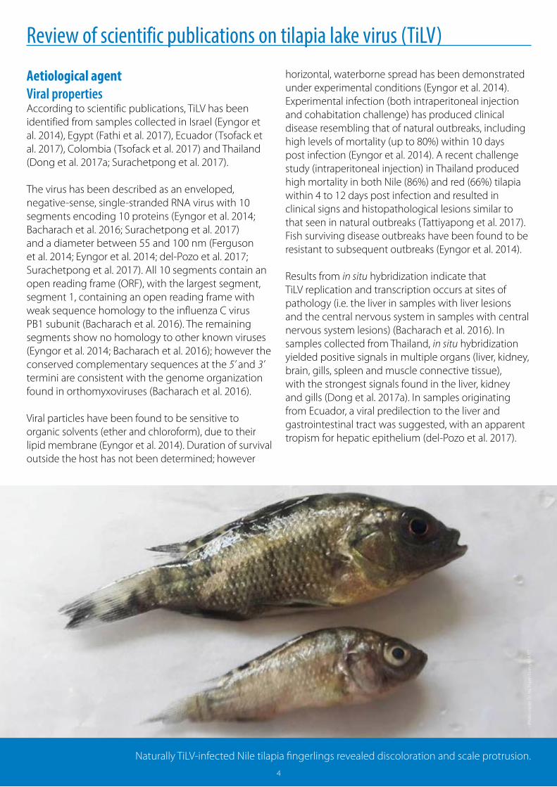
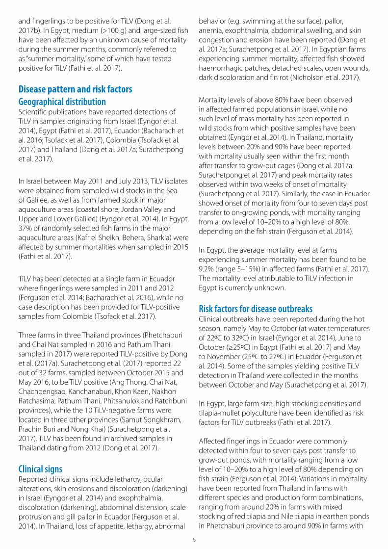
Naturally TiLV-infected Nile tilapia fingerlings revealed discoloration and scale protrusion.
Phot
o cr
edit:
Dr.
Ha
Than
h D
ong/
KMU
TT
4
Aetiological agentViral propertiesAccording to scientific publications, TiLV has been identified from samples collected in Israel (Eyngor et al. 2014), Egypt (Fathi et al. 2017), Ecuador (Tsofack et al. 2017), Colombia (Tsofack et al. 2017) and Thailand (Dong et al. 2017a; Surachetpong et al. 2017).
The virus has been described as an enveloped, negative-sense, single-stranded RNA virus with 10 segments encoding 10 proteins (Eyngor et al. 2014; Bacharach et al. 2016; Surachetpong et al. 2017) and a diameter between 55 and 100 nm (Ferguson et al. 2014; Eyngor et al. 2014; del-Pozo et al. 2017; Surachetpong et al. 2017). All 10 segments contain an open reading frame (ORF), with the largest segment, segment 1, containing an open reading frame with weak sequence homology to the influenza C virus PB1 subunit (Bacharach et al. 2016). The remaining segments show no homology to other known viruses (Eyngor et al. 2014; Bacharach et al. 2016); however the conserved complementary sequences at the 5’ and 3’ termini are consistent with the genome organization found in orthomyxoviruses (Bacharach et al. 2016).
Viral particles have been found to be sensitive to organic solvents (ether and chloroform), due to their lipid membrane (Eyngor et al. 2014). Duration of survival outside the host has not been determined; however
horizontal, waterborne spread has been demonstrated under experimental conditions (Eyngor et al. 2014). Experimental infection (both intraperitoneal injection and cohabitation challenge) has produced clinical disease resembling that of natural outbreaks, including high levels of mortality (up to 80%) within 10 days post infection (Eyngor et al. 2014). A recent challenge study (intraperitoneal injection) in Thailand produced high mortality in both Nile (86%) and red (66%) tilapia within 4 to 12 days post infection and resulted in clinical signs and histopathological lesions similar to that seen in natural outbreaks (Tattiyapong et al. 2017). Fish surviving disease outbreaks have been found to be resistant to subsequent outbreaks (Eyngor et al. 2014).
Results from in situ hybridization indicate that TiLV replication and transcription occurs at sites of pathology (i.e. the liver in samples with liver lesions and the central nervous system in samples with central nervous system lesions) (Bacharach et al. 2016). In samples collected from Thailand, in situ hybridization yielded positive signals in multiple organs (liver, kidney, brain, gills, spleen and muscle connective tissue), with the strongest signals found in the liver, kidney and gills (Dong et al. 2017a). In samples originating from Ecuador, a viral predilection to the liver and gastrointestinal tract was suggested, with an apparent tropism for hepatic epithelium (del-Pozo et al. 2017).
5
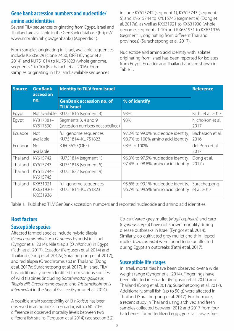
Source GenBank accession no.
Identity to TiLV from Israel Reference
GenBank accession no. of TiLV Israel
% of identify
Egypt Not available KU751816 (segment 3) 93% Fathi et al. 2017
Egypt KY817381–KY817390
Segments 3, 4 and 9(accession numbers not specified)
93% Nicholson et al. 2017
Ecuador Not available
full genome sequences KU751814–KU751823
97.2% to 99.0% nucleotide identity; 98.7% to 100% amino acid identity
Bacharach et al. 2016
Ecuador Not available
KJ605629 (ORF) 98% to 100% del-Pozo et al. 2017
Thailand KY615742 KU751814 (segment 1) 96.3% to 97.5% nucleotide identity;97.4% to 98.8% amino acid identity
Dong et al. 2017aThailand KY615743 KU751818 (segment 5)
Thailand KY615744– KY615745
KU751822 (segment 9)
Thailand KX631921 KX631930– KX631936
full genome sequences KU751814–KU751823
95.6% to 99.1% nucleotide identity;96.7% to 99.5% amino acid identity
Surachetpong et al. 2017
Host factorsSusceptible speciesAffected farmed species include hybrid tilapia (Oreochromis niloticus x O. aureus hybrids) in Israel (Eyngor et al. 2014); Nile tilapia (O. niloticus) in Egypt (Fathi et al. 2017), Ecuador (Ferguson et al. 2014) and Thailand (Dong et al. 2017a; Surachetpong et al. 2017); and red tilapia (Oreochromis sp.) in Thailand (Dong et al. 2017a; Surachetpong et al. 2017). In Israel, TiLV has additionally been identified from various species of wild tilapines (including Sarotherodon galilaeus, Tilapia zilli, Oreochromis aureus, and Tristamellasimonis intermedia) in the Sea of Galilee (Eyngor et al. 2014).
A possible strain susceptibility of O. niloticus has been observed in an outbreak in Ecuador, with a 60–70% difference in observed mortality levels between two different fish strains (Ferguson et al. 2014) (see section 3.2).
Co-cultivated grey mullet (Mugil cephalus) and carp (Cyprinus carpio) have not shown mortality during disease outbreaks in Israel (Eyngor et al. 2014). Similarly, co-cultivated grey mullet and thin-lipped mullet (Liza ramada) were found to be unaffected during Egyptian outbreaks (Fathi et al. 2017).
Susceptible life stagesIn Israel, mortalities have been observed over a wide weight range (Eyngor et al. 2014). Fingerlings have been affected in Ecuador (Ferguson et al. 2014) and Thailand (Dong et al. 2017a; Surachetpong et al. 2017). Additionally, small fish (up to 50 g) were affected in Thailand (Surachetpong et al. 2017). Furthermore, a recent study in Thailand using archived and fresh samples collected between 2012 and 2017 from four hatcheries found fertilized eggs, yolk sac larvae, fries
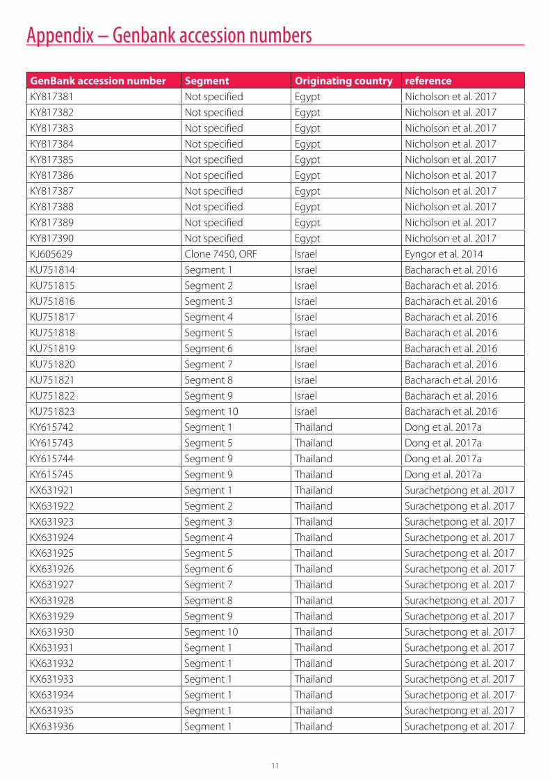
Gene bank accession numbers and nucleotide/amino acid identitiesSeveral TiLV sequences originating from Egypt, Israel and Thailand are available in the GenBank database (https://www.ncbi.nlm.nih.gov/genbank/) (Appendix 1).
From samples originating in Israel, available sequences include KJ605629 (clone 7450, ORF) (Eyngor et al. 2014) and KU751814 to KU751823 (whole genome, segments 1 to 10) (Bacharach et al. 2016). From samples originating in Thailand, available sequences
include KY615742 (segment 1), KY615743 (segment 5) and KY615744 to KY615745 (segment 9) (Dong et al. 2017a), as well as KX631921 to KX631930 (whole genome, segments 1-10) and KX631931 to KX631936 (segment 1, originating from different Thailand provinces) (Surachetpong et al. 2017).
Nucleotide and amino acid identity with isolates originating from Israel has been reported for isolates from Egypt, Ecuador and Thailand and are shown in Table 1.
Table 1. Published TiLV GenBank accession numbers and reported nucleotide and amino acid identities.
6
and fingerlings to be positive for TiLV (Dong et al. 2017b). In Egypt, medium (>100 g) and large-sized fish have been affected by an unknown cause of mortality during the summer months, commonly referred to as “summer mortality,” some of which have tested positive for TiLV (Fathi et al. 2017).
Disease pattern and risk factorsGeographical distributionScientific publications have reported detections of TiLV in samples originating from Israel (Eyngor et al. 2014), Egypt (Fathi et al. 2017), Ecuador (Bacharach et al. 2016; Tsofack et al. 2017), Colombia (Tsofack et al. 2017) and Thailand (Dong et al. 2017a; Surachetpong et al. 2017).
In Israel between May 2011 and July 2013, TiLV isolates were obtained from sampled wild stocks in the Sea of Galilee, as well as from farmed stock in major aquaculture areas (coastal shore, Jordan Valley and Upper and Lower Galilee) (Eyngor et al. 2014). In Egypt, 37% of randomly selected fish farms in the major aquaculture areas (Kafr el Sheikh, Behera, Sharkia) were affected by summer mortalities when sampled in 2015 (Fathi et al. 2017).
TiLV has been detected at a single farm in Ecuador where fingerlings were sampled in 2011 and 2012 (Ferguson et al. 2014; Bacharach et al. 2016), while no case description has been provided for TiLV-positive samples from Colombia (Tsofack et al. 2017).
Three farms in three Thailand provinces (Phetchaburi and Chai Nat sampled in 2016 and Pathum Thani sampled in 2017) were reported TiLV-positive by Dong et al. (2017a). Surachetpong et al. (2017) reported 22 out of 32 farms, sampled between October 2015 and May 2016, to be TiLV positive (Ang Thong, Chai Nat, Chachoengsao, Kanchanaburi, Khon Kaen, Nakhon Ratchasima, Pathum Thani, Phitsanulok and Ratchbuni provinces), while the 10 TiLV-negative farms were located in three other provinces (Samut Songkhram, Prachin Buri and Nong Khai) (Surachetpong et al. 2017). TiLV has been found in archived samples in Thailand dating from 2012 (Dong et al. 2017).
Clinical signsReported clinical signs include lethargy, ocular alterations, skin erosions and discoloration (darkening) in Israel (Eyngor et al. 2014) and exophthalmia, discoloration (darkening), abdominal distension, scale protrusion and gill pallor in Ecuador (Ferguson et al. 2014). In Thailand, loss of appetite, lethargy, abnormal
behavior (e.g. swimming at the surface), pallor, anemia, exophthalmia, abdominal swelling, and skin congestion and erosion have been reported (Dong et al. 2017a; Surachetpong et al. 2017). In Egyptian farms experiencing summer mortality, affected fish showed haemorrhagic patches, detached scales, open wounds, dark discoloration and fin rot (Nicholson et al. 2017).
Mortality levels of above 80% have been observed in affected farmed populations in Israel, while no such level of mass mortality has been reported in wild stocks from which positive samples have been obtained (Eyngor et al. 2014). In Thailand, mortality levels between 20% and 90% have been reported, with mortality usually seen within the first month after transfer to grow-out cages (Dong et al. 2017a; Surachetpong et al. 2017) and peak mortality rates observed within two weeks of onset of mortality (Surachetpong et al. 2017). Similarly, the case in Ecuador showed onset of mortality from four to seven days post transfer to on-growing ponds, with mortality ranging from a low level of 10–20% to a high level of 80%, depending on the fish strain (Ferguson et al. 2014).
In Egypt, the average mortality level at farms experiencing summer mortality has been found to be 9.2% (range 5–15%) in affected farms (Fathi et al. 2017). The mortality level attributable to TiLV infection in Egypt is currently unknown.
Risk factors for disease outbreaksClinical outbreaks have been reported during the hot season, namely May to October (at water temperatures of 22ᵒC to 32ᵒC) in Israel (Eyngor et al. 2014), June to October (≥25ᵒC) in Egypt (Fathi et al. 2017) and May to November (25ᵒC to 27ᵒC) in Ecuador (Ferguson et al. 2014). Some of the samples yielding positive TiLV detection in Thailand were collected in the months between October and May (Surachetpong et al. 2017).
In Egypt, large farm size, high stocking densities and tilapia-mullet polyculture have been identified as risk factors for TiLV outbreaks (Fathi et al. 2017).
Affected fingerlings in Ecuador were commonly detected within four to seven days post transfer to grow-out ponds, with mortality ranging from a low level of 10–20% to a high level of 80% depending on fish strain (Ferguson et al. 2014). Variations in mortality have been reported from Thailand in farms with different species and production form combinations, ranging from around 20% in farms with mixed stocking of red tilapia and Nile tilapia in earthen ponds in Phetchaburi province to around 90% in farms with
7
Nile tilapia in Pathum Thani province and farms with red tilapia in open floating cages in Chai Nat province (Dong et al. 2017a).
Co-infections Reported co-infections in TiLV-positive fish from Thailand were Flavobacterium, Aeromonas and Streptococcus and external monogenean parasites (Gyrodactylus and Dactylogyrus) and ciliated protozoa (Trichodina) (Surachetpong et al. 2017). The relative significance of TiLV and any co-infections have not been determined; however infection experiments have shown mortality levels up to 80% in experimental TiLV-infected populations (Eyngor et al. 2014).
The relationship between summer mortalities in Egypt and TiLV has not yet been determined; however four out of eight farms sampled in 2015 (Nicholson et al. 2017) and three out of seven farms sampled in 2016 (Fathi et al 2017) in Egypt that had experienced summer mortalities were found to be TiLV positive. In addition, multiple Aeromonas spp. (A. veronii, A. ichthiosmia, A. enteropelogenes and A. hydrophilia) were also identified from samples collected from Egyptian fish farms during 2015 (Nicholson et al. 2017).
Diagnostics and diagnostic testsGross pathologyObservations from affected populations in Israel include ocular alterations, skin erosions and discoloration (darkening) (Eyngor et al. 2014), while in Ecuador, abdominal distension, scale protrusion and gill pallor have been observed in addition to exophthalmia and skin discoloration (darkening) (Ferguson et al. 2014). Affected fish from Thailand showed pallor (Dong et al. 2017a), anemia, exophthalmia, abdominal swelling, skin congestion, erosion, brain congestion and paleness of the gills and liver (Surachetpong et al. 2017).
HistopathologyBriefly, cases in Israel showed the most severe central nervous system lesions (Eyngor et al. 2014) while cases in Ecuador and Colombia had major liver lesions (Ferguson et al. 2014; Tsofack et al. 2017; del-Pozo et al. 2017). Both liver and central nervous system lesions have been reported in cases from Egypt (Fathi et al. 2017).
More specifically, observed lesions in affected fish in Israel include congestion of internal organs (kidneys and brain), foci of gliosis and perivascular cuffing in the brain cortex, and ocular lesions (endophthalmitis and cataractous changes of the lens) (Eyngor et al. 2014).
In affected fish from Egypt, histopathological findings include gliosis, encephalitis and mild perivascular cuffing in the brain, multifocal chronic hepatitis and multifocal interstitial hemorrhage in the kidney (Fathi et al. 2017). The case in Ecuador showed hepatocyte necrosis and syncytial cell formation, necrosis of gastric glands and diffuse congestion in multiple tissues (Ferguson et al. 2014). Syncytial hepatitis was also reported in samples from Colombia (Tsofack et al. 2017) and Thailand (Dong et al. 2017a), with additional observations from Thailand including aggregation of lymphocytes and perivascular cuffing in brain tissue (Surachetpong et al. 2017).
Cell culture Experiments have shown multiple cell lines to be suitable for TiLV cell culture (Eyngor et al. 2014; Tsofack et al. 2017).
The E-11 cell line was found to show visible cytopathic effect (CPE) five to seven days after inoculation, with cytoplasmic vacuoles and plaque formation followed by disintegration of cell monolayer nine to 10 days after inoculation (Eyngor et al. 2014). Cell lines of primary tilapia brain cells showed swollen, rounded, granulated cells 10 to 12 days after inoculation, with monolayer detachment 14 to 19 days after inoculation (Eyngor et al. 2014). E-11 cells at 25ᵒC have been reported to provide optimal conditions for TiLV replication (Tsofack et al. 2017). The OmB and TmB cell lines showed similar sensitivities to TiLV infection as the E-11 cell line; however the E-11 cultures were deemed superior due to the clear and rapid CPE development (Tsofack et al. 2017). OmB has been suggested as a useful cell line for end-point dilution (TCID50) assays and both OmB and TmB may reportedly be useful for generating pure TiLV strains as they are snakehead reovirus-free (Tsofack et al. 2017). Pooling of two to three samples of brain tissue have yielded positive TiLV culture results (Tsofack et al. 2017).
Other cell lines (CHSE-214, BF-2, BB, EPC, KF-1, RTG-2 and FHM) have been reported to show inconsistent CPE with TiLV (Eyngor et al. 2014).
Molecular methodsInitially, a RT-PCR method for TiLV detection was published together with information on TiLV specific primers (Eyngor et al. 2014). This was followed by the publication of a nested RT-PCR assay, with a reportedly improved sensitivity for segment 3, enabling detection down to seven copies of TiLV (Tsofack et al. 2017). The nested RT-PCR assay was found to detect TiLV in both fresh and preserved (RNAlater; QIAGEN) samples

8
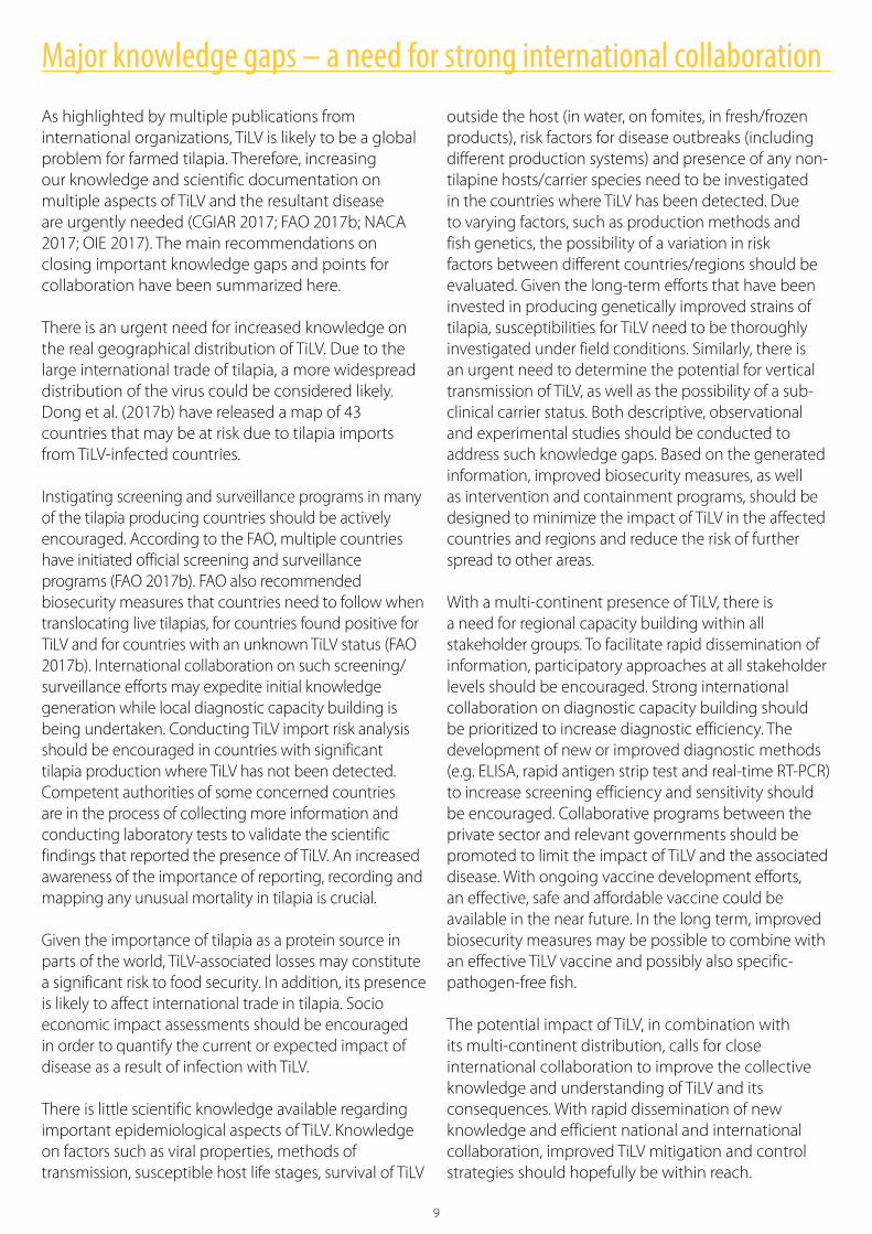
Naturally TiLV-infected red tilapia juveniles (left); skin lesions in TiLV-infected tilapia (right).
8
from diseased fish, and identified TiLV RNA in samples from diseased fish from Israel, Ecuador and Colombia (Tsofack et al. 2017). A real-time PCR method, used for quantification, was additionally described (Tsofack et al. 2017). Subsequently an alternative semi-nested RT-PCR method has been described where the primer Nested ext-2 was omitted (Dong et al. 2017a). Pooling of between two to five samples have been reported to yield successful agent identification (Tsofack et al. 2017; Dong et al. 2017a; Fathi et al. 2017; Surachetpong et al. 2017).
Additionally, the in situ hybridization method has been described and has been used to reveal TiLV tissue tropism (Bacharach et al. 2016; Dong et al. 2017a).
SerologyA reduced packed cell volume (16% versus the normal 48–50%) has been observed in Ecuador, together with an increased number of immature erythrocytes in blood smears (Ferguson et al. 2014).
Socio economic impactFish farms affected by TiLV reported mortality levels that may reach above 80% (Eyngor et al. 2014; Dong et al. 2017a; Surachetpong et al. 2017).
Estimates from Egypt indicate a production loss of 98,000 metric tons, at a value of around USD 100 million,due to the summer mortality syndrome in 2015 (Fathi et al. 2017).
In the Sea of Galilee in Israel, the annual wild catch figures for the main edible fish in the lake, S. galilaeus, decreased from 316 metric tons in 2005 to 8 metric tonsin 2009, with a subsequent increase to 160 metric tonsin 2013 and 140 metric tons in 2014 (Eyngor et al. 2014). The contribution of TiLV infection to this decline has not been determined; however TiLV has been identified in samples from several wild tilapines, including S. galilaeus (Eyngor et al. 2014).
Phot
o cr
edit:
Dr.
Saen
gcha
n Se
napi
n/Ce
ntex
Shr
imp/
Mah
idol
Uni
vers
ity/B
IOTE
C (l
eft)
; Wor
ldFi
sh (r
ight
)
9
Major knowledge gaps – a need for strong international collaboration
As highlighted by multiple publications from international organizations, TiLV is likely to be a global problem for farmed tilapia. Therefore, increasing our knowledge and scientific documentation on multiple aspects of TiLV and the resultant disease are urgently needed (CGIAR 2017; FAO 2017b; NACA 2017; OIE 2017). The main recommendations on closing important knowledge gaps and points for collaboration have been summarized here.
There is an urgent need for increased knowledge on the real geographical distribution of TiLV. Due to the large international trade of tilapia, a more widespread distribution of the virus could be considered likely. Dong et al. (2017b) have released a map of 43 countries that may be at risk due to tilapia imports from TiLV-infected countries.
Instigating screening and surveillance programs in many of the tilapia producing countries should be actively encouraged. According to the FAO, multiple countries have initiated official screening and surveillance programs (FAO 2017b). FAO also recommended biosecurity measures that countries need to follow when translocating live tilapias, for countries found positive for TiLV and for countries with an unknown TiLV status (FAO 2017b). International collaboration on such screening/surveillance efforts may expedite initial knowledge generation while local diagnostic capacity building is being undertaken. Conducting TiLV import risk analysis should be encouraged in countries with significant tilapia production where TiLV has not been detected. Competent authorities of some concerned countries are in the process of collecting more information and conducting laboratory tests to validate the scientific findings that reported the presence of TiLV. An increased awareness of the importance of reporting, recording and mapping any unusual mortality in tilapia is crucial.
Given the importance of tilapia as a protein source in parts of the world, TiLV-associated losses may constitute a significant risk to food security. In addition, its presence is likely to affect international trade in tilapia. Socio economic impact assessments should be encouraged in order to quantify the current or expected impact of disease as a result of infection with TiLV.
There is little scientific knowledge available regarding important epidemiological aspects of TiLV. Knowledge on factors such as viral properties, methods of transmission, susceptible host life stages, survival of TiLV
outside the host (in water, on fomites, in fresh/frozen products), risk factors for disease outbreaks (including different production systems) and presence of any non-tilapine hosts/carrier species need to be investigated in the countries where TiLV has been detected. Due to varying factors, such as production methods and fish genetics, the possibility of a variation in risk factors between different countries/regions should be evaluated. Given the long-term efforts that have been invested in producing genetically improved strains of tilapia, susceptibilities for TiLV need to be thoroughly investigated under field conditions. Similarly, there is an urgent need to determine the potential for vertical transmission of TiLV, as well as the possibility of a sub-clinical carrier status. Both descriptive, observational and experimental studies should be conducted to address such knowledge gaps. Based on the generated information, improved biosecurity measures, as well as intervention and containment programs, should be designed to minimize the impact of TiLV in the affected countries and regions and reduce the risk of further spread to other areas.
With a multi-continent presence of TiLV, there is a need for regional capacity building within all stakeholder groups. To facilitate rapid dissemination of information, participatory approaches at all stakeholder levels should be encouraged. Strong international collaboration on diagnostic capacity building should be prioritized to increase diagnostic efficiency. The development of new or improved diagnostic methods (e.g. ELISA, rapid antigen strip test and real-time RT-PCR) to increase screening efficiency and sensitivity should be encouraged. Collaborative programs between the private sector and relevant governments should be promoted to limit the impact of TiLV and the associated disease. With ongoing vaccine development efforts, an effective, safe and affordable vaccine could be available in the near future. In the long term, improved biosecurity measures may be possible to combine with an effective TiLV vaccine and possibly also specific-pathogen-free fish.
The potential impact of TiLV, in combination with its multi-continent distribution, calls for close international collaboration to improve the collective knowledge and understanding of TiLV and its consequences. With rapid dissemination of new knowledge and efficient national and international collaboration, improved TiLV mitigation and control strategies should hopefully be within reach.

10
Bacharach E, Mishra N, Briese T, Zody MC, KembouTsofack JE, Zamostiano R, Berkowitz A, Ng J, Nitido A, Corvelo A et al. 2016. Characterization of a novel orthomyxo-like virus causing mass die-offs of tilapia. MBio 7, e00431-16.
CGIAR Research Program on Fish Agri-food Systems. 2017. Tilapia lake virus (TiLV): What to know and do? Penang, Malaysia: CGIAR Research Program on Fish Agri-food Systems. Factsheet: FISH-2017-03. http://fish.cgiar.org/publications/tilapia-lake-virus-tilv-what-know-and-do
del-Pozo J, Mishra N, Kabuusu R, Cheetham S, Eldar A, Bacharach E, Lipkin WI and Ferguson HW. 2017. Syncytial hepatitis of tilapia (Oreochromisniloticus L.) is associated with orthomyxovirus-like virions in hepatocytes. Veterinary Pathology 54:164–70.
Dong HT, Siriroob S, Meemetta W, Santimanawong W, Gangnonngiw W, Pirarat N, Khunrae P, Rattanarojpong T, Vanichviriyakit R and Senapin S. 2017a. Emergence of tilapia lake virus in Thailand and an alternative semi-nested RT-PCR for detection. Aquaculture 476:111–18.
Dong HT, Ataguba P, Khunrae T, Rattanarojpong T and Serapin S. 2017b. Evidence of TiLV infection in tilapia hatcheries in Thailand from 2012 to 2017 reveals probable global spread of the disease. Aquaculture, doi: 10.1016/j.aquaculture.2017.2017.06.035.
Eyngor M, Zamostiano R, KembouTsofack JE, Berkowitz A, Bercovier H, Tinman S, Lev M, Hurvitz A, Galeotti M, Bacharach E et al. 2014. Identification of a novel RNA virus lethal to tilapia. Journal of Clinical Microbiology 52:4137–46.
[FAO] Food and Agriculture Organization of the United Nations. 2017a. Global aquaculture production. Rome: FAO. http://www.fao.org/fishery/statistics/global-production/en
[FAO] Food and Agriculture Organization of the United Nations. 2017b. Outbreaks of tilapia lake virus (TiLV) threaten the livelihoods and food security of millions of people dependent on tilapia farming. Rome: FAO. Global Information and Early Warning System (GIEWS) Special Alert No. 338 - Global, 26 May 2017. http://www.fao.org/documents/card/en/c/3ce1da5b-1529-4e7c-8b88-7adfef8d138c/
Fathi M, Dickson C, Dickson M, Leschen W, Baily J, Muir F, Ulrich K and Weidmann M. 2017. Identification of tilapia lake virus in Egypt in Nile tilapia affected by ‘summer mortality’ syndrome. Aquaculture 473:430–32.
Ferguson HW, Kabuusu R, Beltran S, Reyes E, Lince JA and del Pozo J. 2014. Syncytial hepatitis of farmed tilapia, Oreochromisniloticus (L.): A case report. Journal of Fish Diseases 37:583–89.
[NACA] Network of Aquaculture Centres in Asia-Pacific. 2017. Urgent update on possible worldwide spread of tilapia lake virus (TiLV). Accessed 23 May 2017. https://enaca.org/?id=870&title=urgent-update-on-possible-worldwide-spread-of-tilapia-lake-virus-tilv
Nicholson P, Fathi MA, Fischer A, Mohan C, Schieck E, Mishra N, Heinimann A, Frey J, Wieland B and Jores J. 2017. Detection of Tilapia Lake Virus in Egyptian fish farms experiencing high mortalities in 2015. Journal of Fish Diseases https://doi.org/10.1111/jfd.12650
[OIE] World Organisation for Animal Health. 2017. Tilapia lake virus (TiLV) – A novel orthomyxo-like virus. Paris: OIE. OIE technical disease card. Accessed 23 May 2017. http://www.oie.int/fileadmin/Home/eng/Internationa_Standard_Setting/docs/pdf/A_TiLV_disease_card.pdf
Surachetpong W, Janetanakit T, Nonthabenjawan N, Tattiyapong P, Sirikanchana K andAmonsin A. 2017. Outbreaks of tilapia lake virus infection, Thailand, 2015–2016. Emerging Infectious Diseases 23:1031–33.
Tattiyapong P, Dachavichitlead W and Surachetpong W. 2017. Experimental infection of Tilapia Lake Virus (TiLV) in Nile tilapia (Oreochromis niloticus) and red tilapia (Oreochromis spp.). Veterinary Microbiology 207:170–77.
Tsofack JEK, Zamostianoa R, Wattedb S, Berkowitzb A, Rosenbluth E, Mishra N, Briese T, Lipkin WI, Kabuusud RM, Ferguson H et al. Detection of tilapia lake virus (TiLV) in clinical samples by culturing and nested RT-PCR. Journal of Clinical Microbiology 55:759–67.
References
11
Appendix – Genbank accession numbers
GenBank accession number Segment Originating country referenceKY817381 Not specified Egypt Nicholson et al. 2017
KY817382 Not specified Egypt Nicholson et al. 2017
KY817383 Not specified Egypt Nicholson et al. 2017
KY817384 Not specified Egypt Nicholson et al. 2017
KY817385 Not specified Egypt Nicholson et al. 2017
KY817386 Not specified Egypt Nicholson et al. 2017
KY817387 Not specified Egypt Nicholson et al. 2017
KY817388 Not specified Egypt Nicholson et al. 2017
KY817389 Not specified Egypt Nicholson et al. 2017
KY817390 Not specified Egypt Nicholson et al. 2017
KJ605629 Clone 7450, ORF Israel Eyngor et al. 2014
KU751814 Segment 1 Israel Bacharach et al. 2016
KU751815 Segment 2 Israel Bacharach et al. 2016
KU751816 Segment 3 Israel Bacharach et al. 2016
KU751817 Segment 4 Israel Bacharach et al. 2016
KU751818 Segment 5 Israel Bacharach et al. 2016
KU751819 Segment 6 Israel Bacharach et al. 2016
KU751820 Segment 7 Israel Bacharach et al. 2016
KU751821 Segment 8 Israel Bacharach et al. 2016
KU751822 Segment 9 Israel Bacharach et al. 2016
KU751823 Segment 10 Israel Bacharach et al. 2016
KY615742 Segment 1 Thailand Dong et al. 2017a
KY615743 Segment 5 Thailand Dong et al. 2017a
KY615744 Segment 9 Thailand Dong et al. 2017a
KY615745 Segment 9 Thailand Dong et al. 2017a
KX631921 Segment 1 Thailand Surachetpong et al. 2017
KX631922 Segment 2 Thailand Surachetpong et al. 2017
KX631923 Segment 3 Thailand Surachetpong et al. 2017
KX631924 Segment 4 Thailand Surachetpong et al. 2017
KX631925 Segment 5 Thailand Surachetpong et al. 2017
KX631926 Segment 6 Thailand Surachetpong et al. 2017
KX631927 Segment 7 Thailand Surachetpong et al. 2017
KX631928 Segment 8 Thailand Surachetpong et al. 2017
KX631929 Segment 9 Thailand Surachetpong et al. 2017
KX631930 Segment 10 Thailand Surachetpong et al. 2017
KX631931 Segment 1 Thailand Surachetpong et al. 2017
KX631932 Segment 1 Thailand Surachetpong et al. 2017
KX631933 Segment 1 Thailand Surachetpong et al. 2017
KX631934 Segment 1 Thailand Surachetpong et al. 2017
KX631935 Segment 1 Thailand Surachetpong et al. 2017
KX631936 Segment 1 Thailand Surachetpong et al. 2017
www.fish.cgiar.org
This publication should be cited as: Jansen MD and Mohan CV. 2017. Tilapia lake virus (TiLV): Literature review. Penang, Malaysia: CGIAR Research Program on Fish Agri-Food Systems. Working Paper: FISH-2017-04.
© 2017. CGIAR Research Program on Fish Agri-Food Systems. All rights reserved. This publication may be reproduced without the permission of, but with acknowledgment to, the CGIAR Research Program on Fish Agri-Food Systems.
Phot
o cr
edit:
Bac
k co
ver,
Mon
a D
verd
al Ja
nsen
/Vet
erin
ærin
stitu
ttet
100%RECYCLED