Thyroid nodule as a first manifestation of Hodgkin lymphomareport
8
CASE REPORT Open Access Thyroid nodule as a first manifestation of Hodgkin lymphoma–report of two cases and literature review Ewelina Szczepanek-Parulska 1 , Malgorzata Szkudlarek 1 , Przemyslaw Majewski 2 , Jan Breborowicz 3 and Marek Ruchala 1* Abstract Lymphomas account for less than 5% of thyroid malignant lesions. Vast majority of them are B-cell non-Hodgkin lymphomas (NHL), while Hodgkin lymphoma (HL) is extremely rare. Here we present two cases of HL, at baseline manifesting as a thyroid lesion. First patient, 29-year-old pregnant female, initially suspected for metastatic medullary thyroid cancer, was eventually diagnosed with mixed cellularity type of thyroid HL. Second patient, 22- year-old woman with suspicion of advanced thyroid cancer, was in the end diagnosed with an extra-lymphatic classical HL of the thyroid. In both cases, despite repeated fine-needle aspiration biopsy, cytological examination gave inconclusive or misleading results. On histopathological examination, thyroid tumor cells were positive for CD15 and CD30 antigen, which is typical for Reed-Sternberg cells. In the report authors also discuss difficulties in management as well as potential importance of novel methods such as FISH, PCR and other molecular techniques in diagnostics of thyroid lymphomas. Virtual slides: The virtual slide(s) for this article can be found here: http://www.diagnosticpathology.diagnomx.eu/ vs/2896947559559648 Keywords: Thyroid nodule, Hodgkin lymphoma, Fine-needle aspiration biopsy (FNAB), Reed-Sternberg cells Background Hodgkin lymphomas (HL) is a biologically heteroge- neous group of neoplasms. The incidence of HL in European Union is estimated at about 2.2-2.7/100 000 cases per year, constituting 11.7% of all lymphomas diag- nosed in 2006 [1]. The majority of cases is in low-stage disease and presents the nodular sclerosis subtype. An increase in incidence of extra-nodal lymphomas has been observed over the past two decades [2]. Extra-nodal origin is more common in NHL and may reach 33% [3]. On the other hand, HL mainly arises in lymph nodes of the neck and mediastinum, while only approximately 5% develop in extra-nodal sites, including tonsils, nasopha- rynx, parotid glands, thyroid, parathyroid or nasal antrum, with or without concomitant nodal involvement [4]. The head and neck is a third place of extra-nodal localization of the lymphomas [5]. Lymphomas account for less than 5% of malignant lesions diagnosed in the thyroid identified in about two cases per million [6,7]. Vast majority of them are B-cell non-Hodgkin lymph- omas (NHL), developing in the course of autoimmune thyroiditis, while HL primarily localized in the thyroid, is a very rare finding [8,9]. Extra-nodal lymphomas (ENL) represent different pathologic, imaging, and clinical features as well as dis- similar prognosis from nodal lymphomas, and should therefore be distinguished [10]. Extra-nodal involvement is much less common in HL compared with NHL. ENL more often spreads by continuity from contiguous nodal disease, while haematogenous spread is rare, occurring in 5%-10% of patients [11]. Hodgkin lymphoma of the thyroid is uncommon and accounts for 0.6-5% of all thyroid malignancies and 2-7% of all ENL [12-15]. In vast majority primary thyroid * Correspondence: [email protected] 1 Department of Endocrinology, Metabolism and Internal Medicine, Poznan University of Medical Sciences, Poznan, Poland Full list of author information is available at the end of the article © 2013 Szczepanek-Parulska et al.; licensee BioMed Central Ltd. This is an Open Access article distributed under the terms of the Creative Commons Attribution License (http://creativecommons.org/licenses/by/2.0), which permits unrestricted use, distribution, and reproduction in any medium, provided the original work is properly cited. Szczepanek-Parulska et al. Diagnostic Pathology 2013, 8:116 http://www.diagnosticpathology.org/content/8/1/116
Transcript of Thyroid nodule as a first manifestation of Hodgkin lymphomareport
Szczepanek-Parulska et al. Diagnostic Pathology 2013, 8:116http://www.diagnosticpathology.org/content/8/1/116
CASE REPORT Open Access
Thyroid nodule as a first manifestation ofHodgkin lymphoma–report of two cases andliterature reviewEwelina Szczepanek-Parulska1, Malgorzata Szkudlarek1, Przemyslaw Majewski2, Jan Breborowicz3
and Marek Ruchala1*
Abstract
Lymphomas account for less than 5% of thyroid malignant lesions. Vast majority of them are B-cell non-Hodgkinlymphomas (NHL), while Hodgkin lymphoma (HL) is extremely rare. Here we present two cases of HL, at baselinemanifesting as a thyroid lesion. First patient, 29-year-old pregnant female, initially suspected for metastaticmedullary thyroid cancer, was eventually diagnosed with mixed cellularity type of thyroid HL. Second patient, 22-year-old woman with suspicion of advanced thyroid cancer, was in the end diagnosed with an extra-lymphaticclassical HL of the thyroid. In both cases, despite repeated fine-needle aspiration biopsy, cytological examinationgave inconclusive or misleading results. On histopathological examination, thyroid tumor cells were positive forCD15 and CD30 antigen, which is typical for Reed-Sternberg cells. In the report authors also discuss difficulties inmanagement as well as potential importance of novel methods such as FISH, PCR and other molecular techniquesin diagnostics of thyroid lymphomas.Virtual slides: The virtual slide(s) for this article can be found here: http://www.diagnosticpathology.diagnomx.eu/vs/2896947559559648
Keywords: Thyroid nodule, Hodgkin lymphoma, Fine-needle aspiration biopsy (FNAB), Reed-Sternberg cells
BackgroundHodgkin lymphomas (HL) is a biologically heteroge-neous group of neoplasms. The incidence of HL inEuropean Union is estimated at about 2.2-2.7/100 000cases per year, constituting 11.7% of all lymphomas diag-nosed in 2006 [1]. The majority of cases is in low-stagedisease and presents the nodular sclerosis subtype.An increase in incidence of extra-nodal lymphomas has
been observed over the past two decades [2]. Extra-nodalorigin is more common in NHL and may reach 33% [3].On the other hand, HL mainly arises in lymph nodes ofthe neck and mediastinum, while only approximately 5%develop in extra-nodal sites, including tonsils, nasopha-rynx, parotid glands, thyroid, parathyroid or nasal antrum,with or without concomitant nodal involvement [4].
* Correspondence: [email protected] of Endocrinology, Metabolism and Internal Medicine, PoznanUniversity of Medical Sciences, Poznan, PolandFull list of author information is available at the end of the article
© 2013 Szczepanek-Parulska et al.; licensee Biothe Creative Commons Attribution License (htdistribution, and reproduction in any medium
The head and neck is a third place of extra-nodallocalization of the lymphomas [5]. Lymphomas accountfor less than 5% of malignant lesions diagnosed in thethyroid identified in about two cases per million [6,7].Vast majority of them are B-cell non-Hodgkin lymph-omas (NHL), developing in the course of autoimmunethyroiditis, while HL primarily localized in the thyroid, isa very rare finding [8,9].Extra-nodal lymphomas (ENL) represent different
pathologic, imaging, and clinical features as well as dis-similar prognosis from nodal lymphomas, and shouldtherefore be distinguished [10]. Extra-nodal involvementis much less common in HL compared with NHL. ENLmore often spreads by continuity from contiguous nodaldisease, while haematogenous spread is rare, occurringin 5%-10% of patients [11].Hodgkin lymphoma of the thyroid is uncommon and
accounts for 0.6-5% of all thyroid malignancies and 2-7%of all ENL [12-15]. In vast majority primary thyroid
Med Central Ltd. This is an Open Access article distributed under the terms oftp://creativecommons.org/licenses/by/2.0), which permits unrestricted use,, provided the original work is properly cited.
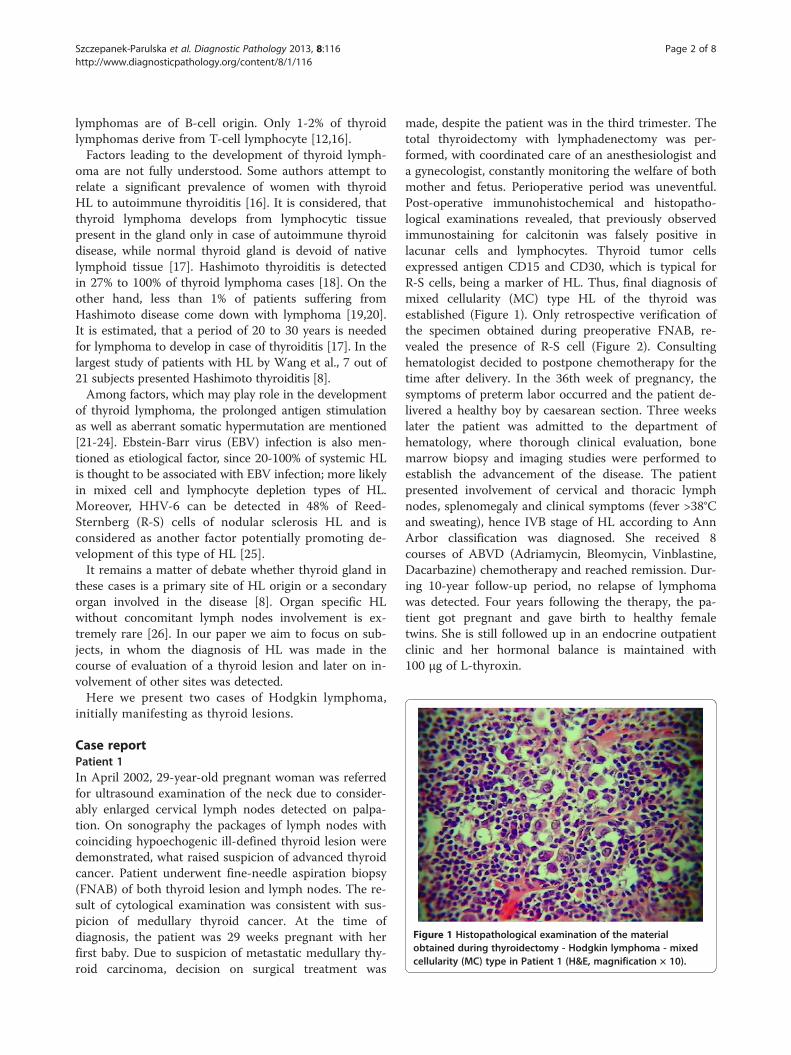
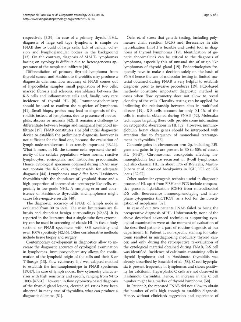
Figure 1 Histopathological examination of the materialobtained during thyroidectomy - Hodgkin lymphoma - mixedcellularity (MC) type in Patient 1 (H&E, magnification × 10).
Szczepanek-Parulska et al. Diagnostic Pathology 2013, 8:116 Page 2 of 8http://www.diagnosticpathology.org/content/8/1/116
lymphomas are of B-cell origin. Only 1-2% of thyroidlymphomas derive from T-cell lymphocyte [12,16].Factors leading to the development of thyroid lymph-
oma are not fully understood. Some authors attempt torelate a significant prevalence of women with thyroidHL to autoimmune thyroiditis [16]. It is considered, thatthyroid lymphoma develops from lymphocytic tissuepresent in the gland only in case of autoimmune thyroiddisease, while normal thyroid gland is devoid of nativelymphoid tissue [17]. Hashimoto thyroiditis is detectedin 27% to 100% of thyroid lymphoma cases [18]. On theother hand, less than 1% of patients suffering fromHashimoto disease come down with lymphoma [19,20].It is estimated, that a period of 20 to 30 years is neededfor lymphoma to develop in case of thyroiditis [17]. In thelargest study of patients with HL by Wang et al., 7 out of21 subjects presented Hashimoto thyroiditis [8].Among factors, which may play role in the development
of thyroid lymphoma, the prolonged antigen stimulationas well as aberrant somatic hypermutation are mentioned[21-24]. Ebstein-Barr virus (EBV) infection is also men-tioned as etiological factor, since 20-100% of systemic HLis thought to be associated with EBV infection; more likelyin mixed cell and lymphocyte depletion types of HL.Moreover, HHV-6 can be detected in 48% of Reed-Sternberg (R-S) cells of nodular sclerosis HL and isconsidered as another factor potentially promoting de-velopment of this type of HL [25].It remains a matter of debate whether thyroid gland in
these cases is a primary site of HL origin or a secondaryorgan involved in the disease [8]. Organ specific HLwithout concomitant lymph nodes involvement is ex-tremely rare [26]. In our paper we aim to focus on sub-jects, in whom the diagnosis of HL was made in thecourse of evaluation of a thyroid lesion and later on in-volvement of other sites was detected.Here we present two cases of Hodgkin lymphoma,
initially manifesting as thyroid lesions.
Case reportPatient 1In April 2002, 29-year-old pregnant woman was referredfor ultrasound examination of the neck due to consider-ably enlarged cervical lymph nodes detected on palpa-tion. On sonography the packages of lymph nodes withcoinciding hypoechogenic ill-defined thyroid lesion weredemonstrated, what raised suspicion of advanced thyroidcancer. Patient underwent fine-needle aspiration biopsy(FNAB) of both thyroid lesion and lymph nodes. The re-sult of cytological examination was consistent with sus-picion of medullary thyroid cancer. At the time ofdiagnosis, the patient was 29 weeks pregnant with herfirst baby. Due to suspicion of metastatic medullary thy-roid carcinoma, decision on surgical treatment was
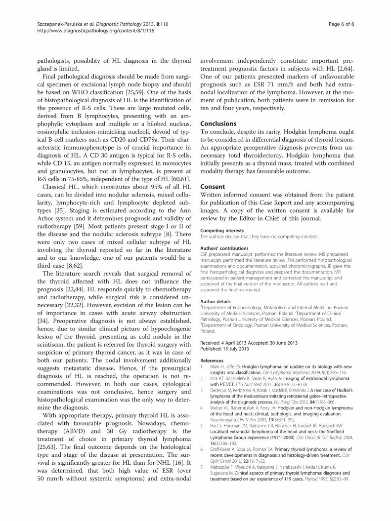
made, despite the patient was in the third trimester. Thetotal thyroidectomy with lymphadenectomy was per-formed, with coordinated care of an anesthesiologist anda gynecologist, constantly monitoring the welfare of bothmother and fetus. Perioperative period was uneventful.Post-operative immunohistochemical and histopatho-logical examinations revealed, that previously observedimmunostaining for calcitonin was falsely positive inlacunar cells and lymphocytes. Thyroid tumor cellsexpressed antigen CD15 and CD30, which is typical forR-S cells, being a marker of HL. Thus, final diagnosis ofmixed cellularity (MC) type HL of the thyroid wasestablished (Figure 1). Only retrospective verification ofthe specimen obtained during preoperative FNAB, re-vealed the presence of R-S cell (Figure 2). Consultinghematologist decided to postpone chemotherapy for thetime after delivery. In the 36th week of pregnancy, thesymptoms of preterm labor occurred and the patient de-livered a healthy boy by caesarean section. Three weekslater the patient was admitted to the department ofhematology, where thorough clinical evaluation, bonemarrow biopsy and imaging studies were performed toestablish the advancement of the disease. The patientpresented involvement of cervical and thoracic lymphnodes, splenomegaly and clinical symptoms (fever >38°Cand sweating), hence IVB stage of HL according to AnnArbor classification was diagnosed. She received 8courses of ABVD (Adriamycin, Bleomycin, Vinblastine,Dacarbazine) chemotherapy and reached remission. Dur-ing 10-year follow-up period, no relapse of lymphomawas detected. Four years following the therapy, the pa-tient got pregnant and gave birth to healthy femaletwins. She is still followed up in an endocrine outpatientclinic and her hormonal balance is maintained with100 μg of L-thyroxin.
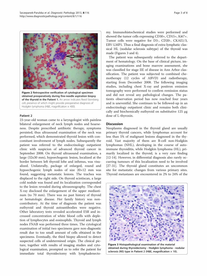
Figure 2 Retrospective verification of cytological specimenobtained preoperatively during fine-needle aspiration biopsyof the thyroid in the Patient 1. An arrow indicates Reed-Sternbergcell, presence of which might provide preoperative diagnosis ofHodgkin lymphoma (H&E, magnification × 400).
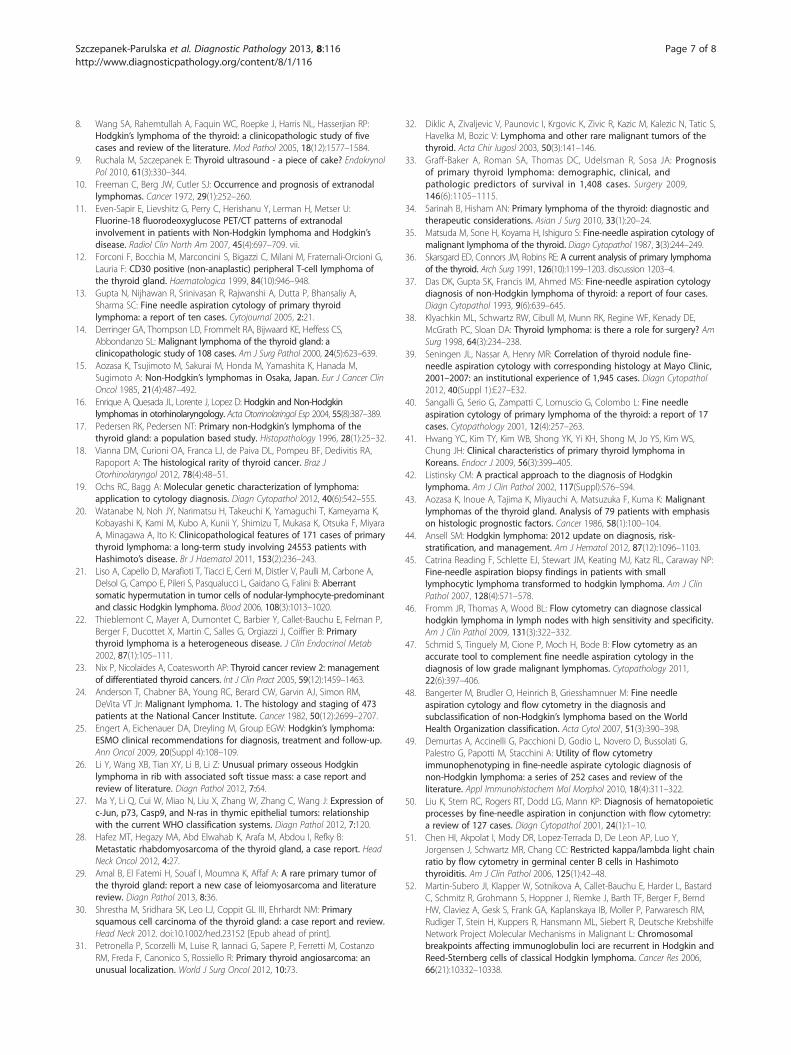
Figure 3 Histopathological examination of the materialobtained during thyroidectomy - Hodgkin lymphoma - nodularsclerosis (NS) type in Patient 2 (H&E, magnification × 10).
Szczepanek-Parulska et al. Diagnostic Pathology 2013, 8:116 Page 3 of 8http://www.diagnosticpathology.org/content/8/1/116
Patient 223-year-old woman came to a laryngologist with painlessbilateral enlargement of neck lymph nodes and hoarse-ness. Despite prescribed antibiotic therapy, symptomspersisted, thus ultrasound examination of the neck wasperformed, which demonstrated thyroid lesion with con-comitant involvement of lymph nodes. Subsequently thepatient was referred to the endocrinology outpatientclinic with suspicion of advanced thyroid cancer inSeptember 2008. On thyroid ultrasound examination, alarge (32x30 mm), hypoechogenic lesion, localized at theborder between left thyroid lobe and isthmus, was visu-alized. Unilaterally, package of enlarged, round andhypoechogenic lymph nodes of size 20×12 mm wasfound, suggesting metastatic lesions. The trachea wasdisplaced to the right side. On thyroid scintiscan, a largecold nodule was found and its localization correspondedto the lesion revealed during ultrasonography. The chestX-ray disclosed the enlargement of the upper mediasti-num (to 70 mm). There was no past history of thyroidor hematologic disease. Her family history was non-contributory. At the time of diagnosis the patient waseuthyroid and thyroid autoantibodies were negative.Other laboratory tests revealed accelerated ESR and in-creased concentration of white blood cells with deple-tion of lymphocytes and eosinophils. Thyroid and lymphnodes FNAB was performed three times. The cytologicalexamination of initial two specimens gave non-diagnosticresult due to too small amount of cells obtained in thespecimens. Eventually, the third biopsy allowed to detectsuspected cells of undetermined origin. The clinical pic-ture, together with results of imaging studies and cyto-logical examination, prompted us to refer the patient forimmediate total thyroidectomy with lymphadenecto-
my. Immunohistochemical studies were performed andshowed the tumor cells expressing CD30+, CD15+, Ki67+.Tumor cells were negative for D3-, CD20-, CKAE1/3,EBV-LMP1. Thus a final diagnosis of extra-lymphatic clas-sical HL (nodular sclerosis subtype) of the thyroid wasmade (Figures 3 and 4).The patient was subsequently referred to the depart-
ment of hematology. On the base of clinical picture, im-aging examinations and bone marrow assessment, shewas classified for stage IIE of disease in Ann Arbor clas-sification. The patient was subjected to combined che-motherapy (12 cycles of ABVD) and radiotherapy,starting from December 2008. The following imagingstudies, including chest X-ray and positron emissiontomography were performed to confirm remission statusand did not reveal any pathological changes. The pa-tients observation period has now reached four yearsand is uneventful. She continues to be followed-up in anendocrinology outpatient clinic and remains both clini-cally and biochemically euthyroid on substitutive 125 μgdose of L-thyroxin.
DiscussionNeoplasms diagnosed in the thyroid gland are usuallyprimary thyroid cancers, while lymphomas account forless than 5% of malignant lesions diagnosed in the thy-roid. Vast majority of them are B-cell non-Hodgkinlymphomas (NHL), developing in the course of auto-immune thyroiditis, while Hodgkin lymphoma (HL), pri-marily localized in the thyroid, is a very rare finding[12-14]. However, in differential diagnosis also rarely oc-curring tumours of this localization need to be involved[27-31]. The thyroid gland constitutes an uncommonsite for metastatic changes from various primary sites.Thyroid metastases are encountered in 2% to 24% of the
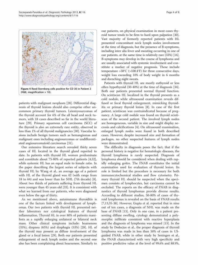
Figure 4 Reed-Sternberg cells positive for CD 30 in Patient 2(H&E, magnification × 10).
Szczepanek-Parulska et al. Diagnostic Pathology 2013, 8:116 Page 4 of 8http://www.diagnosticpathology.org/content/8/1/116
patients with malignant neoplasm [28]. Differential diag-nosis of thyroid lesions should also comprise other un-common primary thyroid tumors. Leiomyosarcomas ofthe thyroid account for 6% of the all head and neck tu-mors, with 18 cases described so far in the world litera-ture [29]. Primary squamous cell carcinoma (SCC) ofthe thyroid is also an extremely rare entity, observed inless than 1% of all thyroid malignancies [30]. Vascular le-sions include benign tumors such as hemangiomas andmalignant ones including angiosarcomas or undifferenti-ated angiosarcomatoid carcinomas [31].Our extensive literature search revealed thirty seven
cases of HL located in the thyroid gland reported todate. In patients with thyroid HL women predominateand constitute about 75-80% of reported patients [4,32],while systemic HL has an equal male to female ratio. Inthe paper describing the largest series of subjects withthyroid HL by Wang et al., an average age of a patientwith HL of the thyroid gland was 42 (with range from18 to 64) and was lower than for NHL (7th decade) [8].About two thirds of patients suffering from thyroid HLwere younger than 45 years old [33]. It is consistent withwhat we learned from our patients, who were diagnosedeven below the age of thirty.As we mentioned above, autoimmune thyroiditis is
one of the factors linked with development of lymph-omas. Our two patients with thyroid HL presented nei-ther laboratory nor cytological features of thyroidinflammation. Thyroid HL in over 80% of patients mani-fests as a rapidly enlarging unilateral or bilateral neckmass. Other clinical symptoms include: hoarseness(35%), dyspnea (65%) and dysphagia (53%) [28]. HL ofthe thyroid may present as diffuse involvement of thegland or a focal lesion [29]. Both our patients presentedenlargement of neck lymph nodes and the second onealso has been complaining about hoarseness. Similarly to
our patients, on physical examination in most cases thy-roid tumor tends to be firm to hard upon palpation [30].Vast majority of formerly reported patients with HLpresented concomitant neck lymph nodes involvementat the time of diagnosis, but the presence of B-symptoms,including inter alia fever and sweating occurring in one ofour patients, at the same time is relatively rare (33%) [16].B-symptoms may develop in the course of lymphoma andare usually associated with systemic involvement and con-stitute a marker of negative prognosis. These includetemperature >38°C (>100.4°F) for three consecutive days,weight loss exceeding 10% of body weight in 6 monthsand drenching night sweats.Patients with thyroid HL are mostly euthyroid or less
often hypothyroid (30-40%) at the time of diagnosis [34].Both our patients presented normal thyroid function.On scintiscan HL localized in the thyroid presents as acold nodule, while ultrasound examination reveals dif-fused or focal thyroid enlargement, mimicking thyroid-itis or primary thyroid lesion [8]. In case of the firstpatient, scintiscan was contraindicated because of preg-nancy. A large cold nodule was found on thyroid scinti-scan of the second patient. The involved lymph nodesare homogeneous, variable in size and might display ne-crosis and calcifications [4]. On ultrasound examination,enlarged lymph nodes were found in both describedcases. However, despite increased size and formation ofpackages, no other suspected features of lymph nodeswere demonstrated.The difficulty in diagnosis poses the fact, that if the
personal history is negative for hematologic diseases, thethyroid lymphoma is rarely suspected. Diagnosis oflymphoma should be considered when dealing with rap-idly enlarging goitre. The FNAB constitutes the initialexamination used for evaluation of thyroid lesion. Itsrole is limited but the procedure is necessary for bothimmunocytochemical studies and flow cytometry. Pri-mary thyroid HL should be suspected when the speci-men consists of lymphocytes, but carcinoma cannot beexcluded. The reports on the efficacy of FNAB in diag-nostics of thyroid lymphomas provide diverse results.According to different studies, 50-80% of cases of thy-roid lymphomas is revealed on the basis of FNAB results[7,13,35-38]. However, Gupta et al. reported that in nineout of ten cases, a diagnosis of NHL was made on thebase of FNAB [13]. Only in one case, in a patient pre-senting diffuse swelling, cytology demonstrated a poly-morphic infiltrate consistent with reactive hyperplasiaand the diagnosis of lymphoma was missed [13]. In thestudy by Dedecjus et al., the proper diagnosis of thyroidlymphoma was made in less than 50% of cases in US-guided FNAB, while in other study by Seningen et al.,the FNAB characterized with very high specificity andpositive predictive value at the level of 99.6% and 88.9%,
Szczepanek-Parulska et al. Diagnostic Pathology 2013, 8:116 Page 5 of 8http://www.diagnosticpathology.org/content/8/1/116
respectively [3,39]. In case of a primary thyroid NHL,diagnosis of large cell type lymphoma is simple onFNAB due to build of large cells, lack of cellular cohe-sion and lymphoglandular bodies in the background[13]. On the contrary, diagnosis of MALT- lymphomasbasing on cytology is difficult due to heterogeneous ap-pearance of the neoplastic infiltrate [40].Differentiation of primary thyroid lymphoma from
thyroid cancer and Hashimoto thyroiditis may produce adiagnostic dilemma. Low accuracy of FNAB comes outof hypocellular samples, small population of R-S cells,marked fibrosis and sclerosis, resemblance between theR-S cells and inflammatory cells and, finally, very rareincidence of thyroid HL [8]. Immunocytochemistryshould be used to confirm the suspicion of lymphoma[41]. Small biopsy probes may lead to diagnosis of thy-roiditis instead of lymphoma, due to presence of neutro-phils, abscess or necrosis [42]. It remains a challenge todifferentiate between benign and malignant lymphoid in-filtrate [19]. FNAB constitutes a helpful initial diagnosticdevice to establish the preliminary diagnosis, however isnot sufficient for the final one, because the evaluation oflymph node architecture is extremely important [43,44].What is more, in HL the tumour cells represent the mi-nority of the cellular population, where normal reactivelymphocytes, eosinophils, and histiocytes predominate.Hence, cytological specimen obtained during FNAB maynot contain the R-S cells, indispensable for adequatediagnosis [44]. Lymphomas may differ from Hashimotothyroiditis with the abundance of lymphoid tissue and ahigh proportion of intermediate centrocyte-like cells, es-pecially in low-grade NHL. A sampling error and coex-istence of Hashimoto thyroiditis and lymphoma mightcause false-negative results [40].The diagnostic accuracy of FNAB of lymph node is
evaluated from 30 to 92%. The main limitations are fi-brosis and abundant benign surroundings [42,45]. It isreported in the literature that a single-tube flow cytome-try can be used in screening of classic HL in tissue bodysections or FNAB specimens with 88% sensitivity andeven 100% specificity [42,46]. Other corroborative methodsinclude tissue biopsy and surgery.Contemporary development in diagnostics allow to in-
crease the diagnostic accuracy of cytological examinationin lymphomas. Immunocytochemistry allows for confir-mation of the lymphoid origin of the cells and their B orT-lineage [13]. Flow cytometry is a well-adapted methodto establish the immunophenotype in FNAB specimens[19,47]. In case of lymph nodes, flow cytometry characte-rizes with high sensitivity and specify, ranging from 94 to100% [47-50]. However, in flow cytometry-based diagnosisof the thyroid gland lesions, elevated κ:λ ratios have beenobserved in many case of thyroiditis, what can produce adiagnostic dilemma [51].
Ochs et. al stress that genetic testing, including poly-merase chain reaction (PCR) and florescence in situhybridization (FISH) is feasible and useful tool in diag-nosis of thyroid lymphomas [19]. Identification of ge-netic abnormalities can be critical to the diagnosis oflymphoma, especially this of unusual site of origin likelymphomas of thyroid gland [19]. Endocrinologists fre-quently have to make a decision solely on the basis ofFNAB hence the use of molecular testing in limited ma-terial obtained during FNAB is very helpful to establishdiagnosis prior to invasive procedures [19]. PCR-basedmethods constitute important diagnostic method incases when flow cytometry does not allow to revealclonality of the cells. Clonality testing can be applied forindicating the relationship between sites in multifocaldisease [19]. R-S cells account for only 0.1-1% of thecells in material obtained during FNAB [52]. Moleculartechniques targeting these cells provide some informationon cytogenetic aberrations in HL [52]. However, immuno-globulin heavy chain genes should be interpreted withattention due to frequency of monoclonal rearrange-ment in thyroiditis [53].Genomic gains in chromosom arm 2p, including REL
gene and gains in 9p are present in 30 to 50% of classicHL [54-57]. Chromosomal breakpoints affecting im-munoglobulin loci are recurrent in B-cell lymphomas,but also classical HL. In about 17% of R-S cells, Martin-Subero et al. observed breakpoints in IGH, IGL or IGKlocus [52,57].Other molecular cytogenic technics useful in diagnostic
process of HL apart from FISH and PCR include compara-tive genomic hybridization (CGH) from microdissectedR-S cells, fluorescence immunophenotyping and inter-phase cytogenetics (FICTION) as a tool for the investi-gation of neoplasms [52].In case of our two patients FNAB failed to bring the
preoperative diagnosis of HL. Unfortunately, none of theabove described advanced techniques supporting cyto-logical examination were at the moment of evaluation ofthe described patients a part of routine diagnosis at ourdepartment. In Patient 1, non-specific staining for calci-tonin resulted in misdiagnosing medullary thyroid can-cer, and only during the retrospective re-evaluation ofthe cytological material obtained during FNAB, R-S cellwas identified. Incidence of calcitonin-containing cells inthyroid lymphoma and in Hashimoto thyroiditis wasalready described by Baschieri et al. [58]. C cell hyperpla-sia is present frequently in lymphomas and shows positiv-ity for calcitonin. Hyperplastic C cells are not observed inHashimoto thyroiditis. Hence, an increase in the C cellnumber might be a marker of thyroid lymphoma [58].In Patient 2, the repeated FNAB did not allow to obtain
the number of cells high enough to establish diagnosis.Hence, without clinician’s suggestion and experience of
Szczepanek-Parulska et al. Diagnostic Pathology 2013, 8:116 Page 6 of 8http://www.diagnosticpathology.org/content/8/1/116
pathologists, possibility of HL diagnosis in the thyroidgland is limited.Final pathological diagnosis should be made from surgi-
cal specimen or excisional lymph node biopsy and shouldbe based on WHO classification [25,59]. One of the basisof histopathological diagnosis of HL is the identification ofthe presence of R-S cells. These are large mutated cells,derived from B lymphocytes, presenting with an am-phophylic cytoplasm and multiple or a bilobed nucleus,eosinophilic inclusion-mimicking nucleoli, devoid of typ-ical B-cell markers such as CD20 and CD79a. Their char-acteristic immunophenotype is of crucial importance indiagnosis of HL. A CD 30 antigen is typical for R-S cells,while CD 15, an antigen normally expressed in monocytesand granulocytes, but not in lymphocytes, is present atR-S cells in 75-85%, independent of the type of HL [60,61].Classical HL, which constitutes about 95% of all HL
cases, can be divided into nodular sclerosis, mixed cellu-larity, lymphocyte-rich and lymphocyte depleted sub-types [25]. Staging is estimated according to the AnnArbor system and it determines prognosis and validity ofradiotherapy [59]. Most patients present stage I or II ofthe disease and the nodular sclerosis subtype [8]. Therewere only two cases of mixed cellular subtype of HLinvolving the thyroid reported so far in the literatureand to our knowledge, one of our patients would be athird case [8,62].The literature search reveals that surgical removal of
the thyroid affected with HL does not influence theprognosis [22,44]. HL responds quickly to chemotherapyand radiotherapy, while surgical risk is considered un-necessary [22,32]. However, excision of the lesion can beof importance in cases with acute airway obstruction[34]. Preoperative diagnosis is not always established,hence, due to similar clinical picture of hypoechogeniclesion of the thyroid, presenting as cold nodule in thescintiscan, the patient is referred for thyroid surgery withsuspicion of primary thyroid cancer, as it was in case ofboth our patients. The nodal involvement additionallysuggests metastatic disease. Hence, if the presurgicaldiagnosis of HL is reached, the operation is not re-commended. However, in both our cases, cytologicalexaminations was not conclusive, hence surgery andhistopathological examination was the only way to deter-mine the diagnosis.With appropriate therapy, primary thyroid HL is asso-
ciated with favourable prognosis. Nowadays, chemo-therapy (ABVD) and 30 Gy radiotherapy is thetreatment of choice in primary thyroid lymphoma[25,63]. The final outcome depends on the histologicaltype and stage of the disease at presentation. The sur-vival is significantly greater for HL than for NHL [16]. Itwas determined, that both high value of ESR (over50 mm/h without systemic symptoms) and extra-nodal
involvement independently constitute important pre-treatment prognostic factors in subjects with HL [2,64].One of our patients presented markers of unfavourableprognosis such as ESR 71 mm/h and both had extra-nodal localization of the lymphoma. However, at the mo-ment of publication, both patients were in remission forten and four years, respectively.
ConclusionsTo conclude, despite its rarity, Hodgkin lymphoma oughtto be considered in differential diagnosis of thyroid lesions.An appropriate preoperative diagnosis prevents from un-necessary total thyroidectomy. Hodgkin lymphoma thatinitially presents as a thyroid mass, treated with combinedmodality therapy has favourable outcome.
ConsentWritten informed consent was obtained from the patientfor publication of this Case Report and any accompanyingimages. A copy of the written consent is available forreview by the Editor-in-Chief of this journal.
Competing interestsThe authors declare that they have no competing interests.
Authors’ contributionsESP preparated manuscipt, performed the literature review. MS preparatedmanuscipt, performed the literature review. PM performed histopathologicalexaminations and documentation, acquired photomicrographs. JB gave thefinal histopathological diagnosis and prepared the documentation. MRparticipated in patient management and corrected the manuscript andapproved of the final version of the manuscript. All authors read andapproved the final manuscript.
Author details1Department of Endocrinology, Metabolism and Internal Medicine, PoznanUniversity of Medical Sciences, Poznan, Poland. 2Department of ClinicalPathology, Poznan University of Medical Sciences, Poznan, Poland.3Department of Oncology, Poznan University of Medical Sciences, Poznan,Poland.
Received: 4 April 2013 Accepted: 30 June 2013Published: 15 July 2013
References1. Mani H, Jaffe ES: Hodgkin lymphoma: an update on its biology with new
insights into classification. Clin Lymphoma Myeloma 2009, 9(3):206–216.2. Ilica AT, Kocacelebi K, Savas R, Ayan A: Imaging of extranodal lymphoma
with PET/CT. Clin Nucl Med 2011, 36(10):e127–e138.3. Dedecjus M, Kedzierska A, Kozak J, Kordek R, Brzezinski J: A rare case of Hodkin’s
lymphoma of the mediastinum imitating retrosternal goiter–retrospectiveanalysis of the diagnostic process. Pol Przegl Chir 2012, 84(7):363–366.
4. Weber AL, Rahemtullah A, Ferry JA: Hodgkin and non-Hodgkin lymphomaof the head and neck: clinical, pathologic, and imaging evaluation.Neuroimaging Clin N Am 2003, 13(3):371–392.
5. Hart S, Horsman JM, Radstone CR, Hancock H, Goepel JR, Hancock BW:Localised extranodal lymphoma of the head and neck: the SheffieldLymphoma Group experience (1971–2000). Clin Oncol (R Coll Radiol) 2004,16(3):186–192.
6. Graff-Baker A, Sosa JA, Roman SA: Primary thyroid lymphoma: a review ofrecent developments in diagnosis and histology-driven treatment. CurrOpin Oncol 2010, 22(1):17–22.
7. Matsuzuka F, Miyauchi A, Katayama S, Narabayashi I, Ikeda H, Kuma K,Sugawara M: Clinical aspects of primary thyroid lymphoma: diagnosis andtreatment based on our experience of 119 cases. Thyroid 1993, 3(2):93–99.
Szczepanek-Parulska et al. Diagnostic Pathology 2013, 8:116 Page 7 of 8http://www.diagnosticpathology.org/content/8/1/116
8. Wang SA, Rahemtullah A, Faquin WC, Roepke J, Harris NL, Hasserjian RP:Hodgkin’s lymphoma of the thyroid: a clinicopathologic study of fivecases and review of the literature. Mod Pathol 2005, 18(12):1577–1584.
9. Ruchala M, Szczepanek E: Thyroid ultrasound - a piece of cake? EndokrynolPol 2010, 61(3):330–344.
10. Freeman C, Berg JW, Cutler SJ: Occurrence and prognosis of extranodallymphomas. Cancer 1972, 29(1):252–260.
11. Even-Sapir E, Lievshitz G, Perry C, Herishanu Y, Lerman H, Metser U:Fluorine-18 fluorodeoxyglucose PET/CT patterns of extranodalinvolvement in patients with Non-Hodgkin lymphoma and Hodgkin’sdisease. Radiol Clin North Am 2007, 45(4):697–709. vii.
12. Forconi F, Bocchia M, Marconcini S, Bigazzi C, Milani M, Fraternali-Orcioni G,Lauria F: CD30 positive (non-anaplastic) peripheral T-cell lymphoma ofthe thyroid gland. Haematologica 1999, 84(10):946–948.
13. Gupta N, Nijhawan R, Srinivasan R, Rajwanshi A, Dutta P, Bhansaliy A,Sharma SC: Fine needle aspiration cytology of primary thyroidlymphoma: a report of ten cases. Cytojournal 2005, 2:21.
14. Derringer GA, Thompson LD, Frommelt RA, Bijwaard KE, Heffess CS,Abbondanzo SL: Malignant lymphoma of the thyroid gland: aclinicopathologic study of 108 cases. Am J Surg Pathol 2000, 24(5):623–639.
15. Aozasa K, Tsujimoto M, Sakurai M, Honda M, Yamashita K, Hanada M,Sugimoto A: Non-Hodgkin’s lymphomas in Osaka, Japan. Eur J Cancer ClinOncol 1985, 21(4):487–492.
16. Enrique A, Quesada JL, Lorente J, Lopez D: Hodgkin and Non-Hodgkinlymphomas in otorhinolaryngology. Acta Otorrinolaringol Esp 2004, 55(8):387–389.
17. Pedersen RK, Pedersen NT: Primary non-Hodgkin’s lymphoma of thethyroid gland: a population based study. Histopathology 1996, 28(1):25–32.
18. Vianna DM, Curioni OA, Franca LJ, de Paiva DL, Pompeu BF, Dedivitis RA,Rapoport A: The histological rarity of thyroid cancer. Braz JOtorhinolaryngol 2012, 78(4):48–51.
19. Ochs RC, Bagg A: Molecular genetic characterization of lymphoma:application to cytology diagnosis. Diagn Cytopathol 2012, 40(6):542–555.
20. Watanabe N, Noh JY, Narimatsu H, Takeuchi K, Yamaguchi T, Kameyama K,Kobayashi K, Kami M, Kubo A, Kunii Y, Shimizu T, Mukasa K, Otsuka F, MiyaraA, Minagawa A, Ito K: Clinicopathological features of 171 cases of primarythyroid lymphoma: a long-term study involving 24553 patients withHashimoto’s disease. Br J Haematol 2011, 153(2):236–243.
21. Liso A, Capello D, Marafioti T, Tiacci E, Cerri M, Distler V, Paulli M, Carbone A,Delsol G, Campo E, Pileri S, Pasqualucci L, Gaidano G, Falini B: Aberrantsomatic hypermutation in tumor cells of nodular-lymphocyte-predominantand classic Hodgkin lymphoma. Blood 2006, 108(3):1013–1020.
22. Thieblemont C, Mayer A, Dumontet C, Barbier Y, Callet-Bauchu E, Felman P,Berger F, Ducottet X, Martin C, Salles G, Orgiazzi J, Coiffier B: Primarythyroid lymphoma is a heterogeneous disease. J Clin Endocrinol Metab2002, 87(1):105–111.
23. Nix P, Nicolaides A, Coatesworth AP: Thyroid cancer review 2: managementof differentiated thyroid cancers. Int J Clin Pract 2005, 59(12):1459–1463.
24. Anderson T, Chabner BA, Young RC, Berard CW, Garvin AJ, Simon RM,DeVita VT Jr: Malignant lymphoma. 1. The histology and staging of 473patients at the National Cancer Institute. Cancer 1982, 50(12):2699–2707.
25. Engert A, Eichenauer DA, Dreyling M, Group EGW: Hodgkin’s lymphoma:ESMO clinical recommendations for diagnosis, treatment and follow-up.Ann Oncol 2009, 20(Suppl 4):108–109.
26. Li Y, Wang XB, Tian XY, Li B, Li Z: Unusual primary osseous Hodgkinlymphoma in rib with associated soft tissue mass: a case report andreview of literature. Diagn Pathol 2012, 7:64.
27. Ma Y, Li Q, Cui W, Miao N, Liu X, Zhang W, Zhang C, Wang J: Expression ofc-Jun, p73, Casp9, and N-ras in thymic epithelial tumors: relationshipwith the current WHO classification systems. Diagn Pathol 2012, 7:120.
28. Hafez MT, Hegazy MA, Abd Elwahab K, Arafa M, Abdou I, Refky B:Metastatic rhabdomyosarcoma of the thyroid gland, a case report. HeadNeck Oncol 2012, 4:27.
29. Amal B, El Fatemi H, Souaf I, Moumna K, Affaf A: A rare primary tumor ofthe thyroid gland: report a new case of leiomyosarcoma and literaturereview. Diagn Pathol 2013, 8:36.
30. Shrestha M, Sridhara SK, Leo LJ, Coppit GL III, Ehrhardt NM: Primarysquamous cell carcinoma of the thyroid gland: a case report and review.Head Neck 2012. doi:10.1002/hed.23152 [Epub ahead of print].
31. Petronella P, Scorzelli M, Luise R, Iannaci G, Sapere P, Ferretti M, CostanzoRM, Freda F, Canonico S, Rossiello R: Primary thyroid angiosarcoma: anunusual localization. World J Surg Oncol 2012, 10:73.
32. Diklic A, Zivaljevic V, Paunovic I, Krgovic K, Zivic R, Kazic M, Kalezic N, Tatic S,Havelka M, Bozic V: Lymphoma and other rare malignant tumors of thethyroid. Acta Chir Iugosl 2003, 50(3):141–146.
33. Graff-Baker A, Roman SA, Thomas DC, Udelsman R, Sosa JA: Prognosisof primary thyroid lymphoma: demographic, clinical, andpathologic predictors of survival in 1,408 cases. Surgery 2009,146(6):1105–1115.
34. Sarinah B, Hisham AN: Primary lymphoma of the thyroid: diagnostic andtherapeutic considerations. Asian J Surg 2010, 33(1):20–24.
35. Matsuda M, Sone H, Koyama H, Ishiguro S: Fine-needle aspiration cytology ofmalignant lymphoma of the thyroid. Diagn Cytopathol 1987, 3(3):244–249.
36. Skarsgard ED, Connors JM, Robins RE: A current analysis of primary lymphomaof the thyroid. Arch Surg 1991, 126(10):1199–1203. discussion 1203–4.
37. Das DK, Gupta SK, Francis IM, Ahmed MS: Fine-needle aspiration cytologydiagnosis of non-Hodgkin lymphoma of thyroid: a report of four cases.Diagn Cytopathol 1993, 9(6):639–645.
38. Klyachkin ML, Schwartz RW, Cibull M, Munn RK, Regine WF, Kenady DE,McGrath PC, Sloan DA: Thyroid lymphoma: is there a role for surgery? AmSurg 1998, 64(3):234–238.
39. Seningen JL, Nassar A, Henry MR: Correlation of thyroid nodule fine-needle aspiration cytology with corresponding histology at Mayo Clinic,2001–2007: an institutional experience of 1,945 cases. Diagn Cytopathol2012, 40(Suppl 1):E27–E32.
40. Sangalli G, Serio G, Zampatti C, Lomuscio G, Colombo L: Fine needleaspiration cytology of primary lymphoma of the thyroid: a report of 17cases. Cytopathology 2001, 12(4):257–263.
41. Hwang YC, Kim TY, Kim WB, Shong YK, Yi KH, Shong M, Jo YS, Kim WS,Chung JH: Clinical characteristics of primary thyroid lymphoma inKoreans. Endocr J 2009, 56(3):399–405.
42. Listinsky CM: A practical approach to the diagnosis of Hodgkinlymphoma. Am J Clin Pathol 2002, 117(Suppl):S76–S94.
43. Aozasa K, Inoue A, Tajima K, Miyauchi A, Matsuzuka F, Kuma K: Malignantlymphomas of the thyroid gland. Analysis of 79 patients with emphasison histologic prognostic factors. Cancer 1986, 58(1):100–104.
44. Ansell SM: Hodgkin lymphoma: 2012 update on diagnosis, risk-stratification, and management. Am J Hematol 2012, 87(12):1096–1103.
45. Catrina Reading F, Schlette EJ, Stewart JM, Keating MJ, Katz RL, Caraway NP:Fine-needle aspiration biopsy findings in patients with smalllymphocytic lymphoma transformed to hodgkin lymphoma. Am J ClinPathol 2007, 128(4):571–578.
46. Fromm JR, Thomas A, Wood BL: Flow cytometry can diagnose classicalhodgkin lymphoma in lymph nodes with high sensitivity and specificity.Am J Clin Pathol 2009, 131(3):322–332.
47. Schmid S, Tinguely M, Cione P, Moch H, Bode B: Flow cytometry as anaccurate tool to complement fine needle aspiration cytology in thediagnosis of low grade malignant lymphomas. Cytopathology 2011,22(6):397–406.
48. Bangerter M, Brudler O, Heinrich B, Griesshamnuer M: Fine needleaspiration cytology and flow cytometry in the diagnosis andsubclassification of non-Hodgkin’s lymphoma based on the WorldHealth Organization classification. Acta Cytol 2007, 51(3):390–398.
49. Demurtas A, Accinelli G, Pacchioni D, Godio L, Novero D, Bussolati G,Palestro G, Papotti M, Stacchini A: Utility of flow cytometryimmunophenotyping in fine-needle aspirate cytologic diagnosis ofnon-Hodgkin lymphoma: a series of 252 cases and review of theliterature. Appl Immunohistochem Mol Morphol 2010, 18(4):311–322.
50. Liu K, Stern RC, Rogers RT, Dodd LG, Mann KP: Diagnosis of hematopoieticprocesses by fine-needle aspiration in conjunction with flow cytometry:a review of 127 cases. Diagn Cytopathol 2001, 24(1):1–10.
51. Chen HI, Akpolat I, Mody DR, Lopez-Terrada D, De Leon AP, Luo Y,Jorgensen J, Schwartz MR, Chang CC: Restricted kappa/lambda light chainratio by flow cytometry in germinal center B cells in Hashimotothyroiditis. Am J Clin Pathol 2006, 125(1):42–48.
52. Martin-Subero JI, Klapper W, Sotnikova A, Callet-Bauchu E, Harder L, BastardC, Schmitz R, Grohmann S, Hoppner J, Riemke J, Barth TF, Berger F, BerndHW, Claviez A, Gesk S, Frank GA, Kaplanskaya IB, Moller P, Parwaresch RM,Rudiger T, Stein H, Kuppers R, Hansmann ML, Siebert R, Deutsche KrebshilfeNetwork Project Molecular Mechanisms in Malignant L: Chromosomalbreakpoints affecting immunoglobulin loci are recurrent in Hodgkin andReed-Sternberg cells of classical Hodgkin lymphoma. Cancer Res 2006,66(21):10332–10338.
Szczepanek-Parulska et al. Diagnostic Pathology 2013, 8:116 Page 8 of 8http://www.diagnosticpathology.org/content/8/1/116
53. Zeppa P, Cozzolino I, Peluso AL, Troncone G, Lucariello A, Picardi M, CarellaC, Pane F, Vetrani A, Palombini L: Cytologic, flow cytometry, andmolecular assessment of lymphoid infiltrate in fine-needle cytologysamples of Hashimoto thyroiditis. Cancer 2009, 117(3):174–184.
54. Joos S, Kupper M, Ohl S, von Bonin F, Mechtersheimer G, Bentz M, MarynenP, Moller P, Pfreundschuh M, Trumper L, Lichter P: Genomic imbalancesincluding amplification of the tyrosine kinase gene JAK2 in CD30+Hodgkin cells. Cancer Res 2000, 60(3):549–552.
55. Joos S, Menz CK, Wrobel G, Siebert R, Gesk S, Ohl S, Mechtersheimer G,Trumper L, Moller P, Lichter P, Barth TF: Classical Hodgkin lymphoma ischaracterized by recurrent copy number gains of the short arm ofchromosome 2. Blood 2002, 99(4):1381–1387.
56. Joos S, Granzow M, Holtgreve-Grez H, Siebert R, Harder L, Martin-Subero JI,Wolf J, Adamowicz M, Barth TF, Lichter P, Jauch A: Hodgkin’s lymphomacell lines are characterized by frequent aberrations on chromosomes 2pand 9p including REL and JAK2. Int J Cancer 2003, 103(4):489–495.
57. Martin-Subero JI, Gesk S, Harder L, Sonoki T, Tucker PW, Schlegelberger B,Grote W, Novo FJ, Calasanz MJ, Hansmann ML, Dyer MJ, Siebert R:Recurrent involvement of the REL and BCL11A loci in classical Hodgkinlymphoma. Blood 2002, 99(4):1474–1477.
58. Baschieri L, Castagna M, Fierabracci A, Antonelli A, Del Guerra P, Squartini F:Distribution of calcitonin- and somatostatin-containing cells in thyroidlymphoma and in Hashimoto’s thyroiditis. Appl Pathol 1989, 7(2):99–104.
59. Linch DC, Gosden RG, Tulandi T, Tan SL, Hancock SL: Hodgkin’s lymphoma:choice of therapy and late complications. Hematology Am Soc Hematol EducProgram 2000, 2000(no. 1):205–221. ASH Education Book January 1, 2000.
60. Kuppers R, Engert A, Hansmann ML: Hodgkin lymphoma. J Clin Invest 2012,122(10):3439–3447.
61. Kuppers R: New insights in the biology of Hodgkin lymphoma.Hematology Am Soc Hematol Educ Program 2012, 2012:328–334.
62. Thomas RM: Hodgkin’s lymphoma presenting as an abscess in thyroidgland. Indian J Pathol Microbiol 2012, 55(1):122–124.
63. Raemaekers JM, van der Maazen RW: Hodgkin’s lymphoma: news from anold disease. Neth J Med 2008, 66(11):457–466.
64. Barros MH, Vera-Lozada G, Soares FA, Niedobitek G, Hassan R: Tumormicroenvironment composition in pediatric classical Hodgkin lymphomais modulated by age and Epstein-Barr virus infection. Int J Cancer 2012,131(5):1142–1152.
doi:10.1186/1746-1596-8-116Cite this article as: Szczepanek-Parulska et al.: Thyroid nodule as a firstmanifestation of Hodgkin lymphoma–report of two cases and literaturereview. Diagnostic Pathology 2013 8:116.
Submit your next manuscript to BioMed Centraland take full advantage of:
• Convenient online submission
• Thorough peer review
• No space constraints or color figure charges
• Immediate publication on acceptance
• Inclusion in PubMed, CAS, Scopus and Google Scholar
• Research which is freely available for redistribution
Submit your manuscript at www.biomedcentral.com/submit