Thyroid Gland, Follicle - Pigment · Thyroid Gland, Follicle – Pigment . Figure Legend: Figure 1...
3
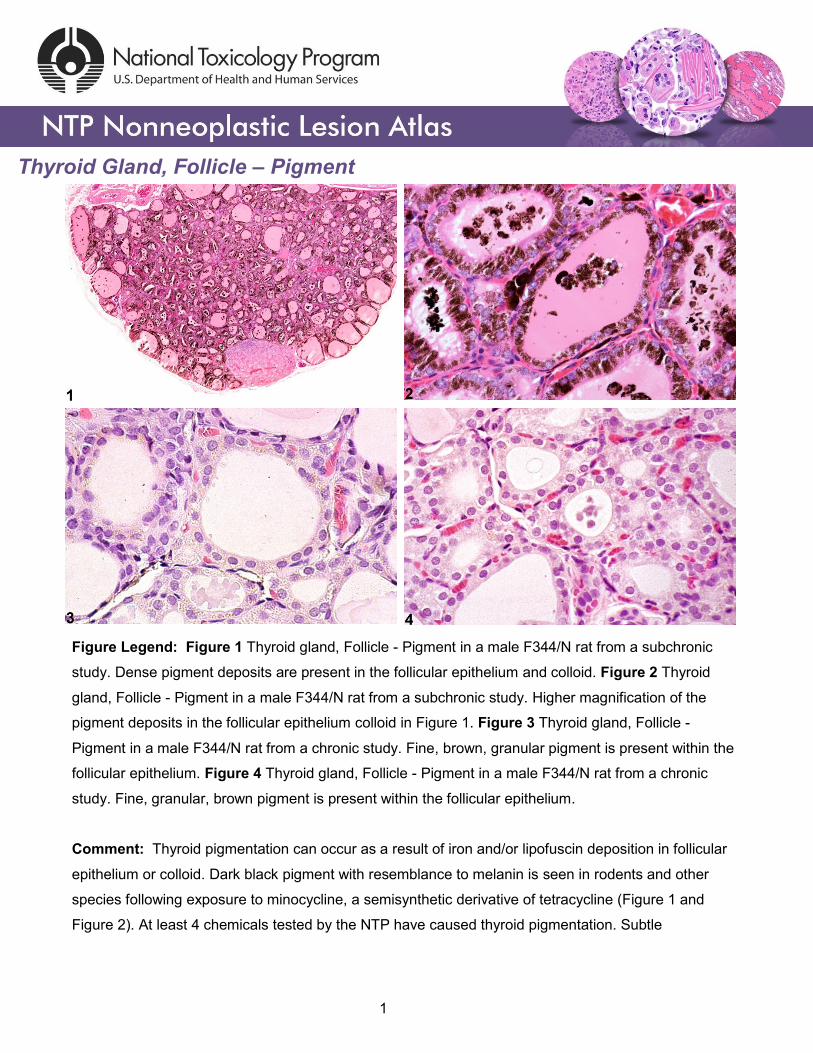
Thyroid Gland, Follicle – Pigment Figure Legend: Figure 1 Thyroid gland, Follicle - Pigment in a male F344/N rat from a subchronic study. Dense pigment deposits are present in the follicular epithelium and colloid. Figure 2 Thyroid gland, Follicle - Pigment in a male F344/N rat from a subchronic study. Higher magnification of the pigment deposits in the follicular epithelium colloid in Figure 1. Figure 3 Thyroid gland, Follicle - Pigment in a male F344/N rat from a chronic study. Fine, brown, granular pigment is present within the follicular epithelium. Figure 4 Thyroid gland, Follicle - Pigment in a male F344/N rat from a chronic study. Fine, granular, brown pigment is present within the follicular epithelium. Comment: Thyroid pigmentation can occur as a result of iron and/or lipofuscin deposition in follicular epithelium or colloid. Dark black pigment with resemblance to melanin is seen in rodents and other species following exposure to minocycline, a semisynthetic derivative of tetracycline (Figure 1 and Figure 2). At least 4 chemicals tested by the NTP have caused thyroid pigmentation. Subtle 1
-
Upload
truongkhue -
Category
Documents
-
view
223 -
download
0
Transcript of Thyroid Gland, Follicle - Pigment · Thyroid Gland, Follicle – Pigment . Figure Legend: Figure 1...
Thyroid Gland, Follicle – Pigment
Figure Legend: Figure 1 Thyroid gland, Follicle - Pigment in a male F344/N rat from a subchronic
study. Dense pigment deposits are present in the follicular epithelium and colloid. Figure 2 Thyroid
gland, Follicle - Pigment in a male F344/N rat from a subchronic study. Higher magnification of the
pigment deposits in the follicular epithelium colloid in Figure 1. Figure 3 Thyroid gland, Follicle -
Pigment in a male F344/N rat from a chronic study. Fine, brown, granular pigment is present within the
follicular epithelium. Figure 4 Thyroid gland, Follicle - Pigment in a male F344/N rat from a chronic
study. Fine, granular, brown pigment is present within the follicular epithelium.
Comment: Thyroid pigmentation can occur as a result of iron and/or lipofuscin deposition in follicular
epithelium or colloid. Dark black pigment with resemblance to melanin is seen in rodents and other
species following exposure to minocycline, a semisynthetic derivative of tetracycline (Figure 1 and
Figure 2). At least 4 chemicals tested by the NTP have caused thyroid pigmentation. Subtle
1
Thyroid Gland, Follicle – Pigment
pigmentation can be seen as a background change in both control and treated rats (Figure 3 and
Figure 4).
Recommendation: Definitive pigment identification is often difficult in histological sections, even with a
battery of special stains. Therefore, it is recommended that a diagnosis of pigment (as opposed to
diagnosing the type of pigment, e.g., hemosiderin or lipofuscin) is most appropriate. The pathology
narrative should describe the morphological features of the pigment. Not all pigments have to be
diagnosed, as some are ubiquitous in aging animals or related to some other disease process and not
toxicologically meaningful. The pathologist should use his or her judgment in deciding whether or not
secondary deposits of pigment are prominent enough to warrant a separate diagnosis.
References:
Greaves P. 2007. Histopathology of Preclinical Toxicity Studies: Interpretation and Relevance in Drug Safety Evaluation, 3rd ed. Academic Press, Amsterdam, 819-839. Abstract: http://www.sciencedirect.com/science/book/9780444527714 Hardisty JF, Boorman GA. 1990. Thyroid gland. In: Pathology of the Fischer Rat: Reference and Atlas (Boorman GA, Eustis SL, Elwell MR, Montgomery CA, MacKenzie WF, eds). Academic Press, San Diego, 519-536. Abstract: http://www.ncbi.nlm.nih.gov/nlmcatalog/9002563 Hardisty JF, Boorman GA. 1999. Thyroid and parathyroid glands. In: Pathology of the Mouse: Reference and Atlas (Maronpot RR, Boorman GA, Gaul BW, eds). Cache River Press, Vienna, IL, 537-554. Abstract: http://www.cacheriverpress.com/books/pathmouse.htm Tajima K, Miyagawa J, Nakajima H, Shimizu M, Katayama S, Mashita K, Tarui S. 1985. Morphological and biochemical studies on minocycline-induced black thyroid in rats. Toxicol Appl Pharmacol 81:393–400. Abstract: http://www.ncbi.nlm.nih.gov/pubmed/3001973 Ward JM, Stinson SF, Hardisty JF, Cockrell BY, Hayden DW. 1979. Neoplasms and pigmentation of thyroid glands in F344 rats exposed to 2,4-diaminoanisole sulfate, a hair dye component. J Natl Cancer Inst 62:1067–1073. Abstract: http://www.ncbi.nlm.nih.gov/pubmed/285280
2
Thyroid Gland, Follicle – Pigment
Authors: Robert R. Maronpot, DVM, MS, MPH, DACVP, DABT, FIATP Senior Pathologist Experimental Pathology Laboratories, Inc. Research Triangle Park, NC Amy Brix, DVM, PhD, DACVP Senior Pathologist Experimental Pathology Laboratories, Inc. Research Triangle Park, NC
3