Clinical Review Barbara Buch, M.D. Orthopaedic Surgeon FDA Orthopaedic Devices Branch.

THYROID CANCER
Mai H. Nguyen, M.D.
Faculty Advisor: Francis B. Quinn, M.D.
The University of Texas Medical Branch
Department of Otolaryngology
Grand Rounds Presentation
Dec. 04, 2002

History
1812: Gay-Lussac discovered iodine as a cause of goiter.
1833: Boussingault prescribed iodized salt for prevention and treatment of goiter.
1836: T.W.King presented anatomical descriptions of thyroid gland.
1870: Fagge described sporadic and congenital cretinism.
History
1882 - 1917: Theodor Kocher (Bern)
introduced techniques of thyroidectomy
(>5000 cases). His mortality rate at the end
of 19th century is as low as 1.8%
1880s: Billroth suggested bilateral partial
thyroidectomy to prevent hypothyroidism.
History
1880: Williams S. Halsted: developed his
thyroidectomy techniques in the US.
Thyroid cancer was first described by
Halsted by the terms “sarcomatous
degeneration”, “thyroid tumor” or “thyroid
cancer cells”
Embryology
• 4th week: thyroid gland appears.
• 5th week: break down of the thyroglossal
duct, thyroid gland continue descending
• 7th week: thyroid gland migrates to its
position, anterior to the trachea
• 10th week: thyroglossal duct disappears

Anatomy
Locate deep to the sternohyoid muscle,
from level C5 to T1 vertebrae or anterior to
the 2nd and 3rd tracheal rings.
Thyroid gland is attached to the trachea by
the lateral suspensory (Berry) ligaments.
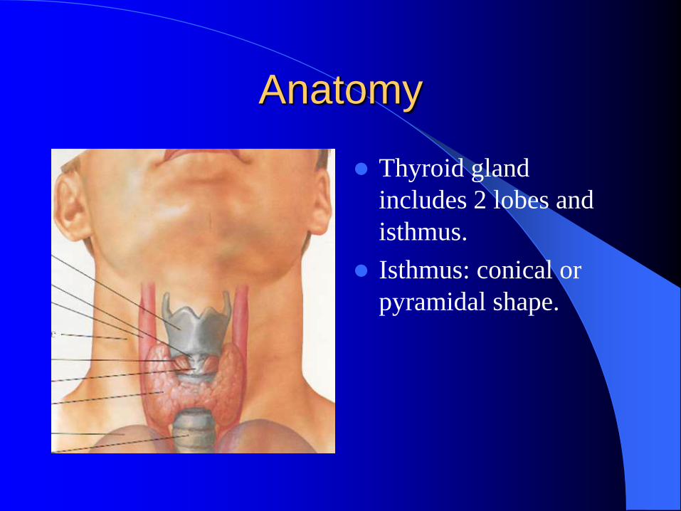
Anatomy
Thyroid gland
includes 2 lobes and
isthmus.
Isthmus: conical or
pyramidal shape.

Anatomy
Blood supply: sup. & inf. thyroid arteries
Anatomy variant: thyroid ima artery, in 1.5% to 12%, in front of the trachea.
Lymph vessels: drain to prelaryngeal, pretracheal and paratracheal nodes.
Innervation: superior, middle, and inferior sympathetic ganglia.
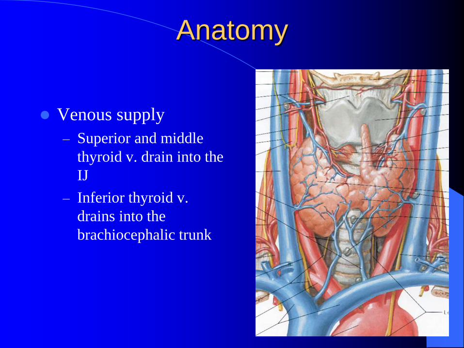
Anatomy
Venous supply
– Superior and middle
thyroid v. drain into the
IJ
– Inferior thyroid v.
drains into the
brachiocephalic trunk

Anatomy-Recurrent Laryngeal Nerve
(RLN)
Sim’s triangle
– Carotid artery
– Trachea
– Inferior pole of thyroid
LRLN runs parallel
with the TEG
RRLN runs diagonal
with the TEG
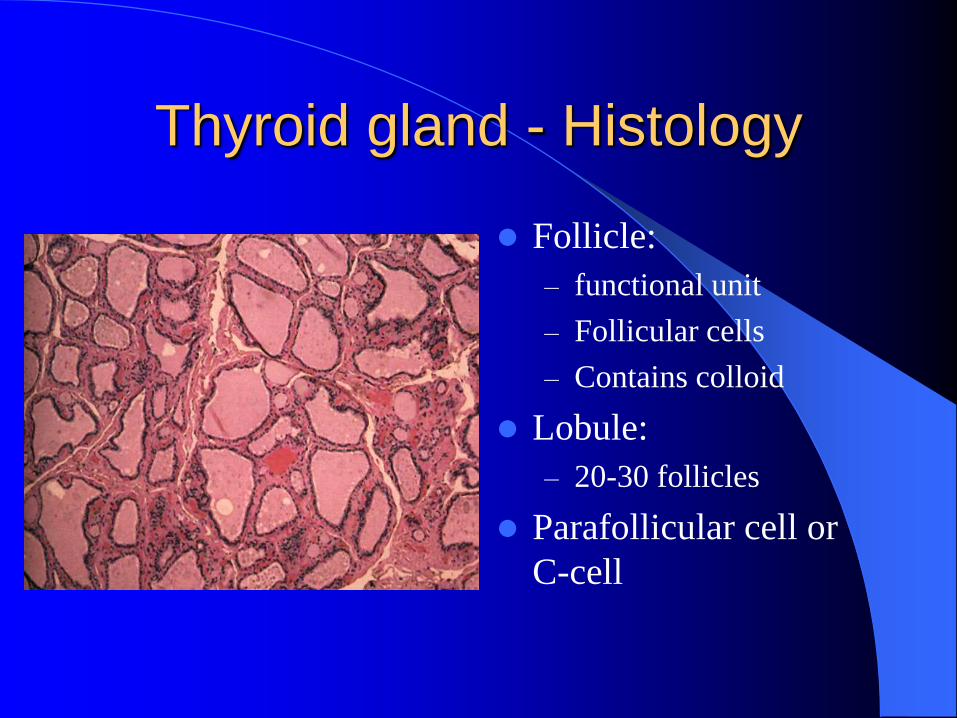
Thyroid gland - Histology
Follicle:
– functional unit
– Follicular cells
– Contains colloid
Lobule:
– 20-30 follicles
Parafollicular cell or
C-cell

Physiology
Euthyroidism control:
1. TRH (thyroid releasing hormone) and TSH (thyroid stimulating hormone)
2. Thyroid gland: synthesis, storage, secretion of thyroxine (T4), triiodothyronine (T3)
3. Peripheral control metabolism of T3, T4
Thyroid Nodule Statistics
3%-7% population, female is 6.5%; male is 1.5%
4% of these nodules are malignant, 1% of all
cancers
Male have a higher risk of being cancer
Single nodule is more likely malignant than
multiple nodules
Nodules in children and the elderly have a higher
risk of malignancy

History Taking
Age, gender
Thyroid mass or nodule (time coarse, growth)
Associated symptoms
– Pain, hoarseness, dysphagia, dyspnea, stridor, hemoptysis
Radiation, goiter, Hashimoto’s, Grave’s, other cancers.
Family history of thyroid and other endocrine tumors.
Physical exam
Complete head and neck exam
– Bimanual palpation of thyroid gland and
cervical chain of lymph nodes
Laryngoscope:
– Evaluate for vocal cord mobility and symmetry

Diagnosis
Needle biopsy:
Core needle biopsy:
– Adequate tissue for diagnosis
– Disadvantages
more difficult
more traumatic
more complications

Diagnosis
Fine needle aspiration (FNA):
– Easy to perform, less morbidity.
– FN: 0.3-10%; FP: 0-2.5%
– Disadvantages
less tissue for diagnosis
limit in differentiation of certain types of thyroid
cancers
– Follicular adenoma vs. carcinoma
– Hurthle cell adenoma vs. carcinoma
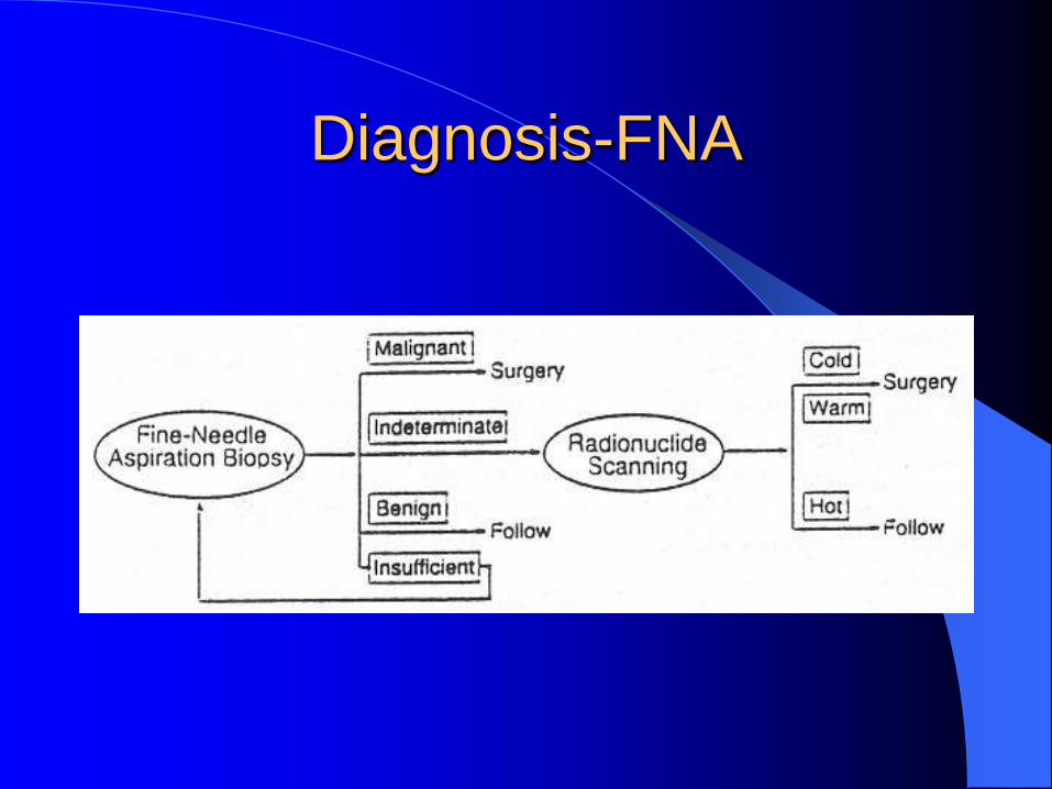
Diagnosis-FNA
Diagnosis
Blood test:
T4,T3, TSH (thyroid function tests)
Ca, P (hyperparathyroidism asso. with TC)
TG (increase in recurrent WDTC)
Calcitonin (increase in MTC)

Diagnosis –U/S
Sensitive (80%)
Detect nodule 2- 3 mm
F/u cystic asp., re-
collection of fluid
FNA guide.

Diagnosis- Imaging
CT: Detect tracheal invasion
Evaluate for cervical met
MRI Useful to detect residual, recurrent and metastatic carcinoma.
T2 differentiates tumor and fibrosis.
CXR: tracheal deviation, airway narrowing, lung
metastasis.

Diagnosis – thyroid scan
Radioactive iodine or technetium uptake
Before FNA – test of choice for initial w/u
Uses today
– Indeterminate FNA
– Large benign nodules (> 4cm)

Thyroid Cancer
Classification:
1. Well-differentiated malignant neoplasms
(85% of thyroid cancer)
*Papillary thyroid carcinoma (PTC)
*Follicular thyroid carcinoma (FTC)
*Hurthle cell carcinoma (HCC)
Pathology Classification
2. Poor differentiated malignant neoplasms
*Medullary thyroid carcinoma (MTC)
*Anaplastic thyroid carcinoma (ATC)
*Insular thyroid carcinoma (ITC)
3. Other malignant tumors:
*Lymphoma
*Metastatic tumors

Papillary Thyroid Carcinoma (PTC)
Most common WDTC - 75%-85%
80%-90% of radiation-induced TC
Peak incidence: 30s-40s
10 year-survival: 84%-90%
Female:male ratio is 3:1
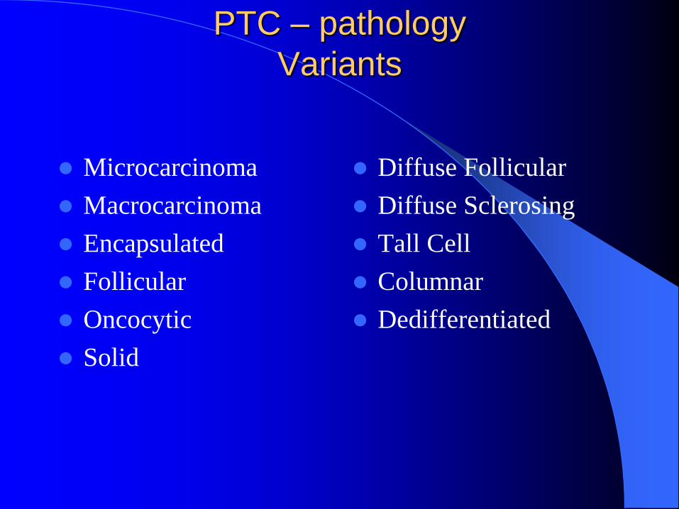
PTC – pathology
Variants
Microcarcinoma
Macrocarcinoma
Encapsulated
Follicular
Oncocytic
Solid
Diffuse Follicular
Diffuse Sclerosing
Tall Cell
Columnar
Dedifferentiated
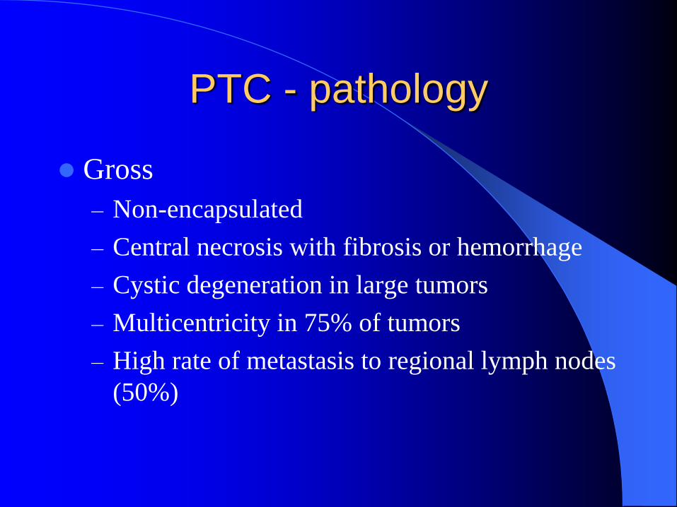
PTC - pathology
Gross
– Non-encapsulated
– Central necrosis with fibrosis or hemorrhage
– Cystic degeneration in large tumors
– Multicentricity in 75% of tumors
– High rate of metastasis to regional lymph nodes
(50%)

PTC - pathology
Histology
– Psammoma bodies
– Columnar thyroid
epithelial
– Well-form
fibrovascular cores

PTC - pathology
Histology
– Papillary projections
– Nuclei
Vesicular and ground-
glass “Orphan Annie”
appearance
High N:C ratio
Mitotic figures
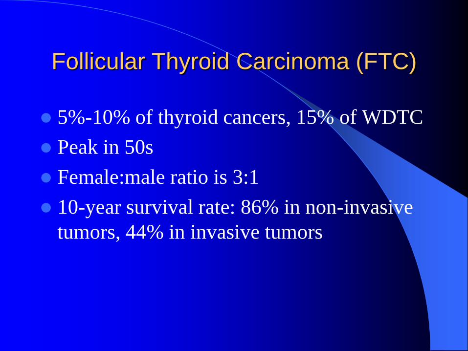
Follicular Thyroid Carcinoma (FTC)
5%-10% of thyroid cancers, 15% of WDTC
Peak in 50s
Female:male ratio is 3:1
10-year survival rate: 86% in non-invasive
tumors, 44% in invasive tumors

FTC - pathology
Gross
– Well-encapsulated
– Cystic degeneration, calcification, hemorrhage
– Tendency invade the thyroid capsule and blood
vessels.

FTC - pathology
Histology
– Follicular pattern with
vesicular nucleolus
cells

FTC - pathology
Histology
– Capsular and vascular
invasion
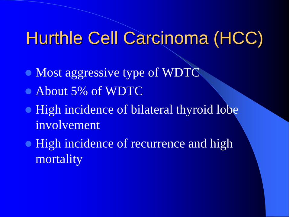
Hurthle Cell Carcinoma (HCC)
Most aggressive type of WDTC
About 5% of WDTC
High incidence of bilateral thyroid lobe
involvement
High incidence of recurrence and high
mortality
Medullary Thyroid Carcinoma (MTC)
Account for 5% to 10 % of all thyroid
cancers
Tumor of the calcitonin-producing
parafollicular or C-cells

MTC
Sporadic
– 80% of MTC
– Poorer prognosis
– Unifocal
– Not associated with other endocrine tumors
– Peak in middle age to elderly

MTC
Familial
– 20% of MTCs
– Autosomal dominant inheritance
– Associated with C-cell hyperplasia
– Associated other endocrine tumors
– Peak in 30s.

MTC
Family traits Sipple’s syndrome (MEN II a)
– MTC
– Pheochromocytoma
– hyperparathyroidism
2. Wermer’s syndrome (MEN II b)
– MTC
– pheochromocytoma
– mucosal neuromas
– marfanoid habitus.

MTC
50% have regional metastases to lymph
nodes.
Distant metastasis include: lung, liver,
adrenal glands, and bone (osteoblastic)

Medullary carcinoma
Gross
– gray to yellow, firm,
well-circumscribed or
invasive with bilateral
multicentric
involvement.
Histology
– Hyperplastic C-cells
contain immunoreative
calcitonin
Anaplastic Thyroid Carcinoma (ATC)
Undifferentiated differentiated CA
3% of thyroid cancers
Most aggressive, poorest prognosis
Uncapsulated, extension out side the gland
Death in several months due to airway obstruction,
vascular invasion, distant metastasis.
Higher incidence in pre-existing multi-nodular
goiter

Anaplatic Carcinoma
Gross
– fleshy, tan-white
appearance, with
hemorrhagic and
necrotic areas.
Histology
– spindle or giant-cell

Malignant Lymphoma
2%-5% of thyroid cancers
Increase in Hashimoto’s or endemic goiter
areas
Most common in > 50s
Prognosis factors: cell types and stages

Malignant Lymphoma
Gross
– large, yellow-tan,
scaly with hemorrhagic
and necrostic areas
Histology
– small cell non-cleaved
type (MC) and large
cell non-cleaved
follicular
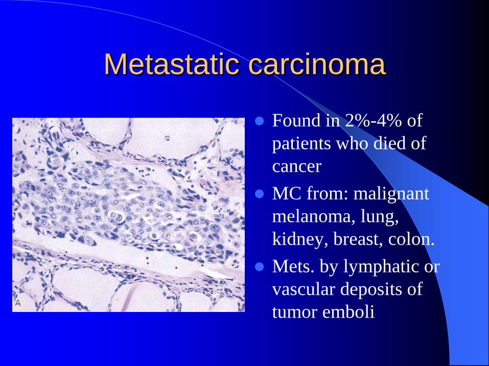
Metastatic carcinoma
Found in 2%-4% of
patients who died of
cancer
MC from: malignant
melanoma, lung,
kidney, breast, colon.
Mets. by lymphatic or
vascular deposits of
tumor emboli
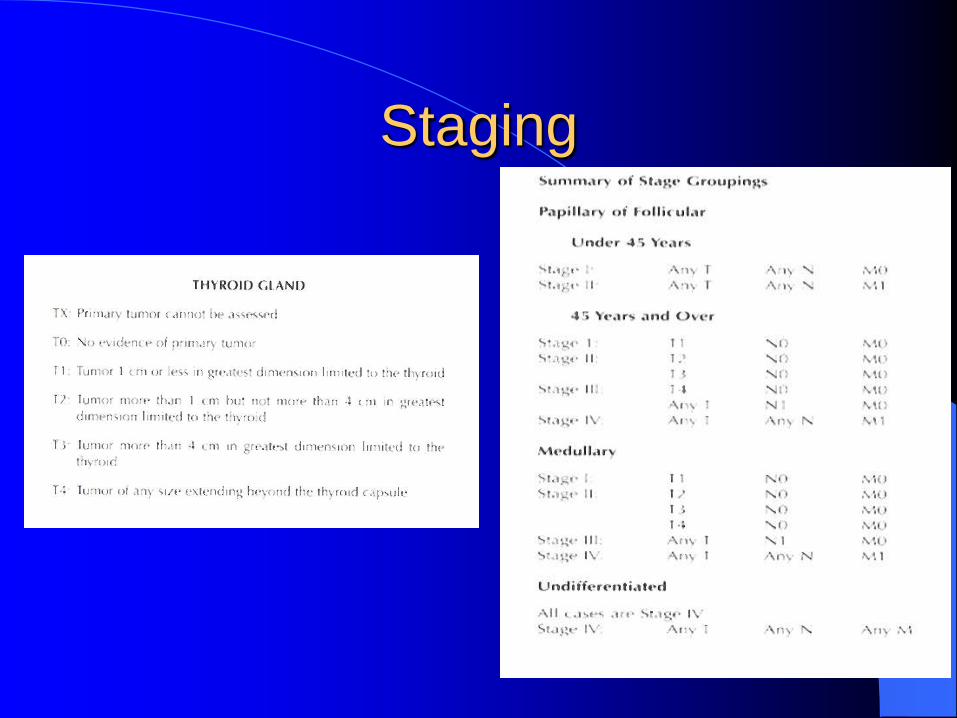
Staging

Management of the Thyroid Nodule
Serial exam
Physical examination
– Benign
– Asymptomatic palpable nodule
U/S
– F/u a benign, nonpalpable nodule
– F/u a cystic nodule for reaccumulation

Management of the Thyroid Nodule
Trial of suppression of TSH
– Benign or indeterminate FNA (controversial)
– Maintain TSH level between 0.1 and 0.5
mlU/L per day
– Decrease tumor volume up to 50% in 40% pts.
– A shrinking tumor is not likely malignant
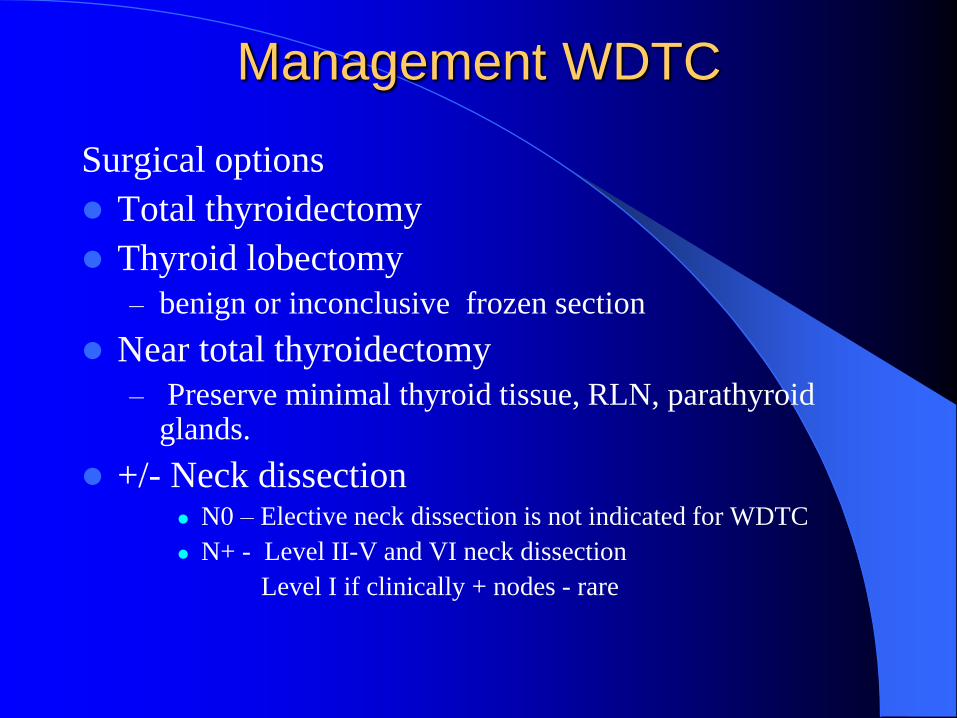
Management WDTC
Surgical options
Total thyroidectomy
Thyroid lobectomy
– benign or inconclusive frozen section
Near total thyroidectomy
– Preserve minimal thyroid tissue, RLN, parathyroid glands.
+/- Neck dissection N0 – Elective neck dissection is not indicated for WDTC
N+ - Level II-V and VI neck dissection
Level I if clinically + nodes - rare
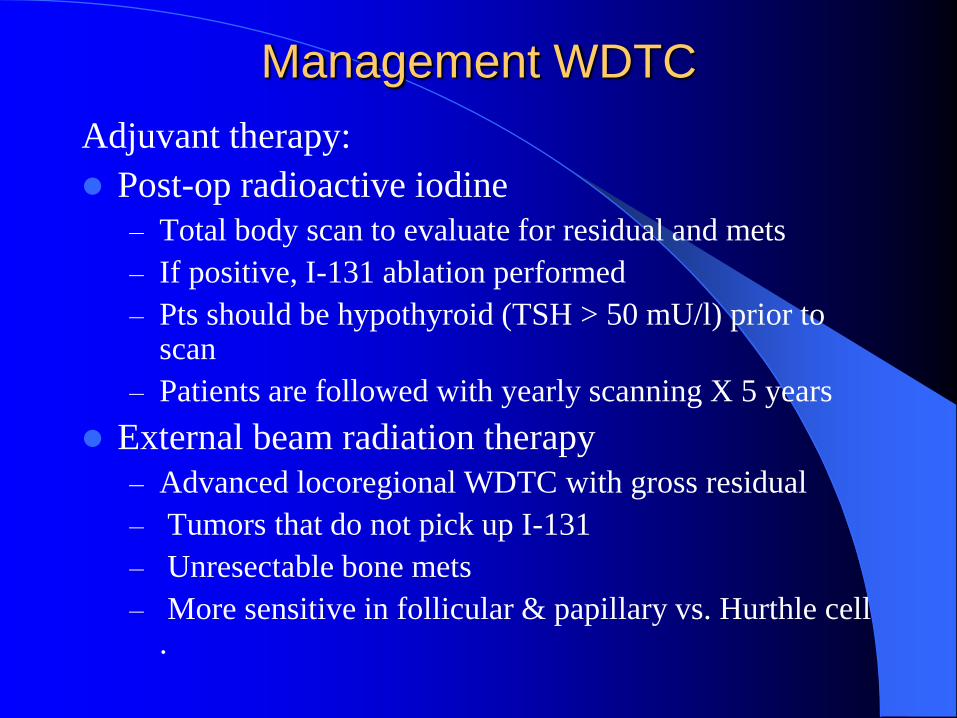
Management WDTC
Adjuvant therapy:
Post-op radioactive iodine
– Total body scan to evaluate for residual and mets
– If positive, I-131 ablation performed
– Pts should be hypothyroid (TSH > 50 mU/l) prior to scan
– Patients are followed with yearly scanning X 5 years
External beam radiation therapy
– Advanced locoregional WDTC with gross residual
– Tumors that do not pick up I-131
– Unresectable bone mets
– More sensitive in follicular & papillary vs. Hurthle cell .
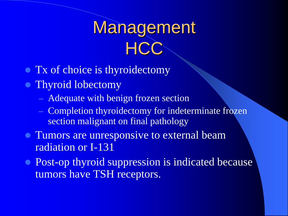
Management
HCC Tx of choice is thyroidectomy
Thyroid lobectomy
– Adequate with benign frozen section
– Completion thyroidectomy for indeterminate frozen section malignant on final pathology
Tumors are unresponsive to external beam radiation or I-131
Post-op thyroid suppression is indicated because tumors have TSH receptors.

Management
MTC
Surgery: Thyroidectomy and SLND (level
II, III, IV), anterior compartment ND
(include level VI, and/or VII).
10-year survival rate is 90%
Recurrent MTC: resistant to chemo and
XRT
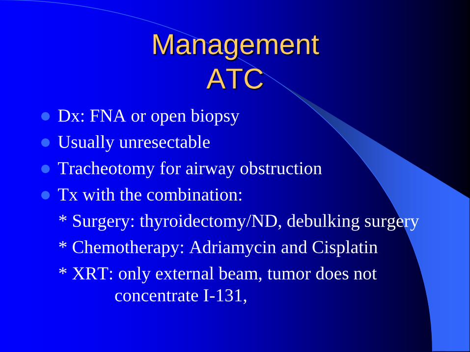
Management
ATC Dx: FNA or open biopsy
Usually unresectable
Tracheotomy for airway obstruction
Tx with the combination:
* Surgery: thyroidectomy/ND, debulking surgery
* Chemotherapy: Adriamycin and Cisplatin
* XRT: only external beam, tumor does not
concentrate I-131,
Surgical complications
Non-metabolic complications
Nerve injury
– SLN (laryngeal sensation) – up to 5% incidence
Unstable voice
Diff. high pitch,
Dysphagia and aspiration
Laryngoscopy:bowing of VCs, ipsilateral rotation or displacement of affected VC.
– RLN up to 1-2% incidence
Unilateral – no treatment vs medialization procedure
Bilateral: re-intubate, tracheotomy

Surgical complications
Non-metabolic complications:
Hemorrhage: thru the drains, neck swelling
Airway obstruction
– Hematoma
– Laryngeal edema
– Bilateral RLN injury
Chyle leak
Pneumothorax

Surgical complications
Metabolic complications:
Hypocalcemia: 5% of thyroidectomy
– Prevention - autotransplatation of parathyroid glands
– Treatment – IV vs PO calcium replacement and Vit. D
Thyroid storm
– More common in pts. with hyperthyroidism or chronic systemic diseases
Tx. supportive
Beta blockers
Muscle relaxants
Prognostic factors
Histology: is an important factor
Age: is a significant factor, e.g. WDTC
Sex: female have more risk of thyroid nodule;
males have more risk of thyroid cancer
Size: tumor > 1.5 cm has poorer prognosis
Extracapsular, vascular invasion or metastases
disease are poor prognosis factors
History of radiation: high risk of papillary CA
Prognostic factors
Mayo clinic: “AGES” including age, grade,
extracapsular tumor, and size.
Lahey clinic: “AMES” including age,
metastasis, extracapsular tumor, and size.
Conclusion
Thyroid cancer is relatively rare (1% of all
cancers), one of the most curable cancer.
Surgery is the treatment of choice for most of
thyroid cancers
Preservation of the RLN and normocalcemia are
the goals for a successful thyroidectomy
Surgical complications are preventable and
treatable