Thymol turbidity test is associated with the risk of ...
7
RESEARCH ARTICLE Open Access Thymol turbidity test is associated with the risk of cyclops syndrome following anterior cruciate ligament reconstruction Yuya Kodama, Takayuki Furumatsu * , Tomohito Hino, Yusuke Kamatsuki, Yoshiki Okazaki, Shin Masuda, Yuki Okazaki and Toshifumi Ozaki Abstract Background: Cyclops nodule formation is a serious complication after anterior cruciate ligament (ACL) reconstruction. The purpose of our study was to investigate whether an increase in thymol turbidity test (TTT) values is involved in the development of cyclops nodule formation or cyclopoid scar formation following ACL reconstruction. Methods: Between 2011 and 2014, 120 cases underwent outside-in ACL reconstruction. Forty-seven patients who had high TTT values were individually matched for age, sex, body mass index, and meniscus injury to a low TTT value group of 47 patients. The primary outcome was the occurrence of cyclops nodule formation or cyclopoid scar formation. All 94 patients were divided into 3 groups using surgical records and intra-operative video to enable a sub- analysis. The groups were a no-cyclops group, a cyclopoid group, and a cyclops group. Blood examinations, including TTT, and knee range of motion evaluations were performed before surgery, 3 months after surgery, and 1 year after surgery. Results: There were no differences in preoperative demographic data between the two groups. TTT values did not significantly influence cyclopoid scar formation (OR, 1.67; 95% CI, 0.62 to 4.66; p = 0.362). However, patients with cyclops nodule formation showed significantly higher TTT values than the control patients. (OR, 9.34; 95% CI, 1.94 to 90. 3; p = 0.002). Knee extension loss was observed in the cyclopoid and cyclops groups 3 months after reconstruction. In the cyclops group, arthroscopic resection of the cyclops nodule was performed 3 months after reconstruction. Eventually, almost full range of motion was restored in all patients. Conclusions: High TTT values before ACL reconstruction were an indicator of cyclops nodule formation. Furthermore, cyclopoid scar formations may not be the result of an individual’s immune reaction but that of extension loss in the early post-reconstruction phase. Keywords: Cyclops syndrome, Anterior cruciate ligament, Thymol turbidity test, Cyclops nodule, Knee extension, Range of motion, Cyclopoid scar Background Cyclops nodule formation is a serious complication after anterior cruciate ligament (ACL) reconstruction, and it is characterized by loss of terminal knee extension due to proliferative fibrous nodule formation in the intercondylar notch [1]. The incidence of post-operative cyclops nodule formation ranges from 1.9 to 10.6% [2, 3], whereas the incidence of cyclops lesions without extension loss varies from 2.2 to 46.8% [4, 5]. This distinction of symptoms is due to 2 distinct types of cyclops lesions, a true cyclops nodule and a cyclopoid scar [6]. Although there are several hypotheses regarding the pathogenesis of cyclops nodule formation, including bone and cartilage residue in the joint following tibial tunnel drilling and preparation for graft passage [1, 6], repeated graft impingement on the notch [1], post-operative hamstring contracture [7], and narrow- ing of the femoral intercondylar notch [8], histologically, a cyclops nodule formation is composed of disorganized * Correspondence: [email protected] Department of Orthopaedic Surgery, Okayama University Graduate School of Medicine, Dentistry, and Pharmaceutical Sciences, 2-5-1 Shikatacho, Kitaku, Okayama 700-8558, Japan © The Author(s). 2018 Open Access This article is distributed under the terms of the Creative Commons Attribution 4.0 International License (http://creativecommons.org/licenses/by/4.0/), which permits unrestricted use, distribution, and reproduction in any medium, provided you give appropriate credit to the original author(s) and the source, provide a link to the Creative Commons license, and indicate if changes were made. The Creative Commons Public Domain Dedication waiver (http://creativecommons.org/publicdomain/zero/1.0/) applies to the data made available in this article, unless otherwise stated. Kodama et al. BMC Musculoskeletal Disorders (2018) 19:367 https://doi.org/10.1186/s12891-018-2286-1
Transcript of Thymol turbidity test is associated with the risk of ...
RESEARCH ARTICLE Open Access
Thymol turbidity test is associated with therisk of cyclops syndrome following anteriorcruciate ligament reconstructionYuya Kodama, Takayuki Furumatsu*, Tomohito Hino, Yusuke Kamatsuki, Yoshiki Okazaki, Shin Masuda,Yuki Okazaki and Toshifumi Ozaki
Abstract
Background: Cyclops nodule formation is a serious complication after anterior cruciate ligament (ACL) reconstruction.The purpose of our study was to investigate whether an increase in thymol turbidity test (TTT) values is involved in thedevelopment of cyclops nodule formation or cyclopoid scar formation following ACL reconstruction.
Methods: Between 2011 and 2014, 120 cases underwent outside-in ACL reconstruction. Forty-seven patients who hadhigh TTT values were individually matched for age, sex, body mass index, and meniscus injury to a low TTT valuegroup of 47 patients. The primary outcome was the occurrence of cyclops nodule formation or cyclopoid scarformation. All 94 patients were divided into 3 groups using surgical records and intra-operative video to enable a sub-analysis. The groups were a no-cyclops group, a cyclopoid group, and a cyclops group. Blood examinations, includingTTT, and knee range of motion evaluations were performed before surgery, 3 months after surgery, and 1 year aftersurgery.
Results: There were no differences in preoperative demographic data between the two groups. TTT values did notsignificantly influence cyclopoid scar formation (OR, 1.67; 95% CI, 0.62 to 4.66; p = 0.362). However, patients withcyclops nodule formation showed significantly higher TTT values than the control patients. (OR, 9.34; 95% CI, 1.94 to 90.3; p = 0.002). Knee extension loss was observed in the cyclopoid and cyclops groups 3 months after reconstruction. Inthe cyclops group, arthroscopic resection of the cyclops nodule was performed 3 months after reconstruction.Eventually, almost full range of motion was restored in all patients.
Conclusions: High TTT values before ACL reconstruction were an indicator of cyclops nodule formation. Furthermore,cyclopoid scar formations may not be the result of an individual’s immune reaction but that of extension loss in theearly post-reconstruction phase.
Keywords: Cyclops syndrome, Anterior cruciate ligament, Thymol turbidity test, Cyclops nodule, Knee extension, Rangeof motion, Cyclopoid scar
BackgroundCyclops nodule formation is a serious complication afteranterior cruciate ligament (ACL) reconstruction, and it ischaracterized by loss of terminal knee extension due toproliferative fibrous nodule formation in the intercondylarnotch [1]. The incidence of post-operative cyclops noduleformation ranges from 1.9 to 10.6% [2, 3], whereas the
incidence of cyclops lesions without extension loss variesfrom 2.2 to 46.8% [4, 5]. This distinction of symptoms isdue to 2 distinct types of cyclops lesions, a true cyclopsnodule and a cyclopoid scar [6]. Although there are severalhypotheses regarding the pathogenesis of cyclops noduleformation, including bone and cartilage residue in the jointfollowing tibial tunnel drilling and preparation for graftpassage [1, 6], repeated graft impingement on the notch[1], post-operative hamstring contracture [7], and narrow-ing of the femoral intercondylar notch [8], histologically, acyclops nodule formation is composed of disorganized
* Correspondence: [email protected] of Orthopaedic Surgery, Okayama University Graduate School ofMedicine, Dentistry, and Pharmaceutical Sciences, 2-5-1 Shikatacho, Kitaku,Okayama 700-8558, Japan
© The Author(s). 2018 Open Access This article is distributed under the terms of the Creative Commons Attribution 4.0International License (http://creativecommons.org/licenses/by/4.0/), which permits unrestricted use, distribution, andreproduction in any medium, provided you give appropriate credit to the original author(s) and the source, provide a link tothe Creative Commons license, and indicate if changes were made. The Creative Commons Public Domain Dedication waiver(http://creativecommons.org/publicdomain/zero/1.0/) applies to the data made available in this article, unless otherwise stated.
Kodama et al. BMC Musculoskeletal Disorders (2018) 19:367 https://doi.org/10.1186/s12891-018-2286-1
fibrous connective tissue with a central region of granula-tion tissue and newly formed vessels [1, 5, 6]. Cyclopoidscar formations are composed of a build-up of fibrous tis-sue showing elements of granulation tissue [6]. However,the same symptoms, along with similar arthroscopic andhistologic findings, also occur in acute ACL injury withoutreconstruction [9, 10]. Therefore, the reparative processesoccurring as an immune reaction of the vital tissue may bethe main triggering factors for the process of cyclops nod-ule formation.The thymol turbidity test (TTT) is a type of colloidal
reaction test that reflects immunoglobulin M [11]. TTTis considered a marker of inflammatory conditions suchas chronic hepatitis, chronic infection, or collagen dis-ease [12, 13]. Before ACL reconstruction surgery, weroutinely perform blood examination, including theTTT. By chance, we discovered that TTT results tendedto be higher in patients with cyclops nodule formation,whereas there were no other blood examination abnor-malities. To the best of our knowledge, a relationship be-tween an immune reaction of the vital tissue and cyclopsor cyclopoid development has been proposed, but notyet proven [6, 9]. In addition, there are no reports onblood examinations in patients with cyclops syndrome.The purpose of our study was to investigate whether anincrease in TTT value was involved in the occurrence ofcyclops nodule formation or cyclopoid scar formation.Furthermore, in order to investigate this in detail, acomparison between 3 groups (a no-cyclops group, acyclopoid group, and a cyclops group) was performedusing blood test results and knee range of motionmeasurements.
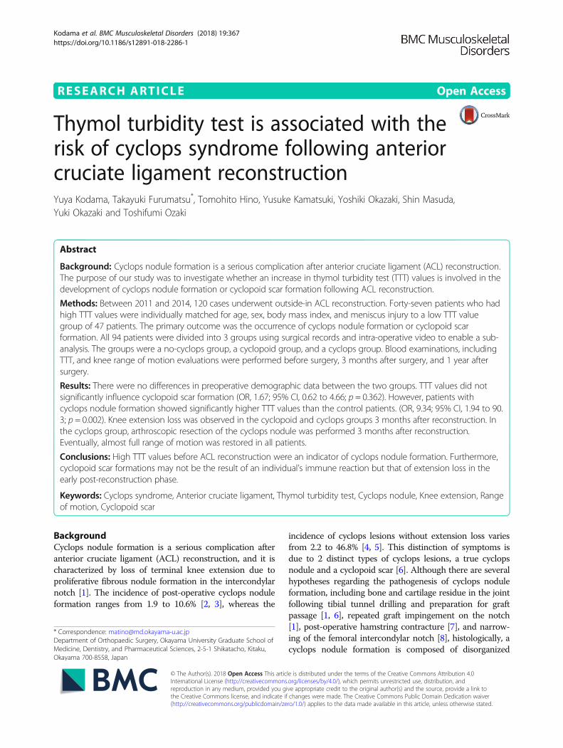
MethodsStudy subjectsThis retrospective study was performed with the approvalof the institutional review board, and all patients signedthe consent form drafted for the study. Between 2011 and2014, 120 consecutive patients underwent outside-in ACLreconstruction [14] performed by two surgeons at ourhospital. Exclusion criteria were patients who had previ-ous ligament injury, and those who had a concomitantmedial collateral ligament injury classified as greater thangrade III. Patients who had undergone revision ACL re-construction were also excluded. Finally, 47 patients withTTT ≥ 4 and 58 patients with TTT < 4 were included. The47 patients in the TTT ≥ 4 group were matched for age,sex, and body mass index (BMI) with 47 patients in theTTT < 4 group (Fig. 1). In order to conduct case-controlresearch, the research design was set as follows. Thepopulation included patients for whom final assessmentscould be made after reconstruction following an ACL tear.Exposure was defined as a TTT value ≥4 and control wasdefined as a TTT value < 4. Outcomes included the
occurrence of cyclops nodule formation or cyclopoid scarformation. Cyclops and cyclopoid lesions were diagnosedusing arthroscopic video based on a previous report [6].All 94 patients were divided into 3 groups using surgical
records and intra-operative video to perform a sub-analysis(Fig. 2). These groups were a cyclops group (case) (n = 16),a no-cyclops group (control 1) (n = 51), and a cyclopoidgroup (control 2) (n = 27). In addition to the TTT, aspar-tate transaminase (AST), alanine transaminase (ALT), andC-reactive protein (CRP) levels were evaluated 1 week be-fore reconstruction. The same inspection was performedafter cyclops resection (3 months after reconstruction) and1 week before second-look arthroscopy. The knee range ofmotion at 3 different time points was determined fromclinical records.
Surgical procedureA double-bundle, outside-in, arthroscopic ACL recon-struction was performed in all patients, using the semi-tendinosus tendon (ST) and, if necessary, the gracilistendon. The harvested tendons were double-looped overan Endobutton fixation device (Smith & Nephew, Ando-ver, MA), with the distal ends anchored using a Krackowsuture, thus recreating the anteromedial (AM) and pos-terolateral (PL) bundles of the ACL. To prevent elong-ation of the grafts, a continuous 30-s loading with 70 Nwas applied twice to the graft (70 N-1 min), and thenthe same loading was applied repeatedly (70 N-2 min)[15]. The femoral tunnel was created using an outside-intechnique. The longitudinal linear resident’s ridge [16]and the posterior cartilage, used as landmarks for theACL femoral footprint, were identified. Two 2.4-mmguide pins were then inserted separately from the out-side into the ACL footprints behind the resident’s ridgeand just anterior to the articular margin, using an an-terolateral entry femoral aimer (Smith & Nephew). A5.5-mm to 6.5-mm tunnel was then created for the AMand PL grafts by over-drilling via the guide pins. The au-togenous tendon was harvested and transected into 2double-looped grafts. Two Endobutton-CLs® (Smith &Nephew) were connected to the end of each loop graft.The appropriate graft length was determined from thelength of the femoral tunnel to allow the introduction ofsufficient graft materials (> 13 mm) into the bone tun-nels. After creation of the femoral tunnel, the ACL tibialtunnel was created. The AM tunnel was created usingthe following intra-articular landmarks: just lateral tothe medial intercondylar ridge and just posterior to theanterior ridge so as not to damage the lateral meniscusanterior insertion [17, 18]. The PL tunnel was createdposterior to the AM tunnel, just lateral to the medialintercondylar ridge. In all cases, tibial fixation of thegraft was performed using double-spike plates (Meira,Aichi, Japan), with the knee flexed at 20°, and an initial
Kodama et al. BMC Musculoskeletal Disorders (2018) 19:367 Page 2 of 7

No-cyclops group (n = 51) Cyclopoid group (n = 27) Cyclops group (n = 16)
105 patients
TTT ≥ 4 group (n = 47) TTT < 4 group (n = 47)
120 patients
MatchedAge, Sex, BMI, meniscus injury
Exclusion• Previous ligament injury• Revision ACL reconstruction• Concomitant MCL injury (> grade III)
94 patients
47 patients with TTT ≥ 4 58 patients with TTT < 4
Subanalysis
• Blood examination data at different time points between 3 groups: Table 3• Range of motion at different time points between 3 groups: Table 4
Fig. 1 Flow diagram of patients screened and grouped. ACL, anterior cruciate ligament; MCL, medial collateral ligament; BMI; body mass index;TTT, thymol turbidity test
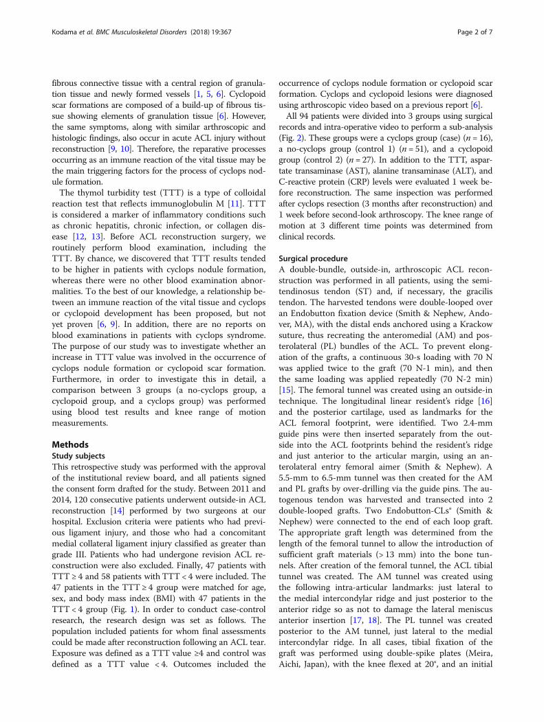
Fig. 2 Arthroscopic findings during second-look arthroscopy after ACL reconstruction. Knee flexion position (a-c) and extension position (d-f) areshown. A patient without cyclops (a, d). A patient with a cyclopoid lesion (b, e). A patient with a cyclops lesion impinging on the intercondylarnotch (c, f)
Kodama et al. BMC Musculoskeletal Disorders (2018) 19:367 Page 3 of 7

tension of 20 N was applied to the PL bundle and 30 Nto the AM bundle. The tension in each bundle was mea-sured independently using a tensiometer. Finally, wechecked for impingement to the notch at full extension.In all cases, there was no impingement to the notch atfull extension. Thus, femoral notchplasty was not per-formed in all cases.
Second-look arthroscopic examination, clinicalevaluations, and post-operative managementSecond-look arthroscopy was performed approximately1 year after reconstruction for the removal of the 2double-spike plates, fixed with screws into the tibia,which were used for tibial fixation of the ACL graft.Knee range of motion was evaluated with a goniometerbefore reconstruction, 3 months after reconstruction,and after second-look arthroscopy. Extension loss wasmeasured in degrees and compared with the normalcontralateral extremity. For post-operative rehabilita-tion, knees without associated meniscal tears weremaintained in a brace for 1 week, and knees with me-niscus sutures were immobilized for 2 weeks. Afterimmobilization, all patients followed the same rehabili-tation protocol including isometric exercises, range ofmotion exercises, and proprioceptive rehabilitation.
Blood examinationBlood examination, including TTT, was performed in allpatients automatically using a reagent (Clinimate TTTreagent, Sekisui Medical, Japan) that does not requireadjustment. This reagent can be used in an automaticanalyzer. The reference standard range was set to 4McLagan units or less.
Statistical analysisDescriptive data were presented as the mean ± stand-ard deviation (SD). We first performed a Fisher’s exacttest to obtain odds ratios (ORs) of the occurrence ofcyclops nodule formation and cyclopoid scar forma-tion in the control and exposure groups. Anindependent-samples Student’s t test was used to com-pare group differences for normally distributed vari-ables. The Mann-Whitney U test was used fornon-normally distributed variables and a one-way ana-lysis of variance with the Fisher protected least signifi-cant difference test for post hoc multiple comparisons.All analyses were performed using SPSS 11.0. Statis-tical significance was set at p < 0.05, a priori.
ResultsAs mentioned above, 47 patients were included in eachgroup in this retrospective study. There were no differ-ences in preoperative patient characteristics between thetwo groups (Table 1). TTT values did not significantly
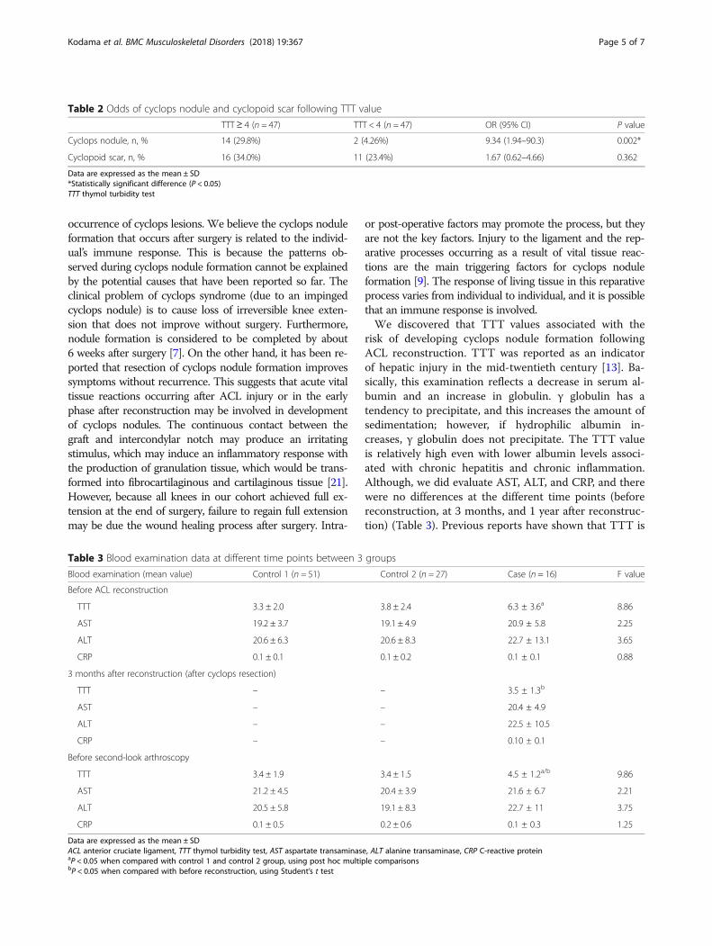
influence cyclopoid scar formation (OR, 1.67; 95% CI,0.62 to 4.66; p = 0.362). However, patients with cyclopsnodule formation showed significantly higher TTTvalues than the control patients. (OR, 9.34; 95% CI, 1.94to 90.3; p = 0.002) (Table 2).Cyclops nodule formation was found in 16 of the 94 pa-
tients (14.9%) and cyclopoid lesions were found in 27 pa-tients (28.7%) during second-look arthroscopy. Bloodexamination data before ACL reconstruction showed thatthe cyclops group (case) had a significant highly TTTvalue compared to the no-cyclops (control 1) and cyclo-poid group (control 2) (6.3 ± 3.6, 3.3 ± 2.0, and 3.8 ± 2.4,respectively; p < 0.05) (Table 3). There was no differencein TTT values in the no-cyclops group (control 1) and thecyclopoid group (control 2). When comparing the differ-ent time point blood examinations, TTT values were sig-nificantly lower after resection of the cyclops lesion(3 months after reconstruction) and before second-lookarthroscopy compared to before reconstruction. After cyc-lops nodule resection, TTT values increased slightly untilbefore second-look arthroscopy, but did not return topre-reconstruction TTT values.Range of motion was compared in the 3 groups
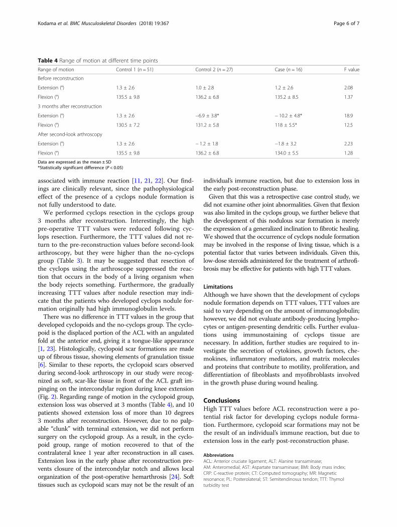
(Table 4). There was no significant difference in the 3groups before reconstruction. Extension loss was observedin the cyclopoid (control 2) and cyclops groups (case)3 months after reconstruction. In addition, knee flexionwas also restricted in the cyclops group (case) comparedto that in the no-cyclops group (control 1). In the cyclopsgroup (case), arthroscopic resection of the cyclops nodulewas performed 3 months after reconstruction. Eventually,almost full range of motion was restored in all patientsand there were no dissatisfied patients.
DiscussionThe most important finding of this study was that high TTTvalues before ACL reconstruction may be a potential riskfactor for developing cyclops nodule formation. Many re-searchers have reported that the cause of cyclops syndrome(due to an impinged cyclops nodule) is multi-factorial, andtherefore, the pathological condition is not completelyunderstood [1, 7, 8]. In fact, although surgical procedurescorresponding to the causative disease condition have beenreported [19, 20], surgical procedures still fail to prevent the
Table 1 Preoperative patient characteristics
TTT≥ 4 (n = 47) TTT < 4 (n = 47) P value
Mean age, years 24.0 ± 7.4 24.0 ± 6.4 0.989
Gender (Male/Female) 26/21 26/21 1.000
BMI, kg/m2 23.7 ± 3.3 23.2 ± 3.0 0.488
Meniscal injury, n, % 33 (70.2%) 25 (53.2%) 0.137
Data are expressed as the mean ± SDTTT thymol turbidity test, BMI body mass index
Kodama et al. BMC Musculoskeletal Disorders (2018) 19:367 Page 4 of 7
occurrence of cyclops lesions. We believe the cyclops noduleformation that occurs after surgery is related to the individ-ual’s immune response. This is because the patterns ob-served during cyclops nodule formation cannot be explainedby the potential causes that have been reported so far. Theclinical problem of cyclops syndrome (due to an impingedcyclops nodule) is to cause loss of irreversible knee exten-sion that does not improve without surgery. Furthermore,nodule formation is considered to be completed by about6 weeks after surgery [7]. On the other hand, it has been re-ported that resection of cyclops nodule formation improvessymptoms without recurrence. This suggests that acute vitaltissue reactions occurring after ACL injury or in the earlyphase after reconstruction may be involved in developmentof cyclops nodules. The continuous contact between thegraft and intercondylar notch may produce an irritatingstimulus, which may induce an inflammatory response withthe production of granulation tissue, which would be trans-formed into fibrocartilaginous and cartilaginous tissue [21].However, because all knees in our cohort achieved full ex-tension at the end of surgery, failure to regain full extensionmay be due the wound healing process after surgery. Intra-
or post-operative factors may promote the process, but theyare not the key factors. Injury to the ligament and the rep-arative processes occurring as a result of vital tissue reac-tions are the main triggering factors for cyclops noduleformation [9]. The response of living tissue in this reparativeprocess varies from individual to individual, and it is possiblethat an immune response is involved.We discovered that TTT values associated with the
risk of developing cyclops nodule formation followingACL reconstruction. TTT was reported as an indicatorof hepatic injury in the mid-twentieth century [13]. Ba-sically, this examination reflects a decrease in serum al-bumin and an increase in globulin. γ globulin has atendency to precipitate, and this increases the amount ofsedimentation; however, if hydrophilic albumin in-creases, γ globulin does not precipitate. The TTT valueis relatively high even with lower albumin levels associ-ated with chronic hepatitis and chronic inflammation.Although, we did evaluate AST, ALT, and CRP, and therewere no differences at the different time points (beforereconstruction, at 3 months, and 1 year after reconstruc-tion) (Table 3). Previous reports have shown that TTT is
Table 2 Odds of cyclops nodule and cyclopoid scar following TTT value
TTT≥ 4 (n = 47) TTT < 4 (n = 47) OR (95% CI) P value
Cyclops nodule, n, % 14 (29.8%) 2 (4.26%) 9.34 (1.94–90.3) 0.002*
Cyclopoid scar, n, % 16 (34.0%) 11 (23.4%) 1.67 (0.62–4.66) 0.362
Data are expressed as the mean ± SD*Statistically significant difference (P < 0.05)TTT thymol turbidity test
Table 3 Blood examination data at different time points between 3 groups
Blood examination (mean value) Control 1 (n = 51) Control 2 (n = 27) Case (n = 16) F value
Before ACL reconstruction
TTT 3.3 ± 2.0 3.8 ± 2.4 6.3 ± 3.6a 8.86
AST 19.2 ± 3.7 19.1 ± 4.9 20.9 ± 5.8 2.25
ALT 20.6 ± 6.3 20.6 ± 8.3 22.7 ± 13.1 3.65
CRP 0.1 ± 0.1 0.1 ± 0.2 0.1 ± 0.1 0.88
3 months after reconstruction (after cyclops resection)
TTT – – 3.5 ± 1.3b
AST – – 20.4 ± 4.9
ALT – – 22.5 ± 10.5
CRP – – 0.10 ± 0.1
Before second-look arthroscopy
TTT 3.4 ± 1.9 3.4 ± 1.5 4.5 ± 1.2a/b 9.86
AST 21.2 ± 4.5 20.4 ± 3.9 21.6 ± 6.7 2.21
ALT 20.5 ± 5.8 19.1 ± 8.3 22.7 ± 11 3.75
CRP 0.1 ± 0.5 0.2 ± 0.6 0.1 ± 0.3 1.25
Data are expressed as the mean ± SDACL anterior cruciate ligament, TTT thymol turbidity test, AST aspartate transaminase, ALT alanine transaminase, CRP C-reactive proteinaP < 0.05 when compared with control 1 and control 2 group, using post hoc multiple comparisonsbP < 0.05 when compared with before reconstruction, using Student’s t test
Kodama et al. BMC Musculoskeletal Disorders (2018) 19:367 Page 5 of 7
associated with immune reaction [11, 21, 22]. Our find-ings are clinically relevant, since the pathophysiologicaleffect of the presence of a cyclops nodule formation isnot fully understood to date.We performed cyclops resection in the cyclops group
3 months after reconstruction. Interestingly, the highpre-operative TTT values were reduced following cyc-lops resection. Furthermore, the TTT values did not re-turn to the pre-reconstruction values before second-lookarthroscopy, but they were higher than the no-cyclopsgroup (Table 3). It may be suggested that resection ofthe cyclops using the arthroscope suppressed the reac-tion that occurs in the body of a living organism whenthe body rejects something. Furthermore, the graduallyincreasing TTT values after nodule resection may indi-cate that the patients who developed cyclops nodule for-mation originally had high immunoglobulin levels.There was no difference in TTT values in the group that
developed cyclopoids and the no-cyclops group. The cyclo-poid is the displaced portion of the ACL with an angulatedfold at the anterior end, giving it a tongue-like appearance[1, 23]. Histologically, cyclopoid scar formations are madeup of fibrous tissue, showing elements of granulation tissue[6]. Similar to these reports, the cyclopoid scars observedduring second-look arthroscopy in our study were recog-nized as soft, scar-like tissue in front of the ACL graft im-pinging on the intercondylar region during knee extension(Fig. 2). Regarding range of motion in the cyclopoid group,extension loss was observed at 3 months (Table 4), and 10patients showed extension loss of more than 10 degrees3 months after reconstruction. However, due to no palp-able “clunk” with terminal extension, we did not performsurgery on the cyclopoid group. As a result, in the cyclo-poid group, range of motion recovered to that of thecontralateral knee 1 year after reconstruction in all cases.Extension loss in the early phase after reconstruction pre-vents closure of the intercondylar notch and allows localorganization of the post-operative hemarthrosis [24]. Softtissues such as cyclopoid scars may not be the result of an
individual’s immune reaction, but due to extension loss inthe early post-reconstruction phase.Given that this was a retrospective case control study, we
did not examine other joint abnormalities. Given that flexionwas also limited in the cyclops group, we further believe thatthe development of this nodulous scar formation is merelythe expression of a generalized inclination to fibrotic healing.We showed that the occurrence of cyclops nodule formationmay be involved in the response of living tissue, which is apotential factor that varies between individuals. Given this,low-dose steroids administered for the treatment of arthrofi-brosis may be effective for patients with highTTT values.
LimitationsAlthough we have shown that the development of cyclopsnodule formation depends on TTT values, TTT values aresaid to vary depending on the amount of immunoglobulin;however, we did not evaluate antibody-producing lympho-cytes or antigen-presenting dendritic cells. Further evalua-tions using immunostaining of cyclops tissue arenecessary. In addition, further studies are required to in-vestigate the secretion of cytokines, growth factors, che-mokines, inflammatory mediators, and matrix moleculesand proteins that contribute to motility, proliferation, anddifferentiation of fibroblasts and myofibroblasts involvedin the growth phase during wound healing.
ConclusionsHigh TTT values before ACL reconstruction were a po-tential risk factor for developing cyclops nodule forma-tion. Furthermore, cyclopoid scar formations may not bethe result of an individual’s immune reaction, but due toextension loss in the early post-reconstruction phase.
AbbreviationsACL: Anterior cruciate ligament; ALT: Alanine transaminase;AM: Anteromedial; AST: Aspartate transaminase; BMI: Body mass index;CRP: C-reactive protein; CT: Computed tomography; MR: Magneticresonance; PL: Posterolateral; ST: Semitendinosus tendon; TTT: Thymolturbidity test
Table 4 Range of motion at different time points
Range of motion Control 1 (n = 51) Control 2 (n = 27) Case (n = 16) F value
Before reconstruction
Extension (°) 1.3 ± 2.6 1.0 ± 2.8 1.2 ± 2.6 2.08
Flexion (°) 135.5 ± 9.8 136.2 ± 6.8 135.2 ± 8.5 1.37
3 months after reconstruction
Extension (°) 1.3 ± 2.6 −6.9 ± 3.8* − 10.2 ± 4.8* 18.9
Flexion (°) 130.5 ± 7.2 131.2 ± 5.8 118 ± 5.5* 12.5
After second-look arthroscopy
Extension (°) 1.3 ± 2.6 − 1.2 ± 1.8 −1.8 ± 3.2 2.23
Flexion (°) 135.5 ± 9.8 136.2 ± 6.8 134.0 ± 5.5 1.28
Data are expressed as the mean ± SD*Statistically significant difference (P < 0.05)
Kodama et al. BMC Musculoskeletal Disorders (2018) 19:367 Page 6 of 7

AcknowledgementsWe would like to thank Editage (http://www.editage.jp) for English languageediting.
Availability of data and materialsThe datasets used and/or analyzed during the current study are availablefrom the corresponding author on reasonable request.
Authors’ contributionsYKo designed the study and wrote the manuscript. TF performed surgeryand designed the study. TH and YKa performed the radiological measurements.YoO and SM performed the arthroscopic evaluations. YuO performed thestatistical analyses. TO organized the laboratory works. All authors have readand approved the final version of the manuscript submitted.
Ethics approval and consent to participateThis retrospective study was performed with the approval of the institutionalreview board (Department of Orthopaedic Surgery, Okayama UniversityGraduate School of Medicine, Dentistry, and Pharmaceutical Sciences. Projectnumber 941), and all patients signed the consent form drafted for the study.
Consent for publicationNot applicable.
Competing interestsThe authors declare that they have no competing interests.
Publisher’s NoteSpringer Nature remains neutral with regard to jurisdictional claims inpublished maps and institutional affiliations.
Received: 9 June 2018 Accepted: 27 September 2018
References1. Jackson DW, Schaefer RK. Cyclops syndrome: loss of extension following
intra-articular anterior cruciate ligament reconstruction. Arthroscopy. 1990;6:171–8.
2. Ahn JH, Yoo JC, Yang HS, Kim JH, Wang JH. Second-look arthroscopicfindings of 208 patients after ACL reconstruction. Knee Surg SportsTraumatol Arthrosc. 2007;15:242–8.
3. Sonnery-Cottet B, Lavoie F, Ogassawara R, Kasmaoui H, Scussiato RG, KidderJF, et al. Clinical and operative characteristics of cyclops syndrome afterdouble-bundle anterior cruciate ligament reconstruction. Arthroscopy. 2010;26:1483–8.
4. Dandy DJ, Edwards DJ. Problems in regaining full extension of the kneeafter anterior cruciate ligament reconstruction: does arthrofibrosis exist?Knee Surg Sports Traumatol Arthrosc. 1994;2:76–9.
5. Delcogliano A, Franzese S, Branca A, Magi M, Fabbriciani C. Light and scanelectron microscopic analysis of Cyclops syndrome: etiopathogenichypothesis and technical solutions. Knee Surg Sports Traumatol Arthrosc.1996;4:194–9.
6. Muellner T, Kdolsky R, Groschmidt K, Schabus R, Kwasny O, Plenk H Jr.Cyclops and cyclopoid formation after anterior cruciate ligamentreconstruction: clinical and histomorphological differences. Knee SurgSports Traumatol Arthrosc. 1999;7:284–9.
7. Guerra-Pinto F, Thaunat M, Daggett M, Kajetanek C, Marques T, Guimares T,et al. Hamstring contracture after ACL reconstruction is associated with anincreased risk of Cyclops syndrome. Orthop J Sport Med. 2017;5:232596711668412.
8. Fujii M, Furumatsu T, Miyazawa S, Okada Y, Tanaka T, Ozaki T, et al.Intercondylar notch size influences cyclops formation after anterior cruciateligament reconstruction. Knee Surg Sports Traumatol Arthrosc. 2015;23:1092–9.
9. Tonin M, Saciri V, Veselko M, Rotter A. Progressive loss of knee extensionafter injury. Cyclops syndrome due to a lesion of anterior cruciate ligament.Am J Sports Med. 2001;29:545–9.
10. Veselko M, Rotter A, Tonin M. Cyclops syndrome occurring after partialrupture of the anterior cruciate ligament not treated by surgicalreconstruction. Arthroscopy. 2000;16:328–31.
11. Ohwada H, Nakayama T, Tomono Y, Yamanaka K. Predictors, includingblood, urine, anthropometry, and nutritional indices, of all-cause mortality
among institutionalized individuals with intellectual disability. Res DevDisabil. 2013 Jan;34(1):650–5.
12. Ohwada H, Nakayama T, Nara N, Tomono Y, Yamanaka K. Anepidemiological study on anemia among institutionalized people withintellectual and/ or motor disability with special reference to its frequency,severity and predictors. BMC Public Health. 2006;6:85.
13. Kunkel HG, Hoagland CL. Mechanism and significance of the thymolturbidity test for liver disease. J Clin Investigation. 1947;26:1060–71.
14. Lubowitz JH, Konicek J. Anterior cruciate ligament femoral tunnel length:cadaveric analysis comparing anteromedial portal versus outside-intechnique. Arthroscopy. 2010;26:1357–62.
15. Fujii M, Furumatsu T, Miyazawa S, Tanaka T, Inoue H, Kodama Y, et al.Features of human autologous hamstring graft elongation after pre-tensioning in anterior cruciate ligament reconstruction. Int Orthop. 2016;40:2553–8.
16. Shino K, Suzuki T, Iwahashi T, Mae T, Nakamura N, Nakata K, et al. Theresident’s ridge as an arthroscopic landmark for anatomical femoral tunneldrilling in ACL reconstruction. Knee Surg Sports Traumatol Arthrosc. 2010;18:1164–8.
17. Kodama Y, Furumatsu T, Miyazawa S, Fujii M, Tanaka T, Inoue H, et al.Location of the tibial tunnel aperture affects extrusion of the lateralmeniscus following reconstruction of the anterior cruciate ligament. JOrthop Res. 2017;35:1625–33.
18. Furumatsu T, Ozaki T. Iatrogenic injury of the lateral meniscus anteriorinsertion following anterior cruciate ligament reconstruction: a case report. JOrthop Sci. 2018;23:197–201.
19. Delince P, Krallis P, Descamps PY, Fabeck L, Hardy D. Different aspects ofthe cyclops lesion following anterior cruciate ligament reconstruction: amultifactorial etiopathogenesis. Arthroscopy. 1998;14:869–76.
20. Imam MA, Abdelkafy A, Dinah F, Adhikari A. Does bone debris in anteriorcruciate ligament reconstruction really matter? A cohort study of a protocolfor bone debris debridement. SICOT J. 2015;1:4.
21. Van Der Sluis JJ, Menke HE. Role of IgG fractions with high isoelectric pointsin the thymol turbidity test in syphilis. Evidence for an increase in basic IgGin early syphilis. Br J Vener Dis. 1975;51(3):158–60.
22. Franklin EC. The role of the basic fraction of γ-globulin in the flocculationtests. Clin Chim Acta. 1959;4:259–64.
23. Plotkin BE, Agarwal VK, Varma R. Stump entrapment of the torn anteriorcruciate ligament. Radiol Case Rep. 2016;4:268.
24. Wang J, Ao Y. Analysis of different kinds of cyclops lesions with or withoutextension loss. Arthroscopy. 2009;25:626–31.
Kodama et al. BMC Musculoskeletal Disorders (2018) 19:367 Page 7 of 7