Management of Central Retinal Artery Occlusion A Statement ...
1761
Thrombotic Occlusion of theMiddle Cerebral Artery
Shinsuke Ueda, MD; Kazuhiko Fujitsu, MD; Shigeo Inomori, MD; and Takeo Kuwabara, MD
Background and Purpose: Epidemiological study of middle cerebral artery occlusion is importantbecause the indication for extracranial-intracranial arterial bypass remains in dispute. To help clarifythis issue, we investigated the prognosis of thrombotic middle cerebral artery occlusion in Japanesepatients.
Methods: We studied 40 patients with thrombotic middle cerebral artery occlusion who were selected onthe basis of clinical features, computed tomographic findings, and angiographic findings. Patients withcauses of embolism (i.e., cardiomyopathy, valvular heart disease, cardiac arrhythmia, and carotidulceration) were excluded. The 40 patients were classified into three groups according to the site of middlecerebral artery occlusion: there were 13 patients with occlusion of the proximal portion of the Ml segment,13 with distal Ml segment occlusion, and 14 with occlusion of the M2 segment.
Results: Good collateral circulation was associated with improved outcomes both clinically and bycomputed tomography in patients with occlusion of the proximal and distal portions of the Ml segmentbut not in those with M2 occlusion.
Conclusions: It is reasonable to assume that not only collateral circulation but also the site of occlusionplays an important role in the outcome of middle cerebral artery occlusion. Our finding that goodcollateral circulation improves the outcome for thrombotic occlusion of the proximal and distal Mlsegments supports the possible benefits of such surgery. (Stroke 1992;23:1761-1766)
KEY WORDS • arterial occlusive diseases • cerebral infarction • collateral circulation •thrombosis
Epidemiological study of middle cerebral artery(MCA) occlusion has become increasingly im-portant because review of the literature suggests
that the indication for extracranial-intracranial arterialbypass surgery remains in dispute.1-3 Strict differentia-tion of thrombosis from embolus is especially importantbecause the clinical course, neuroradiological findings,treatment, and prognosis may be different.4 Thrombosisis usually thought to have a less-sudden onset, to be lesssevere clinically, and to have a computed tomographic(CT) image of an irregular low density area (LDA)without midline shift.5 Embolic cerebral infarction asreported by the Harvard Cooperative Stroke Registry,6
Blackwood et al,7 and Jorgensen and Torvik8 had anincidence of 37%, 48%, and 47%, respectively. Althoughmany authors9"13 have emphasized atheroscleroticthrombosis as the most common cause of cerebral infarc-tion, accounting for 80-95%, some1415 have foundthrombotic etiology of MCA occlusion to be less fre-quent. In the Japanese population, MCA occlusion is 1.8times more common than internal carotid artery (ICA)occlusion.16 In the American Joint Study of ExtracranialArterial Occlusion,17 MCA occlusion had one fourth theincidence of ICA occlusion. The reason for the etiologi-
From the Department of Neurosurgery, Yokohama City Uni-versity, School of Medicine, Yokohama, Japan.
Address for correspondence: Shinsuke Ueda, MD, Departmentof Neurosurgery, Yokohama Red Cross Hospital, 2-85, Negishi-cho, Naka-ku, Yokohama 231, Japan.
Received October 29, 1990; final revision received July 6, 1992;accepted July 28, 1992.
cal dominance of embolus or thrombosis is controversial,but among the Japanese population thrombotic MCAocclusion appears to have a higher incidence.
In this study we investigated the prognosis of throm-botic MCA occlusion in Japanese patients in relation toclinical features, CT findings, and angiographic find-ings. To assess collateral circulation, we determined thepresence or absence of retrograde filling, that is, thepresence or absence of contrast media in MCAbranches through leptomeningeal anastomosis on angi-ography (within 6 seconds after injection).
Subjects and MethodsWe studied 40 patients with MCA occlusion (mean
age, 66.4 years [range, 42-84 years]; 27 men, 13 women)retrospectively. With diagnosis made by CT, angio-graphic, and clinical findings, patients who had atrialfibrillation on electrocardiogram or angiographic find-ings of atherosclerotic ICA ulceration regardless of thepresence of ICA stenosis were excluded from this study.All patients were admitted within 48 hours of the ictus,and the initial CT scans were undertaken within 36hours of admission. Angiography was performed within4 days of admission.
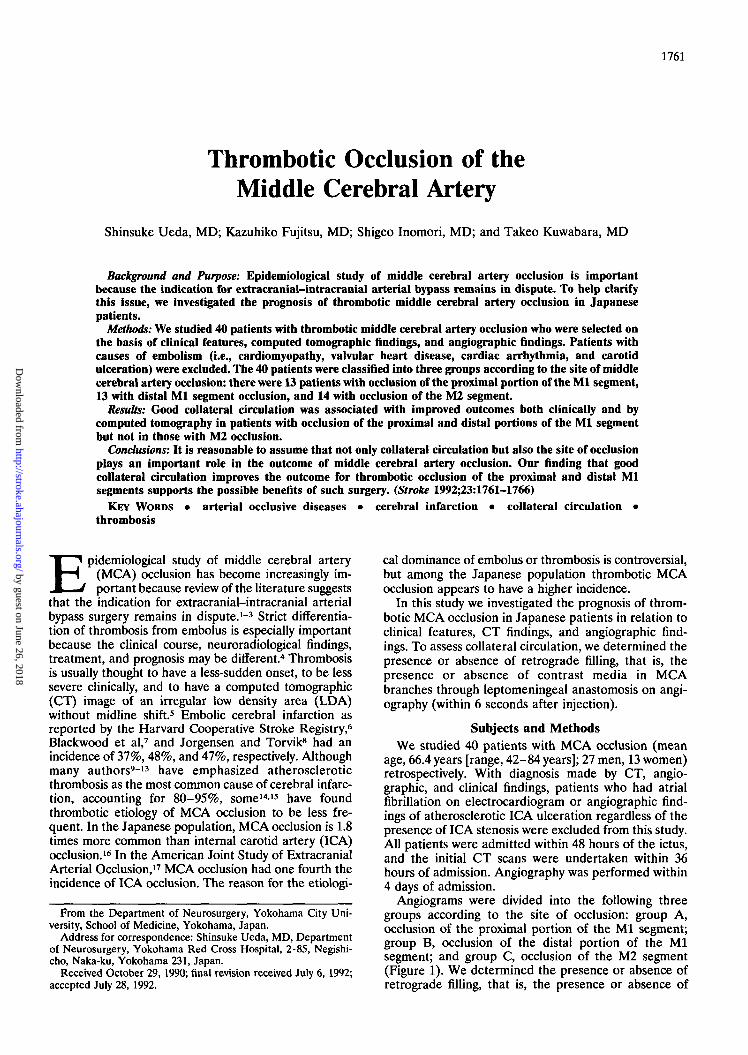
Angiograms were divided into the following threegroups according to the site of occlusion: group A,occlusion of the proximal portion of the Ml segment;group B, occlusion of the distal portion of the Mlsegment; and group C, occlusion of the M2 segment(Figure 1). We determined the presence or absence ofretrograde filling, that is, the presence or absence of
by guest on June 26, 2018http://stroke.ahajournals.org/
Dow
nloaded from
1762 Stroke Vol 23, No 12 December 1992
Proximal Ml Distal Ml M2occlusion occlusion occlusion
FIGURE 1. Angiographic findings of middle cerebral artery occlusion were classified into three groups. Left panel: Occlusion ofthe proximal portion of the Ml segment (group A). Center panel: Occlusion of the distal portion of the Ml segment (group B).Right panel: Occlusion of the M2 segment (group C).
contrast media in MCA branches through leptomenin-geal anastomoses with serial angiography. The presenceor absence of angiographic retrograde filling was alsoanalyzed.
CT scans were classified into the following five cate-gories: basal ganglia-centrum semiovale type, in whichan LDA was localized in the basal ganglia and/orcentrum semiovale; localized cortex-subcortical type, inwhich an LDA was localized in a small area of thecortex and subcortex of the frontal, temporal, or pari-etal lobe; lobular cortex-subcortical type, in which anLDA involved the cortex and subcortex of the frontal,temporal, and parietal lobes; hemispheric type, in whichan LDA extended to one hemisphere including thebasal ganglia; and normal type, in which an LDA wasnot found on CT (Figure 2). For the CT classificationdescribed above, CT scans that showed the maximumextent of LDA during repeated studies were selected.
Degree of hemiparesis on admission was graded asmild, moderate, or severe using DeJong's definition,18 inwhich mild was defined as normal or movement againstgravity and resistance, moderate was movement againstgravity with resistance eliminated, and severe was partialmovement with gravity eliminated, with a trace of musclecontraction. Outcomes were also graded: good outcome,full work and minimal disability, fair outcome, partialdisability; and poor outcome, bed rest, vegetative state, ordeath. The follow-up period was from 43 days to 4 months(with the exception of four patients who died of compli-cations). Correlations were made between the angio-graphic site of occlusion, retrograde filling, CT findings,degree of hemiparesis on admission, and outcome.
ResultsOcclusion of the MCA occurred on the left side in 24
patients and on the right in 16. Eleven of the 40 patientshad hypertension and had been treated by antihyper-tensive medications. The prevalence of diabetes melli-tus was 8% in this study. Mortality was 10%.
At the onset of stroke, 22 patients (55%) had a milddisturbance of consciousness, but no patients werecomatose. Mild hemiparesis was found in 14 patients,moderate hemiparesis in nine, and severe hemiparesisin 17. Two patients (5%) had a transient ischemic attack(TLA), and the remaining 38 patients had a completestroke. Four patients (10%) had had previous ischemicevents in the symptomatic side. Two of these fourpatients had TLA, and the remaining two had completestroke.
In 13 patients the MCA was occluded at the proximalportion of the Ml segment. Another 13 patients showedthe MCA occluded at the distal portion of the Mlsegment, and the remaining 14 had occlusion of the M2segment. Two patients had mild ipsilateral ICA stenosisof less than 50%.
The degree of hemiparesis of each patient on admis-sion is indicated in Figure 3, in relation to the site ofocclusion, retrograde filling, and CT findings. Figure 4shows outcomes of the patients in relation to the site ofocclusion, retrograde fining, and CT findings. All 13patients with occlusion of the proximal Ml (group A)showed basal ganglia-centrum semiovale or hemi-spheric type findings on CT. Of seven patients withbasal ganglia-centrum semiovale type findings on CT,six patients (86%) showed retrograde filling from theanterior cerebral artery (ACA), whereas of six patients
by guest on June 26, 2018http://stroke.ahajournals.org/
Dow
nloaded from

Ueda et al Thrombotic MCA Occlusion 1763
Basal ganglia - Localized cortex- Lobular cortex- Hemispheric
centrum semiovale subcortical subcortical type
type type t y p e
FIGURE 2. Computed tomographic findings were classified into five categories (see text): basal ganglia-centrum semiovale type;localized cortex-subcortical type; lobular cortex-subcortical type; hemispheric type; and normal type (not shown).
with hemispheric type findings on CT only two (33%)showed retrograde filling. In the patients with occlusionof the distal Ml (group B), findings on CT were basalganglia—centrum semiovale in four, localized cortex-subcortical in four, lobular cortex-subcortical in three,
hemispheric in one, and normal in one patient. Thegroup B patients with basal ganglia-centrum semiovaletype findings on CT showed an LDA in the centrumsemiovale but not in the basal ganglia. In the patientswith occlusion of the M2 (group C), findings on CT
N^lta of occluiion
Tjrpa of N ^CT N.
Haul (anil ia -cantruai laalovala
Localizad cortai -subeortieal ^T^
'*** 1 Y 1
Lobalar cortax —•ubeortical / T T .
""* [ Y l
Hwlapharlctypa X^*^
ftoraaltrp*
ProxiM(Sroui
i nA)
Ratrotrada f l l l lnf
Praaant
x x AAA
O
X O
Abaant
O
X X X X
Distal n(Brow B)
Hatrofrada f i l l in *Pratant
x A O
X
X
O
Abaant
A
X
X X
H2(Crow> C)
btrotrada ( I l l l t *Praaant
x x x A O
OOO
Mntnt
AAOOO
O
FIGURE 3. Chart showing degree of hemi-paresis on admission in relation to site ofocclusion, computed tomographic (CT) find-ings, and retrograde filling. O, Mild hemipare-sis; A, moderate hemiparesis; x, severehemiparesis.
by guest on June 26, 2018http://stroke.ahajournals.org/
Dow
nloaded from

1764 Stroke Vol 23, No 12 December 1992
\ X l t a of occlusion
Typa of >v
Baul fanil la -cantrua saaiovala
LocalIzad cortai -tubcortlcal >rTrx
Lobular cortax -•ubcortlcal /^^
tyoa / y%
Haalspharic
•orsaltirpa
Proxlaal K(Group A)
•atrotrada fllllntPraaant
• • • C O
O
* *
Abaant
O
• • • •
Distal HI(Group B)
btrotradt f l l l l mPraaant
•CO
• O O
c
*
o
Abaant
*
*
* *
K2(trout
RatrofradiPraaant
• • •cc
ooo
C)
ft II insAbaant
eoooo
O
FIGURE 4. Chart showing outcome in rela-tion to site of occlusion, computed tomo-graphic (CT) findings, and retrograde filling.O, Good outcome; €>, fair outcome; • , pooroutcome.
were localized cortex-subcortical type in 10 and normaltype in four patients (Figures 3 and 4).
Most patients with severe hemiparesis showed lobularcortex-subcortical and hemispheric type findings onCT. Ah1 patients with normal type findings on CTshowed mild hemiparesis, and most patients with basalganglia-centrum semiovale or localized cortex-subcor-tical type findings on CT had moderate hemiparesis(Figure 3). The degree of hemiparesis on admission waswell correlated to the CT findings.
The outcome was rated good in 12 (30%), fair in eight(20%), and poor, including death, in 20 (50%) patients.Four patients (10%) died of complications at the acutestage of the disease. Outcomes of patients in group Aand group B were poor. However, the outcome wasbetter in patients in group A and group B with retro-grade filling than in those without retrograde filling.Outcomes of patients in group C were generally goodirrespective of the presence or absence of retrogradefilling (Figure 4). Patients with occlusion of the distalMCA branches to the motor cortex, however, showedpoor outcome.
DiscussionMost of the published studies19-24 on MCA occlusion
have not excluded embolic occlusion. The differentialdiagnosis between thrombotic and embolic MCA occlu-sion may be difficult. To improve the reliability of theselection of thrombotic MCA occlusion, patients whohad atrial fibrillation or carotid artery ulceration wereexcluded based on clinical features, CT findings, andangiographic findings.5 It has been reported frequentlythat embolic MCA occlusion is more common thanthrombotic MCA occlusion14-23-26; Lhermitte et al15
found that only 12.5% of 40 MCA occlusions were dueto local thrombosis. However, in the Japanese popula-
tion thrombotic MCA occlusion is reported to be morecommon.13
In this study, the mean age of 66.4 was higher thanthat in other reports.15-21-22-27 The mortality rate of thisseries was essentially the same as that in the study ofBogousslavsky et al.27 Therefore, age did not have asignificant influence on death.27 Male predominancewas noted in this series, as in other reports.27-28 All-cock19 reported no laterality of the side of the lesion,although others have reported left side predominance.21
In this series we reported no laterality of the lesion. Inour patients, 18 of 40 (45%) had hypertension, andthree (8%) had diabetes mellitus resulting in hyperten-sion. Hypertension is an important risk factor in throm-botic MCA occlusion. This finding agrees with otherreports.27-29
TLA has been defined as a temporary, focal neuro-logical deficit presumably related to ischemia, lastingless than 24 hours. Only two patients presented withTLA; the remaining 38 patients presented with completestroke. Caplan et al30 and Sindermann et al24 reportedthat TLA, as an initial symptom, was more frequentlyobserved in ICA occlusion than in MCA occlusion.Thrombotic MCA occlusion has a more benign effectthan embolic MCA occlusion,4-20-31-32 perhaps becausethe development of collateral circulation is better ingradual thrombotic occlusion than in sudden embolicocclusion. Therefore, although disturbance of con-sciousness was found in 21 patients (53%), severedisturbance of consciousness as occurs in the case ofembolic occlusion was not observed.
In group A, most patients with good retrograde fillingshowed basal ganglia-centrum semiovale type findingson CT, whereas four of five patients without retrogradefilling showed hemispheric type findings on CT (Figures3 and 4). We offer two possible explanations for the factthat basal ganglia-centrum semiovale type findings oc-
by guest on June 26, 2018http://stroke.ahajournals.org/
Dow
nloaded from
Veda et al Thrombotic MCA Occlusion 1765
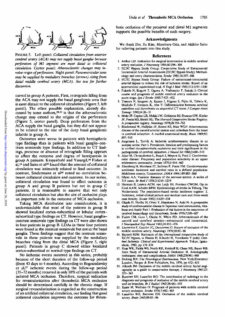
FIGURE 5. Left panel: Collateral circulation from anteriorcerebral artery (ACA) may not supply basal ganglia becauseperforators of Ml segment are most distal to collateralcirculation. Center panel: Atherosclerotic changes may in-volve origin of perforators. Right panel: Paraventricular zonemay be supplied by medullary branches (arrows) rising fromdistal middle cerebral artery (MCA). See text for furtherdiscussion.
curred in group A patients. First, retrograde filling fromthe ACA may not supply the basal ganglionic area thatis most distant to the collateral circulation (Figure 5, leftpanel). The other possible explanation, already dis-cussed by some authors,29-33 is that the atheroscleroticchange may extend to the origins of the perforators(Figure 5, center panel). Deep perforators from theACA supply the basal ganglia, but they did not appearto be related to the size of the deep basal ganglionicinfarcts in group A.
Outcomes were worse in patients with hemispherictype findings than in patients with basal ganglia-cen-trum semiovale type findings. In addition to CT find-ings, presence or absence of retrograde filling appearsto affect the outcome and degree of hemiparesis ingroup A patients. Krayenbuhl and Yasargil,34 Fisher etal,14 and Saito et al29 found that the amount of collateralcirculation was intimately related to final outcome. Incontrast, Sindermann et al24 noted no correlation be-tween collateral circulation and outcome. In our series,collateral circulation was found to affect outcome ingroup A and group B patients but not in group Cpatients. It is reasonable to assume that not onlycollateral circulation but also the site of occlusion playsan important role in the outcome of MCA occlusion.
Taking MCA distribution into consideration, it isunderstandable that most of the patients in group Bshowed localized cortex-subcortical or lobular cortex-subcortical type findings on CT. However, basal ganglia-centrum semiovale type findings on CT were also shownin four patients in group B. LDAs in these four patientswere found in the centrum semiovale but not in the basalganglia. These findings suggest that the centrum semio-vale in these patients was supplied by the medullarybranches rising from the distal MCA (Figure 5, rightpanel). Patients in group C showed either localizedcortex-subcortical or normal type findings on CT.
No ischemic events recurred in this series, probablybecause of the short duration of the follow-up period(from 43 days to 4 months). According to Bogousslavskyet al,27 ischemic events during the follow-up period(35-72 months) recurred in only 10% of the patients withisolated MCA occlusion. Therefore, surgical indicationfor revascularization after thrombotic MCA occlusionshould be determined carefully in the chronic stage. Ifsurgical revascularization is regarded as the constructionof an artificial collateral circulation, our finding that goodcollateral circulation improves the outcome for throm-
botic occlusion of the proximal and distal Ml segmentssupports the possible benefits of such surgery.
AcknowledgmentsWe thank Drs. Ilu Kim, Masaharu Oda, and Akihito Saito
for referring patients into this study.
References1. Arthur LD: Indication for surgical intervention in middle cerebral
artery ocdusion. / Neuwsurg 1984;60:296-3042. EC/IC Bypass Study Group: Cooperative Study of Extracranial/
Intracranial Arterial Anastomosis (EC/IC Bypass Study): Method-ology and entry characteristics. Stroke 1985;16:397-4O6
3. EC/IC Bypass Study Group: Failure of extracranial-intracranialarterial bypass to reduce the risk of ischemic stroke: Result of aninternational randomized trial. N Engl J Med 1985;313:1191-1200
4. Fukada N, Kogure T, Ogawa A, Yoshimoto T, Suzuki J: Clinicalcourse and prognosis of middle cerebral artery occlusion in theacute stage. Jpn J Stroke 1985;7:425-432
5. Tomura N, Inugami A, Kanno I, Higano S, Fijita H, Tabata K,Shishido F, Uemura K, Abe T: Differentiation between cerebralembolism and thrombosis on sequential CT scans. J Comput AssistTomogr 1990;14:26-31
6. Mohr JP, Caplan LR, Melski JW, Goldstein RJ, Duncan GW, KistlerJP, Pessin MS, Bleich HL: The Harvard Cooperative Stroke Registry:A prospective registry. Neurology 1978^8:754-762
7. Blackwood W, Hallpike JF, Kocen RS, Mair WGP: Atheromatousdisease of the carotid arterial system and embolism from the heartin cerebral infarction: A morbid anatomical study. Brain 1969;92;897-910
8. Jorgensen L, Torvik A: Ischemic cerebrovascular disease in anautopsy series: Part 1. Prevalence, location and predisposing factorsin verified thromboembolic occlusions and their significance in thepathogenesis of cerebral infarction. J Neural Set 1966^:490-510
9. Alter M, Christoferson L, Resch J, Meyers G, Ford J: Cerebrovas-cular disease: Frequency and population selectivity in an uppermidwestem community. Stroke 1970;l:454-465
10. Eisenberg H, Morrison JT, Sullivan P, Foote FM: Cerebrovascularaccidents: Incidence and survival rates in a defined population:Middlesex county, Connecticut. JAMA 1964;189:883-888
11. Gh/nn AA: Vascular diseases of the nervous system: A series of315 cases. Br Med J 1956;2:1216-1219
12. Harman B, Leyten ACM, van Luijk JH, Frenken CWGM, Op deCoul AAW, Schulte BPM: Epidemiology of stroke in Tilburg, TheNetherlands: The population-based stroke incidence register: 2.Incidence, initial clinical picture and medical care and three-weekcase fatality. Stroke 1982;13:629-634
13. Okada H, Horibe H, Ohno Y, Hayakawa N, Aoki N: A prospectivestudy of cerebrovascular disease in Japanese rural communities, Aka-bane and Asahi: Part 1. Evaluation of risk factors in the occurrence ofcerebral hemorrhage and thrombosis. Stroke 1976;7:599-6O7
14. Fisher CM, Gore I, Okabe N, White PD: Atherosclerosis of thecarotid and vertebral arteries —extracranial and intracranial./ Neuwpathol Exp Neural 1965;24:455-476
15. Lhermitte F, Gautier JC, Derouesne C: Nature of occlusion of themiddle cerebral artery. Neurology 1970-^0:82-88
16. Barnett HJM: Rationale of the international cooperative study ofEC/IC bypass, in Handa H, Kikuchi H, Yonekawa Y (eds): Cere-bral Ischemia: Clinical and Experimental Approach. Tokyo, Igaku-shoin, 1982, pp 131-134
17. Hass WK, Fields WS, North RR, Kricheff II, Chase NE, Bauer RB:Joint Study of Exlracranial Arterial Occlusion: II. Arteriographytechniques: sites and complications. JAMA 1968;230361-968
18. DeJong RN: The Neurological Examination. New York/Evanston/London, Harper & Row Publishers, Inc, 1976, pp 452-453
19. Allcock JM: Occlusion of the middle cerebral artery: Serial angi-ography as a guide to conservative therapy. J Neuwsurg 1967;27:353-363
20. Burrows EH, Lascelles RG: The contribution of radiology to thediagnosis and prognosis of occlusion of the middle cerebral arteryand its branches. Br J Radial 196538:481-593
21. Kaste M, Waltimo O: Prognosis of patients with middle cerebralartery occlusion. Stroke 1976;7:482-485
22. Lascelles RG, Burrows EH: Occlusion of the middle cerebralartery. Brain 1965;88:85-96
by guest on June 26, 2018http://stroke.ahajournals.org/
Dow
nloaded from

1766 Stroke Vol 23, No 12 December 1992
23. Moulin DE, Lo R, Chiang J, Bamett HTM: Prognosis in middlecerebral artery occlusion. Stroke 1985;16:282-284
24. Sindermann F, Dichgans J, Bergleiter R: Occlusion of the middlecerebral artery and its branches: Angiographic and clinical corre-lates. Brain 1969;92:607-620
25. Lhermitte F, Gautier JC, Derouesne C, Guiraud B: Ischemic acci-dents in the middle cerebral artery territory. Arch Natrol 1968; 19:248-256
26. Olsen TS: Regional cerebral blood flow after occlusion of themiddle cerebral artery. Acta Nairol Scand 1986;73:321-337
27. Bogousslavsky J, Barnett HJM, Fox AJ, Hachinski VC, Taylor W:Atherosclerotic disease of the middle cerebral artery. Stroke 1986;17:1112-1120
28. Rodda RA, Path FRC: The arterial patterns associated withinternal carotid disease and cerebral infarcts. Stroke 1986;17:69-75
29. Saito I, Segawa H, Shiokawa Y, Taniguchi M, Tsutsumi K: Middlecerebral artery: Correlation of computed tomography and angiog-raphy with clinical outcome. Stroke 1987;18:863-868
30. Caplan L, Babikan V, Helgason C, Hier DB, DeWitt D, Patel D,Stein R: Occlusive disease of the middle cerebral artery. Neurology1985^5:975-982
31. Choki J, Yamaguchi T, Hirata Y, Tashiro M, Sawada T: Clinicalfeatures of cerebral embolism: Analysis of 48 cases in the acutestage. Jpn J Stroke 1982;4:54-62
32. Torvik A, Jorgensen L: Thrombotic and embolic occlusion of thecarotid arteries in an autopsy series: Part 2. Cerebral lesions andclinical course. / Neurol Sd 1966;3:401-432
33. Iwamoto T, Katsumuna H, Araki G, Yunoki K: A study of cerebralinfarction of the basal ganglia due to main trunk obstruction. Jpn JStroke 1985;7:291-298
34. Krayenbuhl H, Yasargil G: Der cerebrate kollaterale blutkrcsilauf imangiographischen BUtLActa Neuroctur (Wien) 1958;6J0-80
by guest on June 26, 2018http://stroke.ahajournals.org/
Dow
nloaded from

S Ueda, K Fujitsu, S Inomori and T KuwabaraThrombotic occlusion of the middle cerebral artery.
Print ISSN: 0039-2499. Online ISSN: 1524-4628 Copyright © 1992 American Heart Association, Inc. All rights reserved.
is published by the American Heart Association, 7272 Greenville Avenue, Dallas, TX 75231Stroke doi: 10.1161/01.STR.23.12.1761
1992;23:1761-1766Stroke.
http://stroke.ahajournals.org/content/23/12/1761the World Wide Web at:
The online version of this article, along with updated information and services, is located on
http://stroke.ahajournals.org//subscriptions/
is online at: Stroke Information about subscribing to Subscriptions:
http://www.lww.com/reprints Information about reprints can be found online at: Reprints:
document.
Permissions and Rights Question and Answer information about this process is available in theis located, click Request Permissions in the middle column of the Web page under Services. FurtherEditorial Office. Once the online version of the published article for which permission is being requested
can be obtained via RightsLink, a service of the Copyright Clearance Center, not theStrokepublished in Requests for permissions to reproduce figures, tables, or portions of articles originallyPermissions:
by guest on June 26, 2018http://stroke.ahajournals.org/
Dow
nloaded from