Thromboelastography 4 Devices 4 5 Activation 8 6 Clinical ...
14
25 September 2009 Thromboelastography AZ Mazibuko Commentator: K Keerath Moderator: U Singh Department of Anaesthetics Page 2 of 27 CONTENTS 1 Introduction .................................................................................... 3 2 History............................................................................................. 3 3 Point of care monitoring ............................................................... 3 4 Devices ........................................................................................... 4 5 Activation........................................................................................ 8 6 Clinical uses ................................................................................. 10 7 Problems and limitations of TEG ............................................... 19 8 Conclusion ................................................................................... 20 9 Tests for various POC devices ................................................... 21 10.1 Sonoclot................................................................................... 22 10.2 PFA-100.................................................................................... 22 11 Diagnostic flow chart examples ............................................... 23 12 TEG Patterns .............................................................................. 24 13 References.................................................................................. 25
Transcript of Thromboelastography 4 Devices 4 5 Activation 8 6 Clinical ...
25 September 2009
Thromboelastography
AZ Mazibuko
Commentator: K Keerath Moderator: U Singh
Department of Anaesthetics
Page 2 of 27
CONTENTS 1 Introduction .................................................................................... 3 2 History ............................................................................................. 3 3 Point of care monitoring ............................................................... 3 4 Devices ........................................................................................... 4 5 Activation ........................................................................................ 8 6 Clinical uses ................................................................................. 10 7 Problems and limitations of TEG ............................................... 19 8 Conclusion ................................................................................... 20 9 Tests for various POC devices ................................................... 21 10.1 Sonoclot ................................................................................... 22 10.2 PFA-100 .................................................................................... 22 11 Diagnostic flow chart examples ............................................... 23 12 TEG Patterns .............................................................................. 24 13 References .................................................................................. 25

Page 3 of 27
1 Introduction Perioperative haemostatic monitoring is important to diagnose potential causes of haemorrhage, to guide rational choices of haemostatic therapies, to estimate the risk of bleeding during surgical procedures. Currently, laboratory-based tests are the mainstay in assessing coagulation status. These include typically, prothrombin time (PT), activated partial thromboplastin time (APTT), fibrinogen, international normalised ratio (INR), platelet count.
Problems with routine tests include turnaround time (TAT) but more importantly these are performed on plasma rather than whole blood, yielding no information on platelet function. These tests are performed at standard temperature of 37° rather than patient temperature, which is important under certain circumstances e.g. cardiac surgery.[9]
2 History Thromboelastography was first described in 1948 by Hartert of Heidelburg during World War II [8], as a method to assess global haemostatic function from a single blood sample; this was the original Hellige thromboelastography. Calatzis et al, 1996 have further elaborated on the principles using different activators and inhibitors to localise haemostatic disorders within a short space of time. [1] Rotational thromboelastography (ROTEG/ROTEM) is a refinement of the principle developed in Ludwig-Maximillian University of Munich. 2.1 Terminology Thromboelastography, TEG are used generically in literature. Thromboelastograph and TEG are registered trademarks of the Haemoscope Corporation (Niles, IL) and are used, since 1996 for assays performed by their instruments. ROTEM, TEG/ROTEM are trademarks registered to Pentapharm GmbH (Munich, Germany), refer to assays from their own instrumentation viz. rotation thromboelastometry. 3 Point of care monitoring Point of care (POC) monitoring overcomes several of the limitations. Various devices assessing viscoelastic properties of whole blood are available include thromboelastography (TEG®),rotation thromboelastometry (ROTEM®), Sonoclot® analysis. Benefits of these devices include :
• Bedside analysis, not necessarily central laboratory • faster turnaround times
Page 4 of 27
• coagulation status of whole blood is assessed : in vivo coagulation system interactions with platelets and red blood cells (RBC)
• useful information on platelet function • real-time clot development visually displayed • analysis at patient temperature
Several important limitations must be considered, however : in vivo and in vitro coagulation have important differences
• static, no flow conditions • cuvette is unlike an endothelialised blood vessel
4 Devices 4.1 TEG Assesses viscoelastic properties under low shear conditions. It uses a stationary cylindrical cup that holds the blood sample of 360mcL (0.36ml) and oscillates through an angle of 4°45’. Each rotation lasts 10s. A pin is suspended in the blood sample by a torsion wire and is monitored for motion. The torque of the rotation cu is transmitted to the immersed pin after fibrin-platelet bonding has occurred, linking the cup and pin together. The strength of the fibrin-platelet bonds affects the magnitude of the pin motion. The output is thus directly related to the strength of the formed clot. With clot retraction and lysis the fibrin-platelet bonds are broken and the transfer of motion diminishes. The rotation movement of the pin is converted by an electromechanical to an electrical signal which is displayed as a TEG tracing.
Figure 1
4.2 ROTEM ROTEM uses a modification of the TEG: the signal of the pin is transmitted using an optical detector system and not a torsion wire, the movement originates from the pin itself and not the cup. The ROTEM also has an electronic pipette which improves reproducibility and performance.

Page 5 of 27
A typical device has four independant measuring channels, each providing a thromboelastometry (TEM) trace which can be by a computer.
Figure 2 from [9]
Figure 3
4.2.1 INTEM INTEM is a baseline test using ellagic acid contact activator for analysing general coagulatory status of the patient. Clotting time (CT) and clot formation time (CFT) allow for the analysis of factor deficiencies or detection of the presence of anticoagulants.
Page 6 of 27
One limitation of the standard ROTEM analysis is the inability to measure platelet adhesion defects or aspirin-like defects during primary haemostasis. [8] 4.2.2 HEPTEM HEPTEM add heparinase to demonstrate heparin or a residual heparin effect if compared to INTEM test, or coagulation be tested in the presence of heparin (if present in the sample). 4.2.3 FIBTEM FIBTEM test fibrinogen concentration and polymerisation alone, in the absence of platelets. Cytochalasin D is used as a platelet inhibitor. In the example below patient B has a normal FIBTEM trace, in view of the abnormalities in the INTEM (maximum clot firmness, MCF, CT, CFT) platelets were necessary.(In [1])
Figure 4 from [1]

Page 7 of 27
4.3 Variations and modification 4.3.1 Heparinase coated cups Under conditions where heparin is administered systemically e.g. during CPB, vascular surgery, the effect of heparin can be neutralised by using heparinase coated cups. This will also neutralise heparin like substances. [2] 4.3.2 Celite activation Celite accelerates the activation process, allowing a meaningful result to be obtained quickly. This analysis has supplanted native blood in many situations and has been widely validates.
Table 1 from [11]
4.3.3 Activator F, ADP, arachidonic acid Activator F is a proprietary reagent consisting of Reptilase and FXIIIa. If Act F is added to a sample taken in heparinised tube (thrombin inhibited) a fibrin network is generated independent of thrombin. In the absence of platelet activation there is a minimal response on TEG curve (low MA). Platelet activators ADP and arachidonic acid (AA) can be added to provide alternate mechanisms of platelet activation, and thus increase MA on the trace. The effect of antiplatelet drugs can therefore be assessed and quantified by comparing an unmodified trace and one where platelet activators (ADP, AA) have been added as in Swallow et al.[11, 30]
Page 8 of 27
4.3.4 Platelet-mapping kit System marketed by Haemoscope as Platelet-Mapping Kit which uses the modified TEG tests in table 1 to assess platelet function in the presence of inhibitors like aspirin and clopidogrel. It has been used to map time dependant effect of these agents on platelet function by Swallow et al.[30] The results compare favourably to other platelet function tests like PFA-100 and optical aggregometry – which is considered the gold standard. The benefit of this kit is that it is a bed-side test with result available in 20-30min.
5 Activation Various test, with different activators and reagents are available. Different tests interrogate different aspects of coagulation under varying conditions:
• Extrinsic : tissue factor ex-TEM • Intrinsic : contact activator in in-TEM • Fibrinogen levels in the presence of platelet inhibitor : cytochalasin
D in fib-TEM
5.1 Measurements They measure and display viscoelastic changes at all stages of the evolving and dissolving clot. On a time scale they display :
• time to initial fibrin formation (TEG reaction time, r ; ROTEM clotting time, CT)
• kinetics of fibrin formation and clot development • strength and stability of fibrin clot • clot lysis (fibrinolysis)
Figure 5 from [9]

Page 9 of 27
Table 2 below lists important values and reference ranges for both instruments.
5.1.1 fib-TEM This assay gives a modified MA/MCF, which represents the extent of a fibrin clot in the absence of platelets, i.e. functional fibrinogen. There is good correlation between this assay and laboratory assays of fibrinogen. Furthermore laboratory assays have been shown to report falsely high fibrinogen estimates in the presence of haemodilution with colloids. 5.1.2 Comparisons : TEG and ROTEM Although the TEG and ROTEM tracings look similar there are important differences in the nomenclature and reference ranges. These differences arise because :
• different cups and pins : ROTEM has plastic components resulting in greater surface charge and more contact activation
• proprietary formulas : coagulation activators are of different composition and concentration
Various discrepancies between the assays have been reviewed by Nielsen. [20] are summarised in the table 3. It is vital that these assays are interpreted with based on the specific system that was use, especially in the context of patient management algorithms.
Page 10 of 27
5.1.3 Comparison ROTEM/TEG and Sonoclot The output of sonoclot oscillating probe is sensitive to viscosity changes, therefore monitors viscosity changes during the initiation of coagulation and clot development, while the TEG/ROTEM systems reflect changes once fibrin-platelet bonding has linked cup and pin together. ACT in sonoclot signature reflect initial fibrin formation, whereas R/CT reflect a more developed later stage of initial clot formation. The ACT values have compared favourably to other ACT analyses (8-9%). The Sonoclot assay however is very sensitive to age, sex, platelet count.
6 Clinical uses These devices have been used in various clinical contexts from laboratories initially as a research tool to perioperative coagulation management. The resurgence in popularity began with their application in liver transplantation and cardiac surgery. They have since found emerging role in trauma, obstetric, cardiology and many other fields of medicine. 6.1 Cardiac surgery Complexities of coagulation management in patients undergoing cardiac surgery are well documented. A balance need to be found between anticoagulation for CPB and haemostasis after CPB. Further complexity is added by the aggressive use of anti-platelet therapies; CPB initiates complex inflammatory reactions that result in coagulation abnormalities, platelet dysfunction and fibrinolysis. Known contributors include :

Page 11 of 27
• hypothermia • actions of heparin • excessive or inadequate protamine administration • haemodilution : CPB priming • excessive fibrinolysis • depletion of coagulation factors • reduction in platelet numbers and function : contact activation,
damage (pumps, oxygenators) • pre-operative anti-platelet medication
The complex process of anticoagulation with heparin and heparin antagonism with protamine and management of post-operative haemostasis can be guided by these POC test. Evidence of significant cost benefit, fewer re-explorations, less exposure to blood products is emerging. Avidan et al in their study note wide discrepancy in transfusion practices, mostly based on local beliefs than evidence, some practices being harmful.[2]
Figure 6 From [6]
Page 12 of 27
Table 4 From [6]
Although many coagulation defects have been reported, platelet dysfunction remains the most central and most important. [6] Cardiac surgical units are reported to be among the highest consumers of packed red blood cells (PRBC), reportedly between 4-11% of national stocks.[1,2] Serious hazards accompany transfusion, the commonest being administration of PRBC to the wrong patients; other include transmission of infection, immunosuppression, immunomodulation, TRALI. Appropriate pharmacological manipulation of haemostasis has been shown to decrease blood loss in cardiac surgery; these agents include heparin, protamin, desmopressin, anti-fibrinolytics (aprotinin, tranexamic acid, aminocaproic acid).

Page 13 of 27
6.1.1 Predictive value As a predictor of excessive bleeding post-CPB these POC devices have been found to be poor. However they have a high negative predictive value i.e. where viscoelastic tests are normal bleeding is unlikely to be due to a significant coagulopathy. Their place seems therefore to be early identification and targeted treatment of surgical bleeding. Avidon et al [2] in their study comparing laboratory based algorithm, POC device based algorithm and physician discretion arrived at the following conclusions :
• POC algorithms were not superior to laboratory based algorithms • POC tests do not predict bleeding • POC and laboratory algorithms significantly reduced usage of blood
products compared to physician discretion • POC algorithms may be beneficial in rapid weaning, extubation, and
discharge where laboratory turn around times are high • algorithms and guidelines, strictly adhered to avoid unnecessary
transfusions There were important limitations to the study : single center, historical controls were used, POC algorithm may have been inadequate. The study did however support use of POC devices in reducing transfusion, which has been found by other investigators. [25,27] Anderson et al instituted a similar protocol ( see appendix) in the post-op period and were able to demonstrate significant results summarised in table 5, which they subsequently were able to sustain after the study period. Their ICU discharge data was unchanged in both study periods. They suggested that it was the more targeted, earlier and appropriate interventions guided by ROTEM introduction that mostly accounted for their results, as opposed to the speculative use of blood products which had been prevalent.
Table 5 from [1]
Page 14 of 27
6.1.2 Surgical bleeding Multifactorial and often difficult to identify. Standard laboratory based assays may not identify important causes such as fibrinolysis and platelet dysfunction. Treatment in the post CPB scenario may largely be empirical, with further protamine and blood product (fresh frozen plasma FFP, platelets, cryoprecipitate) being common. 6.1.3 Drug monitoring TEG has been used successfully to monitor clotting during CPB where direct thrombin inhibitors have been used : Pivalizza, 2002 describes a case of a patient with heparin induced thrombocytopaenia (HIT) who needed urgent revascularisation, in the absence of any reliable test for hirudin effect, they were able to use TEG monitoring throughout CPB process.[ 35] 6.2 Cardiology 6.2.1 Aspirin effect The percentage inhibition due to aspirin can be quantified by comparing the unmodified curve in the presence of thrombin, i.e. maximal platelet activation, the heparinised sample with Activator F (Reptilase and Factor XIIIa) alone (no platelet activation) and a modified trace with arachidonic acid (AA) stimulation i.e. residual platelet activation with AA in the presence of aspirin..[11]
6.2.2 Clopidogrel and GPIIb/IIIa antagonist In a similar fashion to aspirin clopidogrel and abciximab effect on platelets can be assessed using ADP-induced platelet aggregation. The Platelet Mapping Kit has been used for this. It has found application in the context of stenting and assessment of stent thrombosis.[11]
Applications for this test kit include • diagnosis & management of clopidogrel and aspirin resistance • tailoring and individualising anti-platelets therapy • optimising withdrawal of anti-platelet therapy in the peri-op period
6.2.3 Thrombotic events McCrath et al[37], 2005 investigated the risk of thrombotic events in 240 non-cardiac post-operative patients using TEG. They found an increased risk in those with MA>68mm. Gurbel et al, 2005 [36] also showed an increased thrombotic risk in patients with increased MA on TEG both pre- and post-loading with clopidogrel at the time of PCI. They developed a predictive tool using TEG parameters of

Page 15 of 27
shortened R time and increased MA correlating to an increased odds ratio for ischaemic events within 6 month of PCI. 6.3 Hepatic surgery Patients undergoing hepatic surgery, particularly orthotopic liver transplant (OLT) may have significant derangements in their coagulation making POC coagulation monitoring ideal. Major contributors to coagulopathy include: [9,16]:
• defective organ : decreased synthesis and clearance clotting factors • platelet defects • systemic complications : sepsis, DIC • anhepatic phase : hyperfibrinolysis • immediately after organ reperfusion : explosive hyperfibrinolysis due
to accumulation of tissue plasminogen activator, release exogenous heparin, release heparin-like substances
• dilutional : massive blood loss, massive transfusion • dynamic blood volume changes
Kang et al, 1985 gave the first account of intraoperative haemostatic management using TEG.[16] They compared two groups of transplant patients : TEG guided transfusion, non-monitored/physician discretion. Results and interventions of a patient from the TEG are shown in figure 7 illustrates the effectiveness of TEG guided monitoring.
Figure 7 from [16]
Page 16 of 27
Notwithstanding improved surgical techniques, Kang et al concluded that TEG monitored patients had required significantly less transfusion (33%), and most of had normalised TEG traces by the end of the procedure.
Table 6 from [16] 6.4 Obstetric care 6.4.1 Post-partum haemorrhage Post-partum haemorrhage (PPH) is a leading cause of morbidity and mortality in both the developed and developing world, responsible for up to 25% of all maternal deaths, more than 50% of which could be preventable. Rapid diagnosis and early management is there imperative to improve prognosis. Uterine atonia usually precedes the development of coagulation disorders, however in some cases e.g. abruption placentae coagulopathy precede delivery, and is direct cause of PPH. Decrease in fibrinogen level is the most rapid change observed among the markers of coagulation, fibrinogen concentration has been found to correlate well with severity of haemorrhage. Huissoud et al studied the use of ROTEM device in PPH in a cohort of 91 women, 54 of which had PPH.[13]Using the FIBTEM test they found significant correlations :

Page 17 of 27
• median CT was higher in the PPH group • CA5 and CA15 (clot amplitude 5, 15 min) and MCF were lower in
PPH group • strong correlation of FIBTEM and fibrinogen levels in both groups • Hb levels did not seem to affect results as previously reported in
literature CA5 was found to correlate well with fibrinogen levels <1g/l (100% sensitivity, 88% specificity), a finding also in trauma patients (91% sensitivity, 85% specificity)..[26] A fibrinogen value of <2g/l has a predictive value for serious PPH of 100%. In this series fibrinogen <2g/l corresponded to CA5-FIBTEM of 6mm (100% sensitivity, 87% specificity).
Table 7 from [13]
6.4.2 Pre-eclampsia Orlikowski et al [22,23] measured platelet counts, TEG parameters and bleeding time in a cohort of healthy pregnant and pre-eclamptic patients. They found MA remained normal (53mm) until the platelet count decreased to less than 54000mm-3 (95% CI, 40-75000mm-3). On this basis they suggested a platelet count 75000mm-3 should be adequate for haemostasis. 6.4.3 Abruption placentae In a small study by Moopanar et al [19] TEG was found to be insensitive to small changes in coagulation, but sensitivity for major clinically relevant changes was much better. Furthermore hypercoagulability, an early phase of DIC was established by TEG. These investigators highlighted the usefulness for monitoring and treating on-going coagulation defects where laboratory tests are not immediately available.
Page 18 of 27
6.5 Trauma Coagulopathy is encountered in 25-30% of trauma patients and is associated with worse outcome. It constitutes one of the components of the classic lethal triad of coagulopathy, metabolic acidosis, hypothermia.[26] 6.5.1 Pathophysiology of coagulopathy Hypothermia act primarily on platelet activation and adhesion by inhibiting the interaction between von Willebrand factor with platelet glycoprotein ib-IX-V complex. It also slows down metabolic rate of coagulation factor enzymes. Clinically significant bleeding occurs in hypothermic and acidotic patients in spite of adequate blood, platelet and plasma replacement. [31] Acidosis has significant effect by inhibiting the action of enzyme complexes on lipid surfaces. At pH 7.0 FVIIa activity is reduced by 90%; FVIIa/TF complex by 55%; the rate of FII (prothrombin) conversion by FXa/FVa conversion is reduced by 70%. The rate kinetics depend on negative charges on exposed phospholipid surface on activated platelets, which are affected by hydrogen ions. The combined effect of acidosis and hypothermia on thrombin generation is profound.
Figure 8
Haemodilution from direct losses (haemorrhage), resuscitation fluid. The situation is aggravated by delays is diagnosing coagulopathy and reconstitution of products. Hallmarks of established coagulopathy include generalised non-surgical bleeding from wounds, serosal surfaces, skin edges, vascular access sites.
Page 19 of 27
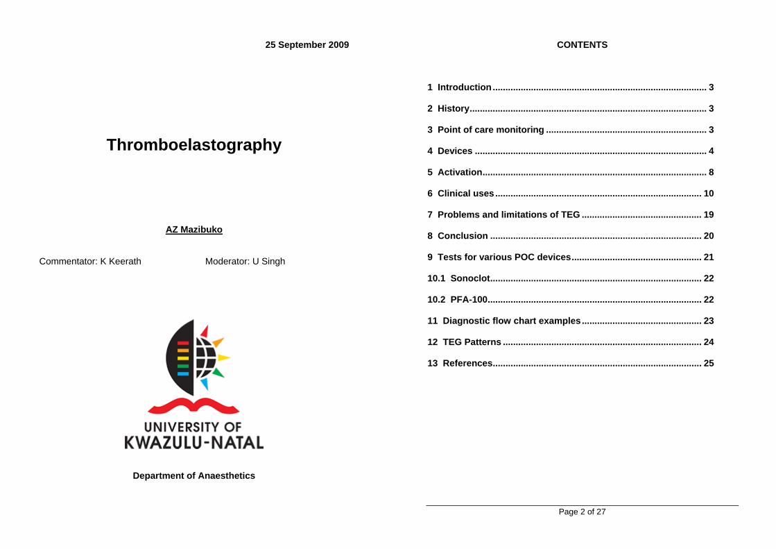
Laboratory tests are often delayed and have inherent limitations : performed on platelet poor plasma, rewarmed to normothermia.[31] 6.5.2 Usefulness of TEG in trauma TEG has a place in trauma providing a bedside, point of care test in a dynamic rapidly evolving situation. It provides functional evaluation of overall coagulation on whole blood. Kaufmann et al[17] in a series of 69 blunt trauma patients found 45 (65%) to be hypercoagulable and 7 (10%) to be hypocoagulable, yet in all patients but one the PTT, PT and platelet count were normal. They found a combination of TEG and ISS to be predictive of transfusion in their cohort. Furthermore in the hypocoagulable group was found to have the highest ISS and the most advanced disease necessitating the most urgent aggressive care. The authors noted the simplicity, immediate availability and ability to repeat test in the emergency suite to be particularly appealing. Rugeri et al found significant alterations in almost all ROTEM tests in a trauma cohort of 88 patients.[26] They were able to determine correlation between standard coagulation tests and ROTEM parameters, and derived transfusion trigger points based on ROTEM parameters alone.
Table 8 from [26]
They also confirmed previous findings of early coagulopathy in up 30% of admission, underlining the relationship between severity of trauma and coagulopathy.
7 Problems and limitations of TEG 7.1 Technical Sampling : the sample needs to be processed within 3-4min of collection, which may necessitate multiple machines in strategic areas of the Hospital rather than a centralised laboratory. [31] Sampling site is important : differences between arterial and venous sample have been reported. The sampling need to be consistent.
Page 20 of 27
Transport of sample to a central area necessitates using citrated blood to prevent clotting, this has been documented to affect the results [31,4] Quality control outside the laboratory may not be adequate unless specifically trained personnel undertake regular calibration and maintenance. Equipment activators and other modifications are often manufacturer specific making standardisation very difficult. [9] 7.2 Patient factors Gender and age have been reported have significant difference in the results, although clinical importance of this remains unclear.[10] Children were found to have different values compared to adults despite otherwise normal coagulation testing. [24] This statistical significance may not necessarily imply clinical significance. Lang et al reported female patients to have faster coagulation activation (shorter CFT and CT), greater clot firmness (higher MCF) and amplitude (CA10 and CA15) compared to males in INTEM, EXTEM and FIBTEM tests.
Table 9
8 Conclusion Viscoelastic POC coagulation analysers are being used in certain clinical situations known for their inherent risk of coagulopathy. especially in cardiac and hepatic surgery. They are finding increasing use in other clinical scenarios like trauma, cardiology in monitoring anticoagulant drugs as well as research. The next phase of development must include easier handling of blood sample, full automation, simultaneous testing with multiple activators, integrated software analysis and more robust devices.[9]
Page 21 of 27
9 Tests for various POC devices
Table 10
Page 22 of 27
10.1 Sonoclot
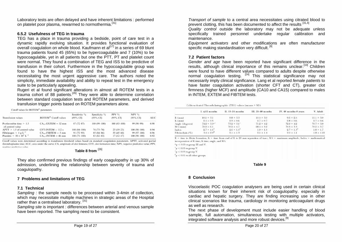
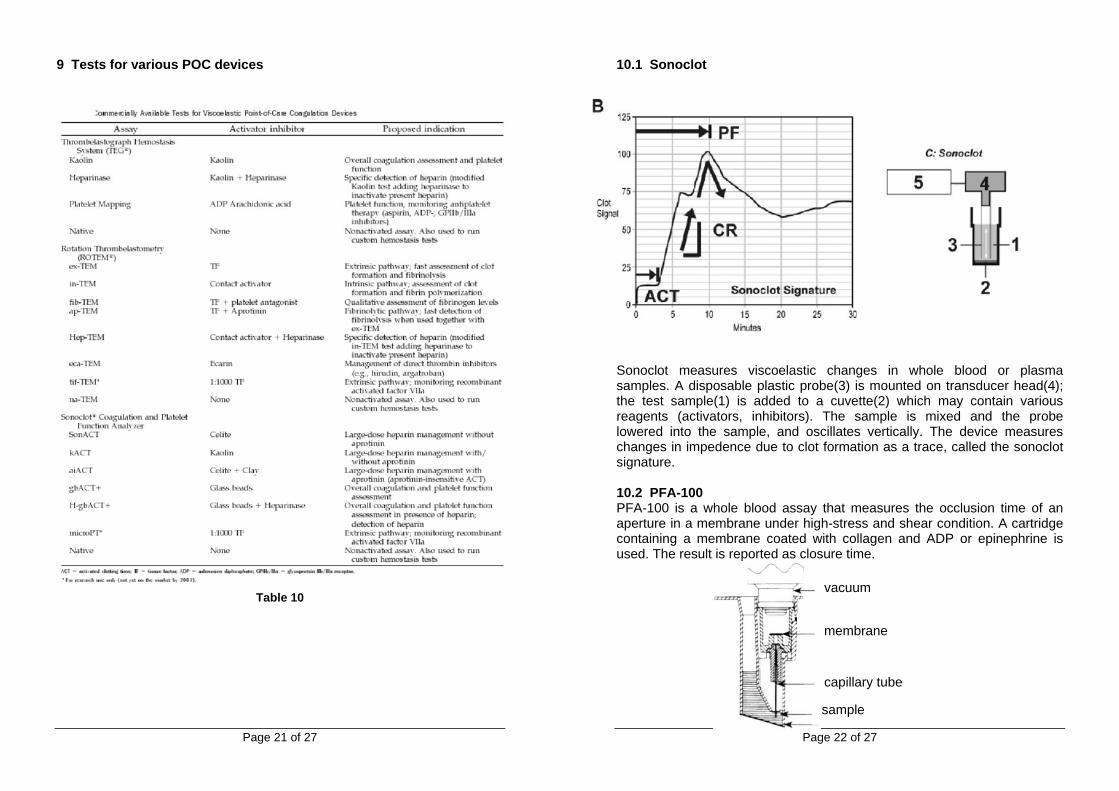
Sonoclot measures viscoelastic changes in whole blood or plasma samples. A disposable plastic probe(3) is mounted on transducer head(4); the test sample(1) is added to a cuvette(2) which may contain various reagents (activators, inhibitors). The sample is mixed and the probe lowered into the sample, and oscillates vertically. The device measures changes in impedence due to clot formation as a trace, called the sonoclot signature. 10.2 PFA-100 PFA-100 is a whole blood assay that measures the occlusion time of an aperture in a membrane under high-stress and shear condition. A cartridge containing a membrane coated with collagen and ADP or epinephrine is used. The result is reported as closure time.
vacuum
membrane
capillary tube
sample

Page 23 of 27
11 Diagnostic flow chart examples
From [1]
Page 24 of 27
12 TEG Patterns
From [32]

Page 25 of 27
13 References [1] L Anderson, I Quasim, R Soutar, M Steven, A Macfie, and W Korte. An
audit of red cell and blood product use after the institution of thromboelastometry in a cardiac intensive care unit. Transfus Med, 16(1):31–9, February 2006.
[2] M S Avidan, E L Alcock, J Da Fonseca, J Ponte, J B Desai, G J Despotis, and B J Hunt. Comparison of structured use of routine laboratory tests or near-patient assessment with clinical judgement in the management of bleeding after cardiac surgery. Br J Anaesth, 92(2):178–86, February 2004.
[3] A Calatzis, M Heesen, and M Spannagl. [point-of-care testing of hemostatic alterations in anaesthesia and intensive care]. Anaesthesist, 52(3):229–37, March 2003.
[4] V Camenzind, T Bombeli, B Seifert, M Jamnicki, D Popovic, T Pasch, and D R Spahn. Citrate storage affects thrombelastograph analysis. Anesthesiology, 92(5):1242–9, May 2000.
[5] T C Collyer, D J Gray, R Sandhu, J Berridge, and G Lyons. Assessment of platelet inhibition secondary to clopidogrel and aspirin therapy in preoperative acute surgical patients measured by thrombelastography platelet mapping. Br J Anaesth, 102(4):492–8, April 2009.
[6] G J Despotis, G Gravlee, K Filos, and J Levy. Anticoagulation monitoring during cardiac surgery: a review of current and emerging techniques. Anesthesiology, 91(4):1122–51, October 1999.
[7] Anna Dyszkiewicz-Korpanty, Horatiu Olteanu, Eugene P Frenkel, and Ravindra Sarode. Clopidogrel anti-platelet effect: an evaluation by optical aggregometry, impedance aggregometry, and the platelet function analyzer (pfa-100). Platelets, 18(7):491–6, November 2007.
[8] R C Franz. Rotem analysis: a significant advance in the field of rotational thrombelastography. S Afr J Surg, 47(1):2–6, February 2009.
[9] Michael T Ganter and Christoph K Hofer. Coagulation monitoring: current techniques and clinical use of viscoelastic point-of-care coagulation devices. Anesth Analg, 106(5):1366–75, May 2008.
[10] H J Gorton, E R Warren, N A Simpson, G R Lyons, and M O Columb. Thromboelastography identifies sex-related differences in coagulation. Anesth Analg, 91(5):1279–81, November 2000.
[11] A R Hobson, R A Agarwala, R A Swallow, K D Dawkins, and N P Curzen. Thrombelastography: current clinical applications and its potential role in interventional cardiology. Platelets, 17(8):509–18, December 2006.
[12] Alex R Hobson, Graham W Petley, Keith D Dawkins, and Nick Curzen. A novel fifteen minute test for assessment of individual time-dependent clotting responses to aspirin and clopidogrel using modified thrombelastography. Platelets, 18(7):497–505, November 2007.
[13] C Huissoud, N Carrabin, F Audibert, A Levrat, D Massignon, M Berland, and R-C Rudigoz. Bedside assessment of fibrinogen level in postpartum haemorrhage by thrombelastometry. BJOG, 116(8):1097–102, July 2009.
Page 26 of 27
[14] C Huissoud, N Carrabin, F Audibert, A Levrat, D Massignon, M Berland, and R-C Rudigoz. Bedside assessment of fibrinogen level in postpartum haemorrhage by thrombelastometry. BJOG, 116(8):1097–102, July 2009.
[15] Cyril Huissoud, Nicolas Carrabin, Mehdi Benchaib, Oriane Fontaine, Albrice Levrat, Denis Massignon, Sandrine Touzet, René-Charles Rudigoz, and Michel Berland. Coagulation assessment by rotation thrombelastometry in normal pregnancy. Thromb Haemost, 101(4):755–61, April 2009.
[16] Y G Kang, D J Martin, J Marquez, J H Lewis, F A Bontempo, B W Shaw, T E Starzl, and P M Winter. Intraoperative changes in blood coagulation and thrombelastographic monitoring in liver transplantation. Anesth Analg, 64(9):888–96, September 1985.
[17] Christoph R. MD Kaufmann, Kevin M. MD Dwyer, John D. BS Crews, Sheila J Dols, and Arthur L. MD Trask. Usefulness of thrombelastography in assessment of trauma patient coagulation. The Journal of Trauma: Injury, Infection, and Critical Care, Volume 42(4):716–722, April 1997.
[18] R J Luddington. Thrombelastography/thromboelastometry. Clin Lab Haematol, 27(2):81–90, April 2005.
[19] D Moopanar, S Naidu, J Moodley, and E Gouws. Thromboelastography in abruptio placentae. J Obstet Gynaecol, 17(3):229–33, May 1997.
[20] Vance G Nielsen. A comparison of the thrombelastograph and the rotem. Blood Coagul Fibrinolysis, 18(3):247–52, April 2007.
[21] G A Nuttall, W C Oliver, P J Santrach, S Bryant, J A Dearani, H V Schaff, and M H Ereth. Efficacy of a simple intraoperative transfusion algorithm for nonerythrocyte component utilization after cardiopulmonary bypass. Anesthesiology, 94(5):773–81; discussion 5A–6A, May 2001.
[22] C E Orlikowski, A J Payne, J Moodley, and D A Rocke. Thrombelastography after aspirin ingestion in pregnant and non-pregnant subjects. Br J Anaesth, 69(2):159–61, August 1992.
[23] C E Orlikowski, D A Rocke, W B Murray, E Gouws, J Moodley, D G Kenoyer, and S Byrne. Thrombelastography changes in pre-eclampsia and eclampsia. Br J Anaesth, 77(2):157–61, August 1996.
[24] E G Pivalizza, P J Pivalizza, L I Gottschalk, S Kee, P Szmuk, and D C Abramson. Celite-activated thrombelastography in children. J Clin Anesth, 13(1):20–3, February 2001.
[25] D Royston and S von Kier. Reduced haemostatic factor transfusion using heparinase-modified thrombelastography during cardiopulmonary bypass. Br J Anaesth, 86(4):575–8, April 2001.
[26] L Rugeri, A Levrat, J S David, E Delecroix, B Floccard, A Gros, B Allaouchiche, and C Negrier. Diagnosis of early coagulation abnormalities in trauma patients by rotation thrombelastography. J Thromb Haemost, 5(2):289–95, February 2007.
[27] L Shore-Lesserson, H E Manspeizer, M DePerio, S Francis, F Vela-Cantos, and M A Ergin. Thromboelastography-guided transfusion algorithm reduces transfusions in complex cardiac surgery. Anesth Analg, 88(2):312–9, February 1999.

Page 27 of 27
[28] G J Spalding, M Hartrumpf, T Sierig, N Oesberg, C G Kirschke, and J M Albes. [bedside thrombelastography. cost reduction in cardiac surgery]. Anaesthesist, 56(8):765–71, August 2007.
[29] Grit J Spalding, Martin Hartrumpf, Tobias Sierig, Nils Oesberg, Christian Günther Kirschke, and Johannes M Albes. Cost reduction of perioperative coagulation management in cardiac surgery: value of “bedside” thrombelastography (rotem). Eur J Cardiothorac Surg, 31(6):1052–7, June 2007.
[30] R A Swallow, R A Agarwala, K D Dawkins, and N P Curzen. Thromboelastography: potential bedside tool to assess the effects of antiplatelet therapy? Platelets, 17(6):385–92, September 2006.
[31] Brandon H Tieu, John B Holcomb, and Martin A Schreiber. Coagulopathy: its pathophysiology and treatment in the injured patient. World J Surg, 31(5):1055–64, May 2007.
[32] K J Tuman, B D Spiess, R J McCarthy, and A D Ivankovich. Comparison of viscoelastic measures of coagulation after cardiopulmonary bypass. Anesth Analg, 69(1):69–75, July 1989.
[33] Nahreen Tynngård, Tomas Lindahl, Sofia Ramström, and Gösta Berlin. Effects of different blood components on clot retraction analysed by measuring elasticity with a free oscillating rheometer. Platelets, 17(8):545–54, December 2006.
[34] L Zuckerman, E Cohen, J P Vagher, E Woodward, and J A Caprini. Comparison of thrombelastography with common coagulation tests. Thromb Haemost, 46(4):752–6, December 1981.
[35] E G Pivalizza. Monitoring of hirudin therapy with the thromboelastograph. Journal of Clinical Anesthesia, 14:456–458, September 2002.
[36] Paul A Gurbel et al. Platelet reactivity in patients and recurrent events post-stenting. JACC, 46(10):1820–1826, November 15 2005.
[37] Douglas J McCrath, Elisabetta Cerboni, Robert J Frumento, Andrew L Hirsh, and Elliott Bennett-Guerrero. Thromboelastography maximum amplitude predicts postoperative thrombotic complications including myocardial infarction. Anesth Analg, 100(6):1576–83, June 2005.