Three-dimensional ultrasonography and power Doppler for ...
6
RESEARCH ARTICLE Open Access Three-dimensional ultrasonography and power Doppler for discrimination between benign and malignant endometrium in premenopausal women with abnormal uterine bleeding Mohamed El-Sharkawy * , Akmal El-Mazny, Wafaa Ramadan, Dina Hatem, Aly Abdel-Hafiz, Mohamed Hammam and Adel Nada Abstract Background: Ultrasonography has been extensively used in women suspected of having a gynecological malignancy. The aim of this study is to evaluate the efficacy of 3D ultrasonography and power Doppler for discrimination between benign and malignant endometrium in premenopausal women with abnormal uterine bleeding. Methods: This cross-sectional study included 78 premenopausal women with abnormal uterine bleeding scheduled for hysteroscopy and endometrial curettage. The endometrial thickness (ET), uterine artery pulsatility index (PI) and resistance index (RI), and endometrial volume (EV) and 3D power Doppler vascularization index (VI), flow index (FI), and vascularization flow index (VFI) were measured and compared with hysteroscopic and histopathologic findings. Results: The ET (P <0.001), EV (P <0.001), and endometrial VI (P <0.001) and VFI (P = 0.043) were significantly increased in patients with atypical endometrial hyperplasia and endometrial carcinoma (n = 10) than those with benign endometrium (n = 68); whereas, the uterine artery PI and RI and endometrial FI were not significantly different between the two groups. The best marker for discrimination between benign and malignant endometrium was the VI with an area under the ROC curve of 0.88 at a cutoff value of 0.81 %. Conclusion: 3D ultrasonography and power Doppler, especially endometrial VI, may be useful for discrimination between benign and malignant endometrium in premenopausal women with abnormal uterine bleeding. Keywords: 3D ultrasonography, Doppler, Endometrial carcinoma, Hysteroscopy, Premenopausal bleeding Background Endometrial carcinoma is the most common form of gynecologic cancer in developed countries, and it is the fourth most common malignant tumor among women worldwide [1]. Abnormal uterine bleeding is usually the first symptom; therefore, appropriate evaluation of women with premenopausal or postmenopausal bleeding will allow for early diagnosis of endometrial carcinoma and the best opportunity for cure [2]. Ultrasonography has been extensively used in women suspected of having a gynecological malignancy, especially in ovarian [3] and endometrial [4] cancer. In fact, transva- ginal ultrasonography is considered the initial imaging procedure for evaluating abnormal vaginal bleeding due to its ability to depict endometrial pathology, its widespread availability, and its excellent safety profile and cost effect- iveness [5]. Three-dimensional ultrasonography is a new imaging technique that has become currently available in gyneco- logic practice [6], specifically in gynecologic oncology [7]. In addition, 3D power-Doppler ultrasonography allows a 3D reconstruction of the vascular network and also * Correspondence: [email protected] Department of Obstetrics and Gynecology, Faculty of Medicine, Cairo University, Cairo, Egypt © 2016 El-Sharkawy et al. Open Access This article is distributed under the terms of the Creative Commons Attribution 4.0 International License (http://creativecommons.org/licenses/by/4.0/), which permits unrestricted use, distribution, and reproduction in any medium, provided you give appropriate credit to the original author(s) and the source, provide a link to the Creative Commons license, and indicate if changes were made. The Creative Commons Public Domain Dedication waiver (http://creativecommons.org/publicdomain/zero/1.0/) applies to the data made available in this article, unless otherwise stated. El-Sharkawy et al. BMC Women's Health (2016) 16:18 DOI 10.1186/s12905-016-0297-3
Transcript of Three-dimensional ultrasonography and power Doppler for ...

RESEARCH ARTICLE Open Access
Three-dimensional ultrasonography andpower Doppler for discrimination betweenbenign and malignant endometrium inpremenopausal women with abnormaluterine bleedingMohamed El-Sharkawy*, Akmal El-Mazny, Wafaa Ramadan, Dina Hatem, Aly Abdel-Hafiz, Mohamed Hammamand Adel Nada
Abstract
Background: Ultrasonography has been extensively used in women suspected of having a gynecological malignancy.The aim of this study is to evaluate the efficacy of 3D ultrasonography and power Doppler for discrimination betweenbenign and malignant endometrium in premenopausal women with abnormal uterine bleeding.
Methods: This cross-sectional study included 78 premenopausal women with abnormal uterine bleeding scheduledfor hysteroscopy and endometrial curettage. The endometrial thickness (ET), uterine artery pulsatility index (PI) andresistance index (RI), and endometrial volume (EV) and 3D power Doppler vascularization index (VI), flow index (FI), andvascularization flow index (VFI) were measured and compared with hysteroscopic and histopathologic findings.
Results: The ET (P <0.001), EV (P <0.001), and endometrial VI (P <0.001) and VFI (P = 0.043) were significantly increased inpatients with atypical endometrial hyperplasia and endometrial carcinoma (n = 10) than those with benign endometrium(n = 68); whereas, the uterine artery PI and RI and endometrial FI were not significantly different between the two groups.The best marker for discrimination between benign and malignant endometrium was the VI with an area under the ROCcurve of 0.88 at a cutoff value of 0.81 %.
Conclusion: 3D ultrasonography and power Doppler, especially endometrial VI, may be useful for discrimination betweenbenign and malignant endometrium in premenopausal women with abnormal uterine bleeding.
Keywords: 3D ultrasonography, Doppler, Endometrial carcinoma, Hysteroscopy, Premenopausal bleeding
BackgroundEndometrial carcinoma is the most common form ofgynecologic cancer in developed countries, and it is thefourth most common malignant tumor among womenworldwide [1]. Abnormal uterine bleeding is usually thefirst symptom; therefore, appropriate evaluation ofwomen with premenopausal or postmenopausal bleedingwill allow for early diagnosis of endometrial carcinomaand the best opportunity for cure [2].
Ultrasonography has been extensively used in womensuspected of having a gynecological malignancy, especiallyin ovarian [3] and endometrial [4] cancer. In fact, transva-ginal ultrasonography is considered the initial imagingprocedure for evaluating abnormal vaginal bleeding due toits ability to depict endometrial pathology, its widespreadavailability, and its excellent safety profile and cost effect-iveness [5].Three-dimensional ultrasonography is a new imaging
technique that has become currently available in gyneco-logic practice [6], specifically in gynecologic oncology [7].In addition, 3D power-Doppler ultrasonography allows a3D reconstruction of the vascular network and also
* Correspondence: [email protected] of Obstetrics and Gynecology, Faculty of Medicine, CairoUniversity, Cairo, Egypt
© 2016 El-Sharkawy et al. Open Access This article is distributed under the terms of the Creative Commons Attribution 4.0International License (http://creativecommons.org/licenses/by/4.0/), which permits unrestricted use, distribution, andreproduction in any medium, provided you give appropriate credit to the original author(s) and the source, provide a link tothe Creative Commons license, and indicate if changes were made. The Creative Commons Public Domain Dedication waiver(http://creativecommons.org/publicdomain/zero/1.0/) applies to the data made available in this article, unless otherwise stated.
El-Sharkawy et al. BMC Women's Health (2016) 16:18 DOI 10.1186/s12905-016-0297-3

calculating vascular indices based on the total and relativeamount of power Doppler information within the volumeof interest [8].The aim of this study is to evaluate the efficacy of 3D
ultrasonography and power Doppler for discriminationbetween benign and malignant endometrium in premen-opausal women with abnormal uterine bleeding.
MethodsThis cross-sectional study was conducted at the Depart-ment of Obstetrics and Gynecology, Faculty of Medicine,Cairo University, during the period from August 2013 toMay 2014. The study protocol was approved by theResearch Ethics Committee, and informed verbal consentwas obtained from all participants.The study population consisted of 78 premenopausal
women with abnormal uterine bleeding scheduled forhysteroscopy and endometrial curettage. They weresubjected to detailed history taking, complete generaland gynecological examination, routine pre-operativelaboratory investigations, and preliminary transvaginalultrasound. The exclusion criteria included uterinefibroids, adenomyosis, endometrial polyps, and anygeneral diseases, hormones or medications that couldpotentially affect pelvic blood flow.Transvaginal ultrasound (Voluson 730; Kretz, Zipf,
Austria) examinations were performed within 24 h prior tosurgery. Using ultrasound in the 2D mode, the endometrialthickness (ET) was measured as the thickest part (doublelayer) in the sagittal plane (Fig. 1). Then, color Doppler wasactivated and the flow velocity waveforms were obtainedfrom the ascending main branch of the uterine artery onboth sides of the internal os (Fig. 2). Three similar
consecutive waveforms of good quality were analyzed, andthe averaged right and left uterine artery pulsatility index(PI) and resistance index (RI) were calculated.The ultrasound was then switched to the 3D mode
with power Doppler. The setting conditions for thisstudy were standardized using a frequency at 3–9 MHz,pulse repetition frequency at 0.6 kHz, gain at −4.0, andwall motion filter at low 1. The Virtual OrganComputer-Aided Analysis (VOCAL™) Imaging Programfor the 3D power Doppler histogram analysis was usedto measure the endometrial volume (EV) and 3D powerDoppler indices within the endometrium (Figs. 3 and 4).Vascularization index (VI) measures the ratio of the
number of color voxels to the total number of voxels (%)and represents the presence of blood vessels (vascularity).Flow index (FI) measures the mean power Doppler signalintensity (0–100) and represents the average intensity ofblood flow. Vascularization flow index (VFI) is calculatedby multiplying VI and FI (0–100) and represents acombination of vascularity and flow intensity.Hysteroscopic examination was performed routinely
before endometrial curettage using a rigid 30° hystero-scope and a 4-mm-diameter diagnostic sheath (Karl StorzGmbH & Co KG, Tuttlingen, Germany). The hystero-scopic diagnosis was based on the following criteria:atrophic endometrium-thin and homogeneous in appear-ance; endometrial hyperplasia-thickened endometrium,easily indented with pressure, with or without multipolypappearance; and endometrial carcinoma-irregular growthwith or without abnormal vascularization.Endometrial sampling was carried out by formal
dilatation and curettage. The histopathological sampleswere examined by two senior pathologists who determined
Fig. 1 Two-dimensional ultrasound measurement of endometrial thickness
El-Sharkawy et al. BMC Women's Health (2016) 16:18 Page 2 of 6

the final diagnosis. Ultrasonographic findings werecompared with hysteroscopic and histopathologic findings.
Statistical analysisData were expressed as mean ± SD or n (%) unless other-wise indicated. Continuous data were compared usingStudent t test or Mann-Whitney U test, as appropriate.Receiver-operating characteristic (ROC) curve analysiswas used to evaluate the optimal cutoff value of ultra-sound markers for prediction of malignant endometriallesions; based on an equivalent sensitivity and specificity,and the highest value of the area under the curve(AUC). A P value <0.05 was considered statistically sig-nificant. The Statistical Package for the Social Science
(SPSS Inc., Chicago, IL, USA), version 16.0, was used fordata analyses.Sample size calculation reveals that with a margin of
error of 4.99 % and a response distribution of 50 %, theconfidence level was 52 %; whereas with a margin oferror of 9.92 % and a response distribution of 50 %, theconfidence level was 84 %.
ResultsPatients’ characteristics and histopathological diagnosisare shown in Table 1. Of the 78 women included in thestudy, 68 (87 %) had benign endometrium and 10 (13 %)had malignant endometrium (atypical hyperplasia andcarcinoma). Hysteroscopic and histopathologic findingswere in agreement in almost all cases.
Fig. 2 Two-dimensional color Doppler of uterine artery flow velocity waveforms
Fig. 3 Virtual Organ Computer-Aided Analysis of the endometrium
El-Sharkawy et al. BMC Women's Health (2016) 16:18 Page 3 of 6

The age was significantly higher (P = 0.032) in patientswith malignant endometrium; however, there were nosignificant differences in the parity (P = 0.954), weight (P =0.952), height (P = 0.244), or body mass index (P = 0.248)between the two groups. The ET (P <0.001), EV (P <0.001),and endometrial VI (P <0.001) and VFI (P = 0.043) weresignificantly increased in patients with malignant endomet-rium; whereas, the uterine artery PI (P = 0.296) andRI (P = 0.922) and endometrial FI (P = 0.474) were notsignificantly different between the two groups (Table 2).The diagnostic performance of the various ultrasound
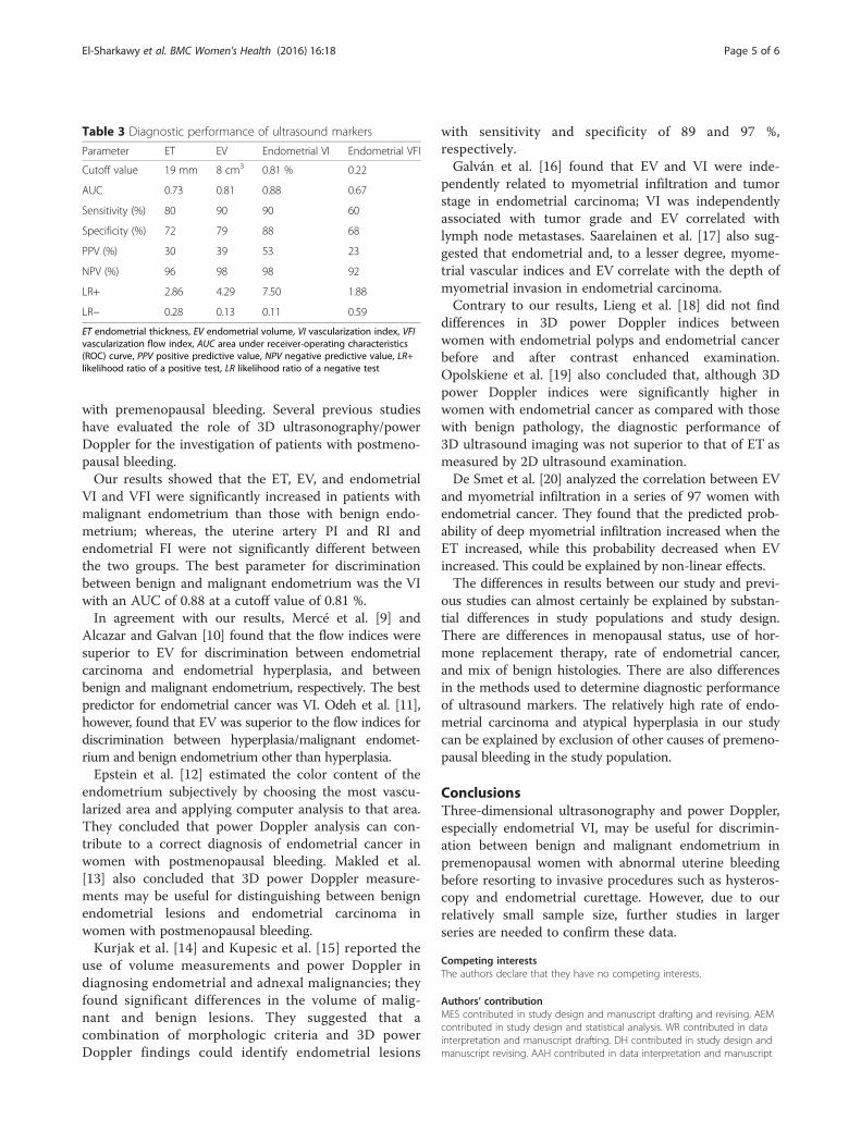
markers is shown in Table 3. The best marker for dis-crimination between benign and malignant endomet-rium was the VI with an AUC of 0.88 at a cutoff value of0.81 %. The sensitivity, specificity, positive predictivevalue (PPV), negative predictive value (NPV), likelihood
ratio of a positive test (LR+), and likelihood ratio of anegative test (LR−) for endometrial VI at 0.81 % (90 %,88 %, 53 %, 98 %, 7.50, and 0.11, respectively) werehigher than those for ET at 19 mm (80 %, 72 %, 30 %,96 %, 2.86, and 0.28, respectively), EV at 8 cm3 (90 %,79 %, 39 %, 98 %, 4.29, and 0.13, respectively) and endo-metrial VFI at 0.22 (60 %, 68 %, 23 %, 92 %, 1.88, and0.59, respectively).
DiscussionTo the best of our knowledge and review of literature,this is the first study to evaluate the efficacy of 3D ultra-sonography and power Doppler for discrimination be-tween benign and malignant endometrium in women
Fig. 4 Three-dimensional power Doppler flow indices of the endometrium
Table 1 Patients’ characteristics and histopathological diagnosis(n = 78)
Characteristic Value
Age (y) 47.46 ± 2.94
Parity 4.18 ± 1.18
Weight (kg) 87.79 ± 11.49
Height (cm) 157.10 ± 5.50
BMI (kg/m2) 35.53 ± 2.09
Endometrial histopathology; n (%)
Normal endometrium 20 (25.64)
Simple hyperplasia 25 (32.05)
Distorted proliferative 16 (20.51)
Atrophic endometrium 7 (8.97)
Atypical hyperplasia 4 (5.13)
Adenocarcinoma 6 (7.69)
BMI body mass index
Table 2 Characteristics of patients with benign and malignantendometrium
Characteristic Benign (n = 68) Malignant (n = 10) P value
Age (y) 46.88 ± 1.95 48.43 ± 2.99 0.032a
Parity 4.18 ± 1.21 4.20 ± 1.03 0.954
Weight (kg) 87.76 ± 10.74 88.00 ± 16.44 0.952
Height (cm) 157.38 ± 5.80 155.20 ± 1.81 0.244
BMI (kg/m2) 35.29 ± 1.84 36.62 ± 8.42 0.248
ET (mm) 7.615 ± 5.493 21.400 ± 9.489 <0.001a
Uterine artery PI 1.847 ± 0.638 2.080 ± 0.755 0.296
Uterine artery RI 1.140 ± 1.583 1.190 ± 0.796 0.922
EV (cm3) 3.410 ± 2.728 7.534 ± 3.622 <0.001a
Endometrial VI (%) 0.310 ± 0.418 1.005 ± 0.597 <0.001a
Endometrial FI (0–100) 22.897 ± 4.547 24.212 ± 9.562 0.474
Endometrial VFI (0–100) 0.100 ± 0.157 0.204 ± 0.072 0.043a
BMI body mass index, ET endometrial thickness, PI pulsatility index,RI resistance index, EV endometrial volume, FI flow index, VI vascularizationindex, VFI vascularization flow indexaStatistically significant
El-Sharkawy et al. BMC Women's Health (2016) 16:18 Page 4 of 6
with premenopausal bleeding. Several previous studieshave evaluated the role of 3D ultrasonography/powerDoppler for the investigation of patients with postmeno-pausal bleeding.Our results showed that the ET, EV, and endometrial
VI and VFI were significantly increased in patients withmalignant endometrium than those with benign endo-metrium; whereas, the uterine artery PI and RI andendometrial FI were not significantly different betweenthe two groups. The best parameter for discriminationbetween benign and malignant endometrium was the VIwith an AUC of 0.88 at a cutoff value of 0.81 %.In agreement with our results, Mercé et al. [9] and
Alcazar and Galvan [10] found that the flow indices weresuperior to EV for discrimination between endometrialcarcinoma and endometrial hyperplasia, and betweenbenign and malignant endometrium, respectively. The bestpredictor for endometrial cancer was VI. Odeh et al. [11],however, found that EV was superior to the flow indices fordiscrimination between hyperplasia/malignant endomet-rium and benign endometrium other than hyperplasia.Epstein et al. [12] estimated the color content of the
endometrium subjectively by choosing the most vascu-larized area and applying computer analysis to that area.They concluded that power Doppler analysis can con-tribute to a correct diagnosis of endometrial cancer inwomen with postmenopausal bleeding. Makled et al.[13] also concluded that 3D power Doppler measure-ments may be useful for distinguishing between benignendometrial lesions and endometrial carcinoma inwomen with postmenopausal bleeding.Kurjak et al. [14] and Kupesic et al. [15] reported the
use of volume measurements and power Doppler indiagnosing endometrial and adnexal malignancies; theyfound significant differences in the volume of malig-nant and benign lesions. They suggested that acombination of morphologic criteria and 3D powerDoppler findings could identify endometrial lesions
with sensitivity and specificity of 89 and 97 %,respectively.Galván et al. [16] found that EV and VI were inde-
pendently related to myometrial infiltration and tumorstage in endometrial carcinoma; VI was independentlyassociated with tumor grade and EV correlated withlymph node metastases. Saarelainen et al. [17] also sug-gested that endometrial and, to a lesser degree, myome-trial vascular indices and EV correlate with the depth ofmyometrial invasion in endometrial carcinoma.Contrary to our results, Lieng et al. [18] did not find
differences in 3D power Doppler indices betweenwomen with endometrial polyps and endometrial cancerbefore and after contrast enhanced examination.Opolskiene et al. [19] also concluded that, although 3Dpower Doppler indices were significantly higher inwomen with endometrial cancer as compared with thosewith benign pathology, the diagnostic performance of3D ultrasound imaging was not superior to that of ET asmeasured by 2D ultrasound examination.De Smet et al. [20] analyzed the correlation between EV
and myometrial infiltration in a series of 97 women withendometrial cancer. They found that the predicted prob-ability of deep myometrial infiltration increased when theET increased, while this probability decreased when EVincreased. This could be explained by non-linear effects.The differences in results between our study and previ-
ous studies can almost certainly be explained by substan-tial differences in study populations and study design.There are differences in menopausal status, use of hor-mone replacement therapy, rate of endometrial cancer,and mix of benign histologies. There are also differencesin the methods used to determine diagnostic performanceof ultrasound markers. The relatively high rate of endo-metrial carcinoma and atypical hyperplasia in our studycan be explained by exclusion of other causes of premeno-pausal bleeding in the study population.
ConclusionsThree-dimensional ultrasonography and power Doppler,especially endometrial VI, may be useful for discrimin-ation between benign and malignant endometrium inpremenopausal women with abnormal uterine bleedingbefore resorting to invasive procedures such as hysteros-copy and endometrial curettage. However, due to ourrelatively small sample size, further studies in largerseries are needed to confirm these data.
Competing interestsThe authors declare that they have no competing interests.
Authors’ contributionMES contributed in study design and manuscript drafting and revising. AEMcontributed in study design and statistical analysis. WR contributed in datainterpretation and manuscript drafting. DH contributed in study design andmanuscript revising. AAH contributed in data interpretation and manuscript
Table 3 Diagnostic performance of ultrasound markers
Parameter ET EV Endometrial VI Endometrial VFI
Cutoff value 19 mm 8 cm3 0.81 % 0.22
AUC 0.73 0.81 0.88 0.67
Sensitivity (%) 80 90 90 60
Specificity (%) 72 79 88 68
PPV (%) 30 39 53 23
NPV (%) 96 98 98 92
LR+ 2.86 4.29 7.50 1.88
LR− 0.28 0.13 0.11 0.59
ET endometrial thickness, EV endometrial volume, VI vascularization index, VFIvascularization flow index, AUC area under receiver-operating characteristics(ROC) curve, PPV positive predictive value, NPV negative predictive value, LR+likelihood ratio of a positive test, LR likelihood ratio of a negative test
El-Sharkawy et al. BMC Women's Health (2016) 16:18 Page 5 of 6

drafting. MH contributed in data interpretation and manuscript revising. ANcontributed in data interpretation and manuscript revising. All authors readand approved the final manuscript.
AcknowledgementNone
FundingNone
Received: 6 January 2015 Accepted: 9 March 2016
References1. Parkin DM, Bray F, Ferlay J, Pisani P. Global cancer statistics, 2002. CA Cancer
J Clin. 2005;55(2):74–108.2. Sorosky JI. Endometrial cancer. Obstet Gynecol. 2008;111(2 Pt 1):436–47.3. Mettler L. The cystic adnexal mass: patient selection, surgical
techniques and long-term follow-up. Curr Opin Obstet Gynecol. 2001;13(4):389–97.
4. Clark TJ, Barton PM, Coomarasamy A, Gupta JK, Khan KS. Investigatingpostmenopausal bleeding for endometrial cancer: cost-effectiveness ofinitial diagnostic strategies. BJOG. 2006;113(5):502–10.
5. Dijkhuizen FP, Mol BW, Brölmann HA, Heintz AP. Cost-effectiveness of theuse of transvaginal sonography in the evaluation of postmenopausalbleeding. Maturitas. 2003;45(4):275–82.
6. Bega G, Lev-Toaff AS, O’Kane P, Becker Jr E, Kurtz AB. Three-dimensionalultrasonography in gynecology: technical aspects and clinical applications.J Ultrasound Med. 2003;22(11):1249–69.
7. Alcázar JL, Jurado M. Three-dimensional ultrasound for assessing womenwith gynecological cancer: a systematic review. Gynecol Oncol. 2011;120(3):340–6.
8. Campbell S. Placental vasculature as visualized by 3D power Dopplerangiography and 3D color Doppler imaging. Ultrasound Obstet Gynecol. 2007;30(6):917–20.
9. Mercé LT, Alcázar JL, Engels V, Troyano J, Bau S, Bajo JM. Endometrialvolume and vascularity measurements by transvaginal three-dimensionalultrasonography and power Doppler angiography in stimulated andtumoral endometria: intraobserver reproducibility. Gynecol Oncol. 2006;100(3):544–50.
10. Alcazar JL, Galvan R. Three-dimensional power Doppler ultrasoundscanning for the prediction of endometrial cancer in women withpostmenopausal bleeding and thickened endometrium. Am J ObstetGynecol. 2009;200(1):44. e1-6.
11. Odeh M, Vainerovsky I, Grinin V, Kais M, Ophir E, Bornstein J. Three-dimensional endometrial volume and 3-dimensional power Doppleranalysis in predicting endometrial carcinoma and hyperplasia. GynecolOncol. 2007;106(2):348–53.
12. Epstein E, Skoog L, Isberg PE, De Smet F, De Moor B, Olofsson PA,Gudmundsson S, Valentin L. An algorithm including results of gray-scaleand power Doppler ultrasound examination to predict endometrialmalignancy in women with postmenopausal bleeding. Ultrasound ObstetGynecol. 2002;20(4):370–6.
13. Makled AK, Elmekkawi SF, El-Refaie TA, El-Sherbiny MA. Three-dimensional powerDoppler and endometrial volume as predictors of malignancy in patients withpostmenopausal bleeding. J Obstet Gynaecol Res. 2013;39(5):1045–51.
14. Kurjak A, Kupesic S, Sparac V, Bekavac I. Preoperative evaluation of pelvictumors by Doppler and three-dimensional sonography. J Ultrasound Med.2001;20(8):829–40.
15. Kupesic S, Kurjak A, Hajder E. Ultrasonic assessment of the postmenopausaluterus. Maturitas. 2002;41(4):255–67.
16. Galván R, Mercé L, Jurado M, Mínguez JA, López-García G, Alcázar JL. Three-dimensional power Doppler angiography in endometrial cancer: correlationwith tumor characteristics. Ultrasound Obstet Gynecol. 2010;35(6):723–9.
17. Saarelainen SK, Vuento MH, Kirkinen P, Mäenpää JU. Preoperativeassessment of endometrial carcinoma by three-dimensional power Dopplerangiography. Ultrasound Obstet Gynecol. 2012;39(4):466–72.
18. Lieng M, Qvigstad E, Dahl GF, Istre O. Flow differences betweenendometrial polyps and cancer: a prospective study using intravenouscontrast-enhanced transvaginal color flow Doppler and three-dimensional power Doppler ultrasound. Ultrasound Obstet Gynecol.2008;32(7):935–40.
19. Opolskiene G, Sladkevicius P, Jokubkiene L, Valentin L. Three-dimensionalultrasound imaging for discrimination between benign and malignantendometrium in women with postmenopausal bleeding and sonographicendometrial thickness of at least 4.5 mm. Ultrasound Obstet Gynecol. 2010;35(1):94–102.
20. De Smet F, De Brabanter J, Van den Bosch T, Pochet N, Amant F, VanHolsbeke C, Moerman P, De Moor B, Vergote I, Timmerman D. New modelsto predict depth of infiltration in endometrial carcinoma based ontransvaginal sonography. Ultrasound Obstet Gynecol. 2006;27(6):664–71.
• We accept pre-submission inquiries
• Our selector tool helps you to find the most relevant journal
• We provide round the clock customer support
• Convenient online submission
• Thorough peer review
• Inclusion in PubMed and all major indexing services
• Maximum visibility for your research
Submit your manuscript atwww.biomedcentral.com/submit
Submit your next manuscript to BioMed Central and we will help you at every step:
El-Sharkawy et al. BMC Women's Health (2016) 16:18 Page 6 of 6