A new electrochemiluminescence biosensor for detection of ...
at SciVerse ScienceDirect
Biomaterials 33 (2012) 1024e1031
Contents lists available
Biomaterials
journal homepage: www.elsevier .com/locate/biomateria ls
Three-dimensional paper-based electrochemiluminescence immunodevicefor multiplexed measurement of biomarkers and point-of-care testing
Lei Ge a, Jixian Yan a, Xianrang Song b, Mei Yan a, Shenguang Ge c, Jinghua Yu a,*
a School of Chemistry and Chemical Engineering, University of Jinan, Jinan 250022, PR ChinabCancer Research Center, Shandong Tumor Hospital, Jinan 250117, PR Chinac Shandong Provincial Key Laboratory of Fluorine Chemistry and Chemical Materials, University of Jinan, Jinan 250022, PR China
a r t i c l e i n f o
Article history:Received 28 August 2011Accepted 24 October 2011Available online 9 November 2011
Keywords:Electrochemiluminescence immunoassayLab on paperScreen-printed electrodeBiomarkerPoint-of-care testing
* Corresponding author. Tel.: þ86 531 82767161; faE-mail address: [email protected] (J. Yu).
0142-9612/$ e see front matter � 2011 Elsevier Ltd.doi:10.1016/j.biomaterials.2011.10.065
a b s t r a c t
In this work, electrochemiluminescence (ECL) immunoassay was introduced into the recently proposedmicrofluidic paper-based analytical device (mPADs) based on directly screen-printed electrodes on paperfor the very first time. The screen-printed paper-electrodes will be more important for further devel-opment of this paper-based ECL device in simple, low-cost and disposable application than commer-cialized ones. To further perform high-performance, high-throughput, simple and inexpensive ECLimmunoassay on mPAD for point-of-care testing, a wax-patterned three-dimensional (3D) paper-basedECL device was demonstrated for the very first time. In this 3D paper-based ECL device, eight carbonworking electrodes including their conductive pads were screen-printed on a piece of square paper andshared the same Ag/AgCl reference and carbon counter electrodes on another piece of square paper afterstacking. Using typical tris-(bipyridine)-ruthenium (Ⅱ) - tri-n-propylamine ECL system, the applicationtest of this 3D paper-based ECL device was performed through the diagnosis of four tumor markers inreal clinical serum samples. With the aid of a facile device-holder and a section-switch assembled on theanalyzer, eight working electrodes were sequentially placed into the circuit to trigger the ECL reaction inthe sweeping range from 0.5 to 1.1 V at room temperature. In addition, this 3D paper-based ECL devicecan be easily integrated and combined with the recently emerging paper electronics to further developsimple, sensitive, low-cost, disposable and portable mPAD for point-of-care testing, public health andenvironmental monitoring in remote regions, developing or developed countries.
� 2011 Elsevier Ltd. All rights reserved.
1. Introduction
While the use of paper for biological assays and point-of-carediagnostics is not a new concept [1], it has never attracted asmuch attention as it does now. Whitesides and coworkers recentlyintroduced a promising concept of using patterned paper substrateas microfluidic platform for multiplex analyte detection [2,3]. Theyaim at developing a simple, inexpensive, portable, disposable andeasy-to-use point-of-care platform for developing countries,resource-limited and remote regions. Microfluidic paper-basedanalytical devices (mPADs) have gained more and more attentionand great interest during the recent years [4e8]. Much effort hasbeen directed toward the development of fabrication [3,9e18],functionalization [19e29] and quantitative methods [27,30e33] formPADs. To date, the primary detection method for the qualitativeand quantitative analysis of multiplex analytes on mPADs is
x: þ86 531 82765969.
All rights reserved.
colorimetric method [2,3,5,9,10,12,13,17,28,33e35] based on visu-ally comparing the color intensity of the reaction spots by nakedeye or camera phones [35] and portable scanners [27]. With the useof a fluidic timer, more accurate glucose levels were determined onmPADs by colorimetric method [28]. However, specifically quanti-tative analysis is still needed when the level of an analyte in realcomplex biological sample is important for simple, rapid, low-costpoint-of-care testing, public health and environmental monitoring.
Recently, there has been an emerging trend of establishing newanalytical methods, such as electrochemical [34,36] and chemilu-minescent [32,37] methods, on paper-based analytical device,which not only retains the simplicity, low cost, portability anddisposability of paper-based analytical devices, but also providesnew opportunities and directions in the development of preciseand sensitive diagnostic devices. Electrochemiluminescence (ECL),which combines the advantages of chemiluminescence and elec-trochemistry, continues to impact diverse areas ranging fromchemical analysis to the molecular-level understanding of biolog-ical processes [38]. In comparison to the conventional electro-chemical and chemiluminescent methods, the ECL assay not only
L. Ge et al. / Biomaterials 33 (2012) 1024e1031 1025
shows high sensitivity and wide dynamic concentration responserange but also is potential- and spatial controlled. Recently, theestablishment of ECL on mPADs [31] based on the integration ofmPADs and commercialized screen-printed electrodes havesubstantially increased the scope of options for detections onmPADs.
Despite their success, there are still pressing needs for mPADsstrategy, especially to separate and enrich analyte from samplescontaining highly abundant nonspecific biological matrixes onmPADs for point-of-care testing. Immunoassay, as an analyticaltechnique based on the highly specific interaction between antigenand antibody, is widely applied in clinical chemistry, pharmaceu-tical analysis, toxicological analysis, bioanalysis, food safety andenvironmental analysis due to its high selectivity, rapid detectionand possible analysis of difficult matrices without extensive pre-treatment. The paper-based ELISA [33], firstly proposed by White-sides, has provided an opportunity and protocol for analyticalmethods established on mPADs. It is based on the colorimetricassays on paper microzone plate. However, colorimetric assay isstill not sufficiently sensitive or specific for accurate point-of-careuse. In addition, the visual readout is usually limited to a yes/noanswer, this is not adequate when the level of an analyte is veryimportant. To overcome these challenges, more sensitive andqualitative methods should be integrated. ECL immunoassay hasbecome a powerful analytical tool for highly sensitive and specificdetection of clinical samples [38]. It also gives a sensitive, rapidalternative to other methods as a detection principle in immuno-assay for the point-of-care determination of molecules (e.g.,proteins, hormones, drugs, nucleic acids and environmentalpollutants) [38]. To the best of our knowledge, no reports aboutestablishing ECL immunoassay on mPADs have been published.
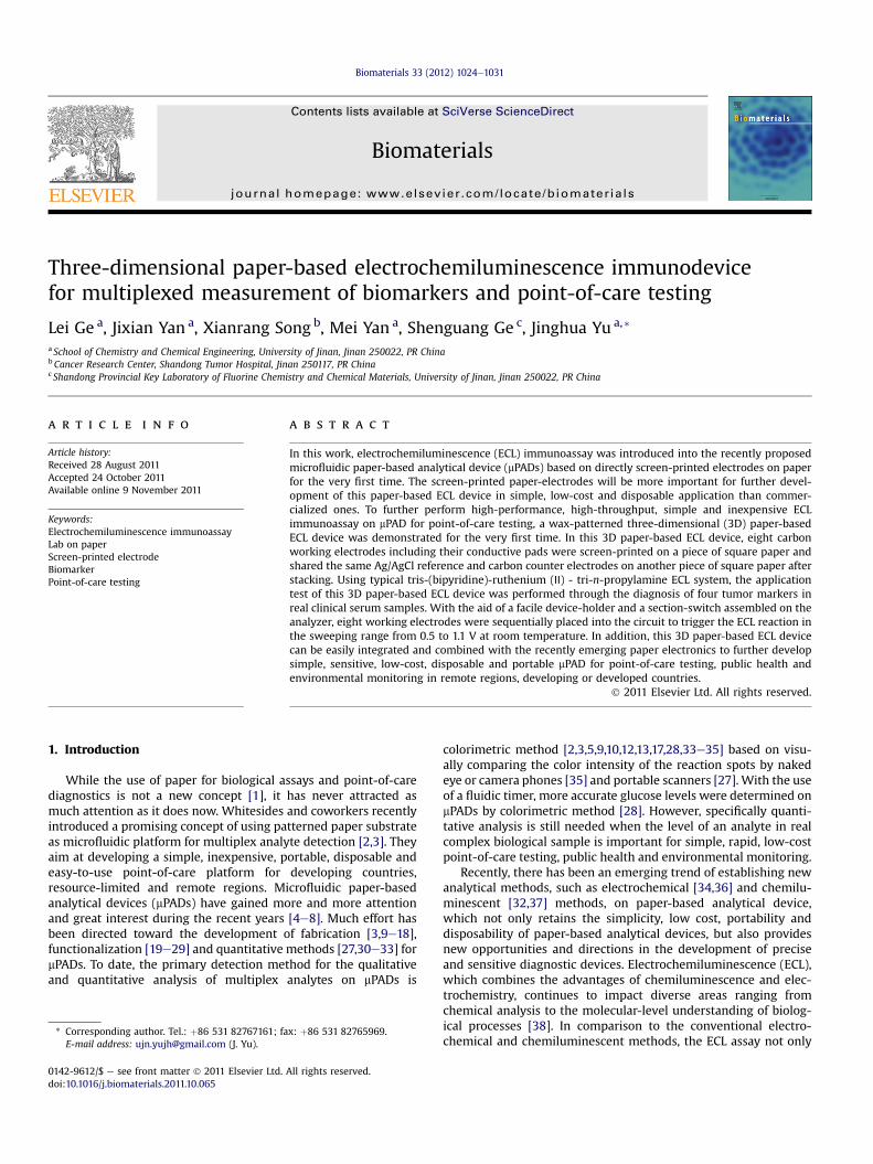
To combine the ECL immunoassay with mPADs for high-throughput, rapid, simple, portable, disposable, low-cost, andsensitive point-of-care testing, we demonstrated a three-dimensional (3D) paper-based ECL device (Scheme 1) based onwax-patterned technology and screen-printed paper-electrodes inthis work. 3D mPADs [20,21] are particularly useful because theypermit fluid movement in the x-, y-, and z-directions, and there-fore, they can accommodate more assays on a smaller footprintthan typical 2D, lateral-flow devices. A 3D mPAD can distributea sample from a single entry point into hundreds of test regions
Sheet-A
Sheet-B
4 mm
30 mm
7 mm
Scheme 1. The schematic representation of process to fabricate 3D paper-based ECL devicescreen-printed on sheet-A and sheet-B respectively in bulk. Finally, sheet-A and sheet-B w
[20]. Wax printing is rapid, inexpensive, insulative and particularlywell-suited for producing large lots (hundreds to thousands) ofprototype mPADs [15e17]. The screen-printed paper-electrodes willbe more important for further development of paper-based ECLdevice in low-cost and disposable application than commercializedones [30,36,39]. In this 3D paper device, carbonworking electrodesincluding their conductive pads were screen-printed on a piece ofsquare paper (named paper-A, 30.0 mm� 30.0 mm) and shared thesame Ag/AgCl reference and carbon counter electrodes on anotherpiece of square paper (named paper-B, 30.0 mm � 30.0 mm) afterstacking. The wax patterns around the electrodes on paper-A andpaper-B constituted reservoirs of the electrochemical cells.
The detection of a panel of biomarkers can significantly improvethe diagnostic value of biomarkers [40]. Thus, as a model, fourtumor markers (r-fetoprotein (AFP), carcinoma antigen 125(CA125), carcinoma antigen 199 (CA199) and carcinoembryonicantigen (CEA)), showing considerable significance in earlyscreening and clinical diagnosis of some tumor diseases [41], wereused as model analytes. In this work, eight electrodes were screen-printed on paper-A for determination of the four tumor marker(every two working electrodes for one tumor marker) to obtainmore exact results. Ultimately, with the aids of a simple home-made device-holder, the concentrations of AFP, CA125, CA199 andCEA in real human serum samples were detected using tris-(bipyridine)-ruthenium (Ⅱ) (Ru(bpy)32þ) - tri-n-propylamine (TPA)ECL system under the optimum conditions. This work could notonly make contribution to further expand detection mode onmPADs, but also can be easily integrated and combined with therecently emerging class of paper electronics [42,43] to furtherdevelop simple, sensitive, low-cost, disposable and portable point-of-care testing device without device-holder in our future work.
2. Materials and methods
2.1. Reagents and materials
Mousemonoclonal capture and Ru(bpy)32þ-labeled signal AFP, CA125, CA199 andCEA antibodies were purchased from Linc-Bio Science Co. Ltd. (Shanghai, China).Chitosan and bovine serum albumin (BSA) was obtained from SigmaeAldrichChemical Co. (St. Louis, MO). Glutaraldehyde (GA, 25% aqueous solution) waspurchased from Alfa Aesar China Ltd. Whatman chromatography paper #1(200.0 mm � 200.0 mm) (pure cellulose paper) was obtained from GE HealthcareWorldwide (Pudong Shanghai, China) and used with further adjustment of size.
A A
B B
Screen-printed carbonworking electrodes
Screen-printed carboncounter electrode
Screen-printed Ag/Agclreference electrode
30 mm
30 mm
. Paper sheets were firstly patterned in bulk using a wax printer. Then electrodes wereere cut to paper-A and paper-B with the same size (30.0 mm � 30.0 mm).
L. Ge et al. / Biomaterials 33 (2012) 1024e10311026
Ultrapure water obtained from aMillipore water purification system (g18 MU, Milli-Q, Millipore) was used in all assays and solutions. Blocking buffer for blocking theresidual reactive sites on the antibody immobilized paper was pH 7.4 phosphatebuffer solution (PBS) containing 0.5% BSA and 0.5% casein. To minimize unspecificadsorption, 0.05% Tween-20 was spiked into 10.0 mM pH 7.4 PBS as washing buffer.Fluorescein isothiocyanate (FITC)-labeled CA125 antibodies and human AFP, CA125,CA199 and CEA standard solutions (0.5mgmL�1) were from Linc-Bio Science Co. Ltd.(Shanghai, China). The clinical serum samples were from Shandong Tumor Hospital.All other reagents were of analytical grade and used as received.
2.2. Fabrication of this 3D paper-based ECL device
Wax was used as the paper hydrophobization and insulation agent in this work.This 3D paper-based ECL device was comprised of two layers of patterned rectan-gular papers with the same size (30.0 mm � 30.0 mm, named paper-A and paper-Brespectively below). Firstly, as shown in Scheme 1, paper-A and paper-B wereproduced in bulk (named sheet-A and sheet-B respectively below). The shape forpatterning this 3D paper-based ECL devicewas designed using Adobe illustrator CS4.On paper-A, the wax-patterned contains a circular contacting zone (7.0 mm indiameter) surrounded by eight working zones (4.0 mm in diameter, 1.0 mm edge-to-edge separation) with paper channels to connect them. On paper-B, there is onlya circular contacting zone (7.0mm in diameter) with the same position as the one onpaper-A. The fabrication process of the wax patterns on sheet-A and sheet-Bincludes only two steps after designing on the computer: (1) print the two waxpatterns onto the surface of sheet-A and sheet-B respectively with wax printer(FUJIXEROX Phaser 8560DN, Japan); (2) bake the wax-printed papers in an oven at130 �C for 150 s to let the printed wax melt and penetrate through the paper to formthe hydrophobic and insulating patterns. These two steps can be finished within2 min.
The wax-patterned sheet-A and sheet-B were then ready for printing electrodeson their hydrophilic zones after cooling to room temperature (within 1 min)(Scheme 1). In addition, due to the small size of this device, the silver wires andcontact pads were unnecessary and can be directly replaced by carbon ink and Ag/AgCl ink respectively. This will be very important for the further development of thisdevice in low-cost application. For sheet-A, eight working electrodes containing thewires and contact pads were screen-printed in the defined area on sheet-A usingcarbon ink. The working electrodes were all aligned to the working zones. For sheet-B, a counter electrode and a reference electrode were screen-printed in the definedcontacting zone on sheet-B using carbon ink and Ag/AgCl ink respectively. After that,sheet-A and sheet-B were cut to paper-A and paper-B with the same size. The eightworking electrodes on paper-Awill share the same reference and counter electrodeson paper-B after stacking. The wax patterns around the electrodes constituteda reservoir of the electrochemical cell with a volume of 35 mL on paper-A and 10 mLon paper-B. The scanning electron microscopy (SEM) images of this 3D paper-basedECL device were recorded on a JEOL JSM-5510 scanning electron microscope. Thecontact angle tests were performed on contact angle measurement (Model OCA40,Dataphysics).
In addition, the front and back surfaces of the wax-patterned electrochemicalcell on paper-A and paper-B are open to atmosphere, thus, for paper-A, the workingelectrodes can be washed by adding PBS or washing buffer to the back of the circularcontacting zone while bending the paper-A to a inverted-U type as shown inFig. S1A. Then a piece of U-type blotting paper was contacted the front of theworking electrodes (Fig. S1C, D). The washing buffer goes through the paper andmigrates along the paper channels by the capillary and gravity action to wash theworking electrodes and carries the unbound reagents with it into the blotting paper.This washing procedure was repeated twice by changing the bend axis, indicated bythe arrow in Fig. S1A, B, to make sure the washing was performed completely. Forpaper-B, the washing procedure can be performed according tomethod proposed byCheng et al. [33]. The printed electrodes will firmly attach to the paper surface due tothe penetration of binding reagents in the inks into the paper matrix. And they willnot break or peel off from the device upon washing and folding [39]. This effectivewashing procedure was used in this work consistently and acquiescently. Thewashing process was important for preventing the nonspecific binding andachieving the best possible signal-to-background ratio. Another purpose for thiswashing procedure was to stop the incubation reaction at exactly same time.
2.3. Preparation of 3D paper-based ECL immunodevice
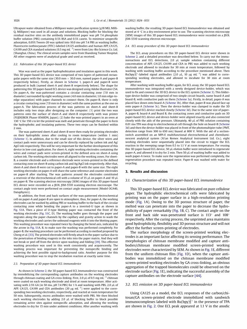
As shown in Scheme 2, the 3D paper-based ECL Immunodevice was constructedby immobilizing the corresponding capture antibodies on the working electrodesthrough chitosan coating and GA cross-linking. First, 2.0 mL of 0.5 mg mL�1 chitosanwere coated on each working electrode and dried at room temperature. After acti-vating with 2.5% GA (in 50 mM, pH 7.4 PBS) for 1 h and washing with PBS, 2.0 mL ofAFP, CA125, CA199 and CEA antibodies (20 mg mL�1) were applied to the corre-sponding twoworking electrodes, respectively, and reacted at room temperature for50 min. Subsequently, excess antibodies were washed with PBS. We then blockedeach working electrodes by adding 2.0 mL of blocking buffer to block possibleremaining active sites against nonspecific adsorption, and allowing the workingelectrodes to dry for 15 min under ambient conditions. After another washing with
washing buffer, the resulting 3D paper-based ECL Immunodevice was obtained andstored at 4 �C in a dry environment prior to use. The scanning electron microscopy(SEM) images of this 3D paper-based ECL immunodevice were recorded on a JEOLJSM-5510 scanning electron microscope.
2.4. ECL assay procedure of this 3D paper-based ECL immunodevice
The ECL assay procedures on this 3D paper-based ECL device were shown inScheme 2, and a detailed procedure was described below. To carry out the immu-noreactions and ECL detections, 2.0 mL sample solution containing differentconcentration of AFP, CA125, CA199 and CEA in PBS was added to each workingelectrode and allowed to incubate for 30 min at room temperature, followed bywashing with washing buffer according to the procedure mentioned above. ThenRu(bpy)32þ-labeled signal antibodies (2.0 mL, 10 mg mL�1) was added to corre-sponding working electrodes, and allowed to incubate for 30 min at roomtemperature.
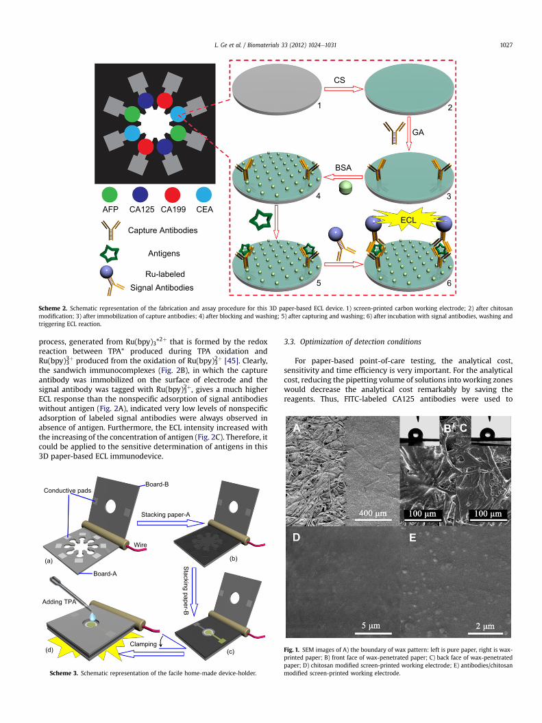
After washing with washing buffer again, for ECL assay, the 3D paper-based ECLimmunodevice was integrated with a newly designed device-holder, which wasused to fix and connect the 3D ECL device to the ECL system (Scheme 3). This folder-type device-holder was comprised of two simple circuit boards, name board-A andboard-B, with conductive pads on them (Scheme 3a). Firstly, in detail, paper-A wasplaced face down onto board-A (Scheme 3b). After that, paper-B was placed face uponto paper-A (Scheme 3c). Then the device-holder was clamped to make the 3Dpaper-based ECL device stacked closely (Scheme 3d). Due to the same size of paper-A, paper-B, board-A and Board B, the contacting zones and conductive pads on 3Dpaper-based ECL device and device-holder were aligned exactly and also connectedclosely with the aids of the pressure. Ultimately, 40 mL of PBS solution containing0.01 mM TPA was drop to electrochemical cell through the hole on board-B (Scheme3d) and then the device-holder was placed in front of the photomultipier tube (PMT,detection range from 300 to 650 nm) biased at 800 V. With the aid of a section-switch assembled on an MPI-E multifunctional electrochemical and chemilumi-nescent analytical system (Xi’an Remex Analytical Instrument Ltd. Co.), eightworking electrodes were sequentially placed into the circuit to trigger the ECLreaction in the sweeping range from 0.5 to 1.1 V at room temperature. For reusingthis 3D paper-based ECL device, 50 mL elution buffer were introduced to regeneratepaper-A, and allowed it to dry for 2 min. Then 50 mL of washing buffer was added towash paper-A twice. To make sure the regeneration was performed completely, theregeneration procedure was repeated twice. Paper-B was washed with water forrecycling.
3. Results and discussion
3.1. Characterization of this 3D paper-based ECL immunodevice
This 3D paper-based ECL device was fabricated on pure cellulosepaper. The hydrophilic electrochemical cells were fabricated byprinting wax on the paper surface in a high-resolution printingmode (Fig. 1A). Owing to the 3D porous structure of paper, themelted wax can penetrate into the paper to decrease the hydro-philicity of paper remarkably (Fig. 1B, C). The contact angle on thefront and back side wax-penetrated surface is 113� and 108�
respectively. After the curing process, the unprinted area maintainsgood hydrophilicity, flexibility and 3D porous structure and will notaffect the further screen-printing of electrodes.
The surface morphology of the screen-printed working elec-trodes is an important factor affecting its sensing performance. Themorphologies of chitosan membrane modified and capture anti-bodies/chitosan membrane modified screen-printed workingelectrodeswere characterized by SEM. As shown in Fig.1E, differentfrom the uniform chitosan film (Fig. 1D), when the capture anti-bodies was immobilized on the chitosan membrane modifiedscreen-printed working electrodes by GA cross-linking, an obviousaggregation of the trapped biomolecules could be observed on theelectrode surface (Fig. 1E), indicating the successful assembly of thecapture antibodies on the electrode surface [44].
3.2. ECL emission on 3D paper-based ECL immunodevice
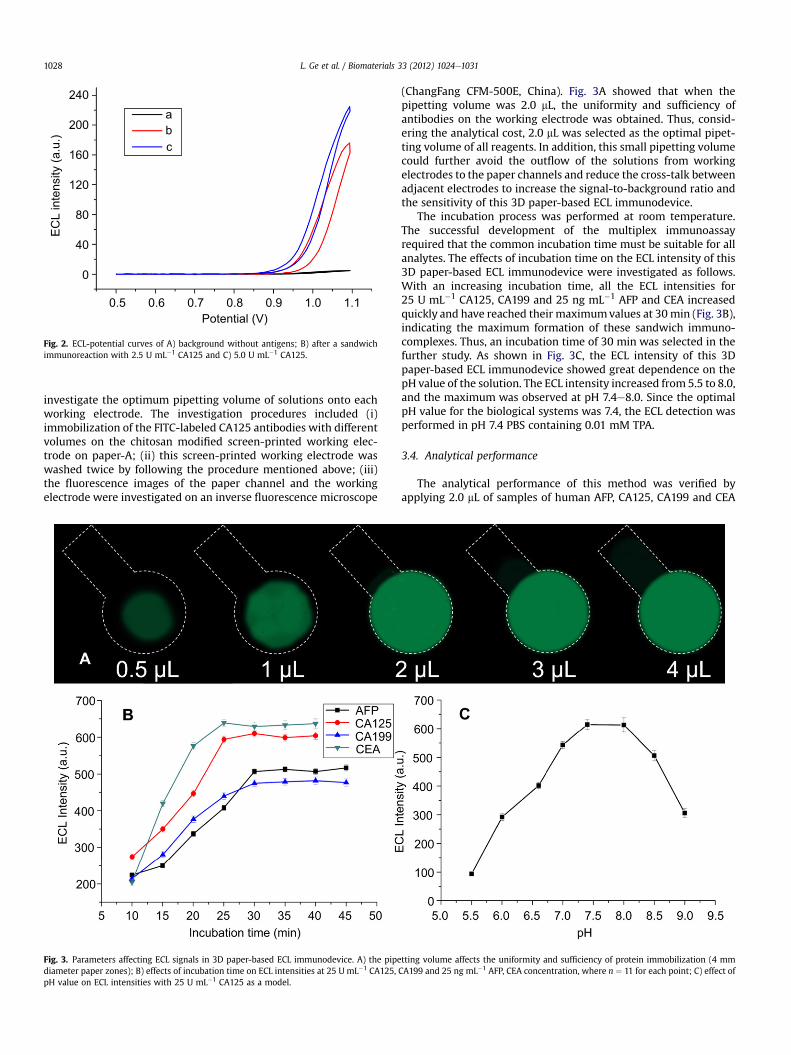
Using CA125 as a model, the ECL responses of the carbon/chi-tosan/GA screen-printed electrode immobilized with sandwichimmunocomplexes labeled with Ru(bpy)32þ in the presence of TPAare shown in Fig. 2. One ECL peak appeared at 1.1 V in the anodic
AFP CA125 CA199
Capture Antibodies
Signal Antibodies
CS
BSA
GA
ECL
5 6
3
21
4
Antigens
Ru-labeled
CEA
Scheme 2. Schematic representation of the fabrication and assay procedure for this 3D paper-based ECL device. 1) screen-printed carbon working electrode; 2) after chitosanmodification; 3) after immobilization of capture antibodies; 4) after blocking and washing; 5) after capturing and washing; 6) after incubation with signal antibodies, washing andtriggering ECL reaction.
L. Ge et al. / Biomaterials 33 (2012) 1024e1031 1027
process, generated from Ru(bpy)3*2þ that is formed by the redoxreaction between TPA* produced during TPA oxidation andRu(bpy)33þ produced from the oxidation of Ru(bpy)32þ [45]. Clearly,the sandwich immunocomplexes (Fig. 2B), in which the captureantibody was immobilized on the surface of electrode and thesignal antibody was tagged with Ru(bpy)32þ, gives a much higherECL response than the nonspecific adsorption of signal antibodieswithout antigen (Fig. 2A), indicated very low levels of nonspecificadsorption of labeled signal antibodies were always observed inabsence of antigen. Furthermore, the ECL intensity increased withthe increasing of the concentration of antigen (Fig. 2C). Therefore, itcould be applied to the sensitive determination of antigens in this3D paper-based ECL immunodevice.
Conductive padsBoard-B
Board-A
(a) (b)
(c)(d)
Adding TPA
Stacking paper-A
Stacking paper-B
Clamping
Wire
Scheme 3. Schematic representation of the facile home-made device-holder.
3.3. Optimization of detection conditions
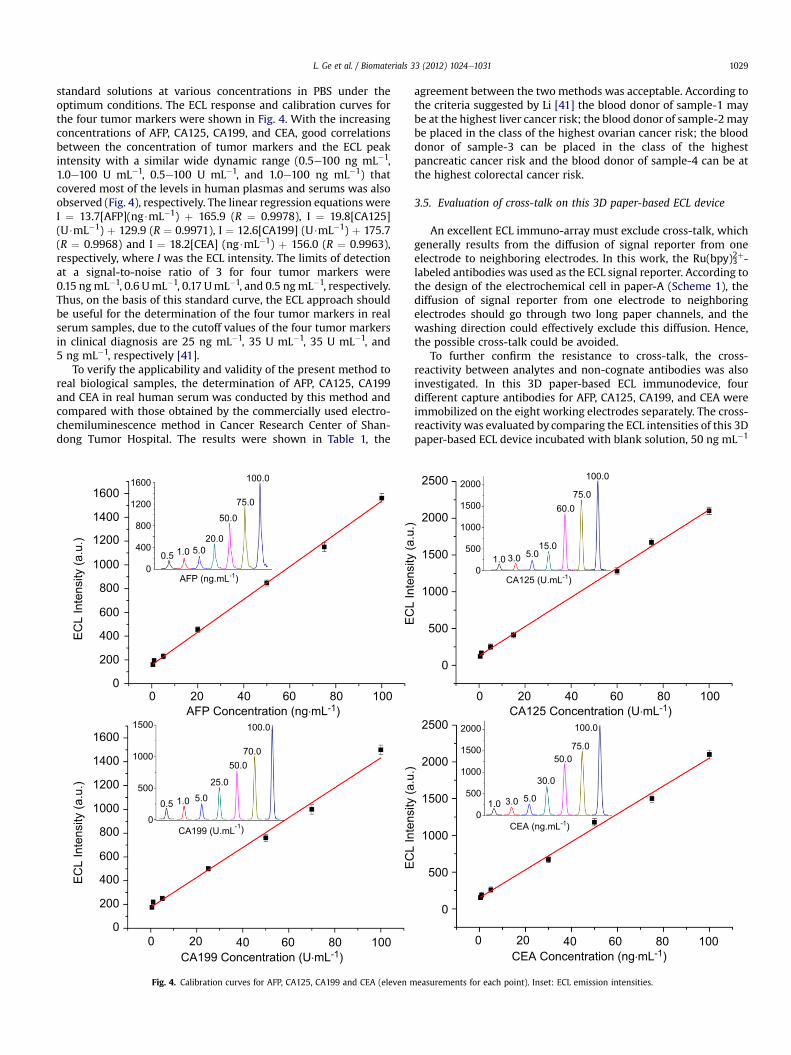
For paper-based point-of-care testing, the analytical cost,sensitivity and time efficiency is very important. For the analyticalcost, reducing the pipetting volume of solutions into working zoneswould decrease the analytical cost remarkably by saving thereagents. Thus, FITC-labeled CA125 antibodies were used to
Fig. 1. SEM images of A) the boundary of wax pattern: left is pure paper, right is wax-printed paper; B) front face of wax-penetrated paper; C) back face of wax-penetratedpaper; D) chitosan modified screen-printed working electrode; E) antibodies/chitosanmodified screen-printed working electrode.
abc
240
200
160
120
80
40
0
Potential (V)
ECL
inte
nsity
(a.u
.)
0.5 0.6 0.7 0.8 0.9 1.0 1.1
Fig. 2. ECL-potential curves of A) background without antigens; B) after a sandwichimmunoreaction with 2.5 U mL�1 CA125 and C) 5.0 U mL�1 CA125.
L. Ge et al. / Biomaterials 33 (2012) 1024e10311028
investigate the optimum pipetting volume of solutions onto eachworking electrode. The investigation procedures included (i)immobilization of the FITC-labeled CA125 antibodies with differentvolumes on the chitosan modified screen-printed working elec-trode on paper-A; (ii) this screen-printed working electrode waswashed twice by following the procedure mentioned above; (iii)the fluorescence images of the paper channel and the workingelectrode were investigated on an inverse fluorescence microscope
Fig. 3. Parameters affecting ECL signals in 3D paper-based ECL immunodevice. A) the pipediameter paper zones); B) effects of incubation time on ECL intensities at 25 U mL�1 CA125,pH value on ECL intensities with 25 U mL�1 CA125 as a model.
(ChangFang CFM-500E, China). Fig. 3A showed that when thepipetting volume was 2.0 mL, the uniformity and sufficiency ofantibodies on the working electrode was obtained. Thus, consid-ering the analytical cost, 2.0 mL was selected as the optimal pipet-ting volume of all reagents. In addition, this small pipetting volumecould further avoid the outflow of the solutions from workingelectrodes to the paper channels and reduce the cross-talk betweenadjacent electrodes to increase the signal-to-background ratio andthe sensitivity of this 3D paper-based ECL immunodevice.
The incubation process was performed at room temperature.The successful development of the multiplex immunoassayrequired that the common incubation time must be suitable for allanalytes. The effects of incubation time on the ECL intensity of this3D paper-based ECL immunodevice were investigated as follows.With an increasing incubation time, all the ECL intensities for25 U mL�1 CA125, CA199 and 25 ng mL�1 AFP and CEA increasedquickly and have reached their maximumvalues at 30min (Fig. 3B),indicating the maximum formation of these sandwich immuno-complexes. Thus, an incubation time of 30 min was selected in thefurther study. As shown in Fig. 3C, the ECL intensity of this 3Dpaper-based ECL immunodevice showed great dependence on thepH value of the solution. The ECL intensity increased from 5.5 to 8.0,and the maximum was observed at pH 7.4e8.0. Since the optimalpH value for the biological systems was 7.4, the ECL detection wasperformed in pH 7.4 PBS containing 0.01 mM TPA.
3.4. Analytical performance
The analytical performance of this method was verified byapplying 2.0 mL of samples of human AFP, CA125, CA199 and CEA
tting volume affects the uniformity and sufficiency of protein immobilization (4 mmCA199 and 25 ng mL�1 AFP, CEA concentration, where n ¼ 11 for each point; C) effect of
L. Ge et al. / Biomaterials 33 (2012) 1024e1031 1029
standard solutions at various concentrations in PBS under theoptimum conditions. The ECL response and calibration curves forthe four tumor markers were shown in Fig. 4. With the increasingconcentrations of AFP, CA125, CA199, and CEA, good correlationsbetween the concentration of tumor markers and the ECL peakintensity with a similar wide dynamic range (0.5e100 ng mL�1,1.0e100 U mL�1, 0.5e100 U mL�1, and 1.0e100 ng mL�1) thatcovered most of the levels in human plasmas and serums was alsoobserved (Fig. 4), respectively. The linear regression equations wereI ¼ 13.7[AFP](ng$mL�1) þ 165.9 (R ¼ 0.9978), I ¼ 19.8[CA125](U$mL�1) þ 129.9 (R ¼ 0.9971), I ¼ 12.6[CA199] (U$mL�1) þ 175.7(R ¼ 0.9968) and I ¼ 18.2[CEA] (ng$mL�1) þ 156.0 (R ¼ 0.9963),respectively, where I was the ECL intensity. The limits of detectionat a signal-to-noise ratio of 3 for four tumor markers were0.15 ngmL�1, 0.6 UmL�1, 0.17 UmL�1, and 0.5 ngmL�1, respectively.Thus, on the basis of this standard curve, the ECL approach shouldbe useful for the determination of the four tumor markers in realserum samples, due to the cutoff values of the four tumor markersin clinical diagnosis are 25 ng mL�1, 35 U mL�1, 35 U mL�1, and5 ng mL�1, respectively [41].
To verify the applicability and validity of the present method toreal biological samples, the determination of AFP, CA125, CA199and CEA in real human serum was conducted by this method andcompared with those obtained by the commercially used electro-chemiluminescence method in Cancer Research Center of Shan-dong Tumor Hospital. The results were shown in Table 1, the
1600
1200
ECL
Inte
nsity
(a.u
.)
800
400
00.5 1.0 5.0
20.0
50.0
75.0
100.01600
1400
1200
1000
800
600
400
200
0
ECL
Inte
nsity
(a.u
.)
1600
1400
1200
1000
800
600
400
200
0
0 20 40 60 80
1500
1000
500
0
0.5 1.0 5.0
25.0
50.070.0
100.0
AFP (ng.mL )
CA199 (U.mL )
100
0 20 40 60 80 100
AFP Concentration (ng.mL )
CA199 Concentration (U.mL )
Fig. 4. Calibration curves for AFP, CA125, CA199 and CEA (eleven m
agreement between the two methods was acceptable. According tothe criteria suggested by Li [41] the blood donor of sample-1 maybe at the highest liver cancer risk; the blood donor of sample-2maybe placed in the class of the highest ovarian cancer risk; the blooddonor of sample-3 can be placed in the class of the highestpancreatic cancer risk and the blood donor of sample-4 can be atthe highest colorectal cancer risk.
3.5. Evaluation of cross-talk on this 3D paper-based ECL device
An excellent ECL immuno-array must exclude cross-talk, whichgenerally results from the diffusion of signal reporter from oneelectrode to neighboring electrodes. In this work, the Ru(bpy)32þ-labeled antibodies was used as the ECL signal reporter. According tothe design of the electrochemical cell in paper-A (Scheme 1), thediffusion of signal reporter from one electrode to neighboringelectrodes should go through two long paper channels, and thewashing direction could effectively exclude this diffusion. Hence,the possible cross-talk could be avoided.
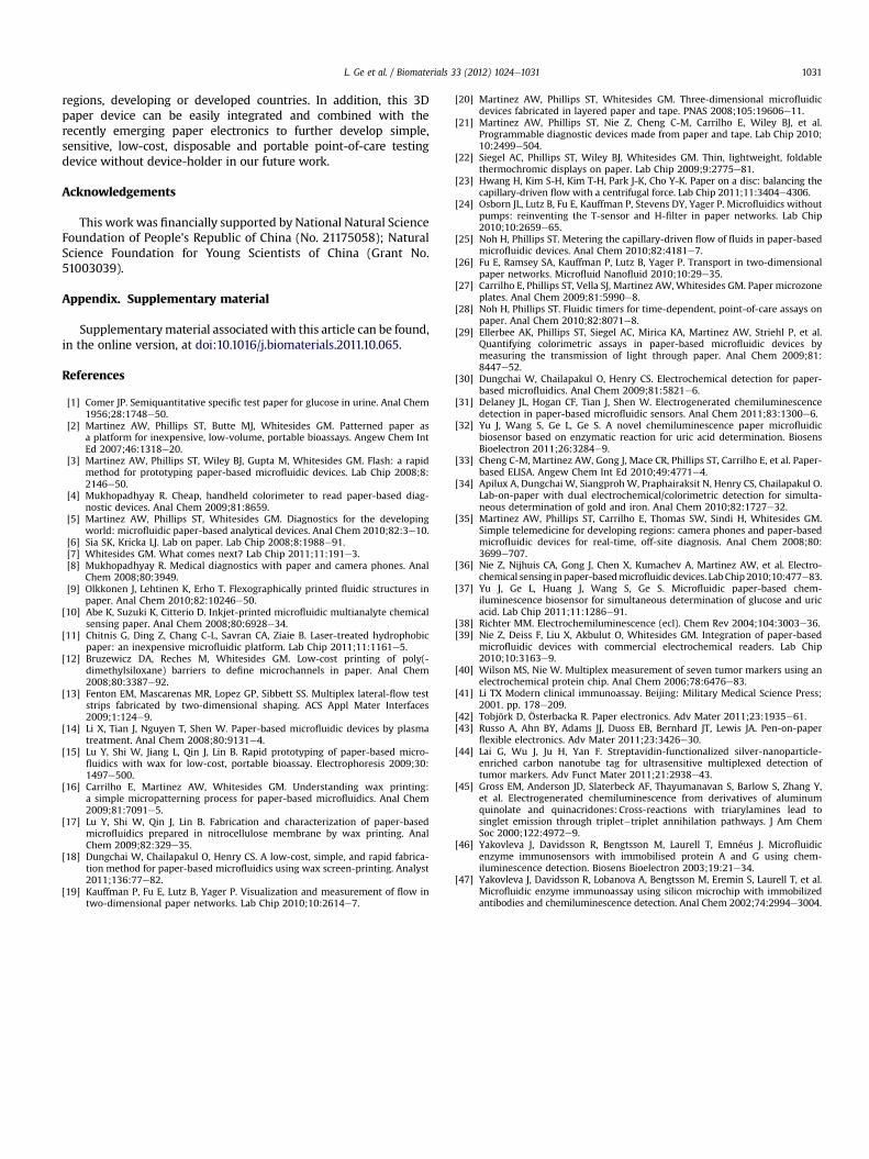
To further confirm the resistance to cross-talk, the cross-reactivity between analytes and non-cognate antibodies was alsoinvestigated. In this 3D paper-based ECL immunodevice, fourdifferent capture antibodies for AFP, CA125, CA199, and CEA wereimmobilized on the eight working electrodes separately. The cross-reactivity was evaluated by comparing the ECL intensities of this 3Dpaper-based ECL device incubated with blank solution, 50 ng mL�1
15.05.03.01.0
60.075.0
100.0
ECL
Inte
nsity
(a.u
.)
2500
2000
1500
1000
500
0
ECL
Inte
nsity
(a.u
.)
2500
2000
1500
1000
500
0
2000
1500
1000
500
0
30.0
5.03.01.0
50.075.0
100.02000
1500
1000
500
0
CA125 (U.mL )
CEA (ng.mL )
0 20 40 60 80 100
0 20 40 60 80
CA125 Concentration (U.mL )
CEA Concentration (ng.mL )100
easurements for each point). Inset: ECL emission intensities.
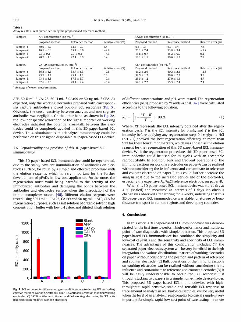
Table 1Assay results of real human serum by the proposed and reference method.
Samples AFP concentration (ng mL�1) CA125 concentration (U$mL�1)
Proposed method Reference method Relative error (%) Proposed method Reference method Relative error (%)
Sample�1 60.9 � 2.2 63.2 � 2.7 3.5 6.2 � 0.3 6.7 � 0.4 7.6Sample�2 16.1 � 0.3 15.4 � 0.6 �4.9 75.1 � 2.4 73.8 � 3.4 �1.7Sample�3 7.9 � 0.2 7.7 � 0.3 �4.3 13.8 � 0.7 15.2 � 0.9 9.2Sample�4 20.7 � 1.0 22.1 � 0.9 6.4 19.1 � 1.1 19.6 � 1.3 2.8
Samples CA199 concentration (U$mL�1) CEA concentration (ng mL�1)Proposed method Reference method Relative error (%) Proposed method Reference method Relative error (%)
Sample�1 36.2 � 1.6 33.7 � 1.3 �7.5 41.2 � 2.0 40.2 � 2.1 �2.5Sample�2 23.9 � 1.1 25.4 � 1.1 5.9 37.9 � 1.7 38.6 � 2.3 3.9Sample�3 93.8 � 3.3 87.6 � 3.7 �7.1 26.5 � 1.2 27.9 � 1.4 4.7Sample�4 52.6 � 2.0 49.4 � 2.4 �6.4 54.1 � 2.2 55.3 � 2.4 2.1
* Average of eleven measurements.
L. Ge et al. / Biomaterials 33 (2012) 1024e10311030
AFP, 50 U mL�1 CA125, 50 U mL�1 CA199 or 50 ng mL�1 CEA. Asexpected, only the working electrodes prepared with correspond-ing capture antibodies showed obvious ECL responses (Fig. 5).Obviously, the cross-reactivity between analytes and non-cognateantibodies was negligible. On the other hand, as shown in Fig. 2A,the low nonspecific adsorption of the signal reporter on workingelectrodes indicated the potential cross-talk between the elec-trodes could be completely avoided in this 3D paper-based ECLdevice. Thus, simultaneous multianalyte immunoassay could beperformed on this designed disposable 3D paper-based ECL device.
3.6. Reproducibility and precision of this 3D paper-based ECLimmunodevice
This 3D paper-based ECL immunodevice could be regenerated,due to the stably covalent immobilization of antibodies on elec-trodes surface, for reuse by a simple and effective procedure withthe elution reagents, which is very important for the furtherdevelopment of mPADs in low-cost application. Furthermore, theregeneration must avoid being harmful to the activity of theimmobilized antibodies and damaging the bonds between theantibodies and electrodes surface when the dissociation of theimmunocomplexes occurs [46]. Different elution reagents weretested using 50 U mL�1 CA125, CA199 and 50 ng mL�1 AFP, CEA forregeneration purposes, such as salt solution of organic solvent, highconcentration, buffer with low pH value, and diluted alkali solution
ECL
Inte
nsity
(a.u
.)
1200
1000
800
600
400
200
0A B
BlankAFPCA125CA199CEA
C D
Fig. 5. ECL response for different antigens on different electrodes. A) AFP antibodies/chitosan modified working electrodes; B) CA125 antibodies/chitosan modified workingelectrodes; C) CA199 antibodies/chitosan modified working electrodes; D) CEA anti-bodies/chitosan modified working electrodes.
of different concentrations and pH, were tested. The regenerationefficiencies (REs), proposed by Yakovleva et al. [47], were calculatedaccording to the following equation.
RE ¼�1� RT � B
T
�� 100% (1)
Where, RT represents the ECL intensity obtained after the regen-eration cycle, B is the ECL intensity for blank, and T is the ECLintensity before applying any regeneration step. 0.1 M glycine-HCl(pH 2.1) showed the best regeneration efficiency at more than97% for these four tumor markers, which was chosen as the elutionreagent for the regeneration of this 3D paper-based ECL immuno-device. With the regeneration procedure, this 3D paper-based ECLimmunodevice could be used for 25 cycles with an acceptablereproducibility. In addition, bulk and frequent operations of theimmunoreactions onworking electrodes on paper-A can be realizedwithout considering the its influence and contaminate to referenceand counter electrode on paper-B, this could further decrease theanalysis cost due to the increased service life of the electrodes,especially the expensive Ag/AgCl reference electrode, on paper-B;
When this 3D paper-based ECL immunodevice was stored dry at4 �C (sealed) and measured at intervals of 3 days, No obviouschange was observed after storing for 3 weeks, indicating that this3D paper-based ECL immunodevice was stable for storage or long-distance transport in remote regions and developing countries.
4. Conclusions
In this work, a 3D paper-based ECL immunodevice was demon-strated for the first time to perform high-performance andmultiplexpoint-of-care diagnostics with simple operation. This proposed 3Dpaper-based ECL immunodevice has combined the simplicity andlow-cost of mPADs and the sensitivity and specificity of ECL immu-noassay. The advantages of this configuration includes: (1) theseparated paper-electrodes systemwill be very beneficial to the highintegration and various distributional pattern of working electrodeson paper without considering the position and pattern of referenceand counter electrode; (2) Bulk operations of the immunoreactionson working electrodes can be realized without considering the itsinfluence and contaminate to reference and counter electrode; (3) Itwill be easily understandable to obtain the ECL response justthrough stacking two papers in a simple home-made device-holder.This proposed 3D paper-based ECL immunodevice, with high-throughput, rapid, sensitive, stable and reusable ECL response totrace amount of analyte in real biological samples, will be very usefulwhen the level of an analyte in real complex biological sample is veryimportant for simple, rapid, low-cost point-of-care testing in remote
L. Ge et al. / Biomaterials 33 (2012) 1024e1031 1031
regions, developing or developed countries. In addition, this 3Dpaper device can be easily integrated and combined with therecently emerging paper electronics to further develop simple,sensitive, low-cost, disposable and portable point-of-care testingdevice without device-holder in our future work.
Acknowledgements
This work was financially supported by National Natural ScienceFoundation of People’s Republic of China (No. 21175058); NaturalScience Foundation for Young Scientists of China (Grant No.51003039).
Appendix. Supplementary material
Supplementarymaterial associatedwith this article can be found,in the online version, at doi:10.1016/j.biomaterials.2011.10.065.
References
[1] Comer JP. Semiquantitative specific test paper for glucose in urine. Anal Chem1956;28:1748e50.
[2] Martinez AW, Phillips ST, Butte MJ, Whitesides GM. Patterned paper asa platform for inexpensive, low-volume, portable bioassays. Angew Chem IntEd 2007;46:1318e20.
[3] Martinez AW, Phillips ST, Wiley BJ, Gupta M, Whitesides GM. Flash: a rapidmethod for prototyping paper-based microfluidic devices. Lab Chip 2008;8:2146e50.
[4] Mukhopadhyay R. Cheap, handheld colorimeter to read paper-based diag-nostic devices. Anal Chem 2009;81:8659.
[5] Martinez AW, Phillips ST, Whitesides GM. Diagnostics for the developingworld: microfluidic paper-based analytical devices. Anal Chem 2010;82:3e10.
[6] Sia SK, Kricka LJ. Lab on paper. Lab Chip 2008;8:1988e91.[7] Whitesides GM. What comes next? Lab Chip 2011;11:191e3.[8] Mukhopadhyay R. Medical diagnostics with paper and camera phones. Anal
Chem 2008;80:3949.[9] Olkkonen J, Lehtinen K, Erho T. Flexographically printed fluidic structures in
paper. Anal Chem 2010;82:10246e50.[10] Abe K, Suzuki K, Citterio D. Inkjet-printed microfluidic multianalyte chemical
sensing paper. Anal Chem 2008;80:6928e34.[11] Chitnis G, Ding Z, Chang C-L, Savran CA, Ziaie B. Laser-treated hydrophobic
paper: an inexpensive microfluidic platform. Lab Chip 2011;11:1161e5.[12] Bruzewicz DA, Reches M, Whitesides GM. Low-cost printing of poly(-
dimethylsiloxane) barriers to define microchannels in paper. Anal Chem2008;80:3387e92.
[13] Fenton EM, Mascarenas MR, Lopez GP, Sibbett SS. Multiplex lateral-flow teststrips fabricated by two-dimensional shaping. ACS Appl Mater Interfaces2009;1:124e9.
[14] Li X, Tian J, Nguyen T, Shen W. Paper-based microfluidic devices by plasmatreatment. Anal Chem 2008;80:9131e4.
[15] Lu Y, Shi W, Jiang L, Qin J, Lin B. Rapid prototyping of paper-based micro-fluidics with wax for low-cost, portable bioassay. Electrophoresis 2009;30:1497e500.
[16] Carrilho E, Martinez AW, Whitesides GM. Understanding wax printing:a simple micropatterning process for paper-based microfluidics. Anal Chem2009;81:7091e5.
[17] Lu Y, Shi W, Qin J, Lin B. Fabrication and characterization of paper-basedmicrofluidics prepared in nitrocellulose membrane by wax printing. AnalChem 2009;82:329e35.
[18] Dungchai W, Chailapakul O, Henry CS. A low-cost, simple, and rapid fabrica-tion method for paper-based microfluidics using wax screen-printing. Analyst2011;136:77e82.
[19] Kauffman P, Fu E, Lutz B, Yager P. Visualization and measurement of flow intwo-dimensional paper networks. Lab Chip 2010;10:2614e7.
[20] Martinez AW, Phillips ST, Whitesides GM. Three-dimensional microfluidicdevices fabricated in layered paper and tape. PNAS 2008;105:19606e11.
[21] Martinez AW, Phillips ST, Nie Z, Cheng C-M, Carrilho E, Wiley BJ, et al.Programmable diagnostic devices made from paper and tape. Lab Chip 2010;10:2499e504.
[22] Siegel AC, Phillips ST, Wiley BJ, Whitesides GM. Thin, lightweight, foldablethermochromic displays on paper. Lab Chip 2009;9:2775e81.
[23] Hwang H, Kim S-H, Kim T-H, Park J-K, Cho Y-K. Paper on a disc: balancing thecapillary-driven flow with a centrifugal force. Lab Chip 2011;11:3404e4306.
[24] Osborn JL, Lutz B, Fu E, Kauffman P, Stevens DY, Yager P. Microfluidics withoutpumps: reinventing the T-sensor and H-filter in paper networks. Lab Chip2010;10:2659e65.
[25] Noh H, Phillips ST. Metering the capillary-driven flow of fluids in paper-basedmicrofluidic devices. Anal Chem 2010;82:4181e7.
[26] Fu E, Ramsey SA, Kauffman P, Lutz B, Yager P. Transport in two-dimensionalpaper networks. Microfluid Nanofluid 2010;10:29e35.
[27] Carrilho E, Phillips ST, Vella SJ, Martinez AW,Whitesides GM. Paper microzoneplates. Anal Chem 2009;81:5990e8.
[28] Noh H, Phillips ST. Fluidic timers for time-dependent, point-of-care assays onpaper. Anal Chem 2010;82:8071e8.
[29] Ellerbee AK, Phillips ST, Siegel AC, Mirica KA, Martinez AW, Striehl P, et al.Quantifying colorimetric assays in paper-based microfluidic devices bymeasuring the transmission of light through paper. Anal Chem 2009;81:8447e52.
[30] Dungchai W, Chailapakul O, Henry CS. Electrochemical detection for paper-based microfluidics. Anal Chem 2009;81:5821e6.
[31] Delaney JL, Hogan CF, Tian J, Shen W. Electrogenerated chemiluminescencedetection in paper-based microfluidic sensors. Anal Chem 2011;83:1300e6.
[32] Yu J, Wang S, Ge L, Ge S. A novel chemiluminescence paper microfluidicbiosensor based on enzymatic reaction for uric acid determination. BiosensBioelectron 2011;26:3284e9.
[33] Cheng C-M, Martinez AW, Gong J, Mace CR, Phillips ST, Carrilho E, et al. Paper-based ELISA. Angew Chem Int Ed 2010;49:4771e4.
[34] Apilux A, Dungchai W, Siangproh W, Praphairaksit N, Henry CS, Chailapakul O.Lab-on-paper with dual electrochemical/colorimetric detection for simulta-neous determination of gold and iron. Anal Chem 2010;82:1727e32.
[35] Martinez AW, Phillips ST, Carrilho E, Thomas SW, Sindi H, Whitesides GM.Simple telemedicine for developing regions: camera phones and paper-basedmicrofluidic devices for real-time, off-site diagnosis. Anal Chem 2008;80:3699e707.
[36] Nie Z, Nijhuis CA, Gong J, Chen X, Kumachev A, Martinez AW, et al. Electro-chemical sensing inpaper-basedmicrofluidic devices. LabChip2010;10:477e83.
[37] Yu J, Ge L, Huang J, Wang S, Ge S. Microfluidic paper-based chem-iluminescence biosensor for simultaneous determination of glucose and uricacid. Lab Chip 2011;11:1286e91.
[38] Richter MM. Electrochemiluminescence (ecl). Chem Rev 2004;104:3003e36.[39] Nie Z, Deiss F, Liu X, Akbulut O, Whitesides GM. Integration of paper-based
microfluidic devices with commercial electrochemical readers. Lab Chip2010;10:3163e9.
[40] Wilson MS, Nie W. Multiplex measurement of seven tumor markers using anelectrochemical protein chip. Anal Chem 2006;78:6476e83.
[41] Li TX Modern clinical immunoassay. Beijing: Military Medical Science Press;2001. pp. 178e209.
[42] Tobjörk D, Österbacka R. Paper electronics. Adv Mater 2011;23:1935e61.[43] Russo A, Ahn BY, Adams JJ, Duoss EB, Bernhard JT, Lewis JA. Pen-on-paper
flexible electronics. Adv Mater 2011;23:3426e30.[44] Lai G, Wu J, Ju H, Yan F. Streptavidin-functionalized silver-nanoparticle-
enriched carbon nanotube tag for ultrasensitive multiplexed detection oftumor markers. Adv Funct Mater 2011;21:2938e43.
[45] Gross EM, Anderson JD, Slaterbeck AF, Thayumanavan S, Barlow S, Zhang Y,et al. Electrogenerated chemiluminescence from derivatives of aluminumquinolate and quinacridones: Cross-reactions with triarylamines lead tosinglet emission through triplet�triplet annihilation pathways. J Am ChemSoc 2000;122:4972e9.
[46] Yakovleva J, Davidsson R, Bengtsson M, Laurell T, Emnéus J. Microfluidicenzyme immunosensors with immobilised protein A and G using chem-iluminescence detection. Biosens Bioelectron 2003;19:21e34.
[47] Yakovleva J, Davidsson R, Lobanova A, Bengtsson M, Eremin S, Laurell T, et al.Microfluidic enzyme immunoassay using silicon microchip with immobilizedantibodies and chemiluminescence detection. Anal Chem 2002;74:2994e3004.