Three-Dimensional Knee Joint Moments During Performance of ...
12
33 Journal of Applied Biomechanics, 2013, 29, 33-43 © 2013 Human Kinetics, Inc. Sivan Almosnino (Corresponding Author) is with the Biome- chanics and Ergonomics Laboratory, School of Kinesiology and Health Studies, Queen’s University, Kingston, ON, Canada, and with the Human Mobility Research Centre, Queen’s University & Kingston General Hospital, Kingston, ON, Canada. David Kingston and Ryan B. Graham are with the Biomechanics and Ergonomics Laboratory, School of Kinesiology and Health Studies, Queen’s University, Kingston, ON, Canada. Three-Dimensional Knee Joint Moments During Performance of the Bodyweight Squat: Effects of Stance Width and Foot Rotation Sivan Almosnino, 1,2 David Kingston, 1 and Ryan B. Graham 1 1 Queen’s University; 2 Queen’s University & Kingston General Hospital The purpose of this investigation was to assess the effects of stance width and foot rotation angle on three- dimensional knee joint moments during bodyweight squat performance. Twenty-eight participants performed 8 repetitions in 4 conditions differing in stance or foot rotation positions. Knee joint moment waveforms were subjected to principal component analysis. Results indicated that increasing stance width resulted in a larger knee flexion moment magnitude, as well as larger and phase-shifted adduction moment waveforms. The knee’s internal rotation moment magnitude was significantly reduced with external foot rotation only under the wide stance condition. Moreover, squat performance with a wide stance and externally rotated feet resulted in a flattening of the internal rotation moment waveform during the middle portion of the movement. However, it is speculated that the differences observed across conditions are not of clinical relevance for young, healthy participants. Keywords: three-dimensional, inverse dynamics, waveform, principal component analysis The bodyweight squat is prescribed in athletic and rehabilitation settings, where it is used for both training and evaluation of movement competency (Butler, et al., 2010; Kiesel, et al., 2007; Kritz, et al., 2009). The attractiveness of this exercise lays in the fact that no specialized equipment is required for performance, the physical space necessary for carrying out the movement is relatively small, the exercise resembles the performance of several everyday tasks (Schoenfeld, 2010), and several simple modifications may be made to affect training dosage and intensity. Although the squat is a multijoint movement involv- ing the recruitment of several muscle groups (Flanagan et al., 2003; Gullett et al., 2009; Schoenfeld, 2010), much of the literature pertaining to the biomechanical description of the exercise is concerned with altera- tions to knee joint mechanics and associated muscular activity (Escamilla, 2001a). In this respect, particular attention has been paid to modifications of the initial stance width and foot rotation angle adopted by the trainee (Boyden, et al., 2000; Escamilla et al., 2001b,c; McCaw & Melrose, 1999; Ninos et al., 1997; Paoli et al., 2009; Singorile et al., 1995). However, the majority of investigations concerned with quantifying the effects of the aforementioned modifications on knee kinetics have primarily used two-dimensional sagittal analyses, with the efforts themselves performed with various magnitudes of external resistance. As such, there appears to be a lack of information regarding three-dimensional (3D) knee joint moments during performance of the bodyweight squat, with specific reference to the effects of exercise modifications on external demands about any of the knee joint’s axes of rotation. Such information may be useful for optimal exercise prescription, as knowledge of which modifications result in higher moments would allow trainers to make educated decisions as to which modification to recommend as a function of a trainee’s health and training status. Considering the lack of information related to 3D knee joint moments during the performance of the body- weight squat in general and specifically with regards to altering initial stance width or foot rotation, a study aimed at quantifying possible effects should be considered exploratory. A suitable statistical procedure to be used in this case is Principal Component Analysis (PCA). The favorable use of PCA for exploratory purposes stems from two underlying characteristics of the procedure. The first is that calculations use data of the entire moment wave- form, and thus essential waveform characteristics that may contribute to the variance between conditions are not overlooked, which may occur if a-priori discrete-time An Official Journal of ISB www.JAB-Journal.com ORIGINAL RESEARCH
Transcript of Three-Dimensional Knee Joint Moments During Performance of ...

33
Journal of Applied Biomechanics, 2013, 29, 33-43 © 2013 Human Kinetics, Inc.
Sivan Almosnino (Corresponding Author) is with the Biome-chanics and Ergonomics Laboratory, School of Kinesiology and Health Studies, Queen’s University, Kingston, ON, Canada, and with the Human Mobility Research Centre, Queen’s University & Kingston General Hospital, Kingston, ON, Canada. David Kingston and Ryan B. Graham are with the Biomechanics and Ergonomics Laboratory, School of Kinesiology and Health Studies, Queen’s University, Kingston, ON, Canada.
Three-Dimensional Knee Joint Moments During Performance of the Bodyweight Squat:
Effects of Stance Width and Foot Rotation
Sivan Almosnino,1,2 David Kingston,1 and Ryan B. Graham1
1Queen’s University; 2Queen’s University & Kingston General Hospital
The purpose of this investigation was to assess the effects of stance width and foot rotation angle on three-dimensional knee joint moments during bodyweight squat performance. Twenty-eight participants performed 8 repetitions in 4 conditions differing in stance or foot rotation positions. Knee joint moment waveforms were subjected to principal component analysis. Results indicated that increasing stance width resulted in a larger knee flexion moment magnitude, as well as larger and phase-shifted adduction moment waveforms. The knee’s internal rotation moment magnitude was significantly reduced with external foot rotation only under the wide stance condition. Moreover, squat performance with a wide stance and externally rotated feet resulted in a flattening of the internal rotation moment waveform during the middle portion of the movement. However, it is speculated that the differences observed across conditions are not of clinical relevance for young, healthy participants.
Keywords: three-dimensional, inverse dynamics, waveform, principal component analysis
The bodyweight squat is prescribed in athletic and rehabilitation settings, where it is used for both training and evaluation of movement competency (Butler, et al., 2010; Kiesel, et al., 2007; Kritz, et al., 2009). The attractiveness of this exercise lays in the fact that no specialized equipment is required for performance, the physical space necessary for carrying out the movement is relatively small, the exercise resembles the performance of several everyday tasks (Schoenfeld, 2010), and several simple modifications may be made to affect training dosage and intensity.
Although the squat is a multijoint movement involv-ing the recruitment of several muscle groups (Flanagan et al., 2003; Gullett et al., 2009; Schoenfeld, 2010), much of the literature pertaining to the biomechanical description of the exercise is concerned with altera-tions to knee joint mechanics and associated muscular activity (Escamilla, 2001a). In this respect, particular attention has been paid to modifications of the initial stance width and foot rotation angle adopted by the
trainee (Boyden, et al., 2000; Escamilla et al., 2001b,c; McCaw & Melrose, 1999; Ninos et al., 1997; Paoli et al., 2009; Singorile et al., 1995). However, the majority of investigations concerned with quantifying the effects of the aforementioned modifications on knee kinetics have primarily used two-dimensional sagittal analyses, with the efforts themselves performed with various magnitudes of external resistance. As such, there appears to be a lack of information regarding three-dimensional (3D) knee joint moments during performance of the bodyweight squat, with specific reference to the effects of exercise modifications on external demands about any of the knee joint’s axes of rotation. Such information may be useful for optimal exercise prescription, as knowledge of which modifications result in higher moments would allow trainers to make educated decisions as to which modification to recommend as a function of a trainee’s health and training status.
Considering the lack of information related to 3D knee joint moments during the performance of the body-weight squat in general and specifically with regards to altering initial stance width or foot rotation, a study aimed at quantifying possible effects should be considered exploratory. A suitable statistical procedure to be used in this case is Principal Component Analysis (PCA). The favorable use of PCA for exploratory purposes stems from two underlying characteristics of the procedure. The first is that calculations use data of the entire moment wave-form, and thus essential waveform characteristics that may contribute to the variance between conditions are not overlooked, which may occur if a-priori discrete-time
An Official Journal of ISBwww.JAB-Journal.comORIGINAL RESEARCH

34 Almosnino, Kingston, and Graham
point variables are subjectively chosen for extraction (Astephen & Deluzio, 2009; Deluzio et al., 1999; Wrigley et al., 2006). The second is that, in human movement related research, it seems that the majority of the vari-ance between conditions is usually contained within the first few principal components (PCs). This allows for a substantial reduction in the amount of data that may be further explored (Deluzio et al., 1997; Reid et al., 2010).
The purpose of this investigation was thus to assess, using an exploratory approach, the effects of stance width and foot rotation angle on three-dimensional knee joint moments during performance of the bodyweight squat exercise.
Methods
Participants
A convenience sample was recruited through direct contact and advertisements posted within the university. Potential participants were screened using a self-report medical questionnaire aimed at identifying any previ-ous injuries to the lower extremities or spine; none of the participants were excluded. The sample consisted of 28 individuals (17 men: age 23 ± 4 years; mass 82 ± 15 kg; height 178 ± 8 cm; and 11 women: age 24 ± 6 years; mass 65 ± 10 kg; height 167 ± 9 cm). On average, the participants were involved in 6.6 ± 3.5 weekly hours of physical activity in a variety of individual and team sports at the recreational or competitive university level. All participants used the squat exercise as part of their regular exercise routines. Procedures for this investiga-tion were approved by the University’s General Research Ethics Board, and written informed consent was obtained from all participants before testing.
Instrumentation
Three-dimensional kinematic data of the participant’s dominant leg, pelvis, and trunk were obtained using a two-camera, Optotrak 3020 motion capture system (Northern Digital, Waterloo, ON, Canada) sampling at 100 Hz. Marker clusters containing three non-collinear infrared light-emitting diodes affixed to small rigid plates were attached to the dorsal aspect of the foot using a fin that was secured to the shoe using the shoelaces and tape; the distal lateral aspect of the shank two inches above the lateral malleolus and to the thigh approximately two inches above the lateral epicondyle, both using flexible neoprene bands; and to the sacrum and trunk at the level of the T-12 vertebra using custom designed fins and flexible bands. A digitizing pointed probe fitted with a cluster of six markers was used to obtain virtual markers from the 1st metatarsal head, 5th metatarsal head, right and left malleoli, right and left femoral epicondyles, and right and left anterior superior iliac spines. These virtual markers were later used to define joint center of rotations and anatomical coordinate systems. During trial perfor-mance, ground reaction force and moment data were obtained from a strain gauge force platform embedded
in the floor (model OR5–7-1000, AMTI, Watertown, MA, USA). Force plate data were sampled at 1000 Hz, and synchronized with kinematic data recordings using a 16 channel Optotrak Digital Acquisition Unit (ODAU).
Procedures
Participants performed 4 squat conditions differing in either stance width or foot rotation angle: 1) Shoulder width parallel (SWP): assuming a shoulder width stance with the feet parallel to the long axis of the force plate; 2) Shoulder width rotated (SWR): assuming a shoulder width stance with feet externally rotated 30° with respect to the long axis of the force plate; 3) Wide stance parallel (WSP): assuming a stance of 140% shoulder width with feet parallel to the long axis of the force plate, and; 4) Wide stance rotated (WSR): assuming a stance of 140% of shoulder width with the feet externally rotated by 30°. To replicate the starting positions between trials, stance width and foot rotation orientations were guided using adhesive tape that was placed on the floor (for the untested leg), and on the force plate (for the tested leg). For all conditions, the participants were instructed to keep their heels in contact with the ground at all times; to squat to a depth such that their inguinal fold was at the same level as the superior aspect of their knee (Fry et al., 2003); to keep their trunk straight and at a constant angle with relationship to the horizontal throughout per-formance; to keep the knees in line with the feet (i.e., not to bend the knees into varus or valgus) and to maintain the anterior aspect of the knee behind the toe line; to cross the arms such that the hands are touching the contra lateral shoulders; and to keep the head in a neutral and constant position (Kritz et al., 2009). A single examiner observed all squat performances, and in the case of any deviations from the predefined movement requirements, the trial was repeated. Movement time across conditions was controlled using an examiner count superimposed on a recording of a digital metronome set to 88 beats per minute (provided in supplementary audio file). The participant performed 4 randomized sets of 8 repetitions in each condition for a total of 32 trials. Within each set, trials were separated by 10 s, while a 2-min rest period was given in between sets. To eliminate fatigue as a poten-tial confounder, a Borg 6–20 rating of perceived exertion (RPE) scale was administered after each repetition (Borg, 1982). If the participant indicated an RPE score that was a minimum of 2 scores above the one indicated for the initial trial, an additional rest period was given before the performance of the next repetition. The length of this additional rest period corresponded to the time needed for the participant to self-report full recovery. It should be noted that only two subjects required additional rest on a single occasion.
Data Analysis
Before calculations, kinematic data were filtered using a zero-phase, 4th order, low pass recursive Butterworth filter with varying cutoff frequencies ranging from 5 to

3D Knee Joint Moments and Performance of the Bodyweight Squat 35
7 Hz, as determined by residual analysis (Winter, 2009). For force plate data, DC bias was removed utilizing an unweighted force plate trial. Joint centers of rotation for the ankle and knee joints were defined as the midpoint between the medial and lateral malleoli and epicondyle, respectively (Brandon and Deluzio, 2010). The hip joint center was defined using a functional method (Camomilla et al., 2006; Gamage & Lasenby, 2002). Three segmen-tal anatomical coordinate systems were subsequently established: The femur anatomical coordinate system was defined based on the hip joint center, the knee joint center and the lateral epicondyle. The tibia anatomical coordinate system was defined by the knee joint center, the ankle joint center and lateral malleolus. The foot anatomical coordinate system was constructed with the ankle joint center, the calcaneus, and the midpoint of the 1st and 5th metatarsals. For each anatomical coordinate system, x-axis was defined to point from the distal end to the proximal end of the body segment, y-axis was defined to point along the anterior direction of the body segment, and the z-axis was defined to point in the medial direc-tion of the body segment. Following, three-dimensional joint angles were calculated using a ZYX Euler rotation sequence; corresponding to flexion-adduction-internal rotation. In addition, 3D segmental trunk angles were also extracted. Knee joint angles and trunk orientation were referenced to a static standing trial.
Next, instantaneous net external knee joint moments were calculated using inverse dynamics procedures. For calculation purposes, the model used ground reaction forces and moments, sex-based segmental inertial charac-teristics (Zatsiorsky, 2002), and segmental displacement, velocity and acceleration data. In the joint moment sign convention, knee flexion, adduction, and internal rotation are defined as positive. Movement start and end periods were defined when the sagittal plane knee angle exceeded and fell below 3 SD of baseline level for a period of 200 ms, and each repetition’s start and end periods were verified by visual inspection by a single examiner. All moment waveforms were amplitude-normalized to the product of the participant’s body weight (kg) and height (m). In addition, each individual repetition period was interpolated using cubic splines to a uniform length of 201 data points, which represented 0–100% of the movement phase in 0.5% increments. All analyses were performed using Matlab version 7.5 (The Mathworks, Natick, MA, USA).
Statistical Analysis
Representative moment waveforms for each participant in each condition were attained by ensemble averaging across trials. These waveforms were subsequently sub-jected to Principal Component Analysis (PCA). Proce-dures for PCA implementation for waveform analyses are detailed elsewhere (Astephen &Deluzio, 2009; Deluzio & Astephen, 2007; Deluzio et al., 1999; Wrigley et al., 2006). For data reduction purposes, the number of PCs retained for further analysis was determined using a 90% trace criterion (Reid et al., 2010). Subsequently,
differences in PC scores between conditions were assessed using a 2-way, repeated-measures analysis of variance (ANOVA), with the within-participant factors being stance width and foot rotation. The alpha level for these comparisons was preset at 0.05 with Bonfer-roni adjustment to account for multiple comparisons. Complementary effect size estimates are reported using generalized omega squared (ω2) for repeated measures experimental design (Olejnik & Algina, 2003).
For interpretation of the results of those PCs found to statistically differ across conditions, two approaches were used: 1) inspection of associated loading vector plots, and 2) inspection of plots of the mean temporal waveform across all conditions, bounded by the contribution of a single PC pattern of variance, which in turn, is scaled by one standard deviation of the group PC-scores (Brandon et al., 2013; Ramsay & Silverman, 1997; O’Connor & Bottum, 2009). This latter technique differs from common method of examination of extreme score subject waveforms (e.g., Astephen & Deluzio, 2009; Reid et al., 2010) in that the variance contributions of other PCs to the representative waveforms being examined are omit-ted (Brandon et al., 2011; Ramsay & Silverman, 1997).
In addition, three separate one-way repeated-mea-sures ANOVAs were conducted to discern any differ-ences in average 3D trunk orientation across conditions (α = .05).
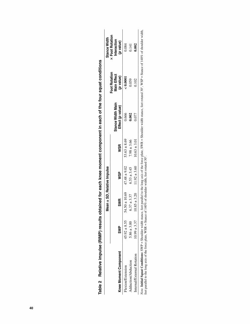
Lastly, to assess whether PCA offers any advantages over conventional moment waveform analysis, the relative absolute impulse (RIMP; absolute area under the ampli-tude and time-normalized moment curve) was extracted from each participants’ ensemble averaged joint moment curve in each squat condition (Hamill et al., 1983). The choice to extract RIMP as a representative measure was due to the fact that the calculation utilizes data of the entire waveform, and hence is similar in this respect to PCA. In addition, the choice of RIMP is in line with the aim of the investigation, namely to assess the influence of certain modifications on external demands about the knee joint throughout the entire movement cycle. Further-more, the strength of the associations between absolute peak moment and RIMP values were quite strong (r2 = .67, .82, and .88 for the association between peak and relative impulse for flexion moment, abduction moment, and internal rotation moment components, respectively), and as such reporting of peak values would be inherently redundant. 2-way repeated-measures ANOVAs were used to assess the influence of the two within-participant factors on RIMP values, with alpha preset at 0.05.
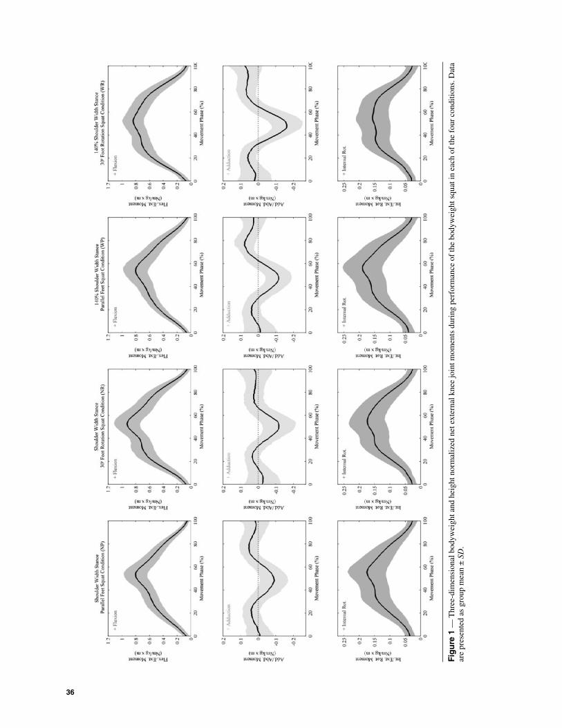
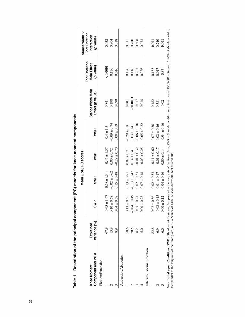
ResultsPrincipal component models were produced for the knee joint moment data (Figure 1) and a total of 10 PC’s were retained utilizing the 90% trace criterion (Table 1). Of these, only 5 PC’s were statistically significant (p ≤ .001). The interpretation of these PC’s was achieved via examination of Figure 2; consisting of loading vectors, ensemble mean waveforms bounded by ± 1 SD single PC variance patterns, and interaction plots.
36
Fig
ure
1 —
Thr
ee-d
imen
sion
al b
odyw
eigh
t and
hei
ght n
orm
aliz
ed n
et e
xter
nal k
nee
join
t mom
ents
dur
ing
perf
orm
ance
of
the
body
wei
ght s
quat
in e
ach
of th
e fo
ur c
ondi
tions
. Dat
a ar
e pr
esen
ted
as g
roup
mea
n ±
SD
.

37
Fig
ure
2 —
Pri
ncip
al c
ompo
nent
ana
lysi
s re
sults
of
3D k
nee
join
t mom
ent w
avef
orm
s. 1
st r
ow: G
roup
mea
n m
omen
t wav
efor
m f
or e
ach
cond
ition
in w
hich
sta
tistic
al s
igni
fican
ce
was
det
ecte
d. 2
nd r
ow: L
oadi
ng v
ecto
r pl
ots
of th
e PC
of
inte
rest
. 3rd
row
: Ens
embl
e m
omen
t cur
ve a
cros
s al
l con
ditio
ns ±
1 S
D o
f th
e va
riat
ion
due
to th
e PC
load
ing
vect
or s
how
n in
the
2nd
row
. 4th
row
: Int
erac
tion
plot
s.
38
Tab
le 1
D
escr
ipti
on
of
the
pri
nci
pal
co
mp
on
ent
(PC
) m
od
els
for
knee
mo
men
t co
mp
on
ents
Kne
e M
omen
t C
ompo
nent
and
PC
#E
xpla
ined
Va
rianc
e (%
)
Mea
n ±
SD
, PC
sco
res
Sta
nce
Wid
th M
ain
Effe
ct ( p
val
ue)
Foot
Rot
atio
n M
ain
Effe
ct
( p v
alue
)
Sta
nce
Wid
th ×
Fo
ot R
otat
ion
Inte
ract
ion
( p
val
ue)
SW
PS
WR
WS
PW
SR
Flex
ion/
Ext
ensi
on
167
.9–0
.65
± 1
.67
0.68
.±1.
34–0
.45
± 1
.37
0.4
± 1
.30.
841
< 0.
0001
0.03
22
13.9
0.10
± 0
.68
–0.0
2 ±
0.6
20.
00 ±
0.7
2–0
.09
± 0
.74
0.19
80.
176
0.80
43
8.9
0.04
± 0
.68
0.15
± 0
.48
–0.2
9 ±
0.7
00.
08 ±
0.5
90.
090
0.01
60.
018
Add
uctio
n/A
bduc
tion
158
.60.
13 ±
0.6
50.
13 ±
0.8
10.
02 ±
0.7
1–0
.29
± 0
.81
0.00
10.
180
0.01
12
20.5
–0.0
4 ±
0.4
9–0
.13
± 0
.47
0.14
± 0
.41
0.03
± 0
.41
< 0.
0001
0.11
60.
780
38.
20.
05 ±
0.2
10.
02 ±
0.3
3–0
.01
± 0
.32
–0.0
6 ±
0.3
60.
017
0.20
70.
808
45.
00.
00 ±
0.2
30.
07 ±
0.1
8–0
.03
± 0
.25
–0.0
5 ±
0.2
20.
014
0.33
60.
073
Inte
rnal
/Ext
erna
l Rot
atio
n
182
.80.
02 ±
0.5
60.
02 ±
0.5
3–0
.11
± 0
.60
0.07
± 0
.50
0.18
20.
153
0.00
12
6.9
–0.0
2 ±
0.1
30.
01 ±
0.1
7–0
.01
± 0
.17
0.02
± 0
.16
0.38
10.
017
0.74
03
6.0
0.00
± 0
.12
0.04
± 0
.16
0.00
± 0
.14
–0.0
4 ±
0.1
60.
020.
870.
001
Not
e. I
niti
al S
quat
Con
diti
ons:
SW
P =
Sho
ulde
r w
idth
sta
nce,
fee
t par
alle
l to
the
long
axi
s of
the
forc
e pl
ate,
SW
R =
Sho
ulde
r w
idth
sta
nce,
fee
t rot
ated
30°
, WSP
= S
tanc
e of
140
% o
f sh
ould
er w
idth
, fe
et p
aral
lel t
o th
e lo
ng a
xis
of th
e fo
rce
plat
e, W
SR =
Sta
nce
of 1
40%
of
shou
lder
wid
th, f
eet r
otat
ed 3
0°.
3D Knee Joint Moments and Performance of the Bodyweight Squat 39
A significant main effect of foot rotation is noted for PC1 of the knee flexion moment (ω2 = 0.31). The magnitude of the corresponding loading vector is positive throughout the entire cycle, with two conspicuous peaks at approximately 35% and 55% of the cycle, signifying an increased variability between the parallel and foot rotated conditions at these instances. Supported by examination of the single PC reconstructed plot; this PC is interpreted as a measure of overall magnitude difference between the parallel and foot rotated conditions, with foot rotation sig-nificantly increasing overall flexion moment magnitude.
With regards to the adduction moment, no effect of foot rotation was noted across conditions. However, a wide stance resulted in significantly lower adduction moment PC1 and PC2 scores (ω2 = 0.18 and 0.19, respec-tively). A lower adduction moment PC1 score indicates a larger adduction moment range. In addition, it is evident that in the initial and last thirds of the movement cycle, those with a low PC1 score experience a knee adduction moment, while in the middle third of the cycle, these par-ticipants experience an abduction moment. Conversely, those with high PC1 scores tend to experience an abduc-tion moment in the initial and final 20% of the cycle, while in the 60% of the cycle a conspicuous double peak occurs with the moment reversing sign from adduction to abduction and back to adduction. In terms of PC2 of the adduction moment, a phase shift is noted whereby during the performance of the squat in the wide stance conditions there is a tendency to reach the peak abduction moment slightly earlier in the movement cycle.
Results for PC1 of the knee internal rotation moment reveal a significant interaction between stance width and foot rotation (ω2 = 0.17). In the wide stance and rotated feet position, internal rotation moment was of lesser magnitude throughout the entire movement cycle, in comparison with performance with parallel feet. There appears to be no difference with respect to the narrower width squat condition, irrespective of foot positioning. In addition, a significant interaction was noted for PC3 of the knee internal rotation moment (ω2 = 0.165). During squat performance with parallel feet, there appears to be no difference in the internal rotation moment. How-ever, during performance of the wide stance with the feet rotated outwards, there appears to be a flattening of the moment waveform during the middle portion of the movement phase. Conversely, during performance of the squat with a shoulder width stance and the feet rotated, the moment waveform exhibits a distinct peak approximately midcycle.
Results for relative impulse (RIMP) values obtained for each knee joint moment component during perfor-mance of the four exercise variations are summarized in Table 2. For the flexion moment, RIMP values were significantly higher when performance was done with rotated feet. These results are in agreement with both the results and interpretation of PC1 as being a magnitude operator.
With regards to the adduction moment component, performance of the exercise at a stance of 140% of
shoulder width resulted in significantly higher RIMP values in comparison shoulder width stance performance. It may be argued that this finding is in agreement with the results and interpretation of adduction moment PC1 as being a range operator. However, RIMP values do not identify the alterations in moment sign throughout the movement cycle between those that scored low or high on PC1, nor are RIMP values sensitive to the phase shift identified by PC2.
Results for RIMP values obtained for the internal rotation joint moment component reveal a significant interaction between stance width and foot rotation. In specifics, during performance with a wide stance and rotated feet, RIMP was smaller than when performance was done with a wide stance and parallel feet. In addi-tion, no differences in RIMP values were noticed when squats were performed with a shoulder width stance, regardless of the foot position employed. These results are concurrent with the interpretation of PC1 scores; however they do not reveal the subtle flattening of the moment waveform in the middle portion of the movement cycle captured by PC3.
Lastly, repeated-measures ANOVA revealed no statistically significant differences in average 3D trunk orientation across conditions (p > .3 for all comparisons). During the performance of each condition, the mean normalized time period where the maximum knee flexion angle was reached was NP 52.5 ± 3.5%, NR 53 ± 4%, WP 53 ± 4%, and WR 52 ± 3.5% of the total cycle, and was not statistically significant across conditions (p = .44).
DiscussionThis investigation is the first to report on the 3D knee joint moment waveforms during the performance of several variants of the bodyweight squat. It should be noted that other investigations have depicted knee flexion/extension moment waveform shapes during performance of the squat exercise; however, these investigations were primarily concerned with descriptions of performance with external resistance. Disregarding expected magni-tude differences between the unweighted and weighted versions of the exercise, it should be noted that there are some differences in waveform shapes. Specifically, in our investigation there is a tendency for an external knee flexion moment throughout the entire movement phase, whereas other investigations explicitly state or are presumed to be reporting an internal extensor moment at the initial and final phases of the movement (e.g., Fla-nagan et al., 2003; Gullett et al., 2009; Robertson, et al., 2008). This difference in waveform shape is most likely attributed to the decision regarding the movement’s start and end points. In terms of the magnitudes of the knee flexion moment, results of the current investigation are approximately 15–20% higher than those reported by Butler et al. (2010). In this latter investigation, which was concerned with analyses of the bodyweight squat, movement was performed through a full range of knee flexion (i.e., deep squat), and at a self selected pace. The
40
Tab
le 2
R
elat
ive
imp
uls
e (R
IMP
) re
sult
s o
bta
ined
for
each
kn
ee m
om
ent
com
po
nen
t in
eac
h o
f th
e fo
ur
squ
at c
on
dit
ion
s
Kne
e M
omen
t Com
pone
nt
Mea
n ±
SD
, Rel
ativ
e Im
puls
e
Sta
nce
Wid
th M
ain
Effe
ct ( p
val
ue)
Foot
Rot
atio
n M
ain
Effe
ct
(p v
alue
)
Sta
nce
Wid
th
× F
oot R
otat
ion
Inte
ract
ion
( p v
alue
)S
WP
SW
RW
SP
WS
R
Flex
ion/
Ext
ensi
on45
.92
± 8
.55
54.5
0 ±
10.
6947
.48
± 9
.02
53.6
3 ±
8.6
90.
666
< 0.
0001
0.08
4A
dduc
tion/
Abd
uctio
n5.
86 ±
3.0
06.
37 ±
3.3
76.
53 ±
3.4
57.
98 ±
3.6
60.
002
0.05
90.
141
Inte
rnal
/Ext
erna
l Rot
atio
n10
.99
± 3
.37
10.8
5 ±
3.2
011
.92
± 3
.60
10.6
3 ±
3.0
10.
077
0.10
20.
002
Not
e. I
niti
al S
quat
Con
diti
ons:
SW
P =
Sho
ulde
r w
idth
sta
nce,
fee
t par
alle
l to
the
long
axi
s of
the
forc
e pl
ate,
SW
R =
Sho
ulde
r w
idth
sta
nce,
fee
t rot
ated
30°
, WSP
= S
tanc
e of
140
% o
f sh
ould
er w
idth
, fe
et p
aral
lel t
o th
e lo
ng a
xis
of th
e fo
rce
plat
e, W
SR =
Sta
nce
of 1
40%
of
shou
lder
wid
th, f
eet r
otat
ed 3
0°.
3D Knee Joint Moments and Performance of the Bodyweight Squat 41
latter has been shown to influence values of inputs serv-ing inverse dynamics calculations (Bentley et al., 2010), and as such is likely to explain the differences in results.
We are aware of only one other investigation that has characterized knee adduction and internal rotation moments during performance of a bodyweight squat (Faber et al., 2010, moments in this investigation are presumed to be reported as internal), and in this inves-tigation movement was performed with the intention of lifting a wide 10-kg box from floor level. Therefore, waveform shapes cannot be readily compared. However, it seems that if considering only the unloaded descent phase in this latter study, than moment magnitudes are fairly similar to the unnormalized magnitudes obtained in our investigation (not reported).
With regards to the differences observed across stance width and foot rotation conditions in the current investiga-tion; it is unclear whether these are of clinical significance specifically for enhancing knee musculature conditioning. If relying on interpretation of effect size estimates, then the differences observed across conditions may be considered small. It is interesting to note in this respect that previous investigations concerned with assessment of superficial thigh musculature activity during weighted squat exercise with either different stance widths or foot rotations have, in general, not found differences across conditions (Boyden et al., 2000; McCaw & Melrose, 1999; Paoli et al., 2009; Singorile et al., 1995). If extrapolating these latter find-ings to the current investigation, then our interpretation regarding the insignificance of the proposed modifications for enhancing aspects of thigh musculature conditioning is supported. Given this, it seems that trainees may adopt the stance width and foot rotation angle that they perceive to be most comfortable during performance. Incidentally, informal questioning of our participants following test-ing revealed that out of the four stance widths and foot rotation variations; the majority preferred performing the exercise with a shoulder width stance and feet rotated outwards by 30°.
It should be noted that there are other possible modifications that can be made to the bodyweight squat exercise to potentially affect training intensity and dosage. Some of these modifications have been assessed to some degree during performance of the weighted squat exercise, including the effects of squat depth (Caterisano et al., 2002) and cadence (Hattin, et al., 1989). Given the lack of information on the matter with regards to the bodyweight squat, examining such effects in future investigations would be of interest.
Several limitations of the current investigation should be noted. First, knee joint kinematic values are prone to errors resulting from misalignment of the joint coordinate system with the axes about which rotation actually occurs (Piazza & Cavanagh, 2000). As kinematics serve as inputs for inverse dynamics calculations, an error in axes align-ment may inherently influence the shape and magnitude of resulting knee joint moments. In this investigation, a single and experienced examiner performed palpation of the anatomical landmarks used as virtual markers, as this
would eliminate interexaminer discrepancies. In addition, the vast majority of the participants in this investigation may be characterized as lean individuals, which aided in locating the aforementioned anatomical landmarks. In any case, as this is the first investigation to depict 3D moment waveforms during the bodyweight squat, we encourage validation of results by others. In addition, although the use of PCA in this investigation enabled objective extraction of joint moment waveform features that would not have been recognized if a parameter based analysis was used; the interpretation of these features is nevertheless subjective. As noted previously, the current method of interpretation using the single reconstructed PC plots appears to offer some advantages with respect to the more common method of extreme waveform evalu-ation (Brandon et al., 2013).
In conclusion, this investigation provides a descrip-tion of 3D knee joint moment waveforms during varia-tions of the bodyweight squat exercise. These variations, which involved modifications of the initial stance width and foot rotation angle, seem to have only a minor and perhaps clinically irrelevant effect with regards to exercise demands. Practically, the findings suggest that healthy participants may adopt a squat position that is most comfortable for performance.. Future research directions that may be of interest involve the effects of other possible bodyweight squat modifications on knee joint moments, as well as expanding to investigations of the version of the exercise commonly performed with external resistance.
Acknowledgments
The authors would like to acknowledge the financial support of the Natural Sciences and Engineering Research Council (NSERC) of Canada. Special thanks are extended to Danielle Bougie and Sarah Stratford for their assistance during data collection, Shuozhi Yang for technical support, and Patrick Costigan for project supervision. Excerpts of this investigation have been presented at the 58th annual American College of Sports Medicine Conference, May 31 to June 4, Denver, CO, USA.
ReferencesAstephen, J.A., & Deluzio, K.J. (2009). Techniques in modern
gait analysis and their application in the study of knee osteoarthritis (OA). In C.T. Leondes & Y. Zhai (Eds.), Biomechanical Systems Technology (pp. 39–72). New Jersey: World Scientific.
Bentley, J.R., Amonette, W.E., De Witt, J.K., & Hagan, R.D. (2010). Effects of different lifting cadences on ground reac-tion forces during the squat exercise. Journal of Strength and Conditioning Research, 24, 1414–1420. PubMed doi:10.1519/JSC.0b013e3181cb27e7
Borg, G.A. (1982). Psychophysical bases of perceived exer-tion. Medicine and Science in Sports and Exercise, 14, 377–381. PubMed
Brandon, S.C.E., Graham, R.B., Almosnino, S., Stevenson, J.M., & Deluzio, K.J. Interpreting principal components
42 Almosnino, Kingston, and Graham
in biomechanics: Representative extremes and single PC reconstruction. Under review: Journal of Electromyogra-phy and Kinesiology.
Brandon, S.C.E., & Deluzio, K.J. (2010). Robust features of knee osteoarthritis in joint moments are independent of reference frame selection. Clinical Biomechanics (Bristol, Avon), 26, 65–70. PubMed doi:10.1016/j.clin-biomech.2010.08.010
Boyden, G., Kingman, J., & Dyson, R.A. (2000). Comparison of quadriceps electromyographic activity with the position of the foot during the parallel squat. Journal of Strength and Conditioning Research, 14, 379–382.
Butler, R.J., Plisky, P.J., Southers, C., Scoma, C., & Kiesel, K.B. (2010). Biomechanical analysis of the different classifica-tions of the Functional Movement Screen deep squat test. Sports Biomechanics, 9, 270–279. PubMed doi:10.1080/14763141.2010.539623
Camomilla, V., Cereatti Vannozzi, G., & Cappozzo, A. (2006). An optimized protocol for hip joint centre determination using the functional method. Journal of Biomechanics, 39, 1096–1106. PubMed doi:10.1016/j.jbiomech.2005.02.008
Caterisano, A., Moss, R.F., Pellinger, T.K., Woodruff, K., Lewis, V.C., Booth, W., & Khadra, T. (2002). The effect of back squat depth on the EMG activity of 4 superficial hip and thigh muscles. Journal of Strength and Conditioning Research, 16, 428–432. PubMed
Deluzio, K.J., & Astephen, J.L. (2007). Biomechanical features of gait waveform data associated with knee osteoar-thritis. An application of principal component analysis. Gait & Posture, 25, 86–93. PubMed doi:10.1016/j.gait-post.2006.01.007
Deluzio, K.J., Wyss, U.P., Costigan, P.A., Sorbie, C., & Zee, B. (1999). Gait assessment in unicompartmental knee arthroplasty patients: principal component modeling of gait waveforms and clinical status. Human Movement Sci-ence, 18, 701–711. doi:10.1016/S0167-9457(99)00030-5
Deluzio, K.J., Wyss, U.P., Costigan, P.A., & Sorbie, C. (1997). Principal component models of knee kinematics and kinetics: normal vs. pathological patterns. Human Movement Science, 16, 201–217. doi:10.1016/S0167-9457(96)00051-6
Escamilla, R.F. (2001a). Knee biomechanics of the dynamic squat exercise. Medicine and Science in Sports and Exercise, 33, 127–141. PubMed doi:10.1097/00005768-200101000-00020
Escamilla, R.F., Fleisig, G.S., Zheng, N., Lander, J.E., Bar-rentine, S.W., Andrews, J.R., . . . Moorman, C.T. (2001b). Effects of technique variations on knee biomechanics during the squat and leg press. Medicine and Sci-ence in Sports and Exercise, 33, 1552–1566. PubMed doi:10.1097/00005768-200109000-00020
Escamilla, R.F., Fleisig, G.S., Lowry, T.M., Barrentine, S.W., & Andrews, J.R. (2001c). A three-dimensional biome-chanical analysis of the squat during varying stance widths. Medicine and Science in Sports and Exercise, 33, 984–998. PubMed doi:10.1097/00005768-200106000-00019
Faber, G.S., Kingma, I., & van Dieen, J.H. (2010). Bottom-up estimation of joint moments during manual lifting using orientation sensors instead of position sensors. Journal of Biomechanics, 43, 1432–1436. PubMed doi:10.1016/j.jbiomech.2010.01.019
Flanagan, S., Salem, G.J., Wang, M.Y., Sanker, S.E., & Greendale, G.A. (2003). Squatting exercises in older adults: Kinematic and kinetic comparisons. Medicine and
Science in Sports and Exercise, 35, 635–643. PubMed doi:10.1249/01.MSS.0000058364.47973.06
Fry, A.C., Smith, J.C., & Schilling, B.K. (2003). Effect of knee position on hip and knee torques during the barbell squat. Journal of Strength and Conditioning Research, 17, 629–633. PubMed
Gamage, S.S., & Lasenby, J. (2002). New least squares solutions for estimating the average centre of rotation and the axis of rotation. Journal of Biomechanics, 35, 87–93. PubMed doi:10.1016/S0021-9290(01)00160-9
Gullett, J.C., Tillman, M.D., Gutierrez, G.M., & Chow, J.W. (2009). A biomechanical comparison of back and front squats in healthy trained individuals. Journal of Strength and Conditioning Research, 23, 284–292. PubMed doi:10.1519/JSC.0b013e31818546bb
Hamill, J., Bates, B.T., Knutzen, K.M., & Sawhill, J.A. (1983). Variations in ground reaction force parameters at differ-ent running speeds. Human Movement Science, 2, 47–56. doi:10.1016/0167-9457(83)90005-2
Hattin, H.C., Pierrynowski, M.R., & Ball, K.A. (1989). Effect of load, cadence, and fatigue on tibio-femoral joint force during a half squat. Medicine and Science in Sports and Exercise, 21, 613–618. PubMed doi:10.1249/00005768-198910000-00019
Hung, Y.J., & Gross, M.T. (1999). Effect of foot position on electromyographic activity of the vastus medialis oblique and vastus lateralis during lower-extremity weight-bearing activities. The Journal of Orthopaedic and Sports Physical Therapy, 29, 93–102. PubMed
Jones, M.C., & Rice, J.A. (1992). Displaying the important fea-tures of large collections of similar curves. The American Statistician, 46, 140–145.
Kiesel, K.B., Plisky, P.J., & Voight, M.L. (2007). Can serious injury in professional football be predicted by a preseason functional movement screen? North American Journal of Sports Physical Therapy, 2, 147–158. PubMed
Kritz, M., Cronin, J., & Hume, P. (2009). The bodyweight squat: A movement screen for the squat pattern. Jour-nal of Strength and Conditioning Research, 31, 76–85. doi:10.1519/SSC.0b013e318195eb2f
McCaw, S.T., & Melrose, D.R. (1999). Stance width and bar load effects on leg muscle activity during the parallel squat. Medicine and Science in Sports and Exercise, 31, 428–436. PubMed doi:10.1097/00005768-199903000-00012
McKean, K.A., Landry, S.C., Hubley-Kozey, C.L., Dunbar, M.J., Stanish, W.D., & Deluzio, K.J. (2007). Gender dif-ferences exist in osteoarthritic gait. Clinical Biomechan-ics (Bristol, Avon), 22, 400–409. PubMed doi:10.1016/j.clinbiomech.2006.11.006
Ninos, J.C., Irrgang, J.J., Burdett, R., & Weiss, J.R. (1997). Electromyographic analysis of the squat performed in self-selected lower extremity neutral rotation and 30 degrees of lower extremity turn-out from the self-selected neutral position. The Journal of Orthopaedic and Sports Physical Therapy, 25, 307–315. PubMed
Olejnik, S., & Algina, J. (2003). Generalized eta and omega squared statistics: measures of effect size for some common research designs. Psychological Methods, 8, 434–447. PubMed doi:10.1037/1082-989X.8.4.434
O’Connor, K.M., & Bottum, M.C. (2009). Differences in cutting knee mechanics based on principal components analysis. Medicine and Science in Sports and Exercise, 41, 867–878. PubMed doi:10.1249/MSS.0b013e31818f8743
Paoli, A., Marcolin, G., & Petrone, N. (2009). The effect of stance width on the electromyographical activity of
http://www.ncbi.nlm.nih.gov/entrez/query.fcgi?cmd=Retrieve&db=PubMed&list_uids=2691822&dopt=Abstract
3D Knee Joint Moments and Performance of the Bodyweight Squat 43
eight superficial thigh muscles during back squat with different bar loads. Journal of Strength and Condition-ing Research, 23, 246–250. PubMed doi:10.1519/JSC.0b013e3181876811
Piazza, S.J., & Cavanagh, P.R. (2000). Measurement of the screw-home motion of the knee is sensitive to errors in axis alignment. Journal of Biomechanics, 33, 1029–1034. PubMed doi:10.1016/S0021-9290(00)00056-7
Ramsay, J.O., & Silverman, B.W. (1997). Functional Data Analysis. New York, NY: Springer.
Reid, S.M., Graham, R.B., & Costigan, P.A. (2010). Dif-ferentiation of young and older adult stair climbing gait using principal component analysis. Gait & Posture, 31, 197–203. PubMed doi:10.1016/j.gaitpost.2009.10.005
Robertson, D.G.E., Wilson, J.M.J., & St. Pierre, T.A. (2008). Lower extremity muscle function during full squats. Journal of Applied Biomechanics, 24, 333–339. PubMed
Shelbourne, K.D., & Nitz, P. (1990). Accelerated rehabilitation after anterior cruciate ligament rehabilitation. Ameri-can Journal of Sports Medicine, 18, 292–299. PubMed doi:10.1177/036354659001800313
Schoenfeld, B.J. (2010). Squatting kinematics and kinetics and their application to exercise performance. Journal of Strength and Conditioning Research, 24, 3497–3506. PubMed doi:10.1519/JSC.0b013e3181bac2d7
Singorile, J.F., Kwiatoski, K., Caruso, J.F., & Robertson, B. (1995). Effect of foot position on the electromyographical activity of the superficial quadriceps muscles during the parallel squat and knee extension. Journal of Strength and Conditioning Research, 9, 182–187.
Winter, D.A. (2009). Biomechanics and Motor Control of Human Movement (4th ed.). Hoboken, NJ: John Wiley & Sons.
Wrigley, A.T., Albert, W.J., Deluzio, K.J., & Stevenson, J.M. (2006). Principal component analysis of lifting waveforms. Clinical Biomechanics (Bristol, Avon), 21, 567–578. PubMed doi:10.1016/j.clinbiomech.2006.01.004
Yack, H.J., Collins, C.E., & Whieldon, T.J. (1993). Com-parison of closed and open kinetic chain exercise in the anterior cruciate ligament-deficient knee. Ameri-can Journal of Sports Medicine, 21, 49–54. PubMed doi:10.1177/036354659302100109
Zatsiorsky, V. (2002). Kinetics of Human Motion. Champaign, Ill: Human Kinetics.
http://www.ncbi.nlm.nih.gov/entrez/query.fcgi?cmd=Retrieve&db=PubMed&list_uids=2372081&dopt=Abstract

Copyright of Journal of Applied Biomechanics is the property of Human Kinetics Publishers, Inc. and its
content may not be copied or emailed to multiple sites or posted to a listserv without the copyright holder's
express written permission. However, users may print, download, or email articles for individual use.