Three-Dimensional Architecture of Grana and Stroma ... · thylakoids are wound around the grana...
11
Three-Dimensional Architecture of Grana and Stroma Thylakoids of Higher Plants as Determined by Electron Tomography 1[W][OA] Jotham R. Austin, II* and L. Andrew Staehelin Department of Molecular Genetics, and Cell Biology and Advance Electron Microscopy Facility, University of Chicago, Chicago, Illinois 60637 (J.R.A.); and Department of Molecular, Cellular and Developmental Biology, University of Colorado, Boulder, Colorado 80309 (L.A.S.) We have investigated the three-dimensional (3D) architecture of the thylakoid membranes of Arabidopsis (Arabidopsis thaliana), tobacco (Nicotiana tabacum), and spinach (Spinacia oleracea) with a resolution of approximately 7 nm by electron tomography of high-pressure-frozen/freeze-substituted intact chloroplasts. Higher-plant thylakoids are differentiated into two interconnected and functionally distinct domains, the photosystem II/light-harvesting complex II-enriched stacked grana thylakoids and the photosystem I/ATP synthase-enriched, nonstacked stroma thylakoids. The grana thylakoids are organized in the form of cylindrical stacks and are connected to the stroma thylakoids via tubular junctions. Our data confirm that the stroma thylakoids are wound around the grana stacks in the form of multiple, right-handed helices at an angle of 20° to 25° as postulated by a helical thylakoid model. The junctional connections between the grana and stroma thylakoids all have a slit- like architecture, but their size varies tremendously from approximately 15 3 30 nm to approximately 15 3 435 nm, which is approximately 5 times larger than seen in chemically fixed thylakoids. The variable slit length results in less periodicity in grana/stroma thylakoid organization than proposed in the original helical model. The stroma thylakoids also exhibit considerable architectural variability, which is dependent, in part, on the number and the orientation of adjacent grana stacks to which they are connected. Whereas some stroma thylakoids form solid, sheet-like bridges between adjacent grana, others exhibit a branching geometry with small, more tubular sheet domains also connecting adjacent, parallel stroma thylakoids. We postulate that the tremendous variability in size of the junctional slits may reflect a novel, active role of junctional slits in the regulation of photosynthetic function. In particular, by controlling the size of junctional slits, plants could regulate the flow of ions and membrane molecules between grana and stroma thylakoid membrane domains. The term thylakoid is the name coined by Menke (1962) to describe the internal photosynthetic mem- branes of chloroplasts. Most of our knowledge of the three-dimensional (3D) architecture of higher-plant thylakoids is based on the analysis of electron micro- graphs of thin-sectioned in situ and isolated chloro- plasts (Staehelin, 2003). The first electron micrographs of thin-sectioned chloroplasts in which the internal thylakoid membranes were clearly resolved indicated that they were organized in the form of flat sheets and membrane stacks (Hodge et al., 1955; Steinmann and Sjo ¨ strand, 1955). Menke (1960) subsequently postu- lated that higher-plant chloroplasts contained many individual, sac-like thylakoids of two types, small thylakoids that gave rise to the grana stacks, and large thylakoids (now called stroma thylakoids) that inter- connected the grana stacks. Menke’s (1960) model was challenged by Weier and coworkers (Weier, 1961; Weier et al., 1963), whose micrographs showed that the stroma thylakoids were not only connected to grana thylakoids, but also formed a tubular network that linked grana thylakoids in adjacent stacks as well as grana thylakoids at different levels in a given stack. Heslop-Harrison (1963) and Wehrmeyer (1964), to our knowledge, were the first to show that the stroma thylakoids were arranged in a spiral-like configuration around grana stacks, and that at each grana-stroma membrane intersection the two membranes were fused together. Their models set the stage for the classic studies of Paolillo and coworkers (Paolillo et al., 1967; Paolillo, 1970), who demonstrated that each grana stack was associated with multiple, parallel spiraling stroma thylakoids (2–12; typically eight), and that the ascending stroma thylakoids always form right-handed helices. One architectural feature highlighted in Paolillo’s (1970) model is the amount of 3D connectedness between all thylakoids of a given chloroplast, thus suggesting that all thylakoid lumen form a continuum. Central predictions of Paolillo’s (1970) model such as the helical organization of multiple parallel stroma thylakoids around a given grana stack, and the tubular 1 This work was supported by the U.S. Department of Agriculture Cooperative State Research, Education, and Extension Service (grant no. 2003–02588 to J.R.A.) and by NIH-NCRR grant P41RR000592. * Corresponding author; e-mail [email protected]. The author responsible for distribution of materials integral to the findings presented in this article in accordance with the policy described in the Instructions for Authors (www.plantphysiol.org) is: Jotham R. Austin, II ([email protected]). [W] The online version of this article contains Web-only data. [OA] Open Access articles can be viewed online without a sub- scription. www.plantphysiol.org/cgi/doi/10.1104/pp.110.170647 Plant Physiology Ò , April 2011, Vol. 155, pp. 1601–1611, www.plantphysiol.org Ó 2011 American Society of Plant Biologists 1601 www.plantphysiol.org on May 17, 2020 - Published by Downloaded from Copyright © 2011 American Society of Plant Biologists. All rights reserved.
Transcript of Three-Dimensional Architecture of Grana and Stroma ... · thylakoids are wound around the grana...

Three-Dimensional Architecture of Grana and StromaThylakoids of Higher Plants as Determined byElectron Tomography1[W][OA]
Jotham R. Austin, II* and L. Andrew Staehelin
Department of Molecular Genetics, and Cell Biology and Advance Electron Microscopy Facility, University ofChicago, Chicago, Illinois 60637 (J.R.A.); and Department of Molecular, Cellular and Developmental Biology,University of Colorado, Boulder, Colorado 80309 (L.A.S.)
We have investigated the three-dimensional (3D) architecture of the thylakoid membranes of Arabidopsis (Arabidopsis thaliana),tobacco (Nicotiana tabacum), and spinach (Spinacia oleracea) with a resolution of approximately 7 nm by electron tomography ofhigh-pressure-frozen/freeze-substituted intact chloroplasts. Higher-plant thylakoids are differentiated into two interconnectedand functionally distinct domains, the photosystem II/light-harvesting complex II-enriched stacked grana thylakoids and thephotosystem I/ATP synthase-enriched, nonstacked stroma thylakoids. The grana thylakoids are organized in the form ofcylindrical stacks and are connected to the stroma thylakoids via tubular junctions. Our data confirm that the stromathylakoids are wound around the grana stacks in the form of multiple, right-handed helices at an angle of 20� to 25� aspostulated by a helical thylakoid model. The junctional connections between the grana and stroma thylakoids all have a slit-like architecture, but their size varies tremendously from approximately 15 3 30 nm to approximately 15 3 435 nm, which isapproximately 5 times larger than seen in chemically fixed thylakoids. The variable slit length results in less periodicity ingrana/stroma thylakoid organization than proposed in the original helical model. The stroma thylakoids also exhibitconsiderable architectural variability, which is dependent, in part, on the number and the orientation of adjacent grana stacksto which they are connected. Whereas some stroma thylakoids form solid, sheet-like bridges between adjacent grana, othersexhibit a branching geometry with small, more tubular sheet domains also connecting adjacent, parallel stroma thylakoids. Wepostulate that the tremendous variability in size of the junctional slits may reflect a novel, active role of junctional slits in theregulation of photosynthetic function. In particular, by controlling the size of junctional slits, plants could regulate the flow ofions and membrane molecules between grana and stroma thylakoid membrane domains.
The term thylakoid is the name coined by Menke(1962) to describe the internal photosynthetic mem-branes of chloroplasts. Most of our knowledge of thethree-dimensional (3D) architecture of higher-plantthylakoids is based on the analysis of electron micro-graphs of thin-sectioned in situ and isolated chloro-plasts (Staehelin, 2003). The first electron micrographsof thin-sectioned chloroplasts in which the internalthylakoid membranes were clearly resolved indicatedthat they were organized in the form of flat sheets andmembrane stacks (Hodge et al., 1955; Steinmann andSjostrand, 1955). Menke (1960) subsequently postu-lated that higher-plant chloroplasts contained manyindividual, sac-like thylakoids of two types, smallthylakoids that gave rise to the grana stacks, and large
thylakoids (now called stroma thylakoids) that inter-connected the grana stacks. Menke’s (1960) model waschallenged by Weier and coworkers (Weier, 1961;Weier et al., 1963), whose micrographs showed thatthe stroma thylakoids were not only connected tograna thylakoids, but also formed a tubular networkthat linked grana thylakoids in adjacent stacks as wellas grana thylakoids at different levels in a given stack.Heslop-Harrison (1963) and Wehrmeyer (1964), to ourknowledge, were the first to show that the stromathylakoids were arranged in a spiral-like configurationaround grana stacks, and that at each grana-stromamembrane intersection the two membranes werefused together. Their models set the stage for theclassic studies of Paolillo and coworkers (Paolillo et al.,1967; Paolillo, 1970), who demonstrated that eachgrana stack was associated with multiple, parallelspiraling stroma thylakoids (2–12; typically eight),and that the ascending stroma thylakoids alwaysform right-handed helices. One architectural featurehighlighted in Paolillo’s (1970) model is the amount of3D connectedness between all thylakoids of a givenchloroplast, thus suggesting that all thylakoid lumenform a continuum.
Central predictions of Paolillo’s (1970) model suchas the helical organization of multiple parallel stromathylakoids around a given grana stack, and the tubular
1 This work was supported by the U.S. Department of AgricultureCooperative State Research, Education, and Extension Service (grantno. 2003–02588 to J.R.A.) and by NIH-NCRR grant P41RR000592.
* Corresponding author; e-mail [email protected] author responsible for distribution of materials integral to the
findings presented in this article in accordance with the policydescribed in the Instructions for Authors (www.plantphysiol.org) is:Jotham R. Austin, II ([email protected]).
[W] The online version of this article contains Web-only data.[OA] Open Access articles can be viewed online without a sub-
scription.www.plantphysiol.org/cgi/doi/10.1104/pp.110.170647
Plant Physiology�, April 2011, Vol. 155, pp. 1601–1611, www.plantphysiol.org � 2011 American Society of Plant Biologists 1601 www.plantphysiol.orgon May 17, 2020 - Published by Downloaded from
Copyright © 2011 American Society of Plant Biologists. All rights reserved.

connections between the grana thylakoids and adjoin-ing stroma thylakoids have been observed by freeze-fracture electron microscopy (Staehelin and Arntzen,1983; Staehelin and van der Staay, 1996). The spiralorganization of the stroma membranes around me-chanically isolated grana stacks has also been visual-ized by means of scanning electron microscopy(Mustardy and Janossy, 1979). Although these freeze-fracture and scanning electron microscopy studiesprovided interesting new perspectives on the 3D ar-chitecture of grana and stroma thylakoids, theydid not yield any new insights necessitating majorrevisions to the Paolillo (1970) model. Thus, themore recently published 3D models of grana andstroma thylakoids (Staehelin and van der Staay, 1996;Mustardy and Garab, 2003) should be viewed asslightly refined iterations of Paolillo’s (1970) thylakoidmodel rather than new thylakoid models.
An alternative model known as the forked or foldedmembrane model originated from an attempt to il-lustrate in a simple diagram how PSI and PSII aresegregated into stroma and grana membrane domains(Andersson and Anderson, 1980; Anderson andAndersson, 1988). More recently, a computerized ver-sion of this model was developed (Arvidsson andSundby, 1999) to explain the rapid, cation-inducedchanges in chlorophyll-a fluorescence (Briantais, 1984),which have been postulated to reflect changes inmembrane architecture related to unstacking andrestacking of the thlakoid membranes. To date, noultrastructural studies have been published in sup-port of the forked/folded membrane model exceptfor some of the data presented in the recent electrontomography article of Shimoni et al. (2005), which arediscussed in greater detail below.
Due to the approximately 20-fold-higher z axisresolution of electron tomography (6–8 nm) comparedto serial thin-section electron microscopy (120–160 nm;McIntosh et al., 2005; Donohoe et al., 2006), electrontomography has the potential of yielding novel infor-mation on the 3D structure of thylakoids. Three elec-tron tomography studies of higher-plant thylakoidshave been published to date (Shimoni et al., 2005;Mustardy et al., 2008; Daum et al., 2010). Unexpect-edly, the first of these investigations (Shimoni et al.,2005) yielded a thylakoid model that differed from thePaolillo (1970) model, whereas the data presented inthe two latter studies (Mustardy et al., 2008; Daumet al., 2010) appear to support the model.
Shimoni et al. (2005) investigated the structureof thylakoids in intact chloroplasts of high-pressure-frozen/freeze-substituted lettuce (Lactuca sativa) leafcells. Although the chloroplasts appear well frozen,the heavy staining of the chloroplast stroma and theresulting negative staining of the thylakoid mem-branes limit the overall resolution of the samples. Inturn, the lack of positive staining of the membranesalso makes it more difficult to produce precise tracingsof the membranes for model building. Nevertheless,the authors state that their tomograms provide sup-
port for a novel thylakoid model that contains ele-ments of the folded membrane model (Andersonand Andersson, 1988). The novel thylakoid model ofShimoni et al. (2005) is described as follows by theauthors: “The granum layers are formed by bifurcationand subsequent fusion of the membranes rather thanby invagination or folding. Adjacent layers in thegranum are not connected to each other through thestroma lamellae. Instead, they are interconnected di-rectly through their edges, which bend toward andfuse with neighboring layers” (p. 2583). Unfortunately,the limited depth of the tomographic reconstructionsreproduced, combined with the poor thylakoid mem-brane staining, raises questions about the validity ofthe interpretation of the data that support the postu-lated novel type of bifurcated grana-stroma thylakoidmodel.
The electron tomographic data sets of Mustardyet al. (2008) illustrate isolated spinach (Spinacia oler-acea) thylakoids chemically fixed with glutaraldehydeand OsO4 prior to embedding in Spurr’s resin. Thepositively stained membranes are clearly seen againstthe unstained background of the buffer solution,and the tomographic slice images appear consistentwith the Paolillo (1970) model. However, the articlelacks a 3D model of reconstructed grana and stromathylakoids. Furthermore, due to the use of isolatedthylakoid membranes that were subjected to mechan-ical and chemical stresses during the thylakoid isola-tion and chemical fixation processes, it is unclear howaccurately the tomographic images reflect the struc-ture of the native membranes. The section of the Daumet al. (2010) article devoted to the 3D architecture of thethylakoid membranes is based on the electron tomog-raphy analysis of vitreous, frozen sections of spinachchloroplasts. Due to technical limitations, the recon-structed thylakoid volumes were limited to the thick-ness of the vitreous sections (approximately 150 nm).However, the 3D models demonstrate a staggeredarrangement of the connections between the stromathylakoids and the grana margins as predicted by thePaolillo (1970) model.
The goal of this study was to produce electrontomography-based models of entire grana stacks andof complete sets of stroma thylakoids between granastacks in intact chloroplasts preserved by high-pres-sure-freezing/freeze-substitution methods. To achievethis objective, we also developed a freeze-substitutionprotocol that enabled us to overcome the problemof negative membrane staining encountered in theShimoni et al. (2005) investigation. Our 3D thylakoidmodels based on the tracing of hundreds of approxi-mately 2-nm-thick slice images of serial thick sectionsare in general agreement with the thylakoid model ofPaolillo (1970). However, they have also led to thediscovery of characteristic irregularities in the organi-zation of the connecting regions of the spiraling stromathylakoidswith themembranes of the grana stacks, andprovide information on the architectural variability ofthe stroma thylakoids between adjacent grana stacks.
Austin and Staehelin
1602 Plant Physiol. Vol. 155, 2011 www.plantphysiol.orgon May 17, 2020 - Published by Downloaded from
Copyright © 2011 American Society of Plant Biologists. All rights reserved.

RESULTS
This study reports on an electron tomography studyof the 3D architecture of grana and stroma thylakoidsof in vivo chloroplasts of tobacco (Nicotiana tobacum)and Arabidopsis (Arabidopsis thaliana) and of isolatedintact spinach chloroplasts (which we found could beused interchangeably) preserved by high-pressure-freezing/freeze-substitution methods. The investiga-tion included the recording of 20 tomograms, theanalysis of approximately 4,500 tomographic sliceimages, and the examination of approximately 300thin-section electron micrographs. The sections usedfor the preparation of the tomograms were either 250-or 300-nm thick, and for the reconstruction of largerchloroplast volumes containing multiple grana stacksand associated stroma thylakoids we produced mon-tages consisting of up to 10 individual tomograms(Austin et al., 2005). As detailed below, the resultingthylakoid models both confirm the central elements ofthe helical grana-stroma thylakoid model of Paolillo(1970), while also revealing architectural details notpreviously reported.
Stroma Thylakoids Form Multiple, Parallel Helices
around the Cylindrical Grana Stacks to Which TheyAre Connected
The type and quality of the tomographic slice imagesproduced during this investigation is documented inFigure 1 and shown in Supplemental Movie S1. Ofparticular importance for this study are the positivestaining of the grana and stroma thylakoid mem-branes, the lack of staining of the clearly delineatedthylakoid lumen, and the lightly stained stroma inwhich individual ribosomes and other molecules arereadily discerned. Figure 2 illustrates the size of thelargest tomographic thylakoid reconstruction pro-duced during the course of this study, which has avolume of approximately 4 3 4 3 1.5 mm (producedfrom five serial sections, two adjacent x/y axis tomo- grams per section). Three examples of tomographic
slice images used for the reconstruction are illustratedin Figure 2, A to C. The modeled chloroplast volumeincluded three grana stacks of different sizes (yellow),and associated stroma thylakoids (green). However, tobetter illustrate the different types of stroma thylakoidconfigurations and grana-stroma thylakoid interac-tions, we have omitted several of the stroma thyla-koids from the model (Fig. 2, D and E).
The models of the cylindrical grana stack and asso-ciated stroma thylakoids shown in Figure 3, A and L,are based on approximately 350 serial tomographicslice images, 10 of which are shown in Figure 3, B to K.To help the viewer follow the changes in architectureof individual thylakoids in the different tomographicslice images, as well as correlate the microscopic imagedata with the structures depicted in the reconstructedthylakoid models, we have labeled the individualstroma thylakoids with different colors and numbers,and the yellow grana thylakoids with letters. Note, for
Figure 1. Electron tomogram of a high-pressure-frozen and freeze-substituted chloroplast. Shown is a composite tomographic slice image(five superimposed serial 2.2-nm optical slices) of the interconnectinggrana stacked thylakoids (gt) and stroma thylakoids (st).
Figure 2. An overview of grana and stroma thylakoid organization. A toC are three composite tomographic slice images (five superimposed2.2-nm optical slices) showing views from the front (A), middle (B), andback (C) of a grana thylakoid stack. D and E are tomographicreconstructed models of the grana stack shown in A to C, with thegrana thylakoids colored yellow and the stroma thylakoids coloredgreen. Front view in D, and the model (E) is rotated 180� to show theback view. Note that for illustrative purposes the model does notdisplay all of the thylakoids associated with the grana stacks.
3D Architecture of Thylakoid Membranes in Higher Plants
Plant Physiol. Vol. 155, 2011 1603 www.plantphysiol.orgon May 17, 2020 - Published by Downloaded from
Copyright © 2011 American Society of Plant Biologists. All rights reserved.

example, that due to its helical geometry, the greenstroma thylakoid 1 is seen close to the top of the granastack as well as near the bottom of the stack in thefront-side view of the model (Fig. 3A), and in adiagonal orientation in the bottom half of the back-side view of the model (Fig. 3L). A total of eight stromathylakoids are wrapped around the grana stack shownin Figure 3. As predicted by the Paolillo (1970) model,the sheet-like stroma thylakoids associated with thestack (Fig. 3, A and L) appear evenly spaced as theyspiral up and around the grana stack.
The Junctional Connections between Stroma and Grana
Thylakoids Have a Slit-Like Geometry and Are ofVariable Size
To characterize the junctional connections betweenindividual spiraling stroma thylakoids and associatedgrana thylakoids within a stack, we have performeddetailed analyses of serial tomographic slice images ofthe junctions (Fig. 4). The image series starts (Fig. 4A,slice 0) with a slice in which a stroma thylakoid(highlighted in green) is seen to be continuous with
the grana thylakoid (yellow) labeled C (arrow). Thenext image (Fig. 4B, slice 10; 22 nm below) shows thebeginning of a membrane branch that links the stromathylakoid to both grana thylakoid C and grana thyla-koid B. A gradual change in geometry of the junctionalconnection is seen as the slice level progresses to slice20 (Fig. 4C) and slice 30 (Fig. 4D). In slice 40, the stromathylakoid appears only connected to grana thylakoid B(Fig. 4E, arrow), and the same type of spatial relation-ship to grana thylakoid B continues on through slice 60(Fig. 4F) for a total change in depth of 44 nm. The onsetof the next branching point is depicted in slice 70 (Fig.4G), where the stroma thylakoid again appears con-nected to two adjacent grana thylakoids (Fig. 4, A andB, arrows). This branching configuration continuesthrough slices 80 and 90 (Fig. 4, H and I). In slice 100(Fig. 4J) the stroma thylakoid appears only connectedto grana thylakoid A. Together these images provideclear evidence for a slit-like architecture of the junc-tional connections between grana and stroma thyla-koids, as well as for the helical configuration (tilt angle20�–25�) of the stroma thylakoids that spiral up andaround the stack (Figs. 2–6). The size of the typical
Figure 3. Serial tomographic slice im-ages through a grana thylakoid stackand reconstructed models of the stack.A is a tomographic model of a granastack. Grana thylakoids are coloredyellow, and interconnecting stoma thy-lakoids are colored and numbered sotheir position can be tracked in theserial tomographic slices B to K. B to Kshow serial 2.2-nm tomographic sliceimages at specific z intervals throughthe grana stack.
Austin and Staehelin
1604 Plant Physiol. Vol. 155, 2011 www.plantphysiol.orgon May 17, 2020 - Published by Downloaded from
Copyright © 2011 American Society of Plant Biologists. All rights reserved.

junctional slits varies from approximately 15 3 30 nmto approximately 15 3 435 nm (Fig. 7). Upon closerexamination of the histogram of slit lengths, it is seenthat the majority of the slits have a length of 30 to 130nm, and a small number are of the larger type, 150 to435 nm in length. The spatial relationship betweensmaller and larger slits on grana stacks is documentedin Figures 5 and 6.A different view of the spiraling architecture of a
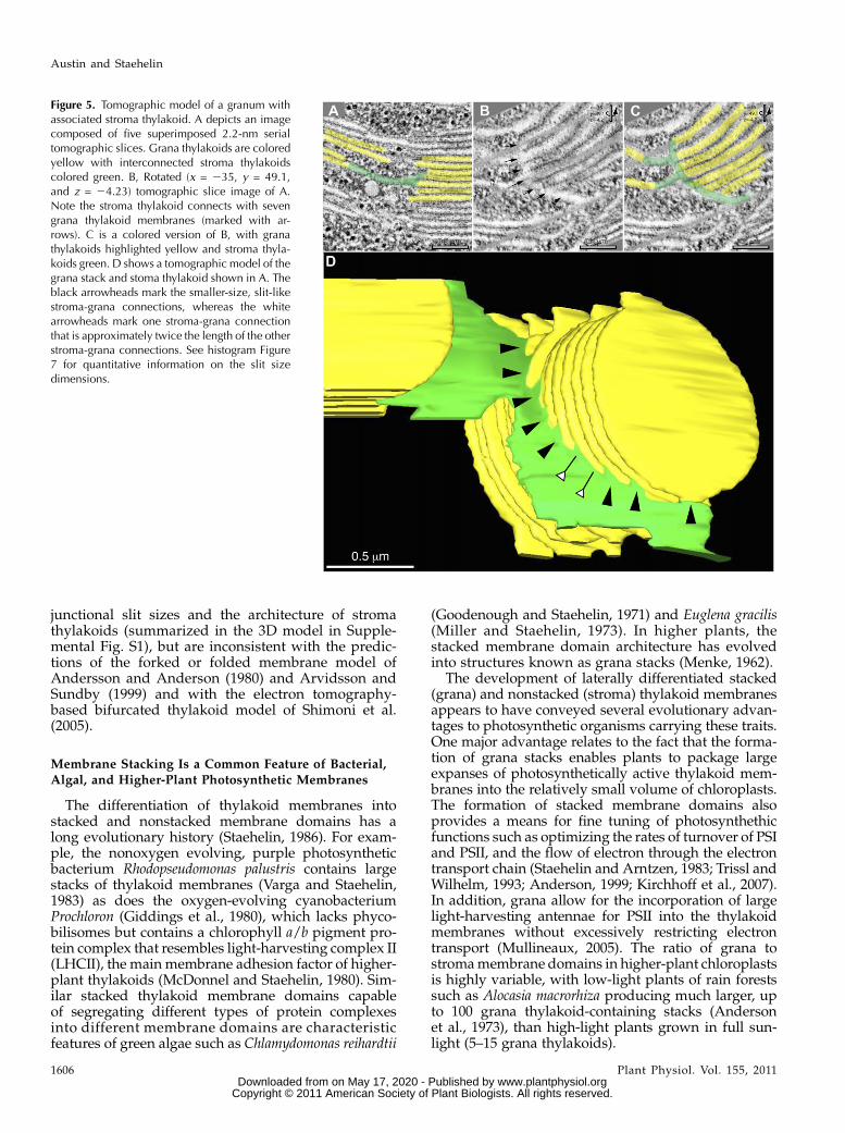
stroma thylakoid connected to a series of stackedgrana thylakoids is presented in Figure 5, A to C. Toobtain the tangential-type view of the stroma thyla-koid with clear connections (arrows) to seven adjacent,stacked grana thylakoids, we used the slicer tool of the3dmod modeling program to orient the slice depictedin Figure 5B to match the plane of the spirally orga-nized connecting regions between the stroma thyla-koid and the adjacent grana thylakoids. A model ofthis association is shown in Figure 5D. Interestingly,when this model is analyzed in greater detail, not all ofthe grana-stroma thylakoid junctional connections areseen to be precisely arranged in a spiral-like configu-ration, and one of the junctional connections is seen tohave twice the slit length (approximately 248 nm) ofthe others (Fig. 5D, white arrowheads). Another ex-ample of a long junctional slit (approximately 307 nm)and the associated interruption of the helical organi-zation of a stroma thylakoid are illustrated in Figure 6(white arrowheads). As seenmost clearly in Figure 6, Band D, the stroma thylakoid adjacent to the granathylakoid with the longer-length slit also becomesparallel to that grana thylakoid, breaking the helicalorganization of the stroma thylakoid.
The Architecture of Stroma Thylakoids ConnectingAdjacent Grana Stacks Is Variable
Figure 8 depicts tomography-based models of twopairs of grana stacks (yellow) and their interconnect-ing stroma thylakoids (multiple colors), which illus-trate the variable architecture of stroma thylakoids inchloroplasts. The first model (Fig. 8, A–D) shows threeparallel, sheet-like stroma thylakoids that form bridg-ing structures between the two grana stacks, but lackany connections to each other. In contrast, the secondmodel (Fig. 8, E–G) displays four spiraling andbranching stroma thylakoids that not only bridge thespace between the grana stacks, but also form a 3Dmembrane network that connects grana thylakoids atdifferent levels within each grana stack. To betterillustrate the geometry of the connecting regions be-tween the differently colored, but physically continu-ous stroma thylakoid membrane domains, we onlyshow the green (semitransparent) and the red stromathylakoids in the tilted model image Figure 8F, and thered and blue stroma thylakoids in Figure 8G.
DISCUSSION
The goal of this study was to produce a detailedunderstanding of the 3D architecture of higher-plantthylakoids by means of electron tomography analysisof high-pressure-frozen/freeze-substituted in situ andin vitro chloroplasts. Our results confirm central fea-tures of the helical thylakoid model of Paolillo (1970)and have added novel insights into the variability of
Figure 4. Electron tomogram serial sections illustrating the structure of the junctional connections between grana thylakoids anda helical stroma thylakoid. A to J show serial 2.2-nm tomographic slice images through grana-stroma thylakoid connections. Thegrana stack thylakoids are colored yellow and lettered A to D, and the stoma thylakoids are colored green. Also, the stroma-granaconnections are marked with arrows. Note that in A the stroma thylakoid connects with the grana thylakoid marked C. In B to Dthe stroma thylakoid connect with grana thylakoids C and B. In E and F the stroma thylakoid is connected with grana thylakoid Bonly. In G to I the stroma thylakoid is connected with grana thylakoids B and A. Finally in J the stroma thylakoid is connected tograna A only.
3D Architecture of Thylakoid Membranes in Higher Plants
Plant Physiol. Vol. 155, 2011 1605 www.plantphysiol.orgon May 17, 2020 - Published by Downloaded from
Copyright © 2011 American Society of Plant Biologists. All rights reserved.
junctional slit sizes and the architecture of stromathylakoids (summarized in the 3D model in Supple-mental Fig. S1), but are inconsistent with the predic-tions of the forked or folded membrane model ofAndersson and Anderson (1980) and Arvidsson andSundby (1999) and with the electron tomography-based bifurcated thylakoid model of Shimoni et al.(2005).
Membrane Stacking Is a Common Feature of Bacterial,Algal, and Higher-Plant Photosynthetic Membranes
The differentiation of thylakoid membranes intostacked and nonstacked membrane domains has along evolutionary history (Staehelin, 1986). For exam-ple, the nonoxygen evolving, purple photosyntheticbacterium Rhodopseudomonas palustris contains largestacks of thylakoid membranes (Varga and Staehelin,1983) as does the oxygen-evolving cyanobacteriumProchloron (Giddings et al., 1980), which lacks phyco-bilisomes but contains a chlorophyll a/b pigment pro-tein complex that resembles light-harvesting complex II(LHCII), the mainmembrane adhesion factor of higher-plant thylakoids (McDonnel and Staehelin, 1980). Sim-ilar stacked thylakoid membrane domains capableof segregating different types of protein complexesinto different membrane domains are characteristicfeatures of green algae such as Chlamydomonas reihardtii
(Goodenough and Staehelin, 1971) and Euglena gracilis(Miller and Staehelin, 1973). In higher plants, thestacked membrane domain architecture has evolvedinto structures known as grana stacks (Menke, 1962).
The development of laterally differentiated stacked(grana) and nonstacked (stroma) thylakoid membranesappears to have conveyed several evolutionary advan-tages to photosynthetic organisms carrying these traits.One major advantage relates to the fact that the forma-tion of grana stacks enables plants to package largeexpanses of photosynthetically active thylakoid mem-branes into the relatively small volume of chloroplasts.The formation of stacked membrane domains alsoprovides a means for fine tuning of photosynthethicfunctions such as optimizing the rates of turnover of PSIand PSII, and the flow of electron through the electrontransport chain (Staehelin and Arntzen, 1983; Trissl andWilhelm, 1993; Anderson, 1999; Kirchhoff et al., 2007).In addition, grana allow for the incorporation of largelight-harvesting antennae for PSII into the thylakoidmembranes without excessively restricting electrontransport (Mullineaux, 2005). The ratio of grana tostromamembrane domains in higher-plant chloroplastsis highly variable, with low-light plants of rain forestssuch as Alocasia macrorhiza producing much larger, upto 100 grana thylakoid-containing stacks (Andersonet al., 1973), than high-light plants grown in full sun-light (5–15 grana thylakoids).
Figure 5. Tomographic model of a granum withassociated stroma thylakoid. A depicts an imagecomposed of five superimposed 2.2-nm serialtomographic slices. Grana thylakoids are coloredyellow with interconnected stroma thylakoidscolored green. B, Rotated (x = 235, y = 49.1,and z = 24.23) tomographic slice image of A.Note the stroma thylakoid connects with sevengrana thylakoid membranes (marked with ar-rows). C is a colored version of B, with granathylakoids highlighted yellow and stroma thyla-koids green. D shows a tomographic model of thegrana stack and stoma thylakoid shown in A. Theblack arrowheads mark the smaller-size, slit-likestroma-grana connections, whereas the whitearrowheads mark one stroma-grana connectionthat is approximately twice the length of the otherstroma-grana connections. See histogram Figure7 for quantitative information on the slit sizedimensions.
Austin and Staehelin
1606 Plant Physiol. Vol. 155, 2011 www.plantphysiol.orgon May 17, 2020 - Published by Downloaded from
Copyright © 2011 American Society of Plant Biologists. All rights reserved.

Stroma Thylakoids Wind around Grana Stacks in aHelical Fashion and Are Connected to the Grana
Thylakoids via Slits in the Grana Margins
To take full advantage of the 3D resolution ofelectron tomography, we have studied exclusivelyintact chloroplasts preserved by high-pressure-freez-ing/freeze-substitution methods (Kiss and Staehelin,1995). The improved structural preservation of thyla-koids prepared in this manner compared to chemicallyfixed and dehydrated samples relates to the fact thatfreezing allows for all of the molecules within a cell tobe immobilized within less than 1 ms (Gilkey andStaehelin, 1986), and that during freeze substitutionthe frozen water molecules are replaced by acetonewhile the sample is maintained at 280�C. By optimiz-ing the freeze-substitution and staining protocols wehave also produced positive staining of the thylakoidmembranes and thereby increased our ability toprecisely trace the membranes in our tomographicslice images (Fig. 1). Compared to the thylakoidmodels derived from serial thin-section reconstruc-tions (Paolillo, 1970; Brangeon andMustardy, 1979) theelectron tomography models produced during thecourse of this study have an approximately 20-fold-higher z axis resolution and a slightly increased x/yaxis resolution, yielding thylakoid models with a 3Dresolution of 6 to 8 nm. Together with the relatively
large size of the reconstructed chloroplast volumes(approximately 4 3 4 3 1.5 mm), these data haveenabled us to generate high-resolution reconstructionsof entire grana stacks and of stroma thylakoids thatbridge the space between adjacent stacks (see Supple-mental Movie S1).
One of the striking features of our new models isthe extent to which they confirm the basic elements ofthe helical thylakoid model of Paolillo (1970). How-ever, considering the many different types of electronmicroscope studies that have provided experimentalsupport for this model during the past 40 years (forreview, see Mustardy and Garab, 2003), this resultis not unexpected. Most clearly seen in our models isthe arrangement of the parallel stroma thylakoidsthat spiral around the grana stacks as right-handedhelices (Figs. 2–6, 8, and Supplemental Figure S1).This helical geometry contradicts central structuralpredictions of the forked/folded thylakoid membranemodel (Andersson and Anderson, 1980; Arvidssonand Sundby, 1999) as well as the bifurcation model ofShimoni et al. (2005).
One of the novel features of our models pertains tothe actual geometry of the stroma thylakoids betweenadjacent grana stacks (Figs. 2, 5, 8, and SupplementalFigure S1). In particular,wedemonstrate thatwhere thestroma thylakoids connect to grana thylakoids in adja-cent grana stacks that are appropriately tilted withrespect to each other, the thylakoids formflat, sheet-likestructures (Fig. 5). In contrast, where the parallel helicalstroma thylakoid membranes are connected both toadjacent grana stacks and to each other through bridg-ing domains, which also form links between differentgrana thylakoidswithin a given grana stack, the stromathylakoids are branched and exhibit a less-expansive,sheet-like, or even tubular geometry (Fig. 8, E–G).
Figure 6. Spatial distribution of differently sized grana-stroma connec-tions on a grana stack. A is a tomographic model showing the top viewof a grana stack (gt), with interconnected stroma thylakoids (st). B is arotated (90�) tomographic model of A. C is a tomographic model of Bwhere the stroma thylakoid has been partially cut away to reveal theconnection site between the stroma and grana membranes (highlightedblack). D is a tomographic model in which all of the stroma thylakoidmembrane on the front side of the grana stack have been removed,revealing all of the stroma-grana connecting sites (highlighted in black).Note the white arrowheads pointing to a stroma-grana connecting sitethat is much longer than the others.
Figure 7. Histogram demonstrating the variation in junctional slitlengths between grana and stroma thylakoids.
3D Architecture of Thylakoid Membranes in Higher Plants
Plant Physiol. Vol. 155, 2011 1607 www.plantphysiol.orgon May 17, 2020 - Published by Downloaded from
Copyright © 2011 American Society of Plant Biologists. All rights reserved.

Another refinement of the helical model necessitatedby our data includes a greater amount of variability inthe periodicity of the organization of the stroma thyla-koids around the grana stacks than previously thought(Figs. 5–7 and Supplemental Fig. S1). As shown inFigure 7, the junctional slits seen in our high-pressure-frozen chloroplasts were not only greater but alsomorevariable in size (30–435 nm) than those reported for thechemically fixed isolated thylakoids (13–70 nm) used inthe electron tomography study by Mustardy et al.(2008). The difference in slit size observed in thetwo studies is most likely due to the use of isolatedthylakoids and of chemical fixation methods in theMustardy et al. (2008) investigation. We do not knowwhat the suspension of isolated thylakoids in a buffersolution does to slit size, but we do know that chemicalfixation typically leads to specimen shrinkage (Gilkeyand Staehelin, 1986). Also, in the study by Daum et al.(2010), which used cryopreservation and cryo-electronmicroscopymethods, the junctional slits were reportedto be larger than those seen in theMustardy et al. (2008)study.
The Variable Size of Junctional Slits Suggests That They
May Participate in the Functional Regulation ofThylakoid Activities
The grana-stroma thylakoid membrane system isunique in its 3D complexity, and based on the helicalthylakoid model Paolillo (1970), which our data con-firmed, it is typically portrayed as a fairly rigid, stable,and inflexible membrane platform. Nevertheless, con-tinuing efforts are being made to explain the regulationof many of the dynamic processes of photosynthesis in
the context of changes in thylakoid ultrastructure. Ofthese, the explanation of state I/II transitions in thecontext of lateral movements of LHCII proteins andchanges in the amount of membrane stacking is bestknown (Staehelin and Arntzen, 1983; Allen, 1992).However, other thylakoid structure-related regulatorymechanisms may exist that have yet to be identified.Based on the unexpectedly large variations in junctionalslit size observed in this study (Fig. 7 and SupplementalFig. S1), and the ability of these slits to shrink dramat-ically when exposed to chemical fixatives such asglutaraldhyde, we propose that junctional slit sizemay be controlled by plants to regulate the movementof protons and of membrane proteins between granaand stroma thylakoid membranes. For example, acti-vation of the kinases that phosphorylate LHCII duringstate I / II transitions may also induce an increase inslits size to expedite the transfer of the phosphorylatedLHCII complexes from grana to stroma thylakoid re-gions. Similarly, photoinhibitory light conditions mightstimulate slit enlargement to facilitate the transfer ofdamaged PSII complexes to stroma thylakoids for re-pair (Aro et al., 2004). Yet other regulatory mechanismsmight exist that have not been discovered. Alterna-tively, the differences in slit dimensions may be relatedto the mechanism of grana thylakoid assembly.
Unstacking of Grana Thylakoids during State I / II
Transitions Differs from Unstacking of IsolatedThylakoids in Low-Salt Solutions
There is much confusion in the literature about howthe thylakoids of higher plants can become unstackedin low-salt solutions, and how the amount of thylakoid
Figure 8. Tomographic models showing the var-iation in stroma architecture. A shows a tomo-graphic model with parallel, sheet-like stromathylakoid membranes (st; colored green, pink,and red) that bridge the space between two granathylakoid stacks (gt; colored yellow), but that arenot connected to each other. B to D is the samemodel as A, but rotated 90� showing the sheet-like architecture of the individual stroma thyla-koids; green (B), pink (C), and red (D) stromathylakoids. E illustrates another tomographicmodel in which quasi-helical stroma thylakoids(st; colored green, red, turquoise, and pink) areseen to connect two grana stacks (gt; coloredyellow). F and G, Same model as E, but F isrotated 60� to better demonstrate the 3D inter-connectivity of the green (semitransparent) andthe red stroma thylakoid membranes; G is rotated45� and shows the interconnectivity between thered and turquoise stroma thylakoid membranes.
Austin and Staehelin
1608 Plant Physiol. Vol. 155, 2011 www.plantphysiol.orgon May 17, 2020 - Published by Downloaded from
Copyright © 2011 American Society of Plant Biologists. All rights reserved.

stacking is reduced during state I / II transitions,considering the complex 3D architecture of the in situmembranes that has been confirmed by this study. Thedifferentiation of thylakoids into stacked grana andnonstacked stroma regions is a reflection of the com-positional and functional differences of these mem-brane domains (Andersson and Anderson, 1980;Staehelin and van der Staay, 1996). Typically, approx-imately 85% of the PSII and 70% to 90% of the LHCIIcomplexes are confined to grana membranes, and.85% of the PSI and 100% of the ATP synthasecomplexes are associated with the stroma membranes.The principal thylakoid membrane adhesion factoris LHCII (McDonnel and Staehelin, 1980). In the chlo-rophyll b-less clorina-f2 mutant of barley (Hordeumvulgare) that lacks LHCII the PSII-associated light-harvesting protein Lhcb5 appears to be responsiblefor stacking, but this stacking requires much higherconcentrations of magnesium than LHCII-mediatedstacking (Bassi et al., 1985; Krol et al., 1995).Isolated thylakoids can be experimentally un-
stacked by transfer to zwitterionic buffers of low ionicstrength, and restacked by the addition of .3 mM
MgCl2 or .150 mM NaCl (Izawa and Good, 1966;Staehelin, 1976). These salt effects can best be ex-plained by neutralization of the negatively chargedgroups on the LHCII molecules that mediate theintermembrane protein-protein interactions by thecations (Barber, 1982; Dekker and Boekema, 2005).Detailed studies of the changes in thylakoid mem-brane architecture induced by the different cationconcentrations have led to the following findings.Experimental unstacking of higher-plant thylakoidsinvolves three steps (Staehelin, 1986). Stage 1: Rapid(5–10 min when the membranes are kept at 4�C)separation of the appressed membrane regions withminimal changes in the lateral distribution of ATPsynthase complexes (Miller and Staehelin, 1976) andPSII complexes (Staehelin et al., 1977). When suchmembranes are immediately restacked by the additionof cations, the resulting reconstituted grana stacks arenearly indistinguishable for those of control thyla-koids. Stage 2: Enlargement of the junctional slitsbetween the grana and stroma thylakoids (15–30min) dramatically increases the lateral movementand intermixing of the different types of integralprotein complexes and bilayer lipids, and results inmembrane unfolding via membrane flow (Staehelinet al., 1977). Stage 3: Complete unfolding of the com-plex 3D thylakoid networks (.30 min) by means ofmembrane flow that results in complete intermixing ofall protein complexes and in the formation of large,interconnected thylakoid membrane sheets (Staehelin,1976; Staehelin et al., 1977). Addition of cations to stage3 type unstacked and unfolded thylakoid membranesleads to the reestablishment of large stacked mem-brane domains (percent stacking equivalent to controlmembranes) and to a complete reseggregation of all ofthe protein complexes (Staehelin, 1976). However,these stacked membrane domains are not organized
in the form of grana stacks. Recently, it has also beenshown that in terms of macromolecular PSII arrange-ment as well as antenna organization the restackedthylakoid domains are virtually identical to stackedcontrol membranes (Kirchhoff et al., 2007). All of thesestudies contradict claims made in an earlier studyof chemically fixed thylakoid samples that low-salt-induced thylakoid unstacking involves membranerupturing (Brangeon, 1974), or requires a forked/foldedmembrane type of grana architecture (Arvidsson andSundby, 1999).
Because state I/II transitions also induce changes inthe amount of thylakoid stacking, many researchershave attempted to explain the state I/II changes inmembrane architecture in terms of the cation concen-tration-dependent changes in thylakoid organization.This analogy has led to wrong conclusions about thechanges in thylakoid architecture that accompanystate I/II changes and to confusing statements aboutthese changes in the literature.
The term state I/II transition refers to a mechanismemployed by algae and higher plants to regulateexcitation energy distribution between PSI and PSII(Allen, 1992). This regulation involves the reversiblephosphorylation of LHCII by a membrane-boundprotein kinase. LHCII phosphorylation reduces theadhesion between LHCII complexes in adjacent thy-lakoid membranes due to electrostatic repulsion andassociated protein conformational changes, and en-ables the LHCII complexes to separate from the PSIIcomplexes and to migrate laterally though the junc-tional slits into the PSI-enriched nonstacked stromathylakoid domains (Staehelin and Arntzen, 1983). Us-ing freeze-fracture electron microscopy to determinethe accompanying changes in membrane stacking anddistribution of the approximately 8 nm in diameterLHCII complexes between stacked and nonstackedmembrane domains, Kyle et al. (1983) have producedquantitative information on state I-state II-inducedmembrane changes. Thus, using thylakoids isolatedfrom peas (Pisum sativum), they observed that between20% and 25% of the grana LHCII complexes migratedinto the stroma thylakoid regions in response to phos-phorylation, and that this net migration of granamembrane proteins resulted in a 23% decrease in theamount of stacked membranes. Both of these phenom-ena were reversed following dephosphorylation of theLHCII complexes. Throughout these events, the basic3D architecture of the thylakoids remained unchangedexcept for the change in the amount of membranestacking (see figures 2 and 3B in Kyle et al., 1983),suggesting that the changes in membrane stacking arebrought about by simple membrane flow betweengrana and stroma thylakoids.
These results contradict the hypothetical model ofstate I/II-induced thylakoid membrane changes re-cently proposed by Chuartzman et al. (2008). Theirhypothetical model is based on the controversial bi-furcated thylakoid membrane model of Shimoni et al.(2005), and postulates that during state I / state II
3D Architecture of Thylakoid Membranes in Higher Plants
Plant Physiol. Vol. 155, 2011 1609 www.plantphysiol.orgon May 17, 2020 - Published by Downloaded from
Copyright © 2011 American Society of Plant Biologists. All rights reserved.

transitions entire stacked membrane domains becomeseparated and that some of the membrane bridgesbetween such grana thylakoids are ruptured duringthe unstacking process. These ruptured membranebridges are postulated to be reformed when the LHCIImolecules are dephosphorylated and the membranesrestack. During the course of our very extensive anal-yses of the effects of state I/II transitions on thylakoidorganization by means of freeze-fracture electron mi-croscopy (Kyle et al., 1983), we never observed anyevidence for ruptured thylakoids.
MATERIALS AND METHODS
Plant Material
Seeds of Arabidopsis (Arabidopsis thaliana; Landsberg erecta) and tobacco
(Nicotiana tobacum) were planted on 0.8% (w/v) agar plates with Murashige
and Skoog medium and 1% Suc for 5 d. Plants were grown at a temperature of
23�C, light intensity of 150 mmol mm22 s21, and a photoperiod of 16/8 light/
dark. Fresh spinach (Spinacia oleracea) was bought at a local supermarket
(Whole Food Market) and used the same day.
Intact Chloroplast Isolation
Fifty grams of washed spinach leaves were added to 125 mL of buffer
1 (0.4 M NaCl, 2 mM MgCl, 0.2% bovine serum albumin, and 20 mM Tricine, pH
8.0) and diced in a blender equipped with razor blades. This was filtered
through four layers of cheesecloth, and centrifuged at 300g for 1 min. The
supernatant was centrifuged at 4,000g to pellet the chloroplasts. The resulting
chloroplast pellet was gently washed with buffer 2 (0.15 M NaCl, 5 mM MgCl,
0.2% bovine serum albumin, and 20 mM Tricine, pH 8.0) without disruption of
the chloroplasts. The supernatant was removed and replaced with buffer 3
(0.4 M Suc, 0.15 mM NaCl, 5 mM MgCl, and 20 mM HEPES, pH 7.5).
Sample Preparation for Electron Tomography
Leaves were removed from plants and transferred to aluminum sample
holders and cryoprotected with 150 mM Suc, or freshly isolated chloroplasts
were placed into an aluminum holder, and frozen in a Baltec HPM 010 high-
pressure freezer (RMC). Samples were then freeze substituted in 2% OsO4 in
anhydrous acetone at280�C for 5 d. The samples were warmed to220�C and
washed with 220�C anhydrous acetone, followed by slow warming to room
temperature over a period of 24 h. The samples were removed from their
holders and infiltrated with increasing concentrations of EPON resin (Ted
Pella). Polymerization was performed at 60�C for 24 h. EPON sections 80 to
350 nm were placed on Formvar (Ted Pella) copper slot grids for electron
microscopy. To prepare grids for electron tomographic data collection, 10 mL
of 15 nm colloidal gold was precipitated onto grids containing thick sections
for 10 min on each side.
Intermediate- and High-Voltage Electron Microscopy and
Acquisition of Tilt Series Images
Seventeen tomograms were collected on a Tecnai TF30 (FEI) intermediate
voltage electron microscope operating at 300 kV. The images were taken at
20,0003 from +60 to 260 at 1� intervals about two orthogonal axes (Ladinsky
et al., 1997), and collected with a Gtan Megascan 795 digital camera that
covered an area of 2.63 2.6 mm2 and had a resolution of 2,0483 2,048 pixels at
a pixel size of 1.26 nm. The remaining tomograms were collected on a JEM-
1000 high-voltage electron microscope (JEOL) operating at 750 kV. The images
were collected as described by Austin et al. (2005).
3D Tomographic Reconstruction, Modeling, and Analysis
The images (single or montaged frames) were aligned using the gold
particles as fiducial markers as described previously (Ladinsky et al., 1999).
Each set of aligned tilts was reconstructed into a single-axis tomogram using
the R-weighted back-projection algorithm (Gilbert, 1972). Merging the two
single-axis tomograms into a dual-axis tomogram involved a warping proce-
dure rather than a single linear transformation to produce the dual-axis
tomogram (Mastronarde, 1997). In addition, dual-axis tomograms computed
from adjacent serial sections were aligned and joined to increase the
reconstructed volume (Austin et al., 2005). Tomograms were displayed and
analyzed with 3dmod, the graphics component of the 3DMOD (formerly
IMOD) software package (Kremer et al., 1996). Membranous structures,
microtubules, and all types of vesicles were modeled as described previously
(Otegui and Austin, 2007). Once a model was completed, meshes of triangles
were computed to define the surface of each object (Kremer et al., 1996).
The image-slicer tool of 3DMOD was used to display and analyze tomo-
graphic slices extracted from the tomogram in any position or tilt around the x,
y, or z axis. This tool allowed us to obtain squeezed images in which a number
of consecutive 2.2-nm tomographic slices were combined, thus generating z
projections of different thicknesses, more similar to conventional electron
microscopy thin sections.
Supplemental Data
The following materials are available in the online version of this article.
Supplemental Figure S1. Three-dimensional model illustrating the spatial
relationship between stacked grana and interconnecting stroma thyla-
koids based on an electron tomography analysis of high pressure frozen
and freeze-substituted intact chloroplasts.
Supplemental Video S1. Tomographic volume of a complete grana
thylakoid stack and associated interconnected stroma thylakoids.
ACKNOWLEDGMENTS
We would like to thank Katie Lundeen for her assistance in preparing
samples for electron microscopy, and David Mastronarde and Richard
Gaudette for providing essential application software and support.
Received December 6, 2010; accepted December 24, 2010; published January
11, 2011.
LITERATURE CITED
Allen JF (1992) Protein phosphorylation in regulation of photosynthesis.
Biochim Biophys Acta 1098: 275–335
Anderson JM (1999) Insights into the consequences of grana stacking of
thylakoid membranes in vascular plants: a personal perspective. Aust J
Plant Physiol 26: 625–639
Anderson JM, Andersson B (1988) The dynamic photosynthetic membrane
and regulation of solar energy conversion. Trends Biochem Sci 13:
351–355
Anderson JM, Goodchild DJ, Boardman NK (1973) Composition of
the photosystems and chloroplast structure in extreme shade plants.
Biochim Biophys Acta 325: 573–585
Andersson B, Anderson JM (1980) Lateral heterogeneity in the distribution
of chlorophyll-protein complexes of the thylakoid membranes of spin-
ach chloroplasts. Biochim Biophys Acta 593: 427–440
Aro EM, Suorsa M, Rokka A, Allahverdiyeva Y, Paakkarinen V, Saleem
A, Battchikova A, Rintamaki E (2004) Dynamics of photosystem II: a
proteomic approach to thylakoid protein complexes. J Exp Bot 56: 347–356
Arvidsson PO, Sundby C (1999) A model for the topology of the chloro-
plast thylakoid membrane. Aust J Plant Physiol 26: 687–694
Austin JR II, Seguı-Simarro JM, Staehelin LA (2005) Quantitative analysis
of changes in spatial distribution and plus-end geometry of microtu-
bules involved in plant-cell cytokinesis. J Cell Sci 118: 3895–3903
Barber J (1982) Influence of surface charges on thylakoid structure and
function. Annu Rev Plant Physiol 33: 261–295
Bassi R, dal Belin Peruffo A, Barbato R, Ghisi R (1985) Differences in
chlorophyll-protein complexes and composition of polypeptides be-
tween thylakoids from bundle sheaths and mesophyll cells in maize.
Eur J Biochem 146: 589–595
Brangeon J (1974) Structural modifications in the lamellar system of
Austin and Staehelin
1610 Plant Physiol. Vol. 155, 2011 www.plantphysiol.orgon May 17, 2020 - Published by Downloaded from
Copyright © 2011 American Society of Plant Biologists. All rights reserved.

isolated Zea mays chloroplasts under different ionic conditions. J
Microsc 21: 75–84
Brangeon J, Mustardy L (1979) The ontogenic assembly of intra-chloroplast
lamellae viewed in 3-dimensions. Biol Cell 36: 71–80
Briantais JM (1984) Kinetics of cation-induced changes of photosystem II
fluorescence and of lateral distribution of the two photosystems in the
thylakoid membrane of pea thylakoids. Biochim Biophys Acta 766: 1–8
Chuartzman SG, Nevo R, Shimoni E, Charuvi D, Kiss V, Ohad I,
Brumfeld V, Reich Z (2008) Thylakoid membrane remodeling during
state transitions in Arabidopsis. Plant Cell 20: 1029–1039
Daum B, Nicastro D, Austin J II, McIntosh JR, Kuhlbrandt W (2010)
Arrangement of photosystem II and ATP synthase in chloroplast mem-
branes of spinach and pea. Plant Cell 22: 1299–1312
Dekker JP, Boekema EJ (2005) Supramolecular organization of thylakoid
membrane proteins in green plants. Biochim Biophys Acta 1706: 12–39
Donohoe BS, Mogelsvang S, Staehelin LA (2006) Electron tomography of
ER, Golgi and related membrane systems. Methods 39: 154–162
Giddings TH, Withers NW, Staehelin LA (1980) Supramolecular structure
of stacked and unstacked regions of the photosynthetic membranes of
Prochloron sp., a prokaryote. Proc Natl Acad Sci USA 77: 352–356
Gilbert PF (1972) The reconstruction of a three-dimensional structure
from projections and its application to electron microscopy: II. Direct
methods. Proc R Soc Lond B Biol Sci 182: 89–102
Gilkey JC, Staehelin LA (1986) Advances in ultrarapid freezing for
the preservation of cellular ultrastructure. J Electron Microsc Tech 3:
177–210
Goodenough UW, Staehelin LA (1971) Structural differentiation of stacked
and unstacked chloroplast membranes: freeze-etch electron microscopy
of wild-type and mutant strains of Chlamydomonas. J Cell Biol 48:
594–619
Heslop-Harrison J (1963) Structure and morphogenesis of lamellar systems
in grana-containing chloroplast. I. Membrane structure and lamellar
architecture. Planta 60: 243–260
Hodge AJ, McLean JD, Mercer FV (1955) Ultrastructure of the lamellae
and grana in the chloroplast of Zea mays L. J Biophys Biochem Cytol 25:
605–614
Izawa S, Good NE (1966) Effects of salts and electron transport on the
conformation of isolated chloroplasts. II. Electron microscopy. Plant
Physiol 41: 544–552
Kirchhoff H, Haase W, Haferkamp S, Schott T, Borinski M, Kubitscheck
U, Rogner M (2007) Structural and functional self-organization of
Photosystem II in grana thylakoids. Biochim Biophys Acta 1767:
1180–1188
Kiss JZ, Staehelin LA (1995) High pressure freezing. In NJ Severs, DM
Shotton, eds, Rapid Freezing, Freeze Fracture and Deep Etching. John
Wiley-Liss Inc., New York, pp 89–104
Kremer JR, Mastronarde DN, McIntosh JR (1996) Computer visualization
of three-dimensional image data using IMOD. J Struct Biol 116: 71–76
Krol M, Spangfort MD, Huner NP, Oquist G, Gustafsson P, Jansson S
(1995) Chlorophyll a/b-binding proteins, pigment conversions, and
early light-induced proteins in a chlorophyll b-less barley mutant. Plant
Physiol 107: 873–883
Kyle DJ, Staehelin LA, Arntzen CJ (1983) Lateral mobility of the light-
harvesting complex in chloroplast membranes controls excitation en-
ergy distribution in higher plants. Arch Biochem Biophys 222: 527–541
Ladinsky MS, Kremer JR, Mastronarde DN, McIntosh JR, Staehelin LA,
Howell KE (1997) HVEM tomography of the Golgi ribbon in cryofixed
NRK cells: the non-compact region, the CGN, and TGN. Mol Biol Cell
8: 2040
Ladinsky MS, Mastronarde DN, McIntosh JR, Howell KE, Staehelin LA
(1999) Golgi structure in three dimensions: functional insights from the
normal rat kidney cell. J Cell Biol 144: 1135–1149
Mastronarde DN (1997) Dual-axis tomography: an approach with align-
ment methods that preserve resolution. J Struct Biol 120: 343–352
McDonnel A, Staehelin LA (1980) Adhesion between liposomes mediated
by the chlorophyll a/b light-harvesting complex isolated from chloro-
plast membranes. J Cell Biol 84: 40–56
McIntosh R, Nicastro D, Mastronarde D (2005) New views of cells in 3D:
an introduction to electron tomography. Trends Cell Biol 15: 43–51
Menke W (1960) Das allgemeine Bauprinzip des Lamellarsystems der
Chloroplasten. Experientia 16: 537–538
Menke W (1962) Structure and chemistry of plastids. Annu Rev Plant
Physiol 13: 27–44
Miller KR, Staehelin LA (1973) Fine Structure of the chloroplast mem-
branes of Euglena gracilia as revealed by freeze-cleaving and deep-
etching techniques. Protoplasma 7: 55–78
Miller KR, Staehelin LA (1976) Analysis of the thylakoid outer surface. J
Cell Biol 68: 30–47
Mullineaux CW (2005) Function and evolution of grana. Trends Plant Sci
10: 521–525
Mustardy L, Buttle K, Steinbach G, Garab G (2008) The three-dimensional
network of the thylakoid membranes in plants: quasihelical model of the
granum-stroma assembly. Plant Cell 20: 2552–2557
Mustardy L, Garab G (2003) Granum revisited: a three-dimensional model
—where things fall into place. Trends Plant Sci 8: 117–122
Mustardy L, Janossy AGS (1979) Evidence of helical thylakoid arrange-
ment by scanning electron microscopy. Plant Sci Lett 16: 281–284
Otegui MS, Austin JR II (2007) Visualization of membrane-cytoskeletal
interactions during plant cytokinesis. Methods Cell Biol 79: 221–240
Paolillo DJ Jr (1970) The three-dimensional arrangement of intergranal
lamellae in chloroplasts. J Cell Sci 6: 243–255
Paolillo DJ Jr, Falk RH, Reighard JA (1967) The effect of chemical fixation
on the fretwork of chloroplasts. Trans Am Microsc Soc 86: 225–232
Shimoni E, Rav-Hon O, Ohad I, Brumfeld V, Reich Z (2005) Three-
dimensional organization of higher-plant chloroplast thylakoid mem-
branes revealed by electron tomography. Plant Cell 17: 2580–2586
Staehelin LA (1976) Reversible particle movements associated with un-
stacking and restacking of chloroplast membranes in vitro. J Cell Biol 71:
136–158
Staehelin LA (1986) Photosynthesis III: Photosynthetic Membranes. In CJ
Arntzen, LA Staehelin, eds, Springer-Verlag, Berlin, pp 1–84
Staehelin LA (2003) Chloroplast structure: from chlorophyll granules to
supra-molecular architecture of thylakoid membranes. Photosynth Res
76: 185–196
Staehelin LA, Armond PA, Miller KR (1977) Chloroplast membrane
organization at the supramolecular levels and its functional implica-
tions. Brookhaven Symp Biol 28: 278–315
Staehelin LA, Arntzen CJ (1983) Regulation of chloroplast membrane
function: protein phosphorylation changes the spatial organization of
membrane components. J Cell Biol 97: 1327–1337
Staehelin LA, van der Staay GWM (1996) Structure, composition, func-
tional organisation and dynamic properties of thylakoid membranes. In
DR Ort, CF Yocum, eds, Oxygenic Photosynthesis: The Light Reactions.
Kluwer Academic Publishers, Dordrecht, The Netherlands, pp 11–30
Steinmann E, Sjostrand FS (1955) The ultrastructure of chloroplasts. Exp
Cell Res 8: 15–23
Trissl HW, Wilhelm C (1993) Why do thylakoid membranes from higher
plants form grana stacks? Trends Biochem Sci 18: 415–419
Varga AR, Staehelin LA (1983) Spatial differentiation in photosynthetic
and non-photosynthetic membranes of Rhodopseudomonas palustris. J
Bacteriol 154: 1414–1430
Wehrmeyer W (1964) Zur klarung der structurellen variabilitat der chloro-
plastengrana des spinats in profil und aufsicht. Planta 62: 272–293
Weier TE (1961) The ultramicrostructure of starch free chloroplasts of
Nicotiana rustica. Am J Bot 48: 615–630
Weier TE, Stocking CR, Thomoson WW, Drever H (1963) The photosyn-
thetic apparatus in chloroplast of higher plants. J Ultrastruct Res 8:
122–143
3D Architecture of Thylakoid Membranes in Higher Plants
Plant Physiol. Vol. 155, 2011 1611 www.plantphysiol.orgon May 17, 2020 - Published by Downloaded from
Copyright © 2011 American Society of Plant Biologists. All rights reserved.


![Multiphoton imaging to identify grana, stroma thylakoid ...cellstudio.org/.../BMCPlant2014_Multiphoton-grana.pdf · and grana in plant [11,16-27]. Inside a chloroplast, both starch](https://static.fdocuments.in/doc/165x107/5fb8149a7316217d4776dc5c/multiphoton-imaging-to-identify-grana-stroma-thylakoid-and-grana-in-plant-1116-27.jpg)