Thousands of previously unknown phages discovered in whole ...
17
RESEARCH Open Access Thousands of previously unknown phages discovered in whole-community human gut metagenomes Sean Benler 1 , Natalya Yutin 1 , Dmitry Antipov 2 , Mikhail Rayko 2 , Sergey Shmakov 1 , Ayal B. Gussow 1 , Pavel Pevzner 2,3 and Eugene V. Koonin 1* Abstract Background: Double-stranded DNA bacteriophages (dsDNA phages) play pivotal roles in structuring human gut microbiomes; yet, the gut virome is far from being fully characterized, and additional groups of phages, including highly abundant ones, continue to be discovered by metagenome mining. A multilevel framework for taxonomic classification of viruses was recently adopted, facilitating the classification of phages into evolutionary informative taxonomic units based on hallmark genes. Together with advanced approaches for sequence assembly and powerful methods of sequence analysis, this revised framework offers the opportunity to discover and classify unknown phage taxa in the human gut. Results: A search of human gut metagenomes for circular contigs encoding phage hallmark genes resulted in the identification of 3738 apparently complete phage genomes that represent 451 putative genera. Several of these phage genera are only distantly related to previously identified phages and are likely to found new families. Two of the candidate families, “Flandersviridae” and “Quimbyviridae”, include some of the most common and abundant members of the human gut virome that infect Bacteroides, Parabacteroides, and Prevotella. The third proposed family, “Gratiaviridae,” consists of less abundant phages that are distantly related to the families Autographiviridae, Drexlerviridae, and Chaseviridae. Analysis of CRISPR spacers indicates that phages of all three putative families infect bacteria of the phylum Bacteroidetes. Comparative genomic analysis of the three candidate phage families revealed features without precedent in phage genomes. Some “Quimbyviridae” phages possess Diversity-Generating Retroelements (DGRs) that generate hypervariable target genes nested within defense-related genes, whereas the previously known targets of phage-encoded DGRs are structural genes. Several “Flandersviridae” phages encode enzymes of the isoprenoid pathway, a lipid biosynthesis pathway that so far has not been known to be manipulated by phages. The “Gratiaviridae” phages encode a HipA-family protein kinase and glycosyltransferase, suggesting these phages modify the host cell wall, preventing superinfection by other phages. Hundreds of phages in these three and other families are shown to encode catalases and iron-sequestering enzymes that can be predicted to enhance cellular tolerance to reactive oxygen species. (Continued on next page) © The Author(s). 2021 Open Access This article is licensed under a Creative Commons Attribution 4.0 International License, which permits use, sharing, adaptation, distribution and reproduction in any medium or format, as long as you give appropriate credit to the original author(s) and the source, provide a link to the Creative Commons licence, and indicate if changes were made. The images or other third party material in this article are included in the article's Creative Commons licence, unless indicated otherwise in a credit line to the material. If material is not included in the article's Creative Commons licence and your intended use is not permitted by statutory regulation or exceeds the permitted use, you will need to obtain permission directly from the copyright holder. To view a copy of this licence, visit http://creativecommons.org/licenses/by/4.0/. The Creative Commons Public Domain Dedication waiver (http://creativecommons.org/publicdomain/zero/1.0/) applies to the data made available in this article, unless otherwise stated in a credit line to the data. * Correspondence: [email protected] 1 National Center for Biotechnology Information, National Library of Medicine, Bethesda, Maryland 20894, USA Full list of author information is available at the end of the article Benler et al. Microbiome (2021) 9:78 https://doi.org/10.1186/s40168-021-01017-w
Transcript of Thousands of previously unknown phages discovered in whole ...

RESEARCH Open Access
Thousands of previously unknown phagesdiscovered in whole-community humangut metagenomesSean Benler1, Natalya Yutin1, Dmitry Antipov2, Mikhail Rayko2, Sergey Shmakov1, Ayal B. Gussow1,Pavel Pevzner2,3 and Eugene V. Koonin1*
Abstract
Background: Double-stranded DNA bacteriophages (dsDNA phages) play pivotal roles in structuring human gutmicrobiomes; yet, the gut virome is far from being fully characterized, and additional groups of phages, includinghighly abundant ones, continue to be discovered by metagenome mining. A multilevel framework for taxonomicclassification of viruses was recently adopted, facilitating the classification of phages into evolutionary informativetaxonomic units based on hallmark genes. Together with advanced approaches for sequence assembly andpowerful methods of sequence analysis, this revised framework offers the opportunity to discover and classifyunknown phage taxa in the human gut.
Results: A search of human gut metagenomes for circular contigs encoding phage hallmark genes resulted in theidentification of 3738 apparently complete phage genomes that represent 451 putative genera. Several of thesephage genera are only distantly related to previously identified phages and are likely to found new families. Two ofthe candidate families, “Flandersviridae” and “Quimbyviridae”, include some of the most common and abundantmembers of the human gut virome that infect Bacteroides, Parabacteroides, and Prevotella. The third proposedfamily, “Gratiaviridae,” consists of less abundant phages that are distantly related to the families Autographiviridae,Drexlerviridae, and Chaseviridae. Analysis of CRISPR spacers indicates that phages of all three putative families infectbacteria of the phylum Bacteroidetes. Comparative genomic analysis of the three candidate phage families revealedfeatures without precedent in phage genomes. Some “Quimbyviridae” phages possess Diversity-GeneratingRetroelements (DGRs) that generate hypervariable target genes nested within defense-related genes, whereas thepreviously known targets of phage-encoded DGRs are structural genes. Several “Flandersviridae” phages encodeenzymes of the isoprenoid pathway, a lipid biosynthesis pathway that so far has not been known to bemanipulated by phages. The “Gratiaviridae” phages encode a HipA-family protein kinase and glycosyltransferase,suggesting these phages modify the host cell wall, preventing superinfection by other phages. Hundreds of phagesin these three and other families are shown to encode catalases and iron-sequestering enzymes that can bepredicted to enhance cellular tolerance to reactive oxygen species.
(Continued on next page)
© The Author(s). 2021 Open Access This article is licensed under a Creative Commons Attribution 4.0 International License,which permits use, sharing, adaptation, distribution and reproduction in any medium or format, as long as you giveappropriate credit to the original author(s) and the source, provide a link to the Creative Commons licence, and indicate ifchanges were made. The images or other third party material in this article are included in the article's Creative Commonslicence, unless indicated otherwise in a credit line to the material. If material is not included in the article's Creative Commonslicence and your intended use is not permitted by statutory regulation or exceeds the permitted use, you will need to obtainpermission directly from the copyright holder. To view a copy of this licence, visit http://creativecommons.org/licenses/by/4.0/.The Creative Commons Public Domain Dedication waiver (http://creativecommons.org/publicdomain/zero/1.0/) applies to thedata made available in this article, unless otherwise stated in a credit line to the data.
* Correspondence: [email protected] Center for Biotechnology Information, National Library of Medicine,Bethesda, Maryland 20894, USAFull list of author information is available at the end of the article
Benler et al. Microbiome (2021) 9:78 https://doi.org/10.1186/s40168-021-01017-w
(Continued from previous page)
Conclusions: Analysis of phage genomes identified in whole-community human gut metagenomes resulted in thedelineation of at least three new candidate families of Caudovirales and revealed diverse putative mechanismsunderlying phage-host interactions in the human gut. Addition of these phylogenetically classified, diverse, anddistinct phages to public databases will facilitate taxonomic decomposition and functional characterization ofhuman gut viromes.
BackgroundThe bulk of the human-associated virome resides in thedistal gastrointestinal tract and is composed of taileddouble-stranded (ds) DNA bacteriophages (dsDNAphages) [1–3] that, in the recent virus megataxonomy,are classified as the class Caudoviricetes under thephylum Uroviricota [4]. The ternary interactions be-tween phages, bacteria, and their human hosts are beingelucidated at an increasing pace through experiments onmodel systems and sequencing of the uncultured com-munity of viruses (virome) [5–9]. Comparisons of thehuman gut virome within and between individuals unveilremarkable longitudinal stability and high diversity ofresident phages [2, 10, 11]. Although the human gut of-fers a rich source of phage genomic diversity, the viromeso far has been explored to a much lesser extent thanthe whole community (metagenome), composed of vi-ruses, bacteria, and archaea. The rapid growth of thepublic whole-community metagenomic data offers theopportunity to identify numerous novel phage genomeslurking in metagenomes.Tailed dsDNA phages encapsidate their genome as
a linear molecule, but depending on the terminalgenomic arrangement, many complete phage ge-nomes assemble into a “circular” contig (i.e., a contigwith direct terminal repeats) [12]. Thus, circularitycan be used as one feature to identify putativecomplete phage genomes in viromes and metagen-omes. However, the comparatively small size ofdsDNA phage genomes (~50 kb, on average) [13]and the estimated low virus-to-microbe ratio in thegut (1:10) [1] jointly translate into a relatively smallamount of phage DNA present in whole communitymetagenomic libraries [14]. Moreover, similar-sizedplasmids also assemble into circular contigs [15]. Arecently developed computational method aims toaddress this problem by focusing specifically on theassembly of circular phage genomes and their auto-matic discrimination from plasmids based on genecontent [16]. The genetic repertoire shared betweenplasmids and phages, for example, the parABS parti-tioning system encoded by both Escherichia coliphage P1 and plasmids [17] can obfuscate their auto-matic annotation-based discrimination and necessi-tate manual curation. Despite these challenges, thereis a pressing need to reduce the amount of viral
“dark matter” in the human gut by identifying andclassifying phages for reference-based analyses [18, 19].The global organization of the virosphere was recently
captured in a comprehensive, unified framework usingprotein domains encoded by viral hallmark genes toinfer evolutionary connections between major groups ofviruses [4] and subsequently approved by the Inter-national Committee on the Taxonomy of Viruses(ICTV) as the comprehensive, multi-rank taxonomy ofviruses. In particular, dsDNA viruses possess either theHK97 fold or the double jelly-roll fold in their majorcapsid proteins, along with distinct ATPases involved incapsid maturation, and thus appear to have independentorigins, justifying their separation into two realms (thehighest virus taxon rank) [4]. Tailed dsDNA phages, withtheir HK97 major capsid proteins, comprise the orderCaudovirales within the class Caudoviricetes, under thephylum Uroviricota (that also include the distantly re-lated herpesviruses of animals) and are further classifiedinto 9 families. With the now formally recognized abilityto classify viruses from sequence data alone [20], phylo-genomic analysis of uncultured phage genomes can de-lineate novel taxa.Here, we describe 3738 completely assembled phage
genomes discovered by analysis of 5742 whole-community human gut metagenomes. Using abundance,taxonomy, and genomic composition as criteria to selectgenomes for further scrutiny, three groups of phages, allinfecting bacteria of the phylum Bacteroidetes compris-ing potential new families, were analyzed in detail. Allthese candidate families, named “Quimbyviridae,”“Flandersviridae,” and “Gratiaviridae” consist of phagesinfecting bacteria of the phylum Bacteroidetes, andthe first two are widely distributed and abundant inhuman gut viromes. The phages in these families andothers yet to be classified encode enzymes that areinvolved in the response of cells to oxidative stress,implicating phages in the tolerance of anaerobes tooxygen. Furthermore, comparative genomic analysisexposed genetic cassettes that are unique to somegenera in each family and thus appear to be relativelyrecent acquisitions involved in phage-host interac-tions. Addition of all the phage genomes identifiedhere to public databases will substantially expand theknown phage diversity and augment taxonomic classi-fication of the human gut virome.
Benler et al. Microbiome (2021) 9:78 Page 2 of 17

MethodsIdentification of phage genomes in human gutmetagenomes5742 whole-community metagenome assemblies gener-ated from human fecal samples were downloaded fromthe NCBI Assembly database (accessed 8/2019). To limitthe search space to likely complete genomes, 95,663 “cir-cular” contigs (50–200 bp direct overlap at contig ends)were extracted from these assemblies. Next, 304 phage-specific protein alignments from the CDD database [21]and 117 custom alignments [22] were converted to Hid-den Markov Models (HMMs) using hmmpress (v. 3.2.1).Proteins in the 95,663 contigs were predicted by Prod-igal (v. 2.6.3) [23] in the metagenomic mode andsearched against the set of 304 phage-specific HMMsusing hmmsearch, with the relaxed e-value cutoff of <0.05. Contigs with at least one hit (n = 4907) were se-lected for a second round of searches after correcting forre-assigned codons, as follows. All contigs were searchedfor the presence of tRNAs using tRNA-scan-SE (v. 2.0)[24]. In 212 contigs, an amber stop codon-suppressortRNA was identified. ORFs were re-predicted for thesecontigs with the amber stop codon re-assigned to glu-tamine, given that this reassignment is most commonlyobserved in human gut phages [25, 26]. The re-translated contigs were added back to the database andall contigs were subjected to a second profile search witha stricter e-value cutoff (< 0.01). Contigs were classifiedas phagepresently organized into 9 families, but 3 of the-ses when exceeding 3 kbp in length and possessing atleast one ORF that matched a capsid, portal, or large ter-minase subunit protein profile below the e value thresh-old. The phage classifications were cross-checked withSeeker [27] and ViralVerify [16]. In cases where bothtools classified a contig as non-phage, the protein anno-tations were examined manually, revealing four contigsof ambiguous identity that were discarded.
Collection of phage genomes in GenBankTaxonomic accession codes corresponding to all pro-karyotic viruses were collected from the NCBI Tax-onomy database and used to extract sequences longerthan 3 kbp from the non-redundant nucleotide database(accessed 09/2019). The protein predictions for eachgenome sequence were retrieved using the “efetch” func-tionality in the entrez direct command line tools [28].Genomic sequences lacking protein predictions werediscarded.
Dereplication and annotation of phage genomesThe collections of GenBank and human gut phage ge-nomes were each dereplicated at 95% average nucleotideidentity across 80% of the genome length using dRep (v.2.6.2) [29] and its associated dependencies, Mash [30]
and FastANI [31], with all other settings left as default.The proteins from these contigs were collected and clus-tered at 95% amino acid identity across 50% of the pro-tein length using mmclust [32]. The representativeprotein sequences were combined into a single BLASTdatabase and compared against the multiple sequencealignments (MSAs) in the CDD database [21] with PSI-BLAST [33] at an e-value cutoff of 0.01. If the represen-tative protein sequence produced a significant result, therepresentative and all constituent members of the pro-tein cluster were annotated using the best hit.
Phylogenetic reconstructionAlignments of the large terminase subunit (TerL), cap-sid, or portal protein were constructed as previouslydescribed [34]. Marker proteins from the metagenomicphages were combined with markers from GenBankphages into a single database and initially clustered to50% amino acid identity using mmclust [32]. The clus-ters were aligned using MUSCLE [35]; cluster align-ments were then compared to each other usingHHsearch (v. 3.0) [36]. The cluster-cluster similarityscores were converted to distances as -ln(SA,B/min(SA,A,SB,B)), where SA,B is similarity between the profiles Aand B, then, an unweighted pair group method witharithmetic mean (UPGMA) dendrogram was con-structed using the estimated cluster distances. Tips ofthe tree (depth <1.5) were used to guide the pairwisealignment of the clusters at the tree leaves with HHalign,creating larger protein clusters. The resulting alignmentswere filtered to remove sites with more than 50% gapsand a homogeneity lower than 0.1 [37]. The filteredalignment was used to construct an approximatemaximum-likelihood tree using FastTree [38], with theWhelan-Goldman models of amino acid evolution andgamma-distributed site rates. Examination of the treesidentified 353 nearly identical PhiX-174 sequences thatwere removed from subsequent analyses as a contamin-ation from a sequencing reagent.
Phage genome analysisA gene-sharing network of phage genomes was con-structed using Vcontact2 (v. 0.9.19) [39], with defaultsearch settings against the database of dereplicated Gen-Bank phage genomes. The results were imported intoCytoscape (v. 3.8) [40] for visualization.The ORFs for selected groups of phages (see the main
text) were additionally annotated through HHblitssearches against the Uniprot database clustered to 30%identity and the PDB database clustered to 70% identity(available at http://wwwuser.gwdg.de/~compbiol/data/hhsuite/databases/hhsuite_dbs/, accessed 02/2020) [41].Genomes encoding a predicted reverse transcriptase(RT) were examined for the presence of repeats
Benler et al. Microbiome (2021) 9:78 Page 3 of 17

corresponding to a diversity-generating retroelementusing DGRScan with default settings [42]. To identify re-peats outside of the 10 kb RT-centered window (the de-fault window of DGRScan), the template repeats wereused as BLASTn queries against the encoding genomewith the following parameters: -dust no -perc_identity75 -qcov_hsp_perc 50 -ungapped -word_size 4.
Fractional abundance of phage genomes inmetagenomesDereplicated phage genomes from the NCBI Genbankdatabase were combined with the dereplicated gutphages into a single database and indexed for read re-cruitment using Bowtie2 [43]. A collection of 1241 hu-man gut viromes were downloaded from the NCBI SRAusing the SRA-toolkit (v. 2.10) and quality filtered withfastp (v. 0.20.1) [44]. The quality-filtered virome readswere mapped to a database containing the referencehuman genome (GCF_000001405), phiX-174 (NC_001422.1) and cloning vectors (available from ftp://ftp.ncbi.nlm.nih.gov/pub/UniVec/) using Bowtie2 with de-fault parameters. Unaligned, “decontaminated” readswere then recruited to the phage database using Bow-tie2 with default parameters, except for the followingadditions “--no-unal --maxins 1000000.” The length-normalized fractional abundance of each phagegenome in each virome was calculated as describedpreviously [1].
Host prediction from CRISPR-spacer matchesA database of CRISPR spacers was compiled from previ-ous surveys of CRISPR-Cas systems [45, 46]. Each spacerwas used as a BLASTN [47] query against the phage ge-nomes, using word size of 8 and low complexity filteringdisabled. A phage-host prediction was inferred if thespacer was 95% identical over 95% of its length to aphage sequence.
Prediction of anti-CRISPR proteinsIdentification of anti-CRISPR proteins (Acrs) was carriedas out as previously described [48]. Briefly, each proteinwas assigned a score by the Acr prediction model ran-ging between 0 and 1, where a higher score correspondsto a higher likelihood of the protein being an Acr. Theproteins were then clustered at 50% amino acid identityand considered a candidate Acr if they satisfied thefollowing criteria: (1) received a mean score of 0.9 orabove, (2) are present in a directon of 5 or fewer genes,(3) at least one of the directons encodes an HTHdomain-containing protein, and (4) the cluster does notproduce a hit with an HHpred probability greater than0.9 to any PDB or CDD database sequence [21].
ResultsIdentification of novel phage genomes from whole-community human gut metagenomesThe collection of 5742 whole-community assembledmetagenomes was searched for the presence of completephage genomes. To limit the search space to likelyclosed genomes, “circular” contigs were extracted fromthese assemblies that contained direct repeats at theirtermini, within the typical k-mer size used to assembleshort reads (50–200 bp, n = 95,663). Each contig wassearched for open reading frames (ORFs) matching aknown phage marker profile (i.e., the terminase largesubunit, major capsid protein, or portal protein). In total,3738 contigs encode at least one ORF that passed the evalue and length cutoff criteria (the “Methods” section)(Additional file 1). Dereplication at approximately 95%mean nucleotide identity reduced the number of phagemarker-matching contigs to 1886 (Additional file 2). Asubset of 664 contigs encoded all three markers, 531encoded two of the three, and the remaining 691 pos-sessed a single detectable marker (Additional file 3). Theputative phage contigs had a median length of 44.9 kb,which is consistent with the recent estimates of the me-dian genome size of dsDNA phages [49]. To exclude anycontaminating contigs (e.g., a plasmid harboring an inte-grated phage), each was assessed with ViralVerify [16]and Seeker [27], and two bioinformatic tools trained todiscriminate phage genomes from other sequences.These tools classified all but 36 of the selected contigs asphages with varying levels of confidence (Additional file2). Upon manual examination for typical phage genesother than the markers, four of the 36 unassigned con-tigs were discarded and the remainder were found torepresent false negative classifications by these tools asjudged by the presence of signature phage genes. Al-though we cannot rule out the possibility that somenon-phage contigs were retained erroneously, the resultscollectively suggest that the set of circular marker-matching contigs predominantly consists of completephage genomes.To determine the host ranges of the phages, a database
of CRISPR spacers from prokaryotic genomes was usedto query the metagenomic phages for potential matches.In total, 553 (29%) of the dereplicated phage genomeswere found to be targeted by at least one CRISPR-Cassystem allowing host prediction (Additional file 4). Themost common predicted hosts were Firmicutes (323phages), followed by Bacteroidetes (143), Actinobacteria(43), Proteobacteria (41), and Verrucomicrobia (4).Among the identified phages, 111 were predicted to in-fect at least two different bacterial genera, consistentwith other studies demonstrating that related bacteriapossess CRISPR spacers targeting the same phage [45,50]. Notably, 359 of the dereplicated phages harbor at
Benler et al. Microbiome (2021) 9:78 Page 4 of 17

least one protospacer identical to another gut phage(Additional file 4), indicating that a single CRISPR spa-cer often can confer immunity against multiple phages.Many phages have been found to encode anti-CRISPR
proteins (Acrs) to parry CRISPR-Cas defenses [51–54].Given their function in counter-defense, Acrs evolverapidly and show limited sequence similarity to experi-mentally characterized Acrs, making inference challen-ging [51]. However, a machine-learning based methodhas been recently developed that utilizes genomic con-text to identify candidate Acrs [48]. Application of thismethod showed that 41 phages, 16 of which were foundto be targeted by a CRISPR-Cas system of their inferredhost, encoded at least one candidate Acr (Additional file5). The highest-scoring Acrs belong to four phages thatare targeted by Bifidobacterium CRISPR-Cas systems.All four phages are > 97% identical over > 90% of theirlength at the nucleotide level to uncharacterized pro-phages in cultured Bifidobacterium isolates (Additionalfile 5), confirming their host-tropism assignment viaCRISPR spacer-protospacer matches. In these phages,two candidate Acr-encoding genes lie between the largeterminase subunit and integrase (Additional file 6). Thelocalization of the Acr-encoding genes suggests they areexpressed not only upon initial entry into the host celland during lysogeny [55], but also upon transition to thelytic program to prevent cleavage of progeny phage ge-nomes by CRISPR-Cas, as demonstrated experimentallyin Listeria-infecting phages [56]. Transcription of Acrs istypically regulated by HTH domain-containing proteinstermed Acr-associated proteins (Acas) [57]. Indeed, inthe Bifidobacterium phages identified here, a short HTHdomain-encoding ORF is located immediately down-stream of the Acrs and can be predicted to regulate theexpression of these two genes throughout the phage life-cycle. While these uncharacterized Bifidobacteriumphages possess the characteristic features of Acr loci, thegreat majority of the phages identified in this work didnot harbor any detectable Acrs yet were targeted byCRISPR-Cas (Additional file 4). Some of these phagesmight encode distinct Acrs undetectable by the methodwe used that was trained on a collection of previouslycharacterized Acrs, whereas others might employ alter-native anti-CRISPR strategies.
Taxonomic decomposition of the gut phages identifiespreviously unknown putative familiesPhylogenetic trees were constructed for the large termi-nase subunit (TerL), major capsid protein (MCP) andportal protein encoded in each phage genome using aniterative approach to construct the underlying align-ments [34]. The trees were constructed alongside refer-ence proteins derived from phage genomes extractedfrom the NCBI GenBank database. Reflecting the set of
protein profiles employed to identify the phage contigs,1480 (78%) genomes were assigned to the phylum Uro-viricota, 360 (19%) to the Phixviricota, and 46 (2%) tothe Loebvirae (Additional file 3). The phylum Phixviri-cota includes Escherichia coli phage phiX174 that is usedas a sequencing reagent; however, the 360 Phixviricotaphages detected in this analysis do not include any se-quences closely related to phiX174 (see the “Methods”section). The remaining analyses focus on the taxonomicdecomposition of the phages that belong to Uroviricota,given that these contigs represent by far the largest frac-tion of the recovered genomes and that the Loebviraeand Phixviricota phyla are the subject of recent taxo-nomic analyses [58, 59].The phylum Uroviricota is organized into a single class
(Caudoviricetes) and order (Caudovirales), but a neworder encompassing the crAss-like phages, a commonand apparently most abundant group of phages in thehuman gut virome, is being proposed [60]. Our profilesrecovered 141 phage genomes (dereplicated from 601total genomes) that displayed phylogenetic relationshipwith the crAss-like phages and are the subject of a sep-arate study [22]. The Caudovirales are presently orga-nized into 9 families, but 3 of these (Myoviridae,Podoviridae, Siphoviridae) are expansive and demon-strably polyphyletic [61–63] and were thus not used forfamily-level taxonomic assignment although theremaining 6 families represent only a small fraction ofthe phages available in GenBank. The phylogenetic treeof TerL, a hallmark protein that is frequently used forphylogenetic reconstruction of Caudovirales phages andappears to be the best phylogenetic marker thanks to itsubiquity among phages and high level of sequence con-servation [63, 64], reveals only 34 gut phages that belongto one of these 6 ICTV-accepted families (Fig. 1 andAdditional file 3). The remainder of these unclassifiedphages are likely to found new families presently com-posed entirely of uncultured phages or belong to familieswith a cultured representative that have yet to be definedunder the new multi-rank taxonomy of viruses.
Selection of candidate families for comparative genomicsThe taxonomic analysis based on phage hallmark pro-teins demonstrates that few phages in the human gut be-long to a currently accepted ICTV-family. To prioritizecandidate families for in-depth analysis, we next comple-mented the hallmark gene-based taxonomic analysiswith whole-genome comparisons and abundance calcu-lations of each phage relative to GenBank phages.A gene-sharing network was constructed with the
phages recovered from metagenomes and those depos-ited in GenBank. Edges are drawn between two viralgenomes, represented as nodes, based on the number ofORFs that share significant sequence similarity [39].
Benler et al. Microbiome (2021) 9:78 Page 5 of 17

Most of the metagenome-recovered phages bore mul-tiple connections within the network to GenBankphages, in agreement with the manual curation of thesecontigs as genuine phage genomes (Fig. 1b). However,two large groups of phages (tentatively labelled “Flan-dersviridae” and “Gratiaviridae”) were weakly connectedto the larger network, reflecting disparate genome con-tent. The divergence of the gene content of these phagesfrom those of previously known phages and their distinctposition in phylogenetic trees (Fig. 1a and see below)
indicate that they represent novel genera, and likely, newfamilies.To quantify the fractional abundance [1] of each phage
in the human gut viral community, reads from a collec-tion of 1241 human gut viromes were competitivelymapped against a database containing the metagenome-recovered and GenBank phages. The majority of the ge-nomes do not recruit any reads (“detection”) from morethan 2% of the viromes (Q1–Q3, 0–2% of viromes)(Fig. 1c-d), consistent with the previously reported
Fig. 1 Three candidate families of Caudovirales phages discovered in human gut metagenomes. a Phylogenetic tree of the large terminasesubunit encoded by Caudovirales phage genomes in GenBank (n = 3931) and in gut metagenomes (n = 1298). Branches are colored accordingto the current ICTV families, except for the Myoviridae, Podoviridae, or Siphoviridae, which are in orange. The outermost ring indicates thelocation of candidate families proposed in this study: 1, “Quimbyviridae” phages; 2, “Flandersviridae” phages; 3, “Gratiaviridae” phages (see maintext). b Gene sharing network of the Urovicota phages. Phage genomes identified in human gut metagenomes (blue nodes) were compared tophages in the GenBank database (colored as in Figure 1, with the addition of the crAss-like phages in brown and the new Caudovirales familiesproposed in this study in black). c Abundance of phages across human gut viromes. The x-axis depicts the fractional abundance of a given phageaveraged across all viromes (n = 1241); the y-axis is the fraction of viromes that a given phage recruits at least one read. Each phage genome (n= 7888 total) is colored at the taxonomic level of order (c) or family (Uroviricota families only) (d)
Benler et al. Microbiome (2021) 9:78 Page 6 of 17

individuality of the human gut viromes [2, 7, 11]. A not-able exception are the crAss-like phages [65] that recruitat least one read from about one third of the viromes(Q1–Q3, 9–28%), in agreement with previous reports oftheir cosmopolitan distribution [66, 67]. One uncharac-terized Caudovirales genome was frequently observed inthe collection of human gut viromes (54%, Fig. 1c),suggesting that this phage is also cosmopolitan. To ruleout the possibility that the observed frequency stemmedfrom non-specific read mapping to one or a few loci,rather than the complete genome, the coverage ofsequencing reads across the genome (accessionOMAC01000147.1) was examined. The broad coverageof this genome in the viromes confirms that its frequentdetection is not an artifact, although several loci presentin the reference sequence were absent in the viromes(Additional file 7). The exceptional detection of thisuncharacterized phage (hereafter referred to as Quimby-virus, after the character Mayor Quimby from the Simp-sons) in the human gut viral community warrants itsdetailed examination.Thus, three groups of phages were selected for in-
depth analysis based on their distinct positions in thephylogenetic trees of the marker genes (all three groups),combined with divergent gene contents (“Flandersviri-dae” and “Gratiaviridae”) and high abundance in thehuman gut viral community (“Flandersviridae” and“Quimbyviridae”). A comparative genomic analysis ofeach candidate family is presented below, case-by-case.
“Quimbyviridae” phages are abundant, hypervariablephages infecting BacteroidesIn the TerL phylogenetic tree, Quimbyvirus belongs to agroup of phages whose closest characterized relatives in-clude the Vequintavirinae and Ounavirinae subfamilies,under the now defunct Myoviridae family. To elucidatethe taxonomic affiliation of Quimbyvirus, genomes fromadjacent branches were examined (Fig. 2). The mediangenome length of Quimby-like phages is 75.2 kb, closeto the genome size of a branch basal to the Quimby-likebranch, “group 4986” (72 kb), but smaller than the ge-nomes of other phages in adjacent branches, Ounaviri-nae (88 kb) and Vequintavirinae (145 kb). Despite thesimilarity in genome size, phylogenetic reconstruction ofthe portal protein and MCP separate the Quimby-likephages from group 4986 (Additional file 8). Moreover,most Quimby-like phages encode a DnaG-family pri-mase and DnaB-family helicase that are both absent ingroup 4986. However, in one branch of Quimby-likephages, the primase was lost from the replication mod-ule. The genomes of this branch encode a proteinadjacent to the DnaB-family helicase with significantstructural similarity to the winged helix-turn-helix do-main of RepA (HHpred probability, 96.5) (Fig. 2). RepA-
family proteins mediate replication of plasmids by inter-acting with host DnaG primases [68], suggesting that theRepA-like protein coopts the host primase during repli-cation, triggering the loss of the phage-encoded dnaG inthis lineage. Consistent with a RepA-mediated episomalreplication strategy, no integrase is identifiable in the ge-nomes on this branch yet the phages encode numerousantirepressors, proteins involved in the lysis-lysogenydecision of temperate phages [69, 70]. The rest of theQuimby-like phages harbor a full-length, three-domaintyrosine integrase, indicating that these phages integrateinto their host cell genome (Fig. 3). Based on thetopologies of the TerL, portal, MCP, and DnaG trees, wepropose that Quimby-like phages represent a noveltaxonomic group at the family rank (henceforth, the“Quimbyviridae”). The potential differences in replica-tion strategies (episomal vs. integrated) combined withthe topologies of the phylogenetic trees of markerproteins suggest that “Quimbyviridae” splits into twodistinct subfamilies.The Quimbyvirus genome aligns with a cryptic pro-
phage of the bacterium Bacteroides dorei (CP011531.1),with 95% nucleotide sequence identity across 92% of itslength, indicating that B. dorei, a common constituent ofhuman gut microbiomes [71], carries a prophage closelyrelated to Quimbyvirus. Inspection of the alignmentshows that Quimbyvirus site-specifically integrates intothe tRNA-Asp gene of B. dorei, a typical site of prophageintegration [72]. The hosts of the other “Quimbyviridae”phages, determined through CRISPR-spacer analysis, in-clude the Prevotella, Bacteroides, and Parabacteroidesgenera within the phylum Bacteroidetes and the Lach-nospiraceae within the phylum Firmicutes. In contrast,the hosts of group 4986 do not include any Bacteroi-detes. The differences in the inferred host ranges supportseparating group 4986 from “Quimbyviridae” phages andsuggests that group 4986 might represent a novel family,but these genomes were not investigated further.Some of the “Quimbyviridae” phages harbor diversity-
generating retroelements (DGRs), a cassette of genesthat selectively mutate a short locus, known as the vari-able repeat, that is part of a C-type lectin or animmunoglobulin-like domain [73, 74]. Targeted muta-tion of these domains yields proteins with altered bind-ing affinities and specificities [75]. The DGR cassette inBordetella phage BPP-1 of the genus Rauchvirus is theonly experimentally studied DGR system in a phage,where diversification of the C-type lectin domain-containing tail fiber gene enables adsorption to differenthost cell receptors [76]. In Quimbyvirus, the RT compo-nent of the DGR is encoded by overlapping ORFs in allthree frames (ORFs 52-54), suggesting that the active RTis produced by two programmed frameshifts. Althoughoverlapping ORFs and programmed frameshifts have
Benler et al. Microbiome (2021) 9:78 Page 7 of 17
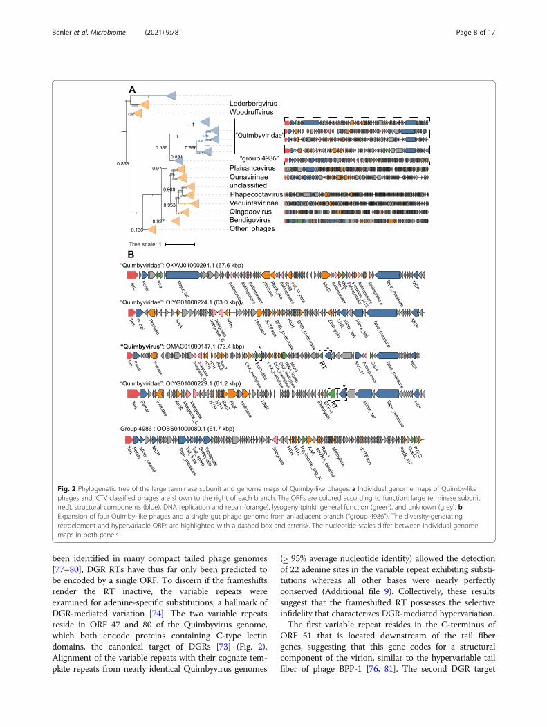
been identified in many compact tailed phage genomes[77–80], DGR RTs have thus far only been predicted tobe encoded by a single ORF. To discern if the frameshiftsrender the RT inactive, the variable repeats wereexamined for adenine-specific substitutions, a hallmark ofDGR-mediated variation [74]. The two variable repeatsreside in ORF 47 and 80 of the Quimbyvirus genome,which both encode proteins containing C-type lectindomains, the canonical target of DGRs [73] (Fig. 2).Alignment of the variable repeats with their cognate tem-plate repeats from nearly identical Quimbyvirus genomes
(> 95% average nucleotide identity) allowed the detectionof 22 adenine sites in the variable repeat exhibiting substi-tutions whereas all other bases were nearly perfectlyconserved (Additional file 9). Collectively, these resultssuggest that the frameshifted RT possesses the selectiveinfidelity that characterizes DGR-mediated hypervariation.The first variable repeat resides in the C-terminus of
ORF 51 that is located downstream of the tail fibergenes, suggesting that this gene codes for a structuralcomponent of the virion, similar to the hypervariable tailfiber of phage BPP-1 [76, 81]. The second DGR target
Fig. 2 Phylogenetic tree of the large terminase subunit and genome maps of Quimby-like phages. a Individual genome maps of Quimby-likephages and ICTV classified phages are shown to the right of each branch. The ORFs are colored according to function: large terminase subunit(red), structural components (blue), DNA replication and repair (orange), lysogeny (pink), general function (green), and unknown (grey). bExpansion of four Quimby-like phages and a single gut phage genome from an adjacent branch (“group 4986”). The diversity-generatingretroelement and hypervariable ORFs are highlighted with a dashed box and asterisk. The nucleotide scales differ between individual genomemaps in both panels
Benler et al. Microbiome (2021) 9:78 Page 8 of 17

locus is in ORF 84 that is distal to the phage structuralgene module and is expressed from the opposite DNAstrand, suggestive of a non-structural protein. Thegenomic neighborhood of ORF 80 includes genes cod-ing for a nuclease, four methyltransferases and a tRNAligase within 7 kb. The nuclease shows significant se-quence and structural similarity to E. coli mutY(HHpred probability, 97.3, Additional file 10), a DNAglycosylase involved in base excision repair. The methyl-transferases are most similar to adenine- and cytosine-modifying enzymes (HHpred probability 100 and 99.9,respectively, Additional file 10) that likely prevent cleav-age by host restriction endonucleases. Similarly, thetRNA ligase might repair tRNAs cleaved by host anti-codon endonucleases [82]. Overall, the adjacency ofORF 84 with defense- and counterdefense-related genesimplies that this hypervariable phage protein plays arole in the phage-host conflicts; however, the exactfunctions of the DGR and hypervariable target proteinsduring the life cycle of “Quimbyviridae” phages remainto be investigated.
“Flandersviridae” phages are common and abundant inwhole-community metagenomesAnalysis of the phylogenetic trees of TerL identified adeep branch of 29 gut phages (dereplicated from 196total genomes) that joins the family Ackermannviridae(Fig. 3a). Annotation of the ORFs encoded by the 29representative contigs demonstrated that the genomesare colinear, confirming that they belong to a cohesivegroup (Fig. 3b). The cohesiveness of this group wasconfirmed by the gene-sharing network, where these ge-nomes form a coherent cluster that has few connectionsto the larger network (Fig. 1b), reflecting distant (if any)similarity between most of the proteins encoded by thesephages and proteins of phages in GenBank. The mediangenome size of the phages in this group is 85.2 kb, com-pared to 157.7 kb among the Ackermannviridae phages.There is a conserved module of structural genes that en-code the MCP, portal, sheath and baseplate proteins,TerL, and the virion maturation proteinase. Thepresence of a contractile tail sheath indicates that theseviruses possess contractile tails similar to those in thefamily Ackermannviridae, in agreement with the TerLphylogeny. Several of the genes within the structural
Fig. 3 Phylogenetic tree of the large terminase subunit and complete genome maps for “Flandersviridae.” a Genome maps of members of the“Flandersviridae and selected ICTV-classified phages were constructed and colored as in Fig 3. b Genome maps of three genera from the“Flandersviridae” family. The dashed box highlights the insertion of licD- and ispD-family enzymes in the replication module of one“Flandersviridae” phage
Benler et al. Microbiome (2021) 9:78 Page 9 of 17

block contain immunoglobulin-like or C-type lectin do-mains (e.g., BACON and GH5, respectively), which arepredicted to play a role in adhesion of the virion to bac-terial cells or host-associated mucosal glycans [83–86].Downstream of the structural block is a module of genesinvolved in DNA replication that includes a DnaB-familyhelicase, DnaG-family primase, and DNA polymerase I(PolA). The polA gene is widely distributed amongdsDNA phages and therefore serves as a useful markerfor delineating the diversity of phage replication modules[87]. Phylogenetic reconstruction of both polA and dnaGencoded by these phages confirmed their monophyly(Additional file 11). Following the replication module isan approximately 20 kb long locus containing ORFs thatshowed no detectable similarity to functionally charac-terized proteins. Two of the phages harbor matches toCRISPR spacers encoded by Bacteroides and Parabacter-oides spp., indicating these bacteria serve as hosts. Basedon the large terminase and polA phylogeny, colinearityof their genomes and differences from known phages inboth genome size and content, we propose that theseBacteroides-infecting phages represent a novel taxo-nomic group, with a family rank hereafter “Flandersviri-dae” (after the region where some of the metagenomeswere sampled).Although all members of the “Flandersviridae” are
syntenic, some contain an insertion of two adjacentgenes encoding nucleotidyltransferase superfamily en-zymes within the DNA replication module. One enzymebelongs to the ispD family that is involved in the biosyn-thesis of isoprenoids [88, 89], and the other is a licDfamily enzyme that is responsible for the addition ofphosphorylcholine to teichoic acids present in bacterialcell walls [90] (Fig. 3). To our knowledge, neither ofthese enzymes has been reported in phages previously.Given that only some members of the “Flandersviridae”possess these genes, they are unlikely to performessential functions in phage reproduction, and insteadcould be implicated in phage-host interactions. The licDfamily enzyme might modify teichoic acids to preventsuperinfection by other phages, given that these polysac-charides serve as receptors for some phages to adsorb tothe host cells [91]. The role of ispD is less clear becauseispD family enzymes catalyze one step in the biosyn-thesis of isopentenyl pyrophosphate, a building block fora large variety of diverse isoprenoids [92]. Phagesmanipulate host metabolic networks including centralcarbon metabolism, nucleotide metabolism andtranslation [93]; the discovery of ispD present in the“Flandersviridae” phage genomes might add to this listthe isoprenoid biosynthetic pathway.Complete “Flandersviridae” phage genomes were re-
covered from 249 whole-community human gut metage-nomic assemblies. Their frequent assembly into closed
contigs suggests that these phages might persist in theirhost cells as extrachromosomal circular DNA molecules,similar to phage P1 [94]. However, neither genes in-volved in DNA partitioning nor lysis-lysogeny switchesare readily identifiable in the “Flandersviridae” genomes.Thus, this group of phages might be obligately lytic al-though discerning the lifestyle of a phage from thegenome sequence alone is challenging [95]. Regardlessof their lifestyle, the frequent recovery of these phagesfrom whole-community metagenomes implies that theyare common members of the human gut virome. Indeed,the “Flandersviridae” phages reach similar detectionfrequency as the crAss-like phages (Fig. 1d) althoughthere are fewer Flanders-like phages in the database.Like the “Quimbyviridae,” the even coverage ofsequencing reads across one “Flandersviridae” genome(accession OLOC01000071.1) confirms its detection isnot artifactual (Additional file 12). The high fractionalabundance and detection of Flanders-like phages inviromes generally agrees with their frequent assemblyfrom whole community metagenomes although theywere not the most abundant (see the “Discussion”section). Overall, Flanders-like phages represent a previ-ously undetected phage group that is widely distributedin human gut viromes.
“Gratiaviridae,” a putative novel family of phagesinfecting BacteroidesA deeply branching cluster of 18 genomes (dereplicatedfrom 45 total) is basal to the families Autographiviridae,Drexlerviridae, and Chaseviridae on the TerL phylogen-etic tree (Fig. 4a). Although not commonly present ingut viromes (Fig. 1d), the deep relationship betweenthese contigs and established phage families prompts indepth genome analysis of these putative phages. All 18genomes encode a DnaG-family primase and a DnaE-family polymerase, and phylogenetic reconstruction forthese genes demonstrates monophyly of these phages;the sole exception is the dnaE gene of bacteriophagephiST, a marine Cellulophaga-infecting phage that be-longs to the polyphyletic, currently defunct Siphoviridaefamily [96] (Additional file 13). The dnaG and dnaEgenes are nested within a module of other replication-associated genes that include superfamily I and IIhelicases, SbcCD exonucleases, and a RecA familyATPase (Fig. 4b). The structural module is composed ofgenes that encode an MCP, capsid maturation protease,portal protein, baseplate proteins, and a contractile tailsheath protein. Although these genomes are not strictlycolinear as observed for the “Flandersviridae” phages,the overall similarity of the proteins encoded by thesephages is apparent in the gene-sharing network wherethey form a coherent cluster that shares some edges withthe crAss-like phages (Fig. 1b). Similar to crAss-like
Benler et al. Microbiome (2021) 9:78 Page 10 of 17

phages, the predicted hosts suggested by CRISPR-spacermatches are the Bacteroides and Parabacteroides genera(Additional file 4). Taken together, the phylogenetic andgenomic organization of these phages indicate that theyrepresent a new family, provisionally named “Gratiaviridae”(after the pioneering phage biologist Dr. Andre Gratia).In addition to structural and replication proteins,
“Gratiaviridae” phages encode several enzymes of theferritin-like diiron-carboxylate superfamily. The ferritin-like enzymes encoded by these phages belong to twofamilies, namely, DNA protecting proteins (DPS) andmanganese-catalases. Manganese-catalases have notbeen documented in phage genomes, and DPS-like en-zymes have only been observed in seven Lactobacillus-infecting phages [97]. Both enzymes are involved in thetolerance of anaerobes to oxidative stress. Catalases de-toxify hydrogen peroxide to oxygen and water, enhan-cing survival of anaerobic Bacteroides in the presence ofoxygen [98]. DPS enzymes catalyze a reaction betweenoxygen and free iron to yield insoluble iron oxide, lower-ing the concentration of both intracellular oxygen andfree iron levels that would otherwise react with hydrogenperoxide and produce a hydroxyl radical, the most toxicreactive oxygen species [99, 100]. “Gratiaviridae” phagesmight deploy catalase- and DPS-like enzymes during
infection to enhance the tolerance of their strictly anaer-obic Bacteroides hosts to oxidative damage. Notably,these enzymes were not restricted to the “Gratiaviridae”but could be identified in 196 (manganese catalase) and36 (DPS) other phage genomes, including the “Flanders-viridae.” The frequent identification of these enzymes ingut phage genomes underscores the importance of intra-cellular iron and reactive oxygen species concentrationfor productive infections in an anaerobic environment.Five of the “Gratiaviridae” phages encode a protein
containing a serine/threonine protein kinase domainwith distant but significant sequence similarity to HipAfamily kinases (HHpred probability 99, Additional file10). Whereas HipA family kinases are present in numer-ous, phylogenetically distinct bacterial genomes as thetoxin component of a distinct variety of type II toxin-antitoxin systems [101, 102], there are only two charac-terized examples of protein kinases encoded by phages.The protein kinase of T7-like phages phosphorylatesRNA polymerase and RNAse III early during infectionas part of the takeover of the host cell transcriptionaland translational machinery [103–105]. In contrast, theprotein kinase of E. coli phage 933W is expressed duringlysogeny and mediates abortive infection upon super-infection of the host cell by phage HK97 [106]. The
Fig. 4 Phylogenetic tree of the large terminase subunit and genome maps of the “Gratiaviridae” phages. a Genome maps of ICTV-classifiedphages were constructed and colored as in Fig. 3. b Genome maps of four genera from the Gratiaviridae family. The dashed box highlights aHipA-family kinase domain-containing protein, AAA-family ATPase, and glycosyltransferase (see main text)
Benler et al. Microbiome (2021) 9:78 Page 11 of 17

HipA-like kinase is unlikely to function early during in-fection like the kinase of T7-like phages because, in allfive “Gratiaviridae” phages, the kinase is encoded be-tween the portal protein and MCP genes, which areexpressed late during infection in numerous culturedphages [107, 108]. Instead, the kinase might confer im-munity to heterotypic phage infection, analogous to thekinase encoded by 933W [106]. In support of animmunity-related role, an AAA-family ATPase and aglycosyltransferase are encoded immediately upstream ofthe kinase in all five phage genomes (Fig. 4). Glycosyl-transferases are encoded within capsular polysaccharidebiosynthetic loci [109] and phase variation of the capsu-lar polysaccharides confers immunity from phages thatrely on these molecules for adsorption [110]. Thespecific roles of the HipA-family kinase, ATPase andglycosyltransferase are unknown, but collectively, theseenzymes might modify host cell capsules, grantingtemporary immunity to heterotypic phage infectionwhile the morphogenesis of “Gratiaviridae” progenyvirions completes.
DiscussionA search of human gut metagenomes identified 3738putative complete phage genomes. In an attempt to re-cover complete phage genomes, this analysis restrictsthe search space to metagenomic contigs with direct ter-minal repeats which are present at the termini of somephage genomes that consequently form circular assem-blies [12]. Circular assemblies can also arise upon se-quencing a concatemer of DNA present during phageDNA replication and packaging [12], in which case thedirect repeats are a technical artifact and are the samelength as the k-mer size used to assemble the contigs.Phages with different replication and DNA packagingstrategies, such as members of the phyla Preplasmiviri-cota, Dividoviricota, or Escherichia phage Mu [12], thatlack direct repeats do not yield circular assemblies andthus were not detected here. As a result, the set of phagegenomes recovered by this strategy is both biased and anunderestimate. The results are also skewed towardssmaller genomes that are more likely to assemble into asingle contig although, in one metagenome, a 294 kbphage genome was identified (Additional file 1). Despitethese limitations, phylogenetic and comparative genomicanalyses suggest that this set of contigs includes manypreviously unnoticed lineages of phages, some, mostlikely, at the family rank.The family-rank phage lineages proposed here were
defined using a combination of approaches. Principally,phylogenetic analysis of the large terminase subunit(TerL), a hallmark gene of the Uroviricota phylum, re-vealed branches of genomes distinct from any of thosereported in the GenBank database (Fig. 1a). Supporting
the phylogenetic results, the genomes of phages onadjacent branches were of similar length and largely syn-tenic, whereas distant branches possess entirely differentarchitectures (Figs. 2, 3, and 4).The phages in two of the three proposed families
(“Flandersviridae” and “Gratiaviridae”), the genomes arelargely disconnected from other phages in the gene-sharing network. The phages in the family “Quimbyviri-dae” share genes with numerous phylogenetically distinctphages (Fig. 1c), although not enough to warrantautomated assignment into the same “viral cluster”(Additional file 2) [39]. Phages that infect phylogenetic-ally related hosts share genes more frequently with oneanother than they do with phages of distantly relatedhosts [111, 112]. The proximity of Quimbyviridae withother phages in the gene sharing network, most likely,reflects a similar preference for Bacteroides hosts (Add-itional file 4). Phylogenetic reconstruction of hallmarkgenes helped to delineate the Quimbyviridae as adistinct group which was otherwise obscured by thenumerous connections in the gene sharing network.Overall, a combination of phylogenetic analysis of hall-mark genes and gene sharing analysis will facilitate thetaxonomic classification of the gut viral community intohigher levels of organization.Two groups of phages were selected for in-depth ana-
lysis based on their frequent recovery in metagenomesand viromes. Complete genomes of “Flandersviridae”and “Quimbyviridae” phages were assembled in 249 and20 whole-community metagenomes, respectively. Yet,Quimbyvirus was more frequently detected in the vir-omes than any “Flandersviridae” phage (Fig. 1d). Thediscrepancy can be attributed to several factors, includ-ing sampling bias, the greater number of “Flandersviri-dae” genomes in the reference database “diluting” thenumber of mapped reads per genome, or the presence ofvariable loci (e.g., the variable repeats of DGRs) thatbreak contig assemblies [113]. Regardless, both groupsencompass abundant members of the human gut vir-ome. Predictably, the hosts of these phages include Bac-teroides spp., which are some of the most dominantbacterial taxa of the human gut [114] and serve as hostsfor other common human gut phages [67, 115]. Much ofthe uncharacterized “dark matter” in these phage ge-nomes is likely to be dedicated to preventing superinfec-tion of the Bacteroides host cells by such phages and tocounter the host defenses. Although in general defensesystems in Bacteroidetes remain poorly characterized,most of the bacteria possess active CRISPR-Cas systems,and numerous CRISPR spacers targeting the phages ana-lyzed here were detected using stringent thresholds witha low estimated false discovery rate (0.06) [45]. This im-plies that many if not most of the phages infecting Bac-teroidetes would encode Acrs. However, the currently
Benler et al. Microbiome (2021) 9:78 Page 12 of 17
available prediction method that was trained on the se-quences of previously identified Acrs detected putativeAcrs only in a small minority of these phages. Theremaining phages of Bacteroidetes might encode distinctAcrs or employ alternative anti-CRISPR strategies.Several phage genera possess DGRs, including Quim-
byvirus. Metagenomic surveys have shown that DGRsare enriched in the viruses that inhabit gastrointestinalenvironments [113, 116]. Combined with the inductionof DGR-carrying phages from human gut bacteria [117,118], these observations reflect a prominent role ofhypervariability underlying phage-host interactions inthe gastrointestinal environment. Notably, Quimbyvirusand another DGR-carrying phage (Hankyphage,BK010646.1), lysogenize the same Bacteroides speciesand both phages are frequently detected in human gutviromes [118]. The commonalities aside, the Quimby-virus DGR RT is encoded by three overlapping readingframes and targets two proteins, one in the structuralmodule and one in a defense-related island. DGRs havebeen associated with putative defense and signaling sys-tems in cyanobacterial and gammaproteobacterial ge-nomes [119, 120], but beyond the presence of the C-typelectin fold, the hypervariable proteins possess few otherrecognizable domains that obfuscate their precise roles.The third group analyzed in this study, the “Gratiaviri-
dae,” is not abundant but occupies a deep position onthe TerL tree relative to the Autographiviridae,Chaseviridae, and Drexlerviridae families. Analysis ofthe “Gratiaviridae” genomes will facilitate the futureorganization of these families into higher taxonomicranks, potentially, at the order level. Furthermore, ana-lysis of the “Gratiaviridae” genomes demonstrated thepresence of catalase- and DPS-family enzymes that arbi-trate cellular responses to oxidative stress [121]. Oxygenconcentrations vary along the length of the gastrointes-tinal tract, where the concentration is lower in the distalvs. proximal gut [122]. Oxygen also diffuses from tissuesradially into the lumen [123], and in combination withother factors, these gradients affect the structure andcomposition of the gastrointestinal microbiota [124].The acquisition of oxygen detoxifying-enzymes by the“Gratiaviridae” and other gut phages signals a need tosupplement their host cell’s tolerance to oxidative dam-age during infection, which might be especially import-ant for cells that reside near the tissue surface whereoxygen exposure is higher.A unique feature of some “Gratiaviridae” phages is a
HipA-family protein kinase. The T7-like phages (withinthe Autographiviridae family) and Escherichia phage933W (currently unclassified at the family level) encodePKC-family protein kinases that function during host celltakeover and abortive infection, respectively [103, 106]. Athird, CotH-family protein kinase domain is occasionally
observed in phage genomes where it is fused to a hyper-variable C-type lectin domain [73, 116], but these proteinsare currently unstudied. The “Gratiaviridae” phages re-cruited a fourth family of protein kinases that, togetherwith the phage encoded glysosyltransferase, might modifythe host cell envelope, contributing to the prevention ofsuperinfection.
ConclusionsIn summary, comparative genomic analysis of the phagesdescribed here, along with the complementary analysisof crAss-like phages [22], substantially increases thecharacterized diversity of phages, primarily, those infect-ing Bacteroidetes bacteria, which are major componentsof the human gut microbiome. These findings also ex-pand the repertoire of phage gene functions, notably, byadding the isoprenoid metabolic pathway, catalase-likeenzymes, HipA family protein kinases, and hypervariablegenes implicated in defense. All of these open multipledirections for experimental study.
Supplementary InformationThe online version contains supplementary material available at https://doi.org/10.1186/s40168-021-01017-w.
Additional file 1. Clustering information for 3,378 gut phages identifiedin the study. Phage genomes were dereplicated at 95% identity over 80%of the contig length. The GenBank accession codes for each phage andthe representative sequence is provided in columns 1 and 3, respectively.
Additional file 2. Taxonomic information for 1,886 representative phagegenomes. Each taxonomically classified phage genome was scored byViralVerify, Seeker and vcontact2 and the assignments are provided incolumns 9-10, 11 and 12-13, respectively.
Additional file 3. Distribution of the marker profiles identified on eachphage genome and pie chart representation of the gut phage taxonomy.(A) Histogram of marker proteins detected on each phage genomerecovered from human gut metagenomes. Abbreviations are as follows:M, major capsid protein; P, portal, T, terminase large subunit. (B)Taxonomic assignments of the dereplicated contigs (n = 1,886), with theoutermost ring corresponding to ICTV families.
Additional file 4. Host ranges inferred from CRISPR-spacer matches. Thenucleotide coordinates of the protospacer are provided in columns 2 and3. The sequence of the CRISPR spacer and protospacer are provided incolumns 4 and 5. The taxonomic information of the host is listed in thesubsequent columns.
Additional file 5. Candidate anti-CRISPR (Acr) proteins encoded on eachphage genome. Proteins predicted to be an Acr with high confidence(score > 0.9) and meeting all other heuristic criteria are listed along withtheir inferred host. For the Bifidobacteria phages (see the main text), thenucleotide coordinates of closely related prophages integrated in theirhost genome are provided in columns 6-8.
Additional file 6. Genome maps of predicted anti-CRISPR proteins (Acrs)in uncharacterized Bifidobacteria phages. Open reading frames are col-ored according to function: large terminase subunit (red), structural com-ponents (blue), replication (orange), integrase (pink), general function(green) and unknown (grey). The candidate Acrs are indicated with adashed box.
Additional file 7. Coverage heatmap of a “Quimbyviridae” genomeacross human gut viromes. The coverage of the most abundant“Quimbyviridae” phage genome (accession OMAC01000147.1) is plottedas a heatmap, scaled from 0 – 100x per 100 bp window.
Benler et al. Microbiome (2021) 9:78 Page 13 of 17
Additional file 8. Phylogenetic tree of the MCP, primase and portalproteins encoded by “Quimbyviridae” phages.
Additional file 9. Alignment of the template and variable repeats fromthe Quimbyvirus DGRs. The template repeat (TR) from Quimbyvirus is thefirst listed sequence and is followed by the variable repeats from eitherORF80 (VR1) or ORF 47 (VR2) encoded in phage genomes that are nearlyidentical to Quimbyvirus (> 95 % average nucleotide identity). A total of21 adenine residues (green) in the template repeat exhibit a least onesubstitution in a corresponding variable repeat.
Additional file 10. HHPred alignments of four Quimbyviridae proteinsand one Gratiaviridae protein with their top-scoring templates, includinga replication initiator protein, a cytosine-specific methyltransferase, anadenine-specific methyltransferase, a MutY nuclease and HipA kinase.
Additional file 11. Phylogenetic tree of the polA and dnaG genes inFlanders-like phages. Branches composed of GenBank phages are coloredin orange and branches of gut metagenomic phages in blue. Brancheswith sequences labelled as bacteria in the GenBank database, likely repre-senting cryptic prophages, are colored in grey.
Additional file 12. Coverage heatmap of a “Flandersviridae” genomeacross human gut viromes. The coverage of the most abundant“Flandersviridae” phage genome (accession OLOC0100071.1) is plotted asa heatmap, scaled from 0 – 100x fold coverage per 100 bp window.
Additional file 13. Phylogenetic tree of the dnaG and dnaE genes inGratiaviridae phages.
AcknowledgementsWe are grateful for the assistance from Yuri Wolf with phylogeneticreconstruction of the phage hallmark genes and to Koonin group membersfor useful discussions. This work utilized the computational resources of theNIH HPC Biowulf cluster (http://hpc.nih.gov).
Authors’ contributionsNY, MR, and DA analyzed the gut metagenomes; SS and AG executed theCRISPR analyses; SB analyzed the phage genomes and wrote the manuscript;PP and EVK conceived the project and wrote the manuscript. The authorsread and approved the final manuscript.
FundingFunding for this project was provided by the Intramural Research Program ofthe National Institutes of Health (National Library of Medicine). MR & DAwere supported by the St. Petersburg State University, Russia (grant ID PURE51555639). The funding body had no role in the collection, analysis, andinterpretation of data or writing of the manuscript.
Availability of data and materialsAll phage genomes are available in the NCBI GenBank database using theaccession numbers listed in Additional file 1. The genomes, proteinpredictions and annotations are also provided at ftp://ftp.ncbi.nih.gov/pub/yutinn/benler_2020/gut_phages/. Underlying alignments of marker proteinsequences used to generate phylogenetic trees and the source data used togenerate Fig. 1c, d are also available on the FTP site.
Declarations
Ethics approval and consent to participateNot applicable.
Consent for publicationNot applicable.
Competing interestsThe authors declare that they have no competing interests.
Author details1National Center for Biotechnology Information, National Library of Medicine,Bethesda, Maryland 20894, USA. 2Center for Algorithmic Biotechnology,Institute for Translational Biomedicine, St. Petersburg State University, St.Petersburg 199004, Russia. 3Department of Computer Science andEngineering, University of California, San Diego, La Jolla, CA 92093, USA.
Received: 6 October 2020 Accepted: 2 February 2021
References1. Cobián Güemes AG, Youle M, Cantú VA, Felts B, Nulton J, Rohwer F. Viruses
as winners in the game of life. Annu Rev Virol. 2016;3(1):197–214. https://doi.org/10.1146/annurev-virology-100114-054952.
2. Reyes A, Haynes M, Hanson N, Angly FE, Heath AC, Rohwer F, Gordon JI.Viruses in the faecal microbiota of monozygotic twins and their mothers.Nature. 2010;466(7304):334–8. https://doi.org/10.1038/nature09199.
3. Breitbart M, Hewson I, Felts B, Mahaffy JM, Nulton J, Salamon P, Rohwer F.Metagenomic analyses of an uncultured viral community from humanfeces. J Bacteriol. 2003;185(20):6220–3. https://doi.org/10.1128/JB.185.20.6220-6223.2003.
4. Koonin EV, Dolja VV, Krupovic M, Varsani A, Wolf YI, Yutin N, Zerbini FM,Kuhn JH. Global organization and proposed megataxonomy of the virusworld. Microbiol Mol Biol Rev. 2020;84(2):e00061–19.
5. Barr JJ, Auro R, Furlan M, Whiteson KL, Erb ML, Pogliano J, Stotland A,Wolkowicz R, Cutting AS, Doran KS, Salamon P, Youle M, Rohwer F.Bacteriophage adhering to mucus provide a non–host-derived immunity.Proc Natl Acad Sci. 2013;110(26):10771–6. https://doi.org/10.1073/pnas.1305923110.
6. Roach DR, Leung CY, Henry M, Morello E, Singh D, Di Santo JP, Weitz JS,Debarbieux L. Synergy between the host immune system andbacteriophage is essential for successful phage therapy against an acuterespiratory pathogen. Cell Host Microbe. 2017;22(1):38–47.e34.
7. Moreno-Gallego JL, Chou S-P, Di Rienzi SC, Goodrich JK, Spector TD, Bell JT,Youngblut ND, Hewson I, Reyes A, Ley RE. Virome diversity correlates withintestinal microbiome diversity in adult monozygotic twins. Cell HostMicrobe. 2019;25(2):261–272.e265.
8. Gogokhia L, Buhrke K, Bell R, Hoffman B, Brown DG, Hanke-Gogokhia C,Ajami NJ, Wong MC, Ghazaryan A, Valentine JF, et al. Expansion ofbacteriophages is linked to aggravated intestinal inflammation and colitis.Cell Host Microbe. 2019;25(2):285–299.e288.
9. Waller AS, Yamada T, Kristensen DM, Kultima JR, Sunagawa S, KooninEV, Bork P. Classification and quantification of bacteriophage taxa inhuman gut metagenomes. ISME J. 2014;8(7):1391–402. https://doi.org/10.1038/ismej.2014.30.
10. Minot S, Bryson A, Chehoud C, Wu GD, Lewis JD, Bushman FD. Rapidevolution of the human gut virome. Proc Natl Acad Sci. 2013;110(30):12450–5.https://doi.org/10.1073/pnas.1300833110.
11. Shkoporov AN, Clooney AG, Sutton TDS, Ryan FJ, Daly KM, Nolan JA,McDonnell SA, Khokhlova EV, Draper LA, Forde A, et al. The human gutvirome is highly diverse, stable, and individual specific. Cell Host Microbe.2019;26(4):527–541.e525.
12. Casjens SR, Gilcrease EB. Determining DNA Packaging Strategy by Analysis ofthe Termini of the Chromosomes in Tailed-Bacteriophage Virions. In:Bacteriophages: Methods and Protocols, Volume 2 Molecular and AppliedAspects. Edited by Clokie MRJ, Kropinski AM. Totowa, NJ: Humana Press;2009:91–111.
13. Al-Shayeb B, Sachdeva R, Chen L-X, Ward F, Munk P, Devoto A, Castelle CJ,Olm MR, Bouma-Gregson K, Amano Y, et al. Clades of huge phages fromacross Earth’s ecosystems. Nature. 2020;578(7795):425–31. https://doi.org/10.1038/s41586-020-2007-4.
14. Shkoporov AN, Ryan FJ, Draper LA, Forde A, Stockdale SR, Daly KM,McDonnell SA, Nolan JA, Sutton TDS, Dalmasso M, et al. Reproducibleprotocols for metagenomic analysis of human faecal phageomes.Microbiome. 2018;6(1):68. https://doi.org/10.1186/s40168-018-0446-z.
15. Antipov D, Raiko M, Lapidus A, Pevzner PA. Plasmid detection and assemblyin genomic and metagenomic data sets. Genome Res. 2019;29(6):961–8.https://doi.org/10.1101/gr.241299.118.
16. Antipov D, Raiko M, Lapidus A, Pevzner PA. metaviralSPAdes: assembly ofviruses from metagenomic data. Bioinformatics. 2020;36(14):4126–9. https://doi.org/10.1093/bioinformatics/btaa490.
17. Gideon SG, Wright A. DNA segregation in bacteria. Annu Rev Microbiol.2000;54(1):681–708.
18. De Sordi L, Lourenço M, Debarbieux L. The battle within: interactions ofbacteriophages and bacteria in the gastrointestinal tract. Cell Host Microbe.2019;25(2):210–8. https://doi.org/10.1016/j.chom.2019.01.018.
Benler et al. Microbiome (2021) 9:78 Page 14 of 17

19. Beller L, Matthijnssens J. What is (not) known about the dynamics of thehuman gut virome in health and disease. Curr Opin Virol. 2019;37:52–7.https://doi.org/10.1016/j.coviro.2019.05.013.
20. Simmonds P, Adams MJ, Benkő M, Breitbart M, Brister JR, Carstens EB,Davison AJ, Delwart E, Gorbalenya AE, Harrach B, Hull R, King AMQ, KooninEV, Krupovic M, Kuhn JH, Lefkowitz EJ, Nibert ML, Orton R, Roossinck MJ,Sabanadzovic S, Sullivan MB, Suttle CA, Tesh RB, van der Vlugt RA, Varsani A,Zerbini FM. Virus taxonomy in the age of metagenomics. Nat Rev Microbiol.2017;15(3):161–8. https://doi.org/10.1038/nrmicro.2016.177.
21. Lu S, Wang J, Chitsaz F, Derbyshire MK, Geer RC, Gonzales NR, Gwadz M,Hurwitz DI, Marchler GH, Song JS, et al. CDD/SPARCLE: the conserveddomain database in 2020. Nucleic Acids Res. 2019;48(D1):D265–8.
22. Yutin N, Benler S, Shmakov SA, Wolf YI, Tolstoy I, Rayko M, et al. Analysis ofmetagenome-assembled viral genomes from the human gut reveals diverseputative CrAss-like phages with unique genomic features. Nat Commun.2021;12:1044
23. Hyatt D, Chen G-L, LoCascio PF, Land ML, Larimer FW, Hauser LJ. Prodigal:prokaryotic gene recognition and translation initiation site identification. BMCBioinformatics. 2010;11(1):119. https://doi.org/10.1186/1471-2105-11-119.
24. Lowe TM, Eddy SR. tRNAscan-SE: a program for improved detection oftransfer RNA genes in genomic sequence. Nucleic Acids Res. 1997;25(5):955–64. https://doi.org/10.1093/nar/25.5.955.
25. Devoto AE, Santini JM, Olm MR, Anantharaman K, Munk P, Tung J, ArchieEA, Turnbaugh PJ, Seed KD, Blekhman R, Aarestrup FM, Thomas BC, BanfieldJF. Megaphages infect Prevotella and variants are widespread in gutmicrobiomes. Nat Microbiol. 2019;4(4):693–700. https://doi.org/10.1038/s41564-018-0338-9.
26. Ivanova NN, Schwientek P, Tripp HJ, Rinke C, Pati A, Huntemann M, Visel A,Woyke T, Kyrpides NC, Rubin EM. Stop codon reassignments in the wild.Science. 2014;344(6186):909–13. https://doi.org/10.1126/science.1250691.
27. Auslander N, Gussow AB, Benler S, Wolf YI, Koonin EV. Seeker: alignment-free identification of bacteriophage genomes by deep learning. NucleicAcids Research 2020;48(21):e121-e121
28. Kans J. Entrez direct: E-utilities on the Unix Command Line. Bethesda:National Center for Biotechnology Information; 2010.
29. Olm MR, Brown CT, Brooks B, Banfield JF. dRep: a tool for fast and accurategenomic comparisons that enables improved genome recovery frommetagenomes through de-replication. ISME J. 2017;11(12):2864–8. https://doi.org/10.1038/ismej.2017.126.
30. Ondov BD, Starrett GJ, Sappington A, Kostic A, Koren S, Buck CB, PhillippyAM. Mash Screen: high-throughput sequence containment estimation forgenome discovery. Genome Biol. 2019;20(1):232. https://doi.org/10.1186/s13059-019-1841-x.
31. Jain C, Rodriguez-R LM, Phillippy AM, Konstantinidis KT, Aluru S. Highthroughput ANI analysis of 90K prokaryotic genomes reveals clear speciesboundaries. Nat Commun. 2018;9(1):5114. https://doi.org/10.1038/s41467-018-07641-9.
32. Steinegger M, Söding J. Clustering huge protein sequence sets inlinear time. Nat Commun. 2018;9(1):2542. https://doi.org/10.1038/s41467-018-04964-5.
33. Altschul SF, Madden TL, Schäffer AA, Zhang J, Zhang Z, Miller W, Lipman DJ.Gapped BLAST and PSI-BLAST: a new generation of protein database searchprograms. Nucleic Acids Res. 1997;25(17):3389–402. https://doi.org/10.1093/nar/25.17.3389.
34. Wolf YI, Kazlauskas D, Iranzo J, Lucía-Sanz A, Kuhn JH, Krupovic M, Dolja VV,Koonin EV. Origins and evolution of the global RNA virome. mBio. 2018;9(6):e02329–18.
35. Edgar RC. MUSCLE: multiple sequence alignment with high accuracy andhigh throughput. Nucleic Acids Res. 2004;32(5):1792–7. https://doi.org/10.1093/nar/gkh340.
36. Steinegger M, Meier M, Mirdita M, Vöhringer H, Haunsberger SJ, Söding J.HH-suite3 for fast remote homology detection and deep proteinannotation. BMC Bioinformatics. 2019;20(1):473. https://doi.org/10.1186/s12859-019-3019-7.
37. Yutin N, Makarova KS, Mekhedov SL, Wolf YI, Koonin EV. The deep archaealroots of eukaryotes. Mol Biol Evol. 2008;25(8):1619–30. https://doi.org/10.1093/molbev/msn108.
38. Price MN, Dehal PS, Arkin AP. FastTree 2 – approximately maximum-likelihood trees for large alignments. PLoS One. 2010;5(3):e9490. https://doi.org/10.1371/journal.pone.0009490.
39. Bin Jang H, Bolduc B, Zablocki O, Kuhn JH, Roux S, Adriaenssens EM, BristerJR, Kropinski AM, Krupovic M, Lavigne R, Turner D, Sullivan MB. Taxonomicassignment of uncultivated prokaryotic virus genomes is enabled by gene-sharing networks. Nat Biotechnol. 2019;37(6):632–9. https://doi.org/10.1038/s41587-019-0100-8.
40. Shannon P, Markiel A, Ozier O, Baliga NS, Wang JT, Ramage D, Amin N,Schwikowski B, Ideker T. Cytoscape: a software environment for integratedmodels of biomolecular interaction networks. Genome Res. 2003;13(11):2498–504. https://doi.org/10.1101/gr.1239303.
41. Mirdita M, von den Driesch L, Galiez C, Martin MJ, Söding J, Steinegger M.Uniclust databases of clustered and deeply annotated protein sequencesand alignments. Nucleic Acids Res. 2016;45(D1):D170–6. https://doi.org/10.1093/nar/gkw1081.
42. Ye Y. Identification of diversity-generating retroelements in humanmicrobiomes. Int J Mol Sci. 2014;15(8):14234–46.
43. Langmead B, Salzberg SL. Fast gapped-read alignment with Bowtie 2. NatMethods. 2012;9(4):357–9. https://doi.org/10.1038/nmeth.1923.
44. Chen S, Zhou Y, Chen Y, Gu J. fastp: an ultra-fast all-in-one FASTQpreprocessor. Bioinformatics. 2018;34(17):i884–90. https://doi.org/10.1093/bioinformatics/bty560.
45. Shmakov SA, Sitnik V, Makarova KS, Wolf YI, Severinov KV, Koonin EV. TheCRISPR spacer space is dominated by sequences from species-specificmobilomes. mBio. 2017;8(5):e01397–17.
46. Shmakov SA, Wolf YI, Savitskaya E, Severinov KV, Koonin EV. Mapping CRISPR spaceromes reveals vast host-specific viromes of prokaryotes. CommunBiol. 2020;3(1):321. https://doi.org/10.1038/s42003-020-1014-1.
47. Morgulis A, Coulouris G, Raytselis Y, Madden TL, Agarwala R, Schäffer AA.Database indexing for production MegaBLAST searches. Bioinformatics.2008;24(16):1757–64. https://doi.org/10.1093/bioinformatics/btn322.
48. Gussow AB, Park AE, Borges AL, Shmakov SA, Makarova KS, Wolf YI, Bondy-Denomy J, Koonin EV. Machine-learning approach expands the repertoire ofanti-CRISPR protein families. Nat Commun. 2020;11(1):3784. https://doi.org/10.1038/s41467-020-17652-0.
49. Luque A, Benler S, Lee DY, Brown C, White S. The missing tailed phages:prediction of small capsid candidates. Microorganisms. 2020;8(12):1944.https://doi.org/10.3390/microorganisms8121944.
50. Soto-Perez P, Bisanz JE, Berry JD, Lam KN, Bondy-Denomy J, Turnbaugh PJ.CRISPR-Cas system of a prevalent human gut bacterium reveals hyper-targeting against phages in a human virome catalog. Cell Host Microbe.2019;26(3):325–335.e325.
51. Hynes AP, Rousseau GM, Agudelo D, Goulet A, Amigues B, Loehr J, RomeroDA, Fremaux C, Horvath P, Doyon Y, Cambillau C, Moineau S. Widespreadanti-CRISPR proteins in virulent bacteriophages inhibit a range of Cas9proteins. Nat Commun. 2018;9(1):2919. https://doi.org/10.1038/s41467-018-05092-w.
52. Marino ND, Zhang JY, Borges AL, Sousa AA, Leon LM, Rauch BJ, Walton RT,Berry JD, Joung JK, Kleinstiver BP, Bondy-Denomy J. Discovery ofwidespread type I and type V CRISPR-Cas inhibitors. Science. 2018;362(6411):240–2. https://doi.org/10.1126/science.aau5174.
53. Pawluk A, Davidson AR, Maxwell KL. Anti-CRISPR: discovery, mechanism andfunction. Nat Rev Microbiol. 2018;16(1):12–7. https://doi.org/10.1038/nrmicro.2017.120.
54. Hwang S, Maxwell KL. Meet the anti-CRISPRs: widespread protein inhibitorsof CRISPR-Cas systems. CRISPR J. 2019;2(1):23–30. https://doi.org/10.1089/crispr.2018.0052.
55. Osuna BA, Karambelkar S, Mahendra C, Christie KA, Garcia B, Davidson AR,Kleinstiver BP, Kilcher S, Bondy-Denomy J. Listeria phages induce Cas9degradation to protect lysogenic genomes. Cell Host Microbe. 2020;28(1):31–40.e9. https://doi.org/10.1016/j.chom.2020.04.001.
56. Osuna BA, Karambelkar S, Mahendra C, Sarbach A, Johnson MC, Kilcher S,Bondy-Denomy J. Critical anti-CRISPR locus repression by a bi-functionalCas9 inhibitor. Cell Host Microbe. 2020;28(1):23–30.e5. https://doi.org/10.1016/j.chom.2020.04.002.
57. Stanley SY, Borges AL, Chen K-H, Swaney DL, Krogan NJ, Bondy-Denomy J,Davidson AR. Anti-CRISPR-associated proteins are crucial repressors of anti-CRISPR transcription. Cell. 2019;178(6):1452–1464.e1413.
58. Roux S, Krupovic M, Daly RA, Borges AL, Nayfach S, Schulz F, Sharrar A,Matheus Carnevali PB, Cheng J-F, Ivanova NN, Bondy-Denomy J, WrightonKC, Woyke T, Visel A, Kyrpides NC, Eloe-Fadrosh EA. Cryptic inovirusesrevealed as pervasive in bacteria and archaea across Earth’s biomes. NatMicrobiol. 2019;4(11):1895–906. https://doi.org/10.1038/s41564-019-0510-x.
Benler et al. Microbiome (2021) 9:78 Page 15 of 17
59. Wang H, Ling Y, Shan T, Yang S, Xu H, Deng X, Delwart E, Zhang W. Gutvirome of mammals and birds reveals high genetic diversity of the familyMicroviridae. Virus Evol. 2019;5(1):vez013.
60. Koonin EV, Yutin N. The crAss-like phage group: how metagenomicsreshaped the human virome. Trends Microbiol. 2020;28(5):349–59. https://doi.org/10.1016/j.tim.2020.01.010.
61. Iranzo J, Krupovic M, Koonin EV. The double-stranded DNA virosphere as amodular hierarchical network of gene sharing. mBio. 2016;7(4):e00978–16.
62. Aiewsakun P, Adriaenssens EM, Lavigne R, Kropinski AM, Simmonds P.Evaluation of the genomic diversity of viruses infecting bacteria, archaeaand eukaryotes using a common bioinformatic platform: steps towards aunified taxonomy. J Gen Virol. 2018;99(9):1331–43. https://doi.org/10.1099/jgv.0.001110.
63. Barylski J, Enault F, Dutilh BE, Schuller MB, Edwards RA, Gillis A, KlumppJ, Knezevic P, Krupovic M, Kuhn JH, et al. Analysis of spounaviruses as acase study for the overdue reclassification of tailed phages. Syst Biol.2019;69(1):110–23.
64. Low SJ, Džunková M, Chaumeil P-A, Parks DH, Hugenholtz P. Evaluation of aconcatenated protein phylogeny for classification of tailed double-strandedDNA viruses belonging to the order Caudovirales. Nat Microbiol. 2019;4(8):1306–15. https://doi.org/10.1038/s41564-019-0448-z.
65. Yutin N, Makarova KS, Gussow AB, Krupovic M, Segall A, Edwards RA, KooninEV. Discovery of an expansive bacteriophage family that includes the mostabundant viruses from the human gut. Nat Microbiol. 2018;3(1):38–46.https://doi.org/10.1038/s41564-017-0053-y.
66. Dutilh BE, Cassman N, McNair K, Sanchez SE, Silva GGZ, Boling L, Barr JJ,Speth DR, Seguritan V, Aziz RK, Felts B, Dinsdale EA, Mokili JL, Edwards RA. Ahighly abundant bacteriophage discovered in the unknown sequences ofhuman faecal metagenomes. Nat Commun. 2014;5(1):4498. https://doi.org/10.1038/ncomms5498.
67. Edwards RA, Vega AA, Norman HM, Ohaeri M, Levi K, Dinsdale EA, Cinek O,Aziz RK, McNair K, Barr JJ, Bibby K, Brouns SJJ, Cazares A, de Jonge PA,Desnues C, Díaz Muñoz SL, Fineran PC, Kurilshikov A, Lavigne R, MazankovaK, McCarthy DT, Nobrega FL, Reyes Muñoz A, Tapia G, Trefault N, Tyakht AV,Vinuesa P, Wagemans J, Zhernakova A, Aarestrup FM, Ahmadov G, Alassaf A,Anton J, Asangba A, Billings EK, Cantu VA, Carlton JM, Cazares D, Cho GS,Condeff T, Cortés P, Cranfield M, Cuevas DA, de la Iglesia R, Decewicz P,Doane MP, Dominy NJ, Dziewit L, Elwasila BM, Eren AM, Franz C, Fu J,Garcia-Aljaro C, Ghedin E, Gulino KM, Haggerty JM, Head SR, Hendriksen RS,Hill C, Hyöty H, Ilina EN, Irwin MT, Jeffries TC, Jofre J, Junge RE, Kelley ST,Khan Mirzaei M, Kowalewski M, Kumaresan D, Leigh SR, Lipson D, LisitsynaES, Llagostera M, Maritz JM, Marr LC, McCann A, Molshanski-Mor S, MonteiroS, Moreira-Grez B, Morris M, Mugisha L, Muniesa M, Neve H, Nguyen NP,Nigro OD, Nilsson AS, O’Connell T, Odeh R, Oliver A, Piuri M, Prussin II AJ,Qimron U, Quan ZX, Rainetova P, Ramírez-Rojas A, Raya R, Reasor K, RiceGAO, Rossi A, Santos R, Shimashita J, Stachler EN, Stene LC, Strain R, StumpfR, Torres PJ, Twaddle A, Ugochi Ibekwe MA, Villagra N, Wandro S, White B,Whiteley A, Whiteson KL, Wijmenga C, Zambrano MM, Zschach H, Dutilh BE.Global phylogeography and ancient evolution of the widespread humangut virus crAssphage. Nat Microbiol. 2019;4(10):1727–36. https://doi.org/10.1038/s41564-019-0494-6.
68. Schumacher MA, Tonthat NK, Kwong SM, Chinnam N, Liu MA, Skurray RA,Firth N. Mechanism of staphylococcal multiresistance plasmid replicationorigin assembly by the RepA protein. Proc Natl Acad Sci. 2014;111(25):9121–6. https://doi.org/10.1073/pnas.1406065111.
69. Botstein D, Lew KK, Jarvik V, Swanson CA. Role of antirepressor in thebipartite control of repression and immunity by bacteriophage P22. J MolBiol. 1975;91(4):439–62. https://doi.org/10.1016/0022-2836(75)90271-5.
70. Argov T, Sapir SR, Pasechnek A, Azulay G, Stadnyuk O, Rabinovich L, Sigal N,Borovok I, Herskovits AA. Coordination of cohabiting phage elementssupports bacteria–phage cooperation. Nat Commun. 2019;10(1):1–4.
71. Davis-Richardson AG, Ardissone AN, Dias R, Simell V, Leonard MT,Kemppainen KM, Drew JC, Schatz D, Atkinson MA, Kolaczkowski B, et al.Bacteroides dorei dominates gut microbiome prior to autoimmunity inFinnish children at high risk for type 1 diabetes. Front Microbiol. 2014;5:678.
72. Campbell A. Comparative molecular biology of lambdoid phages. Annu RevMicrobiol. 1994;48(1):193–222.
73. Wu L, Gingery M, Abebe M, Arambula D, Czornyj E, Handa S, Khan H, Liu M,Pohlschroder M, Shaw KL, et al. Diversity-generating retroelements: naturalvariation, classification and evolution inferred from a large-scale genomicsurvey. Nucleic Acids Res. 2017;46(1):11–24.
74. Handa S, Jiang Y, Tao S, Foreman R, Schinazi RF, Miller JF, Ghosh P.Template-assisted synthesis of adenine-mutagenized cDNA by aretroelement protein complex. Nucleic Acids Res. 2018;46(18):9711–25.https://doi.org/10.1093/nar/gky620.
75. Miller JL, Coq JL, Hodes A, Barbalat R, Miller JF, Ghosh P. Selective ligandrecognition by a diversity-generating retroelement variable protein. PLoSBiol. 2008;6(6):e131. https://doi.org/10.1371/journal.pbio.0060131.
76. Liu M, Deora R, Doulatov SR, Gingery M, Eiserling FA, Preston A, Maskell DJ,Simons RW, Cotter PA, Parkhill J, Miller JF. Reverse transcriptase-mediatedtropism switching in Bordetella bacteriophage. Science. 2002;295(5562):2091–4. https://doi.org/10.1126/science.1067467.
77. Xu J, Hendrix RW, Duda RL. Conserved translational frameshift in dsDNAbacteriophage tail assembly genes. Mol Cell. 2004;16(1):11–21. https://doi.org/10.1016/j.molcel.2004.09.006.
78. Levin ME, Hendrix RW, Casjens SR. A programmed translational frameshift isrequired for the synthesis of a bacteriophage λ tail assembly protein. J MolBiol. 1993;234(1):124–39. https://doi.org/10.1006/jmbi.1993.1568.
79. Baranov PV, Fayet O, Hendrix RW, Atkins JF. Recoding in bacteriophagesand bacterial IS elements. Trends Genet. 2006;22(3):174–81. https://doi.org/10.1016/j.tig.2006.01.005.
80. McNair K, Zhou C, Dinsdale EA, Souza B, Edwards RA. PHANOTATE: a novelapproach to gene identification in phage genomes. Bioinformatics. 2019;35(22):4537–42. https://doi.org/10.1093/bioinformatics/btz265.
81. Doulatov S, Hodes A, Dai L, Mandhana N, Liu M, Deora R, Simons RW,Zimmerly S, Miller JF. Tropism switching in Bordetella bacteriophage definesa family of diversity-generating retroelements. Nature. 2004;431(7007):476–81. https://doi.org/10.1038/nature02833.
82. Meineke B, Shuman S. Determinants of the cytotoxicity of PrrC anticodonnuclease and its amelioration by tRNA repair. RNA. 2012;18(1):145–54.https://doi.org/10.1261/rna.030171.111.
83. de Jonge PA, von Meijenfeldt FAB, van Rooijen LE, Brouns SJJ, Dutilh BE.Evolution of BACON domain tandem repeats in crassphage and novel gutbacteriophage lineages. Viruses. 2019;11(12):1085. https://doi.org/10.3390/v11121085.
84. Pell LG, Gasmi-Seabrook GMC, Morais M, Neudecker P, Kanelis V, Bona D,Donaldson LW, Edwards AM, Howell PL, Davidson AR, Maxwell KL. Thesolution structure of the C-terminal Ig-like domain of the bacteriophage λtail tube protein. J Mol Biol. 2010;403(3):468–79. https://doi.org/10.1016/j.jmb.2010.08.044.
85. Fraser JS, Yu Z, Maxwell KL, Davidson AR. Ig-Like domains onbacteriophages: a tale of promiscuity and deceit. J Mol Biol. 2006;359(2):496–507. https://doi.org/10.1016/j.jmb.2006.03.043.
86. Barr JJ, Auro R, Sam-Soon N, Kassegne S, Peters G, Bonilla N, Hatay M,Mourtada S, Bailey B, Youle M, Felts B, Baljon A, Nulton J, Salamon P,Rohwer F. Subdiffusive motion of bacteriophage in mucosal surfacesincreases the frequency of bacterial encounters. Proc Natl Acad Sci. 2015;112(44):13675–80. https://doi.org/10.1073/pnas.1508355112.
87. Nasko DJ, Chopyk J, Sakowski EG, Ferrell BD, Polson SW, Wommack KE.Family A DNA polymerase phylogeny uncovers diversity and replicationgene organization in the virioplankton. Front Microbiol. 2018;9:3053.
88. Campbell TL, Brown ED. Characterization of the depletion of 2-C-methyl-d-erythritol-2,4-cyclodiphosphate synthase in Escherichia coli and Bacillussubtilis. J Bacteriol. 2002;184(20):5609–18. https://doi.org/10.1128/JB.184.20.5609-5618.2002.
89. Heuston S, Begley M, Gahan CGM, Hill C. Isoprenoid biosynthesis in bacterialpathogens. Microbiology. 2012;158(6):1389–401. https://doi.org/10.1099/mic.0.051599-0.
90. Zhang J-R, Idanpaan-Heikkila I, Fischer W, Tuomanen EI. Pneumococcal licD2gene is involved in phosphorylcholine metabolism. Mol Microbiol. 1999;31(5):1477–88. https://doi.org/10.1046/j.1365-2958.1999.01291.x.
91. Lopez R, Garcia E, Garcia P, Ronda C, Tomasz A. Choline-containingbacteriophage receptors in Streptococcus pneumoniae. J Bacteriol. 1982;151(3):1581–90. https://doi.org/10.1128/JB.151.3.1581-1590.1982.
92. Odom AR. Five questions about non-mevalonate isoprenoid biosynthesis. PLoSPathog. 2011;7(12):e1002323. https://doi.org/10.1371/journal.ppat.1002323.
93. Breitbart M, Bonnain C, Malki K, Sawaya NA. Phage puppet masters of themarine microbial realm. Nat Microbiol. 2018;3(7):754–66. https://doi.org/10.1038/s41564-018-0166-y.
94. Łobocka MB, Rose DJ, Plunkett G, Rusin M, Samojedny A, Lehnherr H,Yarmolinsky MB, Blattner FR. Genome of bacteriophage P1. J Bacteriol. 2004;186(21):7032–68. https://doi.org/10.1128/JB.186.21.7032-7068.2004.
Benler et al. Microbiome (2021) 9:78 Page 16 of 17

95. McNair K, Bailey BA, Edwards RA. PHACTS, a computational approach toclassifying the lifestyle of phages. Bioinformatics. 2012;28(5):614–8. https://doi.org/10.1093/bioinformatics/bts014.
96. Holmfeldt K, Solonenko N, Shah M, Corrier K, Riemann L, VerBerkmoes NC,Sullivan MB. Twelve previously unknown phage genera are ubiquitous inglobal oceans. Proc Natl Acad Sci. 2013;110(31):12798–803. https://doi.org/10.1073/pnas.1305956110.
97. Kyrkou I, Byth Carstens A, Ellegaard-Jensen L, Kot W, Zervas A, Djurhuus AM,Neve H, Hansen M, Hestbjerg Hansen L. Expanding the diversity ofmyoviridae phages infecting Lactobacillus plantarum—a novel lineage oflactobacillus phages comprising five new members. Viruses. 2019;11(7):611.https://doi.org/10.3390/v11070611.
98. Rocha ER, Selby T, Coleman JP, Smith CJ. Oxidative stress response in ananaerobe, Bacteroides fragilis: a role for catalase in protection againsthydrogen peroxide. J Bacteriol. 1996;178(23):6895–903. https://doi.org/10.1128/JB.178.23.6895-6903.1996.
99. Betteken MI, Rocha ER, Smith CJ. Dps and DpsL mediate survival in vitro andin vivo during the prolonged oxidative stress response in Bacteroides fragilis. JBacteriol. 2015;197(20):3329–38. https://doi.org/10.1128/JB.00342-15.
100. Gauss GH, Reott MA, Rocha ER, Young MJ, Douglas T, Smith CJ,Lawrence CM. Characterization of the Bacteroides fragilis gene productidentifies a bacterial DPS-like protein and suggests evolutionary links inthe ferritin superfamily. J Bacteriol. 2012;194(1):15–27. https://doi.org/10.1128/JB.05260-11.
101. Germain E, Castro-Roa D, Zenkin N, Gerdes K. Molecular mechanism ofbacterial persistence by HipA. Mol Cell. 2013;52(2):248–54. https://doi.org/10.1016/j.molcel.2013.08.045.
102. Huang CY, Gonzalez-Lopez C, Henry C, Mijakovic I, Ryan KR. hipBA toxin-antitoxin systems mediate persistence in Caulobacter crescentus. Sci Rep.2020;10(1):2865. https://doi.org/10.1038/s41598-020-59283-x.
103. Rahmsdorf HJ, Pai SH, Ponta P, Herrlich P, Roskoski R, Schweiger M, StudierFW. Protein kinase induction in Escherichia coli by bacteriophage T7. ProcNatl Acad Sci. 1974;71(2):586–9. https://doi.org/10.1073/pnas.71.2.586.
104. Gone S, Alfonso-Prieto M, Paudyal S, Nicholson AW. Mechanism ofribonuclease III catalytic regulation by serine phosphorylation. Sci Rep. 2016;6(1):25448. https://doi.org/10.1038/srep25448.
105. Severinova E, Severinov K. Localization of the Escherichia coli RNApolymerase β′ subunit residue phosphorylated by bacteriophage T7 kinaseGp0.7. J Bacteriol. 2006;188(10):3470–6. https://doi.org/10.1128/JB.188.10.3470-3476.2006.
106. Friedman DI, Mozola CC, Beeri K, Ko C-C, Reynolds JL. Activation of aprophage-encoded tyrosine kinase by a heterologous infecting phageresults in a self-inflicted abortive infection. Mol Microbiol. 2011;82(3):567–77.https://doi.org/10.1111/j.1365-2958.2011.07847.x.
107. Miller ES, Kutter E, Mosig G, Arisaka F, Kunisawa T, Rüger W. BacteriophageT4 genome. Microbiol Mol Biol Rev. 2003;67(1):86–156. https://doi.org/10.1128/MMBR.67.1.86-156.2003.
108. Casjens S. Prophages and bacterial genomics: what have we learned sofar? Mol Microbiol. 2003;49(2):277–300. https://doi.org/10.1046/j.1365-2958.2003.03580.x.
109. Whitfield C, Wear SS, Sande C. Assembly of bacterial capsularpolysaccharides and exopolysaccharides. Annu Rev Microbiol. 2020;74(1):521–43. https://doi.org/10.1146/annurev-micro-011420-075607.
110. Porter NT, Hryckowian AJ, Merrill BD, Fuentes JJ, Gardner JO, Glowacki RWP,Singh S, Crawford RD, Snitkin ES, Sonnenburg JL, Martens EC. Phase-variablecapsular polysaccharides and lipoproteins modify bacteriophagesusceptibility in Bacteroides thetaiotaomicron. Nat Microbiol. 2020;5(9):1170–81. https://doi.org/10.1038/s41564-020-0746-5.
111. Hatfull GF. Actinobacteriophages: genomics, dynamics, and applications.Annu Rev Virol. 2020;7(1):37–61. https://doi.org/10.1146/annurev-virology-122019-070009.
112. Shapiro JW, Putonti C. Gene co-occurrence networks reflect bacteriophageecology and evolution. mBio. 2018;9(2):e01870–17.
113. Minot S, Grunberg S, Wu GD, Lewis JD, Bushman FD. Hypervariable loci inthe human gut virome. Proc Natl Acad Sci. 2012;109(10):3962–6. https://doi.org/10.1073/pnas.1119061109.
114. Huttenhower C, Gevers D, Knight R, Abubucker S, Badger JH, Chinwalla AT,Creasy HH, Earl AM, FitzGerald MG, Fulton RS, et al. Structure, function anddiversity of the healthy human microbiome. Nature. 2012;486(7402):207–14.
115. Hryckowian AJ, Merrill BD, Porter NT, Van Treuren W, Nelson EJ, Garlena RA,Russell DA, Martens EC, Sonnenburg JL. Bacteroides thetaiotaomicron-
infecting bacteriophage isolates inform sequence-based host rangepredictions. Cell Host Microbe. 2020;28(3):371–379.e375.
116. Roux S, Paul BG, Bagby SC, Allen MA, Attwood G, Cavicchioli R,Chistoserdova L, Hallam SJ, Hernandez ME, Hess M, et al. Ecology andmolecular targets of hypermutation in the global microbiome. bioRxiv.2020:2020.2004.2001.020958.
117. Cornuault JK, Petit M-A, Mariadassou M, Benevides L, Moncaut E, Langella P,Sokol H, De Paepe M. Phages infecting Faecalibacterium prausnitzii belongto novel viral genera that help to decipher intestinal viromes. Microbiome.2018;6(1):65. https://doi.org/10.1186/s40168-018-0452-1.
118. Benler S, Cobián-Güemes AG, McNair K, Hung S-H, Levi K, Edwards R,Rohwer F. A diversity-generating retroelement encoded by a globallyubiquitous Bacteroides phage. Microbiome. 2018;6(1):191. https://doi.org/10.1186/s40168-018-0573-6.
119. Kaur G, Burroughs AM, Iyer LM, Aravind L. Highly regulated, diversifying NTP-dependent biological conflict systems with implications for the emergence ofmulticellularity. Elife. 2020;9. https://doi.org/10.7554/eLife.52696.
120. Vallota-Eastman A, Arrington EC, Meeken S, Roux S, Dasari K, Rosen S,Miller JF, Valentine DL, Paul BG. Role of diversity-generatingretroelements for regulatory pathway tuning in cyanobacteria. bioRxiv.2020:2020.2005.2026.117283.
121. Valguarnera E, Wardenburg JB. Good gone bad: one toxin away fromdisease for Bacteroides fragilis. J Mol Biol. 2020;432(4):765–85. https://doi.org/10.1016/j.jmb.2019.12.003.
122. Friedman ES, Bittinger K, Esipova TV, Hou L, Chau L, Jiang J, Mesaros C,Lund PJ, Liang X, FitzGerald GA, et al. Microbes vs. chemistry in the origin ofthe anaerobic gut lumen. Proc Natl Acad Sci. 2018;115(16):4170–5. https://doi.org/10.1073/pnas.1718635115.
123. Albenberg L, Esipova TV, Judge CP, Bittinger K, Chen J, Laughlin A,Grunberg S, Baldassano RN, Lewis JD, Li H, et al. Correlation betweenintraluminal oxygen gradient and radial partitioning of intestinal microbiota.Gastroenterology. 2014;147(5):1055–1063.e1058.
124. Donaldson GP, Lee SM, Mazmanian SK. Gut biogeography of the bacterialmicrobiota. Nat Rev Microbiol. 2016;14(1):20–32. https://doi.org/10.1038/nrmicro3552.
Publisher’s NoteSpringer Nature remains neutral with regard to jurisdictional claims inpublished maps and institutional affiliations.
Benler et al. Microbiome (2021) 9:78 Page 17 of 17