The Planes of the Body Anterior Posterior Midline Midclavicular line Midaxillary.
Upload
truongkhanhCategory
view
213download
0
THORACIC TRAUMA: THE ABCs AND BEYOND
Walter L. Biffl, M.D. Medical Director, Acute Care Surgery, The Queen’s Medical Center
Professor and Associate Chair for Research, Department of Surgery, JABSOM/University of Hawaii Manoa


OUTLINE
• Immediate Threats to Life • Other Stuff

“A” - AIRWAY CONTROL
Intubate for Coma (GCS <8) Consider Intubation if: • Maxillofacial / Neck Trauma • Laryngeal Fracture
(Hoarse, SQ Emphysema, Fracture) • Aspiration

PEARL
If the Patient Says He is Having Difficulty Breathing
BELIEVE HIM!!!!


AIRWAY INJURY DIAGNOSIS Neck Trauma Dyspnea Dysphonia/Aphonia Stridor/Wheezing Hemoptysis Subcutaneous Emphysema Air Leak from Wound Pneumomediastinum Air Leak from Chest Tubes

AIRWAY PITFALL
• Intubation May Obstruct Airway
• Intubate with Bronchoscope if Available
• Secure Airway = No More Emergency

SURGICAL AIRWAY
• Cricothyroidotomy • Size <6.0 • Percutaneous Insufflation (30-45 min) - 12-14 ga - 15 L/min with side-hole (1:4 sec)

B - IMPAIRED VENTILATION
• Tension Pneumothorax • Open Pneumothorax • Pulmonary Contusion /
Flail Chest • Massive Hemothorax

MYTHBUSTING 1. Suspected tension PTX should be
decompressed in the 2nd intercostal space, midclavicular line
2. 36 Fr chest tube should always be used in the trauma patient with HTX or PTX
3. A retained hemothorax should be treated first with a second chest tube
4. Occult PTX must be treated with chest tube in the mechanically ventilated patient

TENSION PNEUMOTHORAX
“One-Way Valve”
↓ Venous Return, Ventilation
Dx: Distended Neck Veins, Tracheal Deviation, Hyperresonance
Rx: Needle Decompression / Tube Thoracostomy

NEEDLE DECOMPRESSION 2nd Intercostal Space, MCL
3rd Rib

NEEDLE DECOMPRESSION 2nd Intercostal Space, MCL
Breast Implant!
3rd Rib

Acad Emerg Med 2004; 11:211
Acad Emerg Med 2004; 11:795
Acad Emerg Med 2004; 11:211

Am J Surg 2013; 205:329

BMJ 2003; 327:1459
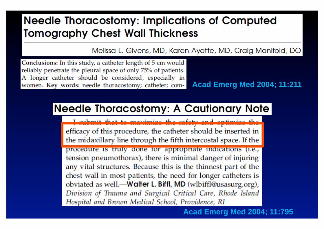
J Trauma Acute Care Surg 2014; 76:1029

J Spec Oper Med 2013; 13:53

J Spec Oper Med 2013; 13:53

J Trauma Acute Care Surg 2013; 75:1071

J Trauma Acute Care Surg 2013; 75:1071

OPEN PNEUMOTHORAX
“Sucking Chest Wound” Pressure Equilibration; Air Passes through Hole if >2/3 Diameter of Trachea
Temporary Occlusive Dressing Taped on 3 Sides
Tube Thoracostomy

PULMONARY CONTUSION / FLAIL CHEST
Flail Segment = >2 Ribs with >2 Fxs
Pain, Disruption of Mechanics Primary Problem is Underlying Contusion Supportive Care; Intubation if Indicated Rib Blocks / Epidural Analgesia Surgical Stabilization?
NEXT LECTURE
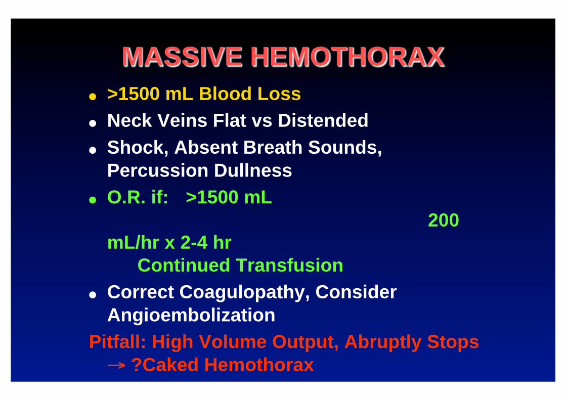
MASSIVE HEMOTHORAX >1500 mL Blood Loss Neck Veins Flat vs Distended Shock, Absent Breath Sounds,
Percussion Dullness O.R. if: >1500 mL
200 mL/hr x 2-4 hr
Continued Transfusion Correct Coagulopathy, Consider
Angioembolization Pitfall: High Volume Output, Abruptly Stops → ?Caked Hemothorax

C - ETIOLOGIES OF SHOCK
Hemorrhagic Cardiac Compressive Cardiogenic Neurogenic Septic

WHERE DOES BLOOD GO?
● Street / Wounds ● Fractures ● Chest ● Pelvis ● Abdomen

CIRCULATION- BLOOD LOSS Class of Hemorrhage I II III IV
Blood Loss (%) 15 15-30 30-40 >40 HR <100 >100 >120 >140 SBP Nl Nl Dec Dec Pulse P Nl / Inc Dec Dec Dec RR 14-20 20-30 30-40 >35 UO (ml/hr) >30 20-30 5-15 Nil
Blood Vol = 7 ml/kg

C - ETIOLOGIES OF SHOCK
• Hemorrhagic • Cardiac Compressive • Cardiogenic • Neurogenic • Septic
CARDIAC COMPRESSIVE SHOCK
Tension Pneumothorax Rx: Tube Thoracostomy


Nagy KK et al. J Trauma 1995; 38:859
THE BOX

PHYSICAL EXAMINATION Vital Signs, Neck Veins, Auscultation Beck’s Triad (Hypotension, JVD,
Muffled Heart Tones) Tachycardia, Narrow Pulse Pressure Pulsus Paradoxus (SBP Drop > 10
mm Hg with Inspiration)

ULTRASONOGRAPHY – FAST
Rozycki et al. J Trauma 1995; 39:492

PERICARDIOCENTESIS
50% False (+): Enter Chamber. Coronary Artery Puncture; Dysrhythmia. 37% False (-): Clot

J Trauma Acute Care Surg 2013; 75:543

SUBXIPHOID PERICARDIAL WINDOW

Ann Surg 2015; 261:573


Ann Surg 2014; 259:438

MANAGEMENT OF CARDIAC WOUNDS
Wall MJ et al. J Trauma 1997; 42:905
X

RESUSCITATIVE THORACOTOMY
Biffl WL et al. Op Tech Gen Surg 2000; 2:168

RESUSCITATIVE THORACOTOMY Objectives • Release Pericardial Tamponade • Repair Cardiac Wounds • Perform Open Cardiac Massage • Cross-Clamp Aorta to Limit Subdiaphragmatic
Hemorrhage and Redistribute Blood Flow to Myocardium and Brain
• Control Intrathoracic Hemorrhage • Control Bronchovenous Air Embolism

J Trauma Acute Care Surg 2012; 73:1359

CARDIOGENIC SHOCK
Myocardial Infarction Dx: ECG, Enzymes
Bronchovenous Air Embolism Dx: Shock with Positive Pressure Vent Rx: Hilar Cross-Clamp, Ventricular /
Aortic Root Venting, Vigorous Cardiac Massage

BLUNT CARDIAC INJURY (Formerly Cardiac Contusion )
Direct Impact Injury to the Heart Right Heart (RV) Most Commonly Affected Clinical Significance: Occult and
Inconsequential to Life-Threatening Dysrhythmias or Pump Failure (“Sig BCI”)
• No Characteristic Presentation • No Diagnostic Gold Standard

No Patient with SIG-BCI Had Elevated CK-MB Without Abnormal ECG
CK-MB Levels Were Not Predictive of SIG-BCI Am J Surg 1994; 169:523

CARDIAC TROPONIN Low Sensitivity and Predictive Value for
SIG-BCI Fulda GJ et al. J Trauma 1997; 43:304 Bertinchant JP et al. J Trauma 2000; 48:924
Real Value: Normal Admission ECG + cTnI at 4-8
Hrs Has Negative Predictive Value for SIG-BCI Approaching 100%:
0/46 Salim A et al. J Trauma 2001; 50:237
0/40 Collins JN et al. Am Surg 2001; 67:821
0/131 Velmahos GC et al. J Trauma 2003; 54:45

J Trauma Acute Care Surg 2012; 73:S301

OTHER TIDBITS

OCCULT PNEUMOTHORAX
Seen on CT but not CXR 2% Trauma Admissions 30% PTXs DeMoya et al. J Trauma 2007; 63:13
If Asymptomatic, No Rx ? Prophylactic Tube Thoracostomy
for Positive-Pressure Ventilation

J Trauma 2011; 70:1019
448 Pts Observed 27 (6%) Chest Tube for PTX
Progression, Resp Distress, or Hemothorax
10/73 (14%) Failed on PPV- No Tension

Kirkpatrick et al. J Trauma Acute Care Surg 2013; 74:747

HEMOTHORAX
Indications for Surgery: • >1500 mL Output • 200 mL/hr Output x 2-4 hr* • Continued Transfusion* *Correct Coagulopathy, Consider Angioembolization
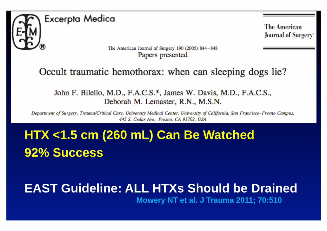
HTX <1.5 cm (260 mL) Can Be Watched 92% Success EAST Guideline: ALL HTXs Should be Drained
Mowery NT et al. J Trauma 2011; 70:510

J Trauma 2012; 72:422
J Trauma Acute Care Surg 2012; 73:1423
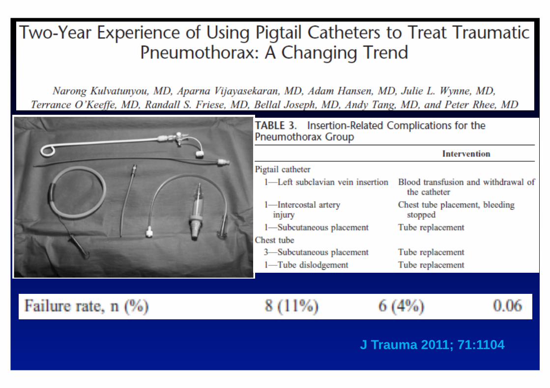
J Trauma 2011; 71:1104

ABX FOR TUBE THORACOSTOMY Prophylactic Abx do not Reduce
Empyema/Pneumonia; Associated with Resistant HAIs
Maxwell et al. J Trauma 2004; 57:742
Cannot Recommend For or Against Abx J Trauma Acute Care Surg 2012; 73:S341

Residual HTX on CXR after CT Placement = 33% Risk of Empyema
Karmy-Jones R et al. Can Respir J 2008; 15:255
RETAINED HEMOTHORAX

J Trauma Acute Care Surg 2012; 73:752

PRCT 2nd Chest Tube vs VATS VATS = Dec Duration of CT Drainage, LOS, Cost 10/24 with 2nd CT (42%) Required Surgery
Meyer DM et al. Ann Thoracic Surg 1997; 64:1396
EAST Guidelines 2011 Persistent retained hemothorax, seen on plain films, after placement of a thoracostomy tube should be treated with early VATS, not a second chest tube (Level 1). J Trauma 2011; 70:510
RETAINED HEMOTHORAX

RIB FRACTURES Common - 9-12% Trauma Admissions 12% Mortality Ziegler, J Trauma 1994; 37:975
Mortality Elderly Young 22% 10% Bulger, J Trauma 2000; 48:1040 20% 9% Bergeron, J Trauma 2003; 54:478 20% 11% Stawicki, J Am Geriatr Soc 2004; 52:805
NEXT LECTURE

Surgery 2004; 136:426
J Trauma 2011; 71:1548

PNEUMOMEDIASTINUM
Sign of Aerodigestive Injury 5% of Chest CTs
10% Have Injuries
If Asymptomatic, Manage Expectantly
Macleod et al. Am Surg 2009; 75:375 Dissanaike et al. J Trauma 2008; 65:1340

TRACHEOBRONCHIAL INJURY Subcu Emphysema; Pneumomediastinum;
PTX w Air Leak Dx by Bronchoscopy
Karmy-Jones et al. Thorac Surg Clin 2007; 17:35
J Trauma Acute Care Surg 2015; 79:1089
CTA Chest Esophagoscopy / Esophagography

Exam + CXR or E-FAST -Hemodynamics, Location of Wound(s); Early
repeat CXR
Unilateral GSW – Chest tube -Drained vs Retained Htx vs Large Air Leak
Transmediastinal GSW – Chest tube(s), CT scan -Add’l W/U based on trajectory
Thoracoabdominal GSW – Chest tube, Laparotomy, ?Pericardial window
THORACIC GSW

DAMAGE CONTROL RESUSCITATION

Damage Control: Keep a Badly Damaged Ship Afloat After Major Penetrating Injury to the Hull

Plug Gaping Holes Extinguish Fires “Dog Down” Watertight
Doors
Keep Ship Afloat Assess Overall Damage Establish a Plan for Definitive Repair
Damage Control: Keep a Badly Damaged Ship Afloat After Major Penetrating Injury to the Hull

1976- Lucas and Ledgerwood
1979- Calne et al
1981- Feliciano et al
1983- Stone et al
1993- Rotondo et al
DAMAGE CONTROL IN TRAUMA

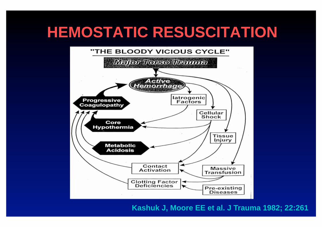
Kashuk J, Moore EE et al. J Trauma 1982; 22:261

“Damage control resuscitation addresses the entire lethal triad immediately upon admission to a combat hospital”
J Trauma 2007; 62:307
Anticipate and Attenuate; Reverse

• Permissive Hypotension • Limit Isotonic Crystalloid • Aggressive Hemostatic Resuscitation • Bleeding Control • Rewarming • Correction of Acidosis, Hypocalcemia
DAMAGE CONTROL RESUSCITATION
Bogert et al. J Intensve Care Med 2016; 31:177 Giannoudi et al. Eur J Trauma Emerg Surg 2016; 42:273 Chang et al. Crit Care Clin 2017; 33:15

• Premise: Avoid Exacerbating Hemorrhage and Dilutional Coagulopathy
• Caveat: Clear Evidence of Benefit and Optimal Perfusion Targets Lacking
• Goal: SBP 70-90; MAP >50; Radial Pulse
• Caution: Severe TBI; Prolonged Shock
PERMISSIVE HYPOTENSION
Bogert et al. J Intensve Care Med 2016; 31:177 Giannoudi et al. Eur J Trauma Emerg Surg 2016; 42:273 Chang et al. Crit Care Clin 2017; 33:15

• Premise: Excess Crystalloid – Dilutional Coagulopathy, ARDS, Cardiac Dysfunction, Compartment Syndromes, Ileus, Anastomotic Leak, Wound Complications, MOF, Death
• Mechanism: Intracellular Edema – Disrupt Biochemical Processes (Pancreatic Insulin, Hepatocyte Glucose Metabolism, Cardiac Myocyte Excitability); Inflammation – Inflammatory Mediators; Endothelial Glycocalyx Degradation
LIMIT ISOTONIC CRYSTALLOID
Bogert et al. J Intensve Care Med 2016; 31:177 Giannoudi et al. Eur J Trauma Emerg Surg 2016; 42:273 Chang et al. Crit Care Clin 2017; 33:15

J Trauma Acute Care Surg 2013; 74:1207
If Not Hypotensive, >500 mL Crystalloid Assoc w/ Higher Mortality and Coagulopathy
J Trauma Acute Care Surg 2013; 74:1215
24 Hr Crystalloid Correlated w/ Vent Days, ICU & Hosp LOS, ARDS, MOF, SSI, Bloodstream Infxn, Compartment Syndromes

Am J Emerg Med 2017; 35:317
● Colloid- Discouraged due to Cost, Coagulopathy, Renal Dysfunction
● Crystalloid- NS vs LR vs Plasmalyte
OPTIMAL FLUID?
Kashuk J, Moore EE et al. J Trauma 1982; 22:261
HEMOSTATIC RESUSCITATION

PRESUMPTIVE FFP
Kashuk J, Moore EE et al. J Trauma 1982; 22:261
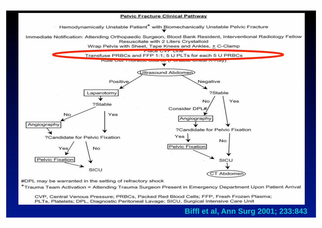
Biffl et al, Ann Surg 2001; 233:843

Post-Injury Life Threatening
Coagulopathy
1:1:1 FFP:PLT:RBC

HEMOSTATIC RESUSCITATION
• 2 Lg Bore IVs Upper Ext • Early MTP • FFP:PLTs:PRBCs 1:1:1-2

MASSIVE TRANSFUSION ● ABC Score >2
• Penetrating Mechanism • SBP <90 • HR >120 • (+) FAST
● Persistent hemodynamic instability ● Active bleeding requiring operation
or angioembolization ● Blood transfusion in trauma bay
TQIP Best Practices

THROMBELASTOGRAPHY
Goal-Directed Resuscitation PRBCs, FFP, PLTs
Fibrinogen, Anti-Fibrinolysis
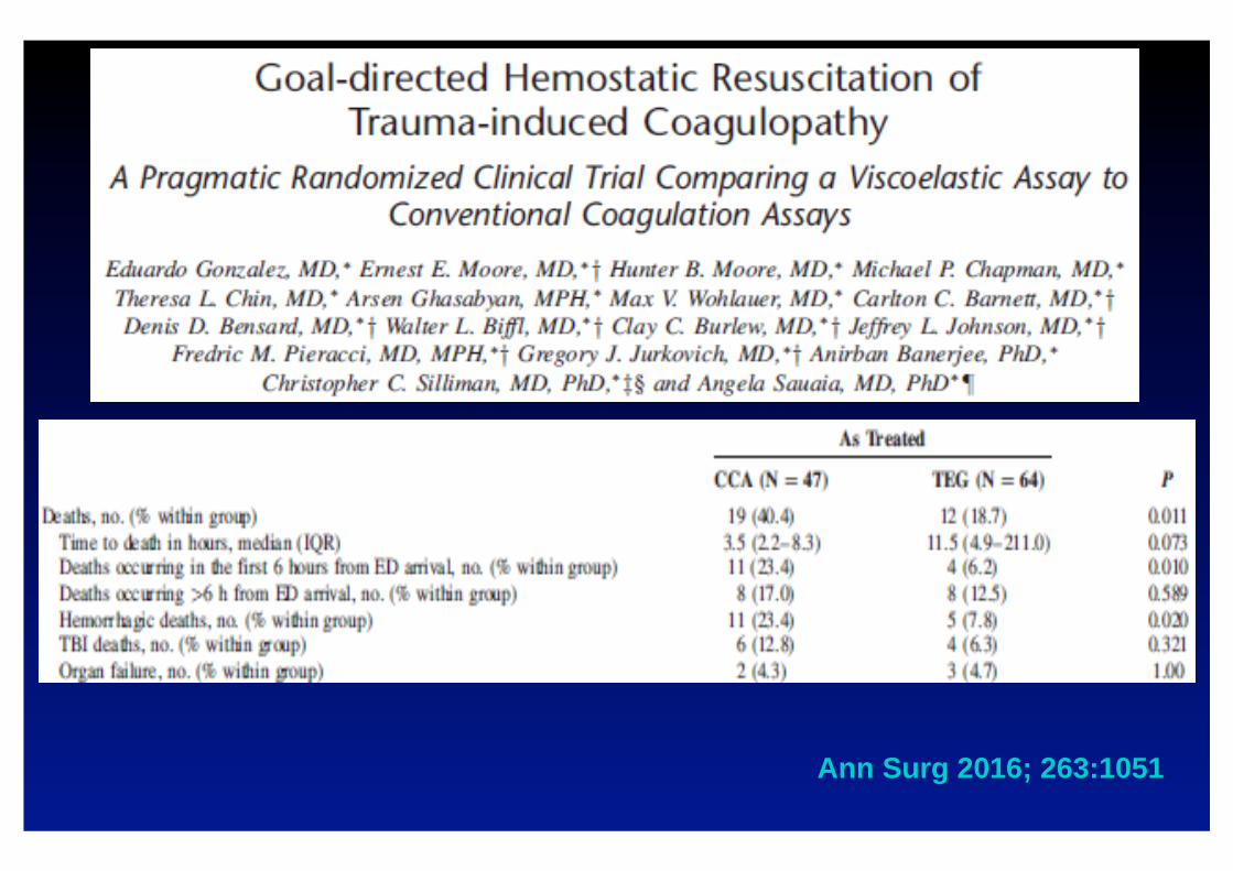
Ann Surg 2016; 263:1051
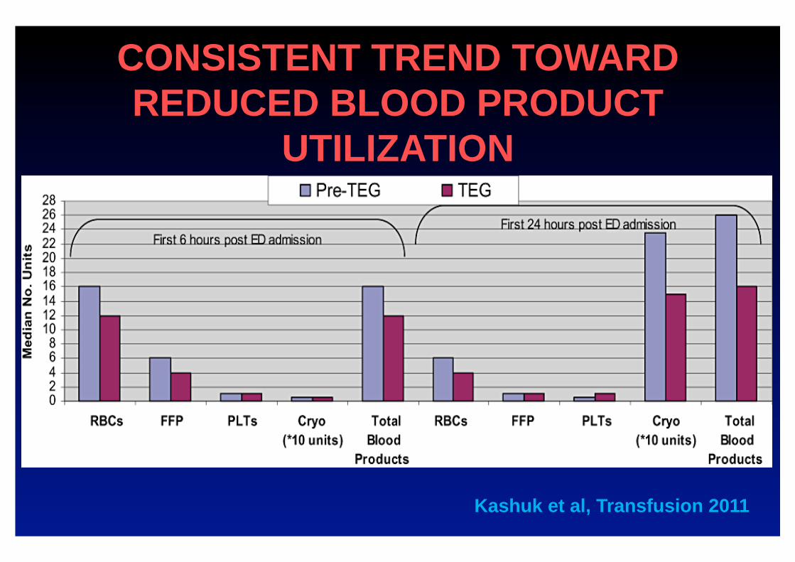
CONSISTENT TREND TOWARD REDUCED BLOOD PRODUCT
UTILIZATION
Kashuk et al, Transfusion 2011

Shock 2014; 41(Suppl 1):35

Shock 2014; 41 Suppl 1:3
• Prehospital Monitoring Shock/Coagulopathy • Hypotensive Resuscitation; Endpoints of Resuscitation • Whole Blood vs Components; Dried Products • Pathogen Reduced Technology for Products • Role of TBI