THOMAS SKÅRUP KRISTENSEN, MD PH.D …bme.elektro.dtu.dk/31545/notes/New trends in X-ray... ·...
42
THOMAS SKÅRUP KRISTENSEN, MD PH.D DEPARTMENT OF RADIOLOGY, RIGSHOSPITALET COPENHAGEN UNIVERSITY HOSPITAL 1 New trends in X-ray and CT-scanning Multislice and fast scanning
Transcript of THOMAS SKÅRUP KRISTENSEN, MD PH.D …bme.elektro.dtu.dk/31545/notes/New trends in X-ray... ·...
T H O M A S S K Å R U P K R I S T E N S E N , M D P H . D D E P A R T M E N T O F R A D I O L O G Y ,
R I G S H O S P I T A L E T
C O P E N H A G E N U N I V E R S I T Y H O S P I T A L
1
New trends in X-ray and CT-scanning Multislice and fast scanning
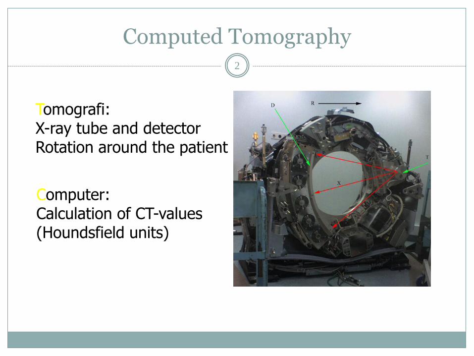
Computed Tomography 2
Tomografi: X-ray tube and detector Rotation around the patient
Computer: Calculation of CT-values (Houndsfield units)

3
• Sir Godfrey Hounsfield : first CT-scanner in 1972 •Nobels price in medicine in 1979 (together with Alan Cormack)

4
1972: Rotationstid: 4min Rekonstruktionstid: ”klar næste morgen”
2014: Rotationstid: 0.27 s Rekonstruktionstid: ” få sekunder ”

5
Fast CT Timing of contrast No breathing artefacts Reduction of motion artefacts (pulsation, breathing…)
Spatial resolution
Visualization of small structures with high resolution in all planes (X-Y-Z)

6

Mutidetector CT/multislice CT (MDCT/MSCT)
7

Mutidetector CT/multislice CT (MDCT/MSCT)
8

MDCT 9
Submillimeter resolution in all planes
Isotropic imaging of larger volumes with an acceptable scantime
Imaging of one or more contrast phases
(non-contrast, arterial, venous, wash-out)

Contrast injection 10

MDCT – performance (64-slice) 11
High spatial resolution (0.5mm x 0.5mm x 0.5 mm)
Coverage: 3,2 – 4 cm per rotation High temporal resolution (175 ms)
(rotation time/2)
Scantime: Heart: 10 sec (320 slice: 135 ms) Thorax and abdomen: 20 sec

Clinical examples
12
Oncology
Angiography
CT of the urinary system
CT of the abdomen
Traumapatients

Abdomen
13

Abdomen
14

15

16

Liver abscess 17

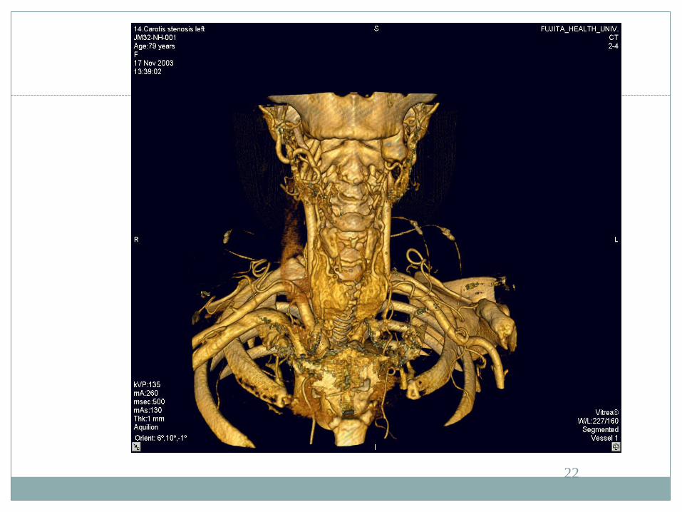
Angiography 18
Stenosis
Rupture
Thrombosis
Aneurismes
Growth of tumors around/in to vessels

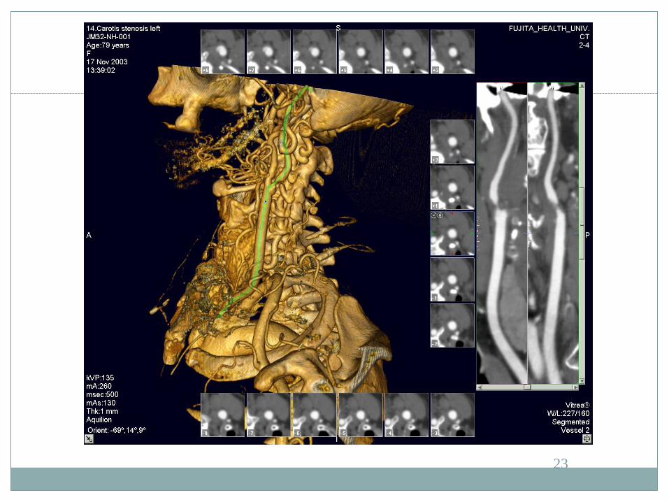
Angiography 19
Fast scantime to ”catch” the contrast in the right phase.
High spatial resolution
High temporal resolution (movement)

Angiography 20

21
22
23

24

25

26

Aortic dissection
27

Vena cava thrombus
28

Lung emboli
29

Lung emboli
30

CT of the urinary system 31

CT of the urinary system
32

CT of the urinary system 33
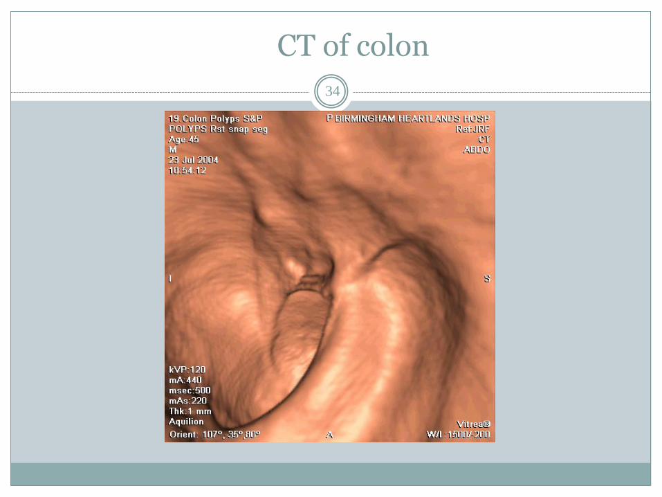
CT of colon
34
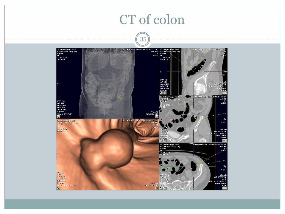
CT of colon
35

Traumapatients 36
Timefactor is crucial
Aqusitiontime/reconstructiontime
MPR reconstruction

Trauma patients
37

Traumapatients
38

Traumapatients 39

Pneumothorax 40

41

42