This thesis/dissertation is submitted to the University of ...
125
UNIVERSITY OF GHANA COLLEGE OF BASIC AND APPLIED SCIENCES GENETIC DIVERSITY OF MYCOLACTONE PRODUCING MYCOBACTERIA CAUSING BURULI ULCER IN GHANA AND CÔTE D’IVOIRE BY MAGDALENE DOGBE (10421622) This thesis/dissertation is submitted to the University of Ghana, Legon in partial fulfilment of the requirement for the award of MPHIL Mol. Cell Biology of Inf. Diseases degree DEPARTMENT OF BIOCHEMISTRY, CELL AND MOLECULAR BIOLOGY JULY, 2019 University of Ghana http://ugspace.ug.edu.gh
Transcript of This thesis/dissertation is submitted to the University of ...

UNIVERSITY OF GHANA
COLLEGE OF BASIC AND APPLIED SCIENCES
GENETIC DIVERSITY OF MYCOLACTONE PRODUCING MYCOBACTERIA
CAUSING BURULI ULCER IN GHANA AND CÔTE D’IVOIRE
BY
MAGDALENE DOGBE
(10421622)
This thesis/dissertation is submitted to the University of Ghana, Legon in
partial fulfilment of the requirement for the award of MPHIL Mol. Cell
Biology of Inf. Diseases degree
DEPARTMENT OF BIOCHEMISTRY, CELL AND MOLECULAR BIOLOGY
JULY, 2019
University of Ghana http://ugspace.ug.edu.gh

i
DECLARATION
I, Magdalene Dogbe, do certify and declare that this project aside other cited works is a product of
my own research undertaken at the Department of Biochemistry, Cell, and Molecular Biology,
University of Ghana, Legon and Department of Biological science, Mississippi State University
by me under the supervision of Dr. Lydia Mosi and Dr. Heather Jordan. Reference made to the
works of others have been duly acknowledged. I certify that no part of this dissertation has been
previously submitted for a degree or any other qualification.
Signature Date……29…/……May…../……2020…..
Magdalene Dogbe
(Student)
Signature Date…29……/…May…./……2020…..
Dr. Lydia Mosi
(Supervisor)
Signature Date…22……/…June……../…2020……..
Dr. Heather Jordan
(Co-Supervisor)
University of Ghana http://ugspace.ug.edu.gh

ii
DEDICATION
I dedicate this work to the memory of my late father, Boniface Dogbe who was keen on providing
the best education for me and my siblings. I also dedicate this work to my lovely family (Ms. Mary
Nyame (Mum), Mrs. Patricia Appiah (sister), Gloria Dogbe (sister), and Kelvin Dogbe (brother),
Roland Dogbe (brother) and Divine Kwabena Appiah Nyame (nephew) for being my greatest
inspiration.
University of Ghana http://ugspace.ug.edu.gh

iii
ACKNOWLEDGEMENT
My profound gratitude goes to the Almighty God for his guidance, protection and the abundance
of grace He bestowed on me throughout the entirety of my study as a graduate student.
I am immeasurably grateful and greatly indebted to my supervisor, Dr. Lydia Mosi of the
Department of Biochemistry, Cell and Molecular Biology for her invaluable support, tutelage and
advice not only for this research project but also for impacting positively to my upbringing for the
duration of my study. It was such a privilege to have worked under her zealous supervision. I also
thank my co-supervisor, Dr. Heather Jordan for her timely advice, guidance and suggestions
towards the successful completion of this thesis. I acknowledge the West African Centre for Cell
Biology of Infectious Pathogens (WACCBIP) for awarding me a fellowship that funded my entire
research work.
My Sincere thanks also goes to all members of the Mosi lab, most especially Mrs. Mabel Sarpong-
Duah, Mrs. Elizabeth Gyamfi Sarkodie and Edwin Kyei-Baffour for their support on this project.
Worthy of mention are Dr. Charles Quaye and Dr. Abel Adjet who supported me throughout my
sample collection period. I am also grateful to Mr. Charles Narh for his great input in my data
analyzes. I also express my profound appreciation to members my family and all others who have
offered diverse assistance towards successful completion of this work.
University of Ghana http://ugspace.ug.edu.gh

iv
TABLE OF CONTENT
DECLARATION…………………………………………………………………………… I
DEDICATION………………………………………………………………………………II
ACKNOWLEDGEMENT…………………………………………………………………...III
TABLE OF CONTENT…………………………...…………………………………………IV
LIST OF FIGURES…………………………………………………………………………..VII
LIST OF TABLES…………………………………………………………………………….X
LIST OF ABBREVIATIONS…………………………………………………………………XI
ABSTRACT…………………………………………………………………………………….XII
CHAPTER ONE ............................................................................................................................. 1
1.0. INTRODUCTION ................................................................................................................... 1
1.1. STUDY RATIONALE ............................................................................................................ 4
1.2. AIM OF STUDY ..................................................................................................................... 5
1.3. SPECIFIC OBJECTIVES ........................................................................................................ 5
CHAPTER TWO ............................................................................................................................ 6
2.0. LITURATURE REVIEW ........................................................................................................ 6
2.1. Mycobacteria............................................................................................................................ 6
2.1.1. Mycobacterium Tuberculosis Complex (MTC) ..................................................................... 7
2.1.2. Mycobacteria leprae ............................................................................................................. 8
2.1.3. Non-Tuberculosis Mycobacteria (NTM) ............................................................................... 8
2.1.4. Mycobacterium ulcerans ....................................................................................................... 8
2.1.4.1. Mycobacterium ulcerans strain Agy 99 ........................................................................... 10
University of Ghana http://ugspace.ug.edu.gh

v
2.1.5. Genetic Classification of Mycobacterium ulcerans ............................................................ 11
2.1.6. Mycolactone and Mycolactone Producing Mycobacteria (MPMs) .................................... 13
2.2. Buruli ulcer disease ................................................................................................................ 18
2.2.1. Epidemiology ...................................................................................................................... 19
2.2.2. Transmission of Buruli ulcer .............................................................................................. 21
2.2.3. Clinical pathology and manifestation ................................................................................. 22
2.2.4. Diagnosis ............................................................................................................................ 24
2.2.5. Treatment and Management ............................................................................................... 24
2.3. Molecular Diagnosis of Buruli ulcer...................................................................................... 26
2.3.1. IS2404 and IS 2606 PCR and Restriction Fragment Length Polymorphism (RFLP) ........ 26
2.3.2. Multi-locus sequence typing (MLST) .................................................................................. 26
2.3.3. Variable Number Tandem Repeats (VNTR) Typing............................................................ 27
CHAPTER THREE ...................................................................................................................... 29
3.0. METHODOLOGY ................................................................................................................ 29
3.1 Ethical Issues .......................................................................................................................... 29
3.2. Study Site and Design ............................................................................................................ 29
3.3. Data Collection and Questionnaires....................................................................................... 30
3.3.1 Inclusion and Exclusion Criteria…………………………………………………………29
3.4. Collection, Storage, Transportation of Samples .................................................................... 32
3.5. Acid Fast Staining .................................................................................................................. 34
3.6. DNA extraction ...................................................................................................................... 34
3.7. Mycobacterial Infection Confirmation of Samples for Insertion Sequence 2404 ................. 34
University of Ghana http://ugspace.ug.edu.gh

vi
3.8. Mycolactone Producing Mycobacteria (MPM) Confirmation for Enoyl Reductase Gene .... 36
3.9. VNTR Typing of Mycolactone Producing Mycobacteria (MPM) ........................................ 36
3.10. Genotype Assignment .......................................................................................................... 40
3.11. Population Genetics ............................................................................................................. 40
3.12. Data and Sequence Analysis ................................................................................................ 40
CHAPTER FOUR ......................................................................................................................... 42
4.0. RESULTS........................................................................................................................... 42
4.1. Sample and Data Collection................................................................................................... 42
4.2. Case Confirmation ................................................................................................................. 45
4.2.1. Microscopy .......................................................................................................................... 45
4.2.2. PCR targeting IS2404 and Enoyl Reductase gene .............................................................. 46
4.2.3. VNTR Typing of Isolates ..................................................................................................... 48
4.2.4. Genotype Assignment .......................................................................................................... 49
4.3. Allelic Frequencies of Loci .................................................................................................... 56
4.4. Sequence Confirmation .......................................................................................................... 57
CHAPTER FIVE .......................................................................................................................... 59
5.0. DISCUSSION, CONCLUSION, AND RECOMMENDATION .......................................... 59
5.1. DISCUSSION ........................................................................................................................ 59
5.2. CONCLUSION ...................................................................................................................... 70
5.3. RECOMMENDATION ......................................................................................................... 72
REFERENCES…………………………………………………………………………………68
APPENDIX ……………………………………………………………………………………………..93
University of Ghana http://ugspace.ug.edu.gh

vii
LIST OF FIGURES
Figure 1: Divergence of Mycobacterium ulcerans from Mycobacterium marinum progenitor into
two distinct lineages analyzed based on region of differences of isolates from diverse geographic
locations. Adapted from Käser et al., (2007) ................................................................................ 12
Figure 2: Structure of mycolactone depicting the various divisions present in the compound.
Adapted from Sarfo et al., (2013). ................................................................................................ 14
Figure 3: Genetic organization of the mycolactone biosynthetic cluster from plasmids present in
MPMs. Adapted from Pidot et al., (2008). ................................................................................... 17
Figure 4: The distribution of BU cases reported as of 2014. Adapted from Zingue et al., (2018)
....................................................................................................................................................... 20
Figure 5: Clinical manifestations of Buruli ulcer. A is the nodular form of the disease. B and
C are the plaque and oedematous form of the disease. D is an ulcerative from with characteristic
cotton-like patches with undermined areas. Images A and D are from this present study whiles
Images B and C were adapted from Dégboé et al., (2019) and Boleira et al., (2010) respectively
....................................................................................................................................................... 23
Figure 6: Location Map of Sample and Information Collection Centers in Ghana and Côte
d’Ivoire. ......................................................................................................................................... 31
Figure 7: A section of nurses and community health workers undergoing training on how to
interview patients and obtain demographic data (A); proper sampling (B); storage (C) in Buruli
ulcer treatment centers in Côte d’Ivoire. In Ghana, patients were interviewed and sampled
directly by our team (D). ............................................................................................................... 33
Figure 8: Age (A) and Gender (B) Distribution of suspected BU participants. Participants were
grouped into three categories; children (less than 18 years), adult (between 18 – 50 years) and aged
(greater than 50 years). ................................................................................................................. 43
Figure 9: Stages of Buruli ulcer disease in suspected participants. Most of the cases recruited
were in the ulcerative stage ........................................................................................................... 44
University of Ghana http://ugspace.ug.edu.gh

viii
Figure 10: Categories (CAT) of lesions of suspected Buruli ulcer patients in Ghana and Côte
d’Ivoire .......................................................................................................................................... 44
Figure 11: Different body parts affected. In Ghana, infections were only on the lower and upper
limbs and the trunk and breast area with Côte d’Ivoire recording infections at the same parts as
well as the genitalia, head and neck area. ..................................................................................... 45
Figure 12: Acid fast stained slide from a sample collected in Côte d’Ivoire. Red arrows shows a
clump of bacilli ............................................................................................................................. 46
Figure 13: Representitive Gels of ethidium bromide stained PCR amplicons. A - amplicons of
IS2404 PCR with band size of 400bp. B - Amplicons for PCR targeting the Enoyl reductase gene
with band size of 720bp. C - amplicons of nested IS2404 PCR with band size of 210bp............ 47
Figure 14: A summary of molecular confirmatory tools used for confirming the presence of
Buruli ulcer in patients. All samples positive for qPCR were further typed at sixteen (16) VNTR
loci................................................................................................................................................. 47
Figure 15: Ethidium bromide stained gels of controls at nine different loci. M- Molecular weight
ladder, 1- M. ulcerans 1615, 2- M. marium hybrid 270995, 3- M. marimum DL 180892, 4- M.
pseudoshottsi, 5- M. marium CL, 6- M. marium SA 200695 ....................................................... 49
Figure 16: Percentage genotype assignment of samples from various communities in Ghana and
Côte d’Ivoire. ................................................................................................................................ 51
Figure 17: Genotypes associated with different disease stages and categories in Ghana (A &C)
and Côte d’Ivoire (B&D). ............................................................................................................. 51
Figure 18: Allelic frequencies of VNTR loci typed for samples collected in Ghana and Côte
d’Ivoire .......................................................................................................................................... 56
Figure 19: Sequence confirmation of VNTR repeats and phylogeny of isolates. M. ulcerans
1615 (MU1615), M. marimum DL 180892 (MDL), M. pseudoshottsii, M. marium hybrid 270995
(MHBD), M. marium CL (MCL), M. marium SA 200695(MSA) were controls included in the
study. NC005916.1 Plasmid is AGY 99 reference strain. PA22, PA05, AM11, T07, PA26, T01
and T18 are cases from Ghana and Côte d’Ivoire. M. ulcerans 1615 (MU1615) clustered closely
University of Ghana http://ugspace.ug.edu.gh

ix
with T07 as well as PA26 and T01. Samples, PA22, PA05 and AM11 clustered together with
T18. All controls besides MU1615 clustered closely with the reference strain. .......................... 58
University of Ghana http://ugspace.ug.edu.gh

x
LIST OF TABLES
Table 1: A list of positive controls included in the VNTR Tying of isolates. ............................. 38
Table 2: Primer sequences length of Amplicon. Primer sequences of different Miru – VNTR
Loci used for typing ...................................................................................................................... 39
Table 3: Genotyping coding for samples based on four standard loci, Miru 1, Locus 6, ST1 and
Locus 19 ........................................................................................................................................ 52
Table 4a: Mycobacteria species identified using Miru-VNTR Profiles of isolates(Indefinitely
assigned) ....................................................................................................................................... 53
Table 4b: Mycobacteria species identified using Miru-VNTR Profiles of isolates (Definitely
assigned) ..................................................................................................................................... 534
Table 5: VNTR typing success at sixteen (16) loci ................................................................... 545
University of Ghana http://ugspace.ug.edu.gh

xi
LIST OF ABBREVIATIONS
BU – Buruli ulcer
CI - Côte d’Ivoire
MIRU – Mycobacterial Interspersed Repetitive Units
VNTR – Variable Number Tandem Repeat
MPM - Mycolactone Producing Mycobacteria
MTC - Mycobacterium tuberculosis complex
NTM - Non-Tuberculous Mycobacteria
MU – Mycobacterium ulcerans
MLF - Mycobacterium liflandii
MHB - Mycobacterium marinum hybrid
MDL - Mycobacterium marinum DL
MCL - Mycobacterium marinum CL
MSA - Mycobacterium marinum SA
MPS - Mycobacterium pseudoshottsii
PCR - Polymerase Chain Reaction
BCG - Bacille Calmette–Guérin
gDNA – genomic DNA
University of Ghana http://ugspace.ug.edu.gh

xii
ABSTRACT
Buruli ulcer remains a neglected tropical disease endemic in Africa especially Ghana and Côte
d’Ivoire. It is a necrotizing skin and soft tissue infection caused by Mycobacterium ulcerans which
produces mycolactone which causes adverse effects associated with disease in humans. Other
mycolactone producing bacteria (MPMs) have been identified to cause granulomas in fish and
frog. There are limited advanced genetic tools for studying transmission of the diseases and for
carrying out molecular epidemiological studies. The mycolactone producing mycobacteria
(MPM), M. shinshuense has been reported to cause Buruli ulcer and other studies have revealed
several MPM infections other than M. ulcerans in clinical samples. It is therefore imperative to
know the exact MPMs that are in circulation by genotypically distinguishing them using molecular
tools. Patients with Buruli ulcer-like disease presentation were recruited from endemic
communities in Ghana and Côte d’Ivoire. Swabs and FNA samples were screened using primers
detecting the insertion sequence 2404 gene and the Enoyl reductase gene. Genotyping was
achieved at 16 MU VNTR loci and length polymorphisms arising from differences in tandem
repeats for samples were validated using six (6) controls and sequencing.
A total one hundred and fifty-nine (159) of the 189 samples were confirmed as Buruli ulcer
positive. Genotyping was successful for all controls (100%) and most samples (69%) at all sixteen
(16) VNTR loci. Seven (7) MU genotypes designated, A, B, C, D, E, F and G and five MPM
genotypes MLF (Mycobacterium liflandii), MHB (Mycobacterium marinum hybrid), MDL
(Mycobacterium marinum DL), MCL (Mycobacterium marinum CL) and MSA (Mycobacterium
marinum SA) were generated samples were assigned genotypes based on their VNTR profiles.
Genotypes C, D and F were present in both Ghana and Côte d’Ivoire. However, genotypes E and
MLF were only found in Ghana whiles Genotype A and G were found in only Côte d’Ivoire.
University of Ghana http://ugspace.ug.edu.gh

xiii
Genotype D has persisted over the years from 2008 to 2019 comparing this study to published
data. These findings support the hypothesis that Mycolactone producing mycobacteria causing
Buruli ulcer in Ghana and Côte d’Ivoire are diverse and affirms VNTR typing as a comparably
useful tool for differentiating MU strains as well as other MPMs in Buruli ulcer endemic
communities.
University of Ghana http://ugspace.ug.edu.gh

1
CHAPTER ONE
1.0. INTRODUCTION
Non-Tuberculous Mycobacteria (NTM) are environmental pathogens that are usually found in
water, aerosols insects, soil, protozoans and small animals (van Ingen et al., 2009; Falkinham,
2011) and are not members of the Mycobacterium Tuberculosis Complex or Mycobacterium
leprae (Falkinham et al., 1996). NTMs can cause infections in humans, other mammals and birds
and usually affects the socio-economic situation of the affected community (Asiedu and Etuaful,
1998; Falkinham, 2011). Nearly thirty-three percent (33%) of all mycobacteria infections causing
diseases occur in humans (Katoch, 2004). Infection with NTMs is difficult to control and
ultimately prevent due to their opportunist nature and their wide distribution in soil, aerosols,
water, insects, protozoans and small animals (Primm et al., 2004; van Ingen et al., 2009). Most
NTM infections such as Mycobacterium kansasii, Mycobacterium avium complex (MAC) and
Mycobacterium intracellulare cause pulmonary disorders (Ahn et al., 1979) whiles NTMs such
as Mycobacterium marinum, Mycobacterium abscessus, Mycobacterium fortuitum and
Mycobacterium chelonae cause cutaneous infections (Lamb and Dawn, 2014).
The epidemiology of certain NTMs of public health importance have not been adequately
addressed. Mycolactone Producing Mycobacteria (MPM) are a subset of NTMs and these MPMs
are of enormous importance due to the debilitating cutaneous disease-causing ability in both
humans and animals. Mycobacterium pseudoshotsii and mycolactone producing Mycobacterium
marinum have been isolated from fish, Mycobacterium liflandii from frogs and Mycobacterium
ulcerans as well as Mycobacterium shinshuense from humans (Rhodes et al., 2001; Rhodes et
al., 2005; Ranger et al., 2006; Funakoshi et al., 2009). Similar studies have also isolated different
University of Ghana http://ugspace.ug.edu.gh

2
strains of MPMs from small mammals such as possum and koala ( Mitchell et al., 1984; Fyfe et
al., 2010) and other domestic animals including cats and horses (Elsner et al., 2008, van Zyl et
al., 2010). The infection of domestic animals raises concern of possible zoonosis; however this
field of research has not received substantial attention. There is the possibility of other
unidentified MPMs besides the isolated strains that produce mycolactone with sufficient potency
to cause infections. A better understanding of distribution of MPMs causing infections would be
essential to accurate disease diagnosis and to a larger extent curtailing the spread of the diseases
they cause, in particular, Buruli ulcer (Yip et al., 2007).
The debilitating and extensive loss of the cutaneous and sub-cutaneous tissue is the hallmark of
the disease, Buruli ulcer (Mosi et al., 2008). It was named after a town in Uganda that had levels
of disease occurrence in the 1960s (Baker et al., 1972). Buruli ulcer is caused by Mycobacterium
ulcerans. It usually affects all body parts indiscriminately, however, there are more infections on
the extremities (lower and upper limbs) and rarely on the genitalia and trunk areas (Zingue et al.,
2018). This disease is a World Health Organization (WHO) reportable disease geographically
present in over thirty-three (33) countries in Southern parts of America, Southeastern parts of
Asia, Africa and Western Pacific and is highly endemic in Australia, some West African countries
such as Côte d’Ivoire, Togo and Ghana (Röltgen et al., 2012; Narh et al., 2014; Dassi et al., 2017;
Zingue et al., 2018). The World Health Organization reported that the total number of Buruli
ulcer cases recorded globally was 5,076 in the year 2012 with Africa being the worst affected
continent and Ghana, the second most endemic African country after Côte d’Ivoire (WHO, 2012).
In Ghana, the Ashanti region usually represents sixty percent (60%) or more of all total cases
with the most affected district, Amansie Central having a prevalence of 151 cases per 100,000
inhabitants (Amofah et al., 2002).
University of Ghana http://ugspace.ug.edu.gh

3
Disease pathogenesis is associated with a lipid toxin, mycolactone produced from a naturally
acquired 174kb plasmid (Stinear et al., 2004; (Adusumilli et al., 2005). This lipid toxin
synthesized by similar plasmids with varying sizes (174kb – 180kb) has also been identified in
other NTM such as M. liflandii, M. pseudoshottsii and M. marinum DL which also cause
debilitating ulcers in both fish and frog (Ranger et al., 2006). These NTM are collectively known
as Mycolactone Producing Mycobacteria. Genetic analysis indicates the emergence of M.
ulcerans from a progenitor, M. marinum following a series of insertion and deletion events and
the acquisition of a virulence plasmid pMUM001 through horizontal gene transfer (Pidot et al.,
2008). The acquisition of insertion sequences IS2404 (213 copies) and IS2606 (91 copies) leading
to the creation of seven hundred and seventy-one (771) pseudogenes, loss of twenty- eight (28)
PE-PPE genes and approximately 1Mb of genome decay differentiates M. ulcerans from its
progenitor, M. marinum. This makes M. ulcerans genome (5.8 MB), 1MB smaller than that of
classical M. marinum (6.6 MB) (Yip et al., 2007).
The mycobacterial 16S rRNA gene has been employed to differentiate NTM strains (Janda and
Abbott, 2007) while SNP typing has been employed to differentiate M. ulcerans globally (Käser
et al., 2009). Enoyl reductase (ER) and keto reductase (KR) are two enzymes encoded by the
virulence plasmid and are involved in biosynthesis of mycolactone (Pidot et al., 2008). These are
usually used in combination with the insertion sequence, IS2404 as genetic markers for the
diagnosis of Buruli ulcer (Johnson et al., 2005). MPMs can be differentiated from one another
using variable number of short tandem repeats located in certain loci of the genome (Ablordey et
al., 2005). The most typed and explored loci for Variable Number Tandem Repeats (VNTR)
include ST1, locus 6, locus 19, and Miru1 (Röltgen et al., 2010; Williamson et al., 2014; Narh et
al., 2015; Dassi et al., 2017).
University of Ghana http://ugspace.ug.edu.gh

4
1.1. STUDY RATIONALE
Currently, Buruli ulcer is a key public health concern in Africa. As the third most common form
of mycobacteriosis after tuberculosis and leprosy, it is the most poorly understood (Amofah et
al., 2002). There have been reported cases of bone deformities and in rare cases death if left
untreated (WHO, 2012). There is also limited advanced genetic tools for studying transmission
of the diseases and for carrying out molecular epidemiological studies (Narh et al., 2015). Most
studies have relied on the detection of genomic material of M. ulcerans as well as other MPMs
in the environment (Stinear and Johnson, 2008) due to the difficulty in culturing environmental
samples in relation to disease burden (Williamson et al., 2008; Merritt et al., 2012). All of these
MPMs also share about ninety-eight percent (98%) nucleotide similarity therefore hindering
proper typing which is reliant on subtle genomic differences (Yip et al., 2007). Advanced
molecular tools such as Variable Number Tandem Repeats (VNTR) and Single Nucleotide
Polymorphism (SNP) typing has been developed to specifically differentiate M. ulcerans from
other MPMs due to the closely shared genome and plasmid sequence similarity (Yip et al., 2007).
Some studies have however been unsuccessful at genotyping both clinical (Dassi et al., 2017)
and environmental samples (Dassi et al., 2017; Tano et al., 2017) due to unsuccessful
amplification at target loci making it prudent to optimize and increase the number of loci to
distinctively genotype MPMs.
Recently, a presumptive Buruli ulcer lesion from a young patient tested negative for M. ulcerans.
The MPM, M. shinshuense was rather identified as the cause of the ulceration (Funakoshi et al.,
2009). Most of the clinical and environmental samples typed by Dassi et al., (2017) using
phylogenetic analysis for IS2404 sequences revealed ninety-six (96) to ninety-nine (99) percent
similarity to MPMs, M. pseudoshottsii and M. liflandii rather than to M. ulcerans. Also, using
University of Ghana http://ugspace.ug.edu.gh

5
two VNTR loci; ST1 and MIRU1, Hilty et al., (2006) identified two strains of M. ulcerans in
circulation within the Amansie Central district (then Amansie West). This suggests that
increasing the loci required for typing by the addition of polymorphic loci would increase the
discriminatory power of this tool and reveal more hidden genotypes masked by not including
such loci. It is therefore necessary to know the exact MPMs that are causing infection by
genotypically distinguishing these isolates using VNTR Tying at 16 Miru VNTR Loci to increase
the discriminatory power and reveal more genotypes in this study.
1.2. AIM OF STUDY
This study aims to assess the genetic diversity of Mycolactone Producing Mycobacteria
infections in, within and between selected communities in Ghana and Côte d’Ivoire.
1.3. SPECIFIC OBJECTIVES
Specifically, this study seeks to,
1. Confirm MPM infection using microscopy and Polymerase Chain Reaction (PCR) assays
2. Genotype human MPM isolates at a panel of 16 VNTR loci present in M. ulcerans Agy99
genome
3. Assess the genetic diversity in the MPM populations from Ghana and Côte d’Ivoire
University of Ghana http://ugspace.ug.edu.gh

6
CHAPTER TWO
2.0. LITURATURE REVIEW
2.1. Mycobacteria
Mycobacteria taxonomically belongs to the kingdom Bacteria and the phylum, Actinobacteria.
Mycobacteria are also grouped under the Order actinomycetales, belongs to the sub-order
Corynebacterineae and ultimately part of the family Mycobacteriaceae (Skerman et al., 1980;
Wayne, 1984; Rastogi et al., 2001;). Presently, Mycobacteria are the only genus in the
Mycobacteriaceae family with about 170 species (Forbes, 2017). Members in this genus can be
grouped into four major categories; Mycobacterium tuberculosis complex (MTC),
Mycobacterium leprae, Mycobacterium ulcerans, and other Non-Tuberculous Mycobacteria
(NTM) based on growth in vitro, epidemiology and disease association (Forbes et al., 2018).
Runyon, (1959) and Tortoli (2006) also groups’ Mycobacteria into four based on growth rate and
pigmentation.
The first group of mycobacteria are typically slow growers and photochromogenic thus they
usually take longer to produce visible colonies which produces pigment only in the presence of
light. Examples include M. kansasii, M. marinum, M. simiae and M. pseudoshottsii (Grange,
2008; Runyon, 1959). The second group of mycobacteria are also slow to grow but produces
pigmentation with or without light (scotochromogenic). Examples in this category also include
M. scrofulaceum and M. gordonae (Runyon, 1959). The third group of mycobacteria are slow
growers but do not produce pigmentation (non-chromogenic). M. ulcerans as well as M. xenopi,
M. avium and M. haemophilium belong to this category (Runyon, 1959). The last group of
mycobacteria unlike the first three groups are fast growers and as such produce visible colonies
University of Ghana http://ugspace.ug.edu.gh

7
in less than one week. Examples include M. fortuitum, M. chelonae and M. abscessus (Grange,
2008; Runyon, 1959).
Mycobacteria have a hydrophobic nature with their hydrophobicity attributed to the possession
of a highly complex cell wall which is essentially rich in lipids. This feature gives them the innate
ability to proliferate on the surfaces of liquid media as mould pellicles (Grange, 2008). The cell
wall has four layers that surrounds a lipid bilayer membrane which contains carotenoids and
usually impart the yellow color formation to bacterial colonies (Grange, 2008). The possession
of a cell wall rich in mycolic acids makes them acid fast and thus resistant to acid alcohol
decolorization. Characteristics of this group of bacteria include non-motility, non-spore-forming,
gram-positive straight or curved to an extent and aerobic growth requirements. They are
characterized by their slow growth as compared to other bacterial groups and some species show
pigmentation (Pfyffer, 2015). Phylogenetic analysis shows that a trade-off between faster growth
rate and their pathogenicity in humans has been made even though the genus encompasses
obligate pathogen, opportunistic and non-pathogenic forms ( Pfyffer, 2015; Forbes et al., 2018).
2.1.1. Mycobacterium Tuberculosis Complex (MTC)
These are similar bacteria causing tuberculosis in a range of animals, including humans. The
tubercle bacilli can either be eliminated by the host immune system or the establishment of an
active or latent infection may occur. Though very similar, members of the group vary and can be
differentiated using genotypic assays (Vernon A., 2013; Forbes et al., 2018). Among this group
is M. tuberculosis, which has caused substantial infections with high mortality in the last decade
though numbers, have decreased over the years. Highly resistant forms have also been recorded
University of Ghana http://ugspace.ug.edu.gh

8
which raises concerns about the disease. M. bovis affect animals and also humans (Forbes et al.,
2018).
2.1.2. Mycobacteria leprae
Mycobacterium leprae cause the most common mycobacterial disease after tuberculosis and
Buruli ulcer (Cambau et al., 2012). Leprosy is a skin disease with chronic granulomatous, which
may be mild or systemic (Cambau et al., 2012). The bacteria cannot be cultivated in vitro in the
lab and is therefore usually diagnosed using observed clinical symptoms (Alotaibi et al., 2016).
With its low prevalence and most cases recorded in India, leprosy cases have decreased (Cambau
et al., 2012).
2.1.3. Non-Tuberculosis Mycobacteria (NTM)
Non-Tuberculosis Mycobacteria (NTM) has been classified in to either slow or fast growing
groups (Runyon, 1959; Forbes et al., 2018). Members of this group include M. abscessus, M.
arcueilense, M. bourgelatii, M. celeriflavum, and M. europaeum having other species and
complexes as close relatives (Runyon, 1959; Tortoli, 2006). They may isolated from humans but
may not necessarily cause disease in humans (Forbes et al., 2018). They are present on a wide
range of niches in the environment from animals to inorganic inanimate surfaces and fomites due
to their cell wall structure and biofilm formation, which serves as protection. This may lead to
pseudoinfections during diagnosis (Griffith et al., 2007; Falkinham et al., 2015).
2.1.4. Mycobacterium ulcerans
Mycobacterium ulcerans causes Buruli ulcer, the disease highly characterized by the debilitating
and extensive loss of the cutaneous and sub-cutaneous tissue (Williamson et al., 2008). This
mycobacterium was first isolated in the year, 1948 by Maccallum et al., (1948) in Bairnsdale,
University of Ghana http://ugspace.ug.edu.gh

9
Australia thus known as Bairnsdale ulcer in some parts of Australia (Zingue et al., 2018). M.
ulcerans infection is however widely known as Buruli ulcer named after a county in Uganda,
Buruli which is now referred to as Nakosongola where high prevalence of disease was observed
(Lunn et al., 1965).
Most studies that looked entirely at the bacterium’s whole genome as well as specific coding and
non-coding genes for phylogenetic analysis suggests that M. ulcerans diverged from a progenitor,
M. marinum (Stinear et al., 2000). One of the two major genomic events that occurred to cause
divergence of M. ulcerans from M. marinum is the acquisition of a 174kb, pMUM001 which
encodes two enzymes, enoyl reductase and keto reductase collectively referred to as the
polyketide synthase required for mycolactone synthesis (Stinear et al., 2004). Secondly, the
acquisition and proliferation of insertion sequences (IS), 2404 and 2606 by M. ulcerans has
contributed to its divergence from M. marinum. There are two hundred and thirteen (213) copies
of IS2404 (205 copies on the circular chromosome and 8 copies on the pMUM001 plasmid) and
ninety-one (91) copies of IS 2606 (88 copies on the circular chromosome and 4 copies on the
pMUM001 plasmid) (Stinear et al., 2007).
Worthy of mention are notable differences that exist between classical, non-mycolactone
producing M. marinum and M. ulcerans due to these genomic events that occurred over time.
Phenotypically, classical M. marinum grows faster (four hours doubling time) than M. ulcerans
(seventy-two hours doubling time) and produces yellow pigmented colonies when exposed to
light (photochromogenic) (Stinear et al., 2007; Yip et al., 2007). In relation to metabolism, M.
marinum unlike M. ulcerans can make use of carbohydrates such as glucose, succinate and
pyruvate as the main carbohydrate source (Stinear et al., 2007). Deletions and genomic
University of Ghana http://ugspace.ug.edu.gh

10
rearrangement events as well as acquisition of these insertion sequences have led to the formation
of seven hundred and six (706) more pseudogenes in M. ulcerans than classical M. marinum
which has only sixty-five (65) pseudogenes and loss of twenty-two (22) to twenty-eight (28) more
PE-PPE genes. These pseudogene formations have interrupted certain activities and processes
such as anaerobic respiration, formation of pigment and expression of potent T-cell antigens
(Stinear et al., 2007). Studies have also shown that the PE-PPE genes present in classical M.
marinum but absent in M. ulcerans supports the latter’s survival in phagocytes (Stinear et al.,
2007; Zingue et al., 2018). These activities can thus confer niche-specific adaptation and
increased pathogenicity for the bacterium.
2.1.4.1. Mycobacterium ulcerans strain Agy 99
Mycobacterium ulcerans strain Agy 99 is a Ghanaian isolated strain which has been extensively
studied and whole genome sequenced (Zingue et al., 2018) and hence serves as a good control
strain for studies in Ghana and surrounding countries. This sequence is available on National
Center for Biotechnology Information (NCBI) (uid62939, accession numbers: NC_005916,
NC_008611). M. ulcerans strain Agy 99 was completely sequenced and readily available on the
April 6, 2006 at the Unite de Genetique Moleculaire Bacterienne, Institut Pasteur, Paris using
Sanger sequencing. This strain was isolated from an ulcer on the right elbow of a female patient
in the Ga district of Ghana, West Africa in the year 1999 (Stinear et al., 2007). It has a circular
genome of a 5.6 Mb chromosome and a 174,155bp plasmid. The chromosome contains four
thousand, one hundred and sixty (4160) coding sequences and seven hundred and seventy-one
(771) pseudogenes and it shelters two (2) prophages. It also has 302 copies of insertion sequences
as well as multiple gene deletions and rearrangements. Studies show that this strain contains two
prophages, phiMU01, a 18kb prophage which encodes an eighteen (18) coding sequence and
University of Ghana http://ugspace.ug.edu.gh

11
phiMU02, a 24kb prophage which encodes a seventeen (17) coding sequence (Stinear et al., 2007;
Yip et al., 2007). The latter’s function is possibly disturbed due to the acquisition and proliferation
of IS 2606 (Stinear et al., 2007). The plasmid pMUM001 encodes eighty-one (81) coding
sequences out of which six (6) code for proteins required for mycolactone synthesis. All these
events have led to the loss of approximately 1Mb of M. ulcerans DNA causing a reduction in
genomic size (5.8Mb) than that of M. marinum (6.6 Mb).
2.1.5. Genetic Classification of Mycobacterium ulcerans
Reports indicate geographic diversity of M. ulcerans into African and Australian subtypes
revealed by partial gene sequencing of the 16S rRNA (Portaels et al., 1996; Huys et al., 2000).
Käser et al., (2007) accessed polymorphic regions contained in twelve (12) region of differences
(RD) in a study involving thirty (30) M. ulcerans strains from different geographical locations.
He reported five (5) haplotypes based on insertion and deletion events and classified isolates as
of ancestral lineage (fairly and closely similar to the progenitor, M. marinum and includes isolates
from South America, Mexico and Asia, specifically China and Japan) and classical lineage
(greatly different from the progenitor, M. marinum and involves the most pathogenic MPM
isolates from South East Asia, Australia and Africa) (Käser et al., 2007; Zingue et al., 2018)
(Figure 1). Stinear et al., (2000) also affirms a similar geographic diversity pattern of isolates
using probe hybridization of IS2404 after restriction fragment length polymorphism (RFLP).
These two lineages have been estimated to have diverged about 250,000 to 400,000 years ago;
Classical African lineages may have emerged in the past 18,000 years (Qi et al., 2009). Molecular
epidemiological studies on classical African isolates also indicate West African isolates (Benin,
Togo, Côte d’Ivoire and Ghana, Togo) and Central African isolates (Angola, Gabon, Congo-
University of Ghana http://ugspace.ug.edu.gh

12
Brazzaville, Democratic Republic of the Congo and Cameroon) shared similar mycobacterial
interspersed repetitive unit-variable number of tandem repeat (MIRU-VNTR) profiles and have
only a small number of single nucleotide polymorphisms (SNPs) separating them genome-wise
(Stragier et al., 2006; Bolz et al., 2015).
Figure 1: Divergence of Mycobacterium ulcerans from Mycobacterium marinum progenitor
into two distinct lineages analyzed based on region of differences of isolates from diverse
geographic locations. Adapted from Käser et al., (2007)
University of Ghana http://ugspace.ug.edu.gh

13
2.1.6. Mycolactone and Mycolactone Producing Mycobacteria (MPMs)
Mycolactone is a diffusible polyketide molecule responsible for lesions observed in the Buruli
ulcer disease (Boulkroun et al., 2009; Bolz et al., 2015). Mycolactone production is a main
distinguishing factor between M. ulcerans and its progenitor, M. marinum, produced by a 174kb
plasmid pMUM001, acquired by M. ulcerans (Farrar et. al., 2014). The chemical structure of
mycolactone shows three regions labelled as the Northern chain (NC), a lactone core (LC) and a
Southern chain (SC) (Sarfo et al., 2016) (Figure 2). Changes based on the SC generates congeners
that are produced by the bacteria, also with differing geographic distribution (Fred Stephen Sarfo
et al., 2016) (Figure 2).
The plasmid is responsible for synthesizing mycolactone by the mlsA1, mlsA2 and mlsB genes,
which synthesize the major components, notably the NC, LC, and SC. The final molecule is
formed through the action of mup045 and mup038 accessory proteins that combine the individual
components (Porter et al., 2013). The different types of mycolactone include mycolactone A/B,
C, D, E and F, with the congener showing the highest cytotoxicity in vitro being A/B (Pidot et
al., 2008). Recent studies show two mechanisms of action of mycolactone; through scaffolding
proteins which leads to cell death by anoikis, and through the inhibition of translocation of
proteins from the endoplasmic reticulum due to the binding of mycolactone to the
Sec61translocon (Hong et al., 2008, Guenin-Macé et al., 2013; B. Hall & Simmonds, 2014;
Demangel & High, 2018). This latter effect shows a time-dependent effect on immune cells and
others cells, along with a different range of effects observed depending on the type of cell (Fred
Stephen Sarfo et al., 2016). Currently, another effect of mycolactone is its ability to activate
angiotensin II receptor which causes the production of prostaglandin E2 leading to its analgesic
effect (Hong et al., 2008; Marion et al., 2014; Song et al., 2017; Guenin-Macé et al., 2019).
University of Ghana http://ugspace.ug.edu.gh

14
Figure 2: Structure of mycolactone depicting the various divisions present in the compound.
Adapted from Sarfo et al., (2013).
M. ulcerans strains from diverse geographic locations produce different congeners of
mycolactone. Isolates from Africa, Australia and China produce mycolactones A/B, C, and D
respectively (Pidot et al., 2008) (Figure 3).
Recently, other mycobacterial species (M. pseudoshottsii, M. liflandii, M. marinum DL, M.
ulcerans subsp. shinshuense) have been identified with this unique toxin production ability and
are thus classified as Mycolactone Producing Mycobacteria (MPM) by the possession of similar
but larger plasmids (Mve-Obiang et al., 2005; Ranger et al., 2006a; Yip et al., 2007). MPMs have
also been categorized under both classical and ancestral lineages. M. ulcerans isolated from South
East Asia, Australia and Africa are categorized under the classical lineage while M. ulcerans
strains isolated from Asia (Japan and China), North America and Mexico. M. pseudoshottsii and
mycolactone producing M. marinum isolated from fish and M. liflandii from frogs are classified
under the ancestral strain (Doig et al., 2012). All of the MPMs share about ninety-eight percent
University of Ghana http://ugspace.ug.edu.gh

15
(98%) nucleotide similarity and contain varying copy numbers of IS2404 and IS2606 (Pidot et
al., 2008).
The first report of mycobacteriosis by mycolactone producing M. marinum was reported in
Dicentrarchus labrax, a European sea bass fish cultured from the Red sea (Ucko et al., 2002,
Colorni, 1992). Subsequently, fish mycobacteriosis have been detected in over twenty (20) fish
species as well as sea turtle (Diamant et al., 2000; Ucko et al., 2002). This species has an optimum
growth temperature between thirty to thirty-two degrees Celsius (30oC-32oC) and has an
incubation period of approximately four (4)weeks and a doubling time of four and a half (4.5)
hours (Ranger et al., 2006a). Mycolactone producing M. marinum possess a similar 210kb
plasmid, pMM23, which is responsible for the production of Mycolactone F (Käser et al., 2007;
Pidot et al., 2008).
Rhodes et al., (2005) first isolated M. pseudoshottsii from Morone saxatilis, a striped bass fish.
Imajoh et al., (2013) in Japan also reports successful isolation of this species from two fish
species, striped jack Pseudocaranx dentex and yellowtail Seriola quinqueradiata. M.
pseudoshottsii is photochromogenic and retains Ziehl Neelsen stain thus is acid fast and is
coccobacilli in shape (Rhodes et al., 2005). M. pseudoshottsii also possess a 210kb plasmid,
pMUM003 which is the largest sized-plasmid amongst the MPMs besides M. marinum DL but
produces however, the smallest mycolactone, Mycolactone F (Ranger et al., 2006a; Pidot et al.,
2008). It grows at twenty-three to twenty-five degrees Celsius (23oC-25oC) and has an incubation
period of approximately four (4) to six (6) weeks (Rhodes et al., 2005; Ranger et al., 2006a). M.
liflandii is the only species of MPM isolated and associated with mycobacteriosis in frogs (Trott
et al., 2004; Suykerbuyk et al., 2007). M. liflandii infection was first reported in 2004 by Trott et
University of Ghana http://ugspace.ug.edu.gh

16
al., (2004) in Xenopus tropicalis, an African tropical clawed frog. This species has an optimum
growth temperature at twenty-eight degrees Celsius (28oC) with 5% carbon dioxide (CO2) and
has an incubation period of approximately four (4) weeks and a doubling time of eighteen (18)
hours (Trott et al., 2004; Ranger et al., 2006). It harbors a 190kb plasmid, pMUM002 which
produces Mycolactone E (Mve-Obiang et al., 2005; Käser et al., 2007; Pidot et al., 2008).
M. ulcerans subsp. shinshuense was first isolated from a nineteen (19) year old Japanese woman
(Mikoshiba et al., 1982), and later from the right elbow of a twenty (20) year old woman in Japan
(Nakanaga et al., 2011). Notably, all subsequently isolated M. ulcerans subsp. shinshuense have
been isolated from Japan and China (Funakoshi et al., 2009; Nakanaga et al., 2011). It also
possesses a 174kb plasmid, pMUM001 and produces mycolactone A/B. The only structural
difference between mycolactone produced by M. ulcerans and its subspecies M. ulcerans subsp.
shinshuense is the side chain which arises from differences within the respective regions encoding
the sidechain (Kim et al., 2005; Nakanaga et al., 2007). Phenotypically, the availability or absence
of light does not perturb the formation of yellow colonies of M. ulcerans subsp. shinshuense
(Nakanaga et al., 2011). It has an optimum growth temperature between twenty-five to thirty-two
degrees Celsius (25oC-32oC) (Nakanaga et al., 2011).
University of Ghana http://ugspace.ug.edu.gh

17
Figure 3: Genetic organization of the mycolactone biosynthetic cluster from plasmids present in
MPMs. Adapted from Pidot et al., (2008).
University of Ghana http://ugspace.ug.edu.gh

18
2.2. Buruli ulcer disease
Buruli ulcer (BU) is an infection cause by M. ulcerans (Williamson et al., 2008). The
pathogenesis of this disease is attributed to the production of mycolactone (George et al., 1999).
The disease has been attributed to humid areas and affects mostly children in Africa (Johnson et
al., 2005) but this might not be the case if there is an active case search which seems to be absent
for the disease in endemic areas, especially Africa (Quaye et al., unpublished data). It has a large
impact on the socioeconomic status of affected communities and families. To a larger extent,
debilitating effect which mostly results in deformities and disability but rarely fatal cases, leads
to social stigmatization to the affected individuals (Stienstra et al., 2002).
According to Walsh et al, (2010), Buruli ulcer is the third most common mycobacterial infection
after tuberculosis (TB) and leprosy; however reports indicate that the burden of leprosy has
diminished making Buruli ulcer second to TB (Cambau et al., 2012; WHO, 2017). In the 1980s,
Buruli ulcer became a public health problem, thus prompting Buruli ulcer to be declared a
neglected tropical disease, and the initiation of the establishment of Global Buruli Ulcer Initiative
(GBUI) by the World Health Organization (WHO) in 1998 (Amofah et al., 2002). GBUI was
implemented for raising disease awareness, improving access to early diagnosis, treatment and
the promotion of research to develop better tools for treatment and prevention of Buruli ulcer
(WHO, 2012). Buruli ulcer mostly affects residents of rural communities who predominantly
farm as an occupation (Asiedu and Etuaful, 1998). Residents in these communities associate
disease occurrences with curses, witchcraft and punishment of sins of affected individuals in the
community (Asiedu and Etuaful, 1998) and as a result prefer to consult and rely upon spiritualists
or herbalists for treatment (Stienstra et al., 2002). Another group also likens disease occurrence
with personal hygiene and thus forgo treatment (Adamba and Owusu, 2011). Buruli ulcer patients
University of Ghana http://ugspace.ug.edu.gh

19
are usually stigmatized and marginalized just like patients with leprosy (Adamba and Owusu,
2011; Cambau et al., 2012).
2.2.1. Epidemiology
This disease has been geographically present in over thirty-three (33) countries in Southern parts
of America, Southeastern parts of Asia, Africa and Western Pacific and is highly endemic in
Australia and West African countries namely Côte d’Ivoire, Togo and Ghana but in recent years,
Australia has recorded an increase in the number of cases especially in Victoria state (Röltgen et
al., 2012; Loftus et al., 2018) (Figure 4) .The disease is also found in the global South East in
Australia (Johnson and Lavender, 2009). A number of cases have also been reported in South
East Asia, North America, Japan, China and Mexico (Oliveira et al., 2005;WHO, 2008; WHO,
2010; Nakanaga et al., 2011).
The World Health Organization in the year 2012 reported that the total number of Buruli ulcer
cases recorded globally was 5,076 with Africa being the worst affected continent and Ghana, the
second most endemic African country after Côte d’Ivoire (WHO, 2012). In Ghana, the Ashanti
region usually represents 60% or more of all total cases with the most affected district, Amansie
Central having a prevalence of 151 cases per 100,000 inhabitants (Amofah et al., 2002).
According to the WHO in 2012, cases have dropped, more than fifty percent (50%), for most of
the countries that consistently report it, in comparison to their report in 2009. Buruli ulcer usually
affects all body parts indiscriminately, however, there are more infections on the extremities
(lower and upper limbs) and rarely the genitalia and trunk areas (Zingue et al., 2018). Most cases
are diagnosed in the final stage (Category III) of the disease (WHO, 2012). These statistics may
be affected by underreporting, especially in African countries where infected individuals do not
University of Ghana http://ugspace.ug.edu.gh

20
report to the hospital and the presence of a poor health system (Farrar et. al., 2014; Quaye et al.,
unpublished data).
In Africa, the majority of Buruli ulcer cases are observed in children below fifteen (15) years of
age and also among individuals in close proximity to slow moving water bodies usually engaging
in farming activities (Van Der Werf et al., 1999; Adamba and Owusu, 2011). Asiedu and Etuaful,
(1998) and Stienstra et al., (2002) attribute this observance in young age groups to their adamant
exposure to aquatic habitats and swamp areas which are contaminated with the bacterium and its
debatable vectors.
Figure 4: The distribution of BU cases reported as of 2014. Adapted from Zingue et al., (2018)
University of Ghana http://ugspace.ug.edu.gh

21
2.2.2. Transmission of Buruli ulcer
Transmission is yet to be fully elucidated as it still remains a mystery to date (Mosi et al., 2008).
Inability of the pathogen to cause diseases in dry and vegetative regions of the globe strongly
implies that disease occurrence may be limited by environmental constraints as the pathogen has
been severally isolated from aquatic ecosystems (Mosi et al., 2008). In addition, rainfall has been
associated with transmission but this varies from place to place (Yerramilli et al., 2018). Several
organisms including invertebrate insects such as Belostomatidae and Naucoridae have been
associated with transmission (Portaels et al., 1999; Mosi et al., 2008). However, the mode of
transmission of the pathogen from environments that pose extremely serious threats of infection
to humans remains unknown and its reservoirs in the environment still investigated (Merritt et
al., 2010; Narh et al., 2015). Johnson et al., (2005, 2007), Quek et al., (2007) and Lavender et al.,
(2011), all studies from Australia suggests that mosquitoes may be probable vectors of M.
ulcerans and reservoirs of Buruli ulcer.
A study in Benin, an endemic Buruli ulcer country however suggests that mosquitoes do not play
any major role in the transmission of the disease (Zogo et al., 2015). Yerramilli et al., (2017)
posits that biting from invertebrate insects is a more probable means of transmission due to the
pattern of distribution of the lesions on different parts of the body (Farrar et. al., 2014). Direct
human contact as a means of transmission has not been recorded but the probability of recording
such cases might be very low as result of the means of infections by the bacteria, which requires
subcutaneous access (George et al., 2000, Marsollier et al., 2003; Tanghe et al., 2008). Johnson
and Lavender, (2009) loosely links infection through the inhalation of aerosols or droplets with
the bacteria. Risk factors of the disease include proximity to wet or swampy, slow moving water
bodies (Debacker et al., 2004). Stienstra et al., (2006) correlates susceptibility of the disease to
University of Ghana http://ugspace.ug.edu.gh

22
polymorphisms in NRAMP1 also known as SLC11A1 gene required for the production of the
natural resistance-associated macrophage protein and reports a thirteen percent (13%) risk in
study population.
2.2.3. Clinical pathology and manifestation
According to the WHO, Buruli ulcer manifests as either a painless nodule (Figure 5A) or plaque
(Figure 5B) to an oedema (Figure 5C), which may be painful and finally to a minimally painful
or painless ulcerative stage (Figure 5D) (Debacker et al., 2004; WHO,2008). WHO categorizes
the ulcerative forms as category I (ulcers less than five (5) cm in diameter), category II (ulcers
between five (5) and fifteen (15) cm in diameter) and category III (ulcers greater than 15 cm in
diameter, multiple lesions and extreme deformities) (Sizaire et al., 2006). The infection is
localized especially to the extremities with rare systemic infections as the body’s temperature is
unfavorable for M. ulcerans growth (Matsumura et al., 2012).
The disease primarily affects fat tissues under the epidermis and spreads. The affected area
contains large clusters of the bacteria, which decreases as the distance increases from the focal
point due to the action of mycolactone (Zingue et al., 2018). Stained sections of excised tissues
that were histopathologically examined showed that M. ulcerans is extracellular (Bolz et al.,
2015), however, some studies also observed the bacteria intracellularly in macrophages, and
attributes that to a transient stage during infection (Schutte et al., 2009, Torrado et al., 2010).
Mycolactone is responsible for the death of immune cells and interferes with innate and adaptive
immune responses (Sarfo et al., 2011). As the disease advances, bone lesions may develop in
addition to the metastatic osteomyelitis (Farrar et. al., 2014). Mortality associated with Buruli
University of Ghana http://ugspace.ug.edu.gh

23
ulcer is very low, however severe disfigurement and permanent disabilities such as contractures
are frequently reported (usually on the limbs) (WHO, 2012).
Figure 5: Clinical manifestations of Buruli ulcer. A is the nodular form of the disease. B and C
are the plaque and oedematous form of the disease. D is an ulcerative from with characteristic
cotton-like patches with undermined areas. Images A and D are from this present study while
Images B and C were adapted from Dégboé et al., (2019) and Boleira et al., (2010) respectively
University of Ghana http://ugspace.ug.edu.gh

24
2.2.4. Diagnosis
Diagnosis of the disease involves culturing on supplemented Lowenstein-Jensen (LJ) or
Middlebrook 7H9 and Middlebrook 7H10 media with specific biochemical tests, microscopy for
acid fast bacilli, polymerase chain reaction (PCR) and histopathological examinations of stained
excised tissues ( Mensah-Quainoo et al, 2003; Sizaire et al., 2006; Beissner et al., 2010; Yeboah-
manu et al., 2011).Fine Needle Aspiration (FNA) and swab specimen are usually taken from
suspected nodules and ulcers respectively for laboratory analysis.
Bacterial isolation upon culture has twenty to sixty percent (20–60%) sensitivity and requires
more than eight (8) weeks of growth on specific medium with tissue biopsy being the preferred
sample (Sizaire et al., 2006). Microscopy which involves bacterial staining using auramine O
acid-fast or Zheil-Nelsen stain is the common point-of-care diagnosis with about forty to eighty
percent (40- 80%) sensitivity (Sizaire et al., 2006). PCR targeting the IS2404 on the other hand,
has become the most common diagnostic in reference labs due to its specificity and sensitivity
(over 90%) (Sizaire et al., 2006). Histopathological examination of excised tissues also have over
ninety percent (90%) sensitivity and involves observation of features such as vasculitis, epidermal
hyperplasia and coagulative necrosis of epidermal cells (Sizaire et al., 2006; Farrar et. al., 2014).
WHO however recommends IS2404 as the standard method of disease confirmation.
2.2.5. Treatment and Management
Early diagnosis and detection of the disease is treatable and terminable within eight weeks course
of antibiotics alone or along surgical excision for more extension ulcers (Asiedu and Etuaful,
1998; WHO, 2012). WHO recommends the use of antibiotic combinations, streptomycin and
rifampicin or clarithromycin and rifampicin for eight weeks as first-line treatment for all active
University of Ghana http://ugspace.ug.edu.gh

25
cases of any form (WHO, 2012). The advent of antimycobacterial therapy has helped curb the
disease but the long duration and close monitoring of patients may undermine its effectiveness as
some antibiotic resistance has been reported in some cases. Surgical excision of necrotic tissue
and skin grafting are measures to prevent and reduce disabilities (WHO, 2012). Other antibiotics
such as streptomycin, kanamycin cycloserine, enviomycin and levofloxacin have been reported
to be effective both in vitro and in vivo (Bretzel et al., 2010; Matsumura et al., 2012).
Thermotherapy is another explored area for BU treatment by the generation and maintenance of
a temperature of forty degrees Celsius (40℃) at the site of infection (usually ulcers) for longer
periods compared to the drug therapy (WHO, 2012; Junghanss et al., 2014).
According to Junghanss et al. (2014), Buruli ulcer treatment has three different phases. The first
phase involves the treatment of the disease, which requires the practice of good nutrition hygiene
preceded by an initial reaction known as the paradoxical reaction, whereby drug therapy
decreases deteriorating of the ulcer. The next phase involves wound dressing and healing serving
as a crucial phase of this treatment regime as co-infection with other bacteria may hinder or slow
down the healing process. The last phase of this treatment involves the gradual restoration to
normal activities involving the affected part of the body (van der Werf et al., 2010). There is
currently no vaccine for Buruli ulcer; however potential antigen candidates have been identifies
for vaccine development (Tanghe et al., 2008; Einarsdottir et al., 2011).
Strategies to curb this disease has been marred by the lack of attention given to the disease. The
interplay between accessible healthcare and the pursue of healthcare by the diseased has also
played a role in the inability to manage the disease (Stienstra et al., 2002).
University of Ghana http://ugspace.ug.edu.gh

26
Prevention of the disease still remains daunting and elusive due to inability to fully understand
and elucidate the transmission of the disease. Transient protection can be achieved with the use
of Bacille Calmette–Guérin (BCG) vaccine Bacille Calmette–Guérin vaccine has been shown to
have transient protection, however prolonged protection is not assured (Junghanss et al.,2014;
Guenin-Macé et al., 2019)
2.3. Molecular Diagnosis of Buruli ulcer
2.3.1. IS2404 and IS 2606 PCR and Restriction Fragment Length Polymorphism (RFLP)
IS2404 and IS2606 have been identified in the genome of M. ulcerans strain, Agy99 on both the
circular chromosome and plasmid (Pidot et al., 2008). Primers flanking this region have been
designed for the molecular detection of the MPMs in clinical, veterinary and environmental
samples (Stinear et al., 2000; Phillips et al., 2005; Ranger et al., 2006b; Yeboah-manu et al.,
2011). RFLP of IS2404 amplicons have been used to genotype M. ulcerans from different
geographical origin into six (6) distinct genotypes (Portaels et al., 2001), however, the emergence
of MPMs with the same insertion sequences require additional markers for definite diagnosis
(Suykerbuyk et al., 2007).
2.3.2. Multi-locus sequence typing (MLST)
MLST involves the simultaneous analysis of aligned sequences from different housekeeping
genes compared simultaneously (Stinear et al., 2000). Stinear et al., (2000) obtained six (6)
different genotypes distributed in six geographical locations of Africa, Australia, China, Surinam,
Papua New Guinea, and Mexico which is consistent with Portaels et al., (2001) IS2404 RFLP
analysis.
University of Ghana http://ugspace.ug.edu.gh

27
2.3.3. Variable Number Tandem Repeats (VNTR) Typing
VNTR is a region in the genome where short nucleotide sequences occur in tandem repeats
(Ablordey et al., 2005). These tandem repeats lead to differences in length polymorphisms of the
gene locus that results in differences in genomes and can thus be used to differentiate related
species. Loci for VNTR typing were identified by two approaches. One approach explored by
Ablordey et al., 2005 and Hilty et al., 2006 was the screening of the whole genome of classical
M. marinum for loci containing sequences repeated in tandem. Stragier et al., 2005 used the same
approach in addition to BLAST searches to find M. tuberculosis MIRU homologs in M. ulcerans.
The completely assembled genome of M. ulcerans for loci containing sequences repeated in
tandem for the first time was explored (Hilty et al., 2007) and this revealed forty-five (45) new
tandem repeat containing loci that showed genetic diversity among eleven (11) isolates used.
MPMs have a number of VNTRs located within both functional and non-functional genomic
regions identified in M. ulcerans strain Agy99 (Ablordey et al, 2005; Ablordey et al., 2005).
VNTR typing is a highly beneficial typing tool for MPMs due to its ability to differentiate
between MPMs based on even four loci as compared to other standard molecular typing tools
(Ablordey et al., 2005; Hilty et al., 2006; Stragier et al., 2005; 2006). The most explored VNTR
loci targeted by PCR reactions include locus 6, locus19, MIRU1 and ST1 which differentiates M.
ulcerans from other MPMs harboring IS2404 and IS2606, and also solve the apparent genetic
homogeneity within/between M. ulcerans geographical isolates (Ablordey et al., 2007). Most
studies have relied on VNTR tying to genotype isolates (Stragier et al., 2005; Hilty et al., 2006;
Williamson et al., 2014; Narh et al., 2015; Tano et al., 2017). Genetic diversity among African
isolates was for the first time achieved using VNTR typing (Hilty et al., 2006; Stragier et al.,
2006).VNTR analysis of two polymorphic loci, MIRU1 and ST1 on seventy-two (72) African
University of Ghana http://ugspace.ug.edu.gh

28
isolates (fifty-seven (57) Ghanaian isolates) revealed three different genotypes with clonal
clustering, suggesting high genetic diversity of M. ulcerans among Ghanaian isolates (Hilty et
al., 2006).
University of Ghana http://ugspace.ug.edu.gh

29
CHAPTER THREE
3.0. METHODOLOGY
3.1 Ethical Issues
Ethical clearance was obtained from the Institutional Review Board (IRB) of Noguchi Memorial
Institute for Medical Research as well as Ghana Health Service Ethical Review Committee
(GHSERC) (Ghana) with reference number GHS-ERC017/07/17 and the “Comite National
D’Ethique des Sciences de la Vie et de la Santé (CNESVS)” in Côte d’Ivoire with reference
number 112-18/MSHP/CNESVS-km.
3.2. Study Site and Design
This study was a longitudinal study aimed at identifying the genetic diversities among
Mycolactone Producing mycobacteria and their ability to cause Buruli ulcer. A total of one
hundred and eighteen (118) samples were obtained over a period of six (6) months in Ghana while
seventy one (71) samples were obtained over a period of three (3) months were collected from
Côte d’Ivoire. Study sites in Ghana included Amasaman in Ga West Municipal district of Greater
Accra, Amansie Central district of Ashanti region and Pakro in the Akuapim South Municipal
district, in the Eastern Region (Figure 6). The primary wound care facility in these districts were
used as sample collection and storage centers; the Pakro Health Facility, the Amasaman District
Hospital and the Amansie Central District Health Directorate at Jacobu. In Côte d’Ivoire, samples
were collected from Buruli ulcer treatment centers located in seven communities. Due to the
endemic nature of this disease, all the hospitals visited had Buruli ulcer treatment centers. The
centers visited for the purpose of this study were located in Yamoussoukro, the country’s capital,
Kongouanou, a small city in Yamoussoukro, Zoukougbeu in the west central, Divo in the south,
Bouaké and Sakassou in the central part and Djenedoufla.
University of Ghana http://ugspace.ug.edu.gh

30
3.3. Data Collection and Questionnaires and Sample size Determination
The purpose of the study was elucidated to participants. Participant’s written consent and parental
consent (in the case of children) were obtained for each patient. The questionnaire touched on the
consented patient’s perception of Buruli ulcer disease, period and mode of possibly contraction,
ethnicity, age, occupation and category and position of lesion as well as treatment options.
Participants with confirmed Buruli ulcer infection, but not on treatment, were referred to a nearby
approved Buruli clinic for treatment.
The minimum samples size were determined using the equation below:
𝑛 = 𝑍2(𝑃)(1 − 𝑃)
𝐸𝑟𝑟𝑜𝑟2
[Sample size, n; Prevalence, P; Standard score, Z (95% confidence level)]
[Z=1.96; P= 0.0015 (Amofah et al., 2002) ; E=0.05]
𝑛 = 3.84(0.0015)(1 − 0.0015)
0.027
n= 2.13
The minimum sample size was approximately 2. However, 189 samples were obtained in both
countries.
3.3.1. Inclusion and Exclusion Criteria
Inclusion: BU patients of all ages and sexes who accept to participate in the study
Exclusion: All non BU patients of all ages and sexes as well as BU patients who refuse to
participant in the study.
University of Ghana http://ugspace.ug.edu.gh

31
Figure 6: Location Map of Sample and Information Collection Centers in Ghana and Côte
d’Ivoire.
University of Ghana http://ugspace.ug.edu.gh

32
3.4. Collection, Storage, Transportation of Samples
Community health workers, nurses and disease control officers were trained in a one day workshop
to aid disease identification and sample collection (Figure 7A). Swabs and Fine needle aspirates
(FNA) samples (depending on the type of lesion) were taken from patients presenting known
clinical presentation and symptoms of Buruli ulcer as specified by WHO (papule, nodule, plaque,
edema or an ulcer with undermined edges and cotton wool-like floors from necrotic slough) and
other lesions to health facilities or identified in the community. Two swabs were taken per patient
and kept in cryotubes on ice each containing 2ml 1x Phosphate buffered saline (PBS) (Figure 7B,
7C and 7D). FNA samples were handled identically. All samples collected in Ghana were
immediately sent to the molecular biology lab of the Department of Biochemistry, Cell and
Molecular Biology, University of Ghana, Legon while the samples collected in Côte d’Ivoire were
sent to Centre Suisse de Recherchés Scientifiques en Côte d’Ivoire (CSRS), Abidjan for storage.
All samples were kept at -4oC until further processing. Demographic data of patients were
collected alongside sample collection (Figure 7D).
University of Ghana http://ugspace.ug.edu.gh

33
Figure 7: A section of nurses and community health workers undergoing training on how to
interview patients and obtain demographic data (A); proper sampling (B); storage (C) in Buruli
ulcer treatment centers in Côte d’Ivoire. In Ghana, patients were interviewed and sampled
directly by our team (D).
University of Ghana http://ugspace.ug.edu.gh

34
3.5. Acid Fast Staining
Bacterial smears from samples were prepared on clean microscopic slides and stained for the
presence of Acid Fast Bacilli (AFB) under sterile conditions. Briefly, smears were heat-fixed after
air drying and covered with few drops of carbon fuchsin stain. Slides were heated until the stain
began to vapourize and were rinsed after five (5) minutes with distilled water. Twenty percent
(20%) sulphuric acid (decolourizer) was added for one minute until the slide appeared light pink
in color, then rinsed with distilled water. The slide was then flooded with methylene blue stain for
two (2) minutes washed off with distilled water, dried and examined under a light microscope in
oil immersion at a magnification of x100.
3.6. DNA extraction
Genomic DNA was extracted from samples stored in 1X PBS. Samples stored in M7H9 containing
PANTA were decontaminated and used for culturing. DNA from clinical samples were directly
extracted using the ZR Quick gDNA MiniPrep kit (Zymo Research Corporation, California, U.S.A
and Catalog Number: D3025) while samples from bacterial isolates (positive controls) were
extracted using ZR Quick DNA Fungal/Bacterial DNA MiniPrep kit (Zymo Research Corporation,
California, U.S.A and Catalog Number: D6005) following the manufacturer’s protocol without any
modifications.
3.7. Mycobacterial Infection Confirmation of Samples by IS2404 Amplification
To confirm the BU status of a recruited patient, conventional and nested PCRs targeting the IS2404
were performed on all clinical samples following Tano et al., (2017) protocol with a few
modifications. The IS2404 oligonucleotide primers used were synthesized by Invitrogen
(California, United States). PCR amplification was carried out on the IS2404 target in a 10 μM PCR
University of Ghana http://ugspace.ug.edu.gh

35
reaction concentration in a total volume of 25μL. Briefly, a 22.5μL PCR master mix consisted of 9
μL of nuclease free PCR water (Thermoscietific, Massachusetts, United States and Catalog number:
K1081), DreamTaq Green PCR Master Mix (2X) (Thermo Scientific, Massachusetts, United States
and Catalog number: K1081) and 0.5ul each of forward primer and reverse primer, (Invitrogen,
California, United States) at a concentration of 10 μM). A 2.5 μL of DNA template was added to
the mastermix and was run in an Eppendorf Mastercycler® nexus thermocycler (Eppendorf, New
York and Catalog Number: 6331000017). Positive Controls (Bacteria strain from culture) and
Negative controls were incorporated in each amplification. Target gene, IS2404 was initially
denatured at 95 °C for 2 minutes. This was further denatured at 95 °C for 1 minute, with primer
annealing at 57 °C for 30seconds and target gene elongation at 72 °C for 1 minute. These three steps
were repeated 34 times and reaction held at 4 °C. This was followed by a final elongation step at
72 °C for 10 minutes.
Amplicons were separated on 1.5% agarose gels containing 4 μL of gel red and visualized with an
Amersham Imager 600UV (29083463) (Wipro GE Healthcare Pvt Ltd, Ahmedabad, India). For the
first amplification round of the nested PCR, the same protocol was used with only the primer
annealing temperature changed to 60.4 °C and DNA amplicon used as template for the second round
of amplification. Positivity of these samples were further confirmed by real time PCR targeting the
same insertion sequence. Final PCR volume was 20 μL containing 7 μL nuclease free water (New
England Biolabs, Massachusetts, United States and Catalog number: M3003S), 10 μL Luna®
Universal qPCR Master Mix (New England Biolabs, Massachusetts, United States and Catalog
number: M3003S), 0.5 μL of both forward and reverse primers (Invitrogen, California, United
States) and 2μL of template DNA. The Amplification was carried out as described by Fyfe et al.,
(2007) with slight modifications. Briefly, the IS2404 gene was denatured at 95 °C for 1 minute.
University of Ghana http://ugspace.ug.edu.gh

36
This was further denatured at 95 °C for 15 seconds, with primer annealing at 60 °C for 30 seconds
and target gene elongation at 72 °C for 30 seconds. These three steps were repeated 29 times. This
was followed by a final elongation step at 72 °C for 5 minutes.
3.8. Mycolactone Producing Mycobacteria Confirmation by ER Gene Amplification
Samples positive for both nested and quantitative PCR were further confirmed as MPM positive
(before VNTR typing) by performing PCR targeting the Enoyl reductase (ER) gene as described
by Williamson et al., (2008). ER PCR was also run using the same protocol described for IS2404
PCR with the annealing temperature modified to 62.1 °C. Briefly, PCR amplification was carried
out on the target gene in a 10 μM PCR reaction concentration in a total volume of 25 μL containing
12.5μL DreamTaq Green PCR Master Mix (2X) (Thermo Scientific, Massachusetts, United States
and Catalog number: K1081), 9 μL of nuclease free water (Thermo Scientific, Massachusetts,
United States and Catalog number: K1081), 0.5 μL of both forward and reverse primer (Invitrogen,
California, United States). DNA template (2.5 μL) was added to tubes and amplification carried out
in an Eppendorf Mastercycler® nexus thermocycler (Eppendorf, New York and Catalog Number:
6331000017). ER Cycling was performed at 95°C for 2 mins, followed by 34 cycles each of,
denaturation at 94°C for 1 minute, annealing at 58°C for 45 seconds and extension at 72°C for 1
minute. A final extension step was included at 72°C for 10minutes and reaction held at 4°C.
3.9. VNTR Typing of Mycolactone Producing Mycobacteria (MPM)
Samples positive for IS2404 and Enoyl Reductase (ER) were profiled for length polymorphisms at
sixteen (16) M. ulcerans VNTR loci. All oligonucleotide VNTR primers used were synthesized by
Invitrogen (California, United States) and are described in Table 2. PCR amplification on 16
different loci were carried out in a 25 μL reaction volume containing 12.5μL DreamTaq Green PCR
University of Ghana http://ugspace.ug.edu.gh

37
Master Mix (Thermo Scientific, Massachusetts, United States and Catalog number: K1081), 9 μL
of nuclease free water (Thermo Scientific, Massachusetts, United States and Catalog number:
K1081), 0.5 μL of both forward and reverse primer and 2.5 μL of template DNA in an Eppendorf
Mastercycler® nexus thermocycler (Eppendorf, New York and Catalog Number: 6331000017).
Cycling conditions for all loci was performed at 95°C for 2mins, followed by 40 cycles each of
denaturation at 95°C for 30 seconds, annealing at different temperatures for different loci (Table 2)
for 30 seconds and extension at 72°C for 1 minute. A final extension step was included at 72°C for
10 minutes. Amplicon sizes of samples as well as controls (M. ulcerans 1615, M. marinum SA
200695, M. marinum hybrid 270995, M. marinum DL 180892, M. marinum CL 240299 and M.
pseudoshotsii L15) were separated on an agarose gel and viewed on a Bio – rad Gel Doc™ XR+
Imaging System (5838) and used to estimate band sizes. PCR amplicons for all controls as well as
randomly selected samples with their respective forward and reverse primers for each locus was
Sanger sequenced (CLAS DNA Laboratory, Arizona State University). Length polymorphisms at
all the VNTR loci were computed based on target loci size and repeat length from published data
(Ablordey et al., 2005, Stragier et al., 2005, Hilty et al., 2006, Hilty et al., 2007, Lavender et al.,
2008, Williamson et al., 2008, Narh et al., 2015) and confirmed by sanger sequencing. Controls
included in the study can be found in Table 1.
University of Ghana http://ugspace.ug.edu.gh

38
Table 1: Positive controls included in the VNTR Typing of MPM isolates.
Species Strain Source
M. ulcerans 1615 Human Ghanaian Isolate (Ashanti Region)
M. marinum hybrid 270995 Red seabream Pagrus major (f) x Sparus aurata (m) (Red
Sea, Israel)
M. marinum DL 180892 Sea bass Dicentrarchus labrax (Ein Yahav, Israel)
M. Pseudoshottsii L15 Sea bass Morone saxatilis
M. marinum CL 240299 Koi Cyprinus carpio (Ma’agan Michael, Israel)
University of Ghana http://ugspace.ug.edu.gh
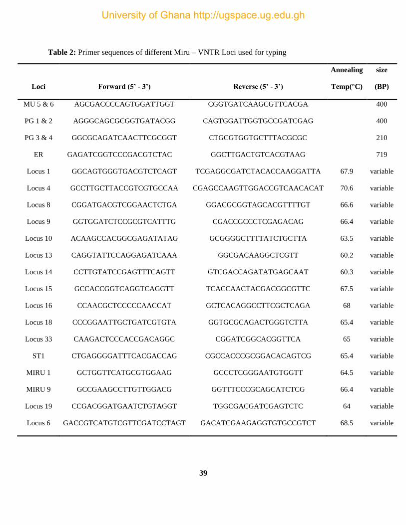
39
Table 2: Primer sequences of different Miru – VNTR Loci used for typing
Loci Forward (5’ - 3’) Reverse (5’ - 3’)
Annealing
Temp(°C)
size
(BP)
MU 5 & 6 AGCGACCCCAGTGGATTGGT CGGTGATCAAGCGTTCACGA
400
PG 1 & 2 AGGGCAGCGCGGTGATACGG CAGTGGATTGGTGCCGATCGAG
400
PG 3 & 4 GGCGCAGATCAACTTCGCGGT CTGCGTGGTGCTTTACGCGC
210
ER GAGATCGGTCCCGACGTCTAC GGCTTGACTGTCACGTAAG
719
Locus 1 GGCAGTGGGTGACGTCTCAGT TCGAGGCGATCTACACCAAGGATTA 67.9 variable
Locus 4 GCCTTGCTTACCGTCGTGCCAA CGAGCCAAGTTGGACCGTCAACACAT 70.6 variable
Locus 8 CGGATGACGTCGGAACTCTGA GGACGCGGTAGCACGTTTTGT 66.6 variable
Locus 9 GGTGGATCTCCGCGTCATTTG CGACCGCCCTCGAGACAG 66.4 variable
Locus 10 ACAAGCCACGGCGAGATATAG GCGGGGCTTTTATCTGCTTA 63.5 variable
Locus 13 CAGGTATTCCAGGAGATCAAA GGCGACAAGGCTCGTT 60.2 variable
Locus 14 CCTTGTATCCGAGTTTCAGTT GTCGACCAGATATGAGCAAT 60.3 variable
Locus 15 GCCACCGGTCAGGTCAGGTT TCACCAACTACGACGGCGTTC 67.5 variable
Locus 16 CCAACGCTCCCCCAACCAT GCTCACAGGCCTTCGCTCAGA 68 variable
Locus 18 CCCGGAATTGCTGATCGTGTA GGTGCGCAGACTGGGTCTTA 65.4 variable
Locus 33 CAAGACTCCCACCGACAGGC CGGATCGGCACGGTTCA 65 variable
ST1 CTGAGGGGATTTCACGACCAG CGCCACCCGCGGACACAGTCG 65.4 variable
MIRU 1 GCTGGTTCATGCGTGGAAG GCCCTCGGGAATGTGGTT 64.5 variable
MIRU 9 GCCGAAGCCTTGTTGGACG GGTTTCCCGCAGCATCTCG 66.4 variable
Locus 19 CCGACGGATGAATCTGTAGGT TGGCGACGATCGAGTCTC 64 variable
Locus 6 GACCGTCATGTCGTTCGATCCTAGT GACATCGAAGAGGTGTGCCGTCT 68.5 variable
University of Ghana http://ugspace.ug.edu.gh

40
3.10. Genotype Assignment
Genotypes were assigned based on length polymorphisms associated with different tandem repeats
copy number calculated based on published data (Narh et al., 2015; Williamson et al.,2014) and
confirmed by sequencing. Isolates that could not be differentiated and genotyped using the four
standard loci (Miru 1, Locus 6, Locus 19 and ST1) were further differentiated at the remaining 12
loci.
3.11. Population Genetics
Data on VNTR profiles of isolates from Narh et al., 2015 and Williamson et al., 2014 at four loci;
ST1, Miru 1, Locus 6 and Locus 19 were included in the study and analyzed for difference in
genotypes across years (2008 – 2019).
Genotypes obtained were compared for differences that existed such as:
Within population (Among communities in a sample site)
Across communities (Different sampling sites)
Across countries ( Côte d’Ivoire & Ghana)
Disease stages and categories (for ulcers)
3.12. Data and Sequence Analysis
Questionnaires were analyzed using Epi Info version 7. The map of study communities was drawn
with Arc GIS. The graphs were drawn using GraphPad Prism. Sequence data was viewed with
SnapGene Viewer and Tandem repeats were analyzed from the sequences using the Tandem
Repeat Finder (Benson, 1999). Sequences were aligned using NCBI basic local alignment of
University of Ghana http://ugspace.ug.edu.gh

41
sequences tool (BLAST) (Altschul et al., 1990). Sequence similarity to database sequences were
based on expect value (E), maximum identity and score, query coverage and total score. Multi
sequence alignments and phylogenetic analyses to reveal genetic relatedness among isolates were
performed with MEGA V5 (Tamura et al., 2011).
University of Ghana http://ugspace.ug.edu.gh

42
CHAPTER FOUR
4.0. RESULTS
4.1. Sample and Data Collection
A total of one hundred and eighty-nine (189) participants with presumptive Buruli ulcer
presentations were recruited [one hundred and eighteen (62.43%) for Ghana from June to
November 2018 and seventy-one (37.57%) for Côte d’Ivoire from August to November 2018].
Community health workers, nurses and disease control officers were trained in one-day workshops
on case identification and sample collection. Over the period reported for sample collection in
Ghana, fifty-six (47.5%), twenty-six (22.03%) and thirty-six (30.51%) suspected cases came from
Amansie District, Amasaman District and Pakro respectively. Similarly, for Côte d’Ivoire,
nineteen (26.76%), eighteen (25.35%), fourteen (19.72%), nine (12.68%) and eleven (15.49%)
suspected cases were sampled from Zoukougbeu, Djekanou, Yamoussoukro, Kongouanou and
Bouake respectively.
Overall, there were more female (52.2%) than male (47.8%) suspected cases recruited in this study
(Figure 8B), however, this difference was not statistically significant when tested by student t-test
(p= 0.202) p < 0.05 (Appendix 2). This scenario was observed for patients in Ghana but in Côte
d’Ivoire the same number of samples were obtained from both males and females. Ages of the
recruited patients ranged from 1 to 90 years for both countries and samples were categorized into
three sections based on age; children (<18 years), adult (18 – 50) and the aged (> 50). Most of the
affected persons were in the adult category (18-50) as supposed to the children and the aged (Figure
8A). The majority of patients were farmers and traders with either primary or no formal education
(Appendix 1). Only four recruits (2.02%) had attained tertiary level education. Age and occupation
University of Ghana http://ugspace.ug.edu.gh

43
status did not offer any correlation with the development of Buruli ulcer disease (p = 0.420 and
0.588 respectively) (Appendix 2)
Ghana
Cote
D'Ivoir
e
0
1 0
2 0
3 0
4 0
5 0
A g e g r o u p s
No
. o
f C
as
es
C h ild ren (< 18)
A du lt(1 8-50)
A g e d (> 5 0 )
Figure 8: Age (A) and Gender (B) Distribution of suspected BU participants. Participants were
grouped into three categories; children (less than 18 years), adult (between 18 – 50 years) and
aged (greater than 50 years).
In Ghana, all presumptive stages of the disease (nodule, plaque, oedema and ulcer) were identified
and sampled unlike Côte d’Ivoire where the majority of cases were in the ulcerative stage with only
two nodular infections identified. One hundred and eighty (180) of the of the infections across both
countries were in the ulcerative stage (95.5%) with only four (4) edematous ulcers (2.1%), four (4)
nodules (2.1%) and one (1) plaque (0.5%) (Figure 9). Out of the one hundred and eighty (180) ulcer
cases and four (4) edematous ulcers, one hundred and five (105) cases were Category II (57.1%)
A B
University of Ghana http://ugspace.ug.edu.gh

44
lesions, followed by forty-seven (47) Category III lesions (25.5%) with just thirty-two (32)
Category I lesions (17.4%)(Figure 10).
Figure 9: Stages of Buruli ulcer disease in suspected participants. Most of the cases recruited
were in the ulcerative stage
Figure 10: Categories (CAT) of lesions of suspected Buruli ulcer patients in Ghana and Côte
d’Ivoire
Most of these infections were observed on the lower and the upper limbs in both countries. Rare infections
on the trunk/breast area were also observed in both Ghana and Côte d’Ivoire with infections on the genitalia
University of Ghana http://ugspace.ug.edu.gh

45
and head/neck observed in only Côte d’Ivoire. One hundred and fifty-six (156) and twenty-seven (27) cases
reported were on the lower (82.5%) and upper (14.1%) limbs respectively, with one (1), two (2) and three
(3) infections observed on the neck/head area (0.5%), genitalia (1.1%) and trunk/breast area (1.6%) (Figure
11)
Figure 11: Different body parts affected. In Ghana, infections were only on the lower and
upper limbs and the trunk and breast area with Côte d’Ivoire recording infections at the same
parts as well as the genitalia, head and neck area.
4.2. Case Confirmation
4.2.1. Microscopy
All one hundred and eighty-nine (189) samples were tested for the presence of Acid-fast bacilli
using Ziehl neelsen’s staining. Only five samples (2.7%) were positive by microscopy and acid
fast coccobacilli clustered together were observed in Figure 12.
University of Ghana http://ugspace.ug.edu.gh

46
Figure 12: Acid fast stained slide from a sample collected in Côte d’Ivoire. Red arrows shows a clump of
bacilli
4.2.2. PCR targeting IS2404 and Enoyl Reductase gene
Conventional and nested PCR targeting the insertion sequence IS2404 were performed and
validated by qPCR targeting the same insertion sequence for the presence of Buruli ulcer among
patients. Out of all the one hundred and eighty-nine(189) samples tested, only twenty-four (24)
samples were positive for IS2404 representing 12.7% positivity. Fifty-eight (58) samples tested
positive by nested IS2404 PCR representing 30.7% positivity. Positivity of samples were further
validated by qPCR which produced one hundred and fifty-nine (159) positive samples for Buruli
ulcder representing 84% positivity.
PCR targeting the Enoyl reductase gene was perform to confirm the presence of MPM infection.
Out of the one hundred and eighty-nine (189) samples tested, thirty-four samples tested positve re
presenting 18% positivity.
University of Ghana http://ugspace.ug.edu.gh

47
Figure 13: Representitive 1.5% Gels of ethidium bromide stained PCR amplicons. A - amplicons
of IS2404 PCR with band size of 400bp. B - Amplicons for PCR targeting the Enoyl reductase
gene with band size of 720bp. C - amplicons of nested IS2404 PCR with band size of 210bp. M
:100bp Molecular Ladder, C+ : Positive Control, C- : Negative Control, 1- 15 : Samples
Figure 14: A summary of molecular confirmatory tools used for confirming the presence of
Buruli ulcer in patients. All samples positive for qPCR were further typed at sixteen (16) VNTR
loci.
University of Ghana http://ugspace.ug.edu.gh

48
4.2.3. VNTR Typing of Isolates
PCR targeting different VNTR loci in the M. ulcerans strain Agy 99 genome was performed for
qPCR IS2404 positive (159/189) samples. Each reaction was run independently for all sixteen (16)
VNTR loci as described in section 3.9. Repeat sizes were confirmed by Sanger sequencing as
described for controls in section 3.12. Table 3 below shows the VNTR profiles and designated
genotypes of isolates based on published data from Williamson et al., 2014; Narh et al., 2015.
Amplification was achieved at all loci for all controls and most isolates (11% - 69% success rate)
(Table 5) with band sizes ranging from 200bp to 900bp (Figure 15).
Figure 15 is a representation of an ethidium bromide stained gel of VNTR profile at nine loci of
six controls. Most of the samples amplified at polymorphic loci Miru 1 (65%), locus 6 (65%),
locus 19 (53%), locus 16 (52%), locus 15 (53%) and ST1 (69%) as well as monomorphic loci such
as locus 4 (68%) and locus 10 (50%). Most samples could not be successfully amplified (>50%
amplification) at loci such as locus 33 (18%), locus 13 (15%), locus 1 (11%), locus 8 (33%), locus
9 (8%), locus 18 (39%), locus 14 (30%) and Miru 9 (13%) (Table 5). All controls and samples
(165) were monomorphic at six (6) loci; locus 1, locus 4, locus 8, locus 9, locus 10 and locus 14
and hence were not used for typing isolates.
Genotyping was achieved by three stages of assignment based on the ten (10) polymorphic loci
out of the sixteen (16) loci. The first stage involved typing at the four (4) standard and most used
loci; Miru 1, locus 6, locus 19 and ST1. Isolates with an indefinite or unassigned genotype at this
first stage were further typed at two loci, locus 15 and locus 33. These two loci distinguished M.
ulcerans strain 1615 from the other MPM controls used in this study and thus was used to
distinguish isolates as either a strain of M. ulcerans or MPM. Isolates without a definite, or
University of Ghana http://ugspace.ug.edu.gh

49
unassigned genotype at this stage, were further typed at the remaining four loci, locus 13, locus
16, locus 18 and Miru 9. These four loci generally differentiated all the controls. Samples with less
than five positively amplified loci out of the sixteen were not assigned a genotype.
Figure 15: Ethidium bromide stained gels of controls at nine different loci. M- Molecular weight
ladder, 1- M. ulcerans 1615, 2- M. marinum hybrid 270995, 3- M. marinum DL 180892, 4- M.
pseudoshottsii, 5- M. marinum CL, 6- M. marinum SA 200695
4.2.4. Genotype Assignment
Seven (7) M. ulcerans genotypes designated A, B, C, D, E, F and G as well as five (5) MPM
genotypes designated MHB, MDL, MCL, MSA and MLF were generated for assigning genotypes
in this study as shown and seen in tables 4 and 5. Most isolates could not be predominantly
assigned genotypes due to unsuccessful amplification at the particular loci required to differentiate
University of Ghana http://ugspace.ug.edu.gh

50
them (Figure 16). Predominant definitely assigned genotypes that were identified in Ghana were
Genotype D present in all three (3) communities, Genotype E present only in Amansie Central
District, Genotype C and F present only in Pakro and MLF present only in Amasaman.
Genotypes prominently observed in Côte d’Ivoire were A, C, D, F and G across all communities
with Genotype F found only in Yamoussoukro and Genotype A and G found only in Djekanou.
Genotype D was observed in Djekanou, Kongouanou and Zoukougbeu while Genotype C was
observed in Kongouanou, Bouake and Zoukougbeu. Genotypes present in both Ghana and Côte
d’Ivoire include Genotypes C, D and F. Genotypes E and MLF were only found in Ghanaian
samples while that of Genotypes A and G were found in Ivorian samples.
In Ghana, Genotype C was associated with Category II lesions while Genotype F was associated
with Category I lesions. Genotype E was also associated with Category I and II lesions while
Genotype D was associated with all categories. Conversely as observed in samples from Côte
d’Ivoire, Genotype A was associated with Category II lesions, Genotype F associated with
Category II and III lesions, Genotype G associated with Category II and III lesions as well as an
edematous ulcer, while Genotype C and D were associated with all categories (Figure 17).
Data from Narh et al., 2015 and Williamson et al., 2014 was included in this study and compared
with genotypes present from 2008 to 2019 and the changes that have occurred over time. These
two studies as well as this current study all observed Genotype D (1, 1, 2, 2) among samples (Table
3). Narh et al., 2015 and Williamson et al., 2014 both observed MDL (1, 3, 2, 1). This current
study observed Genotypes A (1, 1, 1, 2) and C (3, 1,2,2) with Narh et al., 2015 and Genotypes G
( 1, 1, 2, 1) and MLF (1, 2, 2, 1) among isolates. However, there were genotypes peculiar to each
study. Narh et al., 2015 observed Genotype Z (1, 2, 2, 2), Williamson et al., 2014 observed
University of Ghana http://ugspace.ug.edu.gh

51
Genotype B (3, 1, 1, 2) and MPS ( 1, 4, 2, 2) whiles Genotypes E ( 1, 1, 2, 3) and F (2, 1, 2, 2) was
observed in this study only.
Figure 16: Percentage genotype assignment of samples from various communities in Ghana and
Côte d’Ivoire.
Figure 17: Genotypes associated with different disease stages and categories in Ghana (A &C)
and Côte d’Ivoire (B&D).
University of Ghana http://ugspace.ug.edu.gh

52
Table 3: Genotyping coding for samples based on four standard loci, Miru 1, Locus 6, ST1 and
Locus 19
Designated
genotype
VNTR Profiles
Reference (Sample size) MIRU1
Locus
6 ST1
Locus
19
A (MU) 1 1 1 2
Current study (159)
B (MU) 3 1 1 2
C (MU) 3 1 2 2
D (MU) 1 1 2 2
E (MU) 1 1 2 3
F (MU) 2 1 2 2
G (MU) 1 1 2 1
MHB 1 3 2 1
MDL 1 3 2 1
MCL 1 2 2 3
MSA 1 3 3 1
MLF 1 2 2 1
W 1 1 2 1
Narh et al., 2015 (15) X 1 1 2 2
Y 1 2 2 1
Z 1 2 2 2
A 1 1 1
Williamson et al., 2008 (50)
B 3 1 1
C 3 1 2
D (unpublished
strain) 1 1 2 2
MMDL 1 4 2 2
1 2 1 2
MLF 1 2 2 1
MPS 1 4 2 2
University of Ghana http://ugspace.ug.edu.gh

53
Table 4a: Mycobacteria species identified using Miru-VNTR Profiles of isolates (Indefinitely
assigned)
1 M. marinum hybrid 270995; 2M. marinum DL 180892; 3 M. marinum CL; 4M. marinum SA 200695;
5M. liflandii
Country Community/District M. ulcerans
Other Mycolactone Producing
Mycobacteria
UNASSIGNED
A B C D E F G 1MHB 2MDL 3MCL 4MSA 5MLF
Ghana
Amansie Central 11 2 3 40 21 9 0 0 0 0 0 1 4
Amasaman 1 1 2 17 5 2 2 1 0 1 0 2 1
Pakro 1 1 4 11 6 3 3 3 3 1 0 3 4
Côte
d’Ivoire
Kongouanou 1 1 5 7 3 5 0 1 1 1 0 1 0
Djekanou 1 0 5 8 4 3 5 3 3 2 1 2 0
Yamoussoukro 2 1 3 8 8 6 4 5 5 5 1 2 0
Zoukougbeu 3 2 7 12 1 3 9 6 6 1 3 6 1
Bouake 3 2 3 3 2 3 2 1 1 0 1 2 5
TOTAL 23 10 32 106 50 34 25 20 19 11 6 19 15
University of Ghana http://ugspace.ug.edu.gh

54
Table 5b: Mycobacteria species identified using Miru-VNTR Profiles of isolates (Definitely
assigned)
Country Community/
District
M. ulcerans
MU
MU/
MPM
UNASSIGNED
A
B
C D E F G
MLF
MPM
Ghana
Amansie
Central 0 0 0 20 2 0 0 0 17 0 4 8
Amasaman 0 0 0 10 0 0 0 0 6 1 1 0
Pakro 0 0 1 4 0 1 0 1 7 2 0 7
Cote
D'Ivoire
Kongouanou 0 0 2 1 0 0 0 0 5 1 1 0
Djekanou 0 0 1 0 0 0 0 0 2 0 2 5
Yamoussoukro 0 0 0 0 0 2 0 0 4 0 5 0
Zoukougbeu 0 0 3 2 0 0 0 0 8 2 4 0
Bouake 1 0 2 3 0 0 3 0 4 1 2 0
Sakassou 0 0 0 0 0 0 0 0 1 0 0 0
TOTAL 1 0 9 40 2 3 3 1 54 7 19 20
University of Ghana http://ugspace.ug.edu.gh

55
Table 6: VNTR typing success at sixteen (16) loci
LOCI
Ghana (%)
N=88
Côte d’Ivoire (%)
N=70
Total (%)
N=158
Locus 6 51(58) 51(73) 102(65)
Locus 13 14(16) 9(13) 23(15)
Locus1 9(10.2) 8(11) 17(11)
Locus 4 59(67) 49(70) 108(68)
ST1 61(69) 48(69) 109(69)
Locus 8 31(35) 21(30) 52(33)
Locus 9 8(9) 5(7) 13(8)
Locus 15 48(55) 35(50) 83(53)
Locus 16 50(57) 32(46) 82(52)
Locus 18 48(55) 14(20) 62(39)
Locus 19 51(58) 32(46) 83(53)
Locus 33 23(26) 6(9) 29(18)
Miru 1 68(77) 34(49) 102(65)
Miru 9 14(16) 6(9) 20(13)
Locus 10 30(34) 49(70) 79(50)
Locus 14 29(33) 18(26) 47(30)
University of Ghana http://ugspace.ug.edu.gh

56
4.3. Allelic Frequencies of Loci
Most of the isolates and controls were polymorphic at eight loci, Miru 1, ST1, locus 19,locus 8,
locus 13,locus 15, locus 18 and locus 33. The highest number of observed alleles for any loci was
3 with the least being 1.
Figure 18: Allelic frequencies of VNTR loci typed for samples collected in Ghana and Côte
d’Ivoire
University of Ghana http://ugspace.ug.edu.gh

57
4.4. Sequence Confirmation
All controls and randomly selected isolates (10% of 159 isolates) at the sixteen (16) different loci
were Sanger sequenced to confirm tandem repeats. Figure 19 shows sequences of some selected
isolates and controls at locus 6, a polymorphic locus. Repeats are color coded with each color
representing a repeat. All repeats for corresponding isolates confirmed repeat typing using band
sizes. During sequencing, some sequences were trimmed at both ends and hence sequence lengths
do not always correspond exactly with band sizes on agarose gel. Figure 19 (B) shows the
phylogenetic tree of representative isolates plotted using their locus 6 sequences. As shown, there
was clustering of sample T 18 with controls M. Pseudoshottsii, M. marinum DL, M. marinum SA
and M. marinum hybrid. Samples T01, PA26, T07, PA 22 and PA 25 clustered with control MU
1615.
University of Ghana http://ugspace.ug.edu.gh

58
Figure 19: Sequence confirmation of VNTR repeats and phylogeny of isolates. M. ulcerans 1615
(MU1615), M. marinum DL 180892 (MDL), M. pseudoshottsii, M. marinum hybrid 270995
(MHBD), M. marinum CL (MCL), M. marinum SA 200695(MSA) were controls included in the
study. NC005916.1 Plasmid is AGY 99 reference strain. PA22, PA05, AM11, T07, PA26, T01
and T18 are cases from Ghana and Côte d’Ivoire. M. ulcerans 1615 (MU1615) clustered closely
with T07 as well as PA26 and T01. Samples, PA22, PA05 and AM11 clustered together with T18.
All controls besides MU1615 clustered closely with the reference strain.
University of Ghana http://ugspace.ug.edu.gh

59
CHAPTER FIVE
5.0. DISCUSSION, CONCLUSION, AND RECOMMENDATION
5.1. DISCUSSION
Buruli ulcer has remained a neglected tropical disease over decades. The mode of transmission
remains a mystery. Pure cultures of MPMs are difficult to obtain from the environment, hence
most studies fall on genomic material of M. ulcerans and other MPMs in several aquatic
environments to infer transmission with proximity and activities performed in slow moving water
bodies and also as potential reservoirs of these MPMs (Johnson et al., 2005)(Marsollier et al.,
2004; Mosi et al., 2008). MPMs are therefore essentially of immense importance because of their
ability to cause ulcers in both animals and man (Fyfe et al., 2010).
Not until recently, other Mycolactone Producing Mycobacteria other than M. ulcerans causing
infections in fish and frogs have been identified to be somewhat associated with Buruli ulcer in
these animals (Dassi et al., 2017; Tano et al., 2017). Other researchers (Dassi et al., 2017; Tano
et al., 2017) have reported unsuccessful VNTR typing at the four most typed loci, Miru 1, locus
6, locus 19 and ST1 hence the need to increase the loci used for typing to increase the
discriminatory power of this tool. M. ulcerans as well as other MPMs have similar sequence of
various house-keeping genes as well as other very similar genetic elements especially among
isolates from the same geographic region hence hindering substantial genomic studies (Portaels
et al., 1996; Stinear et al., 2000; Ablordey et al., 2005). VNTR Typing has proven to be extremely
beneficial over the years to categorically differentiate isolates even found within the same
geographical locations, hence fine tuning would greatly benefit genomic studies on such isolates.
This section discusses data from the study to support the assertion that other MPMs besides M.
University of Ghana http://ugspace.ug.edu.gh

60
ulcerans may be implicated in Buruli ulcer and are genotypically diverse. In this study, isolates
were typed at 16 VNTR loci to access the genetic diversities at selected loci.
Samples for the study were collected from Ghana and Côte d’Ivoire. These two countries were
explored for this particular studies due to the endemicity of the disease in both countries and ties
to other Buruli ulcer resource centers. Côte d’Ivoire remains the most endemic country with
disease burden closely tailed by Ghana which is the second most endemic country (Zingue et al.,
2018). In Ghana, Amasaman is a small community located in the Ga West Municipal district of
the Greater Accra Region and happens to be the districts’ capital. Resident in that community is
the Amasaman Hospital which has a Buruli ulcer treatment center. This treatment center is one
of the only two Buruli ulcer treatment centers present in the Greater Accra region and hence is
utilized by a large number of Buruli ulcer and other tropical ulcer patients within the region and
from surrounding regions.
This deduction was made from responses obtained from patients on questionnaires administered
during interviews. Pakro is also a community situated in Akuapim South Municipal district in the
Eastern Region. Unlike Amasaman Hospital, the hospital located in Pakro lacked a Buruli ulcer
treatment centre and Buruli ulcer like cases were referred to general Out Patients Department
OPD) and were treated as usual tropical ulcers. Practices on Buruli ulcer patients and other
tropical ulcers in both hospitals were somewhat similar following routine practices such as
periodic wound cleaning and management and drug (mostly antibiotics and NSAIDs)
administration. Patients with severe ulcers that usually affects easy movement to and from the
health center were admitted. The only difference between this two health centers besides
geographical location was that patients in Amasaman hospitals resided in Buruli ulcer treatment
University of Ghana http://ugspace.ug.edu.gh

61
wards while patients admitted in Pakro resided in general wards with patients suffering from other
ailments other that Buruli ulcer or other tropical ulcers. Patients from these two centers were
recruited as and when they visited the hospital and consented to the study and were sampled
passively. This reflects in the low number of samples obtained in both health centers.
Amansie Central District (then Amansie West) is a district located in Ashanti region of Ghana.
Its district capital, Jacobu is home to one of the districts biggest hospitals. Ashanti region alone
has been reported to have more than sixty percent (60%) of all total infections in Ghana (Amofah
et al., 2002) In this particular district and the region at large, high cases of Buruli ulcer disease
have been reported and is a hub for most researchers (Asiedu and Etuaful, 1998; Duker et al.,
2004; Hilty et al., 2006; Owusu-Sekyere et al., 2013; Ablordey et al., 2015). A oneday workshop
was organized in this district which brought about fifty (50) nurses and community health workers
from different parts of the district to sensitize them on disease identification, patient recruitment
and sample collection. This exercise led to an active case search in this particular area and with
active sampling. It is therefore not surprising to have obtained fifty-six (56) samples from
Amansie Central representing 47.5% of the entire samples obtained as supposed to twenty-six
(26) and thirty-six (36) samples representing 22% and 30.5% of the entire samples obtained from
Amasaman and Pakro respectively over a six (6) months period.
In Côte d’Ivoire, seven communities were selected for sampling based on the endemicity of the
disease in such areas. The seven communities include Yamoussoukro, Kongouanou, Zoukougbeu,
Divo, Bouaké, Sakassou and Djenedoufla. Being the most endemic country, all the health facilities
visited had well-furnished Buruli ulcer treatment centers were patients are treated and observed.
The facilities had admission wards for Buruli ulcer as well as other tropical ulcers, wound dressing
University of Ghana http://ugspace.ug.edu.gh

62
area, common play grounds, a small resource laboratory, a kitchen and a common eating area
which is not surprising. Nurses and Community health workers were likewise trained in a oneday
workshop on disease identification, patient recruitment and sample collection. Samples from
patients in Côte d’Ivoire were either collected by trained nurses and community health workers at
the various resource centers and shipped or collected by researchers during periodic visits.
A lot of cases (189) were observed in this short study period (3-6 months) due to the incorporation
of active sampling where patients are identified from endemic communities due to failure to visit
health facilities. Aguiar et al., 1997 identified eight hundred and sixty-seven (867) Buruli ulcer
cases passively by waiting for patients to visit health facilities and recruit as well as sample them
over a five (5) year period (1992-1996). The number of cases identified also in another five (5)
year period study (1997-2001) by Debacker et al., 2004 doubled to one thousand and seven
hundred (1700) cases which could be attributed to either robust passive case search system or
increased number of Buruli ulcer cases. The latter however which is most likely to be cause is
downplayed by underreporting of cases. Active case search is highly recommended for early case
detection and treatment due to the bad roads among other poor infrastructure in remote areas where
the disease usually occurs to manage underreporting.
There were more female than male cases observed in this study but this was not statistically
significant (p= 0.202). This was however in contrast with a study by Debacker et al., 2004 and
case control studies by Raghunathan et al., 2005 and Sopoh et al., 2010 that reported more males
than female cases and Debacker et al., 2004 who reported a 1:1.01 male to female ratio.
Ages of patients with cases ranged from one (1) year to ninety (90) years with most of the cases
reported in the Adult category (18-50). This finding contradicts what is perceived and has been
University of Ghana http://ugspace.ug.edu.gh

63
reported as children especially boys below the ages of fifteen (15) are considered to be the most at
risk of contracting Buruli ulcer. The adult category aged between fifteen (15) and fifty (50) years
however are frequently exposed to wetlands mostly due to their occupation (most being farmers)
and daily activity which validates the preponderance observed among this age group. Incidence
in aged category in this study (50 – 90years) was also high and this could possibly be in relation
to a waning and down regulated immune state allowing the reactivation of latent disease (Debacker
et al., 2004) usually caused by trauma (Meyers et al. 1974). The number of cases in children aged
between zero (0) and fifteen (15) years were lesser and may be attributed to more effective immune
protection of children within this age group.
Lesions in all body areas were observed in this study as observed in other studies (Pluschke et al.,
2005) with more cases reported on the extremities of the body (hundred and fifty-six (156) and
twenty-seven (27) cases reported on the lower (82.5%) and upper (14.1%) limbs respectively).This
preponderance observed was expected and is consistent with reports by Portaels et al., 2001,
Debacker et al., 2004, Merritt et al., 2010 and Adu and Ampadu, 2015. Infections on the trunk and
breast area and the head and neck area were observed but in a few cases as also observed by Adu
and Ampadu, 2015. These observation support reports by Amofah et al., 2002; Portaels et al., 1999
and Wilson et al., 2011 that M. ulcerans is an environmental pathogen found in aquatic habitats
and may possibly be transmitted by close proximity to such environment or surroundings.
Interestingly, two (2) cases on the genitalia which rarely occurs was observed in Côte d’Ivoire in
two (2) children (1 male and 1 female) between the ages of one to ten (1-10). Cases as such are
not often seen in the hospital, as Debacker et al., 2004 reported only one (1) female case (age range
30-44) and six (6) male cases (age range 0-14) on the genitalia in a total of one thousand and seven
hundred (1700) Buruli ulcer cases collected over a five (5) year period (1997-2001).
University of Ghana http://ugspace.ug.edu.gh

64
Gender and age does not significantly affect the presentation of the lesion (Zingue et al., 2018).
The variation in lesion topology by gender and age may be attributed to occupation, daily and
recreational activities. For example, adult males may be more likely to weed and farm with
cutlasses and may sustain injury or trauma on the limbs more as supposed to adult females who
are likely to carry gathered farm produce or even fetch water and carry on their head thus making
that area more prone to infection.
Mycolactone’s analgesic effects make it highly ignored by patients at the earlier stages with most
seeking treatment at the very late stages. This was evident in the cases identified in this study as
the earlier disease presentations such as the nodule (3.8%) and the edema (2.1%) were very few.
These earlier disease stages may also be misdiagnosed as insect bites or allergic reactions by only
relying on clinical diagnosis. The few observed cases were treatable with antibiotics. Most of the
cases were already in the late disease stages possibly requiring surgical excision, skin grafting
and corrective treatment post antibiotic administration. The late forms of the disease were mostly
ulcerative. In Ghana, more of these cases were in Category III (lesions greater than 15cm in
diameter) while more Category II (lesions within 5-15cm in diameter) cases were observed in
Côte d’Ivoire. This may be due to the treatment seeking behavior of the patients as Côte d’Ivoire
had well established Buruli ulcer treatment centers where patients often visited and were treated
and observed whereas most patients in Ghana usually seek traditional healers or treat themselves
at home with just a handful visiting the hospitals.
Cases were confirmed by non-culture based laboratory assays specifically microscopy and PCR.
Culture was not included in the confirmation of cases due to the long period required by the
bacterium for growth (over 4 weeks) and short study period. Only five (5/189) samples (2.7%)
University of Ghana http://ugspace.ug.edu.gh

65
were positive for microscopy with acid fast bacilli and coccobacilli observed in singletons and
clusters. Being the simplest and widely used diagnostic tool, it’s the least sensitive and requires
residence in an endemic community coupled with strong clinical presentations hence may produce
a lot of false negative results (Wansbrough-Jones and Phillips, 2006). WHO recommends PCR
targeting the insertion sequence, IS2404 as the gold standard for disease confirmation. However,
other mycolactone producing mycobacteria also possess this same insertion sequence which may
impede proper diagnosis. IS2404 has been reported in only MPMs and not all NTMs (Yip et al.,
2007). Only twenty-four (24/189) samples (12.7%) were positive for IS2404 PCR using
conventional PCR methodology. The copy number of this insertion sequence is higher in M.
ulcerans (213 copies) than other MPMs (91 copies) thus other MPMs or M. ulcerans strains with
low copy numbers may be missed out. This low amplification outcome was thus validated by
nested PCR targeting the same insertion sequence. PCR amplifications were achieved after several
optimization conditions. This correlates with reports by Williamson et al., (2014) who achieved
successful amplification in a single reaction. This produced fifty-eight (58/189) positive samples
increasing the positivity to 30.7%. Enoyl Reductase PCR was also performed to increase the
chance of detecting other MPMs other than M. ulcerans. Only thirty-four (34/189) representing
18% of the samples were positive. To further increase the validity of these findings, qPCR
targeting IS2404 was used as the last line of sample confirmation as shown and used by Sarpong-
Duah et al., 2017. The low copy numbers of 1S 2404 of MPMs (91 copies) make qPCR more
effective in detecting MPMs which would have been missed by convention PCR by having a
higher cycle threshold (CT) value compared to M. ulcerans which has 213 copies of the same
insertion sequence and hence a lower CT value. Positivity among samples increased from 30.7%
to 84% as one hundred and fifty-nine (159/189) samples were positive using qPCR. The one
University of Ghana http://ugspace.ug.edu.gh

66
hundred and fifty-nine (159) cases were confirmed as positive for Buruli ulcer with laboratory
evidence and corroborates results from other study cases that relied solely on clinical diagnosis
(Kanga and Kacou, 2001). The high rate of laboratory confirmed cases in this study, irrespective
of the stage of the disease indicates that clinical diagnosis is enough to commence treatment in
endemic areas. Diagnosis in most rural areas have however moved from using mere clinical
symptoms only for diagnosis (Bratschi et al., 2013) to the use of laboratory based diagnosis which
is highly recommended by WHO.
SNP typing has been shown to be successful for the typing of MPMs (Yip et al., 2007), however
VNTR typing was used in this particular study to increase its efficiency and discriminatory power
as it is a cheaper and faster tool to use and extensively employed for MPM Typing. VNTR typing
in this study was achieved at sixteen (16) loci; locus 1, locus 4, locus 6, locus 8, locus 9, locus 10,
locus 13, locus 14, locus 15, locus 16, locus 18, locus 19, locus 33, ST1, Miru 1 and Miru 9. VNTR
analysis of two polymorphic loci, MIRU1 and ST1 on seventy-two (72) African isolates (fifty-
seven (57) Ghanaian isolates) revealed three different genotypes with clonal clustering, suggesting
genetic diversity of M. ulcerans in Ghana (Hilty et al., 2006) which could have been more if other
polymorphic loci were included. Using sixteen (16) loci, this study increased the discrimination
power of VNTR typing as against two (2), three (3) and four (4) loci separately reported by Hilty
et al., 2006; Williamson et al., 2014 and Narh et al., 2015 respectively. Controls used (M. ulcerans
1615, M. marinum DL 180892, M. pseudoshottsii, M. marinum hybrid 270995, M. marinum CL,
M. marinum SA 200695) and samples (159) were monomorphic at six (6) loci; locus 1, locus 4,
locus 8, locus 9, locus 10 and locus 14. Data from these loci were not used for genotyping of
samples because it did not distinguish samples.
University of Ghana http://ugspace.ug.edu.gh

67
Samples were classified into seven (7) M. ulcerans genotypes and five (5) MPMs genotypes at ten
(10) loci based on controls and published literature (Williamson et al., 2014; Narh et al., 2015).The
four (4) standard loci, Miru 1, locus 6, ST1, locus 19 were first used to genotype and the other six
(6) used to supplement data for samples that poorly performed at the former. Interestingly, locus
15 and locus 33 were able to distinguish M. ulcerans from the other MPMs. As a results, samples
that could not be categorized as either M. ulcerans or an MPM due to poor amplification at the
standard four loci were identified at this loci. This would serve as another simple PCR based
method of distinguishing M. ulcerans from the other MPMs which only relies on qPCR targeting
IS2404 due to varying copy numbers of this insertion sequences hence reflecting in their CT value.
The remaining four loci, locus 13, locus 15, locus 16 and locus 18 generally distinguished all
MPMs indiscriminatorily and was used when samples could not be genotyped at the first six loci
mentioned above. Loci, Miru 1 and ST1 with repeats 1 and 2 respectively were observed in this
study which is consistent with reports by Hilty et al., (2006) for M. ulcerans strains from Amansie
Central which happens to be one of the sites under this study. Williamson et al., (2014) also
obtained repeats 1, 1 and 1; 3, 1 and 2; 3, 1 and 1 as well as 1, 1 and 2 at loci, Miru 1, locus 6 and
ST1 respectively which were observed in this study as well; however, Repeats 1, 4 and 2 observed
by Williamson et al., (2014) at the above mention loci were absent in this study with samples
monomorphic at locus 19 but polymorphic in this study.
Both Ghana and Côte d’Ivoire had five (5) genotypes in circulation. Genotype C (3, 1, 2, 2), D (1,
1, 2, 2) and F (2, 1, 2, 2) was identified in both Ghana and Côte d’Ivoire. Genotypes A (1, 1, 1, 2)
and G (1, 1, 2, 1) were only found in Côte d’Ivoire while Genotype E (1, 1, 2, 3) and the MPM,
MLF (1, 2, 2, 1) were only identified in Ghana. In Ghana, Genotype D was found in all three
communities. Genotypes E was found only in Amansie Central, Genotype MLF in Amasaman and
University of Ghana http://ugspace.ug.edu.gh

68
Genotype C and F only in Pakro. The presence of two genotypes in Amasaman suggests that
MPMs in circulation in that area is low. This may be attributed to the small of samples (26)
obtained in this area. Three genotypes observed in Pakro in relation to the sample size (36) is
suggests that different species of M. ulcerans exist in Pakro. Amansie central was the community
in Ghana with the most cases (56) due to active case search but only two genotypes were identified.
The high sample size in Amansie central coupled with few genotypes in circulation may be
attributed to the treatment regime amongst patient. In Amansie central, active case searches led to
the early identification of cases and follow up to clinic and treatment commencement. Hence fewer
genotypes observed in this community is inferable.
In Côte d’Ivoire, all the communities sampled had Buruli ulcer treatment centers that are highly
utilized by individuals. Unlike Ghana, no single genotype was found across all communities.
Djekanou had the most genotypes (A, C, D and G) with Yamoussoukro and Bouake having the
least number of genotypes, F and C respectively. Common genotypes (C and D) were found in
Kongouanou and Zoukougbeu. Fewer genotypes were expected in Côte d’Ivoire than in Ghana
due to the approximately 1:2 sample size ratio which was however not observed. A higher number
in the former observed may be linked to the acquisition of infections in rare positions in the body
such as the genitalia as observed in samples from only Côte d’Ivoire. Data from similar studies
conducted by Narh et al., 2015 and Williamson et al., 2014 were included in this study. Both
studies were conducted in Ghana. Genotype D was observed among all studies which makes it the
most prevalent genotype from 2008 to 2019. Apart from Genotype D, Genotype A, B and C were
observed in this current study and Williamson et al., 2014. However, Genotype C was the
prominent genotype in the later study whereas Genotype D was the prominent in the former. There
could possibly be a change from one genotype to another to aid disease establishment and
University of Ghana http://ugspace.ug.edu.gh

69
adaptation to a particular niche over time. Also, Ivorian samples included in this present study may
have influenced this pattern observed as the former study did not have any Ivorian samples.
Genotype G and the MPM genotype MLF represented as W and Y by Narh et al., 2015 were
common to both studies. The absence of these strains in Williamson et al., 2014 study indicates
that there may be possible emerging strains as both studies were conducted between 2015-2019.
Distinctively, Genotype E and F were only found in this present study further validating the
emergence of possible new strains. Genotypes Z was only found in Narh et al., 2015 study whereas
Genotype B and MPM, MPS (M. Pseudoshottsii) suggesting that strains of M. ulcerans of may be
adapting to favor infection and MPMs may be implicated also in Buruli ulcer causation.
In both Ghana and Côte d’Ivoire, Genotype D was associate with all stages of infection. This
observation is consistent with the high number of D genotypes identified in both countries.
Genotypes C and F were associated with the earlier stages of the infection in Ghana and
conversely the late BU stages in Côte d’Ivoire. Similarly, Genotype E was associated with the
earlier disease stage in Ghana whiles Genotype F was associated with late stages of the disease
in Côte d’Ivoire. Genotype A was only involved in nodular infections in Côte d’Ivoire, however
inferring that it’s actually causes nodular infections may be wrong as only a few samples were
Genotype A.
Most of the cases were however either in the late ulcerative stage or had advanced and were
categorized as based on lesion size and presentation. In Ghana, Genotype C was associated with
Category II lesions whiles Genotype F was associated with Category I lesions. Genotype E was
also associated with Category I and II lesions while Genotype D was associated with all
categories. Conversely as observed in Côte d’Ivoire isolates, Genotype A was associated with
University of Ghana http://ugspace.ug.edu.gh

70
Category II lesions, Genotype F associated with Category II and III lesions, Genotype G
associated with Category II and III lesions as well as an edematous ulcer while Genotype C and
D was associated with all categories (Figure 17).
This study addressed three key objectives: Identification of MPMs, MPM genotypes causing
infections in Ghana and Côte d’Ivoire and the dynamics of genotype differences over the years
as well as between two study sites. Genotypes responsible for Buruli ulcer in Ghana and Côte
d’Ivoire were duly accessed.
5.2. CONCLUSION
University of Ghana http://ugspace.ug.edu.gh

71
A higher number of Buruli ulcer patients than expected were identified. More adults were infected
than children and aged. Out of the sixteen (16) VNTR loci used for typing cases, six (locus 1, locus
4, locus 8, locus 10, locus 14 and Miru 9) were monomorphic whiles polymorphic at ten (Miru 1,
locus 6, locus 19, locus 15, locus 13, locus 16, locus 18, locus 33, locus 9 and ST1). Locus 15 and
locus 33 distinguished M. ulcerans from the other MPMs. Five genotypes were observed in Ghana
and Côte d’Ivoire. Genotypes C, D and F were found in both Ghana and Côte d’Ivoire. Genotype
E and MLF found only in Ghana whiles Genotypes A and G were only found in Côte d’Ivoire.
Genotype D was prominent from 2008 to 2019.VNTR typing results with published literature gave
indications for the possible emergence and spreading of new genetic variants of M. ulcerans and
other MPMs within Ghana and Côte d’Ivoire. This study has shown that there is genetic variation
among MPMs, particularly M. ulcerans, and hence additional polymorphic markers can be scanned
from the M. ulcerans genome, Agy99, to differentiate strains.
University of Ghana http://ugspace.ug.edu.gh

72
5.3. RECOMMENDATION
Whole genome metagenomics sequencing of both IS2404 positive and negative will be essential
to see what microbial community may be responsible for varying genotypes observed in the
positives and similar lesion and clinical presentations of the negatives. It would be quite interesting
as more information will be available to definitely assign genotypes. Similar studies can be done
in other endemic communities other than the ones in this study to see if findings will corroborate
that observed in this study. A similar study should also be done in the same communities but this
time around with environmental samples in show if similar genotypes would be observed to infer
local transmission.
University of Ghana http://ugspace.ug.edu.gh

73
REFERENCES
1972 Baker Buruli ulcer and human activities in Uganda.pdf. (n.d.).
Ablordey, A., Fonteyne, P. A., Stragier, P., Vandamme, P., & Portaels, F. (2007). Identification
of a new variable number tandem repeat locus in Mycobacterium ulcerans for potential
strain discrimination among African isolates. Clinical Microbiology and Infection, 13(7),
734–736. https://doi.org/10.1111/j.1469-0691.2007.01716.x
Ablordey, A. S., Vandelannoote, K., Frimpong, I. A., Ahortor, E. K., Amissah, N. A., Eddyani,
M., … Stinear, T. P. (2015). Whole Genome Comparisons Suggest Random Distribution of
Mycobacterium ulcerans Genotypes in a Buruli Ulcer Endemic Region of Ghana. PLoS
Neglected Tropical Diseases, 9(3), 1–15. https://doi.org/10.1371/journal.pntd.0003681
Ablordey, Anthony, Kotlowski, R., & Swings, J. (2005). Reference article, Ablordey et al. 2005.
PCR Amplification with Primers Based on IS2404 and GC-Rich revealed repeateed
sequence plymorphism in MU.pdf, 43(1), 448–451. https://doi.org/10.1128/JCM.43.1.448
Ablordey, Anthony, Swings, J., Hubans, C., Locht, C., Portaels, F., & Chemlal, K. (2005).
Multilocus Variable-Number Tandem Repeat Typing of Mycobacterium ulcerans, 43(4),
1546–1551. https://doi.org/10.1128/JCM.43.4.1546
Adamba, C., & Owusu, A. yaa. (2011). Burden of Buruli Ulcer : How Affected Households in a
Ghanaian District Cope. African Study Monographs, 32(April), 1–23.
Adu, E. J. K., & Ampadu, E. (2015). Mycobacterium ulcerans disease in the middle belt of
Ghana : An eight-year review from six endemic districts. International Journal of
Mycobacteriology, 4(2), 138–142. https://doi.org/10.1016/j.ijmyco.2015.03.006
University of Ghana http://ugspace.ug.edu.gh

74
Adusumilli, S., Mve-Obiang, A., Sparer, T., Meyers, W., Hayman, J., & Small, P. L. C. (2005).
Mycobacterium ulcerans toxic macrolide, mycolactone modulates the host immune
response and cellular location of M. ulcerans in vitro and in vivo. Cellular Microbiology,
7(9), 1295–1304. https://doi.org/10.1111/j.1462-5822.2005.00557.x
Aguiar J, Domingo M, Guédénon A, Meyers W, Steunou C, Portaels F. 1997. L’ulcère de Buruli,
une maladie mycobactérienne importante et en recrudescence au Bénin. Bull Seances Acad
R Sci Outre Mer 3:325–356.
Alotaibi, M. H., Bahammam, S. A., ur Rahman, S., Bahnassy, A. A., Hassan, I. S., Alothman, A.
F., & Alkayal, A. M. (2016). The demographic and clinical characteristics of leprosy in
Saudi Arabia. Journal of Infection and Public Health, 9(5), 611–617.
https://doi.org/10.1016/j.jiph.2015.12.015
Amofah G, B. F., Amofah G, Bonsu F, Tetteh C, “‘et al.,’” Amofah, G., Bonsu, F., Tetteh, C.,
Okrah, J., … Addy, J. (2002). Buruli ulcer in Ghana: Results of a national case search.
Emerging Infectious Diseases, 8(2), 167–170. https://doi.org/10.3201/eid0802.010119
Asiedu, K., & Etuaful, S. (1998). Socioeconomic implications of Buruli ulcer in Ghana: A three-
year review. American Journal of Tropical Medicine and Hygiene, 59(6), 1015–1022.
https://doi.org/10.4269/ajtmh.1998.59.1015
Beissner, M., Herbinger, K.-H., & Bretzel, G. (2010). Laboratory diagnosis of Buruli ulcer
disease. Future Microbiology, 5(3), 363–370. https://doi.org/10.2217/fmb.10.3
Boleira, M., Lupi, O., Lehman, L., Asiedu, K., & Kiszewski, A. (2010). Úlcera de Buruli. Anais
Brasileiros De Dermatologia, 85(3), 281-301. doi: 10.1590/s0365-05962010000300002
University of Ghana http://ugspace.ug.edu.gh

75
Bolz, M., Bratschi, M. W., Kerber, S., Minyem, J. C., Boock, A. U., Vogel, M., … Cabello, A.
L. (2015). Locally confined clonal complexes of Mycobacterium ulcerans in two buruli
ulcer endemic regions of cameroon. PLoS Neglected Tropical Diseases, 9(6), 1–13.
https://doi.org/10.1371/journal.pntd.0003802
Boulkroun, S., Guenin-Mace, L., Thoulouze, M.-I., Monot, M., Merckx, A., Langsley, G., …
Demangel, C. (2009). Mycolactone Suppresses T Cell Responsiveness by Altering Both
Early Signaling and Posttranslational Events. The Journal of Immunology.
Bratschi, M. W., Ruf, M.-T., Noumen, D., Bolz, M., Grize, L., Wantong, F. G., … Pluschke, G.
(2013). Geographic Distribution, Age Pattern and Sites of Lesions in a Cohort of Buruli
Ulcer Patients from the Mapé Basin of Cameroon. PLoS Neglected Tropical Diseases, 7(6),
e2252. https://doi.org/10.1371/journal.pntd.0002252
Bretzel, G., Nienhuis, W. A., Fleischmann, E., Loscher, T., Beissner, M., Siegmund, V., …
Klutse, E. (2010). A Genotypic Approach for Detection, Identification, and Characterization
of Drug Resistance in Mycobacterium ulcerans in Clinical Samples and Isolates from
Ghana. The American Journal of Tropical Medicine and Hygiene, 83(5), 1059–1065.
https://doi.org/10.4269/ajtmh.2010.10-0263
Cambau, E., Chauffour-Nevejans, A., Tejmar-Kolar, L., Matsuoka, M., & Jarlier, V. (2012).
Detection of antibiotic resistance in leprosy using GenoType LepraeDR, a novel ready-to-
use molecular test. PLoS Neglected Tropical Diseases, 6(7).
https://doi.org/10.1371/journal.pntd.0001739
Dassi, C., Mosi, L., Narh, C., Quaye, C., Konan, D., Djaman, J., & Bonfoh, B. (2017).
Distribution and Risk of Mycolactone-Producing Mycobacteria Transmission within Buruli
University of Ghana http://ugspace.ug.edu.gh

76
Ulcer Endemic Communities in Côte d’Ivoire. Tropical Medicine and Infectious Disease,
2(1), 3. https://doi.org/10.3390/tropicalmed2010003
Debacker, M., Aguiar, J., Steunou, C., Zinsou, C., Meyers, W. M., Scott, J. T., … Portaels, F.
(2004). Mycobacterium ulcerans disease: Role of age and gender in incidence and
morbidity. Tropical Medicine and International Health, 9(12), 1297–1304.
https://doi.org/10.1111/j.1365-3156.2004.01339.x
Dégboé, B., Koudoukpo, C., Alimi, M., Sopoh, G. E., Akpadjan, F., Johnson, R. C., …
Atadokpèdé, F. (2019). Buruli Ulcer: Epidemiological, Clinical and Biological Profile of
Patients in the Centre de Depistage et de Traitement d’Allada (Benin) from 2010 to 2014.
Journal of Cosmetics, Dermatological Sciences and Applications, 09(02), 67–82.
https://doi.org/10.4236/jcdsa.2019.92006
Demangel, C., & High, S. (2018). Sec61 blockade by mycolactone: A central mechanism in
Buruli ulcer disease. Biology of the Cell, 110(11), 237–248.
https://doi.org/10.1111/boc.201800030
Diamant, A., Banet, A., Ucko, M., Colorni, A., Knibb, W., & Kvitt, H. (2000). <Mycobacteriosis
in wild rabbitfish associated with cage farming in the gulf of Eilat.pdf>, 39, 211–219.
Doig, K. D., Holt, K. E., Fyfe, J. A. M., Lavender, C. J., Eddyani, M., Portaels, F., … Stinear, T.
P. (2012). On the origin of Mycobacterium ulcerans, the causative agent of Buruli ulcer.
BMC Genomics, 13(1). https://doi.org/10.1186/1471-2164-13-258
Duker, A. A., Carranza, E. J., Hale, M., Asiedu, K., Etuaful, S., Portaels, F., … Kiefer, R.
(2004). Spatial dependency of Buruli ulcer prevalence on arsenic-enriched domains in
University of Ghana http://ugspace.ug.edu.gh

77
Amansie West District, Ghana: implications for arsenic mediation in Mycobacterium
ulcerans infection. International Journal of Health Geographics, 3(1), 19.
https://doi.org/10.1186/1476-072X-3-19
Einarsdottir, T., Huygen, K., & Ulcer, B. (2011). a n d e s i o s c i e n c e o n o t d i s t r i b u t e a
i o, 7(11), 1198–1203.
Falkinham, JOSEPH O. (1996). Epidemiology of infection by nontuberculous mycobacteria .
Epidemiology of Infection by Nontuberculous Mycobacteria, 9(2), 177–215.
https://doi.org/10.1016/S0040-4039(00)79908-4
Falkinham, J. O. (2011). Nontuberculous mycobacteria from household plumbing of patients
with nontuberculous mycobacteria disease. Emerging Infectious Diseases, 17(3), 419–424.
https://doi.org/10.3201/eid1703.101510
Falkinham, J., Pruden, A., & Edwards, M. (2015). Opportunistic Premise Plumbing Pathogens:
Increasingly Important Pathogens in Drinking Water. Pathogens, 4(2), 373–386.
https://doi.org/10.3390/pathogens4020373
Forbes, B. A. (2017). crossm Mycobacterial Taxonomy, 55(2), 380–383.
Forbes, B., Hall, G., Miller, M., Novak, S., Rowlinson, M., & Salfinger, M. et al. (2018).
Practice Guidelines for Clinical Microbiology Laboratories: Mycobacteria. Clinical
Microbiology Reviews, 31(2). doi: 10.1128/cmr.00038-17
Funakoshi, T., Kazumi, Y., Okada, R., Nishimoto, K., Saito, M., Amagai, M., … Ohyama, M.
(2009). Intractable ulcer caused by Mycobacterium shinshuense: Successful identification
of mycobacterium strain by 16S ribosomal RNA 3′-end sequencing. Clinical and
University of Ghana http://ugspace.ug.edu.gh

78
Experimental Dermatology, 34(8), 712–715. https://doi.org/10.1111/j.1365-
2230.2009.03442.x
Fyfe, J. A. M., Lavender, C. J., Handasyde, K. A., Legione, A. R., O’Brien, C. R., Stinear, T. P.,
… Johnson, P. D. R. (2010). A major role for mammals in the ecology of Mycobacterium
ulcerans. PLoS Neglected Tropical Diseases, 4(8).
https://doi.org/10.1371/journal.pntd.0000791
George, K. M., Chatterjee, D., Gunawardana, G., Welty, D., Hayman, J., Lee, R., & Small, P. L.
C. (1999). Mycolactone: A polyketide toxin from mycobacterium ulcerans required for
virulence. Science, 283(5403), 854–857. https://doi.org/10.1126/science.283.5403.854
George, K. M., Pascopella, L., Welty, D. M., & Small, P. L. C. (2000). A Mycobacterium
ulcerans toxin, mycolactone, causes apoptosis in guinea pig ulcers and tissue culture cells.
Infection and Immunity, 68(2), 877–883. https://doi.org/10.1128/IAI.68.2.877-883.2000
Grange, J. M. (2008). The biology of the genus Mycobacterium. Journal of Applied
Microbiology, 81(s25), 1S-9S. https://doi.org/10.1111/j.1365-2672.1996.tb04827.x
Griffith, D. E., Aksamit, T., Brown-Elliott, B. A., Catanzaro, A., Daley, C., Gordin, F., …
Winthrop, K. (2007). An official ATS/IDSA statement: Diagnosis, treatment, and
prevention of nontuberculous mycobacterial diseases. American Journal of Respiratory and
Critical Care Medicine, 175(4), 367–416. https://doi.org/10.1164/rccm.200604-571ST
Guenin-Macé, L., Ruf, M.-T., Pluschke, G., & Demangel, C. (2019). Mycolactone: More than
Just a Cytotoxin BT - Buruli Ulcer: Mycobacterium Ulcerans Disease. In G. Pluschke & K.
Röltgen (Eds.) (pp. 117–134). Cham: Springer International Publishing.
University of Ghana http://ugspace.ug.edu.gh

79
https://doi.org/10.1007/978-3-030-11114-4_7
Guenin-Macé, L., Veyron-Churlet, R., Thoulouze, M. I., Romet-Lemonne, G., Hong, H.,
Leadlay, P. F., … Demangel, C. (2013). Mycolactone activation of Wiskott-Aldrich
syndrome proteins underpins Buruli ulcer formation. Journal of Clinical Investigation.
Hall, B., & Simmonds, R. (2014). Pleiotropic molecular effects of the Mycobacterium ulcerans
virulence factor mycolactone underlying the cell death and immunosuppression seen in
Buruli ulcer . Biochemical Society Transactions, 42(1), 177–183.
https://doi.org/10.1042/bst20130133
Hall, G. S., Rowlinson, M.-C., Somoskövi, A., Miller, M. B., Novak, S. M., Wilson, M. L., …
Salfinger, M. (2018). Practice Guidelines for Clinical Microbiology Laboratories:
Mycobacteria. Clinical Microbiology Reviews, 31(2), 1–66.
https://doi.org/10.1128/cmr.00038-17
Hilty, M., Käser, M., Zinsstag, J., Stinear, T., & Pluschke, G. (2007). Analysis of the
Mycobacterium ulcerans genome sequence reveals new loci for variable number tandem
repeats (VNTR) typing. Microbiology (Reading, England), 153(Pt 5), 1483–1487.
https://doi.org/10.1099/mic.0.2006/004564-0
Hilty, M., Yeboah-manu, D., Boakye, D., Mensah-quainoo, E., Rondini, S., Ofori-adjei, D., …
Schelling, E. (2006). Genetic Diversity in Mycobacterium ulcerans Isolates from Ghana
Revealed by a Newly Identified Locus Containing a Variable Number of Tandem Repeats
Genetic Diversity in Mycobacterium ulcerans Isolates from Ghana Revealed by a Newly
Identified Locus Contai. Society, 188(4), 1462–1465. https://doi.org/10.1128/JB.188.4.1462
University of Ghana http://ugspace.ug.edu.gh

80
Hilty, M., Yeboah-Manu, D., Boakye, D., Mensah-Quainoo, E., Rondini, S., Schelling, E., …
Pluschke, G. (2006). Genetic diversity in Mycobacterium ulcerans isolates from Ghana
revealed by a newly identified locus containing a variable number of tandem repeats.
Journal of Bacteriology, 188(4), 1462–1465. https://doi.org/10.1128/JB.188.4.1462-
1465.2006
Hong, H., Demangel, C., Pidot, S. J., Leadlay, F., & Stinear, T. (2008). Mycolactones :
immunosuppressive and cytotoxic polyketides produced by aquatic mycobacteria, 44, 447–
454. https://doi.org/10.1039/b803101k
Huys, G., Rigouts, L., Chemlal, K., Portaels, F., & Swings, J. (2000). Evaluation of amplified
fragment length polymorphism analysis for inter- and intraspecific differentiation of
Mycobacterium bovis, M. tuberculosis, and M. ulcerans. Journal of Clinical Microbiology,
38(10), 3675–3680.
Imajoh, M., Sugiura, H., Hashida, Y., Hatai, K., Oshima, S. I., Daibata, M., & Kawai, K. (2013).
Genotypic characteristics of a Mycobacterium sp. isolated from yellowtail Seriola
quinqueradiata and striped jack Pseudocaranx dentex in Japan. Microbiology and
Immunology, 57(1), 13–20. https://doi.org/10.1111/j.1348-0421.2012.00514.x
Janda, J. M., & Abbott, S. L. (2007). 16S rRNA gene sequencing for bacterial identification in
the diagnostic laboratory: pluses, perils, and pitfalls. Journal of Clinical Microbiology,
45(9), 2761–2764. https://doi.org/10.1128/JCM.01228-07
Johnson, P., & Lavender, C. (2009). Correlation between Buruli Ulcer and Vector-borne
Notifiable Diseases, Victoria, Australia. Emerging Infectious Diseases, 15(4), 614-615. doi:
10.3201/eid1504.081162
University of Ghana http://ugspace.ug.edu.gh

81
Johnson, P D R, Azuolas, J., Lavender, C. J., Wishart, E., Stinear, T. P., Hayman, J. a, … Fyfe, J.
a M. (2007). Mycobacterium ulcerans in osquitoes captured during outbreak of Buruli
ulcer, southeastern Australia. Emerg.Infect.Dis., 13(11), 1653–1660.
https://doi.org/10.3201/eid1311.061369
Johnson, Paul D R, Stinear, T., Small, P. L. C., Plushke, G., Merritt, R. W., Portaels, F., …
Asiedu, K. (2005). Buruli ulcer (M. ulcerans infection): New insights, new hope for disease
control. PLoS Medicine, 2(4), 0282–0286. https://doi.org/10.1371/journal.pmed.0020108
Junghanss, T., Johnson, R.C., and Pluschke, G. (2014) Mycobacterium ulcerans disease. In
Manson’s Tropical Diseases. Farrar, J., Hotez, P.J., Junghanss, T., Kang, G., Lalloo, D., and
White, N. (eds)(eds.), 23rd edn. Philadelphia, USA: Elsevier Saunders.
Kanga JM, Kacou ED. 2001. Epidemiological aspects of Buruli ulcer in Côte d’Ivoire: results of
a national survey. Bull Soc Pathol Exot 94: 46–51. (In French.)
Käser, M., Hauser, J., & Pluschke, G. (2009). Single nucleotide polymorphisms on the road to
strain differentiation in Mycobacterium ulcerans. Journal of Clinical Microbiology, 47(11),
3647–3652. https://doi.org/10.1128/JCM.00761-09
Käser, M., Rondini, S., Naegeli, M., Stinear, T., Portaels, F., Certa, U., & Pluschke, G. (2007).
Evolution of two distinct phylogenetic lineages of the emerging human pathogen
Mycobacterium ulcerans. BMC Evolutionary Biology, 7. https://doi.org/10.1186/1471-2148-
7-177
Katoch, V. M. (2004). Infections due to non-tuberculous mycobacteria (NTM). Indian Journal of
Medical Research, 120(4), 290–304.
University of Ghana http://ugspace.ug.edu.gh

82
Kim, H., Kim, S. H., Shim, T. S., Kim, M. N., Bai, G. H., Park, Y. G., … Kim, B. J. (2005).
Differentiation of Myocobacterium species by analysis of the heat-shock protein 65 gene
(hsp65). International Journal of Systematic and Evolutionary Microbiology, 55(4), 1649–
1656. https://doi.org/10.1099/ijs.0.63553-0
Lamb, R. C., & Dawn, G. (2014). Cutaneous non-tuberculous mycobacterial infections.
International Journal of Dermatology 2014, 53, 1197–1204.
Lavender, C. J., Brown, K., Evans, R. N., Azuolas, J., Johnson, P. D. R., Fyfe, J. A. M., & Ray,
L. R. (2011). Risk of Buruli Ulcer and Detection of Mycobacterium ulcerans in Mosquitoes
in Southeastern Australia. PLoS Neglected Tropical Diseases, 5(9), e1305.
https://doi.org/10.1371/journal.pntd.0001305
Loftus, M. J., Tay, E. L., Globan, M., Lavender, C. J., Crouch, S. R., Johnson, P. D. R., & Fyfe,
J. A. M. (2018). Epidemiology of buruli ulcer infections, Victoria, Australia, 2011–2016.
Emerging Infectious Diseases, 24(11), 1988–1997. https://doi.org/10.3201/eid2411.171593
Lunn, H. F., CONNOR, D. H., WILKS, N. E., BARNLEY, G. R., KAMUNVI, F., CLANCEY,
J. K., & BEE, J. D. (1965). BURULI (MYCOBACTERIAL) ULCERATION IN
UGANDA. (A NEW FOCUS OF BURULI ULCER IN MADI DISTRICT, UGANDA):
REPORT OF A FIELD STUDY. East African Medical Journal, 42, 275—288. Retrieved
from http://europepmc.org/abstract/MED/14341980
M., B., O., L., L., L., & K.B., A. (2010). Buruli ulcer. Anais Brasileiros de Dermatologia, 85(3),
281–301. https://doi.org/http://dx.doi.org/10.1590/S0365-05962010000300002
M Kanga, J., & D Kacou, E. (2001). [Epidemiologicl aspects of Buruli ulcer in Côte d’Ivoire:
University of Ghana http://ugspace.ug.edu.gh

83
results of a national survey]. Bulletin de La Société de Pathologie Exotique (1990), 94, 46–
51.
Maccallum, P., Tolhurst, J. C., Buckle, G., & Sissons, H. (1948). A new mycobacterial infection
in man: clinical aspects. The Journal of Pathology and Bacteriology, 60(1), 93–102.
https://doi.org/10.1002/path.1700600111
Marion, E., Landier, J., Adeye, A., Rega, A., Vincent, Q. B., Marsollier, L., … Pommelet, V.
(2014). Findings in Patients From Benin With Osteomyelitis and Polymerase Chain
Reaction–Confirmed Mycobacterium ulcerans Infection. Clinical Infectious Diseases,
59(9), 1256–1264. https://doi.org/10.1093/cid/ciu584
Marsollier, L., Aubry, J., Saint-André, J. P., Robert, R., Legras, P., Manceau, A. L., …
Carbonnelle, B. (2003). Écologie et mode de transmission de Mycobacterium ulcerans.
Pathologie Biologie, 51(8–9), 490–495. https://doi.org/10.1016/S0369-8114(03)00151-2
Marsollier, Laurent, Stinear, T., Aubry, J., Saint André, J. P., Robert, R., Legras, P., …
Carbonnelle, B. (2004). Aquatic Plants Stimulate the Growth of and Biofilm Formation by
Mycobacterium ulcerans in Axenic Culture and Harbor These Bacteria in the Environment.
Applied and Environmental Microbiology, 70(2), 1097–1103.
https://doi.org/10.1128/AEM.70.2.1097-1103.2004
Matsumura, Y., Taki, R., & Miyachi, Y. (2012). Case of Buruli ulcer due to Mycobacterium
ulcerans ssp. shinshuense with distal cutaneous involvement and synovitis. Journal of
Dermatology, 39(1), 80–83. https://doi.org/10.1111/j.1346-8138.2011.01215.x
Mensah-Quainoo, E., Rondini, S., Bodmer, T., Pluschke, G., & Troll, H. (2003). Development
University of Ghana http://ugspace.ug.edu.gh

84
and Application of Real-Time PCR Assay for Quantification of Mycobacterium ulcerans
DNA. Journal of Clinical Microbiology, 41(9), 4231–4237.
https://doi.org/10.1128/jcm.41.9.4231-4237.2003
Merritt, R. W., Walker, E. D., Small, P. L. C., Wallace, J. R., Johnson, P. D. R., Benbow, M. E.,
& Boakye, D. A. (2010). Ecology and transmission of buruli ulcer disease: A systematic
review. PLoS Neglected Tropical Diseases, 4(12), 1–15.
https://doi.org/10.1371/journal.pntd.0000911
Merritt, R. W., Williamson, H. R., Sopoh, G., Barogui, Y., Johnson, C. R., Campbell, L. P., …
Benbow, M. E. (2012). Detection of Mycobacterium ulcerans in the Environment Predicts
Prevalence of Buruli Ulcer in Benin. PLoS Neglected Tropical Diseases, 6(1), e1506.
https://doi.org/10.1371/journal.pntd.0001506
Mitchell, P. J., Jerrett, I. V., & Slee, K. J. (1984). Skin ulcers caused by mycobacterium ulcerans
in koalas near bairnsdale, Australia. Pathology, 16(3), 256–260.
https://doi.org/10.3109/00313028409068533
Mosi, L., Williamson, H., Wallace, J. R., Merritt, R. W., & Small, P. L. C. (2008a). Persistent
association of Mycobacterium ulcerans with West African predaceous insects of the family
belostomatidae. Applied and Environmental Microbiology, 74(22), 7036–7042.
https://doi.org/10.1128/AEM.01234-08
Mosi, L., Williamson, H., Wallace, J. R., Merritt, R. W., & Small, P. L. C. (2008b). Persistent
Association of Mycobacterium ulcerans with West African Predaceous Insects of the
Family Belostomatidae ᰔ †, 74(22), 7036–7042. https://doi.org/10.1128/AEM.01234-08
University of Ghana http://ugspace.ug.edu.gh

85
Mve-Obiang, A., Lee, R. E., Umstot, E. S., Trott, K. A., Grammer, T. C., Parker, J. M., … Small,
P. L. C. (2005). A newly discovered mycobacterial pathogen isolated from laboratory
colonies of Xenopus species with lethal infections produces a novel form of mycolactone,
the Mycobacterium ulcerans macrolide toxin. Infection and Immunity, 73(6), 3307–3312.
https://doi.org/10.1128/IAI.73.6.3307-3312.2005
Nakanaga, K., Hoshino, Y., Yotsu, R. R., Makino, M., & Ishii, N. (2011). Nineteen cases of
Buruli ulcer diagnosed in Japan from 1980 to 2010. Journal of Clinical Microbiology,
49(11), 3829–3836. https://doi.org/10.1128/JCM.00783-11
Nakanaga, K., Ishii, N., Suzuki, K., Tanigawa, K., Goto, M., Okabe, T., … Saito, H. (2007).
“Mycobacterium ulcerans subsp. shinshuense” isolated from a skin ulcer lesion:
Identification based on 16S rRNA gene sequencing. Journal of Clinical Microbiology,
45(11), 3840–3843. https://doi.org/10.1128/JCM.01041-07
Narh, C. A., Mosi, L., Quaye, C., Dassi, C., Konan, D. O., Tay, S. C. K., … Bonfoh, B. (2015).
Source Tracking Mycobacterium ulcerans Infections in the Ashanti Region, Ghana. PLoS
Neglected Tropical Diseases, 9(1), 1–18. https://doi.org/10.1371/journal.pntd.0003437
Narh, C. A., Mosi, L., Quaye, C., Tay, S. C. K., & Bonfoh, B. (2014). Europe PMC Funders
Group Genotyping Tools for Mycobacterium ulcerans -Drawbacks and Future Prospects,
4(2), 233–244. https://doi.org/10.4172/2161-1068.1000149.Genotyping
Oliveira, M. S., Fraga, A. G., Castro, G., Pereira, P., Filho, A. L., Milanezi, F., … Pedrosa, J.
(2005). Reference article, Oliveira et al. 2005. Infection with Mycobacterium ulcerans
Induces Persistent Inflammatory response in mice.pdf, 73(10), 6299–6310.
https://doi.org/10.1128/IAI.73.10.6299
University of Ghana http://ugspace.ug.edu.gh

86
Owusu-Sekyere, Kwame, O.-A., Nkuah, J. K., Owusu-Sekyere, E., Kwame, O.-A., & Kofi
Nkuah, J. (2013). Perceptions and Attitudes: The Challenge of Managing Buruli Ulcer
Morbidity in Ghana. International Journal of Sciences, 2(03), 15–24.
Pfyffer GE. 2015. Mycobacterium: general characteristics, laboratory detec- tion, and staining
procedures, p 536–569. In Jorgensen JH, Pfaller MA, Carroll KC, Funke G, Landry ML,
Richter SS, Warnock DW (ed), Manual of clinical microbiology, 11th ed, vol 1. ASM Press,
Washington, DC.
Phillips, R., Horsfield, C., Kuijper, S., Lartey, A., Tetteh, I., Nyamekye, B., … Nyamekye, B.
(2005). Viemester v. White, 179 N.Y. 235 (N.Y. 1904), 43(8), 3650–3656.
https://doi.org/10.1128/JCM.43.8.3650
Pidot, S. J., Hong, H., Seemann, T., Porter, J. L., Yip, M. J., Men, A., … Stinear, T. P. (2008).
Deciphering the genetic basis for polyketide variation among mycobacteria producing
mycolactones, 15, 1–15. https://doi.org/10.1186/1471-2164-9-462
Portaels, F., Fonteyne, P. A., De Beenhouwer, H., De Rijk, P., Guédénon, A., Hayman, J., &
Meyers, W. M. (1996). Variability in 3′ end of 16S rRNA sequence of Mycobacterium
ulcerans is related to geographic origin of isolates. Journal of Clinical Microbiology, 34(4),
962–965.
Portaels, F, Chemlal, K., Elsen, P., Johnson, P. D., Hayman, J. a, Hibble, J., … Meyers, W. M.
(2001). Mycobacterium ulcerans in wild animals. Rev Sci Tech, 20(1), 252–264. Retrieved
fromhttp://www.ncbi.nlm.nih.gov/entrez/query.fcgi?cmd=Retrieve&db=PubMed&dopt=Cit
ation&list_uids=11288515
University of Ghana http://ugspace.ug.edu.gh

87
Portaels, F, Johnson, P., & Meyers, W. (2001). Buruli ulcer: diagnosis of Mycobacterium
ulcerans disease. WHO/CDS/GBUI, Geneva, Switzerland, 4–92.
Portaels, Françoise, Elsen, P., Guimaraes-Peres, A., Fonteyne, P. A., & Meyers, W. M. (1999).
Insects in the transmission of Mycobacterium ulcerans infection. Lancet, 353(9157), 986.
https://doi.org/10.1016/S0140-6736(98)05177-0
Porter, J. L., Tobias, N. J., Pidot, S. J., Falgner, S., Tuck, K. L., Vettiger, A., … Stinear, T. P.
(2013). The Cell Wall-Associated Mycolactone Polyketide Synthases Are Necessary but
Not Sufficient for Mycolactone Biosynthesis. PLoS ONE, 8(7), 1–12.
https://doi.org/10.1371/journal.pone.0070520
Primm, T. P., Lucero, C. A., & Falkinham 3rd, J. O. (2004). Health impacts of environmental
mycobacteria. Clinical Microbiology Reviews, 17(1), 98–106.
https://doi.org/10.1128/cmr.17.1.98-106.2004
Qi, W., Käser, M., Röltgen, K., Yeboah-Manu, D., & Pluschke, G. (2009). Genomic diversity
and evolution of Mycobacterium ulcerans revealed by next-generation sequencing. PLoS
Pathogens, 5(9). https://doi.org/10.1371/journal.ppat.1000580
Quek, T. Y. J., Athan, E., Henry, M. J., Pasco, J. A., Redden-Hoare, J., Hughes, A., & Johnson,
P. D. R. (2007). Risk factors for Mycobacterium ulcerans infection, southeastern Australia.
Emerging Infectious Diseases, 13(11), 1661–1666. https://doi.org/10.3201/eid1311.061206
Quek, T. Y. J., Henry, M. J., Pasco, J. A., O’Brien, D. P., Johnson, P. D. R., Hughes, A., …
Athan, E. (2007). Mycobacterium ulcerans infection: Factors influencing diagnostic delay.
Medical Journal of Australia, 187(10), 561–563.
University of Ghana http://ugspace.ug.edu.gh

88
Raghunathan, P. L., Whitney, E. A. S., Asamoa, K., Stienstra, Y., Taylor, T. H., Amofah, G. K.,
… Ashford, D. A. (2005). Risk Factors for Buruli Ulcer Disease ( Mycobacterium ulcerans
Infection ): Results from a Case-Control Study in Ghana, 30333(January), 1445–1453.
Ranger, B. S., Mahrous, E. A., Mosi, L., Adusumilli, S., Lee, R. E., Colorni, A., … Small, P. L.
C. (2006a). Globally distributed mycobacterial fish pathogens produce a novel plasmid-
encoded toxic macrolide, mycolactone F. Infection and Immunity, 74(11), 6037–6045.
https://doi.org/10.1128/IAI.00970-06
Ranger, B. S., Mahrous, E. A., Mosi, L., Adusumilli, S., Lee, R. E., Colorni, A., … Small, P. L.
C. (2006b). Globally Distributed Mycobacterial Fish Pathogens Produce a Novel Plasmid-
Encoded Toxic Macrolide , Mycolactone F ᰔ, 74(11), 6037–6045.
https://doi.org/10.1128/IAI.00970-06
Rastogi, N., Legrand, E., & Sola, C. (2001). The mycobacteria : an introduction to nomenclature
and pathogenesis, 20(1), 21–54.
Rhodes, M. W., Kator, H., McNabb, A., Deshayes, C., Reyrat, J. M., Brown-Elliott, B. A., …
Ottinger, C. A. (2005). Mycobacterium pseudoshottsii sp. nov., a slowly growing
chromogenic species isolated from Chesapeake Bay striped bass (Morone saxatilis).
International Journal of Systematic and Evolutionary Microbiology, 55(3), 1139–1147.
https://doi.org/10.1099/ijs.0.63343-0
Röltgen, K., Qi, W., Ruf, M. T., Mensah-Quainoo, E., Pidot, S. J., Seemann, T., … Pluschke, G.
(2010). Single nucleotide polymorphism typing of mycobacterium ulcerans reveals focal
transmission of buruli ulcer in a highly endemic Region of Ghana. PLoS Neglected Tropical
Diseases, 4(7). https://doi.org/10.1371/journal.pntd.0000751
University of Ghana http://ugspace.ug.edu.gh

89
Röltgen, K., Stinear, T. P., & Pluschke, G. (2012). The genome, evolution and diversity of
Mycobacterium ulcerans. Infection, Genetics and Evolution, 12(3), 522–529.
https://doi.org/10.1016/j.meegid.2012.01.018
Runyon, E. H. (1959). Anonymous mycobacteria in pulmonary disease. The Medical Clinics of
North America, 43(1), 273–290. https://doi.org/10.1016/S0025-7125(16)34193-1
Sarfo, Fred S., Le Chevalier, F., Aka, N., Phillips, R. O., Amoako, Y., Boneca, I. G., …
Demangel, C. (2011). Mycolactone Diffuses into the Peripheral Blood of Buruli Ulcer
Patients - Implications for Diagnosis and Disease Monitoring. PLoS Neglected Tropical
Diseases, 5(7), e1237. https://doi.org/10.1371/journal.pntd.0001237
Sarfo, Fred Stephen, Converse, P. J., Almeida, D. V., Zhang, J., Robinson, C., Wansbrough-
Jones, M., & Grosset, J. H. (2013). Microbiological, Histological, Immunological, and
Toxin Response to Antibiotic Treatment in the Mouse Model of Mycobacterium ulcerans
Disease. PLoS Neglected Tropical Diseases, 7(3), 1–11.
https://doi.org/10.1371/journal.pntd.0002101
Sarfo, Fred Stephen, Phillips, R., Wansbrough-Jones, M., & Simmonds, R. E. (2016). Recent
advances: Role of mycolactone in the pathogenesis and monitoring of Mycobacterium
ulcerans infection/Buruli ulcer disease. Cellular Microbiology, 18(1), 17–29.
https://doi.org/10.1111/cmi.12547
Sarpong-Duah, M., Frimpong, M., Beissner, M., Saar, M., Laing, K., Sarpong, F., … Phillips, R.
O. (2017). Clearance of viable Mycobacterium ulcerans from Buruli ulcer lesions during
antibiotic treatment as determined by combined 16S rRNA reverse transcriptase /IS2404
qPCR assay. PLoS Neglected Tropical Diseases, 11(7), 1–15.
University of Ghana http://ugspace.ug.edu.gh

90
https://doi.org/10.1371/journal.pntd.0005695
Schutte D, Umboock A, Pluschke G. 2009. Phagocytosis of Mycobacte- rium ulcerans in the
course of rifampicin and streptomycin chemother- apy in Buruli ulcer lesions. Br J
Dermatol 160:273–283. https://doi.org/ 10.1111/j.1365-2133.2008.08879.x
Sizaire, V., Nackers, F., Comte, E., & Portaels, F. (2006). Mycobacterium ulcerans infection:
control, diagnosis, and treatment. Lancet Infectious Diseases, 6(5), 288–296.
https://doi.org/10.1016/S1473-3099(06)70464-9
Song, O. R., Kim, H. B., Jouny, S., Ricard, I., Vandeputte, A., Deboosere, N., … Brodin, P.
(2017). A bacterial toxin with analgesic properties: Hyperpolarization of DRG neurons by
mycolactone. Toxins, 9(7). https://doi.org/10.3390/toxins9070227
Sopoh, G. E., Barogui, Y. T., Johnson, R. C., Dossou, A. D., Makoutodé, M., Anagonou, S. Y.,
… Portaels, F. (2010). Family relationship, water contact and occurrence of buruli ulcer in
Benin. PLoS Neglected Tropical Diseases, 4(7).
https://doi.org/10.1371/journal.pntd.0000746
Stienstra, Y., van der Werf, T. S., Oosterom, E., Nolte, I. M., van der Graaf, W. T. A., Etuaful,
S., … van der Steege, G. (2006). Susceptibility to Buruli ulcer is associated with the
SLC11A1 (NRAMP1) D543N polymorphism. Genes and Immunity.
Stienstra, Ymkje, Van Der Graaf, W. T. A., Asamoa, K., & Van Der Werf, T. S. (2002). Beliefs
and attitudes toward Buruli ulcer in Ghana. American Journal of Tropical Medicine and
Hygiene, 67(2 SUPPL.), 207–213.
Stinear, T., & Johnson, P. D. R. (2008). First Isolation of Mycobacterium ulcerans from an
University of Ghana http://ugspace.ug.edu.gh

91
Aquatic Environment: The End of a 60-Year Search? PLoS Neglected Tropical Diseases,
2(3), e216. https://doi.org/10.1371/journal.pntd.0000216
Stinear, T. P., Jenkin, G. A., Johnson, P. D. R., & Davies, J. K. (2000). Comparative genetic
analysis of mycobacterium ulcerans and Mycobacterium marinum reveals evidence of
recent divergence. Journal of Bacteriology, 182(22), 6322–6330.
https://doi.org/10.1128/JB.182.22.6322-6330.2000
Stinear, T. P., Mve-Obiang, A., Small, P. L. C., Frigui, W., Pryor, M. J., Brosch, R., … Cole, S.
T. (2004). Giant plasmid-encoded polyketide synthases produce the macrolide toxin of
Mycobacterium ulcerans. Proceedings of the National Academy of Sciences, 101(5), 1345–
1349. https://doi.org/10.1073/pnas.0305877101
Stinear, Timothy P., Seemann, T., Pidot, S., Frigui, W., Reysset, G., Garnier, T., … Cole, S. T.
(2007). Reductive evolution and niche adaptation inferred from the genome of
Mycobacterium ulcerans, the causative agent of Buruli ulcer. Genome Research, 17(2),
192–200. https://doi.org/10.1101/gr.5942807
Stragier, P., Portaels, F., Swings, J., Hilty, M., & Ablordey, A. (2005). Comparative Nucleotide
Sequence Analysis of Polymorphic Variable-Number Tandem-Repeat Loci in
Mycobacterium ulcerans. Journal of Clinical Microbiology, 43(10), 5281–5284.
https://doi.org/10.1128/jcm.43.10.5281-5284.2005
Stragier, Pieter, Ablordey, A., Bayonne, L. M., Lugor, Y. L., Sindani, I. S., Suykerbuyk, P., …
Portaels, F. (2006). Heterogeneity among Mycobacterium ulcerans isolates from Africa.
Emerging Infectious Diseases, 12(5), 844–847. https://doi.org/10.3201/eid1205.051191
University of Ghana http://ugspace.ug.edu.gh

92
Stragier, Pieter, Ablordey, A., Meyers, W. M., & Portaels, F. (2005). Using Mycobacterial
Interspersed Repetitive Units. Journal of Bacteriology, 187(5), 1639–1647.
https://doi.org/10.1128/JB.187.5.1639
Suykerbuyk, P., Vleminckx, K., Pasmans, F., Stragier, P., Ablordey, A., Hong, T. T., … Portaels,
F. (2007). Mycobacterium liflandii infection in European colony of Silurana tropicalis.
Emerging Infectious Diseases, 13(5), 743–746. https://doi.org/10.3201/eid1305.060625
Tanghe, A., Dangy, J. P., Pluschke, G., & Huygen, K. (2008). Improved protective efficacy of a
species-specific DNA vaccine encoding mycolyl-transferase Ag85A from Mycobacterium
ulcerans by homologous protein boosting. PLoS Neglected Tropical Diseases, 2(3), 1–10.
https://doi.org/10.1371/journal.pntd.0000199
Tano, M. B., Dassi, C., Mosi, L., Kouss�mon, M., & Bonfoh, B. (2017). Molecular
characterization of mycolactone producing mycobacteria from aquatic environments in
buruli ulcer non-endemic areas in c�te d’ivoire. International Journal of Environmental
Research and Public Health, 14(2), 1–10. https://doi.org/10.3390/ijerph14020178
Torrado E, Fraga AG, Logarinho E, Martins TG, Carmona JA, Gama JB, Carvalho MA, Proença
F, Castro AG, Pedrosa J. 2010. IFN-ᰔ-dependent activation of macrophages during
experimental infections by Myco- bacterium ulcerans is impaired by the toxin mycolactone.
J Immunol 184:947–955. https://doi.org/10.4049/jimmunol.0902717.
Tortoli, E. (2006). The new mycobacteria: An update. FEMS Immunology and Medical
Microbiology, 48(2), 159–178. https://doi.org/10.1111/j.1574-695X.2006.00123.x
Trott, K. A., Stacy, B. A., Lifland, B. D., Diggs, H. E., Harland, R. M., Khokha, M. K., …
University of Ghana http://ugspace.ug.edu.gh

93
Parker, J. M. (2004). Characterization of a Mycobacterium ulcerans-like infection in a
colony of African tropical clawed frogs (Xenopus tropicalis). Comparative Medicine, 54(3),
309–317.
Quaye, C., AbeL, A., Gyamf i, E., Dogbe, M., Sarpong-Duah, M., Mosi,
L.(2020). Active Case Search enhances early detection of Buruli ulcer. (unpublished data)
Ucko, M., Colorni, A., Kvitt, H., Diamant, A., Zlotkin, A., & Knibb, W. R. (2002). Strain
variation in Mycobacterium marinum fish isolates. Applied and Environmental
Microbiology, 68(11), 5281–5287. https://doi.org/10.1128/AEM.68.11.5281-5287.2002
van der Werf, T. S., Abass, K. M., Bretzel, G., Awuah, P. C., Thompson, W. A., Nienhuis, W.
A., … Stienstra, Y. (2010). Antimicrobial treatment for early, limited Mycobacterium
ulcerans infection: a randomised controlled trial. The Lancet, 375(9715), 664–672.
https://doi.org/10.1016/s0140-6736(09)61962-0
Van Der Werf, T. S., Van Der Graaf, W. T. A., Tappero, J. W., & Asiedu, K. (1999).
Mycobacterium ulcerans infection. Lancet, 354(9183), 1013–1018.
https://doi.org/10.1016/S0140-6736(99)01156-3
van Ingen, J., Boeree, M. J., Dekhuijzen, P. N. R., & van Soolingen, D. (2009). Environmental
sources of rapid growing nontuberculous mycobacteria causing disease in humans. Clinical
Microbiology and Infection, 15(10), 888–893. https://doi.org/10.1111/j.1469-
0691.2009.03013.x
Vernon A. (2013). Treatment of latent tuberculosis infection. Seminars in Respiratory and
Critical Care Medicine, 34(1), 67–86. Retrieved from
University of Ghana http://ugspace.ug.edu.gh

94
http://www.embase.com/search/results?subaction=viewrecord&from=export&id=L5247082
7
Walsh, D. S., Portaels, F., & Meyers, W. M. (2010). Recent advances in leprosy and Buruli ulcer
(Mycobacterium ulcerans infection). Current Opinion in Infectious Diseases, 23(5), 445–
455. https://doi.org/10.1097/QCO.0b013e32833c2209
Wansbrough-Jones, M., & Phillips, R. (2006). Buruli ulcer: emerging from obscurity. Lancet,
367(9525), 1849–1858. https://doi.org/10.1016/S0140-6736(06)68807-7
Williamson, H., Phillips, R., Sarfo, S., Wansbrough-Jones, M., & Small, P. (2014). Genetic
diversity of PCR-Positive, culture-negative and culture-positive Mycobacterium ulcerans
isolated from Buruli ulcer patients in Ghana. PLoS ONE, 9(2), 1–5.
https://doi.org/10.1371/journal.pone.0088007
Wilson, M., Boakye, D., Mosi, L., & Asiedu, K. (2011). In the case of transmission of
Mycobacterium ulcerans in Buruli ulcer disease Acanthamoeba species stand accused.
Ghana Medical Journal, 45(1), 1–4. https://doi.org/10.4314/gmj.v45i1.68920
World Health Organization. (2017). Weekly epidemiological record. Global leprosy update,
2016: accelerating reduction of disease burden. Weekly Epidemiological Record, 35(35),
501–520. Retrieved from
http://apps.who.int/iris/bitstream/handle/10665/258841/WER9235.pdf;jsessionid=7764138
E12C95871EDE601B064AF07D2?sequence=1
World Health Organization (WHO). (2012). Treatment of Mycobacterium Ulcerans Disease
(Buruli Ulcer). Geneva: World Health Organization, 1–73.
University of Ghana http://ugspace.ug.edu.gh

95
Yeboah-manu, D., Asante-poku, A., Asan-ampah, K., Danso, E., Ampadu, E., & Pluschke, G.
(2011). Combining PCR with Microscopy to Reduce Costs of Laboratory Diagnosis of
Buruli Ulcer, 85(5), 900–904. https://doi.org/10.4269/ajtmh.2011.11-0362
Yerramilli, A., Tay, E. L., Stewardson, A. J., Fyfe, J., O’Brien, D. P., & Johnson, P. D. R.
(2018). The association of rainfall and Buruli ulcer in southeastern Australia. PLoS
Neglected Tropical Diseases, 12(9). https://doi.org/10.1371/journal.pntd.0006757
Yerramilli, A., Tay, E. L., Stewardson, A. J., Kelley, P. G., Bishop, E., Jenkin, G. A., …
Johnson, P. D. R. (2017). The location of Australian Buruli ulcer lesions—Implications for
unravelling disease transmission. PLoS Neglected Tropical Diseases, 11(8), 1–16.
https://doi.org/10.1371/journal.pntd.0005800
Yip, M. J., Porter, J. L., Fyfe, J. A. M., Lavender, C. J., Portaels, F., Rhodes, M., … Stinear, T.
(2007). Evolution of Mycobacterium ulcerans and other mycolactone-producing
mycobacteria from a common Mycobacterium marinum progenitor. Journal of
Bacteriology, 189(5), 2021–2029. https://doi.org/10.1128/JB.01442-06
Zingue, D., Bouam, A., Tian, R. B. D., & Drancourt, M. (2018). Buruli ulcer, a prototype for
ecosystem-related infection, caused by mycobacterium ulcerans. Clinical Microbiology
Reviews.
Zogo, B., Djenontin, A., Carolan, K., Babonneau, J., Guegan, J.-F., Eyangoh, S., & Marion, E.
(2015). A Field Study in Benin to Investigate the Role of Mosquitoes and Other Flying
Insects in the Ecology of Mycobacterium ulcerans. PLOS Neglected Tropical Diseases,
9(7), e0003941. Retrieved from https://doi.org/10.1371/journal.pntd.0003941
University of Ghana http://ugspace.ug.edu.gh

96
APPENDICES
Appendix 1: Demographic characteristics of participants
Country Ghana Côte d'Ivoire Total
Frequency (%)
Education
No Formal Education 33 (21.7) 33 (50) 66 (37.2)
Primary 47 (42.3) 18 (27.3) 65 (36.7)
Secondary 27 (24.3) 13 (19.7) 40 (22.6)
Tertiary 4 (3.6) 0 (0.0) 4 (2.3)
Koranic School 0 (0.0) 2 (3.0) 2 (1.1)
Total 111 66 177
Occupation
None 14 (12.5) 17 (26.1) 31 (17.5)
Driver 5 (4.5) 0 (0.0) 5 (2.8)
Farmer 42 (37.5) 13(20) 55 (31.1)
Student 15 (13.4) 14 (21.5) 29 (16.4)
Artisan 9 (8.0) 5 (7.7) 14 (7.9)
Trader 14 (12.5) 9 (13.8) 23 (13.0)
Other 13 (11.6) 7 (10.8) 20 (11.3)
Total 112 65 177
Gender
Male 52 (46.4) 33 (50.0) 85 (47.8)
Female 60 (53.6) 33 (50.0) 93 (52.2)
Total 112 66 178
Age Categories
0-10 5 (5.7) 12 (21.4) 17 (11.9)
11-20 10 (11.5) 9 (16.1) 19 (13.3)
21-30 11 (12.6) 5 (8.9) 16 (11.2)
31-40 14 (16.1) 9 (16.1) 23 (16.1)
41-50 15 (17.2) 7 (12.5) 22 (15.4)
51-60 10 (11.5) 9 (16.1) 19 (13.3)
61-70 11 (12.6) 4 (7.1) 15 (10.5)
71-80 9 (10.3) 1 (1.8) 10 (7.0)
81-90 2 (2.3) 0 (0.0) 2 (1.4)
Total 87 56 143
University of Ghana http://ugspace.ug.edu.gh

97
Appendix 2: Correlation table; Infection Status by the other Variables. Negative sign means
negative correlation and Positive sign means positive correlation.
Infection
status
Lesion
category sex
Marital
status age
Level of
education profession
Infection
status 1.000 Lesion
category -0.170 1.000
(-0.103) sex -0.123 0.217 1.000
(0.202) (0.036) Marital status 0.074 -0.096 -0.076 1.000
(0.449) (0.359) (0.428) age -0.101 0.077 -0.061 -0.023 1.000
(0.294) (0.461) (0.522) (0.809) Leve of
education 0.104 -0.184 0.180 0.096 -0.229 1.000
(0.288) (0.083) (0.062) (0.329) (0.017) profession 0.028 0.006 -0.020 0.016 -0.450 0.029 1.000
(0.773) (0.952) (0.838) (0.872) (0.000) (0.768)
Infections status by profession
Infections Status
Profession First Time reinfection Total
Artisan 7 2 9
Driver 5 0 5
Farmer 39 3 42
None 13 0 13
Other 11 1 12
Student 13 2 15
Trader 12 2 14
Total 100 10 110
Pearson chi2(6) = 4.6628 Pr = 0.588
University of Ghana http://ugspace.ug.edu.gh

98
Infections status by lesion category
Infection status
Lesion category First time reinfection Total
I 11 3 14
II 25 2 27
III 49 3 52
Total 85 8 93
Pearson chi2(2) = 3.5093 Pr = 0.173
Infections status by age
Infection status
Age First time reinfection Total
children 9 1 10
adult 50 7 57
aged 41 2 43
Total 100 10 110
Pearson chi2(2) = 1.7373 Pr = 0.420
University of Ghana http://ugspace.ug.edu.gh

99
Appendix 3: Buruli ulcer project Questionnaire
DATE:………. /………../………..
INTERVIEWER…………………………………………
Has consent been sought Yes/ No
PART ONE: SOCIOLOGY
SECTION A - DETAILS OF SUSPECTED CASE
A1. Name (Surname, First Name(s))
……………………………………………………….
A2. Age …………..
A4. Profession ……………………………………………
A5. Marital Status ………………………………………
A6. Ethnic group (Optional)…………………….….....
A7. Level of education.
A8. Where do you stay? ………………………………………..
A9. Duration of stay in the community.……………
(If duration is less than 6 months, ask question A8)
A10. Where were you previously staying? …………………………………………
A11. Do you know of other skin diseases prevalent in the community? Mention them.
………………….……………………………………………………………
A12. Is this your first time of contracting the disease? Yes / No
A13. If yes, how do you think you contracted it? ………………………………………
University of Ghana http://ugspace.ug.edu.gh

100
A14. How long did it take before it progressed to an ulcer (sore) ………………………
SECTION B - HISTORY OF LESION/ ULCER PRESENTATION (Both Bu and Non BU)
B1. When did you first notice the lesion/ ulcer?
Establish approximate date through a seasonal calendar. (__/__/____) or describe date
………………………………………………………………………………
B2. Where is the lesion / ulcer located? Circle the portion.
B3. Describe your behavioral activities when the lesion/ulcer was first noticed (Be as specific as
possible.) ………………………………………………………………………………
B4. How did the lesion present itself during its development? Provide a brief narrative of the order
of development stages. Note whether the ulcer first appeared from trauma.
A. Nodule B. Plaque C. Ulcer D. Nodule>Ulcer E. Plaque>Ulcer
F. Oedema>Ulcer G. Nodule>Oedema>Ulcer
B5. Are you undergoing any treatment?
B6. If Yes, which form of treatment?
A. Hospital/Clinic B. Traditional healer C. Self-treatment
B7. What medication do you take? ……………………………………….....
B8. How long have you been on treatment? …………………………………………….
SECTION C
SOCIAL FACTORS RELATED TO REPORTING AND TREATMENT OF BURULI ULCER
C1. Do you know
If participant answers no, move to section D C2. What is BU called in your local language? ............
C4. If yes, do you k
University of Ghana http://ugspace.ug.edu.gh

101
C4.1. Is the person a family member, friend, neighbor? …………………………………………….
C4.2. If it’s a family member, identify their relationship to you.
Child Sibling (same mother) Sibling (same father)
Other genetic relationship Non-genetic relationship
C4.2. If it’s a friend or neighbor, identify his/her relationship to
you…………………………………………………………..
C5. What are some symptoms of this disease that you know?
I
C6. Which part of the body is particularly affected?
C7. Do you know what can happen to you if you have the
C8. If yes, cite at least one (1) of them …………………………………………………...
C9. Do you know if it is treatable?
C10. If yes, How? …………………………………………………………………
SECTION D - TYPE OF SAMPLE TAKEN (TICK THE BOX)
D1. Nodule : FNA Methanol 1 Glass slide
Skin 1 Methanol 1 Glass slide
2 P Glass slide
D2. Has patient’s incentive been given?
……………………………………. (Signature of Patient)
Thank you
University of Ghana http://ugspace.ug.edu.gh

102
Appendix 4: Consent form for patients
CONSENT FORM
Title: Metabolites in MPM and BU infection: Diagnostic Biomarkers through Metabolomics.
Principal Investigator: Dr. Lydia Mosi
Address: Department of Biochemistry, Cell and Molecular Biology, University of Ghana. P. O.
Box LG 54, Legon.
General Information about Research
This study involves research aimed at determining the ‘complete’ set of metabolites (small
molecules, such as sugars, amino acids or hormones) of Mycobacterium ulcerans, which will be
used to find metabolites of Buruli ulcer patients. This will lead to the identification of markers
which could serve as a means of diagnosing this disease. This study will run for 2years. If you
agree to be in this study, you will be asked to provide two types of samples; a small portion of the
skin around your wound and also a small amount of fluid that will be drawn near your wound.
Your engagement in the study for the taking your samples and answering of questionnaire will last
about 20 to 30 minutes
Possible Risks and Discomforts
There is no major risk associated with this study apart from the slight discomfort you may get from
parts of your wounds when the sample is being taken. When you are emotionally disturbed in the
course of the study, you will need to inform the researcher. In such instance, you will be required
to take a break and resume when you feels better. However if you wants to discontinue due to the
slight discomfort, you can freely do so.
Possible Benefits
The study will be of no direct benefit to you. However, the results of the sample to be taken and
the information you provide will contribute significantly in the identification of differences that
exist among the bacteria causing Buruli ulcer as well as the best way of treating them. In addition,
you will be offered the opportunity to test for Buruli ulcer which will help you know your status.
Confidentiality
We will protect information about you to the best of our ability. Your records will be kept in a
secure location at the Department of Biochemistry Cell and Molecular Biology for 2 years after
University of Ghana http://ugspace.ug.edu.gh

103
which it will be destroyed. All information collected during the study will be stored in a file which
will not have your name on it, but a study number assigned to your name. Only the research team
will have access to your name associated with the study number and for special reasons such as
treatments. It is likely that data obtained from tests done on you may be published in medical
journals. However, your identity will not be disclosed. The samples taken will not be used for any
future work.
Compensation
You will be given an incentive in a form of provisions which will amount to Gh₵25.00 as
appreciation for partaking in the study.
Voluntary Participation and Right to Leave the Research
Your participation in this research is voluntary and you are free to withdraw at any point without
penalty.
Notification of Significant New Findings
Any significant new findings that will be obtained in the course of the research that concerns you
and requires further participation will be communicated to you.
Contacts for Additional Information
Lydia Mosi PhD,
Department of Biochemistry, Cell and Molecular Biology,
University of Ghana.
P. O. Box LG 54, Legon
054 089 0352, [email protected]
Your rights as a Participant
This research has been reviewed and approved by the Institutional Review Board of Noguchi
Memorial Institute for Medical Research (NMIMR-IRB). If you have any questions about your
rights as a research participant you can contact the IRB Office between the hours of 8am-5pm
through the landline 0302916438 or email addresses:
University of Ghana http://ugspace.ug.edu.gh

104
VOLUNTEER AGREEMENT
The above document describing the benefits, risks and procedures for the research titled
Metabolites in MPM and BU infection: Diagnostic Biomarkers through Metabolomics has
been read and explained to me. I have been given an opportunity to have any questions about the
research answered to my satisfaction. I agree to participate as a volunteer.
_______________________ _________________________________________________
Date Name and signature or mark of volunteer
If volunteers cannot read the form themselves, a witness must sign here:
I was present while the benefits, risks and procedures were read to the volunteer. All questions
were answered and the volunteer has agreed to take part in the research.
_________________________________________________ ______________________
Name and signature of witness Date
I certify that the nature and purpose, the potential benefits, and possible risks associated with
participating in this research have been explained to the above individual.
_________________________________________________ ______________________
Name and signature of Person Who Obtained Consent Date
University of Ghana http://ugspace.ug.edu.gh

105
Appendix 5: Parental consent form for parents with infant patients
PARENTAL CONSENT FORM
Title: Metabolites in MPM and BU infection: Diagnostic Biomarkers through Metabolomics.
Principal Investigator: Dr. Lydia Mosi
Address: Department of Biochemistry, Cell and Molecular Biology,
University of Ghana.
P. O. Box LG 54, Legon
054 089 0352, [email protected]
General Information about Research
This study involves research aimed at determining the ‘complete’ set of metabolites (small
molecules, such as sugars, amino acids or hormones) of Mycobacterium ulcerans, which will be
used to find metabolites of Buruli ulcer patients. This will lead to the identification of markers
which could serve as a means of diagnosing this disease. This study will run for 2years. If you
agree for your child to be in this study, you will be asked to provide two types of samples; a small
portion of the skin around your wound and also a small amount of fluid that will be drawn near
your wound. Your engagement in the study for the taking your samples and answering of
questionnaire will last about 20 to 30 minutes.
Possible Risks and Discomforts
There is no major risk associated with this study apart from the slight discomfort your child may
get from parts of their wounds when the sample is being taken. When your child is emotionally
disturbed in the course of the study, you will need to inform the researcher. In such instance, he/
she will be required to take a break and resume when he/she feels better.
Possible Benefits
University of Ghana http://ugspace.ug.edu.gh

106
The study will be of no direct benefit to your child. However, the results of the sample to be taken
and the information your child provides will contribute significantly in the identification of
biomarkers (markers relating to living organism) associated with the study. In addition, your child
will be offered the opportunity to test for Buruli Ulcer which will help you know his/her status.
Confidentiality
We will protect information about your child to the best of our ability. Your child’s records will
be kept in a secure location at the Department of Biochemistry Cell and Molecular Biology for a
year after which it will be destroyed. All information collected during the study will be stored in a
file which will not have your child’s names on it, but a study number assigned to each of your
child’s names. Only the research team will have access to your child’s names associated with the
study numbers and for special reasons such as treatments. It is likely that data obtained from tests
done on you may be published in medical journals. However, your identity will not be disclosed.
The samples taken will not be used for any future work.
Compensation
You will be given an incentive in a form of provisions which will amount to Gh₵25.00 as
appreciation for partaking in the study.
Voluntary Participation and Right to Leave the Research
Your child’s participation in this research is voluntary and he/she is free to withdraw at any point
without penalty.
Notification of Significant New Findings
Any significant new findings that will be obtained in the course of the research that concerns your
child and requires further participation will be communicated to you.
University of Ghana http://ugspace.ug.edu.gh

107
Contacts for Additional Information
Magdalene Dogbe, Department of Biochemistry, Cell and Molecular Biology, University of
Ghana. P. O. Box LG 54, Legon 0209337398, [email protected]
Your Child’s Rights as a Participant
This research has been reviewed and approved by the Noguchi Memorial Institute for Medical
Research Institutional Review Board (NMIMR-IRB). If you have any questions about your child’s
rights as a research participant you can contact the IRB Office between the hours of 8am-5pm
through the landline 0302916438 or email addresses:
VOLUNTEER AGREEMENT
The above document describing the benefits, risks and procedures for the research title:
“Metabolites in
MPM and BU infection: Diagnostic Biomarkers through Metabolomics” has been read and
explained to me. I have been given an opportunity to have any questions about the research
answered to my satisfaction. I agree that my child should participate as a volunteer.
_________________________________________________ ______________________
Name and signature or mark of parent or guardian Date
If volunteers cannot read the form themselves, a witness must sign here:
_________________________________________________
University of Ghana http://ugspace.ug.edu.gh

108
I was present while the benefits, risks and procedures were read to the child’s parent or guardian.
All questions were answered and the child’s parent has agreed that his or her child should take part
in the research.
_________________________________________________ ______________________
Name and signature or mark of parent or guardian Date
I certify that the nature and purpose, the potential benefits, and possible risks associated with
participating in this research have been explained to the above individual.
_________________________________________________ ______________________
Name and signature of Person Who Obtained Consent Date
Appendix 6: Child consent form for juveniles
CHILD ASSENT FORM
Introduction
University of Ghana http://ugspace.ug.edu.gh

109
My name is Dr. Lydia Mosi and I am from the Department of Biochemistry, Cell and Molecular
Biology, at University of Ghana. I am conducting a research study entitled Metabolites in MPM
and BU infection: Diagnostic Biomarkers through Metabolomics. I am asking you to take part
in this research study because I am trying to learn more about the ‘complete’ set of metabolites
(small molecules, such as sugars, amino acids or hormones) of Mycobacterium ulcerans, which
will be used to find metabolites of Buruli ulcer patients. This will lead to the identification of
markers which could serve as a means of diagnosing this disease. This study will run for a 2years.
If you agree to be in this study, you will be asked to provide two types of samples; a small portion
of the skin around your wound and a small amount of fluid that will be drawn near your wound.
Your engagement in the study for the taking your samples and answering of questionnaire will last
about 20 to 30 minutes.
Possible Benefits
The study will be of no direct benefit to you. However, the results of the sample to be taken and
the information you provide will contribute significantly in the identification of biomarkers
(markers relating to living organism) associated with the study. In addition, you will be offered
the opportunity to test for Buruli Ulcer which will help you know your status.
Possible Risks and Discomforts
There is no major risk associated with this study however, there may be slight discomfort you may
get from parts of the wounds when the sample is being taken. When you are emotionally disturbed
in the course of the study, you will need to inform the researcher. In such instance, you will be
required to take a break and resume when you feel better.
Voluntary Participation and Right to Leave the Research
You can stop participating at any time if you feel uncomfortable without any persuasion.
University of Ghana http://ugspace.ug.edu.gh

110
Confidentiality
We will protect information about you to the best of our ability. Your records will be kept in a
secure location at the Department of Biochemistry Cell and Molecular Biology for a year after
which it will be destroyed. All information collected during the study will be stored in a file which
will not have your names on it, but a study number assigned to your sample and data. Only the
research team will have access to your name associated with the study number and for special
reasons such as treatments. It is likely that data obtained from tests done on you may be published
in medical journals. However, your identity will not be disclosed. The samples taken will not be
used for any future work.
Contacts for Additional Information
You may ask me any questions about this study. You can call me at any time on
0262211948/0540890352 or talk to me the next time you see me.
Please talk about this study with your parents before you decide whether or not to participate. I
will also ask permission from your parents before you are enrolled into the study. Even if your
parents say “yes” you can still decide not to participate.
Your rights as a Participant
This research has been reviewed and approved by the Institutional Review Board of Noguchi
Memorial Institute for Medical Research (NMIMR-IRB). If you have any questions about your
rights as a research participant, you can contact the IRB Office between the hours of 8am-5pm
through the landline 0302916438 or email addresses: [email protected].
VOLUNTARY AGREEMENT
By making a mark or thumb printing below, it means that you understand and know the issues
concerning this research study. If you do not want to participate in this study, please do not sign
this assent form. You and your parents will be given a copy of this form after you have signed it.
University of Ghana http://ugspace.ug.edu.gh

111
This assent form which describes the benefits, risks and procedures for the research titled
‘Metabolites in MPM and BU infection: Diagnostic Biomarkers through Metabolomics has
been read and or explained to me. I have been given an opportunity to have any questions about
the research answered to my satisfaction. I agree to participate.
Child’s Name:…………………………………
Researcher’s Name:……………………………
Date:………………………………………………
Child’s Mark/Thumbprint………………………..
Researcher’s Signature:………………………
Date:……………………………………………………
University of Ghana http://ugspace.ug.edu.gh


![Thesis and Dissertation - University of West Georgia · 2019-02-06 · similar statement should be added, “A [Thesis or Dissertation] Submitted to the Graduate Faculty of the University](https://static.fdocuments.in/doc/165x107/5ece4d50b1af104f892b67a0/thesis-and-dissertation-university-of-west-georgia-2019-02-06-similar-statement.jpg)