Evaluation of the angulation of the nasal septum deviation ...

30
The St. Jude ValveThrombolysis as the First Line of Therapy for
Cardiac Valve Thrombosis
Haim Silber, MD; Steven S. Khan, MD; Jack M. Matloff, MD; Aurelio Chaux, MD;Michele DeRobertis, RN; and Richard Gray, MD
Background. Thrombolytic therapy is a promising alternative to valve replacement in the managementof prosthetic valve thrombosis. We sought to determine the short- and long-term results of treatingthrombosed St. Jude heart valves with thrombolytic therapy as the primary treatment modality.Methods and Results. Between March 1978 and December 1991, 988 patients underwent implantation of St.
Jude prosthetic valves at our medical center, and all patients with thrombosed valves were identifiedprospectively. During this period, 17 patients (13 women; mean age, 66.8±19.0 years) developed prostheticvalve thrombosis (11 aortic, six mitral). In six patients, Coumadin was stopped in preparation for electivesurgery. The clinical presentation was congestive heart failure in 13, syncope and fatigue in two, and acerebrovascular accident in one; one patient was asymptomatic. The average duration of symptoms was11.7±12.0 days (range, 1-45 days). Anticoagulation was subtherapeutic in all but one patient at the time ofpresentation. Cinefluoroscopy was the primary method used for diagnosis and was also used to follow theresponse to therapy. Twelve patients were treated medically (10 with thrombolytic therapy and two withheparin), three were treated surgically, and two were diagnosed at autopsy. Of the 12 medically treatedpatients, 10 had marked improvement in leaflet movement and symptoms within 12 hours. Thus, 10 of 12patients (83%) had a satisfactory response to medical therapy alone. No medically treated patient died or hada major complication resulting in permanent damage. However, four of the 12 medically treated patients hadminor complications, including a transient episode of facial weakness in one patient, hematomas in two, andepistaxis in one. Late rethrombosis recurred in two patients in the medically treated group and was
successfuly retreated with thrombolytic therapy. At 3 months, all patients were alive and well.Conclusions. Thrombolytic therapy can be used as the first line of therapy for thrombosed St. Jude valves
with a low risk of permanent side effects and excellent chances of success. In most cases, surgery can bereserved for patients who do not respond to thrombolytic therapy. (Circulation 1993;87:30-37)KEYWORDS * thrombolysis * prosthetic valve * St. Jude heart valve * cinefluoroscopy * thrombosis
V alvular thrombosis, systemic emboli, and hem-orrhage from anticoagulation are major com-plications of heart valve replacement with me-
chanical prostheses. The reported incidence of valvularthrombosis with currently available mechanical devicesvaries from 0.03% to 4.3% per year.'-10 Although thetraditional treatment has been emergent reoperation,surgery is associated with the risk of operative death,significant morbidity, and high costs. Reported mortal-ity rates for valve rereplacement in this setting varyfrom 4.5% to 20%."1-15 Surgical valve debridement isoccasionally sufficient and may be associated with alower operative mortality rate,16 although the rate ofrethrombosis may be significantly higher.17
In 1971, Luluaga et al'8 first reported successfultreatment of a thrombosed Starr-Edwards prosthesis in
From the Division of Cardiology and the Department of Car-diothoracic Surgery, Cedars-Sinai Medical Center, Los Angeles,Calif.
Supported by the Save-A-Heart Foundation (H.S.) and fundedin part by the Mrs. Dora B. Herbert fellowship.Address for correspondence: Steven S. Khan, MD, Department
of Cardiothoracic Surgery, Cedars-Sinai Medical Center, 8700Beverly Boulevard, Room 6215, Los Angeles, CA 90048.
Received April 30, 1992; revision accepted September 15, 1992.
the tricuspid position with thrombolytic therapy, andBaille et al19 subsequently reported the use of throm-bolytic therapy for relief of left-sided prosthetic valveobstruction (a Cutter prosthetic valve in the aorticposition) in 1974. Since these case reports, another 69patients receiving thrombolytic therapy for cardiac valvethrombosis have been reported with satisfactory re-sults.20-24 Thus, thrombolytic therapy is emerging as analternative to surgery. However, the patients in thesereported series have had a variety of different prostheticvalves, which makes it difficult to draw conclusionsabout the preferred treatment for thrombosis of aspecific valve. Since our first experience with thrombo-lytic treatment of a thrombosed St. Jude aortic valve,24we have used this approach as our primary mode oftherapy and have collected data on our results prospec-tively. The purpose of this report is to analyze ourexperience with medical and surgical management ofthrombosed St. Jude valves during a 13-year period.
MethodsPatientsFrom March 1978 to December 1991, 988 patients
underwent heart valve replacement with a low-profile
by guest on May 2, 2017
http://circ.ahajournals.org/D
ownloaded from
Silber et al Thrombolysis and St. Jude Valves 31
E ~
LEAR.ET POSMONt W~~~E OPFEN
nD cioss
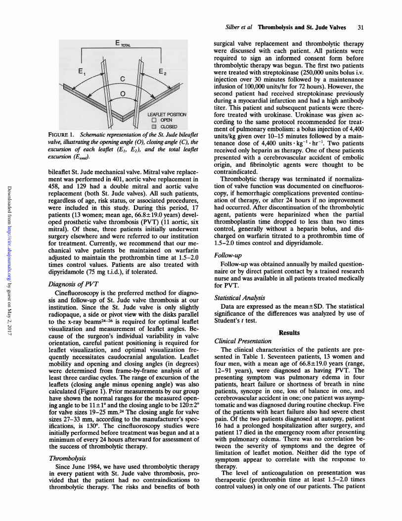
FIGURE 1. Schematic representation of the St. Jude bileafletvalve, illustrating the opening angle (0), closing angle (C), theexcursion of each leaflet (El, E2), and the total leafletexcursion (Etotad.
bileaflet St. Jude mechanical valve. Mitral valve replace-ment was performed in 401, aortic valve replacement in458, and 129 had a double mitral and aortic valvereplacement (both St. Jude valves). All such patients,regardless of age, risk status, or associated procedures,were included in this study. During this period, 17patients (13 women; mean age, 66.8+19.0 years) devel-oped prosthetic valve thrombosis (PVT) (11 aortic, sixmitral). Of these, three patients initially underwentsurgery elsewhere and were referred to our institutionfor treatment. Currently, we recommend that our me-
chanical valve patients be maintained on warfarinadjusted to maintain the prothrombin time at 1.5-2.0times control values. Patients are also treated withdipyridamole (75 mg t.i.d.), if tolerated.
Diagnosis ofPVTCinefluoroscopy is the preferred method for diagno-
sis and follow-up of St. Jude valve thrombosis at ourinstitution. Since the St. Jude valve is only slightlyradiopaque, a side or pivot view with the disks parallelto the x-ray beams24-26 is required for optimal leafletvisualization and measurement of leaflet angles. Be-cause of the surgeon's individual variability in valveorientation, careful patient positioning is required forleaflet visualization, and optimal visualization fre-quently necessitates caudocranial angulation. Leafletmobility and opening and closing angles (in degrees)were determined from frame-by-frame analysis of atleast three cardiac cycles. The range of excursion of theleaflets (closing angle minus opening angle) was alsocalculated (Figure 1). Prior measurements by our grouphave shown the normal ranges for the measured open-ing angle to be 11± 10 and the closing angle to be 120+2°for valve sizes 19-25 mm.24 The closing angle for valvesizes 27-33 mm, according to the manufacturer's spec-ifications, is 130°. The cinefluoroscopy studies were
initially performed before treatment was begun and at aminimum of every 24 hours afterward for assessment ofthe success of thrombolytic therapy.
ThrombolysisSince June 1984, we have used thrombolytic therapy
in every patient with St. Jude valve thrombosis, pro-vided that the patient had no contraindications tothrombolytic therapy. The risks and benefits of both
surgical valve replacement and thrombolytic therapywere discussed with each patient. All patients wererequired to sign an informed consent form beforethrombolytic therapy was begun. The first two patientswere treated with streptokinase (250,000 units bolus i.v.injection over 30 minutes followed by a maintenanceinfusion of 100,000 units/hr for 72 hours). However, thesecond patient had received streptokinase previouslyduring a myocardial infarction and had a high antibodytiter. This patient and subsequent patients were there-fore treated with urokinase. Urokinase was given ac-cording to the same protocol recommended for treat-ment of pulmonary embolism: a bolus injection of 4,400units/kg given over 10-15 minutes followed by a main-tenance dose of 4,400 units . kg.* hr-'. Two patientsreceived only heparin as therapy. One of these patientspresented with a cerebrovascular accident of embolicorigin, and fibrinolytic agents were thought to becontraindicated.
Thrombolytic therapy was terminated if normaliza-tion of valve function was documented on cinefluoros-copy, if hemorrhagic complications prevented continu-ation of therapy, or after 24 hours if no improvementhad occurred. After discontinuation of the thrombolyticagent, patients were heparinized when the partialthromboplastin time dropped to less than two timescontrol, generally without a heparin bolus, and dis-charged on warfarin titrated to a prothrombin time of1.5-2.0 times control and dipyridamole.
Follow-upFollow-up was obtained annually by mailed question-
naire or by direct patient contact by a trained researchnurse and was available in all patients treated medicallyfor PVT.
Statistical AnalysisData are expressed as the mean±SD. The statistical
significance of the differences was analyzed by use ofStudent's t test.
ResultsClinical PresentationThe clinical characteristics of the patients are pre-
sented in Table 1. Seventeen patients, 13 women andfour men, with a mean age of 66.8±19.0 years (range,12-91 years), were diagnosed as having PVT. Thepresenting symptom was pulmonary edema in fourpatients, heart failure or shortness of breath in ninepatients, syncope in one, loss of balance in one, andcerebrovascular accident in one; one patient was asymp-tomatic and was diagnosed during routine checkup. Fiveof the patients with heart failure also had severe chestpain. Of the two patients diagnosed at autopsy, patient16 had a prolonged hospitalization after surgery, andpatient 17 died in the emergency room after presentingwith pulmonary edema. There was no correlation be-tween the severity of symptoms and the degree oflimitation of leaflet motion. Neither did the type ofsymptom appear to correlate with the response totherapy.The level of anticoagulation on presentation was
therapeutic (prothrombin time at least 1.5-2.0 timescontrol values) in only one of our patients. The patient
by guest on May 2, 2017
http://circ.ahajournals.org/D
ownloaded from
32 Circulation Vol 87, No 1 January 1993
TABLE 1. Characteristics of Patients at Presentation
Thrombosed Time from Duration of Adequatevalve/size surgery to symptoms Clinical anticoagulation at Method of
Patient Age Sex (mm) event (months) (days) presentation presentation diagnosis
1 79 F Ao/21 54 14 Syncope No Cine2 83 M Mi/27* 4 1 Fatigue Not Cine3 74 F Ao/21 26 1 CVA No Cine4 66 F Mi/31* 30 12 CHF/CP Not Cine5 77 F Ao/23 38 21 CHF/CP No Cine6 58 F Ao/19 72 5 CHF/CP No Cine7 49 F Mi/27* 99 45 CHF/CP No Cine8 76 F Ao/19 55 7 CHF Yes Cine9 91 F Ao/19 108 7 CHF No Cine10 87 F Ao/19 37 1 CHF Not Cine11 75 F Ao/21 106 24 CHF Not Cine12 76 M Ao/23 106 7 CHF/CP Not Cine13 36 F Ao/23 43 25 CHF No Cine14 12 M Ao/21 60 1 Asymptomatic Not Cine15 70 F Mi/27 48 4 CHF Not Cine16 62 M Mi/31 5 ... CHF No Autopsy17 64 F Mi/31 26 ... CHF Not Autopsy
Mean 66.8 53.9 11.7SD 19.0 33.0 12.0
Ao, aortic; Mi, mitral; CVA, cerebrovascular accident; CHF, congestive heart failure; CP, chest pain; Cine, cinefluoroscopy.*Double valve replacement.tCoumadin withheld for surgical intervention.tTreated only with aspirin+dipyridamole.
with a therapeutic prothrombin time was also the onlyactive smoker in the series. In all other patients, one ormore prothrombin times were documented to be lessthan the target level within the preceding weeks. In 35%of the patients (six of 17), Coumadin was stopped <3weeks earlier as preparation for elective surgery (pace-maker insertion, repair of femoral aneurysm, cholecys-tectomy, facial trauma, epidural injection, prostatec-tomy). Two patients were not being treated withCoumadin before the thrombolytic event because ofprevious life-threatening bleeding.The mean time elapsed from valve replacement to
the thrombotic event was 53.9+33.0 months (range,4-108 months). The duration of symptoms rangedfrom 1 to 45 days, similar to the diverse mode ofpresentation of prosthetic valve thrombosis noted inother studies.9'15Outcome of Thrombolytic TreatmentOf 12 initial events of valve thrombosis treated by
thrombolytic agents in our hospital, both leaflets of theprosthetic valve were limited in motion in 10 patients(83%), and one leaflet was entirely immobilized ineight patients (66%). Treatment was defined as suc-cessful if the valve leaflets resumed more than 90% ofthe normal range of motion (Figure 2). In nine pa-tients (75%), treatment was successful, and in one(8%) there was partial success. This patient's symp-toms improved significantly, but cinefluoroscopy dem-onstrated residual limitation in leaflet motion. Twopatients (17%) failed to demonstrate any change inleaflet motion; however, their clinical state was stabi-
lized with conservative therapy. The opening anglesbefore and after therapy were 60.8±+19.30 and28.8+19.1° (p<0.001), and the closing angles were116.0+9.00 and 123.1±3.50 (p<0.01) before and aftertherapy, respectively (Table 2). The range of leafletmotion increased from 55.2+18.4° to 95.9±16.70 de-grees (p<0.001) after treatment (Figure 3). All pa-tients were released from the hospital and were fol-lowed by their cardiologists. None of these patientsrequired subsequent surgery. At 3-month follow-up,all were alive and well.
Patients With Recurrent EventsRecurrent events are outlined in Table 3. During
17.2±17.6 months of follow-up (range, 1-65 months),two patients had cinefluoroscopic evidence of recur-rent thrombosis. Patient 5 was initially successfullytreated with urokinase, but 8 months later a secondepisode of thrombosis was diagnosed and again suc-cessfully treated with urokinase. The patient returnedtwice more with recurrent thrombus and was success-fully treated 3 months and 7 months later with uroki-nase. This patient had metastatic breast cancer andwas approaching senility, and her physicians had greatdifficulty keeping her Coumadin in a therapeuticrange. At each admission, her prothrombin time wasessentially normal (nontherapeutic). Twelve monthsafter her last event, the patient died of heart failureafter a prolonged hospitalization for significant gastro-intestinal bleeding, during which her Coumadin ther-apy had been discontinued for at least three weeksbefore her death. Patient 10 had recurrent PVT 6
by guest on May 2, 2017
http://circ.ahajournals.org/D
ownloaded from

Silber et al Thrombolysis and St. Jude Valves 33
B
PRE POSTFIGURE 2. Cinefluoroscopy showing leaflet motion before (PRE) (panel A) and after (POST) (panel B) thrombolytic therapy(patient 9). The top panels are views of the open valve, and the bottom panels are views of the closed valves. In the pretreatmentviews (panelA), one leaflet is seen to be immobilized (arrow). The measured opening angle is 59°(normal, 11±1°), and the closing
angle is 110" (normal, 120±2°). After treatment with urokinase (panel B), leaflet motion is seen to have returned almost entirelyto normal. The opening angle is now 14° and the closing angle 124".
months after her first episode, which was again suc-cessfully treated with urokinase. It is interesting thatthis patient had developed thrombosis of a Bjork-Shiley valve before implantation of her St. Jude valve.
The patient is currently stable, with no evidence ofPVT. Patient 3 failed thrombolytic therapy and wasmanaged conservatively because of his high risk forreoperation. He died of progressive congestive heart
TABLE 2. Effect of Thrombolysis on Valve Function
Thrombolytic Opening angle Closing angle Leaflet motion Outcome ofPatient agent Pre Post Pre Post Pre Post therapy
1 Streptokinase 35 22 122 122 87 100 Success2 Streptokinase 79 79 130 130 51 51 Failure3 Heparin 67 46 109 124 42 98 Success4 Urokinase 106 26 131 131 25 105 Success5 Urokinase 84 42 107 122 23 80 Partial
success6 Urokinase 46 14 102 122 56 108 Success7 Urokinase 53 12 120 120 67 108 Success8 Heparin 54 22 120 120 66 98 Success9 Urokinase 59 14 110 124 51 110 Success10 Urokinase 54 14 107 120 53 106 Success11 Urokinase 41 41 121 121 80 80 Failure12 Urokinase 52 14 113 121 61 107 Success
Mean 60.8 28.8 116.0 123.1 55.2 95.9SD 19.3 19.1 9.0 3.6 18.4 16.7
p<O.00l p<0.02 p<O.0001
Pre, before treatment; post, after treatment.
A
by guest on May 2, 2017
http://circ.ahajournals.org/D
ownloaded from

34 Circulation Vol 87, No 1 January 1993
14n.
1201
100CO)W 80
CDw 60~0
40
20
0
PRE POSTFIGURE 3. Graph showing valve leaflet excursion before(PRE) and after (POST) thrombolytic therapy. The average
change in leaflet excursion was 55.2±16. 70 (p<0.001) aftertreatment.
failure 24 months later at the age of 84. This patientalso did not take his Coumadin regularly, and it isquite possible that his death was related to progressivePVT.
Adverse EventsNo patient died as a result of thrombolytic therapy or
suffered permanent major sequelae. However, four pa-
tients developed hemorrhagic complications requiringtreatment. One patient (patient 4) developed a groinhematoma at the site of a recent catheterization thatrequired surgical repair. This same patient also had theonly thromboembolic event, consisting of transient leftfacial weakness lasting 2-3 hours, with no residualabnormality. A 91-year-old patient developed severeecchymoses of both arms requiring a 2-unit blood
TABLE 3. Follow-up of Patients After Thrombolytic Therapy
Duration of Interval tofollow-up 3-Month Late rethrombosis
Patient (months) status rethrombosis (months)1 65 A/W No ...
2 24 A/W Possible* 243 23 A/W No ...
4 33 A/W No ...
5 31 A/W Yest 86 25 A/W No ...
7 7 A/W No ...
8 5 A/W No ...
9 6 A/W No ...
10 22 A/W Yest 611 3 A/W No ...
12 1 ... No ...
Mean 20.4SD 17.3
A/W, Alive and well.*Patient died of heart failure, autopsy not performed.tRethrombosis treated by thrombolysis.
transfusion. Thrombolytic therapy had to be modified intwo patients, one for hemarthrosis of the shoulder andthe second for epistaxis requiring nasal packing. Theoccurrence of complications was not clearly related to aspecific thrombolytic agent.
Surgical TreatmentThree patients (patients 13, 14, and 15) underwent
valve replacement for PVT (two aortic valve replace-ment, one mitral valve replacement). Patient 13 hadsurgery before our routine use of thrombolytic therapyfor PVT. Patient 15 was suspected to have thrombusextending upward from a previous ventricular septaldefect patch repair, a diagnosis that was confirmedsurgically. Patient 14 presented 3 weeks after abdominalsurgery (cholecystectomy), and thrombolytic therapywas thought to be contraindicated. All these patientshad uneventful recoveries. One patient in this groupdied at home 23 months after surgery of a stroke aftershe had stopped taking her Coumadin regularly.
Incidence ofPVTTo calculate the incidence of PVT, the total number
of those at risk for this complication must be known;therefore, we used only patients whose original surgerywas at our institution (n=988) to calculate the throm-bosis rate. Of the 17 patients in this series, 14 patientswere operated on here at Cedars-Sinai (nine aorticvalve replacement, three mitral valve replacement, andtwo double mitral and aortic valve replacement). Themean interval from the time of surgery to the occur-rence of thrombosis was 53.9 months (range, 1-108months). This results in a linearized rate of PVT of0.2% per patient-year, consistent with the literature.
DiscussionResults of Thrombolytic Therapy for PVT
During the past years, several reports of successfulthrombolytic therapy of PVT in selected patients haveappeared. This is the first study in which thrombolytictherapy has been studied prospectively as the first lineof therapy for PVT in a single valve type in all patientswho did not have a contraindication to thrombolytictherapy.Ten of 12 initial events (83%) responded to throm-
bolytic therapy. The age of patients, anatomic locationof the prosthetic valve, and duration of symptoms didnot predict the success of therapy. Within the first 6months of follow-up, there were no episodes of re-thrombosis. All patients, including the two in whichtreatment failed, were managed conservatively duringthe follow-up period. Our success rate is similar to theexpected incidence of thrombus as the main cause ofvalve obstruction (70%) as described by Deviri et al.15This suggests that failure or only partial success ofthrombolytic therapy may be a result of pannus forma-tion. Deviri reported pannus formation to be the solecause of obstruction in 11% of valves and to be presentin combination with thrombus in another 12% of valves.Most patients could be stabilized hemodynamically,
probably because the St. Jude valve is a bileaflet tiltingdisk and thrombus formation can totally occlude motionof one disk while continuing to allow flow through theother. The mechanism for restricted opening of both
* AORTICA MITRAL
by guest on May 2, 2017
http://circ.ahajournals.org/D
ownloaded from

Silber et al Thrombolysis and St. Jude Valves 35
leaflets is not clear and may involve larger or multiplethrombi or could result from thrombosis of one leafletonly, with restriction of the second leaflet resultingsolely from hemodynamic factors (i.e., alteration of thenormal flow patterns resulting in incomplete leafletopening). The low incidence of embolization in thisseries may result from preferential formation of throm-bus at the disk pivot points in St. Jude valves. Formationof pivot point thrombus means that relatively smallamounts of thrombus can result in restriction of leafletopening.15 These relatively small amounts of thrombusmay dissolve completely or substantially during throm-bolytic therapy with a lower risk of embolization. Thismay result in a larger safety margin for thromboseddual-disk valves compared with single-tilting-diskvalves, in which larger amounts of thrombus may berequired to obstruct the valve. It should be noted thatsome patients may demonstrate only partial or minimalimprovements in opening angles but have a good clinicalresponse (resolution of heart failure, etc.). It is possiblethat those patients may have had thrombus obstructingthe valve orifice that was lysed by thrombolytic therapy.Thus, it is conceivable that hemodynamic and clinicalimprovement could occur despite persistent restrictionof leaflet motion.
EmbolizationOnly one patient had a transient ischemic attack
related to embolization, which resolved completelywithin 2 hours. This experience is similar to that ofKurzrok et al,21 who found that all manifestation ofembolization disappeared within 2 days. Thus, it seemsthat after thrombolytic therapy, the majority of patientswith a thrombosed prosthesis in the left side of the heartwill not develop permanent neurological sequelaecaused by systemic embolization. These data are also inaccordance with observations of patients receivingurokinase for left ventricular thrombus after myocardialinfarction.27
Choice of Thrombolytic AgentsTo decrease the risk of embolization, we believe a less
intense but more prolonged thrombolytic state is desir-able than is used for treatment of acute myocardialinfarction. This is the primary reason that we haveselected the urokinase protocol discussed in the "Meth-ods" section. Urokinase has been used successfully inthis manner for dissolution of peripheral arterial as wellas pulmonary artery thrombi and also has the advantageof a lack of hemodynamic and allergenic effects. In twoof our patients, heparin alone resulted in significantimprovement, and its value as sole agent of treatmentshould be evaluated in further studies.
Role of Cinefluoroscopy andDoppler Echocardiography
In our experience, cinefluoroscopy is a simple andaccurate method both for diagnosis of St. Jude PVT andto follow the response to thrombolytic treatment. Dopp-ler echocardiography may also have an important role,provided that accurate measurements of gradients andvalve area can be made.28 Doppler gradients are notsufficient for diagnosis unless they are very high, andserial changes in gradient may not be useful, becausethey may be caused by changes in cardiac output and
hemodynamic state.29 In addition, Doppler gradientsmay be significantly higher than catheter gradients in St.Jude valves because of the phenomena of pressurerecovery and high localized gradients in the St. Judevalve.30 However, Doppler echocardiography can pro-vide important hemodynamic information if valve areasare assessed.A further factor complicating the use of Doppler
measured gradients and valve areas is the possibilitythat the relation between Doppler and catheter gradi-ents may change as valve function returns to normal.Preliminary in vitro results from our lab suggest that thephenomena of pressure recovery and high localizedgradients do not occur in thrombosed St. Jude valves.31As valves become progressively more stenotic, flowthrough the valve becomes more turbulent, and lesspressure recovery occurs. Thus, the discrepancy be-tween Doppler and catheter gradients may progres-sively disappear as a valve becomes stenotic. This raisesthe possibility that when one measures a high Dopplergradient in a St. Jude valve, it may be a result of eithera true high gradient in a stenotic valve or the recognizedDoppler overestimation of gradient because of pressurerecovery in a normally functioning valve prosthesis.
In assessing the relative roles of cinefluoroscopy andDoppler, it must be remembered that each methodprovides different kinds of information. Cinefluoros-copy provides information solely about restriction ofleaflet motion. In contrast, Doppler echocardiographyprovides hemodynamic information about pressure gra-dients and valve areas. Therefore, Doppler echocardi-ography and cinefluoroscopy may have complementaryroles in assessing prosthetic valve function. Furtherstudies are needed to establish the preferred method fordiagnosis and follow-up in patients with thrombosedprosthetic valves.
Incidence and Characteristics ofPVTThe linearized incidence of St. Jude PVT for patients
operated on in our institution is low (0.2% per patient-year).3 It is important to note that in all but one of thecases, PVT occurred in association with subtherapeuticanticoagulation (prothrombin time <1.2 times control)or nonuse of warfarin (two patients). In six of 17 (35%)of these patients, Coumadin treatment was stopped ormodified because of elective surgical intervention. Elec-tive surgery therefore emerges as a major risk factor forPVT in our study population. Although 13 of 17 pa-tients with PVT were female, we cannot conclude thatwomen are at a higher risk for developing PVT becauseof the small number of patients studied.These statistics also highlight a major rationale for
using thrombolytic therapy instead of surgical valverereplacement. In all but one case, there was a reasonfor PVT related to the patient (i.e., a nontherapeuticprothrombin time). Thus, in each of these cases, there isno reason to suspect that there is a problem intrinsic tothe prosthetic valve. If clot lysis can be achieved and thepatient maintained on therapeutic Coumadin levels,there should be little likelihood of recurrent PVT.Therefore, we believe that in cases in which there is aclearly defined episode of subtherapeutic anticoagula-tion, thrombolytic therapy should be considered asinitial therapy.
by guest on May 2, 2017
http://circ.ahajournals.org/D
ownloaded from

36 Circulation Vol 87, No 1 January 1993
Unexplained chest pain, shortness of breath,or abnormal neurological symptoms
CinefluoroscopyPedent stable Patient unstable~- Abnormal leaflet motion
Contraindications Yes Contraindications Yes
to Thrombolytics to Heparin
No No4
Urokinase Heparin
Repeat Cinefluoroscopy q24 hrs
Normalization of No
valve function -within 72 hrs
Yes
Coumadin Valve replacement
FIGURE 4. Flow chart showing treatment strategy forthrombosed St. Jude valves (see text for details).
Limitations of StudyIn this study, no patients with a thrombosed tricuspid
valve were identified, and we therefore cannot drawconclusions about management of these patients. Be-cause of the small number of events, we also could notassess the roles of atrial fibrillation or smoking as riskfactors for PVT, nor could we compare the efficacy ofdifferent thrombolytic agents. Our estimate of the inci-dence of thrombosis is probably an underestimate,because it is possible that some patients may have beentreated elsewhere or may have died suddenly beforereaching medical care. Finally, it should be stressed thatour low thromboembolic rate may be valid only for theSt. Jude valve. It is possible that caged-ball and single-disk valves may have a different incidence of throm-boembolism or rate of successful thrombolysis. Wewould be cautious in extrapolating these results to othertypes of prosthetic valves.
Clinical ImplicationsOur results suggest that thrombolytic therapy should
be the first line of treatment for thrombosed St. Judevalves. Medical treatment of thrombosed St. Jude valvesis safe, has a low complication rate, and, in this series,resulted in no deaths related to treatment comparedwith the reported 4.7-20% perioperative mortality.Figure 4 outlines our present protocol for managingpatients with suspected thrombosis of a St. Jude valve.Initial diagnosis is by cinefluoroscopy but might also bedone by echocardiography. The key to diagnosis isrestriction of leaflet motion, however, and not thedemonstration of high gradients, because Dopplerechocardiography may overestimate gradients in St.Jude valves30 as well as underestimate valve areas.32Once significant leaflet obstruction is confirmed, pa-tients without contraindications should receive throm-bolytic therapy. Patients should provide informed con-
sent and be aware of the risks of embolism andhemorrhage as well as the alternative treatments andtheir risks (i.e., surgery). After thrombolytic therapy isbegun, cinefluoroscopy (or possibly Doppler echocardi-ography) should be performed daily to evaluate theimprovement in valve function. If "normalization" ofvalve function occurs (defined as either normal opening
and closing angles or significant improvement in theopening angles as defined in the "Methods" section),patients should be switched to heparin and then Cou-madin and dipyridamole for the long term. Patients whodo not respond within 48-72 hours should be referredfor surgery, because these patients may have tissueingrowth obstructing the valve (pannus formation) andprobably will not respond to continued thrombolytictreatment. Gratifyingly, none of the patients receivingthrombolytic therapy in our series required surgery(although it was offered to one), and their clinical statusgreatly improved. If surgery is needed for PVT, it is alsopossible that thrombolytic therapy may allow surgery ina more stable clinical setting, although the improvementin hemodynamics must be balanced against the increasedrisk of hemorrhage perioperatively. Patients who returnwith repeated episodes of thrombosis can usually betreated successfully with thrombolytic therapy. However,these patients may be at a continuing risk for furtherthrombotic episodes, probably related to patient factors(senility, noncompliance, etc.), and consideration shouldalso be given to prosthetic valve replacement with a tissuevalve. Patients with residual limitation of leaflet motionafter thrombolytic therapy remain a clinical challenge. Inour experience, they may be managed conservatively, butthis decision should be individualized.
ConclusionsThrombosis of a St. Jude prosthetic valve is a rare but
potentially lethal clinical situation. The clinical presen-tation varies from minimal or absent symptoms tocirculatory collapse, and prompt diagnosis is essential.Many patients with PVT give a recent history of stop-ping anticoagulation to undergo surgery. Thrombolysismay be used as the first line of therapy in patients withthrombosed St. Jude valves and appears to be aneffective and safe treatment. We believe that surgeryshould be reserved for patients who cannot be stabilizedmedically and for patients who have contraindicationsto thrombolytic therapy.
AcknowledgmentsWe wish to express special thanks to Dr. Lawrence Czer for
many helpful discussions and to Dr. Robert Kass, Dr. CarlosBlanche, Dr. Alfredo Trento, Dr. Tsung-Po Tsai, and Dr.Sharo Raissi for their help in caring for these patients, as wellas Kathleen Farrington for her expert secretarial assistance.
References1. Arom KV, Nicoloff DM, Kersten TE, Northrup WF III, LindsayWG, Emery RW: Ten years experience with the St. Jude Medicalvalve prosthesis. Ann Thorac Surg 1989;47:831-837
2. Czer LS, Matloff JM, Chaux A, DeRobertis M, Stewart ME, GrayRJ: The St. Jude valve: Analysis of thromboembolism, warfarinrelated hemorrhage and survival. Am Heart J 1987;114:389-397
3. Czer LSC, Chaux A, Matloff JM, DeRobertis MA, Nessim SA,Scarlata D, Khan SS, Kass RM, Tsai TP, Blanche C, Gray RJ:Ten-year experience with the St. Jude Medical valve for primaryvalve replacement. J Thorac Cardiovasc Surg 1990;100:44-55
4. Ryder SJ, Bradley H, Brannan JJ, Turner MA, Bain WH: Throm-botic obstruction of the Bjork-Shiley valve: The Glasgow experi-ence. Thorax 1984;39:487-492
5. Balram A, Kaul U, Rama Rao BV, Iyer KS, Rajani M, Rao IM,Bhatia ML, Gopinath N, Venugopal P: Thrombotic obstruction ofBjork-Shiley valves: Diagnostic and surgical considerations. Int JCardiol 1984;6:61-73
6. Edmunds LH Jr: Thrombotic and bleeding complications of pros-thetic heart valves. Ann Thorac Surg 1987;44:430-445
by guest on May 2, 2017
http://circ.ahajournals.org/D
ownloaded from

Silber et al Thrombolysis and St. Jude Valves 37
7. Metzdorff MT, Grunkemeier GL, Pinson CW, Starr A: Thrombo-sis of mechanical cardiac valves: A qualitative comparison of theSilastic ball valve and the tilting disc valve. J Am Coll Cardiol1984;4:50-53
8. Macmanus Q, Metzdorff MT, Grunkemeier GL, Starr A: Throm-botic and embolic complications with Silastic ball prosthetic valves.Eur Heart J 1984;5(suppl D):59-63
9. Kontos GJ, Schaff HV, Orszulak TA, Puga FJ, Pluth JR, DanielsonGK: Thrombotic obstruction of disc valves: Clinical recognitionand surgical management. Ann Thorac Surg 1989;48:60-65
10. Karp RB, Cyrus RJ, Blackstone EH, Kirklin JM, Kouchoukos NT,Pacifico AD: The Bjork-Shiley valve: Intermediate-term follow-up.J Thorac Cardiovasc Surg 1981;81:602-614
11. Spencer FC, Trinkler JK, Reeves JT: Successful replacement of athrombosed mitral ball prosthesis. JAMA 1975;194:1249-1251
12. Martinell J, Fraile J, Artiz V, Cortina J, Fresneda P, Rabago D:Reoperations for left-sided low-profile mechanical prostheticobstructions. Ann Thorac Surg 1987;43:172-175
13. Copans H, Lakier JB, Kinsley RH, Colsen PR, Frilz MD, BarlowJB: Thrombosed Bjork-Shiley mitral prostheses. Circulation 1980;61:169-174
14. Husebye DG, Pluth JR, Piehler JM, Schaff HV, Orszulak TA, PugaFJ, Danielson GK: Reoperation on prosthetic heart valves: Anal-ysis of risk factors in 552 patients. J Thorac Cardiovasc Surg 1983;86:543-552
15. Deviri E, Sareli P, Wisenbaugh T, Cronje SL: Obstruction ofmechanical heart valve prostheses: Clinical aspects and surgicalmanagement. JAm Coll Cardiol 1991;17:646-650
16. Montero CG, Mula N, Burgos R, Pradas G, Figuera D: Throm-bectomy of the Bjork-Shiley prosthetic valve revisited: Long termresults. Ann Thorac Surg 1989;48:824-828
17. Martinell J, Jimenez A, Rabago G, Artiz V, Fraile J, Farre J:Mechanical cardiac valve thrombosis: Is thrombectomy justified?Circulation 1991;84(suppl III):III-70-III-75
18. Luluaga IT, Carrera D, D'Oliveira J, Cantaluppi CG, Santin H,Molteni L, Ferreira R, Zwolinski E, Luluaga II: Successful throm-bolytic therapy after acute tricuspid-valve obstruction. Lancet 1971;1:1067-1068
19. Baille Y, Choffel J, Sicard MP, Malmejac C, Metras D, Delaye A,Manuel C, Houel J: Traitement thrombolytique des thromboses deprothese valvulaire. (letter) Nouv Presse Med 1974;11:1233
20. Graver LM, Gelber PM, Tyras DH: The risks and benefits ofthrombolytic therapy in acute aortic and mitral prosthetic valvedysfunction: Report of a case and review of the literature. AnnThorac Surg 1988;46:85-88
21. Kurzrok S, Singh AK, Most AS, Williams DO: Thrombolytic ther-apy for prosthetic cardiac valve thrombosis. J Am Coll Cardiol1987;9:592-598
22. Ledain LD, Ohayon JP, Colle JP, Lorient-Roudaut FM, RoudautRP, Besse PM: Acute thrombotic obstruction with disc valve pros-theses: Diagnostic considerations and fibrinolytic treatment. JAmColl Cardiol 1986;7:743-751
23. Witchitz S, Veyrat C, Moisson P, Scheinman N, Rozenstayn L:Fibrinolytic treatment of thrombus on prosthetic heart valves. BrHeart J 1980;44:545-554
24. Czer LSC, Weiss M, Bateman TM, Pfaff JM, DeRobertis M, EiglerN, Vas R, Matloff JM, Gray RJ: Fibrinolytic therapy of St. Judevalve thrombosis under guidance of digital cinefluoroscopy. JAmColl Cardiol 1985;5:1244-1249
25. Castaneda-Zuniga W, Nocoloff D, Jorgensen C, Nath P, ZollikoferC, Amplatz K: In vivo radiographic appearance of the St. Judevalve prosthesis. Radiology 1980;134:775-776
26. Mehlman D: A guide to the radiographic identification of pros-thetic heart valves: An addendum. Circulation 1984;69:102-105
27. Kremer P, Ranier F, Tilsner V, Bleifeld W, Mahey DG: Lysis ofventricular thrombi with urokinase. Circulation 1985;72:112-118
28. Chafizadeh ER, Zoghbi WA: Doppler echocardiographic assess-ment of the St. Jude Medical prosthetic valve in the aortic positionusing the continuity equation. Circulation 1991;83:213-223
29. Tatineni S, Barner HB, Pearson AC, Halbe D, Woodruff R, Labo-vitz AJ: Rest and exercise evaluation of St. Jude Medical andMedtronic Hall prostheses: Influence of primary lesion, valvulartype, valvular size, and left ventricular function. Circulation 1989;80(suppl I):I-16-I-23
30. Baumgartner H, Khan S, DeRobertis M, Czer L, Maurer G: Pres-sure recovery: A cause of discrepancy between Doppler and cath-eter gradients in St. Jude valves. Circulation 1990;82:1467-1475
31. Silber H, Khan S, DeRobertis M, Gray R, Matloff J, Maurer G:Effect of prosthetic valve obstruction on the Doppler-cathetergradient correlation for St. Jude valves: In vitro studies. (abstract)Circulation 1992;86(suppl I):I-806
32. Baumgartner H, Khan S, DeRobertis M, Czer L, Maurer G: Dopp-ler assessment of prosthetic valve orifice area: An in vitro study.Circulation 1992;85:2275-2283
by guest on May 2, 2017
http://circ.ahajournals.org/D
ownloaded from

H Silber, S S Khan, J M Matloff, A Chaux, M DeRobertis and R GrayThe St. Jude valve. Thrombolysis as the first line of therapy for cardiac valve thrombosis.
Print ISSN: 0009-7322. Online ISSN: 1524-4539 Copyright © 1993 American Heart Association, Inc. All rights reserved.
is published by the American Heart Association, 7272 Greenville Avenue, Dallas, TX 75231Circulation doi: 10.1161/01.CIR.87.1.30
1993;87:30-37Circulation.
http://circ.ahajournals.org/content/87/1/30World Wide Web at:
The online version of this article, along with updated information and services, is located on the
http://circ.ahajournals.org//subscriptions/
is online at: Circulation Information about subscribing to Subscriptions:
http://www.lww.com/reprints Information about reprints can be found online at: Reprints:
document. Permissions and Rights Question and Answer available in the
Permissions in the middle column of the Web page under Services. Further information about this process isOnce the online version of the published article for which permission is being requested is located, click Request
can be obtained via RightsLink, a service of the Copyright Clearance Center, not the Editorial Office.Circulation Requests for permissions to reproduce figures, tables, or portions of articles originally published inPermissions:
by guest on May 2, 2017
http://circ.ahajournals.org/D
ownloaded from