Thesis: Duty ratio of cooperative molecular motors.
60
BEN-GURION UNIVERSITY OF THE NEGEV FACULTY OF ENGINEERING SCIENCES DEPARTMENT OF BIOMEDICAL ENGINEERING Duty ratio of cooperative molecular motors Thesis submitted in partial fulfillment of the requirements for the M.Sc. degree By: Nadiv Dharan September, 2012
Transcript of Thesis: Duty ratio of cooperative molecular motors.
BEN-GURION UNIVERSITY OF THE NEGEV
FACULTY OF ENGINEERING SCIENCES
DEPARTMENT OF BIOMEDICAL ENGINEERING
Duty ratio of cooperative molecular motors
Thesis submitted in partial fulfillment of the requirements for the M.Sc. degree
By: Nadiv Dharan
September, 2012
September, 2012
BEN-GURION UNIVERSITY OF THE NEGEV
FACULTY OF ENGINEERING SCIENCES
DEPARTMENT OF BIOMEDICAL ENGINEERING
Duty ratio of cooperative molecular motors
Thesis submitted in partial fulfillment of the requirements for the M.Sc. degree
By: Nadiv Dharan
Supervised by: Prof. Oded Farago
Author:................................................ Date:...................
Supervisor:........................................... Date:...................
Chairman of Graduate Studies Committee:............................Date:...................
September, 2012
2

Abstract
Motor proteins are specialized molecules that convert chemical energy into useful mechanical
work and govern many important biological processes. They bind to actin filaments and
microtubules and use them as tracks, on which they propagate in order to transport different
types of cargo across the cell. Among these molecules we find myosin II motors that interact
with elastic actin filaments. Myosin II are non processive motors that exhibit a low duty
ratio, which means that in order to work effectively they need to cooperate with each other.
The collective work of myosin II motors on elastic actin tracks suggests that these filaments
may be subjected to significant tensile stresses. It has been found that the stress applied
on the actin may induce an indirect crosstalk between the motors in order to diminish
the elastic energy, which is expressed by changes in their binding / unbinding statistics.
This type of indirect communication between motors via the elastic track, which has been
termed the elasticity mediated crosstalk (EMC) effect, may significantly affect the motors’
effective processivity. In this thesis, we use a statistical mechanical analysis and Monte
Carlo computer simulations, to explore the magnitude and role of the EMC effect in two
types of systems where the collective action of myosin II motors on actin filament is present:
(i) gliding motility assays and (ii) muscle contraction. In motility assays, we find that the
EMC effect has a small impact on the collective action of motors, and that the duty ratio of
the motors remains effectively unchanged. In muscle contraction, where the actin filament
is subjected to an external force opposing the action of the motors, we find that the EMC
effect leads to a non uniform attachment probability along the actin track. Such duty ratio
variations between motors may have a serious negative influence on the ability of the motors
to perform effectively. Nevertheless, we find that this feature becomes significant only when
the size of the system is larger than the size of the sarcomere (which is the basic contractile
unit of the muscle cell). The EMC effect may, thus, serve as an explanation for the specific
dimensions of the sarcomere, which are found to be essentially identical across different
vertebrate species.
3

Acknowledgments
I wish to thank my supervisor, Prof. Oded Farago, for all the guidance, support and assis-
tance throughout my thesis project.
A special thanks to my lab colleagues Noam Weil and Yotam Avital for all the assistance
and useful discussions we had over the past two years.
4

Contents
1 Introduction 7
1.1 The cytoskeleton . . . . . . . . . . . . . . . . . . . . . . . . . . . . . . . . . . . . . . . . . . . 7
1.2 Motors proteins . . . . . . . . . . . . . . . . . . . . . . . . . . . . . . . . . . . . . . . . . . . . 9
1.3 Cooperativity between motor proteins . . . . . . . . . . . . . . . . . . . . . . . . . . . . . . . 11
1.3.1 Motility assays . . . . . . . . . . . . . . . . . . . . . . . . . . . . . . . . . . . . . . . . 12
1.3.2 Skeletal muscles . . . . . . . . . . . . . . . . . . . . . . . . . . . . . . . . . . . . . . . 13
1.4 Cooperative bidirectional motion . . . . . . . . . . . . . . . . . . . . . . . . . . . . . . . . . . 15
1.5 The elasticity-mediated crosstalk (EMC) effect . . . . . . . . . . . . . . . . . . . . . . . . . . 18
1.5.1 System model . . . . . . . . . . . . . . . . . . . . . . . . . . . . . . . . . . . . . . . . . 19
1.5.2 Calculating the elastic energy . . . . . . . . . . . . . . . . . . . . . . . . . . . . . . . . 20
1.5.3 The dimensionless parameter β∗ . . . . . . . . . . . . . . . . . . . . . . . . . . . . . . 22
1.5.4 Stress fluctuations . . . . . . . . . . . . . . . . . . . . . . . . . . . . . . . . . . . . . . 23
1.6 Outline . . . . . . . . . . . . . . . . . . . . . . . . . . . . . . . . . . . . . . . . . . . . . . . . 24
2 The EMC effect in gilding motility assays 26
2.1 Statistical-mechanical analysis . . . . . . . . . . . . . . . . . . . . . . . . . . . . . . . . . . . . 27
2.2 Gliding assay computer simulations . . . . . . . . . . . . . . . . . . . . . . . . . . . . . . . . . 31
2.3 Actin-Myosin II systems . . . . . . . . . . . . . . . . . . . . . . . . . . . . . . . . . . . . . . . 33
2.4 Summary . . . . . . . . . . . . . . . . . . . . . . . . . . . . . . . . . . . . . . . . . . . . . . . 34
3 The EMC effect in muscle contraction 36
3.1 Muscle contraction models . . . . . . . . . . . . . . . . . . . . . . . . . . . . . . . . . . . . . . 37
3.2 A simple derivation of Hill’s equation . . . . . . . . . . . . . . . . . . . . . . . . . . . . . . . . 42
3.3 System model . . . . . . . . . . . . . . . . . . . . . . . . . . . . . . . . . . . . . . . . . . . . . 44
3.4 Monte Carlo computer simulations . . . . . . . . . . . . . . . . . . . . . . . . . . . . . . . . . 45
5

3.4.1 A Polarized attachment probability due to the EMC effect . . . . . . . . . . . . . . . . 46
3.4.2 Sarcomere shortening simulations . . . . . . . . . . . . . . . . . . . . . . . . . . . . . . 47
3.5 Summary . . . . . . . . . . . . . . . . . . . . . . . . . . . . . . . . . . . . . . . . . . . . . . . 50
4 Discussion 52
6
Chapter 1
Introduction
1.1 The cytoskeleton
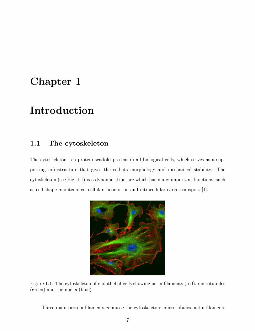
The cytoskeleton is a protein scaffold present in all biological cells, which serves as a sup-
porting infrastructure that gives the cell its morphology and mechanical stability. The
cytoskeleton (see Fig. 1.1) is a dynamic structure which has many important functions, such
as cell shape maintenance, cellular locomotion and intracellular cargo transport [1].
Figure 1.1: The cytoskeleton of endothelial cells showing actin filaments (red), microtubules(green) and the nuclei (blue).
Three main protein filaments compose the cytoskeleton: microtubules, actin filaments
7

and intermediate filaments (see Fig. 1.2). The thickest and most rigid filaments are mi-
crotubules, which are hollow, pipe-like filaments. The cylindrical structure of microtubules
is made of 13 circularly arranged protofilaments, each made of successive units of α- and
β-tubulin dimers [2]. The successive assembly of tubulin dimers into microtubules defines
its polarity, where the “plus” and “minus” ends are at the fast and slow growing ends of the
filament respectively. The rigid microtubules provide the cell important mechanical support
and have a key role in intracellular transport [3, 4], cell division [5] and other important
cellular processes. Actin filaments, whose diameter is ∼ 7 nm, are the thinnest filaments of
the cytoskeleton. Actin filaments are relatively flexible cable-like filaments, with a double
helical structure repeating every ∼ 38 nm, composed of G-actin monomers. Actin filaments
are also polar in nature, and they play a significant role in numerous cellular processes,
including cell crawling [6] and cell division [7]. The third type of cytoskeletal filaments are
intermediate filaments, which are a broad class of fibrous crossed-linked proteins with diam-
eter of ∼ 10 nm. The presence and composition of intermediate filaments varies between
cells of different tissues. Different intermediate filaments include cytoplasmic proteins, such
as vimentin and keratin, and nuclear proteins, such as lamins. Like actin filaments, inter-
mediate filaments are relatively flexible rope-like structures. Intermediate filaments do not
have a primary role in cellular processes, but rather provide the cell its physical strength
and serve as an anchoring scaffold for the nucleus and other organelles [8].
While rigid microtubules function as compression resisting filaments to maintain the
cell’s structure, the flexible intermediate and actin filaments do so by functioning as ten-
sion bearing elements. Other than providing physical support and maintaining the cell’s
shape and structure, the polar microtubules and actin filaments also serve as tracks for the
propagation of motor proteins.
8

(A) (B) (C)
Figure 1.2: The biological cell has three main cytoskeletal filaments: (A) Microtubules arecomposed of tubulin dimers that form a hollow cylindrical structure with diameter ∼ 25 nm.(B) Actin filaments have a double stranded structure composed of G-actin monomers. (C)Intermediate filaments are made of tetramer subunits arranged in an anti-parallel fashion,which makes intermediate filaments non-polar.
1.2 Motors proteins
Motor proteins are specialized molecules that possess the ability to convert chemical energy,
derived from adenosine triphosphate (ATP) hydrolysis, into useful mechanical work [9]. The
biological cell utilizes the action of these nano machines to transport vesicles, organelles
and other kinds of cargo across the cell. This type of active transport is achieved by the
propagation of the motors along microtubule and actin filament tracks within the cell.
Motor proteins can be divided into different classes, characterized by the filament with
which they interact and the preferred direction of motion along the filament. They also vary
in their degree of processivity, which is determined by the duty ratio, i.e. the fraction of
time out of the entire ATP hydrolysis cycle in which a motor remains bound to the filament.
For example, kinesin motors “walk” along microtubules, while myosin motors interact with
actin filaments. Most kinesins and myosins move towards the plus end of the microtubules
and actin filaments, respectively [27]. In contrast, Ncd motors (kinesin-related microtubule
motors) and myosin VI (which propagate along actin filaments) move towards the minus
9

end of their associated filament tracks [11,12]. Finally, while processive motors like kinesins
can cover long distances along microtubules without detaching from the track [13], non-
processive motors with a low duty ratio, like most myosin family motors, take only single
steps along the actin filament before disconnection occurs [14].
One abundant non-processive motor protein is myosin II, which plays a significant role
in a diversity of cellular processes. The low duty ratio of myosin II implies that these motors
have a rather poor performance as individuals (unlike kinesins) and, hence, they need to
work in cooperation with each other in order to produce substantial motion. The structure
of myosin II consists of three major parts [see Fig. 1.3(A)]: (i) a head domain, which binds
to binding sites located on the actin filament. This part is responsible for ATP hydrolysis,
which triggers a conformational change that causes the motor head to rotate; (ii) a neck
domain, which serves as a lever arm that amplifies the motion of the rotating head; (iii) a
tail domain, which can attach cargo and organelles for transport.
(A) (B)Head
Neck
Tail
Figure 1.3: (A) Schematics of a myosin II molecule, which consists of three main regions:a head domain with an ATP hydrolysis site; a neck region that acts as a lever arm for therotating head; a tail domain which binds cargo. (B) Myosin II molecules pack together toform thick filaments from which motor heads project. A single thick filament may consist ofseveral hundreds of myosin II molecules.
In vertebrate striated muscles myosin II molecules pack together into bipolar myosin
filaments with the tails forming a roughly cylindrical filament backbone and the myosin
heads arranged in a quasi-helical array on the filament surface [see Fig. 1.3(B)].
10
1.3 Cooperativity between motor proteins
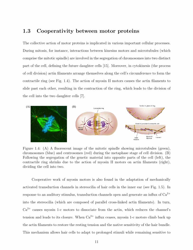
The collective action of motor proteins is implicated in various important cellular processes.
During mitosis, for instance, interactions between kinesins motors and microtubules (which
comprise the mitotic spindle) are involved in the segregation of chromosomes into two distinct
part of the cell, defining the future daughter cells [15]. Moreover, in cytokinesis (the process
of cell division) actin filaments arrange themselves along the cell’s circumference to form the
contractile ring (see Fig. 1.4). The action of myosin II motors causes the actin filaments to
slide past each other, resulting in the contraction of the ring, which leads to the division of
the cell into the two daughter cells [7].
(A) (B)
Figure 1.4: (A) A fluorescent image of the mitotic spindle showing microtubules (green),chromosomes (blue) and centrosomes (red) during the metaphase stage of cell division. (B)Following the segregation of the genetic material into opposite parts of the cell (left), thecontractile ring shrinks due to the action of myosin II motors on actin filaments (right),dividing the cell into two.
Cooperative work of myosin motors is also found in the adaptation of mechanically
activated transduction channels in stereocilia of hair cells in the inner ear (see Fig. 1.5). In
response to an auditory stimulus, transduction channels open and generate an influx of Ca2+
into the stereocilia (which are composed of parallel cross-linked actin filaments). In turn,
Ca2+ causes myosin 1-c motors to dissociate from the actin, which reduces the channel’s
tension and leads to its closure. When Ca2+ influx ceases, myosin 1-c motors climb back up
the actin filaments to restore the resting tension and the native sensitivity of the hair bundle.
This mechanism allows hair cells to adapt to prolonged stimuli while remaining sensitive to
11

new ones [16].
Cooperativity between myosin II motors can also be found in muscle contraction and
in vitro motility assays. These two cases are further discussed below.
Figure 1.5: (A) The cochlea of the inner ear has a spiral structure consisting of three fluidfilled chambers. (B) Cross section of the cochlea showing the hair cells that lie on top ofthe basilar membrane. (C) Stereocilia of hair cells arranged in rows of increasing height aredeflected due to the movement of the basilar membrane, increasing tip link tension. (D)Increased tension within the tip links, connecting stereocilia, opens transduction channels.The action of myosin 1-c on actin filament restores the resting tension for new stimuli.
1.3.1 Motility assays
In vitro motility assays constitute a very useful experimental technique for studying motor
dynamics in reconstituted model systems. The two main geometries used in motility assays
are the bead assay and the gilding assay. In the former, the filament tracks are fixed to a
coverslip, and motors are adsorbed to micro-sized beads, which are suspended in a solution
with the presence of ATP. The beads first diffuse until they encounter a filament track,
allowing the motors to attach to the track and propagate along it. In gliding motility
assays the filaments glide over a surface covered by immobilized yet active motors (see
Fig. 1.6). In both cases, either the beads (in the bead assay) or the filaments (in the
gilding assay) are flouroscently labeled, and the motion is detected and analyzed by means
of video microscopy. Tracking the motion of the bead or the filament provides important
information, such as motor directionality, filament gliding velocities, and motor proteins
12

Figure 1.6: Schematic of a gliding motility assay, in which a flouroscently labeled actinfilament lies on a bed of immobilized, yet active, myosin II motors. The filament’s motiondue to motor forces is recorded and analyzed to extract different data.
kinetic parameters. Motility assays, thus, offer an important and powerful experimental tool
for understanding the nature and dynamics of motor proteins under controlled conditions.
1.3.2 Skeletal muscles
One of the more fascinating examples for the cooperative action of myosin II motors is their
role in skeletal muscle contraction. Skeletal muscles are organized in a hierarchical fashion,
in which the basic contractile unit is the sarcomere. Repeating units of sarcomeres comprise
the myofibril which, in turn, lie parallel to other myofibrils to compose the multi-nucleated
cylindrical muscle cell [also referred as a muscle fiber, or a myocyte (see Fig. 1.7)]. The
sarcomere itself is composed of two types of filaments, actin (thin) and myosin (thick), in an
arrangement that allows them to slide past each other [see Fig. 1.8(A)]. The thick filament
consists of about 300 myosin II molecules, and is surrounded by 12 actin filaments, 6 around
each half [see Fig. 1.8(B)]. The motors heads project out of the thick myosin filament every
14.3 nm, and the angular difference between adjacent motor heads is 40 [17]. The thin
filaments in each half sarcomere lie parallel to each other, and their orientation is opposite
to the thin filaments on the other half sarcomere, so that the plus ends of all thin filaments
are found at the ends of the sarcomere (i.e., the Z-line). While thin actin filaments are
anchored at the Z-line, thick myosin filaments are crosslinked at the M-line, which is located
at the center of the sarcomere [see Fig. 1.8(A)].
13

Figure 1.7: The skeletal muscle consists of bundles of fascicles made of muscle fibers, eachcomposed of myofibrils, which are made of repeating units of sarcomeres.
When the muscle is at rest the actin filament is covered by tropomyosin, making the
binding sites unavailable for the attachment of motors. Upon nervous stimulation, Ca2+
ions flow into the muscle cell and unlock the actin-covering tropomyosin, which results in
the exposure of binding sites to the attachment of myosin II motors. Once attached to a
binding site, ATP hydrolysis causes the motor head to rotate and pull the actin filament
towards the M-line. The resulting sliding of thin filaments past thick filaments leads to the
shortening of the sarcomere and muscle contraction.
Thin filaments Thick filamentM−line
(B)
(A)
Z−lineZ−line
Figure 1.8: (A) The sarcomere is made of thin actin and thick myosin II filaments that slidepast each other, causing the sarcomere to shorten. Actin filaments are anchored at the Z-lineat both ends of the sarcomere, while myosin filaments are connected at the M-line, which islocated in the center of the sarcomere. (B) Various cross-section views along the sarcomere,showing the spatial arrangement of thin and thick filaments.
14

1.4 Cooperative bidirectional motion
One of the more interesting outcomes of the cooperative action of molecular motors is their
ability to induce bidirectional (“back and forth”) motion. This dynamical pattern emerges
when filaments (or other objects such as liposomes and organelles) are subjected to the action
of two groups of motors that engage in a “tug-of-war” (TOW) contest, exerting forces in
opposite directions. The “victory” of the motor party that exerts the larger force determines
the instantaneous direction of motion. If the two opposite forces are nearly similar, the
balance of forces may shift between the two motor parties as a result of stochastic binding
and unbinding events of motors to the filament track. Bidirectional motion is not restricted
to cases where two classes of motors apply forces in opposite directions, but may also occur
when a single group of motors interacts with a filament with mixed polarities. In a recent
gliding motility, actin filaments were severed into smaller fragments and then randomly fused
together to form globally a-polar actin with alternating polarities [18]. The actin filament
exhibited bidirectional movement as depicted in Fig. 1.9(A). The velocity histogram of the
actin shown in Fig. 1.9(B) clearly exhibits a bimodal distribution centered around zero.
Fig. 1.9(C) plots a histogram of the time duration of unidirectional intervals of motion. The
histogram is fitted to an exponential distribution p(t) = (1/τrev)exp(−t/τrev), from which
the typical reversal time τrev between unidirectional intervals of motion was extracted. The
measured reversal times were found to be macroscopically large, τrev ∼ 3 − 10 seconds [see
Fig. 1.9(D)], which is two order of magnitude larger than the ATP-ase cycle time [9]. This
observation has indicated that some cooperativity is involved in the dynamics.
One of the earlier models for cooperative bidirectional motion has been presented in
1995 by Julicher and Prost [19] (see also later versions in [20, 21]). The model has been in-
spired by several ratchet models for single motor proteins [22–24], in which a motor is viewed
as a particle moving in a two state system: In the attached state it experiences a periodic
asymmetric potential, while in the detached state it moves freely (see Fig. 1.10). Julicher
and Prost extended this model for collective motor dynamics by connecting the motors to
15

(A) (B)
(D)(C)
Figure 1.9: (A) The position of the a-polar actin filament with alternating polarities (top).The filament’s instantaneous direction of motion (bottom). Bar size is 5 µm. (B) Velocityhistogram of the actin filament with alternating polarities, exhibiting a bimodal distributioncentered around zero. (C) Distribution of reversal times of a single filament with alternatingpolarities. The distribution is fitted by a single exponential decay function with a charac-teristic reversal time τrev. (D) The characteristic reversal time τrev of 19 different bundles asa function of the number of motors N .
a rigid rod that enforces the particles to move together. The fact that the system is not in
thermodynamic equilibrium (due to constant ATP consumption by the motors) is introduced
via the assumption that the attachment and detachment rates (ω1 and ω2, respectively) do
not satisfy conditions of detailed balance - w1(x)/w2(x) 6= exp [−∆U(x)/kBT ]. Specifically,
in ref. [21] the authors assumed that
w1(x)
w2(x)= exp
(−∆U(x)
kBT
)+ ΩΘ(x), (1.1)
where the function Θ(x) and the amplitude Ω characterize the non-equilibrium (ATP-driven)
16

(A)
(B)
Figure 1.10: Schematics representation of the ratchet model for collective motor dynamicsintroduced by Julicher and Prost. (A) The motors are connected to a rigid rod at fixedspacing q. In the attached state, the motors interact with an asymmetric potential W1
with period l. The detached state is represented by a uniform potential W2. (B) The motorsattachment rate w2 (detachment rate w1) is localized around the potential maxima (minima).
transitions. Bidirectional motion was observed when Θ(x) is defined such as that detach-
ments occur around the minima of the potential wells, while attachments take place around
the potential maxima. For Ω < Ωc (i.e., near thermal equilibrium) the system rapidly os-
cillates between left and right movement. Above a critical value Ωc, the motion becomes
bidirectional with reversal times that diverge exponentially with the number of motors N1.
This is reminiscent of a para-ferro magnetic transition at zero external field, where below
the critical temperature the up-down symmetry is spontaneously broken, with a “flip” prob-
ability that diminishes exponentially with the system size.
The experimental results reported in [18] challenged the prediction regarding the ex-
ponential growth of τrev with N . It was found that the characteristic reversal times of
the bidirectional motion in this motility assay were macroscopically large, but practically
independent of the number of motors [see Fig. 1.9(D)]. This observation implies that the
motors interact (crosstalk) with each other - a feature that was absent in earlier theoretical
studies. Crosstalk between motors may arise from direct interactions between motors that
lead to correlations between their attachment states. Another possibility is that the motors
1The critical value Ωc depends on the form of the ratchet potential. For the asymmetric saw-toothpotential used in ref. [21] the authors found Ωc ' 0.009ω2
17

indirectly influence each other. This latter scenario is discussed below.
1.5 The elasticity-mediated crosstalk (EMC) effect
A recent theoretical model for bidirectional motion which incorporated the filament’s com-
pliance showed that indirect interaction between motors can be mediated by the elasticity
of the actin filament. According to this model, binding and unbinding events of motors
change the elastic energy stored within the elastic filament, thus altering the transition rates
of motors between states. During bidirectional motion, the elastic filament is subjected to a
tug-of-war between motors that exert opposite forces, which creates large stress fluctuations
along the elastic filament. In order to relieve the tension within the actin and reduce these
stress fluctuations, the attachment and detachment rates of the motors are modified. This
type of crosstalk between motors, in which they indirectly communicate with each other
through the elastic filament, has been termed the elasticity-mediated crosstalk (EMC) effect.
Mathematically, the EMC effect can be understood by considering Eq. 1.1 and noticing that
in the equilibrium term, the energy change includes two contributions,
∆U =N∑i=1
∆Umotori + ∆Eel. (1.2)
The first term in Eq. 1.2 is the change in the energy of the motor whose state is varied. The
second term ∆Eel is the change in the elastic energy, occurring due to the change in the
force that the motor exerts on the filament. The elasticity term is a collective term because
the elastic energy depends on the states of all the motors (see section 1.5.2 below). It can
be treated as an equilibrium degree of freedom since mechanical equilibrium is established
on time scales that are several orders of magnitude smaller than the ATP-ase cycle.
18

1.5.1 System model
In order to quantify the EMC effect, we propose the following model: The actin filament
is represented as a chain of N nodes connected by N − 1 identical elastic springs with
spring constant ks (see Fig. 1.11). In the chain’s reference frame the i-th node is located at
xi = (i− 1)∆l, and, for brevity, we set ∆l = 1. Each node, which represents a binding site
for the myosin II motors, can be in one of two states - detached or attached. In the detached
state there is no interaction between the node and a motor, and the node experiences no
force. In the attached state, the motor exerts a force of magnitude f0 on the node, in a
direction that depends on the local polarity of the filament 1.
Figure 1.11: A schematics of our system model: The elastic actin track is represented bya chain of N nodes connected by identical springs with spring constant ks. The nodescan attach to and detach from myosin II motors. An attached node experiences a forceof magnitude f0 in a directions that depends on the local polarity, while a detached oneexperiences no force.
The probability of a motor to be in the attached state will be denoted by r, which is
the duty ratio of a single motor. It is important to note that our model uses fixed values for
r and f0. Actually, the values of r and f0 depend on several parameters, most importantly
the instantaneous relative position of the motor head to its binding site. Our model utilizes
a simplified picture, in which the temporal and spatial variations in the duty ratio between
motors (which arise due to the motion of the actin filament and the incommensurability
between the motors and the binding sites) are neglected. A possible way to include variations
between motors is to assume that these values are chosen from certain distributions, which
are centered around r and f0. However, for a large number of working motors N and large
1Depending on the setup, the number of motors may be larger than the number of nodes (see, e.g., ourschematic Fig. 1.11). However, each node can be attached to no more than one motor and, therefore, theexcess motors (which, at a given instance, are spatially hindered from binding to one of the sites) have noimpact on the chain. Thus, we will assume that the number of motors is also N , effectively ignoring themotors that cannot connect to the nodes.
19

times, these variations are expected to average out, thus not altering our results. This is
analogous to the diffusion problem of a particle, whose step size (per unit time) is drawn from
a symmetric Gaussian distribution. One can reproduce the same diffusive behavior using
a different model with steps of fixed size that are made with equal probability in opposite
directions.
Different configurations of the system are defined according to which nodes are con-
nected to motors and which are not. The statistical weight of a configuration with n “at-
tached” motors is given by
w = rn(1− r)N−nexp
(− Eel
kBT
), (1.3)
where T is the temperature and kB is the Boltzmann constant. By averaging over all possible
configurations, one can find the effective duty ratio of the i-th motor 〈r(i)〉, and the mean
effective duty ratio 〈r〉 =∑N
i=1〈r(i)〉/N .
1.5.2 Calculating the elastic energy
Three types of forces are exerted on the chain’s nodes: (i) the motor forces fmotori , (ii) the
spring forces Fi, and (iii) friction drag forces fdragi . Because the motion is highly overdamped,
the total instantaneous force on each node vanishes. Thus, for the i-th mass the equation of
motion reads
fmotori − fdrag
i + Fi − Fi−1 = 0, (1.4)
with F0 = FN = 0. The drag force includes two contributions - one is due to motor friction
(MF), fMFi , and the other is due to friction with the surrounding viscous medium, fviscous
i .
The motor friction forces act only on the particles which are connected to the motors.
20

Therefore, by redefining the motor forces fi = fmotori − fMF
i , we can rewrite Eq. 1.4 as
fi − fviscousi + Fi − Fi−1 = 0, (1.5)
where fi = f0 or fi = 0 if the i-th motor is attached or detached respectively. For the chain’s
center of mass (CoM), the equation of motion reads
FCoM =N∑i=1
fi −N∑i=1
fviscousi = 0. (1.6)
Assuming that the viscous drag force is distributed uniformly along the chain (i.e., it is equal
for all nodes) and, therefore, Eq. 1.6 gives
fviscousi =
∑Ni=1 fiN
= f . (1.7)
Using this last result, Eq. 1.5 reads
Fi − Fi−1 = −f ∗i , (1.8)
where f ∗i = fi − f is the excess force. Since F0 = 0, we find that F1 = −f ∗1 . Then,
F2 = F1 − f ∗2 = −f ∗1 − f ∗2 ; and, in general,
Fi = −i∑
j=1
f ∗j . (1.9)
The elastic energy of the elastic chain, which is the sum of energies of all the springs, is given
by
Eel =N−1∑i=1
F 2i /2ks. (1.10)
If the motion is only partially overdamped (including in the limit of zero friction), all
21

the nodes move together at the same acceleration. One can repeat the above calculation and
show that Eq. 1.9 remains valid.
1.5.3 The dimensionless parameter β∗
Substituting Eq. 1.10 into Eq. 1.3 yields
w = rn(1− r)N−nexp
(−β∗
N−1∑i=1
Fi2
), (1.11)
where Fi is the reduced force exerted on the i-th spring (in units of f0), and the dimensionless
parameter
β∗ =f 2
0
2kskBT(1.12)
is the ratio between the typical elastic energy of a spring f 20 /2ks and the thermal energy
kBT . Eq. 1.12 relates the dimensionless parameter β∗ to three physical parameters of the
system: the temperature T , the typical force exerted by a motor f0, and the effective spring
constant of the actin filament segment between two binding sites of the motors, ks. The
latter parameter can be further expressed as
ks =Y A
l, (1.13)
where Y is Young’s modulus of the actin, A is the cross-sectional area of an actin filament
and l is the distance between binding sites. For myosin II motors, forces in the range of
f0 = 5 − 10 pN have been measured experimentally [25, 26]. The actin-cross sectional area
(including the tropomyosin wrapped around the actin helix) is A = 23 nm2, and the Young’s
modulus of the actin-tropomyosin filament is Y = 2.8 GPa [27,28].
The value of l is somewhat more difficult to assess. One possibility is that l ' 5.5 nm,
which is simply the size of the G-actin monomers, each of which includes one binding site
22

for myosin motors [29]. Another possible value is related to the double helical structure of
F-actin and the fact that it completes half a wist about every 7 monomers, i.e. every ' 38.5
nm [30]. Since the binding sites follow a twisted spatial path along the double helix, many
of them remain spatially unavailable to the motors in motility assays, where the motors are
located underneath the F-actin. Thus, in gliding motility assays the distance between the
binding sites along the line of contact with the bed of motors is l ' 38.5 nm. Two other
choices for l, which are more relevant to skeletal muscles, arise from the spatial arrangement
of thin and thick filaments in the sarcomere, where an average of three thick myosin filaments
surround each thin actin filament. Along the thick filament, the separation between collinear
motor heads is ' 43 nm [31–33]. On the other hand, the half pitch of the thin actin filament
is ' 38.5 nm. The last two structural attributes of thick and thin filaments lead to two
other possible values for the effective distance between nodes - l = 43/3 ' 14.3 nm ,and
l = 38.5/3 ' 12.6 nm. Using the above mentioned values of the system parameters, we find
that β∗ lies within the range of 2.5× 10⁻⁴ . β∗ . 7× 10⁻³.
1.5.4 Stress fluctuations
According to Eq. 1.10, the elastic energy of a given spring i is directly related to the sum
of excess forces actin on all the nodes located on one side of the specific spring. Generally
speaking, the elastic energy of the springs becomes larger when the force fluctuations are
larger. In the motility assay described in ref. [18], featuring actin filaments with alternating
polarities, the motors exert forces in opposite directions along the filament, which implies
that the force fluctuations can be quite large [see Fig. 1.12(B)]. This is not the case in
conventional motility assays, where the forces act in the same directions, leading to smaller
force fluctuations [see Fig. 1.12(A)] . Using a scaling argument based on the statistics of
random walks, the authors of ref. [18] show that the elastic energy of a bipolar actin track
23

with N nodes and n attached motors scales as 1
Eel
kBT∝ Nn. (1.14)
Thus, the detachment of a single motor (n→ n− 1) leads, on average, to an energy gain of
∆Eel
kBT= −αN, (1.15)
where α is some dimensionless parameter (that is closely related to the dimensionless pa-
rameter β∗ described above). Eq. 1.15 implies that if N is large, the elastic energy released
due to the detachment of even a single motor can be large, which is the reason why the EMC
effect is significant in tug-of-war situations. The authors of ref. [18] have incorporated the
EMC effect into their model by introducing an additional detachment rate that depends on
the elastic energy: w3 = w03exp
(−∆Eel/kBT
)= w0
3exp (−αN). This additional detachment
rate eliminates the exponential dependence of the bidirectional motion reversal time, τrev,
on the system size N , which was predicted by earlier theoretical studies.
f f f f(A) (B)
Figure 1.12: (A) In a conventional motility assays, the motors exert forces in the samedirection and the springs are barely stretched. (B) In contrast, when the motors exert forcesin opposite directions, the springs are more stretched, and have a much larger elastic energy.
1.6 Outline
In bidirectional motion, the origin of the EMC effect are large stress fluctuations along the
actin filament due to opposite motor forces. Overall, the EMC effect reduces the attachment
probabilities of motor, making each motor effectively less processive. In chapter 2 of this
1In chapter 2 we show that, in contrast, in a conventional motility assay with polar filaments the elasticenergy scales only as Eel/kBT ∝
√Nn.
24

thesis we investigate the role of the EMC in the more common case of gliding motility
assays, where polar actin filaments move directionally under the action of unidirectional
forces. We present a statistical-mechanical analysis and derive an analytical expression for
the effective duty ratio of myosin II motors in gliding motility assays. We also conduct
Monte Carlo (MC) computer simulations to verify our model. We show that, in contrast to
the case of bidirectional motion (where the attachment probability is greatly influenced by
the EMC effect), in gliding motility assays the EMC effect has a much weaker impact, and
the effective attachment probability of myosin II motors, 〈r〉, is only slightly smaller than
their duty ratio, r.
In chapter 3 we investigate the role of the EMC effect in muscle contraction. We first
present a simple derivation of Hill’s equation for muscle contraction, based on Newton’s first
law and the experimental data in ref. [34] . We conduct MC simulation to show that the
EMC effect leads to a non-uniform attachment probability along the actin filament. We
argue that while these non-uniformities may impair myosin II to cooperate efficiently, this
effect becomes significant only when the size of the system exceeds that of the sarcomere in
skeletal muscles. This observation may serve as an explanation for the remarkable similarities
in the size of sarcomeres across different vertebrate species. In chapter 4 we summarize our
research and discuss future possible extensions.
25

Chapter 2
The EMC effect in gilding motility
assays
For the actin filament with alternating polarities, the EMC is a cooperative effect that
reduces the degree of cooperativity between motors by decreasing their duty ratio (i.e., their
attachment probability). As discussed above, much of the strength of this effect is related
to the large stress fluctuations that develop in the elastic filament due to the opposite
forces applied by the antagonistic motors. Conversely, in conventional motility assays a
single group of motors exert forces in the same direction, leading to much smaller stress
fluctuations along the elastic actin track. Thus, one can expect the EMC effect to have only
a minor impact on the effective attachment probability of the motors. In this chapter we
investigate the importance of the EMC effect in conventional motility assays. In section 2.1
we calculate the effective (mean) attachment probability of the motors, 〈r〉, as a function of
the “bare” attachment probability r and the dimensionless parameter β∗ (see section 1.5).
Our analytical predictions are compared to Monte Carlo simulations in section 2.2. In
section 2.3 we discuss the model parameter values characteristic for actomyosin motility
assays. Specifically, we show that β∗ 1, which implies that the EMC effect indeed plays
only a minor role in such systems. We summarize and discuss our results in section 2.4.
26

2.1 Statistical-mechanical analysis
To analyze the role of the EMC effect in conventional gliding motility assays, we use the
model depicted in section 1.5.1. To properly examine the case of motility assays, we assume
that the chain of nodes represents a polar filament ,i.e., all the motors exert forces in the
same direction. As it is more convenient to analyze this problem using continuous function,
we introduce the function h(x) which, for xi < x < xi+1, has slope +1 if the monomer at xi
is connected to a motor, and a slope 0, otherwise. Thus, h(x) gives the total force applied
on the chain up to the point x (in units of f0), with h(x = 0) = 0 and h(x = N) = n, where
n is the number of nodes connected to motors in a given configuration. The solid line in
Fig. 2.1 shows the function h(x) corresponding to the attachment configuration of five nodes
depicted in Fig. 1.11. To calculate the elastic energy of a configuration, we introduce the
function g(x) = h(x) − (n/N)x, which is depicted by the dashes line in Fig. 2.1 and gives
the total excess force accumulated up to x (in units of f0). From Eq. 1.10 we find that the
Figure 2.1: The function h(x) and g(x) which correspond to the attachment configurationdepicted in Fig. 1.11, with all attached motors exerting forces in the same direction.
elastic energy can then be expressed as:
27

Eelj
kbT= β∗
N∑i=1
g2(xi) ' β∗∫ N
0
g2(x)dx, (2.1)
To determine the mean number of connected motors, one needs to calculate the parti-
tion function
Z =N∑n=0
rn(1− r)N−nzn, (2.2)
where r is the “bare” attachment probability of a single motor (i.e., its duty ratio) and zn is
the partition function of all the configurations with exactly n connected motors. The function
zn can be calculated by tracing over all the functions g(x) corresponding to configurations
with n connected motors. Mathematically, the condition that exactly n motors are connected
can be expressed through the following constraint on the function h(x).
limα→∞
∫ N
0
∣∣∣∣dhdx∣∣∣∣α dx = n. (2.3)
To allow an analytical solution, we approximate this constraint by setting α = 2, in
which case Eq. 2.3 can be expressed in terms of g(x) = h(x)− (n/N)x as
∫ N
0
(dg
dx
)2
dx = Nn
N
(1− n
N
)(2.4)
With Eq. 2.4, the partition function zn is given by
zn = B (n,N)
∫D [g(x)] exp
(−β∗
∫ N
0
g2(x)dx
)δ
[∫ N
0
(dg
dx
)2
dx−N n
N
(1− n
N
)](2.5)
where δ is Dirac’s delta-function, and D [g(x)] denotes a trace over all possible realizations
of the function g(x). The function B (n,N) is introduced in Eq. 2.5 in order to compensate
for the error introduced by the approximated constraint Eq. 2.4. We will determine this
28

function through the requirement that for β∗ = 0, i.e. in the absence of elastic crosstalk
between the motors,
zn|β∗=0 =
(N
n
)=
N !
n!(N − n)!, (2.6)
which is simply the number of ways to choose n out of N monomers.
In order to calculate the partition function zn, we use the Fourier space representation
of δ(x),
δ(x− a) =1
2πi
∫ i∞
−i∞ew(x−a)dw, (2.7)
and the Fourier series of g(x),
g(x) =
N/2−1∑k=−N/2
gkei 2πNkx. (2.8)
Substituting Eqs. 2.7 and 2.8 into eq. 2.5 yields:
zn = B (n,N)1
2πi
∫ i∞
−i∞dw
∫D [gk] exp
[wN
n
N
(1− n
N
)]×exp
[−∑k
g2k
(8π2
Nk2w + 2Nβ∗
)].
(2.9)
Tracing over gk can be readily performed, giving
zn = B (n,N)1
2πi
∫ i∞
−i∞dw exp
[wN
n
N
(1− n
N
)]N/2∏k=0
π
2Nβ∗ + 8π2k2w/N
. (2.10)
The integral over w can be evaluated using the method of steepest descent, which yields:
zn ' B (n,N) eG(w0), (2.11)
29

where,
G(w) = n(
1− n
N
)w −
N/2∑k=0
ln
[1
π
(8π2
Nk2w + 2Nβ∗
)](2.12)
' N
wn
N
(1− n
N
)− 1
2ln
[2e−2N
π
(π2w + β∗
)]−√
β∗
π2wtan−1
(√π2w
β∗
),
and w0 satisfying
dG
dw
∣∣∣∣∣w0
=n
N
(1− n
N
)− 1
2w0
+1
2
√β∗
π2w30
tan−1
(√π2w0
β∗
)= 0. (2.13)
For β∗ 1, one gets
w0 'N
2n
(N
N − n
)−
√N
8n
(N
N − n
)β∗. (2.14)
From Eqs. 2.6, 2.11, 2.12, and 2.14, one finds that
B (n,N) =
(N
n
)e−G(w0) =
(N
n
)( πe3
)(N/2)(
N3
n(N − n)
)N/2. (2.15)
Inserting Eq. 2.14 into Eq. 2.12, and expanding G(w0) in powers of√β∗, yields
G(w0) ' G(w0)|β∗=0 −√n(N − n)
2β∗. (2.16)
Finally, for β∗ 1, the partition function zn is obtained by substituting Eqs. 2.15 and 2.16
into Eq. 2.11, which gives:
zn '(N
n
)exp
(−√n(N − n)
2β∗
). (2.17)
In order to calculate the partition function Z, one needs to substitute Eq. 2.17 into
30

Eq. 2.2, which gives:
Z =N∑n=0
(N
n
)rn(1− r)N−n exp
(−√n(N − n)
2β∗
). (2.18)
In the thermodynamic limit (N 1), the sum in Eq. 2.18 is dominated by one term which
corresponds to the mean number of attached motors 〈n〉. This term is given by
〈n〉 = N
[r − (1− 2r)
√r(1− r)
8β∗
]. (2.19)
From Eq. 2.19 we identify the effective attachment probability as
〈r〉 ≡ 〈n〉N
= r − (1− 2r)
√r(1− r)
8β∗. (2.20)
Notice that the second term on the right hand side of Eq. 2.20 is anti-symmetric
around r = 1/2, and that for r < 1/2 (r > 1/2), the effective attachment probability 〈r〉
is smaller (larger) than r. This observation is directly related to the fact the elasticity-
mediated crosstalk effect is driven by the tendency to reduce the force fluctuations along the
elastic filament. For r < 1/2 (r > 1/2) the force fluctuations are reduced by the detachment
(attachment) of motors, which brings the system closer to the limiting case r = 0 (r = 1)
where the force fluctuations vanish.
2.2 Gliding assay computer simulations
To test the validity and range of applicability of Eq. 2.20, we conducted Monte Carlo (MC)
simulations of elastic chains of N = 1000 monomers with r = 0.05, which is the typi-
cal duty ratio of myosin II motors [26]. Systems corresponding to different values of β∗
were simulated using the parallel tempering method. The simulations include two types of
elementary moves (which are attempted with equal probability) - one in which the state (con-
31

nected/disconnected) of a randomly chosen node changes, and the other in which two ran-
domly chosen nodes with opposite states change their states simultaneously. For each move
attempt, the model energy of the chain Eel is recalculated, and the move is accepted/rejected
according to the conventional Metropolis criterion with the statistical weights given by
Eq. 1.3. As discussed in section 1.5, we treat the elastic energy of the actin Eel as an
equilibrium degree of freedom since the mechanical response of the filament to changes in
the attachment states of motors is extremely rapid and occurs on time scales which are far
shorter than the typical attachment time of motors. The assumption that the actin fila-
ment is at mechanical equilibrium is also made in theoretical studies of intracellular cargo
transport [35] and muscle contraction [36].
Figure 2.2: The effective attachment probability as a function of the dimensionless parameterβ∗. The circles denote he results of the MC simulations, while the solid line depicts theanalytical approximation for β∗ 1, Eq. 2.20.
Our MC results are summarized in Fig. 2.2. For β∗ < 0.2, we find an excellent agree-
ment between our computational results and Eq. 2.20 (which has been derived for β∗ 1).
Notice that Eq. 2.20 does not include any fitting parameters. For larger values of β∗, Eq. 2.20
overestimates the decrease in 〈r〉.
32

2.3 Actin-Myosin II systems
For the duty ratio r, the range of experimental values is scattered and varies from r =
0.01− 0.02 [9] to r = 0.05 [26]. The uncertainty may be partially related to the EMC effect
discussed here. It also stems from the fact that r also depends on the distance and orientation
of the motor head (both in space and time) with respect to the associated binding site. These
may vary between the motors which, hence, should have different attachment probabilities 1.
Setting the duty ratio of myosin II to r = 0.05 (as used in the MC simulations) we find
that for the range of β∗ estimated in section 1.5.3, the effective duty ratio is slightly lower
than r and lies within the range of 〈r〉 = 0.97r (for β∗ = 2.5 × 10⁻⁴) and 〈r〉 = 0.88r (for
β∗ = 7 × 10⁻³). Using a lower estimate for the duty ratio r = 0.02 [9], we find that the
effective duty ratio drops to 0.80r . 〈r〉 . 0.96r for the same range of β∗.
As stated earlier, the cooperative action of myosin motors “compensates” for the non-
processive character of the individual motors. The mean force generated by a group of N
motors is 〈F 〉 = N〈r〉f0. Which force f0 maximizes the effective force per motor feff = 〈F 〉/N
and, hence, the force production of the collectively working motors? From Eq. 1.12 and 2.20
we find that the maximum value of feff is achieved when the force of the individual motors
is
fmax0 =
2r
1− 2r
√kskBT
r(1− r)(2.21)
Setting the values of the system parameters as above (Y = 2.8 GPa, A = 23 nm2, l = 38 nm)
and taking r = 0.02 as the duty ratio of a single motor, we find that fmax0 ' 25 pN, for which
feff ' 0.25 pN. For forces in the range of f0 ' 5 − 10 pN, which are typically measured
for myosin II motors [26], the effective mean force per motor is feff ' 0.1 − 0.15 pN, which
is about half of the optimal effective force feff (fmax0 ). We, thus, conclude that myosin II
1In our statistical-mechanical analysis, r represents the typical duty ratio of individual myosin II motors,which are all assumed to have the same r. It is possible that accounting for the variations in r in the modelwill result in a slight increase in the magnitude of the elasticity crosstalk effect.
33

motors work quite close to conditions that maximize their cooperative force generation.
2.4 Summary
The growing interest in the collective behavior of molecular motors have led to numerous
theoretical studies over the past years, especially in relation to cooperative dynamics of
cytoskeletal filaments in motility assays. Most of these studies have focused on the bidirec-
tional motion arising when the filament is driven by two groups of antagonistic motors, or
when a single motor party works against an external force. The findings of ref. [18] have
demonstrated that the duty ratio of motors (and, hence, the degree of cooperativity between
them) is effectively reduced by the elasticity of the actin filament.
In this chapter we have extended our studies and analyzed the role of the EMC effect
in gliding motility assays, where similar motors act on a polar filament in the absence of a
counter external force. We used a statistical-mechanical calculation to derive an analytical
expression for the effective attachment probability 〈r〉. from the partition function associated
with the filament elastic energy. The analysis shows that due to the EMC effect, 〈r〉 takes
a slightly smaller value than the “bare” attachment probability of a motor r. This result is
quite different from the previous findings for motility assays of bidirectional motion, where
the EMC effect has a much greater impact. Within the range of small values of β∗ (which
is the case for myosin II motors), the presented model is in a very good agreement with the
results from our computer simulations.
In order to single out the EMC effect from other possible collective effects (e.g. those
associated with non-equilibrium ATP-assisted processes and the elasticity of the motors
themselves [37–39]), we neglect motor to motor variations and use a model in which the
motors are characterized by two mean quantities: the bare attachment probability to a non-
elastic rigid filament r, and the mean applied force f0 (see discussion in section 1.5.1). This
mean field description is expected to hold when the number of motors N becomes large.
34

However, the characterized mean duty ratio of a motor r and the motor force f0 actually do
depend on the filament sliding velocity v. In the next chapter we discuss how this notion
is related to the EMC effect and what does it imply on the effective attachment probability
of motors 〈r〉, when the filament is subjected to an external force, as in the case of muscle
contraction.
35

Chapter 3
The EMC effect in muscle contraction
Skeletal muscle contractility is governed by interactions between thick myosin II filaments
and thin actin filament tracks. Over the past century, several important theoretical studies
have emerged in the attempt to better understand the underlying mechanisms of myosin
II cooperative work in skeletal muscle. Similar to the case of bidirectional motion where
antagonistic motor parties exert forces in opposite directions on the elastic actin filament,
muscle contraction also displays a tug-of-war contest between a single group of motors and
an external force. What effect does the EMC has in this kind of a setup, and what are the
consequences regarding muscle contraction? The following chapter aims to address these
questions. In section 3.1 we review the key developments in our current understanding of
muscle contractility, starting from the early work of Hill and his phenomenological equation
for the load-velocity relationship of muscles, and reaching to modern mechano-chemical
models. In section 3.2 we return to Hill’s equation and rederive it in light of some new
experimental findings (which we will investigate in our review section 3.1. Finally, we will
investigate the relevance of the EMC to the problem of muscle contraction in sections 3.3
and 3.4. Our results are summarized and discussed in section 3.5.
36

3.1 Muscle contraction models
Comparison of skeletal muscle cells in different vertebrates reveals that the lengths of their
sarcomeres are almost identical. More specifically, the length of the thick filament is usually
found to be close to 1.6 µm, while the length of the thin filament is typically in the 0.95−
1.25 µm range [40]. The fact that the dimensions of the sarcomere have been preserved
through the course of evolution is remarkable, considering the different tasks that different
muscles perform in different species. As discussed in section 1.3.2, coordinated action of
motors is ubiquitous for muscle contractility (as well as other processes described above) due
to the low processivity of myosin II.
The structure of the acto-myosin sarcomere was fully resolved by the late 60s’ [41];
but our current understanding of the mechanics of muscle contraction is actually very much
influenced by two earlier classical works. The first one is A. V. Hill’s work (1938) [42], in
which the muscle was represented through a combination of elastic, contractile, and resistive
(viscous) elements. Hill postulated that in overcoming the viscous resistance, the contracting
muscle does work and produces heat. Through a general notion of energy balance and some
empirical relations between the rate of heat production during muscle contraction and the
contraction velocity, Hill derived his famous equation:
(P + a)(v + b) = (P0 + a)b, (3.1)
where P is the load opposing the contraction, v is the contraction velocity, P0 is the isometric
load (i.e., the load for v = 0) and a and b are constants. Eq. 3.1 describes a hyperbolic
relation between P and v. Although generally considered as a phenomenological force-
velocity relationship rather than a thermo-mechanical expression, Hill’s equation has drawn
much attention because of its simplicity and the agreement it shows with experimental
measurements [43].
The second milestone paper was published in 1957 by Huxley, where he laid out his
37
crossbridge theory [44]. The crossbridge model provides a molecular-level interpretation for
muscle contraction. Within the model, the myosin motor heads interact with specific binding
sites along the actin filament to form elastic crossbridges. When a motor is attached to a
binding site, the crossbridge stretches and force is applied on the actin filament, resulting
in the relative movement of the actin (thin) and myosin (thick) filaments past each other.
The attachment and detachment of motor heads to and from the actin filament are governed
by “on” and “off” rate functions that regulate the fraction of crossbridges (i.e., attached
motors), and which depend on the stretching energy of the crossbridges. The “on” and
“off” rates were chosen by Huxley to obtains a good fit with Hill’s experimental data for the
force-velocity (P − v) relationship (Eq. 3.1).
Huxley’s work, together with the development of improved methods for experimental
determination of the sarcomere’s micro-structure [45], as well as new biochemical measure-
ments of ATP activity [46], have provided a fruitful field for further investigations. A large
body of work has been devoted to expanding Huxley’s two-state model into more sophis-
ticated schemes that describe more adequately the working cycle of the myosin motors.
The most important chemical states comprising the mechanochemical cycle of myosin II
are described by Lymn-Taylor four-state scheme [47]. According to this scheme, which is
schematically depicted in Fig. 3.1, a myosin motor captures an ATP molecule and hydrolyzes
it into a phosphate unit (Pi) and ADP. It then binds to a proximal binding site of the actin.
Release of the Pi triggers a conformational change during which the motor head rotates.
This stage is known as the “power stoke” that propels the myosin II motor along the actin
filament. At the end of the power stroke, ADP is released, ATP rebinds to the myosin
motor that rapidly dissociates from the actin, ready to repeat the cycle. One of the first
models that integrated the Lymn-Taylor scheme into Huxley’s crossbridge theory was intro-
duced by Duke [36]. Duke’s stochastic-elastic model (which inspired several other works) is
based on two key principles which already existed in Huxley’s theory. The first one is the
assumption that the motor heads are elastic elements whose chemical on and off rates are
38

Figure 3.1: The Lymn-Taylor model for the cross bridge cycle. (1) The motor head bindsATP and dissociates from the actin. (2) ATP is hydrolyzed into ADP and Pi. (3) The motorhead binds to the actin. (4) Release of the hydrolysis products causes the motor head torotate and pull the actin filament. At the end of the cycle myosin rebinds ATP and the cycleis ready to repeat.
strain-dependent. The second key assumption is to neglect the viscous friction force in the
equation of motion of the contracting muscle. The last assumption can be justified by a
simple fluid-mechanics calculation for the motion of a thin rod in a medium with viscosity
similar to water [48]. One can easily check that the friction force on a thin rod (with diameter
' 5.5 nm and length ' 1.1 µm) is in the fN-range, which is 3-4 order of magnitude smaller
than the typical motor forces which are in the pN-range [26]. The last assumption implies
that when the muscle contracts at constant velocity , the sum of motor forces exactly balances
the external force opposing the contraction. To solve his stochastic model and obtain the
force-velocity relationship, Duke used kinetic Monte Carlo (MC) simulations [49,50] in which:
(i) the elastic energy and the associated reaction rates are calculated for each motor, (ii) the
state of one randomly chosen (with probability proportional to the corresponding reaction
rate) motor is changed, which disrupts the balance of forces in the system, and (iii) the actin
filament is advanced to a new position where mechanical equilibrium is reestablished.
Following Duke’s work, several stochastic-elastic models with increased level of com-
plexity were published [51–56], which succeeded in reproducing Hill’s force-velocity relation-
ship. Another major success of this class of models is their ability to show that the fraction
of working (force-producing) motors, r, increases with the load P . This interesting feature
39
of skeletal muscles has been recently observed by Lombardi and co-workers [34]. Using X-ray
scattering and mechanical measurements on tibialis anterior muscles of frogs, they showed
that r increases from roughly r = 0.05 for P = 0 (which is comparable to the duty ratio
measured for myosin II motors in motility gliding assays [57]) to r = 0.3 for the maximal
isometric load. However, despite the successes of the stochastic-elastic models, controversy
remains concerning the purely elastic nature of these models. How can the sarcomere func-
tion as a low Reynolds number system with constant velocity in the absence of significant
friction forces? To restate this puzzle, consider the aforementioned kinetic MC simulations
of the stochastic-elastic model, where a change in the state of one motor takes the actin
filament out of mechanical equilibrium and causes it to move. But if friction forces can be
neglected in favor of the motor forces, why should the actin filament stop at the new position
of mechanical equilibrium and not continue past this point with inertia? Obviously, one may
argue that the motion is erratic only on the microscopic time scales of the motor cycle, but
appears smooth on the larger time scales corresponding to sarcomere shortening. However,
this explanation is somewhat unsatisfactory considering the fact that the elastic-stochastic
models are based on detailed descriptions of the transitions between different mechanochem-
ical states of the motors. Thus, it is not surprising that alternative theories incorporating
viscous effects have been also proposed for the contractile process. One of the first such
theories is the “impulsive model” of Worthington [58] which, in stark contrast to Huxley’s
view, assumes that the motors operate by exerting impulsive forces on the actin. Accord-
ing to this theoretical approach, the viscous force for active muscles is comparable to the
motor forces, i.e. 3-4 orders of magnitude larger than Huxley’s hydrodynamic estimate [59].
Presumably, the relevant viscosity is that of the cytoplasmatic environment, which is indeed
much larger than the viscosity of water [60,61]. More recently, Landesberg et al. introduced
a four-state model in which the motors are represented as viscoelastic, rather than purely
elastic, elements [62]. The transition rates between the different states of the viscoelastic
motors depend on the sliding velocity (strain rate) and not on the displacement (strain) of
40

the motor heads. This model has been capable of reproducing many features of muscles,
including the derivation of Hill’s equation, as well as the increase in the number of working
motors with the load. The model neglects the viscous friction with the embedding medium
(which is taken to be water), and proposes that the friction originates from the crossbridges
themselves. Accordingly, the force applied on the filament by the motors decrease linearly
with the sliding velocity v, and vanishes in the absence of an external load. Nevertheless,
this feature of the model is not supported by Lombardi’s finding that the force per motor
depends only weakly on v (see Fig. 4(B) in ref. [34]).
The increase in the fraction of attached motors with the load can be considered as a
reflection of motor cooperativity. A careful inspection of the different existing models reveals
that the origin of this effect is the increase in the duration of the power stroke step of the
motor cycle with P or, in other words, the increase in the detachment rate of the motors with
v. As discussed in section 1.4, related patterns of collective behavior also emerge in physical
ratchet models for motor protein motility assays [20,21]. Specifically, several ratchet models
have shown that when motors are engaged in “tug-of-war” competitions, their detachment
rates may be considerably varied [35,63].
One of the factors that significantly contributes to the changes in the detachment rates
during tug-of-war is the EMC effect [31]. The opposite forces engaged in a tug-of-war contest
can be applied by two groups of antagonistic motors (as in the motility assay described in
ref. [18]) but also by a single class of motors that work against the force produced by an
optical trap [30]. The latter scenario is obviously very closely related to muscle contractility.
In this chapter, we consider the latter scenario which is more relevant to muscle contractility,
where the tug-of-war contest takes place between one class of similar motors working against
an opposite load pulling at the end of the actin filament. In section 3.2 we discuss in more
detail the observed increase in r with P , and show that this feature is directly related to
Hill’s equation for muscle contraction. In sections 3.3 and 3.4 we present our model for
sarcomere shortening, based on the system model described in section 1.5.1, and present our
41

computer simulations of muscle contraction. As will be shown, we find that the EMC effect
leads to a polarized attachment probability along the actin filament, which might disrupt
muscle performance. Interestingly, these feature becomes significant only in system whose
size is larger than that of the sarcomere.
3.2 A simple derivation of Hill’s equation
In this section we examine more closely the experimental results of ref. [34] regarding
the increase in r with P . To this end, it is useful to define the dimensionless variables
0 ≤ x ≡ P/P0 ≤ 1, and 0 ≤ y ≡ v/vmax ≤ 1, where vmax is the maximum contraction
velocity at P = 0. When expressed in terms of these variables, Hill’s equation (3.1) takes
the dimensionless form:
y =1− x1 + cx
, (3.2)
where c is a constant. Notice that Eq. 3.2 satisfies both the relation that y = 1 for x = 0
(load-free contraction) and y = 0 for x = 1 (isometric contraction). The other important
notion in relation to muscle contraction is the observed (see, e.g., Fig. 3(D) in ref. [34])
increase in r with P , which is well approximated by the linear relationship
r = r0 + (r1 − r0)x, (3.3)
where r0 and r1 denote the attachment probability for x = 0 and x = 1, respectively. To
match the experimental data in ref. [34], we set r0 = 0.05 and r1 ' 0.3. From Newton’s first
law of motion, the fact that the sarcomere contracts at a constant velocity implies that the
forces generated by the motors are balanced by the external load P and the friction forces in
system. The latter originate from two sources: the surrounding medium and the attachment
42

of motors. The balance of forces reads
r(fm − λmv) = λv +P
N. (3.4)
The expression in brackets on the left hand side of Eq. 3.4 can be identified as the force per
motor
f0(v) = fm − λmv. (3.5)
It includes a positive “active force”, fm, and a negative “motor-friction force” characterized
by the motor friction coefficient λm. On the right hand side of Eq. 3.4 we have the counter
external load per motor P/N , and the friction force due to the viscous drag, λv (λ 6= λm),
normalized per motor.
An interesting point, which to the best of our knowledge has not been recognized
before, is the fact that Hill’s dimensionless equation (Eq. 3.2) can be derived from Eqs. 3.3
and 3.4 without any further assumptions. Explicitly, upon substitution of Eq. 3.3 in Eq. 3.4
and rearrangement of the resulting equation, one arrives to the following expression for the
contraction velocity
v =fmr0(1− x)
λ+ λm[r0 + (r1 − r0)x]. (3.6)
Also, for P = 0, Eq. 3.4 takes the form
fmr0
vmax
= λ+ λmr0. (3.7)
Dividing Eq. 3.6 by vmax, and using Eq. 3.7 as well as the relation P/N = xP0/N = xfmr1,
43
one arrives to Eq. 3.2 with
c =λm(r1 − r0)
λ+ λmr0
. (3.8)
We further note that for y = 1 (v = vmax), the following expression can be derived for f0 if
Eq. 3.7 is used in Eq. 3.5
f0|y=1 = fmλ
λ+ r0λm. (3.9)
The experimental data [34] gives fm = 6pN, f0|y=1 = 4pN, and r0 = 0.05 which, upon
substitution in Eq. 3.9, yields λm = 10λ. When this last result, together with the values for
r0 and r1 are used in Eq. 3.8, one arrives to c = 5/3, which is within the range 1.2 < c < 4
where the constant c is typically found for skeletal muscles [64]. Notice that we included
viscous damping terms in our derivation which, as discussed earlier, is a matter of controversy.
Nevertheless, it is important to recognize that Eq.(3.8) has a well defined limit when both
λ and λm vanish, provided that λ/λm converges to a finite limit. The experimental results
of ref. [34] seem to support the idea that the viscous forces are non-negligible.
3.3 System model
We now turn to our discussion on the relevance of the EMC effect to skeletal muscle con-
traction. To model muscle contraction we use the same model described in section 1.5.1 and
add an external force P , that opposes the action of the motors, at the end of the elastic
chain (i.e., the load P is applied on the N -the monomer of the chain). We set the distance
between the monomers to l ' 43/3 = 14.3 nm, where the numerator is set by distance
between collinear motor heads along the thick filament and the denominator comes from the
fact that each thin filament is surrounded by three thick filaments [64]. As in our motility
assay model presented in chapter 2, we use the term “attached” to refer to a motor head
44

that exerts force on the actin filaments at a given instance. In doing so, we adopt Huxley’s
simple two-state scheme that only distinguishes between force-producing (“attached”) and
non force-producing (“detached”) motors. As discussed above, more recent models expand
this scheme by introducing more states (e.g., strongly and weakly attached states), but this
expansion is not necessary for our purpose, which is to examine the relevance of the EMC
effect to muscle contraction. Thus, we assume that a motor exerts a force of magnitude f0
on the actin filament in the attached state, and no force in the detached state. The other
model parameter is the attachment probability of the motors, r. As discussed above, both f0
and r vary with v. We do not, however, consider the spatial (motor-to-motor) and temporal
variations of r and f0 at a given v. Thus, these two quantities represent the typical attach-
ment probability and motor force, respectively. As we will demonstrate below, it is the EMC
effect that leads to spatial variations in the effective (mean) attachment probability of the
motors, 〈r〉. Strictly speaking, the bare attachment probability r in Eqs. 3.3 and 3.4 should
be replaced with the effective attachment probablity 〈r〉, which is, in fact, the experimentally
measured quantity. However, as depicted later on, we find that the difference between the
two is quite small in the case of muscle contraction.
3.4 Monte Carlo computer simulations
We study the system by using Monte Carlo simulations that include two types of elementary
moves (as in the gliding assay simulations): one in which the state (connected or discon-
nected) of a randomly chosen motor changes, and the other in which two randomly chosen
motors with opposite states change their states simultaneously. For each move attempt, the
elastic energy Eel of the chain is recalculated, and the move is accepted or rejected according
to the conventional Metropolis criterion with the statistical weights given by Eq. 1.3.
45

3.4.1 A Polarized attachment probability due to the EMC effect
In half sarcomere, each thick filament has about 150 motor heads [1]. To simulate muscles
operating under conditions of optimal force generation, we assume that there is a full overlap
between the thick and thin filaments [65], and consider a chain with N = 150/2 = 75
monomers (where the factor of 2 in the denominator originating from the 1:2 ratio between
thin and thick filaments) 1. We also simulate larger systems of N = 150 and N = 300 nodes
and fix the model parameters to r = 0.05, f0 = 6 pN, and ks ' 4.5 N/m 2. Our simulation
results for the mean attachment probability, 〈r(i)〉, as a function of i (1 ≤ i ≤ N), the
position of the monomer along chains, are depicted in Fig. 3.2 for N = 75 (A), N = 150
(B), and N = 300 (C). The simulations reveal that due to the EMC effect, the attachment
probability becomes a monotonically increasing function of i. The origin of this feature is the
fact that the springs are not equally stretched (as can be inferred from the derivation of the
elastic energy in section 1.5.2). Generally speaking, attachment of a motor to a certain node
i leads to a reduction in the energy of the springs with j < i. This reduction in the elastic
energy makes statistically more favorable the configurations with polarized attachment of
motors. For each N , there is a single node (i = i∗) where the attachment probability, 〈r(i∗)〉,
is independent of P and takes a value which is very close to the bare attachment probability
r. The difference between the attachment probabilities at both ends of the chain (i.e., for
i = 1 and i = N) increases with both N and P . For N = 75, the variation in 〈r(i)〉 is
quite small, becoming meaningful only at near-stall forces P/(f0N) ' 0.05. In contrast, for
1Due to the incommensurability between the motor heads and the actin binding sites there is, at eachinstance, a small fraction of motors that are practically unable to bind to the actin. Thus, the number ofmotors per filament in half sarcomere should be probably set to a somewhat smaller number (N ∼ 65). Wealso ignore the fact that the length of the thin filament (∼ 1 µm) is larger that half the length of the thickfilament (∼ 0.8 µm), which means that, even at full overlap, the external force is applied a certain distanceaway from the position of the nearest motor head. Introducing these features into the simulations does notalter significantly our results.
2The spring constant is given by ks = Y A/l, where Y is the filament Young’s modulus, A the crosssectional area, and l the distance between motors. The Young’s modulus of the thick filament is similar tothat of the thin filament, Ythick ∼ Ythin. However, Athick ∼ 10Athin, and lthick = 0.5lthin (due to the 2:1number ratio between them). Thus, ks−thick ks−thin, which means that to a very good approximation thecompliance of the thick filament can be ignored.
46

N = 150, the variations in 〈r(i)〉 are significant and may be as large as 〈r(N)〉/〈r(1)〉 & 4.
The mean attachment probability, 〈r〉 ≡ [∑N
i=1〈r〉(i)]/N , is plotted in Fig. 3.2(D) as a
function of P/(f0N). For both N = 75 and N = 150 and for all values of P , we find that
〈r〉 ' r, which shows that the decrease in 〈r(i)〉 for i < i∗ is almost offset by the increase
in 〈r(i)〉 for i > i∗. This is not the case for N = 300, where 〈r〉 exhibits a steady increase
with P . The increase in 〈r〉 is due to the fact that at large forces (notice that the applied
loads are proportional to N), the attachment probability 〈r(i)〉 becomes a rapidly increasing
function of i. The implication of the rise in 〈r〉 is that the stall force per motor increases to
' 0.1f0 (from ' 0.05f0 for N = 75 and N = 150), which explains why in Fig. 3.2(D), the
range of forces simulated for N = 300 is larger than for N = 75 and N = 150.
3.4.2 Sarcomere shortening simulations
The above results seem to indicate that for N > 75, the variations in 〈r(i)〉 may be sufficient
to disrupt muscle performance. However, one needs to recall that the simulation results
depicted in Fig. 3.2 correspond to fixed values of r and f0, while in reality the values of these
quantities vary with the shortening velocity v. To correctly evaluate the significance of the
EMC effect in muscle contraction, one needs to run simulations where for each value of P ,
the appropriate value of v is chosen and, accordingly, the values of r and f0 are set. This
is done by using Eqs. 3.2, 3.3 and 3.5. Specifically, for a given value of y, the value of x is
determined from Hill’s Eq. 3.2
y =1− x
1 + (5/3)x, (3.10)
while the value of f0 is set according to Eq. 3.5
f0 = 6(1− y
3) [pN]. (3.11)
The determination of r is slightly more complicated. The complication arises due to the
47

0 25 50 75 100 125 150i-position of the node along the chain
0
0.02
0.04
0.06
0.08
0.1
0.12
0.14
<r(
i)>
P/(f0N)=0
P/(f0N)=0.01
P/(f0N)=0.03
P/(f0N)=0.05
0 10 20 30 40 50 60 70i-position of the node along the chain
0
0.01
0.02
0.03
0.04
0.05
0.06
0.07
<r(i
)>
P/(f0N)=0
P/(f0N)=0.01
P/(f0N)=0.03
P/(f0N)=0.05
0 50 100 150 200 250 300i-position of the node along the chain
0
0.1
0.2
0.3
0.4
0.5
0.6
<r(
i)>
P/(f0N)=0
P/(f0N)=0.01
P/(f0N)=0.03
P/(f0N)=0.05
P/(f0N)=0.08
0 0.02 0.04 0.06 0.08 0.1P/(f
0N)
0.04
0.06
0.08
0.1
<r>
N=300N=150N=75
(A) (B)
(C) (D)
Figure 3.2: The attachment probability 〈r(i)〉 to the i-th chain node, calculated for chainsconsisting of (A) N = 75, (B) N = 150, and (C) N = 300 nodes. For each N , the attachmentprobability is plotted for various values of the load P . (D) The effective attachment proba-bility 〈r〉 as a function of the dimensionless load per motor P/(f0N) for N = 75 (triangles),N = 150 (squares), and N = 300 (circles).
fact that the bare attachment probability r appearing in Eqs. 3.3 and 3.4 should be replaced
with the effective attachment probability, 〈r〉, which is the experimentally measured quantity
and which is not known a-priori. Therefore, for each value of y we simulated different values
of r and, using linear interpolation, determined the value of r that yields the desired 〈r〉.
Our results for the sarcomere simulations are summarized in Fig. 3.3 which shows the
position-dependent attachment probability, 〈r(i)〉, calculated for a chain of size N = 75 and
for different loads. It is important to note that in the simulations presented in Fig. 3.2 the
motor force is taken as f0 = fm = 6 pN, while the spring constant ks = 4.5 N/m. This
means that at physiological temperature of T = 310K, β∗ ' 10−3, which is the value of
48
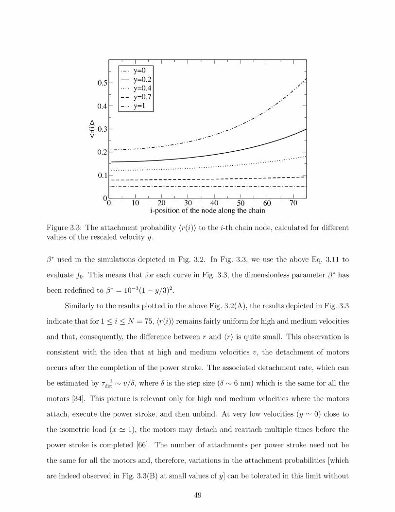
Figure 3.3: The attachment probability 〈r(i)〉 to the i-th chain node, calculated for differentvalues of the rescaled velocity y.
β∗ used in the simulations depicted in Fig. 3.2. In Fig. 3.3, we use the above Eq. 3.11 to
evaluate f0. This means that for each curve in Fig. 3.3, the dimensionless parameter β∗ has
been redefined to β∗ = 10−3(1− y/3)2.
Similarly to the results plotted in the above Fig. 3.2(A), the results depicted in Fig. 3.3
indicate that for 1 ≤ i ≤ N = 75, 〈r(i)〉 remains fairly uniform for high and medium velocities
and that, consequently, the difference between r and 〈r〉 is quite small. This observation is
consistent with the idea that at high and medium velocities v, the detachment of motors
occurs after the completion of the power stroke. The associated detachment rate, which can
be estimated by τ−1det ∼ v/δ, where δ is the step size (δ ∼ 6 nm) which is the same for all the
motors [34]. This picture is relevant only for high and medium velocities where the motors
attach, execute the power stroke, and then unbind. At very low velocities (y ' 0) close to
the isometric load (x ' 1), the motors may detach and reattach multiple times before the
power stroke is completed [66]. The number of attachments per power stroke need not be
the same for all the motors and, therefore, variations in the attachment probabilities [which
are indeed observed in Fig. 3.3(B) at small values of y] can be tolerated in this limit without
49

negative consequences for muscle contractility.
3.5 Summary
In this chapter we examined the thoroughly studied problem of muscle contraction in light
of the growing interest in collective dynamics of motor proteins. The basic contractile unit
of the muscle is the sarcomere which appears to have almost the same length in different
muscle cells of different vertebrates. We argue that the origin of this remarkable feature
of muscle cells may be the elasticity-mediated crosstalk (EMC) effect, which is related to
the changes in the attachment/detachment rates of the myosin II motors in response to the
tensile stress that they (collectively) generate in the F-actin. The associated changes in
the duty ratio of the motors can impair their ability to cooperate efficiently during muscle
contraction. Specifically, the major problem originating from the EMC effect is that it
leads to an increases in the attachment probability of motors located near the Z-line while,
simultaneously, decreasing the attachment probability of the motors in the central part of
the sarcomere (M-line). The resulting variations in the duty ratios of the motors along
the thick filament pose a serious problem in light of the fact that the detachment rate of
the motors is primarily a function of the step size and the muscle shortening velocity and,
therefore, should be fairly uniform. Using computer simulations of a simple bead-spring
model we demonstrated that this undesired effect can be almost neglected for half-sarcomeres
consisting of less than approximately 2N = 150 motors. Remarkably, this is precisely the
“universal” size of half a sarcomere in muscle cells across vertebrates.
Another notable result of our study is the derivation of the celebrated Hill’s equation
for the muscle force-velocity relationship. The basic assumptions in our derivation are given
by Eqs. 3.3 and 3.4. The former describes the experimentally measured linear relationship
between the attachment probability and the load, while the latter is simply Newton’s first
law for an object (the actin filament in this case) moving at constant velocity. As a bonus,
50
we obtain an expression relating the constant c in the dimensionless form of Hill’s equation
to some of the system parameters.
51

Chapter 4
Discussion
Motor proteins are a key component in virtually all eukaryotic cells. They are involved in
various important biological processes such as cell division and muscle contraction. Over
the past years, our knowledge concerning how these molecules operate and interact with
the cytoskeletal filament has increased dramatically, as a result of theoretical studies and
improved experimental techniques. Recently, there has been an increased interest in the
collective action of motor proteins, particularly in the way that cooperative effects change
their duty ratios. One notable example is the elasticity-mediated crosstalk (EMC) coupling
between myosin II motors, whose origin is the tensile stress that they collectively generate
in actin filaments. The EMC effect was originally proposed in order to explain the observed
distribution of reversal times in motility assays of myosin II motors and actin filaments with
alternating polarities [18]. Such filaments exhibit bidirectional motion due to the action of
motors exerting opposite forces on actin segments with opposite polarities. The “tug-of-war”
between the two antagonistic motor parties leads to large stress fluctuations along the elastic
actin track. As a result, the motors change their attachment/detachment statistics in order
to relieve the strain energy in the filaments. More specifically, during bidirectional motion,
the motors duty ratio decreases and, on average, each one of them becomes effectively less
processive. The EMC between motors in bidirectional motion, thus, is a collective effect that
52

decreases their degree of cooperativity.
The aforementioned motility assay for bidirectional motion constitute a rather unusual
experimental setup. In this thesis we explored the role of the EMC effect in two more com-
mon scenarios. The first one is that of conventional motility gliding assays where myosin
II motors work on polar actin filaments. In this case, the filaments move unidirectionally
because all the motors exert forces in the same direction. Unlike in the “tug-of-war” case,
the force fluctuations along the filament are limited when all the forces are applied in the
same direction and, therefore, the EMC effect is expected to be quite small. Our statistical
mechanical analysis of this problem, presented in chapter 2, demonstrates this point. The
magnitude of the EMC effect is quantified by introducing the dimensionless parameter β∗
[see Eq. 1.12] that characterizes the ratio between the typical elastic energy per actin seg-
ment and the thermal energy. Our analysis reveals that the effective attachment probability
〈r〉 of non processive myosin II motors drops linearly with√β∗ (see Eq. 2.20) from the value
of r, the bare attachment probability, at β∗ = 0 (i.e., for an infinetly rigid filament that does
not exhibit EMC). Our calculation holds for small values of β∗, which is indeed the case for
actomyosin motility assays where β∗ ∼ 0.002. This means that in this conventional acto-
myosin gliding assays the EMC effect has only a small impact on the attachment probability
of the motors, i.e. 〈r〉 ' r.
The second system under investigation in this thesis is skeletal muscles. As in the case
of the motility assays for bidirectional motion, this system features a tug-of-war competition
between opposite forces. But while in bidirectional motion two motor parties work against
each other, in the case of muscle contraction, the tug-of-war takes place between the motors
and an external opposite force that pulls at the end of the sarcomere. This type of tug-of-ear
also creates larger stress fluctuations along the actin track, specifically near the end where
the external force is applied. Our analysis of this system, presented in chapter 3, reveals
that the stress fluctuations can be reduced by inhomogeneous attachment of the motors.
Specifically, the motors will tend to attach at a higher (lower) rate close to (away from) the
53

end of the sarcomere where the external load is applied. In other words, the EMC leads
to a polarized attachment probability, where the motors in proximity to the last monomer
attach with higher probability than motors far away from this point. The problem with
this observation, however, is that the duty ratio variations between motors may seriously
impair their ability to efficiently cooperate with each other (see discussion in chapter 3).
Nevertheless, we find that the polarization of the attachment probability becomes significant
either when a large external force is applied, or when the system size is larger than the
“universal” size of sarcomeres in vertebrates (i.e., N > 75). Thus, our analysis proposes
a possible solution for the long standing puzzle concerning the similarity of the sarcomere
length in different species in nature. Recent advances in single molecule techniques [67]
open the opportunity for testing our theoretical predictions concerning the polarization of
the attachment probability. Specifically, optical tweezers provide the mean to apply pico-
newton forces against the gliding motion of the thin filament, while the duty ratio may be
directly measured using high resolution atomic force microscopy (AFM).
Motor proteins are the ”engines of biology”, converting chemical energy into mechanical
work. To gain a better understanding of their action, one needs to develop models that
integrate and couple the biochemical cycle of these molecules with their mechanical features
and with the elasticity of the associated cytoskeletal filaments. Over the past century, this
problem has drawn attention from different scientific disciplines. One of the main issues that
has been largely debated within the community is the role of friction forces in the process
of muscle contraction. Some theoretical studies support the notion that friction forces play
a minor role in muscle contraction, while others argue that friction is a non-negligible force
that may emerge from the medium surrounding the filaments and / or the motors themselves.
Our simple derivation of Hill’s equation (see section 3.2) interprets the experimental findings
in ref. [34] as if friction forces are indeed non-negligible (see Eq. 3.4). Nevertheless, it is
important to emphasize that our approach does not rely on this notion. What we have
presented in this thesis is a unique statistical mechanical approach for investigating the
54

collective action of myosin II motors. Our theoretical approach bypasses the aforementioned
controversies by introducing only two variables that do not depend on friction at all, namely
the duty ratio r and the dimensionless energy β∗. The resulting EMC effect can be easily
integrated into other theoretical model. Specifically, with the steady improvement in imaging
techniques and computer modeling, more atomistic models for muscles may be ”just around
the corner”.
55

Bibliography
[1] B. Alberts, D. Bray, J. Lewis, M. Raff, K. Roberts, and J. D. Watson, Molecular Biology
of the Cell (Garland, New York, 1994).
[2] E. Mandelkow and E.M. Mandelkow, Curr. Opin. Struct. Biol. 4, 171 (1994).
[3] K.J. Palmer, P. Watson and D.J. Stephens, Biochem. Soc. Symp. 72, 1 (2005).
[4] B. A. Rowning et. al., Proc. Natl. Acad. Sci. 94, 1224 (1997)
[5] K. Bloom, Curr. Biol. 3, R430 (2003).
[6] T.P. Stossel, Sci. Am. 271 54 (1994).
[7] B. Feierbach and F. Chang, Curr. Opin. Microbiol. 4, 713 (2001).
[8] D.A.D. Parry and P.M. Steinert, Intermediate Filament Structure (Springer-Verlag,
New York, 1995).
[9] J. Howard, Mechanics of Motor Proteins and the Cytoskeleton (Sinauer, Sunderland
MA, 2001).
[10] H. Higuchi and S. A. Endow, Curr. Opin. Cell Biol. 14, 50 (2002).
[11] H. B. McDonald, R. J. Stewart and L. S. Goldstein, Cell 63, 1159 (1990).
[12] M. Schliwa, Nature, 401, 431 (1999).
[13] H. Miki, Y. Okada, and N. Hirokawa, Trends. Cell Biol.15, 467 (2005).
56

[14] L. M. Coluccia (ed.), Myosins: A Superfamily of Moleculare Motors (Springer, Dor-
drecht, 2008).
[15] Cooper G.M. The Cell: A Molecular Approach. 2nd edition (Sinauer Associates, Sun-
derland, MA, 2000).
[16] M. LeMasurier and P. G. Gillespie, Neuron 48, 403 (2005).
[17] H. A. AL-Khayat, L. Hudson, M. K. Reedy, T. C. Irving and J. M. Squire, Biophys. J.
85, 1063 (2003).
[18] B. Gilboa, D. Gillo, O. Farago, and A. Bernheim-Groswasser, Soft Matter 5, 2223
(2009).
[19] F. Julicher and J. Prost, Phys. Rev. Lett. 75, 2618 (1995).
[20] F. Julicher and J. Prost, Phys. Rev. Lett. 78, 4510 (1997).
[21] M. Badoual, F. Julicher, and J. Prost, Proc. Natl. Acad. Sci. USA 99, 6696 (2002).
[22] M. O. Magnasco, Phys. Rev. Lett. 71, 1477 (1993).
[23] A. Ajdari, D. Mukamel, L. Peliti and J. Prost, J. Phys. I 4, 1551 (1994).
[24] R. D. Astumian and M. Bier, Phys. Rev. Lett. 72, 1766 (1994).
[25] J. E. Molloy, J. E. Burns, J. Kendrick-Jones, R. T. Tregear, and D. C. S. White, Nature
378, 209 (1995).
[26] J. T. Finer, R. M. Simmons, and J. A. Spudich, Nature 368, 113 (1994).
[27] H. Higuchi, T. Yanagida, and Y. E. Goldman, Biophys. J. 69, 1000 (1995).
[28] H. Kojima, A. Ishijima, and T. Yanagida, Proc. Natl. Acad. Sci. USA 91, 12962 (1994).
[29] K. C. Holmes, D. Popp, W. Gebhard, and W. Kabsch, Nature 347, 44 (1990).
57

[30] S. L. Hooper, K. H. Hobbs, and J. B. Thuma, Prog. Neurobiol. 86, 72 (2008).
[31] H. E. Huxley, A. Stewart, H. Sosa, and T. Irving, Biophys. J. 67, 2411 (1994).
[32] K. Wakabayashi, Y. Sugimoto, H. Tanaka, Y. Ueno, Y. Takezawa, and Y. Amemiya,
Biophys. J. 67, 2422 (1994).
[33] T. L. Daniel, A. C. Trimble, and P. B. Chase, Biophys. J. 74, 1611 (1998).
[34] G. Piazzesi, et. al., Cell 131, 784 (2007).
[35] M. J. I. Muller, S. Klumpp, and R. Lipowsky, Proc. Natl. Acad. Sci. USA 105, 4609
(2008).
[36] T. A. J. Duke, Proc. Nat. Acad. Sci. USA 96, 2770 (1999).
[37] K. Kruse and D. Riveline, Curr. Top. Dev. Biol. 95, 67 (2011).
[38] T. Guerin, J. Prost, P. Martin, and. J.-F. Joanny, Curr. Opin. Cell Biol. 22, 14 (2010).
[39] S. Banerjee, M. C. Marchetti, and K. Muller-Nedebock, Phys. Rev. E. 84, 011914 (2011).
[40] T. J. Burkholder, R. L. Lieber, J. Exp. Biol. 204, 1529 (2001).
[41] H. E. Huxley, Science 164, 1356 (1969).
[42] A.V. Hill, Proc. R. Soc. London B Biol. Sci. 126, 136 (1938).
[43] R. V. Mastrigt, IEEE Trans. Biomed. Eng. 27, 413 (1980).
[44] A. F. Huxely, Prog. Biophys. Biophys. Chem. 7, 255 (1957).
[45] H. E. Huxley, J. Mol. Biol. 7, 281 (1963).
[46] J. Kendrick-Jones, W. Lehman, A. G. Szent-Gyorgyi, J. Mol. Biol. 54, 313 (1970).
[47] R. W. Lymn, E. W. Taylor, Biochemistry 10, 4617 (1971).
58

[48] B. R. Munson, D. F. Young, T. Okiishi, W. W. Huebsch Fundamentals of Fluid Me-
chanics (Wiley, Hoboken NJ, 2010).
[49] D. Bernstein, Phys. Rev. E. 71, 041103 (2005).
[50] A. F. Voter Radiation Effects in Solids (Springer, Dordrecht, 2005).
[51] G. Lan, X. Sun, Biophys. 88, 1407 (2005).
[52] K. Kitamura, M. Tokunaga, S. Esaki, A.H. Iwane, T. Yanagida, Biophysics 1, 1 (2005).
[53] S. Walcott, S. X. Sun, Phys. Chem. Chem. Phys. 11, 4871 (2009).
[54] A. Mansson, Biophys. J. 98, 1237 (2010).
[55] L. Marcucci, L. Truskinovsky, Eur. Phys. J. E. 32, 411 (2010).
[56] B. Chen, H. Gao, Biophys. J. 101, 396 (2011).
[57] S. J. Kron, J. A. Spudich, Proc. Natl. Acad. Sci. USA 83, 6272 (1986).
[58] C. R. Worthington, Nature 193, 1283 (1962).
[59] G. F. Elliott, C. R. Worthington, Int. J. Biol. Macromol. 29, 213 (2001).
[60] A. R. Bausch, W. Moller, E. Sackmann, Biophys. J. 76, 573 (1999).
[61] K. Keren, P. T. Yam, A. Kinkhabwala, A. Mogilner, J. A. Theriot, Nat. Cell Biol. 11,
1219 (2009).
[62] A. Landesberg, S. Sideman, Am. J. Physiol. 278, H1274 (2000).
[63] D. Hexner, Y. Kafri, Phys. Biol. 6, 036016 (2009).
[64] Y. C. Fung, Biomechanics: Mechanical Properties of Living Tissues (Springer-Verlag,
New York, 1993).
59

[65] J. Keener, J. Sneyd, Mathematical Physiology, (Springer-Verlag, New York, 1998).
[66] M. Linari, M. Caremani, V. Lombardi, Proc. R. Soc. B. 277, 19 (2010).
[67] C. Veigel, C. F. Schmidt, Nat. Rev. Mol. Cell Biol. 12, 163 (2011).
60