Thermotolerance induced at a mild temperature of 40°C alleviates heat shock-induced ER stress and...
11
Thermotolerance induced at a mild temperature of 40 °C alleviates heat shock-induced ER stress and apoptosis in HeLa cells Ahmed Bettaieb 1,2 , Diana A. Averill-Bates ⁎ Département des sciences biologiques, TOXEN, Université du Québec à Montréal, CP 8888, Succursale Centre-Ville, Montréal, Québec H3C 3P8, Canada abstract article info Article history: Received 14 August 2014 Received in revised form 16 September 2014 Accepted 17 September 2014 Available online 26 September 2014 Keywords: Hyperthermia ER stress Apoptosis Calpain Caspase Hsp72 Hyperthermia (39–45 °C) has emerged as an alternate prospect for cancer therapy in combination with radiation and chemotherapy. Despite promising progress in the clinic, molecular mechanisms involved in hyperthermia- induced cell death are not clear. Hyperthermia causes protein denaturation/aggregation, which results in cell death by apoptosis and/or necrosis. Hyperthermia also induces thermotolerance, which renders cells resistant to subsequent exposure to lethal heat shock. This study investigates the role of both lethal (42–43 °C) and mild (40 °C) hyperthermia in regulating ER stress and ER stress-induced apoptosis in HeLa cells. The ability of mild thermotolerance induced at 40 °C to alleviate either or both of these processes is also determined. Hyper- thermia (42–43 °C) induced ER stress, revealed by phosphorylation of PERK, eIF2α and IRE1α, cleavage of ATF6 and increased expression of BiP and sXBP1. Real-time PCR revealed that mRNA levels of ATF6, ATF4, BiP, sXBP1 and CHOP increased in cells exposed to hyperthermia. Moreover, hyperthermia caused disruption of calcium homeostasis and activated the calpain-calpastatin proteolytic system and ER resident caspase 4. Pre-exposure to mild hyperthermia (40 °C) alleviated the induction of cytotoxicity and ER stress by hyperthermia (42–43 °C) and protected cells against ER stress-induced apoptosis. ShRNA-mediated depletion of Hsp72 abro- gated protective effects of mild thermotolerance (40 °C) against heat-shock induced ER stress and sensitized cells to ER stress-mediated apoptosis. Our findings show that Hsp72 contributes to the protective effects of mild hyperthermia (40 °C) against hyperthermia-induced ER stress and apoptosis. © 2014 Elsevier B.V. All rights reserved. 1. Introduction Over the past decades, the struggle against cancer has led to the dis- covery of new strategies to fight this disease and to bring hope to pa- tients. Among these new strategies, hyperthermia (39–45 °C) (also known as thermal therapy or thermotherapy) has emerged as a promising alternative that can treat a wide range of tumor types with minimal injury to normal tissues [1]. Furthermore, unlike healthy cells, tumors act as a heat reservoir when subjected to local hyperther- mia. Tumors are unable to increase blood flow in response to thermal stress, which makes them more vulnerable to heat damage than the sur- rounding normal tissue [1,2]. This results in collapse of the tumor vascu- lar system and destruction of tumor cells. In addition, hyperthermia appears to be a potent modifier of tumor response to radiation and sev- eral chemotherapy agents by increasing and targeting their cytotoxic effects in the tumor volume. A multitude of randomized studies showed that hyperthermia combined with radiotherapy, chemotherapy or both, resulted in significant improvement in clinical outcome in cancer pa- tients [1–7]. The tumor sites include cervix, soft-tissue sarcoma, breast, head and neck, rectum, brain, bladder, lung, esophagus, liver, appendix, prostate and melanoma. Several larger phase III randomized trials showed that hyperthermia improved the rate of clinical complete re- sponse in patients treated with radiotherapy for superficial breast can- cer and chest wall recurrence [7]. Hyperthermia was beneficial when combined with re-irradiation for breast cancer recurrences [5]. For high-risk soft tissue sarcoma, the addition of regional hyperthermia to a multimodal treatment of surgery, radiotherapy, and chemotherapy was shown to improve local recurrence- and disease-free survival [4]. Several randomized trials showed an improvement by adding Biochimica et Biophysica Acta 1853 (2015) 52–62 Abbreviations: ASK1, apoptotic-signaling kinase-1; ATF4, activating transcription factor 4; ATF6, activating transcription factor 6; Bcl-2, B cell lymphoma 2; BiP, binding immuno- globulin protein; cATF6, cleaved ATF6; CHOP, transcriptional factor C/EBP homologous pro- tein; DMEM, Dulbecco's modification of Eagle's medium; DTT, dithiothreitol; eIF2α, eukaryotic translation initiation factor; ER, endoplasmic reticulum; GADD34, growth arrest and DNA damage-inducible 34; GAPDH, glyceraldehyde 3-phosphate dehydrogenase; HeLa, human cervical carcinoma cells; HRP, horseradish peroxidase; Hsp, heat shock protein; IRE1α, inositol-requiring protein-1; JNK, c-Jun N terminal kinase; MOPS, 3-(N-morpholino)- propane sulfonic acid; PBS, phosphate-buffered saline; PERK, protein kinase RNA (PKR)-like ER kinase; PI, propidium iodide; PMSF, phenylmethylsulfonyl fluoride; SDS-PAGE, sodium dodecyl sulphate polyacrylamide gel electrophoresis; SEM, standard error of mean; sXBP1, spliced XBP1; TBP, TATA-box binding protein; TT, thermotolerant; XBP1, X-Box binding pro- tein 1 ⁎ Corresponding author. Tel.: +1 514 987 3000 (4811); fax: +1 514 987 4647. E-mail address: [email protected] (D.A. Averill-Bates). 1 Tel.: +1 514 987 3000 (4811); fax: +1 514 987 4647. 2 Present address: Department of Nutrition, University of California, Davis, CA, USA. http://dx.doi.org/10.1016/j.bbamcr.2014.09.016 0167-4889/© 2014 Elsevier B.V. All rights reserved. Contents lists available at ScienceDirect Biochimica et Biophysica Acta journal homepage: www.elsevier.com/locate/bbamcr
Transcript of Thermotolerance induced at a mild temperature of 40°C alleviates heat shock-induced ER stress and...

Biochimica et Biophysica Acta 1853 (2015) 52–62
Contents lists available at ScienceDirect
Biochimica et Biophysica Acta
j ourna l homepage: www.e lsev ie r .com/ locate /bbamcr
Thermotolerance induced at a mild temperature of 40 °C alleviates heatshock-induced ER stress and apoptosis in HeLa cells
Ahmed Bettaieb 1,2, Diana A. Averill-Bates ⁎Département des sciences biologiques, TOXEN, Université du Québec à Montréal, CP 8888, Succursale Centre-Ville, Montréal, Québec H3C 3P8, Canada
Abbreviations:ASK1, apoptotic-signaling kinase-1; ATF4; ATF6, activating transcription factor 6; Bcl-2, B cell lympglobulin protein; cATF6, cleaved ATF6; CHOP, transcriptiontein; DMEM, Dulbecco's modification of Eagle's mediueukaryotic translation initiation factor; ER, endoplasmic retand DNA damage-inducible 34; GAPDH, glyceraldehydeHeLa, human cervical carcinoma cells; HRP, horseradish perIRE1α, inositol-requiring protein-1; JNK, c-JunN terminal kipropane sulfonic acid; PBS, phosphate-buffered saline; PERKER kinase; PI, propidium iodide; PMSF, phenylmethylsulfododecyl sulphate polyacrylamide gel electrophoresis; SEMspliced XBP1; TBP, TATA-box binding protein; TT, thermototein 1⁎ Corresponding author. Tel.: +1 514 987 3000 (4811)
E-mail address: [email protected] (D.A. Averill-Ba1 Tel.: +1 514 987 3000 (4811); fax: +1 514 987 46472 Present address: Department of Nutrition, University
http://dx.doi.org/10.1016/j.bbamcr.2014.09.0160167-4889/© 2014 Elsevier B.V. All rights reserved.
a b s t r a c t
a r t i c l e i n f oArticle history:Received 14 August 2014Received in revised form 16 September 2014Accepted 17 September 2014Available online 26 September 2014
Keywords:HyperthermiaER stressApoptosisCalpainCaspaseHsp72
Hyperthermia (39–45 °C) has emerged as an alternate prospect for cancer therapy in combinationwith radiationand chemotherapy. Despite promising progress in the clinic, molecular mechanisms involved in hyperthermia-induced cell death are not clear. Hyperthermia causes protein denaturation/aggregation, which results in celldeath by apoptosis and/or necrosis. Hyperthermia also induces thermotolerance, which renders cells resistantto subsequent exposure to lethal heat shock. This study investigates the role of both lethal (42–43 °C) andmild (40 °C) hyperthermia in regulating ER stress and ER stress-induced apoptosis in HeLa cells. The ability ofmild thermotolerance induced at 40 °C to alleviate either or both of these processes is also determined. Hyper-thermia (42–43 °C) induced ER stress, revealed by phosphorylation of PERK, eIF2α and IRE1α, cleavage ofATF6 and increased expression of BiP and sXBP1. Real-time PCR revealed that mRNA levels of ATF6, ATF4, BiP,sXBP1 and CHOP increased in cells exposed to hyperthermia. Moreover, hyperthermia caused disruptionof calcium homeostasis and activated the calpain-calpastatin proteolytic system and ER resident caspase 4.Pre-exposure tomild hyperthermia (40 °C) alleviated the induction of cytotoxicity and ER stress byhyperthermia(42–43 °C) and protected cells against ER stress-induced apoptosis. ShRNA-mediated depletion of Hsp72 abro-gated protective effects of mild thermotolerance (40 °C) against heat-shock induced ER stress and sensitizedcells to ER stress-mediated apoptosis. Our findings show that Hsp72 contributes to the protective effects ofmild hyperthermia (40 °C) against hyperthermia-induced ER stress and apoptosis.
© 2014 Elsevier B.V. All rights reserved.
1. Introduction
Over the past decades, the struggle against cancer has led to the dis-covery of new strategies to fight this disease and to bring hope to pa-tients. Among these new strategies, hyperthermia (39–45 °C) (alsoknown as thermal therapy or thermotherapy) has emerged as a
4, activating transcription factorhoma 2; BiP, binding immuno-al factor C/EBP homologous pro-m; DTT, dithiothreitol; eIF2α,iculum; GADD34, growth arrest3-phosphate dehydrogenase;
oxidase;Hsp, heat shockprotein;nase;MOPS, 3-(N-morpholino)-, protein kinase RNA (PKR)-likenyl fluoride; SDS-PAGE, sodium, standard error of mean; sXBP1,lerant; XBP1, X-Box binding pro-
; fax: +1 514 987 4647.tes)..of California, Davis, CA, USA.
promising alternative that can treat a wide range of tumor types withminimal injury to normal tissues [1]. Furthermore, unlike healthycells, tumors act as a heat reservoir when subjected to local hyperther-mia. Tumors are unable to increase blood flow in response to thermalstress, whichmakes themmore vulnerable to heat damage than the sur-rounding normal tissue [1,2]. This results in collapse of the tumor vascu-lar system and destruction of tumor cells. In addition, hyperthermiaappears to be a potent modifier of tumor response to radiation and sev-eral chemotherapy agents by increasing and targeting their cytotoxiceffects in the tumor volume. Amultitude of randomized studies showedthat hyperthermia combinedwith radiotherapy, chemotherapy or both,resulted in significant improvement in clinical outcome in cancer pa-tients [1–7]. The tumor sites include cervix, soft-tissue sarcoma, breast,head and neck, rectum, brain, bladder, lung, esophagus, liver, appendix,prostate and melanoma. Several larger phase III randomized trialsshowed that hyperthermia improved the rate of clinical complete re-sponse in patients treated with radiotherapy for superficial breast can-cer and chest wall recurrence [7]. Hyperthermia was beneficial whencombined with re-irradiation for breast cancer recurrences [5]. Forhigh-risk soft tissue sarcoma, the addition of regional hyperthermia toa multimodal treatment of surgery, radiotherapy, and chemotherapywas shown to improve local recurrence- and disease-free survival [4].Several randomized trials showed an improvement by adding

53A. Bettaieb, D.A. Averill-Bates / Biochimica et Biophysica Acta 1853 (2015) 52–62
hyperthermia to radiation for cervical cancer patients, even at 12 yearsfollow-up [3,6].
Despite promisingprogress in the clinic, themechanisms involved inheat stress-induced cell death are not well understood [8]. Hyperther-mia causesmany changes in cells aswell as a loss of cellular homeostasis[9–13]. A key event appears to be protein denaturation and aggregation[14], which results in cell cycle arrest, inactivation of protein synthesis,and inhibition of DNA repair processes. The correct structure and con-formation of proteins is essential for their function in the cell. A small in-crease in temperature can cause protein unfolding, entanglement andaggregation leading to an imbalance in proteostasis. This can result inincreased degradation of aggregated/misfolded proteins through theproteasomal and lysosomal pathways. Other cellular effects of hyper-thermia include the: (1) inhibition of DNA synthesis, transcription,RNA processing and translation; (2) disruption of the membranecytoskeleton; (3)metabolic changes (e.g. uncoupling of oxidative phos-phorylation) that lead to decreased ATP levels; and (4) alterations inmembrane permeability that cause increases in intracellular levels ofNa+, H+ andCa2+ [9,10,12]. Furthermore, changes occur to intracellularorganelles: the Golgi system and endoplasmic reticulum (ER) werefragmented during heat shock andmodest swelling ofmitochondria oc-curred [15].
The ER is an organelle that is highly responsive to the nutrient andenergy status of the cell and plays an important role in the folding ofnewly synthesized proteins. When the folding capacity of the ER isexceeded, misfolded/unfolded proteins accumulate and lead to ERstress [16]. Cells use an adaptive mechanism to counter the deleteriouseffects of ER stress, known as the unfolded protein response (UPR) [17].The UPR consists of three major branches that are controlled by the ERtransmembrane proteins PKR-like ER-regulated kinase (PERK), inositolrequiring protein 1α (IRE1α) and activating transcription factor 6(ATF6) [18–20]. In particular, the PERK/eukaryotic translation initiationfactor 2α (eIF2α) sub-arm of ER stress signaling is critical for the UPR incancer cells and their adaption to hypoxia, as well as their resistance totherapy [21]. PERK phosphorylates eIF2α, which inhibits general pro-tein translation. This allows selective translation of ATF4, which acti-vates the transcription of ER chaperones such as BiP. The distant UPRarms (IRE1α/X-Box binding protein 1 (XBP1)) synergize to attenuatestress by increasing the folding capacity of the ER [16]. IRE1α catalyzesthe alternative splicing of XBP1 mRNA, leading to expression of thesXBP1 transcription factor, which activates ER chaperone genes. Thethird arm is mediated through the transcription factor ATF6. ATF6 un-dergoes proteolysis in the Golgi apparatus which leads to activationfollowed by its translocation to the nucleus. One of the ATF6 targetgenes is XBP1. However, if the compensatory mechanisms fail to facili-tate the adaptation of cells to ER stress, induction of the UPR can leadto the elimination of stressed cells by apoptosis [22–24]. C/EBP homolo-gous protein (CHOP), also known as growth arrest- and DNA damage-inducible gene 153 (GADD153), plays a convergent role in the UPRand is as an important mediator of ER stress-induced apoptosis [24,25]. CHOP-mediated activation of growth arrest and DNA damage-inducible 34 (GADD34) promotes protein dephosphorylation of eIF2α,reversing the translational inhibition to allow the recovery of proteinsynthesis following a stress insult.
The induction of apoptosis following ER stress can occur in variousways including the activation of caspase 12/4, cleavage of transmem-brane ER protein Bcl-2-associated protein-31 (BAP31) by active caspase8, and the activation of c-Jun N terminal kinase (JNK) via the apoptosissignaling kinase 1 (Ask1)/IRE1α/TNF receptor-associated factor 2(TRAF2) complex [24,26]. In addition, cytosolic free calcium levelshave been reported to increase during ER stress-induced apoptosis,thereby leading to the activation of calpains and subsequent cleavageof the anti-apoptotic B cell lymphoma 2 (Bcl-2) family member Bcl-XL
[27]. Besides these direct pathways, activation of caspase 8 and/or cas-pase 7 may occur during ER stress-induced apoptosis, leading to cyto-chrome c release and caspase 9 activation. Additionally, ER stress may
activate the traditional mitochondrial pathway through a crosstalk be-tween both compartments involving the Bcl-2 family proteins [28].
Hyperthermia is cytotoxic at temperatures above 42.5 °C and selec-tively lethal to cancer cellswhilemild hyperthermia is known to be ben-eficial to tissues and organs [1]. Hyperthermia-range temperaturescause protein denaturation and aggregation [9,14] and therefore, it islikely that hyperthermia could induce ER stress.We previously reportedthat heat preconditioning at a mild temperature (40 °C for 3 h) inhuman adenocarcinoma cervical HeLa cells led to the development ofmild thermotolerance, whichwas associatedwith an increase in the ex-pression of several heat shock proteins (Hsp) includingHsp72 [29]. Thiswork aims to determine whether mild doses of hyperthermia (40 °C)can activate the ER stress survival response, and whether more severeheat exposure can tip the balance towards ER stress-mediated apopto-sis. This study also determines whether pre-conditioning with mild hy-perthermia (40 °C) could protect cells against ER stress-mediatedapoptosis induced by lethal hyperthermia (42–43 °C), and whetherHsp72 plays a role in this adaptive survival response.
2. Experimental procedures
2.1. Reagents
Dulbecco's Modified Eagle Medium (DMEM), penicillin/streptomy-cin, fetal bovine serum (FBS), trypsin and Fluo 3-AM (1-[2-Amino-5-(2,7-dichloro-6-hydroxy-3-oxo-9-xanthenyl)phenoxy]-2-(2-amino-5-methylphenoxy))were purchased from Invitrogen (Carlsbad, CA). Anti-bodies for tubulin were purchased from Upstate Biotechnology (LakePlacid, NY). Antibodies for pPERK (Thr980), PERK, peIF2α (Ser51),eIF2α, sXBP1, ATF6, IRE1α, BiP, caspase 4 and vinculin were purchasedfrom Santa Cruz Biotechnology (Santa Cruz, CA) while calpain,calpastatin and cleaved caspase-3 antibodies were from Cell Signaling(Beverly, MA). Antibodies for pIRE1α (Ser724) were purchased fromAbcam (Cambridge, MA). Horseradish peroxidase (HRP)-conjugatedsecondary antibodies were purchased from BioResources Interna-tional (Carlsbad, CA). BAPTA-AM (1,2-Bis(2-aminophenoxy)ethane-N,N,N′,N′-tetraacetic acid tetrakis(acetoxymethyl ester), Hoechst 33258,propidium iodide (PI), sulforhodamine B and all other chemicals, unlessotherwise indicated, were purchased from the Sigma Chemical Co. (St.Louis, MO).
2.2. Cell culture
HeLa cells (ATCC #CCL-2) were grown in monolayer in DMEM con-taining 10% FBS, 50 U/ml penicillin and 50 μg/ml streptomycin at 37 °Cin a humidified atmosphere of 5% CO2 in awater jacketed incubator [30].The cells were grown to near confluence and cell culture medium wasreplaced with fresh medium 24 h before experiments. To induce ther-motolerance (TT), cells were grown to near confluence, culturemediumwas replacedwith freshmedium for 24 h, and then confluent cells weretransferred to an identical incubator for 3 h at 40 °C (±0.1 °C), follow-ing a period of 20 min to allow the temperature of the culture mediumto reach 40 °C [26]. Confluent cells were harvested using 0.25% (w/v)trypsin-0.02% (w/v) EDTA solution, and washed by centrifugation(1000×g, 3 min).
At 40 °C, maximum levels of thermotolerance and induction of Hspsoccur after 3 hours [29]. Protein synthesis continues at 40 °C and doesnot stop for several hours as occurs when thermotolerance is inducedby exposure to higher temperatures such as 43–45 °C. There was noloss of viability in cells heated at 40 °C for 3 h, evaluated by trypanblue exclusion (data not shown).
Hsp72 silencing in HeLa cells was achieved by testing four differenthairpins. Packaging (psPAX2) and envelope (pMD2.G) vectors were ob-tained from Addgene (Boston, MA). Lentiviruses were generated by co-transfection of vectors in 293FT cells (#R700-07, Life Technologies)using Lipofectamine 2000 (Invitrogen) following manufacturer's

Table 1Primer sequences used for real time PCR to determine mRNA expression of ER stressmarkers.
Gene Name Forward primer (5′–3′) Reverse primer (5′–3′)
ATF4 GGGTTCTCCAGCGACAAGGCTAAG AACAGGGCATCCAAGTCGAACTCATF6 ATGTCTCCCCTTTCCTTATATGGT AAGGCTTGGGCTGAATTGAABiP TGCTTGATGTATGTCCCCTTA CCTTGTCTTCAGCTGTCACTCHOP CATCACCACACCTGAAAGCA TCAGCTGCCATCTCTGCAeIF2α GAAGAGTGTGTTGGGCAGGT TGGCTAGCAATCATGGCACTGADD34 TCCTCTGGCAATCCCCCATA TGGTTTTCAGCCCCAGTGTTIRE GCAGCAGACTTTGTCATCGG GTGATCACACACTCCCCCTTGTPERK TGTCGCCAATGGGATAGTGACGAA AATCCGGCTCTCGTTTCCATGTCTTBP TATAATCCCAAGCGGTTTGC GCTGGAAAACCCAACTTCTGXBP1 TGCTGAGTCCGCAGCAGGTG GCTGGCAGGCTCTGGGGAAG
54 A. Bettaieb, D.A. Averill-Bates / Biochimica et Biophysica Acta 1853 (2015) 52–62
guidelines and then used to infect HeLa cells. Cells were selected usingpuromycin (2 μg/ml) and drug-resistant pools were propagated.
2.3. Heat treatment
Freshly harvested thermotolerant (3 h at 40 °C) and non-thermotolerant cells (3 h at 37 °C) in phosphate-buffered saline(PBS)-1% bovine serum albumin (BSA) supplemented with 10 mM ofglucose in a final volume of 1.0 ml were heated for 3 h at 42 or 43 °C,relative to controls at 37 °C, in temperature-controlled precisionwaterbaths (±0.02 °C) (Haake D8, Fisher Scientific, Montreal, QC)[29]. One ml of cell suspension reached a temperature within 0.1 °C ofthe waterbath temperaturewithin 3min. There was no recovery periodbetween development of thermotolerance (3 h at 40 °C) and hyper-thermia treatment at 42–43 °C.
2.4. Cytotoxicity
Cytotoxicity assays were performed using sulforhodamine B to staincellular proteins as described previously [29]. Freshly harvested normaland thermotolerant cells (5 × 10⁶/ml) were incubated in 1 ml of PBS-1% BSA supplemented with 10 mM glucose in tubes in a waterbath(Haake D8) at 43 °C for up to 180 min relative to controls at 37 °C. Thecells were then diluted and seeded in 96-well microplates in DMEMmedium and incubated at 37 °C for 96 h. The cells were fixed with 17%trichloroacetic acid in PBS and cellular protein was stained with 0.4%sulforhodamine B in 1% acetic acid. Quantification of sulforhodamine Bwas carried out by spectrofluorimetry using a UVmax kineticmicroplatereader (Spectra Max Gemini, Molecular Devices, Sunnyvale, CA) at awavelength of 540 nm. The relative plating efficiency of each cell typewas determined by dividing the absorbance observed for a given treat-ment by the absorbance detected in the normal cells incubated at 37 °Cfor 180 min and expressed as a percentage.
2.5. Morphological assessment of apoptosis
Briefly, thermotolerant and non thermotolerant cells were heatedat 42 and 43 °C for 3 h. Where indicated, cells were pretreated for 2 hwith 100 μM calcium chelator BAPTA-AM (Sigma-Aldrich), 20 μMcalpain inhibitor I (Ac-LLnL-CHO) (Sigma-Aldrich), 20 μM caspase-4 inhibitor (LEVD-CHO) or 50 μM caspase-7/3 inhibitor I (5-[(S)-(+)-2-(methoxymethyl)pyrrolidino]sulfonylisatin) (Calbiochem,La Jolla, CA). Thereafter, Hoechst 33258 and propidium iodide (PI) wereadded to visualize apoptotic and necrotic cells, respectively, by fluores-cencemicroscopy [29,30]. For each image, at least 300 cells were countedusing Northern Eclipse software (Empix Imaging Inc., Mississauga, ON).The percentages of apoptotic and necrotic cells were determined relativeto total cells (obtained using bright field illumination).
2.6. Biochemical analyses
Cells were homogenised in radio-immunoprecipitation assay buffer(RIPA: 10mMTris-HCl, pH 7.4, 150 mMNaCl, 0.1% sodium dodecyl sul-fate [SDS], 1% Triton X-100, 1% sodiumdeoxycholate, 5mMEDTA, 1mMNaF, 1 mM sodium orthovanadate and protease inhibitors). Lysateswere clarified by centrifugation at 13,000 rpm for 10 min, and proteinconcentrations were determined using a bicinchoninic acid assay kit(Pierce Chemical Co.). Proteins (30–90 μg) were resolved by SDS-PAGE and transferred to PVDF membranes [29,30]. Immunoblotting oflysates was performed with primary antibodies, and after incubationwith secondary antibodies, proteins were visualized using enhancedchemiluminescence (Amersham Biosciences Corp., Piscataway, NJ).Pixel intensities of immunoreactive bands were quantified usingFluorChem Q Imaging software (Alpha Innotech).
Total RNA was extracted from cells using TRIzol reagent(Invitrogen). mRNA of PERK, IRE1α, ATF6, GADD34, BiP, spliced (s)
XBP1, CHOP, and ATF4 was assessed by quantitative reverse transcrip-tion PCR (iCycler, Bio-Rad Laboratories, Hercules, CA) with appropriateprimers (Integrated DNA Technologies, Coralville, Iowa) (Table 1) andnormalized to glyceraldehyde 3-phosphate dehydrogenase (GAPDH).
For nuclear preparations, cells were washed in buffer A [100mM su-crose, 1 mM EGTA, 20 mM 3-(N-morpholino)-propane sulfonic acid(MOPS), pH 7.4] and then resuspended in buffer B [buffer A plus 5%Percoll, 0.01% digitonin, and a cocktail of protease inhibitors: 10 μMaprotinin, 10 μM pepstatin A, 10 μM leupeptin, 25 μM calpain inhibitorI, 1 mM phenylmethylsulfonyl fluoride (PMSF), pH 7.4] containing0.1 mM dithiothreitol (DTT) (Bettaieb and Averill-Bates, 2005). Mem-braneswere broken using a dounce homogenizer (200 strokes/sample).After 30min incubation on ice, debris and unbroken cells were removedby centrifugation (500 x g, 10 min) and then supernatants were centri-fuged (2,500 x g, 5min) to separate nuclei (pellet). The purity of nuclearfractions (89%) was confirmed by Western blotting using lamin A(Calbiochem, La Jolla, CA, USA) (data not shown).
2.7. Measurement of caspase and calpain activities
Cells were lysed in 20 mM piperazine-N,N′-bis(2-ethanesulfonicacid) (PIPES), 100 mM NaCl, 10 mM DTT, 1 mM EDTA, 0.1% 3-[(3-cholamidopropyl)-dimethylammonio]-2-hydroxy-1-propane-sulfonicacid (CHAPS), 10% sucrose, pH 7.2). Activities of calpain, caspase 3, cas-pase 4 and caspase 7were determined using their fluorogenic substrates:Suc-LY-AM, Ac-DEVD-AMC, Ac-LEVD-AFC, andMCA-VDQVDGWK(DNP)-NH2 (Calbiochem), respectively, by spectrofluorometry [29,30]. Activitiesof proteases are represented as relative cumulative fluorescence of thekinetic reaction and compared to untreated non-thermotolerant controlcells (Normal).
2.8. Intracellular Free Ca2+ measurement
The intracellular Ca2+ transients were monitored using the Ca2+-sensitive fluorescent indicator, Fluo 3-AM as previously described [31]with modifications. Briefly, where indicated, cells were pre-incubatedwith calcium chelator BAPTA-AM (100 μM) for 2 in a serum-free medi-um. Cells were then heated for 1 h at 42 and 43 °C. Forty five minutesbefore the end of the heat shock exposure, Fluo 3-AM (5 μM) wasadded to the cells. Cells were then washed and resuspended in PBSsupplementedwith PI (25 μM). Fluo 3-AM and PI fluorescence were an-alyzed using a FACScan cytometer (FACScan, BD Biosciences, Mississau-ga, ON, Canada) equipped with an argon ion laser emitting at 488 nmand a fluorescence detector band pass filter of 530/30 (for Fluo 3-AM)and 682/33 (for PI). At least 10,000 live cells were counted for each sam-ple. WinMDI software was used to measure fluorescence intensity.
2.9. Statistical analyses
Data are expressed as means + standard error of the mean (SEM).Statistical analyses were performed using JMP program (SAS Institute).

55A. Bettaieb, D.A. Averill-Bates / Biochimica et Biophysica Acta 1853 (2015) 52–62
Comparisons between treatments were performed using one-wayANOVA and the Bonferroni-Holmes adjustment was used to controlfor the Family-wise error rate at a desired level (α = 5%). Softwareused was JMP Statistical Discovery 4.0 (SAS Institute Inc., Cary, NC).For significant differences, a symbol (such as *) indicates P b 0.05,whereas a duplicate symbol (such as **) indicates P b 0.01.
3. Results
3.1. Mild thermotolerance induced at 40 °C protects HeLa cells againsthyperthermia-induced cytotoxicity
Mild thermotolerance was developed by heating cells for 3 h at40 °C, whereas controlswere incubated for 3 h at 37 °C. Thermotolerantcellsweremore resistant to cytotoxicity causedby subsequent exposureto lethal heat shock for up to 180 min at 43 °C, compared to controls(Fig. 1).
3.2. Hyperthermia (42–43 °C) activates the ER stress response in HeLa cells
The expression of several ER stressmarkers and the activation of keycomponents of the ER stress signaling pathways by lethal hyperthermiawere evaluated in normal non-thermotolerant HeLa cells. Immunoblotanalysis showed that phosphorylation of PERK (Thr980) and its down-stream target eIF2α (Ser51)were significantly increased upon exposureto lethal hyperthermia (42 - 43 °C) for 2 h, compared to control cells(37 °C) (Fig. 2A). Additionally, Ire1α phosphorylation at Ser724 in-creased at 42 and 43 °C compared to controls (37 °C). Phosphorylationof PERK and IRE1α, and cleavage of ATF6 (cATF6) were increased after1 h at 43 °C (data not shown). Protein and mRNA levels of PERK,eIF2α and Ire1α were comparable in heat-shocked and control cells(Fig. 2A and B). Similarly, the cleaved form of ATF6 protein (Fig. 2A)and mRNA levels of ATF6 (Fig. 2B) were significantly higher in cellsupon thermal stress. Moreover, elevated temperatures also increasedmRNA levels of the eIF2α downstream target ATF4, whileGADD34 levelsdecreased (Fig. 2B). Heat shock at 42 and 43 °C also significantly in-creased total protein and mRNA levels of spliced XBP1, a downstreamtarget of IRE1α, as well as the ER chaperone BiP (Fig. 2A and B).
Fig. 1. Mild thermotolerance induced at 40 °C protects HeLa cells against hyperthermia-induced cytotoxicity. Normal and thermotolerant (TT) cells (5 × 10⁶/ml) were incubatedat 43 °C for up to 180 min in PBS – 1% BSA-10 mM glucose. The relative plating efficiencyof each cell type was calculated by dividing the absorbance measured for a given time bythe value measured for the normal control cells incubated at 37 °C for 180 min. Data(mean ± SEM, n ≥ 3) are expressed as percentage of cell survival relative to controls.(*: P b 0.05; **: P b 0.01) indicate significant difference between the hyperthermia-treatedversus control cells (37 °C for 180min) for each cell type. (#: P b 0.05; ##: P b 0.01) indi-cate significant difference between normal and TT cells for the indicated treatment time.
3.3. Mild thermotolerance at 40 °C mitigates hyperthermia-induced ERstress in HeLa cells
The effects of mild thermotolerance induced at fever range temper-ature (40 °C) on ER stress signalingwere evaluated. The induction of ERstress markers in thermotolerant cells by subsequent exposure toelevated temperatures of 42 and 43 °C was significantly reduced(Fig. 2A). Indeed, mild thermotolerance mitigated hyperthermia-induced PERK (Thr980), eIF2α (Ser51) and Ire1α (Ser727) phosphory-lation and decreased sXBP1 and cleaved ATF6 expression whencompared to non-thermotolerant control cells (Fig. 2A). In accordancewith these biochemical findings, mild thermotolerance alleviatedhyperthermia-induced increases in BiP, sXBP1, ATF4 and ATF6mRNA ex-pression (Fig. 2B). GADD34 mRNA levels were higher at 37 °C inthermotolerant cells and remained elevated upon exposure to 42 and43 °C (Fig. 2B). The function of GADD34 is the recovery from the shut-down of protein synthesis induced by ER stress [32].
3.4. Hyperthermia (42–43 °C) induces ER stress-mediated apoptosis inHeLa cells: protective effect of mild thermotolerance at 40 °C
Next we assessed the role of the ER in hyperthermia-induced apo-ptosis. CHOP is known to be an important component in ER stress-mediated apoptosis [33]. Hyperthermia (42–43 °C) caused a significantincrease in mRNA and protein levels of CHOP (Fig. 3A and B). There wasa corresponding increase in the nuclear translocation of CHOP (Fig. 3C).Mild thermotolerance (40 °C) significantly decreased the induction ofCHOP by lethal hyperthermia at both the mRNA and protein levels(Fig. 3A and B), as well as its nuclear expression (Fig. 3C).
3.4.1. Heat shock (42–43 °C) activates the calpain-calpastatin proteolyticsystem: attenuation by mild thermotolerance at 40 °C
Alterations in Ca2+ homeostasis appear to play a role in ER stress-associated apoptosis [24]. Rising temperatures from 37 °C to 43 °Ccaused a significant increase in intracellular free calcium levels thatwere detected by increased Fluo-3 fluorescence (Fig. 4A and B). Alter-ations in free calcium levels can activate the calcium-dependent proteo-lytic system calpain-calpastatin [34]. In accordance, the calpain inhibitorcalpastatin underwent cleavage at 42 to 43 °C, while calpainwas cleavedto generate its active form (Fig. 4C), which was confirmed by increasedenzymatic activity at 42 and43 °C, compared to 37 °C (Fig. 4D). Vinculin,a cytoskeletal protein that is a calpain substrate [35], underwent cleavageat 42 and 43 °C (Fig. 4C). The hyperthermia-induced increase in calpainactivity was inhibited by BAPTA-AM, whereas vinculin cleavage wasinhibited by a calpain inhibitor (data not shown). Nevertheless, thermo-tolerance induced at 40 °C significantly inhibited hyperthermia-inducedalterations in calcium levels (Fig. 4A and B), as well as activation of thecalpain-calpastatin proteolytic system, compared to normal non-thermotolerant cells (Fig. 4C and D).
3.4.2.Mild thermotolerance at 40 °C diminishes ER stress-mediated caspaseactivation by heat shock
Next, we investigated the role of ER-mediated caspase activation inhyperthermia-induced apoptosis in HeLa cells. The cleavage and activa-tion of ER initiator caspase 4were examined. Procaspase 4 is localized atthe cytosolic side of the ERmembrane [36] and can be activated by sev-eralmechanisms, including cleavage by calpain and by caspase 7 [37,38]and by the Ask1/IRE1α/TRAF2 complex [24,26]. The exposure of cells tolethal hyperthermia (42–43 °C) for 3 h caused the cleavage ofprocaspase 4 and procaspase 7 (Fig. 5A), as well increases in their enzy-matic activities (Fig. 5B). Caspase 4 activation by heat was inhibited byBAPTA-AM and by a calpain inhibitor, but not by a caspase 7 inhibitor(Fig. 5B). The immunoprecipitation of IRE1α and Western blotting forAsk1, pro-caspase 4 and TRAF2 showed that the Ask1/IRE1α/TRAF2complex was not involved in the activation of caspase 4 at 42 and43 °C (data not shown). Once activated, caspase 4 can directly activate
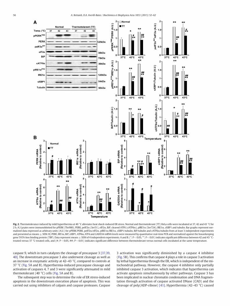
Fig. 2. Thermotolerance induced bymild hyperthermia at 40 °C alleviates heat shock-induced ER stress. Normal and thermotolerant (TT) HeLa cells were incubated at 37, 42 and 43 °C for2 h. A) Lysateswere immunoblotted for pPERK (Thr980), PERK, peIF2α (Ser51), eIF2α, BiP, cleaved ATF6 (cATF6α), pIRE1α (Ser724), IRE1α, sXBP1 and tubulin. Bar graphs represent nor-malized data expressed as arbitrary units (A.U.) for pPERK/PERK, peIF2α/eIF2α, pIRE1α/IRE1α, sXBP1/tubulin, BiP/tubulin and cATF6α/tubulin from at least 3 independent experimentsand presented asmeans± SEM. B) PERK, IRE1α, BiP, sXBP1, ATF6α, ATF4 andGADD34mRNA levelsweremeasured by quantitative real-time PCR and normalized against the housekeepinggene TATA-boxbinding protein (TBP). Data representmeans±SEMof 4 independent experiments. A andB. (*; P b 0.05, **; P b 0.01) indicates significant difference between 42 and 43 °C-treated versus 37 °C-treated cells, and (#; P b 0.05, ##; P b 0.01) indicates significant difference between thermotolerant versus normal cells incubated at the same temperature.
56 A. Bettaieb, D.A. Averill-Bates / Biochimica et Biophysica Acta 1853 (2015) 52–62
caspase 9, which in turn catalyzes the cleavage of procaspase 3 [37,39,40]. The downstream procaspase 3 also underwent cleavage as well asan increase in enzymatic activity at 42–43 °C, compared to controls at37 °C (Fig. 5A and B). Hyperthermia-induced procaspase cleavage andactivation of caspases 4, 7 and 3 were significantly attenuated in mildthermotolerant (40 °C) cells (Fig. 5A and B).
The subsequent step was to determine the role of ER stress-inducedapoptosis in the downstream execution phase of apoptosis. This wascarried out using inhibitors of calpain and caspase proteases. Caspase
3 activation was significantly diminished by a caspase 4 inhibitor(Fig. 5B). This confirms that caspase 4 plays a role in caspase 3 activationby lethal hyperthermia through the ER, which is independent of themi-tochondrial pathway. However, the caspase 4 inhibitor only partiallyinhibited caspase 3 activation, which indicates that hyperthermia canactivate apoptosis simultaneously by other pathways. Caspase 3 hasbeen implicated in nuclear chromatin condensation and DNA fragmen-tation through activation of caspase activated DNase (CAD) and thecleavage of poly(ADP-ribose) [41]. Hyperthermia (42–43 °C) caused

Fig. 3.Mild thermotolerance at 40 °C decreases heat shock-induced CHOP expression and nuclear translocation. Normal and thermotolerant HeLa cells were incubated at 37, 42 and 43 °Cfor 3 h. A) CHOPmRNA levelsweremeasured byquantitative real-time PCR and normalized against TATA boxbinding protein (TBP) as housekeeping gene. Data representmeans±SEMof4 independent experiments. Total (B) and nuclear (C) protein levels of CHOP. Bar graphs represent normalized data expressed as arbitrary units (A.U.) for CHOP/tubulin (B) or CHOP/histone B (C) from at least 3 independent experiments and presented as means ± SEM. (*; P b 0.05, **; P b 0.01) indicates significant difference between 42 and 43 °C-treated versus37 °C-treated cells, and (#; P b 0.05, ##; P b 0.01) indicates significant difference between thermotolerant versus normal cells incubated at the same temperature.
57A. Bettaieb, D.A. Averill-Bates / Biochimica et Biophysica Acta 1853 (2015) 52–62
chromatin condensation in cells, relative to controls at 37 °C (Fig. 5Cand D). Mild thermotolerance at 40 °C decreased the percentage ofapoptotic cells at 42 °C and 43 °C (Fig. 5C and D). Interestingly,hyperthermia-induced chromatin condensation was inhibited signifi-cantly by a calcium chelator BAPTA-AM and by a caspase 4 inhibitor,but not by a calpain inhibitor (Fig. 5C and D). Apoptosis was only par-tially inhibited by BAPTA-AM and the caspase 4 inhibitor. Propidium io-dide was used to distinguish between cell death by apoptosis andnecrosis. The percentages of necrotic cells at 42 and 43 °C were muchlower than those of apoptotic cells (Fig. 5E), and they were not alteredby BAPTA-AM or the caspase 4 and calpain inhibitors.
3.5. Hsp72 plays a role in the protective effect of mild thermotoleranceagainst activation of ER stress and apoptosis by hyperthermia
The development of thermotolerance by exposure of HeLa cells tomild hyperthermia (40 °C) for 3 to 24 h led to the accumulation of sev-eral Hsps [29,30]. Among them, Hsp72 protected rat pheochromocyto-ma PC12 cells from apoptosis induced by the ER stressors thapsigarginand tunicamycin [42]. To investigate themolecular mechanisms under-lying regulation of ER stress and ER stress-mediated apoptosis by mildthermotolerance (40 °C), we determined the effects of Hsp72 deficien-cy on these processes. HeLa cells with stable knockdown (KD) of Hsp72(sh-Hsp72) and control cells using sh-scramble (sh-SCR) were generat-ed using lentiviral shRNA. Immunoblot analysis demonstrated a signif-icant decrease in Hsp72 expression by about 40.3% in knockdown cellsat 37 °C (Fig. 6A, white bars). Furthermore, there was no increase inHsp72 in thermotolerant KD cells (TT + sh-Hsp72) as compared tothermotolerant WT cells (TT + sh-SCR) (Fig. 6A). The total Hsp72 (in-ducible + constitutive) expression in thermotolerant KD cells after
24 h was only 16.7% compared to thermotolerant cells (black bars:TT + sh-SCR versus TT + sh-Hsp72).
Hyperthermia (42–43 °C for 2 h) increased ER stress in normal(non-thermotolerant, 3 h at 37 °C) cells (sh-SCR) that was diminishedin thermotolerant (3 h at 40 °C) (TT + sh-SCR control) cells (Fig. 6B).To determine the role of Hsp72 in the protective effects of mild thermo-tolerance at 40 °C, we compared the magnitude of the induction of ERstress by hyperthermia in thermotolerant (TT + sh-SCR control) andthermotolerant Hsp72 KD (TT + sh-Hsp72) cells. Hsp72 deficiency inthermotolerant cells increased ER stress and counteracted the protec-tive effects of mild hyperthermia (40 °C) as evidenced by increasedphosphorylation of PERK (Thr980), eIF2α (Ser51) and IRE1α (Ser724),and increased expression of sXBP1 (Fig. 6B). Additionally, Hsp72 KD al-leviated the protective effects of mild thermotolerance against the in-duction of ER stress-mediated apoptosis by hyperthermia (42–43 °C)and sensitized thermotolerant KD cells to the activation of caspases 4,7 and 3 (Fig. 6C). Together, these data indicate that Hsp72 plays an im-portant role inmediating the protective effects of mild thermotolerance(40 °C) against ER stress-mediated apoptosis triggered by lethal hyper-thermia at 42–43 °C.
4. Discussion
This study shows that the exposure of human cervical cancer cells tolethal hyperthermia at 42 to 43 °C for 2 h activated the 3 branches of theER stress response; therewas increased phosphorylation of PERK, eIF2αand IRE1α, cleavage of ATF6, and increased expression of sXBP1. WhenHeLa cells were exposed to lethal hyperthermia for a longer time of 3 h,they underwent apoptosis through the ER. This involved the inductionof CHOP, alterations in calcium levels and the activation of ER proteases:

Fig. 4.Mild thermotolerance protects against heat shock-induced calcium release and activation of calcium-dependent proteases. A)Normal and thermotolerantHeLa cellswere incubatedat 37, 42 and 43 °C for 1 h and then loadedwith Fluo 3-AM for 20min. Histograms represent relative levels of cytoplasmic Ca2+. B) Bar graph represents green fluorescence (FL-1) thatwasquantified usingWinMDI software and normalized to normal cells incubated at 37 °C (100%) and presented asmeans± SEM (n≥ 3). C) Immunoblots of calpastatin, calpain and vinculinin lysates of normal and thermotolerant cells that were incubated at 37, 42 and 43 °C for 3 h. Bar graphs represent normalized data for cleaved (c) forms of each protein, presented asmeans ± SEM (n ≥ 3). D) Enzymatic calpain activity in normal and thermotolerant HeLa cells incubated at 37, 42 and 43 °C for 3 h. Bar graph represents normalized data presented asmeans± SEM. B, C and D. (*; P b 0.05, **; P b 0.01) indicates significant difference between 42 and 43 treated versus 37 °C treated cells, and (#; P b 0.05, ##; P b 0.01) indicates significantdifference between thermotolerant versus normal cells incubated at the same temperature.
58 A. Bettaieb, D.A. Averill-Bates / Biochimica et Biophysica Acta 1853 (2015) 52–62
the calpain-calpastatin proteolytic system and caspase 4. On the otherhand, mild thermotolerance induced by pre-conditioning of HeLa cellsat a non-lethal temperature of 40 °C diminished activation of the ERstress response and protected cells against apoptosis induced by lethaldoses of hyperthermia (42–43 °C). These protective effects ofmild ther-motolerance were mediated at least in part through the induction ofHsp72. shRNA mediated KD of Hsp72 mitigated the protective effectsof mild thermotolerance against the induction of ER stress and ERstress-mediated apoptosis by lethal hyperthermia. Together, these find-ings reveal an important role for Hsp72 in mediating the adaptive sur-vival response induced by heat preconditioning of tumor cells at a lowdose, fever temperature of 40 °C.
Hyperthermia has been shown to activate the ER stress response inseveral cell types including cancer cells. An in vivo study showed that hy-perthermia (45 °C for 50min) activated ER stressmarkers (XBP1 splicing
and eIF2αphosphorylation) in the rat cortex [43]. In humangliomaA172cells, exposure to heat at 40.5, 42 or 43.5 °C for 40min induced ER stressthrough increased eIF2α phosphorylation and XBP1 splicing resulting inthe induction ofGADD34, CHOP and BiP gene expression [43].We obtain-ed similar ER stress responses in hyperthermia-treated HeLa cells,although the 3 branches, PERK/eIF2α, ATF6 and IRE1α/XBP1, were acti-vated. Conversely, hyperthermia (43 °C for 3 to 12 h) inhibited the in-duction of ER stress response genes in AD293 cells, whereas several ERstress genes including BiP, CHOP and GADD34 as well as XBP1 splicingwere induced by mild hyperthermia (5 h at 40 °C) [44]. These findingsseem to be specific to AD293 cells since responses were low or absentin hepatic cells, insulin-secreting cells and mouse embryonic fibroblasts[44]. Findings for heat–induced ER stress responses seem to vary be-tween different cell and tissue types and warrant additionalinvestigation.

Fig. 5.Mild thermotolerance at 40 °C protects cells against heat shock-induced apoptosis. Normal and thermotolerant HeLa cells incubated at 37, 42 and 43 °C for 3 h. A) Immunoblots ofcleaved forms of caspases 4, 7 and 3 in cell lysates. Bar graphs represent normalized data presented asmeans±SEM from4 independent experiments. B) Enzymatic activity of caspases 4, 7and 3 in cell lysates. For caspase 3 activity, cells were pretreated with a caspase 4 inhibitor. Bar graphs represent normalized data presented as means ± SEM. C) Normal cells were pre-treated with the indicated inhibitor as described in the Methods section. Cells were stained for apoptosis (Hoechst 33258, green) and necrosis (PI, red). Bar graphs represent apoptosis(D) and necrosis (E) levels and presented as means ± SEM from at least 3 independent experiments. A, B and D. (*; P b 0.05, **; P b 0.01) indicates significant difference between 42and 43 treated versus 37 °C treated cells, and (#; P b 0.05, ##; P b 0.01) indicates significant difference between indicated conditions versus normal cells incubated at the same temper-ature. (^; P b 0.05, ^^; P b 0.01) indicates significant difference with and without inhibitor.
59A. Bettaieb, D.A. Averill-Bates / Biochimica et Biophysica Acta 1853 (2015) 52–62

Fig. 6.Hsp72 deficiencymitigates heat shock-induced ER stress and caspase activation. A) Immunoblots of Hsp72 expression in normal non-thermotolerant and thermotolerant (TT) HeLa cellsexpressing a scrambled (SCR) control-shRNAor shRNA targeted toHsp72 (sh-Hsp72)upon3hexposure to40 °C followedby0hor 24h recovery at 37 °C. Bar graphs represent normalizeddataexpressed as arbitrary units (A.U.) for Hsp72/tubulin from 3 independent experiments. (*) indicates significant difference between cells incubated at 42 or 43 °C versus cells incubated at 37 °C.B) Lysates from normal and thermotolerant cells expressing shRNA targeted to Hsp72 (sh-Hsp72) or a scrambled control-shRNA and exposed for 2 h to 42 or 43 °C were immunoblotted forpPERK (Thr980), PERK, peIF2α (Ser51), eIF2α, BiP, pIRE1α (Ser724), IRE1α, spliced XBP1 (sXBP1), and tubulin. Bar graphs represent normalized data expressed as arbitrary units (A.U.) forpPERK/PERK, peIF2α/eIF2α, BiP/tubulin, pIRE1α/IRE1α and sXBP1/tubulin from 3 independent experiments and presented as means + SEM. C) Lysates from normal and thermotolerantcells expressing sh-Hsp72 or a scrambled control-shRNA and exposed for 2 h to 42 or 43 °Cwere immunoblotted for active caspases 4, 7 and 3. B and C. (*; P b 0.05, **; P b 0.01) indicates sig-nificant difference between 42 and 43 °C versus 37 °C for each cell type. (#; P b 0.05, ##; P b 0.01) indicates significant difference between indicated cell type and sh-SCR for the same tem-perature. (^; P b 0.05, ^^; P b 0.01) indicates significant difference between TT + sh-Hsp72 and TT + sh-SCR cells.
60 A. Bettaieb, D.A. Averill-Bates / Biochimica et Biophysica Acta 1853 (2015) 52–62
Apoptosis and necrosis are the best describedmechanisms bywhichdifferent stresses can cause cell death. The induction of apoptosis andnecrosis by hyperthermia is dependent on the dose and duration ofheat stress [30]. Hyperthermia (41–45 °C) can induce apoptosisthrough thedeath receptor,mitochondrial [8,29,30,45–47] and ER path-ways [43,44,48]. The mechanisms through which lethal hyperthermiacauses ER stress-induced cell death are poorly understood. Inmelanomaand non-melanoma skin cancer cells, hyperthermia caused ER stress-mediated apoptosis that involved caspase 4/12 [48]. Our current studyprovides new insights into the molecular mechanisms underlying ERstress-mediated apoptosis in HeLa cells. Hyperthermia (42–43 °C) in-creased the expression and nuclear translocation of the pro-apoptotictranscription factor CHOP. The activation of PERK and ATF6 leads totranscriptional induction of CHOP, which appears to mediate apoptosis
by up-regulating the expression of genes such as GADD34 and BH3-onlypro-apoptotic proteins Bim, PUMA and Bax and/or by inhibiting gene ex-pression of Bcl-2 [49]. Hyperthermia is known to alter the permeabilityof plasmamembranes resulting in a calcium spike [10].We showed thatER stress-induced apoptosis was mediated by the calcium-dependentproteolytic system calpain-calpastatin, which resulted in calpain-dependent activation of the ER resident caspase 4. Furthermore, caspase4 played a role in caspase 3 activation and DNA fragmentation by lethalhyperthermia at 42 to 43 °C.
Thermotolerance is an adaptive survival response induced by lowdose heat preconditioning in which cells become resistant to exposureto a subsequent toxic stress such as heat shock, oxidative stress or envi-ronmental toxins [29,37,50,51]. Adaptive survival responses allow cellsand organisms to continue their normal functions despite exposure to

61A. Bettaieb, D.A. Averill-Bates / Biochimica et Biophysica Acta 1853 (2015) 52–62
an adverse stimulus. These responses involve a variety of survival strat-egies that appear to bemediated by a group of anti-apoptotic genes andtheir products (e.g. Hsps, antioxidants), which protect cells against di-verse toxic and environmental stresses [52]. If the adaptive survival re-sponse cannot protect the cell against a toxic stress exposure, then thedamaged cell will be removed by death processes such as apoptosisand/or necrosis. This study shows that mild thermotolerance inducedby lowdosehyperthermia (40 °C) diminished the activation of ER stressmarkers by subsequent lethal heat shock at 42–43 °C in HeLa cells. Inaddition, mild thermotolerance (40 °C) protected cells against lethalhyperthermia-induced pro-apoptotic events including the disruptionof calcium homeostasis, the activation of calpain and caspase 4, and nu-clear chromatin condensation.
It is worth noting that Hsp72, the stress inducible form of cytosolicHsp70, inhibits several features of the intrinsic and extrinsic apoptoticpathways [45–47,53]. However, the detailed molecular mechanisms bywhich Hsp72 inhibits ER stress and apoptosis through the ER are not en-tirely clear. This study shows thatHsp72 deficiency alleviated the protec-tive effects of mild thermotolerance (40 °C) against hyperthermia inHeLa cells. This suggests that Hsp72 is a key element that diminisheshyperthermia-induced ER stress, which occurred at the level of PERK/eIF2α, ATF6 and IRE1α/XBP1. In addition, Hsp72 deficiency abrogatedthe protective effect of mild thermotolerance against hyperthermia-induced apoptosis through the ER in HeLa cells. Further, Hsp72 wasshown to protect PC12 cells from apoptosis caused by the ER stress in-ducers thapsigargin and tunicamycin [42]. The protective effect involveda direct physical interaction between Hsp72 and the cytosolic domain ofIRE1α that enhanced IRE1α/XBP1 signalling at the ER, promoting adap-tation to ER stress and cell survival. Hsp72 enhanced cell survival underER stress conditions, in particular the IRE1α/XBP1 axis. The overexpres-sion of Hsp72 decreased the induction of ER stress markers (BiP, CHOP,XBP1) by tumor necrosis factor (TNF)-α in insulinoma-derived MIN6β-cells [54]. Moreover, other studies have shown that Hsp72 inhibitsCHOP-induced apoptosis through binding to Bax and preventing itstranslocation to mitochondria in RAW 264.7 macrophages [55].
It appears that the harsh conditions of the tumormicroenvironmentsuch as low nutrient supply, hypoxia, low extracellular pH, nutrientdeprivation and metabolic changes could lead to ER stress [56]. In tu-mors, the balance between the level of ER stress and the capacity ofthe cell towithstand toxic insults could determine cell outcome: surviv-al versus cell death [57,58]. The ER stress response could therefore pro-vide a survival and proliferative advantage whereas apoptosis throughthe ER could inhibit tumor growth. Treatments that aim to diminishthe adaptation response of ER stress and increase the pro-apoptotic as-pects of the ER signalling pathway could therefore be beneficial for theelimination of tumors [58,59]. In this regard, hyperthermia could beuseful in cancer therapy through regulating ER stress pathways intumor cells.
Acknowledgements
The authors thank Mr Bertrand Fournier (Service de consultation enanalyse de données, Université du Québec à Montréal) for statisticalanalyses, Mr Denis Flipo (MSc) for assistance with FACscan analyses,and NSERC Canada for financial support (DAB). AB receives researchsupport from NIDDK (K99DK100736).
References
[1] J. van der Zee, Heating the patient: a promising approach? Ann. Oncol. 13 (2002)1173–1184.
[2] R.D. Issels, High-risk soft tissue sarcoma: clinical trial and hyperthermia combinedchemotherapy, Int. J. Hyperthermia 22 (2006) 235–239.
[3] M. Franckena, J. van der Zee, Use of combined radiation and hyperthermia for gyne-cological cancer, Curr. Opin. Obstet. Gynecol. 22 (2010) 9–14.
[4] L.H. Lindner, R.D. Issels, Hyperthermia in soft tissue sarcoma, Curr. Treat. Options inOncol. 12 (2011) 12–20.
[5] J. Van Der Zee, M. De Bruijne, J.W. Mens, A. Ameziane, M.P. Broekmeyer-Reurink, T.Drizdal, M. Linthorst, G.C. Van Rhoon, Reirradiation combined with hyperthermia inbreast cancer recurrences: overview of experience in Erasmus MC, Int. J. Hyperther-mia 26 (2010) 638–648.
[6] A. Westermann, O. Mella, J. Van Der Zee, E.L. Jones, E. Van Der Steen-Banasik, P.Koper, A.L. Uitterhoeve, R. De Wit, J. Van Der Velden, C. Burger, B.C. Schem, C. VanDer Wilt, O. Dahl, L.R. Prosnitz, H. Van Tinteren, Long-term survival data of triplemodality treatment of stage IIB-III-IVA cervical cancer with the combination of ra-diotherapy, chemotherapy and hyperthermia - an update, Int. J. Hyperthermia 28(2012) 549–553.
[7] T.M. Zagar, J.R. Oleson, Z. Vujaskovic, M.W. Dewhirst, O.I. Craciunescu, K.L. Blackwell,L.R. Prosnitz, E.L. Jones, Hyperthermia combined with radiation therapy for superfi-cial breast cancer and chest wall recurrence: a review of the randomised data, Int. J.Hyperthermia 26 (2010) 612–617.
[8] R.S. Milleron, S.B. Bratton, ‘Heated’ debates in apoptosis, Cell. Mol. Life Sci. 64 (2007)2329–2333.
[9] K. Richter, M. Haslbeck, J. Buchner, The heat shock response: life on the verge ofdeath, Mol. Cell 40 (2010) 253–266.
[10] J.L. Roti Roti, Cellular responses to hyperthermia (40–46 degrees C): cell killing andmolecular events, Int. J. Hyperthermia 24 (2008) 3–15.
[11] B. Hildebrandt, P. Wust, O. Ahlers, A. Dieing, G. Sreenivasa, T. Kerner, R. Felix, H.Riess, The cellular and molecular basis of hyperthermia, Crit. Rev. Oncol. Hematol.43 (2002) 33–56.
[12] L.A. Sonna, J. Fujita, S.L. Gaffin, C.M. Lilly, Invited review: Effects of heat and cold stresson mammalian gene expression, J. Appl. Physiol. (1985) 92 (2002) 1725–1742.
[13] S. Lindquist, The heat-shock response, Annu. Rev. Biochem. 55 (1986) 1151–1191.[14] J.R. Lepock, How do cells respond to their thermal environment? Int. J. Hyperther-
mia 21 (2005) 681–687.[15] W.J. Welch, J.P. Suhan, Morphological study of the mammalian stress response:
characterization of changes in cytoplasmic organelles, cytoskeleton, and nucleoli,and appearance of intranuclear actin filaments in rat fibroblasts after heat-shocktreatment, J. Cell Biol. 101 (1985) 1198–1211.
[16] M. Schroder, R.J. Kaufman, The mammalian unfolded protein response, Annu. Rev.Biochem. 74 (2005) 739–789.
[17] R.J. Kaufman, D. Scheuner, M. Schroder, X. Shen, K. Lee, C.Y. Liu, S.M. Arnold, The un-folded protein response in nutrient sensing and differentiation, Nat. Rev. Mol. CellBiol. 3 (2002) 411–421.
[18] D. Ron, P. Walter, Signal integration in the endoplasmic reticulum unfolded proteinresponse, Nat. Rev. Mol. Cell Biol. 8 (2007) 519–529.
[19] G.S. Hotamisligil, Endoplasmic reticulum stress and the inflammatory basis of met-abolic disease, Cell 140 (2010) 900–917.
[20] S. Hummasti, G.S. Hotamisligil, Endoplasmic reticulum stress and inflammation inobesity and diabetes, Circ. Res. 107 (2010) 579–591.
[21] K.M. Rouschop, L.J. Dubois, T.G. Keulers, T. van den Beucken, P. Lambin, J. Bussink, A.J. van der Kogel, M. Koritzinsky, B.G. Wouters, PERK/eIF2alpha signaling protectstherapy resistant hypoxic cells through induction of glutathione synthesis andprotection against ROS, Proc. Natl. Acad. Sci. U. S. A. 110 (2013) 4622–4627.
[22] H. Zinszner, M. Kuroda, X. Wang, N. Batchvarova, R.T. Lightfoot, H. Remotti, J.L.Stevens, D. Ron, CHOP is implicated in programmed cell death in response to im-paired function of the endoplasmic reticulum, Genes Dev. 12 (1998) 982–995.
[23] H. Nishitoh, A. Matsuzawa, K. Tobiume, K. Saegusa, K. Takeda, K. Inoue, S. Hori, A.Kakizuka, H. Ichijo, ASK1 is essential for endoplasmic reticulum stress-induced neu-ronal cell death triggered by expanded polyglutamine repeats, Genes Dev. 16(2002) 1345–1355.
[24] R. Sano, J.C. Reed, ER stress-induced cell death mechanisms, Biochim. Biophys. Acta1833 (2013) 3460–3470.
[25] K.D. McCullough, J.L. Martindale, L.O. Klotz, T.Y. Aw, N.J. Holbrook, Gadd153 sensi-tizes cells to endoplasmic reticulum stress by down-regulating Bcl2 and perturbingthe cellular redox state, Mol. Cell. Biol. 21 (2001) 1249–1259.
[26] G.C. Shore, F.R. Papa, S.A. Oakes, Signaling cell death from the endoplasmic reticu-lum stress response, Curr. Opin. Cell Biol. 23 (2011) 143–149.
[27] T. Nakagawa, J. Yuan, Cross-talk between two cysteine protease families. Activationof caspase-12 by calpain in apoptosis, J. Cell Biol. 150 (2000) 887–894.
[28] P. Pinton, R. Rizzuto, Bcl-2 and Ca2+ homeostasis in the endoplasmic reticulum,Cell Death Differ. 13 (2006) 1409–1418.
[29] A. Bettaieb, D.A. Averill-Bates, Thermotolerance induced at a mild temperature of 40degrees C protects cells against heat shock-induced apoptosis, J. Cell. Physiol. 205(2005) 47–57.
[30] A. Bettaieb, D.A. Averill-Bates, Thermotolerance induced at a fever temperature of40 degrees C protects cells against hyperthermia-induced apoptosis mediated bydeath receptor signalling, Biochem. Cell Biol. 86 (2008) 521–538.
[31] B. Jakubczak, M. Wasik, K. Popko, U. Demkow, Kinetics of calcium ion concentrationaccompanying signal transduction in neutrophils from children with increased sus-ceptibility to infections, J. Physiol. Pharmacol. 57 (Suppl. 4) (2006) 131–137.
[32] E. Kojima, A. Takeuchi, M. Haneda, A. Yagi, T. Hasegawa, K. Yamaki, K. Takeda, S.Akira, K. Shimokata, K. Isobe, The function of GADD34 is a recovery from a shutoffof protein synthesis induced by ER stress: elucidation by GADD34-deficient mice,FASEB J. 17 (2003) 1573–1575.
[33] S. Oyadomari, H.P. Harding, Y. Zhang, M. Oyadomari, D. Ron, Dephosphorylation oftranslation initiation factor 2alpha enhances glucose tolerance and attenuateshepatosteatosis in mice, Cell Metab. 7 (2008) 520–532.
[34] M.A. Smith, R.G. Schnellmann, Calpains, mitochondria, and apoptosis, Cardiovasc.Res. 96 (2012) 32–37.
[35] K. Serrano, D.V. Devine, Vinculin is proteolyzed by calpain during platelet aggrega-tion: 95 kDa cleavage fragment associates with the platelet cytoskeleton, CellMotil. Cytoskeleton 58 (2004) 242–252.

62 A. Bettaieb, D.A. Averill-Bates / Biochimica et Biophysica Acta 1853 (2015) 52–62
[36] J. Hitomi, T. Katayama, Y. Eguchi, T. Kudo, M. Taniguchi, Y. Koyama, T. Manabe, S.Yamagishi, Y. Bando, K. Imaizumi, Y. Tsujimoto, M. Tohyama, Involvement ofcaspase-4 in endoplasmic reticulum stress-induced apoptosis and Abeta-inducedcell death, J. Cell Biol. 165 (2004) 347–356.
[37] P. Pallepati, D.A. Averill-Bates, Activation of ER stress and apoptosis by hydrogenperoxide in HeLa cells: protective role of mild heat preconditioning at 40 degreesC, Biochim. Biophys. Acta 1813 (2011) 1987–1999.
[38] S. Matsuzaki, T. Hiratsuka, R. Kuwahara, T. Katayama, M. Tohyama, Caspase-4 is par-tially cleaved by calpain via the impairment of Ca2+ homeostasis under the ERstress, Neurochem. Int. 56 (2010) 352–356.
[39] M.V. Fiandalo, N. Kyprianou, Caspase control: protagonists of cancer cell apoptosis,Exp. Oncol. 34 (2012) 165–175.
[40] A. Yamamuro, T. Kishino, Y. Ohshima, Y. Yoshioka, T. Kimura, A. Kasai, S. Maeda,Caspase-4 directly activates caspase-9 in endoplasmic reticulum stress-inducedapoptosis in SH-SY5Y cells, J. Pharmacol. Sci. 115 (2011) 239–243.
[41] S. Nagata, Apoptotic DNA fragmentation, Exp. Cell Res. 256 (2000) 12–18.[42] S. Gupta, A. Deepti, S. Deegan, F. Lisbona, C. Hetz, A. Samali, HSP72 protects cells
from ER stress-induced apoptosis via enhancement of IRE1alpha-XBP1 signalingthrough a physical interaction, PLoS Biol. 8 (2010) e1000410.
[43] Y. Liu, H. Sakamoto, M. Adachi, S. Zhao, W. Ukai, E. Hashimoto, M. Hareyama, T.Ishida, K. Imai, Y. Shinomura, Heat stress activates ER stress signals which suppressthe heat shock response, an effect occurring preferentially in the cortex in rats, Mol.Biol. Rep. 39 (2012) 3987–3993.
[44] X. Xu, S. Gupta, W. Hu, B.C. McGrath, D.R. Cavener, Hyperthermia induces the ERstress pathway, PLoS One 6 (2011) e23740.
[45] H.M. Beere, Death versus survival: functional interaction between the apoptotic andstress-inducible heat shock protein pathways, J. Clin. Invest. 115 (2005) 2633–2639.
[46] R. Steel, J.P. Doherty, K. Buzzard, N. Clemons, C.J. Hawkins, R.L. Anderson, Hsp72 in-hibits apoptosis upstream of the mitochondria and not through interactions withApaf-1, J. Biol. Chem. 279 (2004) 51490–51499.
[47] A.R. Stankiewicz, G. Lachapelle, C.P. Foo, S.M. Radicioni, D.D. Mosser, Hsp70 inhibitsheat-induced apoptosis upstream of mitochondria by preventing Bax translocation,J. Biol. Chem. 280 (2005) 38729–38739.
[48] Y.G. Shellman,W.R. Howe, L.A. Miller, N.B. Goldstein, T.R. Pacheco, R.L. Mahajan, S.M.LaRue, D.A. Norris, Hyperthermia induces endoplasmic reticulum-mediated apopto-sis in melanoma and non-melanoma skin cancer cells, J. Investig. Dermatol. 128(2008) 949–956.
[49] J.D. Malhotra, R.J. Kaufman, The endoplasmic reticulum and the unfolded protein re-sponse, Semin. Cell Dev. Biol. 18 (2007) 716–731.
[50] J. Landry, P. Chretien, D. Bernier, L.M. Nicole, N. Marceau, R.M. Tanguay, Thermotol-erance and heat shock proteins induced by hyperthermia in rat liver cells, Int. J.Radiat. Oncol. Biol. Phys. 8 (1982) 59–62.
[51] J.L. Martindale, N.J. Holbrook, Cellular response to oxidative stress: signaling forsuicide and survival, J. Cell. Physiol. 192 (2002) 1–15.
[52] L. Portt, G. Norman, C. Clapp, M. Greenwood, M.T. Greenwood, Anti-apoptosis andcell survival: a review, Biochim. Biophys. Acta 1813 (2011) 238–259.
[53] D.D. Mosser, L.H. Martin, Induced thermotolerance to apoptosis in a human Tlymphocyte cell line, J. Cell. Physiol. 151 (1992) 561–570.
[54] T. Kondo, K. Sasaki, R. Matsuyama, S. Morino-Koga, H. Adachi, M.A. Suico, J.Kawashima, H. Motoshima, N. Furukawa, H. Kai, E. Araki, Hyperthermia with mildelectrical stimulation protects pancreatic beta-cells from cell stresses and apoptosis,Diabetes 61 (2012) 838–847.
[55] T. Gotoh, K. Terada, S. Oyadomari, M. Mori, hsp70-DnaJ chaperone pair preventsnitric oxide- and CHOP-induced apoptosis by inhibiting translocation of Bax to mi-tochondria, Cell Death Differ. 11 (2004) 390–402.
[56] J.D. Blais, C.L. Addison, R. Edge, T. Falls, H. Zhao, K. Wary, C. Koumenis, H.P. Harding,D. Ron, M. Holcik, J.C. Bell, Perk-dependent translational regulation promotes tumorcell adaptation and angiogenesis in response to hypoxic stress, Mol. Cell. Biol. 26(2006) 9517–9532.
[57] B. Mollereau, Establishing links between endoplasmic reticulum-mediated hormesisand cancer, Mol. Cell. Biol. 33 (2013) 2372–2374.
[58] F. Martinon, Targeting endoplasmic reticulum signaling pathways in cancer, ActaOncol. 51 (2012) 822–830.
[59] A.H. Schonthal, Pharmacological targeting of endoplasmic reticulum stress signalingin cancer, Biochem. Pharmacol. 85 (2013) 653–666.