Therapeutic Embolization for Vascular Trauma of the Head and Neck
6
C. Mark Mehringer 1 Grant B. Hieshima 1 Verity S. Grinnell' Fong Y. Tsai 2 John R. Bentson 3 Anton N. Hasso 4 Joseph R. Thompson s Charles Strother s Henry F. Pribram 6 Received February 1 8, 1982; accepted after rev ision September 30, 1982. Presented at the annual meet in g of the Ameri- can Society of Neuroradiology, Los Angeles, March 1 980. 'Department of Radiology, Harbor-UCLA Med- ical Center, Torrance, CA 90509. Address reprint requests to C. M. Mehringer. 2Department of Radiology, University of Califor- nia, Irvine Medi ca l Center, Irvine, CA 92664. ' Department of Radiology, UCLA Center for Health Scienc es, Los Angeles, CA 90024 . ' Department of Radiology, Loma Linda Univer- sity Medical Center, Loma Linda, CA 92354 . 5Department of Radiology , University of Wis- cons in Medical Center, Madison, WI 53706. 6Department of Radiology , University of Califor- ni a, Irvine, CA 92664, and Long Beach V.A. Hos- pital, Long Beach, CA 90822 . AJNR 4:137-142, March / April 1983 0195-6108 / 83 / 0402-0137 $00 . 00 © American Roentgen Ray Society Therapeutic Embolization for Vascular Trauma of the Head and Neck 137 Therapeutic embolization is an effective and relatively safe method for managing many cases of head and neck trauma. In the last 5 years , 78 traumatic vascular lesions-10 arterial transections and 68 arteriovenous fistula-were treated by intra- vascular embolization at four medical centers . Selection of embolic materials is dis- cussed and different types of lesions are illustrated. Treatment was successful in every instance. Complications were limited to one case of cerebral infarction and two cases of temporary oculomotor weakness . The indications for embolization have widened beyond life-threatening hemorrhage alone , and continued improvement in techniques and embolic agents should see an increased use of this form of treatment . Traumatic vascular injuries of the head and neck include vessel tr an sect ion and laceration, false aneurysm formation , and arteriovenous fistula . Such injuries may result from either closed or penetrating trauma and their pr esentation can be either immediate or delayed . In the past , management of these injur ies had consisted mostly of surgery or observation . In many instance s surg ica l treatment is efficacious, but in others the location of the injury may make surgery difficult or unacceptably dangerous. In such cir cumsta nces we have used therapeutic embolization techniques as an ad jun ct to facilitate surgery or as the definitive treatment. Our experience in treat ing 78 patients with traumatic vascular lesions of the head and neck over the past 5 years forms the basis of this report. Many of the patients were bleeding actively at the time of angiography, and this has been one of the most common indi cations for e mbolization therapy in the past . The indications for vascular occ lusive techniques are widening, how- ever, and patients with other lesions such as arteriovenous fistulas and false aneurysms have been treated in creasingly with these methods . Compl ex injuries with comb in ations of bleeding, fistulas, and false aneurysms may be particul ar ly suitab le for embolization therapy . The spec ific lesions treated in this series are summarized in table 1. Ten involved major vessel transection; the rest were arteriovenous fistulas, the most common involving the caverous sinus. Materials and Methods The technique s of vascular occl usive therapy have been descr ibed elsew here f1- 8). A number of diffe rent materials have been used success full y for vessel occ lusion. The materials th at we have used inc lude particu late embo li and balloon catheters. For particu late emboli, we prefer polyvinyl al co hol f oam (PVA, Unipoint, Ind., High Point, NC), Gelfoam (Upjohn, Kalamazoo, MI) , or barium-impr egnated Silastic sphe res (American Heyer-Sc hulte Corp ., Goleta, CAl . The balloon ca theters used have been both fixed and detachable. In early cases, a fi xed balloon (Edwards Labs. , Santa Ana, CAl was used . More recently , we have used a det ac hable Sil astic balloon catheter system [3). Th e cho ice of agent va ri es with different lesions, as no one agent is op timal for all lesions . We have chos en to ca t egorize this se ri es into groups based on the vessel size and type of injury . This, in lu rn , helps to determine the choice of agen ts used f or embo li zation. The gro ups in c lu de small or
Transcript of Therapeutic Embolization for Vascular Trauma of the Head and Neck

C. Mark Mehringer 1
Grant B. Hieshima 1
Verity S. Grinnell' Fong Y. Tsai 2
John R. Bentson3
Anton N. Hasso4
Joseph R. Thompsons Charles Strothers
Henry F. Pribram6
Received Febru ary 18, 1982; accepted after revision September 30, 1982.
Presented at the annual meeting of the American Society of Neuroradiology, Los Angeles, March 1980.
'Department of Radiology, Harbor-UCLA Medical Center, Torrance, CA 90509. Address reprint requests to C. M. Mehringer.
2Department of Radiology, University of California, Irvine Medical Center, Irvine, CA 92664.
' Department of Rad iology, UCLA Center for Health Sciences, Los Angeles, CA 90024.
' Department of Radiology, Loma Linda University Medical Center, Loma Linda, CA 92354.
5Department of Radiology, University of Wisconsin Medical Center, Madison, WI 53706.
6Department of Rad iology , University of Cali fornia, Irvine, CA 92664, and Long Beach V.A. Hospital, Long Beach, CA 90822 .
AJNR 4:137-142, March/ April 1983 0 195-6108/ 83 / 0402-0137 $00.00 © American Roentgen Ray Society
Therapeutic Embolization for Vascular Trauma of the Head and Neck
137
Therapeutic embolization is an effective and relatively safe method for managing many cases of head and neck trauma. In the last 5 years, 78 traumatic vascular lesions-10 arterial transections and 68 arteriovenous fistula-were treated by intravascular embolization at four medical centers . Selection of embolic materials is discussed and different types of lesions are illustrated. Treatment was successful in every instance. Complications were limited to one case of cerebral infarction and two cases of temporary oculomotor weakness . The indications for embolization have widened beyond life-threatening hemorrhage alone, and continued improvement in techniques and embolic agents should see an increased use of this form of treatment.
Traumatic vascular injuries of the head and neck include vesse l transection and laceration, false aneurysm formation , and arteriovenous fistula . Such injuries may result from either c losed or penetrating trauma and their presentation can be either immediate or delayed . In the past , management of these injuries had consisted mostly of surgery or observation . In many instances surg ical treatment is efficacious, but in others the location of the injury may make surgery difficult or unacceptably dangerous. In such c ircumstances we have used therapeutic embolization techniques as an adjunct to facilitate surgery or as the definitive treatment. Our experience in treat ing 78 patients with traumatic vascular lesions of the head and neck over the past 5 years forms the basis of this report.
Many of the patients were bleeding actively at the time of angiography, and this has been one of the most common indications for embolization therapy in the past . The indications for vascular occ lusive techniques are widening, however, and patients with other lesions such as arteriovenous fistulas and false aneurysms have been treated increasingly with these methods. Complex injuries with combinations of bleeding , fistulas, and false aneurysms may be particularly suitable for embolization therapy. The spec ific lesions treated in thi s series are summarized in table 1. Ten involved major vessel transection; the rest were arteriovenous f istu las, the most common involving the caverous sinus .
Materials and Methods
The techniques of vascular occlusive th erapy have been described elsewhere f1-8). A number of different material s have been used successfull y for vessel occlusion. The materials th at we have used inc lude particu late emboli and balloon catheters. For particu late emboli, we prefer polyvinyl alcohol foam (PVA, Unipoint , Ind ., High Point, NC) , Gelfoam (Upjohn, Kalamazoo, MI) , or barium-impregnated Silastic spheres (American Heyer-Schulte Corp ., Goleta, CAl. Th e balloon catheters used have been both fixed and detachable. In early cases, a fi xed balloon (Edwards Labs. , Santa Ana, CAl was used . More recently , we have used a detachable Silastic balloon catheter system [3). Th e choice of agent varies with different lesions, as no one agent is optimal for all lesions . We have chosen to categorize this series into groups based on the vessel size and type of injury . This, in lu rn , helps to determine the choice of agents used for embolization . The groups inc lude small or

138 MEHRINGER ET AL. AJNR:4, Mar. l Apr. 1983
A B
TABLE 1: Embolization for Head and Neck Trauma over 5 Years
Reason for Emboliza tion No.
Internal carot id-cavernous sinus fistula 51 Internal carot id-jugular fi stu la Internal carotid transection / laceration 4 Middle cerebral artery transection 1 External carotid-cavernous fi stula ........... 3 External carotid -jugu lar fi stula 2 External carot id branch transection 5 Meningeal arteriovenous fistula 8 Vertebral arteriovenous fistula 3
Total 78
larg e vessel lacerat ions and small or large vessel arteriovenous fistu las. Patients with complex injuries are classified under th e more c linically sign ificant injury at the time of th e procedure.
Representative Case Reports
Case 1
A 32-year-old man suffered a stab wound in th e left neck. Upon arrival at the hospital the patient was spitting up copious amounts of blood and there was an expanding cerv ica l hematoma with impending compromise of the airway . An emergency angiogram showed extravasation of contrast material from a branch of the left superior thyroid artery into a false aneurysm, which in turn fed into an arteriovenous fistula draining into the jugular vein (fig. 1 A) .
The tip of the 5 French ang iograph ic catheter was positioned near the site of the fistula. Because of the short d istance from the catheter tip to the fistu la and the desire for permanent occlusion,
Fig . 1.-Case 1 . Small vessel lacerati on. A, Lobu lated false aneurysm of left superior thyroid artery with small inc idental fistula to jugular vein. B, Occlusion of aneurysm (arrow) with sparing of nearby branches after PYA foam embolization .
PVA foam was selected for embolization. It has been our experience that this agent shows more of a tendency to lodge proximally in an artery than Gelfoam or Silastic spheres. An occlusion that is too proximal may allow continued patency of the artery via collaterals beyond the site of occlusion. This would negate the effect of embolization, and the co llateral vessels themselves might be of such size and geometry that further embolization through them would be impossible. The short distance from catheter tip to injury
site and the small size of th e artery in this case were ideal for PVA foam.
The fi stula and false aneurysm were easily occluded with 1 mm PVA foam particles injec ted through the angiographic cath eter. Th ere was no occlusion of nearby vessels (fig . 1 B) . Th e patient underwent an uneventful recovery and no surgery was performed .
Case 2
A 40-year-old man received a closed head injury. Ten weeks later he noted onset of decreased vision and diplopia bilaterally with an intracranial bruit. Carotid angiography was performed and bilateral ex ternal carotid-cavernous fi stulas were demonstrated (fig . 2A). Embolization of both extern al carotid arteries was accom
plished using about 60 1 mm Silastic spheres (fig. 2B). Within 3 days of th e embolization, all the symptoms had resolved.
Silast ic spheres were chosen in this case because of the need for an embolus with the potential to travel for some distance from the catheter tip without becoming lodged en route to the fi stula. In our experience , spheres have been more effective in this setting than Gelfoam or PVA foam.
Case 3
A 30-year-old man was shot in the right med ial orbit and developed massive epistaxis while being transported to the hospital. The pat ient was found comatose with a dense right hemipleg ia and

AJNR:4 , Mar. / Apr. 1983 THERAPEUTIC EMBOLIZATION FOR TRAUMA 139
Fig . 2. - Case 2. Small vessel arleriovenous fistula. A, Exte rnal carotid to cavernous sinus fistul a, left-sided injection . B , Closure o f fi stul a after embolization w ith Silas tic spheres. Similar result was obtained on right side.
A
A
B
B
(
c Fig. 3. -Case 3. Large vessel laceration. Frontal (A) and lateral (B) projec tions. Lett middle cerebral artery transection with ex travasation through bu llet
tract. C, Plain fi lm. Two detachable balloons in place in prox imal left midd le ce rebral artery. Hemorrhage stopped; some ex travasation remains from preembolization injecti ons.
ophthalmoplegias involving right c ranial nerves III, IV, and VI. He
was rapid ly transfused and treated with nasal packs. A sku ll series revealed that the bu llet had entered the righ t
ethmoid sinus anteriorly and exited the left sphenoid sinus to lodge in the left posterior temporal area. An arteriog ram was obtained which revealed a transection of the left middle cerebral artery and extravasation of contrast material in to the bu llet tract and out through the sphenoid sinus (fig s. 3A and 38).
Due to continued arterial hemorrhage and deteriorating neurolog ic status, an emergency embolization of the left middle cerebral artery was performed with two detachable balloons (fig . 3C).
Although the hemorrhage was stopped, the patient continued to deteriorate as a resu lt of the massive brain injury and subsequently died.
Case 4
A 22-year-old man suffered a c losed head injury in an automobile accident. Signs of a carot id-cavernous fi stula developed soon th ereafter. The right eye showed proptosis and chemosis and there was an intracranial bruit . A cerebral angiog ram (fig. 4A) documented the
right internal carot id-cavernous fi stula. Vision in the right eye began
to deteriorate and extraocular muscle palsy developed . It was decided to treat the fi stul a by use of an inflatable and detachable balloon. Two balloon s were placed through th e fistu la in the cavern ous sinus. Flow through the fi stula was marked ly slowed and at follow-up angiography 2 weeks later th e fi stula was c losed (figs. 48 and 4C). Vision and ocular movemen ts returned to normal.
This case demonstrates treatmen t of a large vessel fi stula. Inflatable and detachable balloons are ideal for such a situation. Th ey are controllable and have an expansibility greater than other emboli . The balloon can be controlled before detachment by ft ow d irec tion and mechanica l man ipu lat ion of the coax ial ca theter system. This makes precise embolus placemen t possible . The expansi le characteri st ics of balloons offer the advan tage of a large embolus withou t requiring a large catheter and arteriotomy for delivery .
Case 5
A 28-year-old man sustained a gunshot wound to his left eye . He was comatose wi th a severe orbit al injury. A cranial computed tomographic (CT) scan showed subarachnoid hemorrh age but was
140 MEHRINGER ET AL. AJNR:4 , Mar. l Apr. 1983
A B c Fig. 4. -Case 4. Large vessel arteriovenous fistul a. A , Right internal carotid to cavernous sinus fistula. B , Detachable balloon in place. C , Fistula is c losed
and internal carotid is patent. Small false aneurysm.
A B Fig. 5.-Case 5. Large vessel lacerati on and arteri ovenous fistula. A,
Lateral view of high cervica l ca rot id preembolization. Large false aneurysm of left intern al ca ro tid artery and a caro tid-cavern ous fistula (arrowlJeads ). Carotid bifurcation (arrow) at bottom of fi lm. Carotid to jugular fistula also
otherwise negative. His cond ition rapid ly deteriorated and cerebral
angiog raphy was performed . The angiogram showed a complex vascular injury (fig. 5A). There
was a large traumatic aneu rysm of the left internal carotid artery and an internal carotid-cavernous fistu la. In addition , there was a left internal carotid-jugular fistu la. The jugular vein was occluded di stal to the fistu la and the fistu lized left jugu lar venous flow was
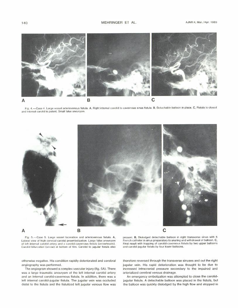
c present. B, Dislodged detachable balloon in right transverse sinus with 5 French cath eter in sinus preparatory to snaring and withdrawal of balloon. C , Final result with trapping of carotid-cavernous fistu la by two upper balloons and carot id-jugular fistula by four lower balloons.
therefore reversed through the transverse sinuses and out the r ight
jugular vein . His rapid deterioration was thought to be due to increased intracranial pressure secondary to the impaired and arterialized cerebral venous drainage.
An emergency embolization was attempted to close the carotidjugu lar fistula. A detachable balloon was placed in the fistula, but the balloon was qu ickly dislodged by the high flow and stopped in

AJNR :4 , Mar. l Apr. 1983 THERAPEUTIC EMBOLIZATION FOR TRAUMA 141
the ri ght transverse sinus. This resul t could have been catastroph ic because it further comprom ised the already abnormal cerebral venous outflow. It was considered imperative to remove the balloon. This was accomplished by manipu lation with a J gu ide wire via a percutaneous right jugular approach (fi g. 5B). The balloon was snared and withdrawn into the right jugu lar vein where it was percutaneously punctu red and allowed to pass in to the lung. Subsequentl y, the carotid-cavernous and carotid-j ugu lar fi stul as were
treated by trapping with detachable balloons (fig . 5C) . After this the patient made a slow recovery .
This rather complex case involved large vessel laceration with pseudoaneurysm formation and fistu las. The carot id-jugular fistula was thought to be contri buting to th e patient 's c linical deterioration and a prompt and complete occ lusion of the fi stu la was needed . Inflatable and detachable balloons were considered more li kely
than smaller particulate emboli to effec t rap id c losure in vessels of this size.
Discussion
Embolization therapy for traumatic vascular lesions has a number of advantages over more conventional methods of treatment. It can be performed in conjunction w ith the in iti al ang iog raphic workup, thus sav ing t ime in patients with li fethreatening lesions. It can be used to treat lesions whose anatomic locati on makes them unsuitable for surgical treatment. Morbidity after embolizati on is usuall y less than with surgery and , although there are defin ite ri sks of emboli zati on therapy , if carefull y performed , it is a safe proced ure.
The choice of embolic agents used in thi s series is based on our own earl y experi ence and has been fairly consistent since that t ime. Thi s does not imply that agents other than the ones used might not have been used successfully in any given case, however, results w ith these agents have been satisfactory thus far . Our approach to the choice of embolic agents is that small vessel lacerati ons and fi stulas at a distance from the catheter tip are treated initiall y w ith Silasti c spheres of 1 or 1.5 mm size, If th is does not effect closure of the injured vesse l or fi stul a, larger pieces of moist PVA
foam are injected . As mentioned in case 1, PVA foam in our experience has shown a tendency to become lodged more prox imall y in arteri es than Si last ic spheres, perh aps due to its more irregular surface with a tendency for part ic les to c lump together. Therefore, when the catheter can be placed very near the injury site PVA foam may be used as the ini t ial agent. Gelfoam alone is used instead of PVA foam only in those instances where temporary occ lusion is acceptable, as there is ev idence that recanalizati on can occur soon after embolizati on when Gelfoam is used [ 9].
For the large vessel arterial injuries that have invo lved vertebral, common carotid , intern al carot id , or midd le cerebral arteries , we have used Silastic balloon catheters [7]. These have been almost entirely detachable balloons of 2 mm to 2 cm in diameter when inflated. In cases of arteri ovenous fi stu la the balloons are preferably placed in the fi stul a without compromise of the arteri al lumen. When thi s is not possible the f istul a is trapped by placi ng a balloon
distall y and then proximall y on the arte ri al side. If the safety of arteri al occ lusion is questi oned the arte ry may be temporarily occ luded with the balloon before detachment and the pati ent observed for ischem ic symptoms. If there are
none, the balloon is then detached. Almost all of the balloon embolizations have been perform ed with the pati ents anticoagulated to decrease the risks of unwanted embolizati on distal to the lesion. When the pati ent is acti ve ly bleeding the value of anticoagulati on may have to be weighed against the ri sk of hemorrh age.
If a large-bore catheter can be pl aced c lose to a large vessel lesion, an effective c losure might be obtained by inject ing large pieces of sponge or Gelfoam. Thi s technique could be used in treating large vesse l lacerati ons, but offers litt le advantage over balloons and is less desi rable for large vessel fistu las because of the ri sk of prox imal occ lusion w ith continued fi stul a patency via collaterals. Al so, the lack of control over a large part iculate embolus when it is injected compared with balloon placement before detachment is a distinc t d isadvantage. The maneuverability of balloons offers the best chance for prec ise embolus placement, whi c h is necessary to c lose fi stu las.
The c lin ica l presentation of pati ents with head and neck trauma receiving embolization therapy is vari able. Acute hemorrh age is a frequent ind ication for embo li zati on therapy in general, and it is also an important presentati on in head and neck trauma. However, hemorrhage alone has not been the dominant indicati on for treatment in our experi ence, and we have seen more pati ents with arteri ovenous fi stulas than with hemorrhage . The arteriovenous f istu las are usuall y managed electively because symptoms deve lop graduall y as the f istu la increases and there is usuall y no immediate threat to life. An exception to thi s is seen in case 5 where increased intracranial pressure due to arteri ali zati on of an obstructed cerebral venous outf low was causing acute neurolog ic deteri orat ion. A carotid-cavernous fi stu la may also req uire 'emergency embolization if vision is deteri orat ing or cerebral ischemic changes develop.
Complications of embolizat ion in our seri es were limited to one patient who developed a cerebral infarc tion from thrombi, wh ich formed on a detachable balloon during its placement in a carotid-cavernous f istula. Th is patient was not ant icoagulated because of previous life-threatening epistaxis re lated to the carot id injury. Two patients developed third nerve palsies after treatment of carotid-cavern ous fi stulas but responded to exerc ise and extraocular musc leshortening procedures , the on ly subsequent surgery requi red , The proced ures have otherwise been free of any permanent seq uelae and all pat ients rece ived a total c ure of their vascular lesions.
REFERENCES
1. Athanasoulis CA. Therapeutic applications of angiog raphy:
medica l progress, part 1 . N Engl J Med 1980;302: 11 1 7 -11 25 2. Athanasou li s CA. Therapeutic applica tions of ang iog raph y:
medical progress, part 2. N Engl J Med 1980;302: 1 1 74-11 79 3. Berenstein A, Kricheff II . Catheter and materia l se lection for
transarteri al embolization: technica l considerations. I. Catheter. Radiology 1979;132: 61 0-630
4 . Berenstein A, Kricheff II . Catheter and material selec tion for transarterial embolization: technica l consideration. II . Mate
rials. Radiology 1979 ;132:631-639 5. Debrun G, Lacour P, Caron JP, et al. Detachable balloon and

142 MEHRINGER ET AL. AJNR :4, Mar. / Apr. 1983
ca librated leak balloon techniques in the treatment of cerebral vascular lesions. J Neurosurg 1978;49: 635
6. Hieshima GB, Mehringer CM , Grinnell VS, Hasso AN , Siegel NH , Pribram HFW. Emergency occlusive techniques . Surg Neuro l 1978;9: 293-302
7. Hieshima GB, Grinnell VS, Mehringer CM . A detachable balloon for therapeutic transcatheter occlusions. Radio logy
1981 ; 138: 227 -228 8. Serbinenko FA. Balloon catheterization and occlusion of major
cerebral vessels. J Neurosurg 1974;42: 125 9. Vlahos L, Benakis V, Dimakakos P, Dimopoulos C, Pontifex G.
A comparative study of degree of arterial recanalization in
kidney of dogs following transcatheter embolization with eight different materials. Eur Uro l 1980;6: 1 80-1 8 5