Therapeutic effect of quercetin and resveratrol in an in ... · Therapeutic effect of quercetin and...
82
Therapeutic effect of quercetin and resveratrol in an in vitro model of Graves ’ ophthalmopathy Jin Sook Yoon Department of Medicine The Graduate School, Yonsei University
-
Upload
hoangkhanh -
Category
Documents
-
view
219 -
download
0
Transcript of Therapeutic effect of quercetin and resveratrol in an in ... · Therapeutic effect of quercetin and...
Therapeutic effect of quercetin and resveratrol
in an in vitro model of Graves’ ophthalmopathy
Jin Sook Yoon
Department of Medicine
The Graduate School, Yonsei University
Therapeutic effect of quercetin and resveratrol
in an in vitro model of Graves’ ophthalmopathy
Directed by Professor Sang Yeul Lee
The Doctoral Dissertation
Submitted to the Department of Medicine ,
the Graduate School of Yonsei University
in partial fulfillment of the requirements for the degree
of Doctor of Philosophy
Jin Sook Yoon
June 2010

This certifies that the Doctoral Dissertation of
Jin Sook Yoon is approved.
------------------------------------ Thesis Supervisor : Sang Yeul Lee
------------------------------------ Thesis Committee Member#1: Eun Jig Lee
------------------------------------ Thesis Committee Member#2 : Joon H. Lee
------------------------------------ Thesis Committee Member#3 : Hang Seok Chang
------------------------------------ Thesis Committee Member#4 : Kyung-Soo Park
The Graduate School
Yonsei University
June 2010

ACKNOWLEDGEMENTS
My gratitude to Prof. Sang Yeul Lee is beyond description. As a mentor,
he always leads my steps in the path of righteousness and is dedicated to
supporting me. And I am deeply appreciative of Prof. Eun Jig Lee for his
great help and guidance for my search. I would further like to offer my
greatfulness to Prof. Joon H. Lee for his admirable advice and support for
me to obtain research fund for my research. I wish to express my grat itude
to Prof. Hang Se ok Cha ng and Prof. Kyung-Soo Park for their
knowledgeable advice to my thesis. I am especially grateful to Soo Hyun
Choi and Hyunjung Lee for their great help for my experiments.
I would like to express my heartful thanks to my parents providing their
endless support and unwavering love.
I would like to thank my husband, Sung Jun Lee, who always shares all
the good and bad things with me. I was able to accomplish my research
because of his great support. And most importantly, I thank God for him to
give me an adorable son, Yoon Sung, the best gift from God. I dedicate this
thesis to my husband and my only son.
Jin Sook Yoon

T ABLE OF CONTE NT S
ABSTRACT 1
I. INTRODUCTION 3
II. MATERIALS AND METHODS 8
1. Reagents and chemicals 8
2. Cell culture and differentiation protocol 9
3. Cell viability and proliferation assay 10
4. Annexin V assay for cytotoxicity 11
5. Semiquantitative RT-PCR 12
6. Hyaluronan ELISA 13
7. Oil red O stain 14
8. Western blot assay of PPAR, C/EBP, C/EBP 14
9. Statistical analysis 15
III. RESULTS 16
1. The effect of quercetin and resveatrol on cell viability 16
2. Determination of IL-1 concentration to study the maximal
inhibitory effect of quercetin and resveratrol on the inflammation
25
3. Inhibition of ICAM-1, IL-6, IL-8 and COX-2 mRNA by
quercetin and resveratrol
27
4. Quercetin inhibited hyaluronic acid production in orbital

fibroblasts 31
5. Effect of quercetin and resveratrol on the accumulation of lipid
droplets and the expression of transcriptional regulators of
adipogenesis
34
6. Quercetin’s effect on accumulation of lipid droplet and cell
viability in differentiating fibroblasts treated with IL-1 or H2O2
43
IV. DISCUSSION 52
V. CON CLUSION 60
VI. REFERENCES 61
ABSTRACT(IN KOREAN) 70

LIST OF FIGURES
Figure 1. Main pathologic mechanism of Graves’ ophthalmopathy5
Figure 2. Structure of the quercetin (A) and resveratrol (B) 9
Figure 3. Effect of quercetin on cell viability in normal and Graves’
ophthalmopathy orbital fibroblasts17
Figure 4. Effect of resveratrol on cell viability in normal and Graves’
ophthalmopathy orbital fibroblasts18
Figure 5. Effect of quercetin on cell viability during adipogenesis20
Figure 6. Effect of resveratrol on cell viability during adipogenesis·························21
Figure 7. Effects of quercetin and resveratrol on cell proliferation in normal and
Graves’ ophthalmopathy orbital fibroblasts································································23
Figure 8. Effect of quercetin and resveratrol on apoptosis in orbital preadipocyte
fibroblasts······················································································································24
Figure 9. Determination of IL-1 concentration to examine the maximal suppressive
effect of quercetin and resveratrol on the expression of proinflammatory cytokines
induced by IL-1 in Graves’ ophthalmopathy orbital fibroblasts·····························26
Figure 10. Effects of various concentrations of quercetin on IL-1 induced ICAM-1,
IL-6, IL-8 and COX-2 mRNA expression···································································28
Figure 11. Effects of quercetin for various duration of treatment on IL-1 induced
ICAM-1, IL-6, IL-8 and COX-2 mRNA expressions·················································29
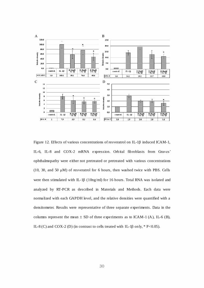
Figure 12. Effects of various concentrations of resveratrol on IL-1 induced ICAM-
1, IL-6, IL-8 and COX-2 mRNA expression······························································30
Figure 13. The effect of quercetin on hyaluronan production induced by IL-1 and
TNF- in orbital fibroblasts from normal and Graves’ ophthalmopathy
subjects··························································································································32
Figure 14. Examination of prestained orbital fibroblasts cultured in adipogenic
medium under light microscopy··················································································35
Figure 15. Microscopic examination of prestained cultures grown in adipogenic
medium containing either quercetin or resveratrol·················································37

Figure 16. Examination of oil red O stained cultures exposed to quercetin and
resveratrol with various concentrations·································································39
Figure 17. Microscopic examination of oil red O stained cultures exposed to
quercetin and resveratrol·····························································································40
Figure 18. Effect of quercetin and resveratrol on the expression of adipogenic
transcriptional regulators of differentiated orbital fibroblasts from Graves’
ophthalmopathy patients······························································································42
Figure 19. The effects of quercetin on adipocyte differentiation in cells exposed to
adipogenic medium including IL-1 and quercetin···············································45
Figure 20. Microscopic results of oil red O stain showing suppressive effect of
quercetin on adipogenesis in differentiating cells grown under adipogenic medium
including IL-1···································································································46
Figure 21. Microscopic results of oil red O stain showing protective effects of
quercetin on cell viability in differentiating orbital fibroblasts exposed to H2O2··· 48
Figure 22. MTT analyses after the treatment of quercetin in fully differentiated

orbital fibroblasts in different adipogenic condition··············································50
LIST OF TABLES
Table 1. Primer used for semiquantitative RT-PCR····················································13

1
ABST RACT
Therapeutic effect of quercetin and resveratrol
in an in vitro model of Graves’ ophthalmopathy
Jin Sook Yoon
Department of Medicine
The Graduate School, Yonsei University
(Directed by Professor Sang Yeul Lee)
As the management of Graves’ ophthalmopathy is challenging and often
not satisfactory, a new effective and safe treatment on intractable Graves’
ophthalmopathy is mandatory. Flavonoids, which are polyphenolic
compounds with a wide distribution throughout the plant kingdom, are found
to have anti-inflammatory and anti-adipogenic activities. We investigated the
therapeutic effect of quercetin and resveratrol in an in vitro model of Graves’
ophthalmopathy. Primary cultures of orbital fibroblasts were treated with
interleukin-1 (IL-1) β with quercetin or resveratrol in nontoxic concentrations.
The inhibitory effect of quercetin and resveratrol on the IL-1β induced
proinflammatory cytokine expression and the hyaluronan production by IL-1β
or tumor necrosis factor (TNF)- were determined by semiquantitative
reverse transcriptase polymerase chain reaction (RT-PCR) and by hyaluronan
ELISA, respectively. To evaluate anti-adipogenic activities, confluent

2
fibroblasts were subjected to differentiation protocol including peroxisome
proliferator activator gamma (PPAR) agonist for 10 days, and exposed to
quercetin and resveratrol during adipocyte differentiation. Differentiated cells
were stained with oil red O, and the expression of PPAR and
CCAAT-enhancer-binding proteins (C/EBP) , were determined by western
blot. Quercetin attenuated IL-1β (10ng/ml) induced intercellular adhesion
molecule-1 (ICAM-1), IL-6, IL-8 and cyclo-oxygenase (COX-2) mRNA
expression in a dose- and time- dependent manner. Resveratrol also had a
significant suppressive effect on IL-1β induced proinflammatory cytokine
expressions. Hyaluronan production was only inhibited by quercetin treatment
in a dose- and time-dependent manner. Treatment of both quercetin and
resveratrol in the process of adipogenic differentiation inhibited lipid
accumulation in the cytoplasm of cells stained with oil red O and resulted in
decreased expression of adipogenesis-related factors, PPARγ and C/EBP in
western blot analyses in a dose-dependent manner. The results suggest that
quercetin and resveratrol possess significant anti-inflammatory and
anti-adipogenic effects in vitro. In addition, quercetin significantly inhibited
hyaluronan production. Our study results provided a basis for further study of
the potential usage of quercetin and resveratrol for the treatment of Graves’
ophthalmopathy.
-------------------------------------------------------------------------------------------------------
Key words: adipogenesis, Graves’ ophthalmopathy, hyaluronan, inflammation,
orbital fibroblasts, quercetin, resveratrol
3
Therapeutic effect of quercetin and resveratrol
in an in vitro model of Graves’ ophthalmopathy
Jin Sook Yoon
Department of Medicine
The Graduate School, Yonsei University
(Directed by Professor Sang Yeul Lee)
I. INTRODUC TION
Graves’ disease is a well-known autoimmune disease of thyroid gland in which
autoantibodies bind to thyrotropin receptor on thyroid follicular endothelial cells and
thereby activate gland function, leading to excess production of thyroid hormo nes.
Over 25-50% of Graves’ disease patients develop manifestation of the eye, known as
Graves’ ophthalmopathy.1, 2
Most common features include upper eyelid retract ion,
edema, and erythema of periorbital t issues, and proptosis. Almos t 3-5% of individuals
with Graves’ ophthalmopathy suffer from intense pain and inflammat ion, dip lopia and
sight-threatening compressive optic neuropathy.
En largement of extraocular muscle bodies together with an increase of orb ital
connective / fatty tissues within the bony orbits is responsible for most of the orbital
complications in patients with severe active Graves’ ophthalmopathy.3 Tissue

4
enlargement is affected by marked infiltration of immunocompetent cells, main ly T
and B lymphocytes and mast cells, and by abundant collagen and hydrophilic
glycosaminoglycans, predominantly hyaluronan. In addit ion, overabundance of
adipose tissue within the orb its is another prominent feature of Graves’
ophthalmopathy. It is likely that orb ital adipose tissue in Graves’ ophthalmopathy is
more cellu lar and comprises a higher proportion of preadipocytes capable of
differentiat ing into adipocytes.4, 5
The process of adipocyte differentiation appears to
be phenotypic attribute of orbital fibroblasts, which is not observed in dermal and
perimysial fib roblasts (Figure 1). The mechanistic connection of those pathogenic
components in Graves’ ophthalmopathy is poorly understood. Clin ical course of
Graves’ ophthalmopathy are heterogeneous among patients and often culminates in
ocular dysfunction, including restricted eye motility after inflammat ion subsides.6

5
Figure 1. Main pathologic mechanism of Graves’ ophthalmopathy
Glucocorticoids have been used for decades and still are indicated as the first-line
treatment due to their anti-inflammatory and immunosuppressive actions, alone or in
combination with orbital radiotherapy.2, 7
However, the management of moderate to
severe Graves’ ophthalmopathy is challenging, often not satisfactory. They are mostly
effective in patients with severe and active eye diseas e.7 Soft tissue inflammatory
changes, recent onset extraocular muscle involvement, and optic neuropathy are most
responsive, while proptosis and longstanding extraocular muscle involvement
associated with fibrotic changes are poorly influenced. A major d rawback of systemic
glucocorticoid therapy is indeed represented by its possible side effects and
complications, although transient, cushingoid features, diabetes, depression,
reactivation of chronic d iseases, infections, hypertension, osteoporosis, increased
body weight, peptic ulcer, hirsutism, and cataract, have been reported during
6
prolonged glucocorticoid therapy for Graves’ ophthalmopathy.
Salvi et al. compared rituximab, anti CD 20 antibody with intravenous
glucocorticoid therapy in a small, open label nonrandomized pilot study in patients
with mild to moderate Graves’ ophthalmopathy, but the study was uncontrolled and
inadequately powered.8 Somastatin analogues were init ially considered promising
treatment for Graves’ ophthalmopathy based on nonrandomized open trials.9 However,
this drug was abandoned on the basis of recent randomized prospective trials showing
no efficacy.10, 11
Agents interfering with TNF- action, which have been used to treat
rheumatoid arthritis and inflammatory bowel disease, were found to have efficacy in
active Graves’ ophthalmopathy in several uncontrolled studies, but there are
limitat ions of high costs and potential side effects.12, 13
No reliab le, specific and safe
medical therapeutic agents have yet been developed for Graves’ ophthalmopathy. The
challenge of developing specific therapies targeting pathways of inflammat ion,
adipose tissue expansion, aberrant accumulat ion of extracellular matrix
macromolecules and fib rosis are mandatory.
Recently, much attention has been focused on natural antioxidants in foods.
Flavonoids are a class of natural biological products that are structurally
heterogeneous, polyphenolic compounds, present in high concentrations in fruits,
vegetables, and other plant-derived foods. It is reported that flavonoids show
pharmacologic effects such as antioxidant,14, 15
antitumor16
and anti-inflammatory17
and therapeutic potential for ischemic heart disease and atherosclerosis .18

7
Quercetin (3,3,4,5,7-pentahydroxyl flavonone) is a flavonoid, phytoestrogen,
abundantly found in soybeans, vegetables and fruits. Quercetin affects cell cycle
kinetics and pro liferat ion and induces apoptosis.19, 20
It has also been found to possess
anti-oxidant,21
anti-inflammatory,22, 23
and anti-adipogenic effects. 24-26
Resveratrol
(3,4’,5-trihydroxystilbene) is a naturally occurring phytoalexin found in red wines and
grape juice, and has also been reported to have antioxidant,21
anti-inflammatory,27
and
anticarcinogenic,28
and anti-adipogenic effects.24, 29
In this study, we investigated the effect of quercetin and resveratrol on the
expression of proinflammatory gene, hyaluronan production induced by interleukin
(IL)-1, and on the adipocyte differentiation in the primary cultures of orbital
fibroblasts from patients with Graves’ ophthalmopathy.

8
II. MATERIALS AND MET HODS
1. Reagents and chemicals
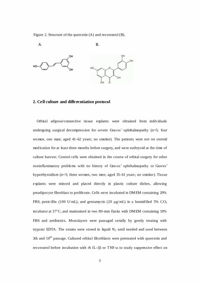
Quercetin (Q 0125), resveratrol (R 5010) were purchased from Sigma-Aldrich, Inc
(St. Louis, MO). Structures of the flavonoids used in this study are shown in Figure 2.
Dulbecco’s modified Eagle’s mediam (DMEM), fetal bovine serum (FBS), penicillin,
and gentamycin were purchased from Hyclone Laborotaries, Inc (Logan, UT).
3-(4,5-dimethyl-thiazo l-2-y l)-2,5-diphenyl-tetrazolium bromide (MTT) assay, the
fluorescent probe propidium iodide (PI) and Oil Red O were purchased from
Sigma-Aldrich. Bromodeoxyuridine (5-bromo-2-deoxyuridine, BrdU) cell
proliferation assay kit was purchased from Chemicon (Temecular, CA). The annexin
V-fluorescence isothiocyanate (FITC) apoptosis kit was purchased from BD
Biosciences (Franklin Lakes, NJ). Hyaluronan enzyme-linked immunosorbent assay
(ELISA) kit was purchased from Echelon Biosciences (Salt Lake, UT).
Recombinant human (rh) IL-1 and tumor necrosis factor (TNF)- were purchased
from R&D Systems (Minneapolis, MN). Anti-peroxisome pro liferator activator
gamma (PPAR) antibody, anti-CCAAT-enhancer-binding proteins (C/EBP)
antibody, anti-C/EBP β antibody and anti-β-actin antibody were all obtained from
Santa Cruz Biotechnology (Santa Cruz, CA).
9
Figure 2. Structure of the quercetin (A) and resveratrol (B).
A. B.
2. Cell culture and differentiation protocol
Orbital adipose/connective tissue explants were obtained from indiv iduals
undergoing surgical decompression for severe Graves’ ophthalmopathy (n=5; four
women, one man; aged 41-62 years; no smoker). The patients were not on steroid
medication for at least three months before surgery, and were euthryoid at the time of
culture harvest. Control cells were obtained in the course of orbital surgery for other
noninflammatory problems with no history of Graves’ ophthalmopathy or Graves’
hyperthyroidism (n=5; three women, two men; aged 35-61 years; no smoker). Tissue
explants were minced and placed directly in plastic culture dishes, allowing
preadipocyte fibroblast to proliferate. Cells were incubated in DMEM containing 20%
FBS, penicillin (100 U/mL), and gentamycin (20 g/mL) in a humidified 5% CO2
incubator at 37C, and maintained in two 80-mm flasks with DMEM containing 10%
FBS and antibiotics. Monolayers were passaged serially by gently treating with
trypsin/ EDTA. The strains were stored in liquid N2 until needed and used between
3th and 10th
passage. Cultured orbital fib roblasts were pretreated with quercetin and
resveratrol before incubation with rh IL-1 or TNF- to study suppressive effect on

10
inflammat ion and hyaluronan production.
To init iate differentiation, cells were grown to confluence in six-well plates.
Cultures were changed to serum-free DMEM supplemented with 33 M biotin, 17
M pantothenic acid, 10 g/ml transferrin , 0.2 nM T3, 1 M insulin
(Boehringer-Mannheim, Manheim, Germany), 0.2 M carbaprostaglandin (cPGI2;
Calbiochem, La Jolla, CA), and a PPAR agonist, thiazolid inediones (TZD).
Rosiglitazone 10 M (Cayman) was used in the TZD class of drugs in this study. For
the first 4 days only, 10 M dexamethasone and 0.1 mM isobutylmethylxanthine
(IBMX) was included in the media . The differentiat ion protocol was continued for 10
days, during which the media was replaced every 3-4 days. To evaluate the effect of
quercetin and resveratrol on ad ipocyte differentiat ion, we exposed cultures to
quercetin (10, 50, 100 M) or resveratrol (10, 30, 50 M) for the entire 10-day
differentiation period.
3. Cell viability and proliferation assay
Cell viab ility was assessed by using the MTT assay according to the manufacturer's
(Sigma-Aldrich) protocol. Briefly, MTT (5 mg/ml) was added to equal 1/10 of the
culture volume and incubated for 3 hours. Medium was removed, and the converted
dye was solubilized in ice-co ld isopropanol. Absorbency of the dye was measured at
560 nm with background subtraction at 630 nm on a microplate reader
(EL 340 Bio
Kinetics Reader; Bio-Tek Instruments, Winooski, VT)
Cell p roliferat ion was determined with a nonisotopic enzyme immunoassay for
11
BrdU incorporation, fo llowing the manufacturer’s instruction. Cells were labeled with
BrdU for 24 hours. The medium was removed, and cells were fixed with fixing
solution at room temperature fo r 30 minutes. The fixing solution was aspirated and
anti-BrdU antibody was added for 1 hour. After washing, 100 l goat anti-mouse IgG
peroxidase conjugate was added for 30 minutes. After repeated washing, 100 l of
substrate was added, and the plate was incubated in the dark for 6 and 24 hours
followed by the supply of 100 l of stop solution. Absorbance was read on a
spectrophotometer microplate reader set at dual wavelength of 450/550 nm. The
background absorbance of cells receiving no primary antibody was subtracted.
4. Annexin V assay for cytotoxicity
Cells were washed with phosphate buffered saline (PBS) and incubated in
serum-free DMEM in the presence of quercetin at 10- 100 M for 24 hours and
resveratrol at 1- 50 M for 24 hours. An annexin V- FITC kit was used to detect
phosphatidylserine externalization, as an index of apoptosis. Cells were washed and
incubated for 15 minutes at room temperature in the presence of annexin V labeled
with FITC and PI. In total, 10,000 cells were excited at 488 nm, and emission was
measured at 530 and 584 nm to assess FITC and PI fluorescence, respectively. Cells
were analyzed with a flow cytometer (FACSn; BD Bioscience). Gated cell numbers
were p lotted on a dot plot with reference to both annexin V and PI staining.
12
5. Semiquantitative RT-PCR
RNA isolation and semiquantitative reverse transcription polymerase chain reaction
(RT-PCR) were performed as described previously. Total RNA was prepared using
TriZol (Invitrogen, Carlsbad, CA). cDNA was synthesized from 1 g of total RNA
using 10mM of dNTP mixture (2 l), recombinant RNasin®
ribonuclease inhib itor
(0.5 l), AMV reverse transcriptase (15u), reverse transcription 10X buffer,
Oligo(dT)15 primer (0.5 μg/μl) (Promega Corporation, Madison, WI).
PCR was performed using 0.25 mM dNTP, 0.25 U Taq polymerase (iNtRON
Biotechnology, Inc., Korea), 10 pmole primer pair and 3 l cDNA with a thermal
cycler (PerkinElmer, NY). PCR cycling conditions were as follows: 30 cycles with
glyceraldehyde 3-phosphate dehydrogenase (GAPDH), IL-6, IL-8 at 94C for 30
seconds, 55C for 1 minute, 72C for 1 minute, 34 cycles with intercellular adhesion
molecule-1 (ICAM-1): 94C for 30 seconds, 65C for 30 seconds, 72C for 1 minute,
and 35 cycles with cyclo-oxygenase (COX)-2: 93C for 30 seconds, 60C for 30
seconds, 72C for 30 seconds. Primer sequences specific for amplification genes
encoding ICAM-1, IL-6, IL-8, and COX-2 were designed from available human gene
sequences (Table 1). The levels of mRNAs, quantified by densitometry scanning of
the amplified products electrophoresed on agarose gels, are expressed as the ratio
between the density of each gene products and coamplified GAPDH.

13
Table 1. Primers used for semiquantitative RT-PCR
Target Gene Primer Sequence
ICAM-1
forward
reverse
5’-GGC CTC AGC ACG TAC CTC TA-3’
5’-TGC TCC TTC CTC TTG GCT TA-3’
IL-6
forward
reverse
5’-TCA ATG AGG AGA CTT GCC TG-3’
5’-GAT GAG TTG TCA TGT CCT GC-3’
IL-8
forward
reverse
5’-TTG GCA GCC TTC CTG ATT TC-3’
5’-AAC TTC TCC ACA ACC CTC TG-3’
COX-2
forward
reverse
5’- GTT CCA CCC GCA GTA CAG-3’
5’-GGA GCG GGA AGA ACT TGC-3’
GAPDH
forward
reverse
5’-GCC AAG GTC ATC CAT GAC AAC-3’
5’-GTC CAC CAC CCT GTT GCT GTA-3’
6. Hyaluronan ELISA
Orbital preadipocyte fibroblasts were grown to confluence in 12-well p lates and
then incubated for the indicated time periods with variab le concentrations of quercetin
and resveratrol before stimulation with IL-1. Supernatants from the cell cultures
were collected, and hyaluronan concentrations were determined using a competit ive
binding hyaluronan ELISA kit (Echelon Biosciences) according to the manufacturer ’s
instructions. The concentration of hyaluronan in the sample was determined using a
standard curve generated using known amounts of hyaluronan. Samples were d iluted
1:10 before analysis, and the average of triplicate measurements was taken.
14
7. Oil red O stain
Cells were stained with Oil red O as described by Green and Kehinde .30
A stock
solution of Oil red O (0.5g in 100 ml isopropanol) was prepared and passed through a
0.2 m filter. To prepare the working solution, 6 ml of the stock solution was mixed
with 4 ml of d istilled water, left for 1 hour at room temperature, and filtered through a
0.2 m filter. Cells were washed twice with 1X PBS, fixed with 10% formalin in PBS
for 1 hour at 4 C and stained with 300 l of the Oil red O working solution for 1 hour
at room temperature. The dishes were washed with distilled water before being
visualized using Axiovert (Carl Zeiss) light microscope and photographed at X40 and
X100 using an Olympus Corp. BX60 light microscope (Olympus, Melville, NY) .
8. Western blot assay of PPAR, C/EBP, C/EBP
Differentiated cells were washed with ice -cold PBS and lysed with cell lysis buffer
(20 mM HEPES [pH 7.2], 10% [/] glycerol, 10 mM Na3VO4, 50 mM NaF, 1 mM
phenylmethylsulfonyl fluoride, 0.1 mM dithiothreitol, 1 g/ml leupeptin, 1 g/ml
pepstatin, and 1% [/] Triton X-100; Sigma- A ldrich) on ice for 30 minutes. Lysates
were centrifuged for 10 minutes at 12,000 g and cell homogenate fractions stored at
-70C before use.
Protein concentrations in supernatant fractions were determined by the Bradford
assay. Equal amounts of protein (50 g) were boiled in sample buffer and resolved by
10% [w/] SDS-PAGE. Proteins were transferred to polyvinylidene fluoride (PVDF)

15
membranes (Immobilon; Millipore, Billerica, MA), probed overnight with primary
antibodies in TBST, and washed three times with TBST. Anti-PPAR antibody,
anti-C/EBP antibody, anti-C/EBP antibody, and anti--actin antibody were all
obtained from Santa Cruz Biotechnology (Santa Cruz, CA, USA). Immunoreactive
bands were detected with horseradish peroxidase-conjugated secondary antibody and
developed using an enhanced chemiluminescence kit (Amersham Pharmacia Biotech)
and exposed to X-ray film (Amersham Pharmacia Biotech).
9. Statistical analysis
For the purpose of statistically analyses, each level of mRNAs were quantified by
densitometry scanning and expressed as the ratio relative to GAPDH. Each data point
represents the mean SD of three separate experiments, using cells from three
different individuals. Data were analyzed by the Mann-Whitney U test, Student’s
t-test using the SPSS program for Windows, version 12.0.1 (SPSS, Chicago, IL, USA).
P-values less than 0.05 were considered to be significant.

16
III. RESULTS
1. The effect of quercetin and resveratrol on cell viability
The cytotoxicity of quercetin and resveratrol to orbital fibroblasts was detected
through the loss of cell viability using MTT assay. More than 95% of cells were
viable after challenge with 10-100 M quercetin for both 6 and 24 h in both normal
(Figure 3 A) and Graves’ ophthalmopathy (Figure 3B) orb ital fib roblasts . Nearly more
than 90% of cells were v iable after t reatment of 10-50 M resveratrol fo r both 6 and
24 hours in normal (Figure 4 A) and Graves’ ophthalmopathy (Figure 4 B) cells.
Interestingly, treatment of quercetin at low concentrations (10-30 M) for 24 hours
promoted mild pro liferation up to 130-140% in both normal and Graves’
ophthalmopathy orbital fibroblasts. The cell viab ility was nearly 100% after treatment
of 100 M quercetin in both normal and Graves’ ophthalmopathy cells (Figure 3 A,
B), whereas cell v iability significantly decreased below 65% after challenge of 100
M resveratrol for both 6 and 24 hours in both normal and Graves’ ophthalmopathy
orbital fibroblasts by MTT analysis (Figure 4 A, B).
We could also observe the difference of cell viability according to exposure time of
chemicals. Long exposure (24 hours) of compounds at low concen trations induced
slightly more pro liferation than the short exposure (6 hours) after treatment with both
compounds.

17
Figure 3. Effect of quercetin on cell v iability in normal and Graves’ ophthalmopathy
orbital fibroblasts. MTT analyses results after the treatment with quercetin in normal
(A) and Graves’ ophthalmopathy (B) orbital fibroblasts . Data are average of three
independent experiments SD.

18
Figure 4. Effect of resveratrol on cell v iability in normal and Graves’ ophthalmopathy
orbital fibroblasts. MTT analyses results after the treatment with resveratrol in normal
(A) and Graves’ ophthalmopathy (B) orbital fibroblasts . Data are average of three
independent experiments SD.
19
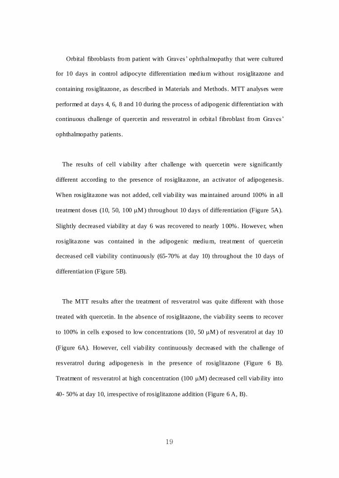
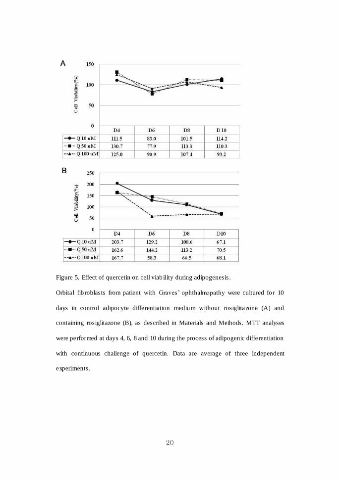
Orbital fibroblasts from patient with Graves’ ophthalmopathy that were cultured
for 10 days in control adipocyte differentiation medium without rosiglitazone and
containing rosiglitazone, as described in Materials and Methods. MTT analyses were
performed at days 4, 6, 8 and 10 during the process of adipogenic differentiat ion with
continuous challenge of quercetin and resveratrol in orbital fibroblast from Graves’
ophthalmopathy patients.
The results of cell v iability after challenge with quercetin were significantly
different according to the presence of rosiglitazone, an activator of adipogenesis.
When rosiglitazone was not added, cell viab ility was maintained around 100% in all
treatment doses (10, 50, 100 M) throughout 10 days of differentiation (Figure 5A).
Slightly decreased viability at day 6 was recovered to nearly 100%. However, when
rosiglitazone was contained in the adipogenic medium, treatment of quercetin
decreased cell viability continuously (65-70% at day 10) throughout the 10 days of
differentiat ion (Figure 5B).
The MTT results after the treatment of resveratrol was quite different with those
treated with quercetin. In the absence of rosiglitazone, the viab ility seems to recover
to 100% in cells exposed to low concentrations (10, 50 M) of resveratrol at day 10
(Figure 6A). However, cell viab ility continuously decreased with the challenge of
resveratrol during adipogenesis in the presence of rosiglitazone (Figure 6 B).
Treatment of resveratrol at high concentration (100 M) decreased cell viab ility into
40- 50% at day 10, irrespective of rosiglitazone addition (Figure 6 A, B).
20
Figure 5. Effect of quercetin on cell viab ility during adipogenesis .
Orbital fib roblasts from patient with Graves’ ophthalmopathy were cultured fo r 10
days in control adipocyte differentiation medium without rosiglitazone (A) and
containing rosiglitazone (B), as described in Materials and Methods. MTT analyses
were performed at days 4, 6, 8 and 10 during the process of adipogenic differentiation
with continuous challenge of quercetin. Data are average of three independent
experiments.

21
Figure 6. Effect of resveratrol on cell v iability during adipogenesis .
Orbital fib roblasts from patient with Graves’ ophthalmopathy were cultured fo r 10
days in control adipocyte differentiation medium without rosiglitazone (A) and
containing rosiglitazone (B), as described in Materials and Methods. MTT analyses
were performed at days 4, 6, 8 and 10 during the process of adipogenic differentiation
with continuous challenge of resveratrol. Data are average of three independent
experiments.

22
As shown in Figure 7, BrdU analyses results were somewhat different from the
MTT analyses. Proliferation of normal and Graves’ ophthalmopathy orbital fibroblasts
was inhibited dose dependently at 6 hours treatment of quercetin, whereas
proliferation was promoted at 24 hours treatment of quercetin at low concentration
(10-30 M) up to 180% in normal and 140% in Graves’ ophthalmopathy orbital
fibroblasts (Figure 7 A, B). In contrast, s ignificant dose dependent-inhibition of
proliferation was observed at both 6 and 24 hours of resveratrol treatment in normal
orbital fibroblasts (Figure 7 C). In orbital fibroblasts from Graves’ ophthalmopathy
patients, similar pattern of inhibition of proliferat ion was observed at 6 hours
treatment of resveratrol, and minimally promoted proliferat ion (up to 118%) was
observed at 24 hours treatment of resveratrol at 30 M concentrations (Figure 7 D).
Long exposure (24 hours) of low concentrations (10-30 M) of quercetin induced
mild proliferation of orb ital fib roblasts, which results were similar to MTT analyses.
Inhibition effects on proliferat ion of orbital fibroblasts were prominent after the
treatment of resveratrol.
To confirm nontoxic concentrations of chemicals in preadipocyte fibroblasts,
annexin V-FITC/PI binding assay was performed. The results showed that treatment
of quercetin at 10-100 M and resveratrol at 1-50 M for 24 hours did not induce
significant apoptosis or necrosis in both normal and Graves’ ophthalmopathy orbital
fibroblasts. Figure 8 shows representative figure from the results in Graves’
ophthalmopathy orbital fibroblasts.

23
Figure 7. Effects of quercetin and resveratrol on cell p roliferat ion in normal and
Graves’ ophthalmopathy orbital fibroblasts. BrdU analyses results of orbital fibrob last
after treatment with quercetin in normal (A) and Graves’ ophthalmopathy (B) orbital
fibroblasts, with resveratrol in normal (C) and Graves’ ophthalmopathy (D) orbital
fibroblasts are shown. BrdU incorporation was expressed as viability (%). Data in the
columns represent the mean SD of three experiments
24
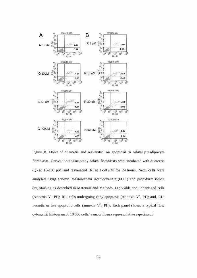
Figure 8. Effect of quercetin and resveratrol on apoptosis in orbital p readipocyte
fibroblasts. Graves’ ophthalmopathy orbital fibroblasts were incubated with quercetin
(Q) at 10-100 M and resveratrol (R) at 1-50 M for 24 hours. Next, cells were
analyzed using annexin V-fluorescein isothiocyanate (FITC) and propidium iodide
(PI) staining as described in Materials and Methods. LL; viable and undamaged cells
(Annexin V-, PI
-); RL: cells undergoing early apoptosis (Annexin V
+, PI
-); and, RU:
necrotic or late apoptotic cells (annexin V+, PI
+). Each panel shows a typical flow
cytometric h istogram of 10,000 cells/ sample from a representative experiment.

25
Therefore, maximal nontoxic concentraion of quercetin and resveratrol was
determined as 100 M and 50 M, respectively. We determined to use these
concentrations as a maximal level to study any suppressive effect of chemicals on the
inflammat ion, hyaluronan production and adipogenesis of orbital fibroblasts from
normal and Graves’ ophthalmopathy subjects.
2. Determination of IL-1 concentration to study the maximal inhibitory
effect of quercetin and resveratrol on the inflammation
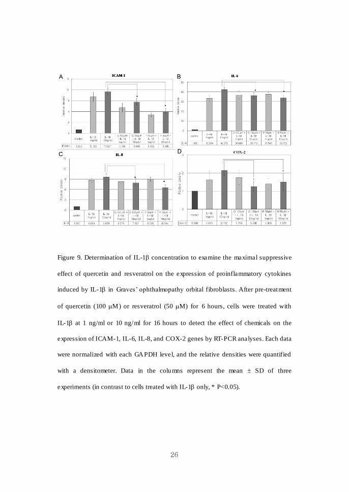
As Figure 9 ind icates, both pretreatment of quercetin at 100 M and resveratrol at
50 M for 6 hours in orbital fibroblasts from Graves’ ophthalmopathy induced strong
inhibit ion of ICAM-1 (A), IL-6 (B), IL-8 (C) and COX-2 (D) expression (*P<0.05),
when the cells were stimulated with IL-1 at a dose of 10 ng/ml. ICAM-1, IL-6, IL-8,
and COX-2 mRNA gene expressions were more significantly induced, and inhibited
by quercetin at 100 M and resveratrol at 50 M, with the stimulation of 10 ng/ml of
IL-1, in comparison with that of 1 ng/ml of IL-1. Thus, 10 ng/ml of IL-1 was
determined as a stimulation dose for experiments of dose- and time- dependent effect
of chemicals.
26
Figure 9. Determination of IL-1 concentration to examine the maximal suppressive
effect of quercetin and resveratrol on the expression of proinflammatory cytokines
induced by IL-1 in Graves’ ophthalmopathy orbital fibroblasts. After pre-treatment
of quercetin (100 M) or resveratrol (50 M) for 6 hours, cells were treated with
IL-1 at 1 ng/ml or 10 ng/ml for 16 hours to detect the effect of chemicals on the
expression of ICAM-1, IL-6, IL-8, and COX-2 genes by RT-PCR analyses. Each data
were normalized with each GAPDH level, and the relative densities were quantified
with a densitometer. Data in the columns represent the mean SD of three
experiments (in contrast to cells treated with IL-1 only, * P<0.05).
27
3. Inhibition of ICAM-1, IL-6, IL-8 and COX-2 mRNA by quercetin and
resveratrol
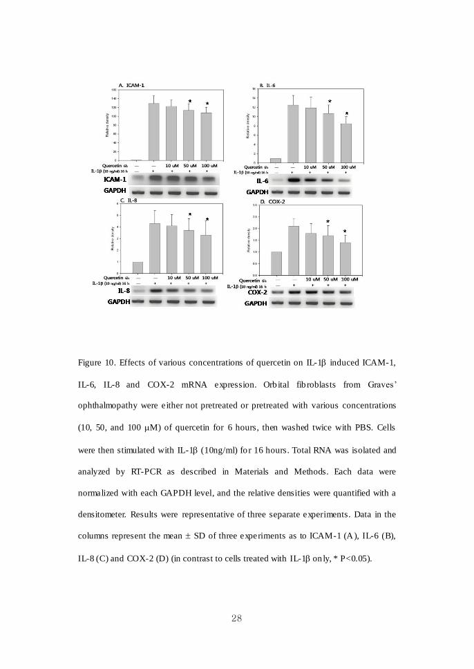
We investigated the effects of increasing treatment doses and times of quercetin and
resveratrol on ICAM-1, IL-6, IL-8 and COX-2 mRNA expression in response to
IL-1 challenge of orbital fibroblasts from Graves’ ophthalmopathy patients. We
chose three concentrations of quercetin (10, 50, and 100 M) and resveratrol (10, 30,
and 50 M) for 6 hours before treatment with IL-1 (10 ng/ml) for 16 hours to study
their dose-dependent effects. Also, to study time-dependent effects, cells were treated
with quercetin (100 M) and resveratrol (50 M) for 3, 6, 9, and 24 hours before
treatment with IL-1 (10 ng/ml) for 16 hour.
As shown in Figure 10 and 11, pret reatment of quercetin showed a significant
inhibit ion of ICAM-1, IL-6, IL-8 and COX-2 mRNA expression induced by IL-1 in
a dose- and time- dependent manner. Resveratrol also had a significant suppressive
effect on IL-1β induced proinflammatory cytokine e xpressions in mRNA levels
(Figure 12). The inhib itions of IL-8 and COX-2 mRNA expression by resveratrol
were dose dependent. Time-dependent manners of inhibit ion were not observed after
the treatment of resveratrol (data not shown).
28
Figure 10. Effects of various concentrations of quercetin on IL-1 induced ICAM-1,
IL-6, IL-8 and COX-2 mRNA expression. Orb ital fib roblasts from Graves’
ophthalmopathy were either not pretreated or pretreated with various concentrations
(10, 50, and 100 M) of quercetin for 6 hours, then washed twice with PBS. Cells
were then stimulated with IL-1 (10ng/ml) fo r 16 hours. Total RNA was isolated and
analyzed by RT-PCR as described in Materials and Methods. Each data were
normalized with each GAPDH level, and the relative densities were quantified with a
densitometer. Results were representative of three separate experiments. Data in the
columns represent the mean SD of three experiments as to ICAM-1 (A), IL-6 (B),
IL-8 (C) and COX-2 (D) (in contrast to cells treated with IL-1 on ly, * P<0.05).

29
Figure 11. Effects of quercetin for various duration of treatment on IL-1 induced
ICAM-1, IL-6, IL-8 and COX-2 mRNA expressions. Orbital fib roblasts from Graves’
ophthalmopathy was either not pretreated or pret reated with quercetin (100 M) for
various time periods (3, 6, 9, 24 h), and then washed twice with PBS. Cells were then
stimulated with IL-1 (10 ng/ml) for 16 hours. Total RNA was isolated and analyzed
by RT-PCR as described in Materials and Methods. Each data were normalized with
each GAPDH level, and the relative densities were quantified with a densitometer.
Results were representative of three separate experiments. Data in the columns
represent the mean SD of three experiments as to ICAM-1 (A), IL-6 (B), IL-8 (C)
and COX-2 (D) (in contrast to cells treated with IL-1 only, * P<0.05).
30
Figure 12. Effects of various concentrations of resveratrol on IL-1 induced ICAM-1,
IL-6, IL-8 and COX-2 mRNA expression. Orb ital fib roblasts from Graves’
ophthalmopathy were either not pretreated or pretreated with various concentrations
(10, 30, and 50 M) of resveratrol for 6 hours, then washed twice with PBS. Cells
were then stimulated with IL-1 (10ng/ml) fo r 16 hours. Total RNA was isolated and
analyzed by RT-PCR as described in Materials and Methods. Each data were
normalized with each GAPDH level, and the relative densities were quantified with a
densitometer. Results were representative of three separate experiments. Data in the
columns represent the mean SD of three experiments as to ICAM-1 (A), IL-6 (B),
IL-8 (C) and COX-2 (D) (in contrast to cells treated with IL-1 on ly, * P<0.05).

31
4. Quercetin inhibited hyaluronic acid production in orbital fibroblasts
Orbital fibroblasts produced high level of hyaluronic acid with the stimulat ion of
IL-1 or TNF- (10 ng/ml, 16 hours) in ELISA analyses. The mean concentration of
hyaluronan induced by IL-1 was higher, albeit not significant, in cells from Graves’
ophthalmopathy (n=3) than in cells from normal individuals (n=3) (Figures 13 A, B).
Cells were treated with various concentrations (10, 50, 100 M) of quercetin or
resveratrol (10, 30, 50 M) for 24 hours, before incubation with IL-1 (10ng/ml, 16
hours) for hyaluronan ELISA analyses. Quercetin showed a significant inhib ition of
hyaluronan production induced by IL-1 in a dose- and time- dependent manner in
both normal and Graves’ ophthalmopathy orbital fibroblasts (Figures 13 A, B). The
inhibit ion effects of quercetin on the hyaluronan production were similarly observed
in a dose-dependent manner in both IL-1 and TNF- (same condition, 10 ng/ml, 16
hours) stimulated orbital fibroblasts from Graves’ ophthalmopathy subjects (Figure 13
C).
However, resveratrol did not show any inhibitory or stimulatory effect on
hyaluronan production when stimulated by either IL-1 or TNF- (data not shown).

32
33
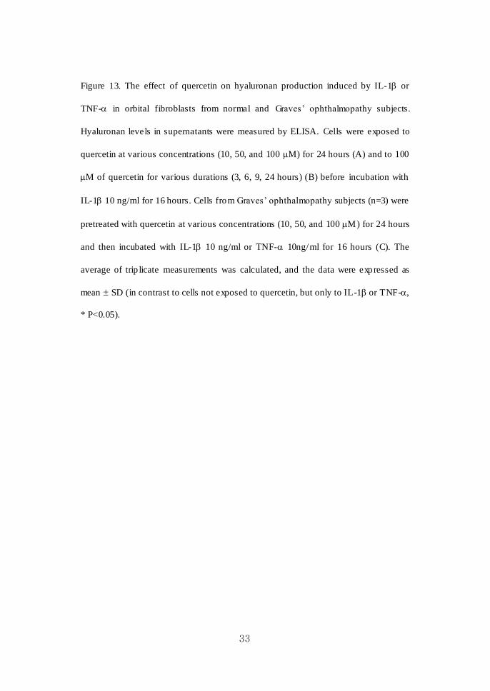
Figure 13. The effect of quercetin on hyaluronan production induced by IL-1 or
TNF- in orbital fibroblasts from normal and Graves’ ophthalmopathy subjects.
Hyaluronan levels in supernatants were measured by ELISA. Cells were exposed to
quercetin at various concentrations (10, 50, and 100 M) for 24 hours (A) and to 100
M of quercetin for various durations (3, 6, 9, 24 hours) (B) before incubation with
IL-1 10 ng/ml for 16 hours. Cells from Graves’ ophthalmopathy subjects (n=3) were
pretreated with quercetin at various concentrations (10, 50, and 100 M) for 24 hours
and then incubated with IL-1 10 ng/ml or TNF- 10ng/ml for 16 hours (C). The
average of trip licate measurements was calculated, and the data were expressed as
mean SD (in contrast to cells not exposed to quercetin, but only to IL-1 or TNF-,
* P<0.05).

34
5. Effect of quercetin and resveratrol on the accumulation of lipid
droplets and the expression of transcriptional regulators of adipogenesis
Confluent orbital fibroblasts from Graves ’ Ophthalmopathy patients were subjected
to the differentiated protocol for 10 days. Cells were first examined under light
microscopy. Under control adipogenic conditions, preadipocyte fibrob lasts lost their
stellate fibroblastic appearance and converted to a spherical adipocytic shape, and a
fraction of these cells accumulated small lipid droplets (Figure 14 A). Visib le from
day 3, lipid droplets increased in number and en larged in size during the 10 days of
differentiat ion. The addition of rosiglitazone (10 M), a PPAR agonist significantly
increased adipogenesis in light microscopy compared with baseline conditions
without rosiglitazone (Figure 14 B). When preadipocyte orbital fibroblasts were
cultured under adipogenic condition, and exposed to exogenous IL-1 (10ng/ml) for
the first 3 days, accumulation of lipid droplets increased microscopically compared
with baseline control conditions (Figure 14 C). Combination of rosiglitazone and
IL-1 further stimulated adipogenesis compared with adipogenic condition containing
rosiglitazone or IL-1 only (Figure 14 D).

35
Figure 14. Examination of prestained orbital fibroblasts cultured in adipogenic
medium under light microscopy. Orbital fibroblasts from a subject with Graves’
ophthalmopathy were cu ltured in control ad ipogenic medium (A), adipogenic medium
containing rosiglitazone 10 M for 10 days (B), IL-1 10ng/ml for first 3 days (C)
and both rosiglitazone and IL-1 (D) during differentiation as described in Materials
and Method. TZD = Thiazolidinediones (X40, X400: s mall box).
. control . control + TZD
. control + IL-1 . control + IL-1 + TZD

36
To examine whether quercetin and resveratrol has any suppressive effects on
adipogenesis, those chemicals were added at day 1 in ad ipogenic medium including
rosiglitazone, and continued for the 10 day differentiation period, being replaced
whenever media was replaced. Both quercetin and resveratrol decreased number of
adipocyte and suppressed accumulat ion of lip id droplets dose dependently (Figure 15).
High power (X400) microscopic examination showed significant reduction of size
and number of lipid droplets according to treatment dose of quercetin and resveratrol
(Figure 15). Pread ipocyte fibroblasts which did not convert into adipocytes were
uniform in size and stellate in shape, which maintained viable fibroblastic
morphology.

37
Quercetin Resveratrol
. control + TZD
. Q 10 M
. Q 50 M
. Q 100 M
. R 10 M
. R 30 M
. R 50 M

38
Figure 15. Microscopic examination of prestained cultures grown in adipogenic
medium containing either quercetin o r resveratrol. Orb ital fibroblasts from a subject
with Graves’ ophthalmopathy were cu ltured for 10 days in adipogenic medium
containing rosiglitazone 10 M (A). Quercetin (B: 10, C: 50, D: 100 M) and
resveratrol (E: 10, F: 30, G: 50 M) were exposed to cultures with adipogenic
condition in (A) for the entire 10-day differentiation period as described in Materials
and Methods. Microscopic examination showed a dose-dependent inhibition of
adipogenesis by quercetin and resveratrol. TZD = Thiazo lid inediones (X40, X400:
small box).
Cells were fixed with 10% formalin for 1 hour and then stained with oil red O to
measure lip id accumulat ion. As shown in Figure 16 and 17, rosiglitazone enhanced,
but both quercetin and resveratrol inhibited adipogenesis in a dose dependent manner,
as detected by oil red O staining. When the stained cells were examined under
microscope with X 400 magnification, significant decrease of cytoplasmic lip id
droplets in size and number were observed by treatment of both quercetin and
resveratrol, dose dependently.

39
Figure 16. Examination of oil red O stained cultures exposed to quercetin and
resveratrol with various concentrations. Orbital fib roblasts from a subject with
Graves’ ophthalmopathy were cultured for 10 days in control ad ipogenic medium and
containing rosiglitazone 10 M. Quercet in (10, 50, 100 M) and resveratrol (10, 30,
50 M) were exposed to cultures with adipogenic medium containing rosiglitazone
for the entire 10-day differentiation. Cells were fixed with 10% formalin for 1 hour
and then stained with oil red O to measure lipid accumulation. A dose-dependent
inhibit ion of ad ipogenesis by quercetin and resveratrol was visible. TZD =
Thiazo lid inediones (X40, X400: s mall box).

40
Quercetin Resveratrol
. control . control + TZD
. Q 10 M . R 10 M
. R 30 M
. R 50 M
. Q 50 M
. Q 100 M

41
Figure 17. Microscopic examination of oil red O stained cultures exposed to quercetin
and resveratrol. Orbital fibroblasts from a single individual with Graves’
ophthalmopathy were cultured for 10 days in control adipogenic medium without
rosiglitazone (A) and containing rosiglitazone 10 M (B), and were fixed with 10%
formalin for 1 hour and then stained with oil red O to measure lip id accumulat ion.
Quercetin (C: 10, D: 50, E: 100 M) and resveratrol (F: 10, G: 30, H: 50 M) with
varying concentrations were added to the differentiation medium containing
rosiglitazone for the entire 10 day differentiation period, and were examined by light
microscopy after oil red O stain. TZD = Thiazo lid inediones (X40, X400: s mall box).
Western blot analysis was performed to investigate whether various concentrations
of quercetin and resveratrol (10, 50, and 100 M) affect the expression of adipogenic
transcription factors such as PPAR , C/EBP and C/EBP. PPAR and C/EBP
proteins were highly expressed in control differentiated fib roblasts without treatment
of quercetin and resveratrol. The increases in PPAR and C/EBP were marked ly
attenuated dose dependently by quercetin and resveratrol as shown in Figure 18.

42
43
Figure 18. Effect of quercetin and resveratrol on the expression of adipogenic
transcriptional regulators of differentiated orbital fibroblasts from Graves’
ophthalmopathy patients. Various doses of quercetin (50, 100 M) and resveratrol (30,
50 M) were co-applied with d ifferentiation medium including rosiglitazone 10 M
for 10 days. After differentiation was completed, cell lysates were prepared and
subjected to western blot for PPAR and C/EBP , β protein. Representative figure of
western blot analysis is shown in (A). Significant dose dependent -inhibitions of
PPAR (B) and C/EBP β (C) proteins by both quercetin and resveratrol were noted.
Relative densities of PPAR, C/EBP β p rotein contents (%) normalized by reprobing
with anti--actin Abs were quantified with a densitometer. Data in the columns
represent the mean SD of three experiments.
6. Quercetin’s effect on accumulation of lipid droplet and cell viability in
differentiating fibroblasts treated with IL-1 or H2O2
Our results indicate that resveratrol induce a dose dependent cytotoxicity and
inhibit pro liferation both in preadipocyte fibroblasts and differentiating fibroblasts as
shown in figure 4-7. As shown in figure 15-17, inhibit ion effect of resveratrol on the
accumulat ion of lipid droplets both in prestained and stained cultures was more
apparent than that of quercetin even at low dose (10 M) treatment, which might be
associated with suppressive action of resveratrol on proliferat ion.
In MTT analyses of quercetin treated cells , viability was maintained around 100%

44
in all treatment doses (10, 50, 100 M) of quercetin at the 10 day of d ifferentiation
when grown under adipogenic medium without rosiglitazone. However, t reatment of
10-100 M quercetin suppressed viability significantly when cultured in adipogenic
medium containing rosiglitazone. The effect of quercetin on cell viab ility seems to be
dependent on culture condition. Quercetin decreased adipocyte numbers and clearly
suppressed lipid droplet accumulat ion in d ifferentiating orbital fibroblasts with
minimal cytotoxicity. Author further evaluated the quercetin’s effect on adipocyte
differentiat ion depending on different cell condition. The condition was determined to
be inflammatory by IL-1 treatment and to be stressed by oxidants, H2O2 in the early
period of differentiat ion.
When preadipocyte orbital fibroblasts were cultured under adipogenic condition,
exposed to exogenous IL-1 (10ng/ml) for the first 3 days, adipogenesis was
enhanced as shown in figure 19 and 20. More cells became round and accumulated
lip id droplets. Combination of TZD and IL-1 further stimulated adipogenesis
compared with adipogenic condition containing IL-1 only (Figure 19B, 20B). A
dose-dependent inhibition of adipogenesis by quercetin in cultures grown under
adipogenic medium including IL-1 and TZD was visible as shown in figure 19 C,D
and 20 C,D. The remained preadipocyte fibroblasts preserved their fibroblast-like
morphology (figure 20).
45
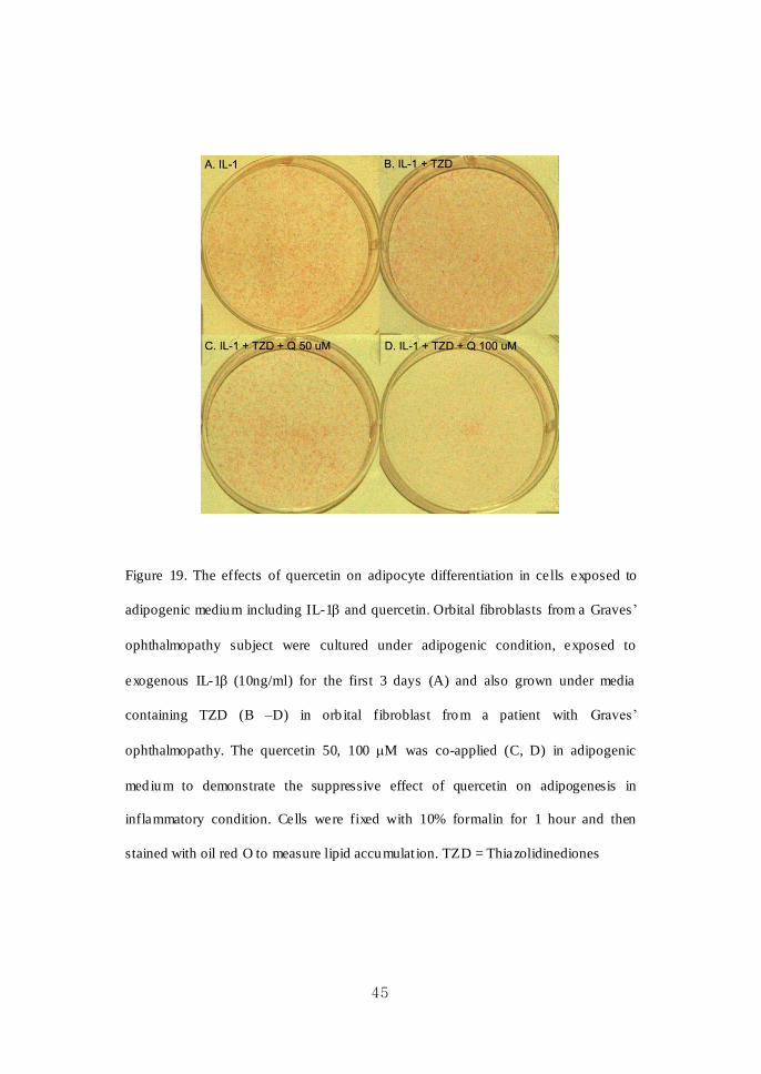
Figure 19. The effects of quercetin on adipocyte differentiation in cells exposed to
adipogenic medium including IL-1 and quercetin. Orbital fibroblasts from a Graves’
ophthalmopathy subject were cultured under adipogenic condition, exposed to
exogenous IL-1 (10ng/ml) for the first 3 days (A) and also grown under media
containing TZD (B –D) in orb ital fibroblast from a patient with Graves’
ophthalmopathy. The quercetin 50, 100 M was co-applied (C, D) in adipogenic
medium to demonstrate the suppressive effect of quercetin on adipogenesis in
inflammatory condition. Cells were fixed with 10% formalin for 1 hour and then
stained with oil red O to measure lipid accumulat ion. TZD = Thiazolidinediones

46
Figure 20. Microscopic results of oil red O stain showing suppressive effect of
quercetin on adipogenesis in d ifferentiating cells grown under ad ipogenic medium
including IL-1. Orbital fibroblasts from a Graves’ ophthalmopathy subject were
cultured under the adipogenic condition described in figure 19 (A: exposed to
exogenous IL-1 10ng/ml for the first 3 days only, B: IL-1 + rosiglitazone 10 M, C:
IL-1 + rosiglitazone + quercetin 50 M, D: IL-1 + rosiglitazone + quercetin 100
M, TZD = Thiazo lid inediones).
. IL-1 . IL-1 + TZD
. IL-1 + TZD + Q 50 M . IL-1 + TZD + Q 100 M

47
H2O2 100 M was exposed to differentiating orbital f ibroblasts from a patient with
Graves’ ophthalmopathy for the first 3 days only, and the quercetin was co-applied in
the adipogenic medium during 10 days of differentiat ion (Figure 21). As shown in
figure 21, H2O2 100 M treatment in early differentiation induced significant cell
death. Interestingly, however, in quercetin treated cells, not only accumulation of lip id
droplets decreased significantly in numbers , but also a considerable number of orbital
fibroblasts preserved their normal fibroblastic morphology. We could observe a
protective effect of quercetin on cell v iability from the oxidative stress by H2O2 during
adipocyte differentiation.
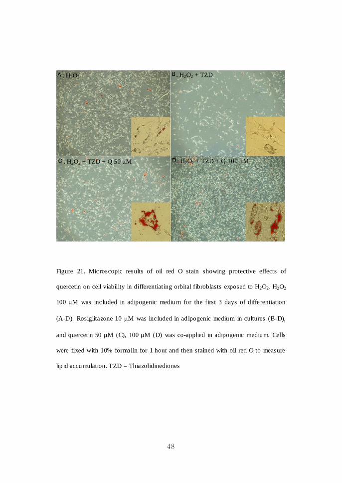
48
Figure 21. Microscopic results of oil red O stain showing protective effects of
quercetin on cell viability in differentiat ing orbital fibroblasts exposed to H2O2. H2O2
100 M was included in adipogenic medium for the first 3 days of differentiation
(A-D). Rosiglitazone 10 M was included in ad ipogenic medium in cultures (B-D),
and quercetin 50 M (C), 100 M (D) was co-applied in adipogenic medium. Cells
were fixed with 10% formalin for 1 hour and then stained with oil red O to measure
lip id accumulation. TZD = Thiazolidinediones
. H2O2 + TZD + Q 100 M . H2O2 + TZD + Q 50 M
. H2O2 . H2O2 + TZD
49
To demonstrate the effect of quercetin on viability of fu lly differentiated orbital
fibroblasts, MTT analyses were performed after quercetin treatment in fully
differentiated cells in various conditions , not in the process of adipocyte
differentiat ion (Figure 22). Varying concentrations of quercetin (1-100 M) were
exposed for 48 hours in cells from a Graves’ ophthalmopathy subject at day 10 of
adipocyte differentiat ion (Figure 22A). In addition, quercetin 100 M was treated for
48 hours in fully d ifferentiated cells which were exposed to varying concentrations of
IL-1β (10, 100 ng/ml) (Figure 22B) and H2O2 (100, 500, 1000 M) (Figure 22 C) for
the first 3 days of adipogenesis.
As shown in figure 22 A, quercetin decreased viability of fully differentiated
fibroblasts dose dependently, which were similar to the MTT results in cells cultured
in adipogenic medium containing quercetin. In contrast, cell viab ility was preserved
with the treatment quercetin in cells exposed to IL-1β (10 ng/ml, 100 ng/ml both), and
the quercetin did not decrease cell viab ility in these conditions (Figure 22 B). In other
words, the inhibit ion effect of quercetin on adipogenesis in an in vitro inflammatory
condition by IL-1β does not seem to be related to cytotoxicity. In concordance with
microscopic results , H2O2 induced cell death dose dependently, and the application of
quercetin increased viability significantly in a dose dependent manner (Figure 22 C).
Quercetin treatment during and after adipogenesis both attenuated H2O2-induced
cytotoxicity, as demonstrated in microscopic examination and MTT analyses.

50

51
Figure 22. MTT analyses after the treatment of quercetin in fully d ifferentiated orbital
fibroblasts in d ifferent adipogenic condition. Orbital fibroblasts from a single
individual with Graves ’ ophthalmopathy were cultured fo r 10 days in control
adipocyte differentiation medium including rosiglitazone, as described in Materials
and Methods. After full adipocyte differentiation, quercetin with various
concentrations (1-100 M) was treated for 48 hours (A). To study the effect the
quercetin on viability in cells treated with proinflammatory cytokine and oxidant,
quercetin (10, 50, 100 M) was treated for 48 hours in adipocyte fibroblasts which
were stimulated with IL-1β (10, 100 ng/ml) (B) and H2O2 (100, 500, 1000 M) (C)
only for the first 3 days of differentiation. Data are average of three independent
experiments SD.

52
IV. DISCUSSION
Graves’ ophthalmopathy is the most frequent extrathyroida l manifestation of
autoimmune hyperthyroidism. Many clin ical signs and symptoms of Graves ’
ophthalmopathy arise from the enlargement of soft tissues in the orbit, resulting in
increased pressure within the bony orbit.31, 32
Enlargement of both extraocular muscle
and adipose tissue is found in some patients, with a predominance of one or the other
in some.32
Current evidence points to orbital fibroblasts as the target cells, and the
thyrotropin receptor in orbital fibroblasts is the primary autoantigen in Graves ’
ophthalmopathy. Orbital fibroblasts particularly exhib it robust response to many
proinflammatory mediators.33
They critically orchestrate the recruitment of
immunocompetent cells and thus initiation of t issue remodeling.33
These orbital
fibroblasts secrete large amount of hyaluronan in response to various cy tokines,33
and a subgroup of orbital fib roblasts can differentiate into mature ad ipocytes,4 that
have increased expression of thyrotropin receptor.34, 35
Graves’ ophthalmopathy is a disfiguring and often incapacitating disease which is
difficult to treat. Glucocorticoids have been the mainstay in the treatment of Graves’
ophthalmopathy for a long time in spite of several frustrating complications. Orbital
radiotherapy is an alternative treatment of Graves’ ophthalmopathy with few adverse
events. However, both treatments have a limited role when Graves’ ophthalmopathy is
not in active inflammat ion, especially with mild severity. Other immunosuppressants
such as cyclosporine and methotrexate are inferio r to glucocorticoids in reducing
inflammat ion and halting the progression into severe course of Graves’
ophthalmopathy. There is a need to improve the efficacy and tolerability of

53
immunosuppressive treatment of Graves’ ophthalmopathy. Otherwise, new treatments
with minimal adverse effects are eagerly awaited, which are effective in the major
known pathogenesis of Graves’ ophthalmopathy, including inflammat ion, hyaluronan
production, and adipogenesis. In this study, we wished to determine whether quercetin
and resveratrol could modify the proinflammatory cytokines expression,
glycosaminoglycan synthesis and adipogenesis in primary cu ltured orbital fibroblasts
from patients with Graves’ ophthalmopathy. We have found these two chemicals
showed somewhat d ifferent patterns of therapeutic effects of Graves’ ophthalmopathy
in the in vitro experiments.
Quercetin, a flavonol found in fruit and vegetables, has unique biological properties
that include anti-inflammatory act ivity.22
In the present investigation, we have
observed that quercetin showed a treatment dose- and time-dependent inhibition of
IL-1β induced proinflammatory cytokines expression in mRNA levels. It is reported
that quercetin suppressed the production of TNF and nitric oxide by macrophages,
microglial cells and mast cells stimulated with lipopolysaccharide.36, 37
Different
mechanis ms are involved in the anti-inflammatory action of quercetin in many cell
systems, as it targets multiple intracellular signaling pathways such as signal
transducer and activator of transcription 1 and NF -kB activations and mitogen-
activated protein kinase family phosphorylatons.38, 39
Natural products have potential for inducing apoptosis, inhibit ing adipocyte
differentiation, and stimulating lipolysis in adipocytes. Various flavonoids including
genistein, docosahexanoic acid, ep igallocathechin gallate, quercetin and resveratrol

54
affect adipocytes during specific stages of development, resulting in either inhib ition
of adipogenesis or induction of apoptosis. Quercetin is reported to affect ad ipocytes
during specific stages of development, resulting in either inhibition of adipogenesis or
induction of apoptosis.24, 40
It caused a dose- and time-dependent increase in lipolysis
in rat adipocytes, which was reported to be potent phosphodiesterase inhibitors .41
In
this study, treatment of quercetin during adipogenesis of orbital fib roblasts from
Graves’ ophthalmopathy inhibited accumulat ion of lipid d roplet and decreased
number of round shape adipocytes in a dose-dependent manner in microscopic
examination. The expressions of transcriptional regulators of adipogenesis, PPAR
and C/EBP p roteins were also marked ly attenuated by quercetin. In MTT analyses,
quercetin did not reduce cell viability in differentiating cells in the absence of PPAR
agonist. However, quercetin decreased cell v iability by 20-30% similarly both in
differentiat ing and fully differentiated orbital fibrob lasts under adipogenic condition
containing PPAR agonist. In microscopic examination, quercetin treatment preserved
viable fibroblastic morphology of remained pread ipocyte fibroblasts which were not
differentiated. Orbital fibroblasts from Graves’ ophthalmopathy might have different
sensitivities regarding cytotoxicity of phytochemicals depending on cell status, such
as different stage of adipocyte life cycles, inflammatory or oxidative stressed
condition.
Quercetin significantly inhibited ad ipogenesis without induction of apoptosis in cells
exposed to IL-1β . Stimulat ion with IL-1 in vitro can mimic orbital in flammation in
Graves’ ophthalmopathy. Consistent with a previous reports,42, 43
we found IL-1 had
a stimulatory effect on adipogenesis in orbital fibroblasts, which may have important

55
clin ical implications. IL-1 promoted all three pathological aspects of Graves ’
ophthalmopathy, those are inflammat ion, hyaluronan production and adipogenesis in
our investigations. IL-1 might present an attractive therapeutic target in Graves ’
Ophthalmopathy. Quercet in not only suppressed IL-1 induced proinflammatory
cytokines, hyaluronan production, but also inhibited adipocyte differentiation
enhanced by IL-1. The cytotoxic effects of quercetin in cells exposed to IL-1
during and after adipogenesis were minimal in microscopic examination and MTT
analyses. Quercetin is believed to be a strong inhibitor of adipogenesis of orbital
fibroblasts which target different stages of adipocyte life cycle, however not a strong
inducer of apoptosis.
The particularly interesting result was that the quercetin protected cells from H2O2
induced cytotoxicity. Stimulat ion with H2O2 in vitro can mimic condition of cigarette
smoking in vivo, which is well known as the strongest risk factor for developing
severe Graves’ ophthalmopathy. We could also observe that the differentiating orbital
fibroblasts were highly vulnerable to H2O2, the oxidant. However, treatment of
quercetin during and after adipogenesis significantly attenuated H2O2-induced
cytotoxicity in orbital fibroblasts in microscopic results and MTT analyses. Cell
viability recovered with the treatment of quercetin in a dose-dependent manner. This
protective effect from acute stress may be associated with antioxidant property of
quercetin, which is reported to show structural features that have been related to the
antioxidant potency of flavonoids and also shows protection in different models of
oxidative death.18, 44-46
A novel mechanism for quercetin-induced cytoprotection has
been described involving the sterol regulatory element-binding protein-2

56
(SREBP-2)-mediated sterol synthesis that decreases lipid peroxidation by maintaining
membrane integrity in the presence of oxidative stress.47
It is required to further
investigate a cytoprotective mechanis m of quercetin in orb ital fibroblasts.
Resveratrol, a naturally-occurring molecule known as a phytoalexin, is synthesized
by plants in response to attacks by fungi, bacteria, or other injurious substances.36, 48, 49
Resveratrol has positive effects on metabolis m and can increase the lifespan of
various organisms.49
Its effects arise from its capacity to interact with multiple
molecular targets involved in diverse intracellu lar pathways. Most well known is the
ability of resveratrol to activate sirtuins, a class of NAD(+) -dependent deacetylases
that affect multip le transcription factors and other protein targets.49
In our study,
resveratrol had a significant suppressive effect on IL-1β induced proinflammatory
cytokine expressions in mRNA levels in orbital fibroblasts. The inhibit ions of IL-1
induced IL-8 and COX-2 mRNA expression were dose dependent. In previous
experiments in other cell systems and in vivo, resveratrol dampened inflammation in
arthritis and immune responsiveness in autoimmune disease such as rheumatoid
arthritis.49, 50
It could also down regulate inflammatory biomarkers such as TNF,
COX-2, inducible n itric oxide synthase, C-reactive protein, interferon- and several
interleukins.49
Resveratrol is reported to decrease fat mass by inhibiting adipogenesis and induce
apoptosis by affecting expression of genes that modulate mitochondrial function.29
It
has been shown to inhibit adipogenesis by repressing PPAR act ivity with activation
of Sirt 1 in 3T3-L1 adipocytes.51
In our study, as expected, resveratrol suppressed
57
accumulat ion of lipid droplet in d ifferentiating cells dose dependently. It also blocked
the expression of PPAR and C/EBP proteins in a dose-dependent manner. The
inhibit ion effects were even stronger than those of quercetin at low dose treatment.
The difference of the effect of resveratrol from the quercetin was the inhibit ion of
proliferation as shown in MTT and BrdU analyses . Cell v iability decreased almost by
50% after treatment of resveratrol 100 M both in preadipocyte fibroblasts and
differentiat ing fibroblasts regardless of addition of PPAR agonist. The mechanisms
by which resveratrol induces apoptosis may be mediated through any of numerous
mechanis ms that involve activation of mitochondria and of death caspases. In 3T3 -L1
adipocytes, treatment of resveratrol in maturing cells inhib ited l ipid accumulation and
downregulated the expression of PPAR, C/EBP, SREBP-1c and lipoprotein lipase,
and also increased apoptosis in mature adipocytes dose dependently.29
Recently, both
quercetin and resveratrol inh ibited lip id accumulation and induced apoptosis in early–
and mid– phases maturing and lipid-filled mature primary human adipocytes .24
The
inhibitory effects on adipogenesis and apoptosis were greater when treated in
combination including genistein.24
In this study, resveratrol seems be a strong
inhibitor of adipogenesis and also an inducer of apoptosis of maturing and matured
orbital fibroblasts.
Thiazo lid inediones (TZD), which are also called glitazones, are commonly used as
oral hypoglycemic agents in the treatment of type 2 diabetes mellitus. These agents
are shown to be potent agonists of the nuclear hormone receptor, PPAR which plays
a dominant role in adipocyte differentiation. A primary cause of proptosis in Graves’
ophthalmopathy is the expansion of ad ipose tissue volume in the orbit.52
In primary

58
cultured orbital fibroblasts, the activation of PPAR by agonists was reported to
stimulate functional TSH receptor expression and to induce recruitment and
differentiat ion of orbital fibroblasts into mature lipid-laden adipocytes.53-55
Several
case studies found that Graves’ ophthalmopathy was exacerbated by the use of TZD
and stabilized after discontinuation.56-59
These studies suggest that inhibition of
adipogenic pathway through the use of PPAR inhibitor might be a potential therapy
for Graves’ ophthalmopathy. In our study, orbital preadipocyte fibroblasts derived
from Graves’ ophthalmopathy patients were treated with flavonoid quercetin and
resveratrol at various concentrations during the differentiat ion period. The
differentiat ion media contained rosiglitazone, one of the members of TZD to induce
strong adipogenic differentiation. Decrease in the lip id accumulat ion by oil red O
stain was accompanied by attenuated expression of this adipogenic transcription
factor, PPAR and C/EBP by treatment of quercetin and resveratrol in a
dose-dependent manner. PPAR, C/EBP transcription factors expressed in distinct
phases of adipogenesis have shown to play important roles. There is a positive
feedback loop between PPAR and C/EBP during the terminal stages of
adipogenesis.60
Our data suggest that quercetin and resveratrol exert anti -adipogenic
effects by suppressing these adipogenic transcription factors.
Graves’ ophthalmopathy is characterized by an inflammat ion of retrobulbar t issues,
leading to accumulat ion of hydrophilic glycosaminoglycan, which attracts water into
surrounding tissues and thereby increase volume of orbital connective tissue and
extraocular muscles.31, 61
Orbital fibroblasts in vitro respond to various mediators of
inflammat ion, such as IL-1 by producing excessive amounts of hyaluronan, which is
59
a major glycosaminoglycan in orbital tissues of Graves ’ ophthalmopathy patients.61, 62
In this study, both IL-1β and TNF- increased production of hyaluronan as previously
reported, 61, 62
and the pretreatment of quercetin suppressed the level of hyaluronan in
orbital fibroblasts from both normal and Graves ’ ophthalmopathy individuals. We
could not found any previous reports regarding the effect of quercetin on the
production of glycosaminoglycan in any cell systems. Contrary to our expectation,
resveratrol could not attenuate IL-1β or TNF induced hyaluronan production.
Quercetin is reported to have the capacity to reduce acute, chronic, and subclinical
inflammatory processes, the latter being associated with lifestyle diseases such as
obesity and diabetes.46
It is now available in high grade purified form, and clin ical
phase I-III studies can be readily done in the near fugure .46
Resveratrol, the most
potent natural sirtuin act ivator, together with the much more potent synthetic sirtuin
activators, have considerable potential in the prevention and treatment of several
common conditions of aging. The beneficial effects are supported by detailed findings,
at the molecular and cellular level, of the specific pathways and molecules affected.
However, there are many questions regarding flavoniods that remain to be
investigated. Whether flavoniods act as pro- or anti-inflamamtory and pro- or
anti-oxidants may depend on the differences in flavonoid concentration, cell type,
and/or culture conditions. It is unknown whether these flavonoids contribute to the
clin ical benefits seen in the epidemiologic studies. However, we believe the results in
this study are noteworthy as specific phytochemicals could be used as lead molecules
to develop a new generation of drugs for the treatment of Graves’ ophthalmopathy.
The flavonoids could be safer and more natural with minimal side effects than high

60
dose glucocorticoids. Further research and more clin ical studies are necessary in order
to ensure the safety of these and for ascertaining the optimum doses for prevention
and treatment of Graves’ ophthalmopathy, bearing in mind that these molecules seem
to have tissue and concentration-specific effects.
V. CONCL USION
Flavonoids, quercetin and resveratrol possessed significant anti-inflammatory and
anti-adipogenic effects in primary cultured orbital fibroblasts from Graves ’
ophthalmopathy. Quercetin also inhibited hyaluronan production induced by
proinflammatory mediators . Our study results provided a basis for further study on the
potential use of quercetin and resveratrol for the treatment of Graves’
ophthalmopathy.

61
REFE RE NCES
1. Garrity JA, Bahn RS. Pathogenesis of graves ophthalmopathy: implications
for predict ion, prevention, and treatment. Am J Ophthalmol 2006 Ju l;142(1):147 -53.
2. Kuriyan AE, Phipps RP, Feldon SE. The eye and thyroid disease. Curr Opin
Ophthalmol 2008 Nov;19(6):499-506.
3. Lehmann GM, Feldon SE, Smith TJ, Phipps RP. Immune mechanisms in
thyroid eye disease. Thyroid 2008 Sep;18(9):959-65.
4. Sorisky A, Pardasani D, Gagnon A, Smith TJ. Evidence of adipocyte
differentiation in human orb ital fibroblasts in primary cu lture. J Clin Endocrinol
Metab 1996 Sep;81(9):3428-31.
5. Crisp M, Starkey KJ, Lane C, Ham J, Ludgate M. Adipogenesis in thyroid
eye disease. Invest Ophthalmol Vis Sci 2000 Oct;41(11):3249-55.
6. Bart ley GB, Fatourechi V, Kadrmas EF, Jacobsen SJ, Ilstrup DM, Garrity
JA, et al. Long-term fo llow-up of Graves ophthalmopathy in an incidence cohort.
Ophthalmology 1996 Jun;103(6):958-62.
7. Bartalena L, Pinchera A, Marcocci C. Management of Graves'
ophthalmopathy: reality and perspectives. Endocr Rev 2000 Apr;21(2):168 -99.
8. Salvi M, Vannucchi G, Campi I, Curro N, Dazzi D, Simonetta S, et al.
Treatment of Graves' disease and associated ophthalmopathy with the anti-CD20
monoclonal antibody rituximab: an open study. Eur J Endocrinol 2007
Jan;156(1):33-40.
9. Kung AW, Michon J, Tai KS, Chan FL. The effect o f somatostatin versus
corticosteroid in the treatment of Graves' ophthalmopathy. Thyroid 1996
Oct;6(5):381-4.
62
10. Dickinson AJ, Vaidya B, Miller M, Coulthard A, Perros P, Baister E, et al.
Double-blind, placebo-controlled trial of octreotide long-acting repeatable (LAR) in
thyroid-associated ophthalmopathy. J Clin Endocrinol Metab 2004
Dec;89(12):5910-5.
11. Stan MN, Garrity JA, Brad ley EA, Woog JJ, Bahn MM, Brennan MD, et al.
Randomized, double-blind, placebo-controlled trial of long-acting release octreotide
for treatment o f Graves' ophthalmopathy. J Clin Endocrinol Metab 2006
Dec;91(12):4817-24.
12. Paridaens D, van den Bosch WA, van der Loos TL, Krenning EP, van
Hagen PM. The effect of etanercept on Graves' ophthalmopathy: a pilot study. Eye
(Lond) 2005 Dec;19(12):1286-9.
13. Durrani OM, Reuser TQ, Murray PI. Infliximab: a novel t reatment for
sight-threatening thyroid associated ophthalmopathy. Orb it 2005 Jun;24(2):117-9.
14. Pietta PG. Flavonoids as antioxidants. J Nat Prod 2000 Ju l;63(7):1035-42.
15. Cotelle N. Role of flavonoids in oxidative stress. Curr Top Med Chem 2001
Dec;1(6):569-90.
16. Formica JV, Regelson W. Review of the b iology of Quercetin and related
bioflavonoids. Food Chem Toxicol 1995 Dec;33(12):1061-80.
17. Nijveldt RJ, van Nood E, van Hoorn DE, Boelens PG, van Norren K, van
Leeuwen PA. Flavonoids: a review of probable mechanisms of action and potent ial
applications. Am J Clin Nutr 2001 Oct;74(4):418-25.
18. Perez-Vizcaino F, Duarte J, Andriantsitohaina R. Endothelial function and
cardiovascular disease: effects of quercetin and wine polyphenols. Free Radic Res
2006 Oct;40(10):1054-65.
63
19. Ong CS, Tran E, Nguyen TT, Ong CK, Lee SK, Lee JJ, et al.
Quercetin-induced growth inhibition and cell death in nasopharyngeal carcinoma cells
are associated with increase in Bad and hypophosphorylated retinoblastoma
expressions. Oncol Rep 2004 Mar;11(3):727-33.
20. Mertens-Talcott SU, Talcott ST, Percival SS. Low concentrations of
quercetin and ellagic acid synergistically influence proliferation, cytotoxicity and
apoptosis in MOLT-4 human leukemia cells. J Nutr 2003 Aug;133(8):2669-74.
21. Kawada N, Seki S, Inoue M, Kuroki T. Effect of antioxidants, resveratrol,
quercetin, and N-acetylcysteine, on the functions of cultured rat hepatic stellate cells
and Kupffer cells. Hepatology 1998 May;27(5):1265-74.
22. Comalada M, Camuesco D, Sierra S, Ballester I, Xaus J, Galvez J, et al. In
vivo quercitrin anti-in flammatory effect involves release of quercetin, which inhibits
inflammat ion through down-regulation of the NF-kappaB pathway. Eur J Immunol
2005 Feb;35(2):584-92.
23. Ying B, Yang T, Song X, Hu X, Fan H, Lu X, et al. Quercetin inhib its IL-1
beta-induced ICAM-1 expression in pulmonary epithelial cell line A549 through the
MAPK pathways. Mol Biol Rep 2009 Sep;36(7):1825-32.
24. Park HJ, Yang JY, Ambati S, Della -Fera MA, Haus man DB, Rayalam S, et
al. Combined effects of genistein, quercetin, and resveratrol in human and 3T3-L1
adipocytes. J Med Food 2008 Dec;11(4):773-83.
25. Ahn J, Lee H, Kim S, Park J, Ha T. The anti-obesity effect of quercetin is
mediated by the AMPK and MAPK signaling pathways. Biochem Biophys Res
Commun 2008 Sep 5;373(4):545-9.
26. Visentin V, Prevot D, Marti L, Carpene C. Inhibit ion of rat fat cell lipolysis

64
by monoamine oxidase and semicarbazide-sensitive amine oxidase substrates. Eur J
Pharmacol 2003 Apr 18;466(3):235-43.
27. Donnelly LE, Newton R, Kennedy GE, Fenwick PS, Leung RH, Ito K, et al.
Anti-inflammatory effects of resveratrol in lung epithelial cells: molecular
mechanis ms. Am J Physiol Lung Cell Mol Physiol 2004 Oct;287(4):L774-83.
28. ElAttar TM, Virji AS. Modulating effect of resveratrol and quercetin on
oral cancer cell growth and proliferation. Anticancer Drugs 1999 Feb;10(2):187-93.
29. Rayalam S, Yang JY, Ambati S, Della -Fera MA, Baile CA. Resveratrol
induces apoptosis and inhibits adipogenesis in 3T3-L1 adipocytes. Phytother Res
2008 Oct;22(10):1367-71.
30. Green H, Kehinde O. An established preadipose cell line and its
differentiation in culture. II. Factors affecting the adipose conversion. Cell 1975
May;5(1):19-27.
31. Bahn RS, Heufelder AE. Pathogenesis of Graves' ophthalmopathy. N Engl J
Med 1993 Nov 11;329(20):1468-75.
32. Bahn RS. Graves' ophthalmopathy. N Engl J Med Feb 25;362(8):726-38.
33. Smith TJ. Orbital fibroblasts exh ibit a novel pattern of responses to
proinflammatory cytokines: potential basis for the pathogenesis of thyroid-associated
ophthalmopathy. Thyroid 2002 Mar;12(3):197-203.
34. Valyasevi RW, Erickson DZ, Harteneck DA, Dutton CM, Heufelder AE,
Jyonouchi SC, et al. Differentiat ion of human orbital preadipocyte fibroblasts induces
expression of functional thyrotropin receptor. J Clin Endocrinol Metab 1999
Jul;84(7):2557-62.
35. Starkey KJ, Janezic A, Jones G, Jordan N, Baker G, Ludgate M. Adipose
65
thyrotrophin receptor expression is elevated in Graves' and thyroid eye diseases ex
vivo and indicates adipogenesis in progress in v ivo. J Mol Endocrinol 2003
Jun;30(3):369-80.
36. Bureau G, Longpre F, Mart inoli MG. Resveratrol and quercetin, two natural
polyphenols, reduce apoptotic neuronal cell death induced by neuroinflammation. J
Neurosci Res 2008 Feb 1;86(2):403-10.
37. Kumazawa Y, Kawaguchi K, Takimoto H. Immunomodulating effects of
flavonoids on acute and chronic inflammatory responses caused by tumor necrosis
factor alpha. Curr Pharm Des 2006;12(32):4271-9.
38. Hamalainen M, Nieminen R, Vuorela P, Heinonen M, Moilanen E.
Anti-inflammatory effects of flavonoids: genistein, kaempferol, quercetin, and
daidzein inhib it STAT-1 and NF-kappaB act ivations, whereas flavone, isorhamnetin,
naringenin, and pelargonidin inhibit only NF-kappaB activation along with their
inhibitory effect on iNOS expression and NO production in activated macrophages.
Mediators Inflamm 2007;2007:45673.
39. Lee KW, Kang NJ, Heo YS, Rogozin EA, Pugliese A, Hwang MK, et al.
Raf and MEK protein kinases are direct molecular targets for the chemopreventive
effect of quercetin, a major flavonol in red wine. Cancer Res 2008 Feb
1;68(3):946-55.
40. Rayalam S, Della-Fera MA, Baile CA. Phytochemicals and regulation of
the adipocyte life cycle. J Nutr Biochem 2008 Nov;19(11):717-26.
41. Kuppusamy UR, Das NP. Potentiation of beta-adrenoceptor
agonist-mediated lipolysis by quercetin and fisetin in isolated rat adipocytes. Biochem
Pharmacol 1994 Feb 9;47(3):521-9.
66
42. Cawood TJ, Moriarty P, O'Farrelly C, O'Shea D. Smoking and
thyroid-associated ophthalmopathy: A novel explanation of the biological link. J Clin
Endocrinol Metab 2007 Jan;92(1):59-64.
43. Cawood TJ, Moriarty P, O'Farrelly C, O'Shea D. The effects of tumour
necrosis factor-alpha and interleukin1 on an in v itro model of thyroid-associated
ophthalmopathy; contrasting effects on adipogenesis. Eur J Endocrinol 2006
Sep;155(3):395-403.
44. Loke WM, Proudfoot JM, McKinley AJ, Needs PW, Kroon PA, Hodgson
JM, et al. Quercet in and its in vivo metabolites inhibit neutrophil-mediated
low-density lipoprotein oxidation. J Agric Food Chem 2008 May 28;56(10):3609-15.
45. Huk I, Brovkovych V, Nanobash Vili J, Weigel G, Neumayer C, Partyka L,
et al. Bioflavonoid quercetin scavenges superoxide and increases nitric oxide
concentration in ischaemia -reperfusion inju ry: an experimental study. Br J Surg 1998
Aug;85(8):1080-5.
46. Bischoff SC. Quercetin : potentials in the p revention and therapy of d isease.
Curr Opin Clin Nutr Metab Care 2008 Nov;11(6):733-40.
47. Soundararajan R, Wishart AD, Rupasinghe HP, Arcellana-Pan lilio M,
Nelson CM, Mayne M, et al. Quercetin 3-glucoside protects neuroblastoma
(SH-SY5Y) cells in vitro against oxidative damage by inducing sterol regulatory
element-binding protein-2-mediated cholesterol biosynthesis. J Biol Chem 2008 Jan
25;283(4):2231-45.
48. Pace-Asciak CR, Hahn S, Diamandis EP, Soleas G, Goldberg DM. The red
wine phenolics trans-resveratrol and quercetin block human platelet aggregation and
eicosanoid synthesis: implications for protection against coronary heart disease. Clin
67
Chim Acta 1995 Mar 31;235(2):207-19.
49. Shakibaei M, Harikumar KB, Aggarwal BB. Resveratrol addiction: to d ie or
not to die. Mol Nutr Food Res 2009 Jan;53(1):115-28.
50. Csaki C, Mobasheri A, Shakibaei M. Synergistic chondroprotective effects
of curcumin and resveratrol in human articu lar chondrocytes: inhibition of
IL-1beta-induced NF-kappaB-mediated inflammation and apoptosis. Arthritis Res
Ther 2009;11(6):R165.
51. Picard F, Kurtev M, Chung N, Topark-Ngarm A, Senawong T, Machado De
Oliveira R, et al. Sirt1 promotes fat mobilizat ion in white adipocytes by repressing
PPAR-gamma. Nature 2004 Jun 17;429(6993):771-6.
52. Kumar S, Coenen MJ, Scherer PE, Bahn RS. Evidence for enhanced
adipogenesis in the orbits of patients with Graves' ophthalmopathy. J Clin Endocrinol
Metab 2004 Feb;89(2):930-5.
53. Valyasevi RW, Harteneck DA, Dutton CM, Bahn RS. St imulat ion of
adipogenesis, peroxisome pro liferator-act ivated receptor-gamma (PPARgamma), and
thyrotropin receptor by PPARgamma agonist in human orb ital p readipocyte
fibroblasts. J Clin Endocrinol Metab 2002 May;87(5):2352-8.
54. Pistrosch F, Herbrig K, Oelschlaegel U, Richter S, Passauer J, Fischer S, et
al. PPARgamma-agonist rosiglitazone increases number and migratory activity of
cultured endothelial progenitor cells. Atherosclerosis 2005 Nov;183(1):163-7.
55. Mimura LY, Villares SM, Monteiro ML, Guazzelli IC, Bloise W.
Peroxisome proliferator-activated receptor-gamma gene expression in orbital
adipose/connective tissues is increased during the active stage of Graves'
ophthalmopathy. Thyroid 2003 Sep;13(9):845-50.

68
56. Levin F, Kazim M, Smith TJ, Marcovici E. Rosiglitazone-induced proptosis.
Arch Ophthalmol 2005 Jan;123(1):119-21.
57. Dorkhan M, Lantz M, Frid A, Groop L, Hallengren B. Treatment with a
thiazolidinedione increases eye protrusion in a subgroup of patients with type 2
diabetes. Clin Endocrinol (Oxf) 2006 Ju l;65(1):35-9.
58. Starkey K, Heufelder A, Baker G, Joba W, Evans M, Davies S, et al.
Peroxisome proliferator-activated receptor-gamma in thyroid eye d isease:
contraindication for thiazolidinedione use? J Clin Endocrinol Metab 2003
Jan;88(1):55-9.
59. Lee S, Tsirbas A, Goldberg RA, McCann JD. Thiazolidinedione induced
thyroid associated orbitopathy. BMC Ophthalmol 2007;7:8.
60. Zielen iak A, Wojcik M, Wozniak LA. Structure and physiological functions
of the human peroxisome proliferator-activated receptor gamma. Arch Immunol Ther
Exp (Warsz) 2008 Sep-Oct;56(5):331-45.
61. Kaback LA, Smith TJ. Expression of hyaluronan synthase messenger
ribonucleic acids and their induction by interleukin-1beta in human orb ital fib roblasts:
potential insight into the molecular pathogenesis of thyroid-associated
ophthalmopathy. J Clin Endocrinol Metab 1999 Nov;84(11):4079-84.
62. Smith TJ, Wang HS, Evans CH. Leukoregulin is a potent inducer of
hyaluronan synthesis in cultured human orb ital fibroblasts. Am J Physiol 1995
Feb;268(2 Pt 1):C382-8.
63. Hubbard GP, Wolffram S, Lovegrove JA, Gibbins JM. Ingestion of
quercetin inhibits platelet aggregation and essential components of the
collagen-stimulated platelet activation pathway in humans. J Thromb Haemost 2004

69
Dec;2(12):2138-45.

70
< ABST RACT (IN KORE AN) >
갑상선안병증 in vitro 모델에서 quercetin과 resveratrol의 치료적 역할
<지도교수 이상열>
연세대학교 대학원 의학과
윤 진 숙
갑상선안병증은 아직까지 그 발생기전이 명확하지는 않으나, 전형적인
자가면역 염증질환으로, 안와섬유모세포의 급성 염증반응, hyaluronan
생성 및 지방분화의 증가로 인한 안와내 지방결체조직과 외안근의 팽창이
주요 기전으로 생각되고 있다. 고용량 스테로이드와 방사선치료가 활동성
염증기의 갑상선안병증에는 진행억제 효과가 있지만 비활동성, 만성염증
시기에는 효과가 없으며, 부작용으로 인해 사용에 한계가 있다. 본
연구자는 갑상선안병증을 일으키는 여러 병태생리학적 기전을 전반적으로
억제하면서 부작용이 적은 치료 방법을 찾아보고자 하였고 가장 유력한
물질로 Flavonoid, 이중 quercetin 과 resveratrol 을 선택하였다.
갑상선안병증 환자에서 안와감압술 시행후 버려지는 조직인 안와
지방결체조직으로부터 안와 섬유모세포를 일차 배양하고 비교실험을 위해
갑상선안병증이 없는 정상인을 대상으로 시행한 안와 수술 등을 통해 안와
지방결체조직을 얻어, 정상 안와섬유모세포도 일차 배양하였다. MTT, BrDU
assay 와 annexin V-FITC 를 이용한 flow cytometry 실험을 통해 두
flavonoid 가 세포의 생존, 증식 및 세포사에 미치는 영향을 평가하였으며,
다양한 농도의 quercetin (0-100 M)과 resveratrol (0-50 M)을
안와섬유모세포에 전처치후, IL-1β 자극에 의해 증가된 ICAM-1, IL-6,
71
IL-8, COX-2 mRNA 표현이 억제되는지를 RT-PCR 을 이용하여 확인하였다.
또한, Hyaluronan ELISA를 이용하여 quercetin 과 resveratrol 의 전처치후,
IL-1β 또는 TNF- 자극에 의해 증가한 hyaluronan 농도가 감소하는지
측정하였다. 마지막으로, peroxisome proliferator activator gamma (PPAR)
agonist (Rosiglitazone)을 포함한 지방분화 유도 배양액에 quercetin 또는
resveratrol을 10 일간 같이 처리한 세포에서 지방분화가 끝난 후, Oil red
O 염색 결과 및 western blot 을 이용하여 PPAR, C/EBP 단백질발현을
확인하였다.
MTT 분석결과, quercetin (0-100 M), resveratrol (0-50 M) 24 시간
처리시 quercetin 은 undifferentiated orbital fibroblast 의 생존에
악영향이 없었으나 resveratrol 은 농도 비례하여 세포사를 유도하였다.
BrdU 분석결과, 저농도의 quercetin 은 약한 증식을 일으켰고
resveratrol은 농도에 비례하여 증식억제효과가 강하였다. Annexin V/FITC
FACS 결과 quercetin 100 M, resveratrol 50 M 까지 세포독성이 발생하지
않아, 최대 농도를 이와 같이 정하였다. 두 약물 모두 IL-1β (10ng/ml)
자극에 의한 ICAM-1, IL-6, IL-8 과 COX-2 mRNA 발현을 억제하였고
Hyaluronan 생성 억제 효과는 quercetin 에서만 관찰되었다. 두 약물 모두
지방분화를 유도하는 세포에 처리한 후 oil red O 염색한 결과, 농도
의존적으로, lipid droplet 의 크기와 수가 감소하였고, 농도의존적으로
PPARγ 와 C/EBP 단백질 발현을 억제하였다. Quercetin 추가실험결과,
IL-1β 10ng/ml 자극으로 지방분화를 증가시킨 세포에 quercetin 을 농도별
처리한 결과, 세포 독성없이 농도의존적으로 지방분화를 감소시켰으며,
H2O2 을 지방분화도중 혹은 지방분화 후 처리한 세포에서 quercetin 이
농도의존적으로 세포생존을 증가시킴을 관찰할 수 있었다.
Quercetin 과 resveratrol 은 in vitro 갑상선안병증 모델, 즉 일차
배양 안와 섬유모세포에서, 효과는 다르지만 치료약제로서의 가능성을
보여주었다. Quercetin 은 항염증효과, hyaluronan 생성 및 지방분화에
대한 전반적인 억제 효과를 나타냈으며, resveratrol 은 quercetin 과
마찬가지로 항염증효과 및 지방분화 억제 효과가 있었고, 농도의존적인

72
증식억제효과가 뚜렷하였다. 본 연구는 갑상선안병증의 새로운 치료제의
in vitro 에서의 가능성을 입증하는 연구로, 향후 동물 실험 또는
임상연구를 통해 약물의 유효성과 안전성 평가가 필요할 것이다.
---------------------------------------------------------------------
핵심되는 말: 갑상선안병증, 안와섬유모세포, 염증, 지방분화, hyaluronan,
quercetin, reveratrol