THERAPEUTIC APPROACHES FOR PRION DISEASES AND …
134
Candidate Supervisor Tran Hoang Ngoc Ai Prof. Giuseppe Legname Scuola Internazionale Superiore Studi Avanzati (S.I.S.S.A.) International School for Advanced Studies (I.S.A.S.) Trieste, Italy THERAPEUTIC APPROACHES FOR PRION DISEASES AND RELATED NEURODEGENERATIVE DISORDERS Thesis submitted for the degree of Doctor of Philosophy (Ph.D.) Academic Year 2010/2011
Transcript of THERAPEUTIC APPROACHES FOR PRION DISEASES AND …
Candidate Supervisor
Tran Hoang Ngoc Ai Prof. Giuseppe Legname
Scuola Internazionale Superiore Studi Avanzati (S.I.S.S.A.)
International School for Advanced Studies (I.S.A.S.)
Trieste, Italy
THERAPEUTIC APPROACHES
FOR PRION DISEASES AND RELATED
NEURODEGENERATIVE DISORDERS
Thesis submitted for the degree of Doctor of Philosophy (Ph.D.)
Academic Year 2010/2011
THERAPEUTIC APPROACHES
FOR PRION DISEASES AND RELATED
NEURODEGENERATIVE DISORDERS
Candidate Supervisor
Tran Hoang Ngoc Ai Prof. Giuseppe Legname
i
Contents
List of abbreviations 1
1. Introduction 2
1.1 Protein misfolding 2
1.2 Protein aggregation and fibrillation 4
1.3 Prion proteins and diseases 6
1.4 Prion replication 11
1.5 Prion infectivity 16
1.6 Therapies for prion diseases 18
1.7 Therapies for other neurodegenerative disorders 29
1.8 Aim of the present work 34
2. Polyelectrolyte multilayer-coated gold nanoparticles as multi-target
compounds for treatment of prion diseases and related neurodegenerative
disorders 36
2.1 Introduction 36
2.2 Materials and methods 36
2.3 Results 45
2.4 Discussion 56
2.5 Conclusion 58
3. Discovery of a class of diketopiperazines as anti-prion compounds 60
3.1 Introduction 60
3.2 Materials and methods 61
3.3 Results and discussion 64
3.4 Conclusion 72
4. Anti-prion activity and preliminary structure-ac tivity relationship of
benzoquinones 73
4.1 Introduction 73
4.2 Synthesis and evaluation of a library of 2,5-bisdiamino-benzoquinone
derivatives as probes to modulate protein–protein interactions in prions 75
4.3 Evaluation and preliminary structure-activity relationship of 2,5-diamino-
1,4-benzoquinones as a novel class of bivalent anti-prion compound 79
5. Synthesis and biological evaluation of lipoic acid hybrids as novel compounds
against prion diseases 92
ii
5.1 Introduction 92
5.2 Materials and methods 93
5.3 Results and discussion 97
5.4 Conclusion 102
6. Concluding remarks 103
List of publications 105
Acknowledgements 106
References 107

To My Dad and Mom
To My Husband Ha M.Tuan
1
List of abbreviations
Aβ, β-amyloid;
α-syn, alpha-synuclein;
AuNP, gold nanoparticle;
BBB, blood-brain barrier;
BC, BiCappa;
BQ, 2,5-diamino-1,4-benzoquinones;
CoQ, coenzyme Q;
DKP, diketopiperazine;
GT1, mouse hypothalamus cells;
HOBt , N-hydroxybenzotriazole;
MTDL , multi-target directed ligand;
N2a, mouse neuroblastoma cells;
NQO1, NAD(P)H/quinone oxidoreductase 1;
OS, oxidative stress;
PK, proteinase K;
PPIs, protein-protein interactions;
PrP, prion protein;
PrPC, normal cellular prion protein;
PrPSc, infectious conformational form of prion protein;
recMoPrP, recombinant mouse prion protein;
ROS, reactive oxygen species;
ScGT1, scrapie-infected mouse hypothalamus cells;
ScN2a, scrapie-infected mouse neuroblastoma cells;
TBARS, thiobarbituric acid-reactive substances;
TSE, transmissible spongiform encephalopathy.
2
1. Introduction
1.1 Protein misfolding
In protein folding, the three-dimensional (3D) structure of a protein is determined
by its amino acid sequence and the biological function of a protein depends on its 3D
structure. However, the conformational changes in the secondary and/or tertiary
structure of a normal protein may promote diseases including several
neurodegenerative diseases such as Alzheimer’s disease (AD), transmissible
spongiform encephalopathies (TSEs), Hungtinton’s disease (HD), Parkinson’s disease
(PD), amyotrophic lateral sclerosis (ALS) and other amyloidoses such as diabetes
type II, etc.1
The protein misfolding may be associated to disease by either gain of a toxic
activity by the misfolded protein or by the lack of biological function of the natively
folded protein. The misfolded protein is rich in β-sheets which are formed of
alternating peptide pleated strands linked by hydrogen bonding between the NH and
CO groups of the peptide bond. In β-sheets the bonds are between one strand and
another and formation of β-sheets is usually stabilized by protein oligomerization or
aggregation since the second β-strand can come from a different region of the same
protein or from a different molecule. In contrast to the misfolded protein, the natively
folded protein is rich in α-helices with the hydrogen bonds are between groups within
the same strand.1-3 The role of protein misfolding in disease is provided by
neuropathologic and genetic studies as well as the development of transgenic animal
models that the end point of protein misfolding is aberrant protein aggregation and
accumulation as amyloid-like deposits in diverse organs.2,4-6
Three different hypotheses have been proposed to describe the relationship
between conformational changes and aggregation (Fig. 1). The critical event in
protein conformational disease is the formation of protein oligomers that act as seeds
to induce protein misfolding. In this model, the polymerization hypothesis has been
shown that misfolding occurs as a consequence of protein aggregation (Fig. 1A).7 An
alternative conformational hypothesis is that the underlying protein is stable in both
the folded and misfolded forms in solution (Fig. 1B). In this model, protein
misfolding is independent of aggregation, which is a non-necessary end point of
conformational changes, can be an accompanying consequence rather than a direct
cause of the disease.3,8,9 Moreover, the conformation/oligomerization hypothesis
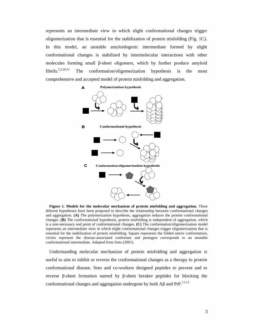
3
represents an intermediate view in which slight conformational changes trigger
oligomerization that is essential for the stabilization of protein misfolding (Fig. 1C).
In this model, an unstable amyloidogenic intermediate formed by slight
conformational changes is stabilized by intermolecular interactions with other
molecules forming small β-sheet oligomers, which by further produce amyloid
fibrils.2,3,10,11 The conformation/oligomerization hypothesis is the most
comprehensive and accepted model of protein misfolding and aggregation.
Figure 1. Models for the molecular mechanism of protein misfolding and aggregation. Three
diferent hypotheses have been proposed to describe the relationship between conformational changes and aggregation. (A) The polymerization hypothesis, aggregation induces the protein conformational changes. (B) The conformational hypothesis, protein misfolding is independent of aggregation, which is a non-necessary end point of conformational changes. (C) The conformation/oligomerization model represents an intermediate view in which slight conformational changes trigger oligomerization that is essential for the stabilization of protein misfolding. Square represents the folded native conformation, circles represent the disease-associated conformer and pentagon corresponds to an unstable conformational intermediate. Adapted from Soto (2001).
Understanding molecular mechanism of protein misfolding and aggregation is
useful to aim to inhibit or reverse the conformational changes as a therapy to protein
conformational disease. Soto and co-workers designed peptides to prevent and to
reverse β-sheet formation named by β-sheet breaker peptides for blocking the
conformational changes and aggregation undergone by both Aβ and PrP.11-13
4
1.2 Protein aggregation and fibrillation
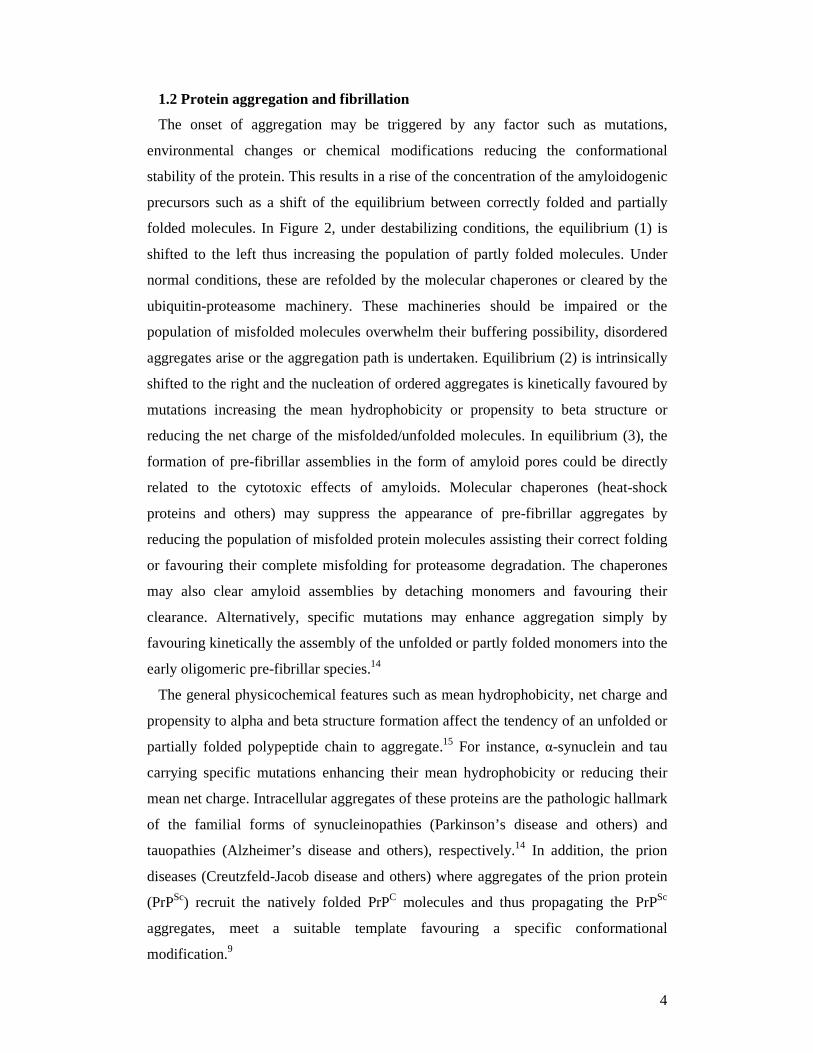
The onset of aggregation may be triggered by any factor such as mutations,
environmental changes or chemical modifications reducing the conformational
stability of the protein. This results in a rise of the concentration of the amyloidogenic
precursors such as a shift of the equilibrium between correctly folded and partially
folded molecules. In Figure 2, under destabilizing conditions, the equilibrium (1) is
shifted to the left thus increasing the population of partly folded molecules. Under
normal conditions, these are refolded by the molecular chaperones or cleared by the
ubiquitin-proteasome machinery. These machineries should be impaired or the
population of misfolded molecules overwhelm their buffering possibility, disordered
aggregates arise or the aggregation path is undertaken. Equilibrium (2) is intrinsically
shifted to the right and the nucleation of ordered aggregates is kinetically favoured by
mutations increasing the mean hydrophobicity or propensity to beta structure or
reducing the net charge of the misfolded/unfolded molecules. In equilibrium (3), the
formation of pre-fibrillar assemblies in the form of amyloid pores could be directly
related to the cytotoxic effects of amyloids. Molecular chaperones (heat-shock
proteins and others) may suppress the appearance of pre-fibrillar aggregates by
reducing the population of misfolded protein molecules assisting their correct folding
or favouring their complete misfolding for proteasome degradation. The chaperones
may also clear amyloid assemblies by detaching monomers and favouring their
clearance. Alternatively, specific mutations may enhance aggregation simply by
favouring kinetically the assembly of the unfolded or partly folded monomers into the
early oligomeric pre-fibrillar species.14
The general physicochemical features such as mean hydrophobicity, net charge and
propensity to alpha and beta structure formation affect the tendency of an unfolded or
partially folded polypeptide chain to aggregate.15 For instance, α-synuclein and tau
carrying specific mutations enhancing their mean hydrophobicity or reducing their
mean net charge. Intracellular aggregates of these proteins are the pathologic hallmark
of the familial forms of synucleinopathies (Parkinson’s disease and others) and
tauopathies (Alzheimer’s disease and others), respectively.14 In addition, the prion
diseases (Creutzfeld-Jacob disease and others) where aggregates of the prion protein
(PrPSc) recruit the natively folded PrPC molecules and thus propagating the PrPSc
aggregates, meet a suitable template favouring a specific conformational
modification.9
5
Figure 2. The possible fates of newly synthesized polypeptide chains. Modifications of protein
structure or medium conditions may favour protein-protein interactions into fibers or into crystalline lattices. DANGER! indicates the processes generating the pre-fibrillar assemblies presently considered mostly associated with cell impairment. The question mark indicates that it is not known whether amyloid pores (when formed) are on path or dead end intermediates of fibril formation. Adapted from Stefani (2004).
Protein aggregation may be favoured under conditions resulting in the impairment
or overwhelming of the molecular machineries. These molecular machineries
comprise the molecular chaperones of the endoplasmic reticulum (ER) such as Bip,
Grp94, calnexin and of the cytosol (heat-shock proteins, crystallins, prefoldin, Hsc70)
and the ubiquitin-proteasome pathway in ensuring the quality control of protein
folding.16-18
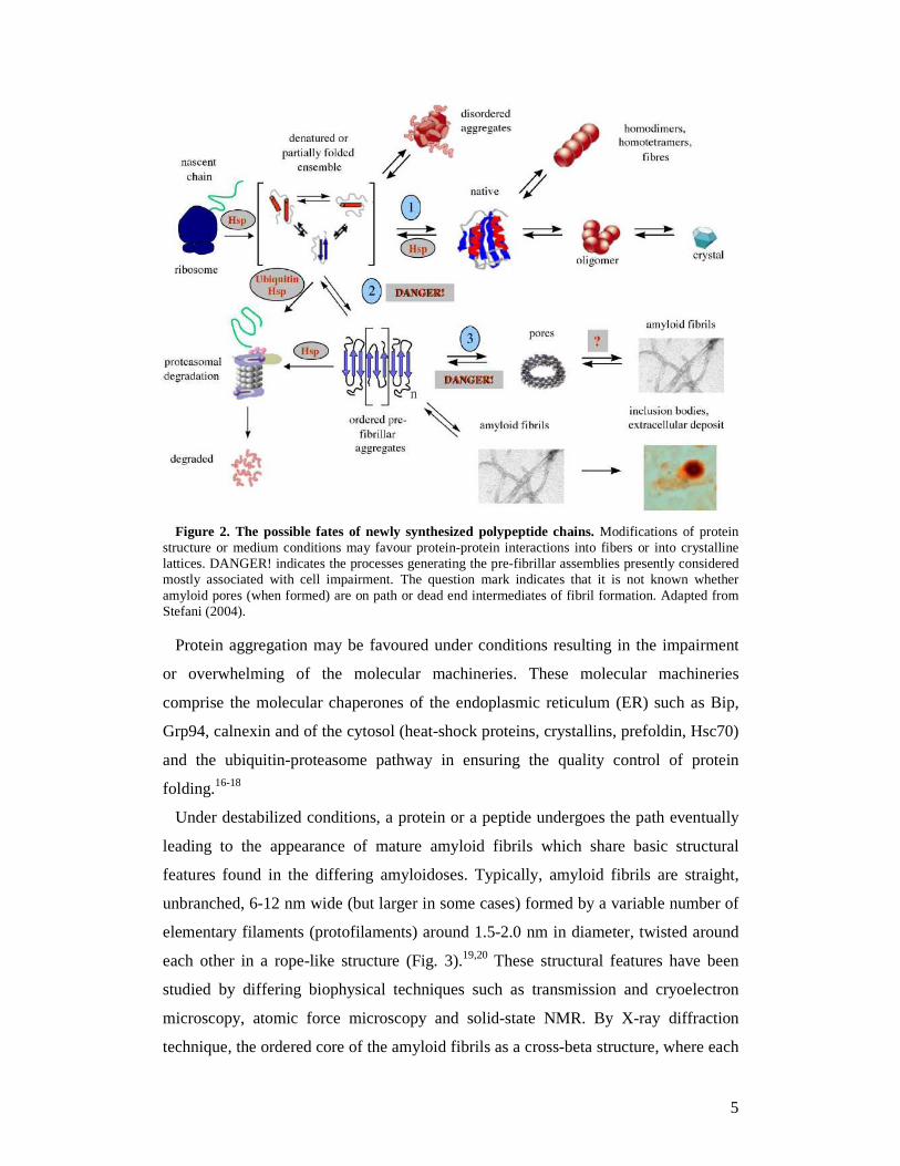
Under destabilized conditions, a protein or a peptide undergoes the path eventually
leading to the appearance of mature amyloid fibrils which share basic structural
features found in the differing amyloidoses. Typically, amyloid fibrils are straight,
unbranched, 6-12 nm wide (but larger in some cases) formed by a variable number of
elementary filaments (protofilaments) around 1.5-2.0 nm in diameter, twisted around
each other in a rope-like structure (Fig. 3).19,20 These structural features have been
studied by differing biophysical techniques such as transmission and cryoelectron
microscopy, atomic force microscopy and solid-state NMR. By X-ray diffraction
technique, the ordered core of the amyloid fibrils as a cross-beta structure, where each
6
protofilament results from a double row of beta-sheets provided by each monomer,
has been descripted. The strands of the cross-beta structure of the core of amyloid
aggregates run parallel to each other and perpendicular to the main fibril axis (Fig.
3).14
Figure 3. Close-up view of the structural organization of an amyloid fibril. The four
protofilaments are wound around each other and their core structure is a row of β-sheets where each strand runs perpendicular to the fibril axis. Adapted from Stefani (2004).
The studies have been reported that the pathogenic protein aggregates are the
destabilised monomeric, or the non-fibrillar oligomeric species of distinct morphology
(protofibrils) preceding mature fibrils in the aggregation pathway. The earliest
protofibrils typically appear as globular assemblies 2.5-5.0 nm in diameter
spontaneously organizing into chains and variously sized rings comprising small
bdoughnutsQ with a central pore, further organising into ribbons, protofilaments and
mature fibrils.21,22
1.3 Prion proteins and diseases
Prion diseases, also known as transmissible spongiform encephalopathies (TSEs)
are fatal and incurable neurodegenerative disorders of animals and humans.9 They can
manifest as genetic, infectious and sporadic illnesses and they include bovine
spongiform encephalopathy (BSE) of cattle, scrapie of sheep, chronic wasting disease
(CWD) of deer, moose and elk, Creutzfeldt-Jakob (CJD) and Gerstmann-Sträussler-
Scheinker (GSS) diseases of humans.23
The prion hypothesis
In 1967, Alper and co-workers demonstrated that the infectious materials was not
destroyed under very high doses of ionizing radiation and ultraviolet (UV) which
7
obliterate nucleic acids.24 And also in this year, Griffith demonstrated that a protein
can act as the infectious agent causing scrapie.25 However, until 1982, Prusiner first
proposed the prion (proteinaceous infectious particles) hypothesis.26 Over decades of
research, there have been crucial evidences for this hypothesis by starting with the
initial indication that prion diseases can be transmissible, owing to the accidental
transmission of scrapie in sheep and ending with the demonstration that infectious
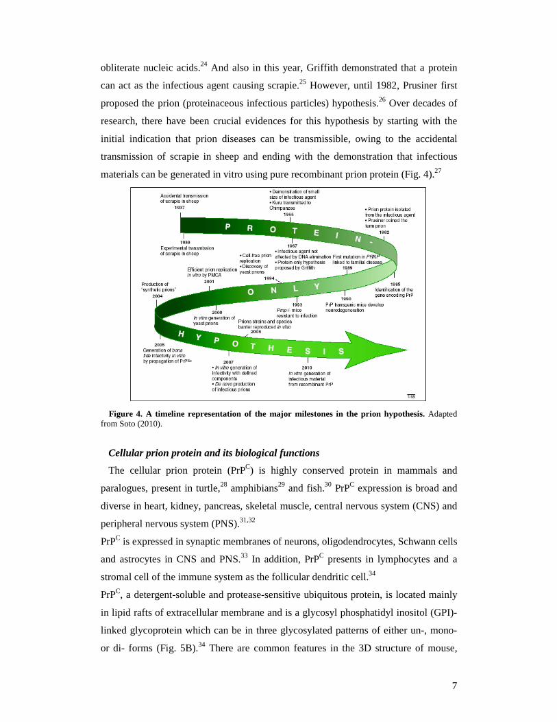
materials can be generated in vitro using pure recombinant prion protein (Fig. 4).27
Figure 4. A timeline representation of the major milestones in the prion hypothesis. Adapted
from Soto (2010).
Cellular prion protein and its biological functions
The cellular prion protein (PrPC) is highly conserved protein in mammals and
paralogues, present in turtle,28 amphibians29 and fish.30 PrPC expression is broad and
diverse in heart, kidney, pancreas, skeletal muscle, central nervous system (CNS) and
peripheral nervous system (PNS).31,32
PrPC is expressed in synaptic membranes of neurons, oligodendrocytes, Schwann cells
and astrocytes in CNS and PNS.33 In addition, PrPC presents in lymphocytes and a
stromal cell of the immune system as the follicular dendritic cell.34
PrPC, a detergent-soluble and protease-sensitive ubiquitous protein, is located mainly
in lipid rafts of extracellular membrane and is a glycosyl phosphatidyl inositol (GPI)-
linked glycoprotein which can be in three glycosylated patterns of either un-, mono-
or di- forms (Fig. 5B).34 There are common features in the 3D structure of mouse,
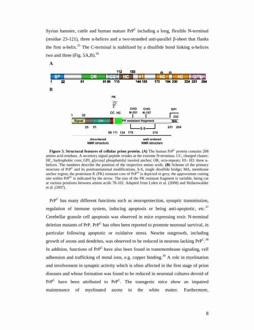
8
Syrian hamster, cattle and human mature PrPC including a long, flexible N-terminal
(residue 23-121), three α-helices and a two-stranded anti-parallel β-sheet that flanks
the first α-helix.35 The C-terminal is stabilized by a disulfide bond linking α-helices
two and three (Fig. 5A,B).36
A
B
Figure 5. Structural features of cellular prion protein. (A) The human PrPC protein contains 208
amino acid residues. A secretory signal peptide resides at the extreme N-terminus. CC, charged cluster; HC, hydrophobic core; GPI, glycosyl phosphatidyl inositol anchor; OR, octa-repeats; H1- H3: three α-helices. The numbers describe the position of the respective amino acids. (B) Scheme of the primary structure of PrPC and its posttranslational modifications. S-S, single disulfide bridge; MA, membrane anchor region; the proteinase K (PK) resistant core of PrPSc is depicted in grey; the approximate cutting site within PrPSc is indicated by the arrow. The size of the PK resistant fragment is variable, being cut at various positions between amino acids 78-102. Adapted from Liden et al. (2008) and Heikenwalder et al. (2007).
PrPC has many different functions such as neuroprotection, synaptic transmission,
regulation of immune system, inducing apoptosis or being anti-apoptotic, etc.37
Cerebellar granule cell apoptosis was observed in mice expressing toxic N-terminal
deletion mutants of PrP. PrPC has often been reported to promote neuronal survival, in
particular following apoptotic or oxidative stress. Neurite outgrowth, including
growth of axons and dendrites, was observed to be reduced in neurons lacking PrPC.38
In addition, functions of PrPC have also been found in transmembrane signaling, cell
adhension and trafficking of metal ions, e.g. copper binding.39 A role in myelination
and involvement in synaptic activity which is often affected in the first stage of prion
diseases and whose formation was found to be reduced in neuronal cultures devoid of
PrPC have been attributed to PrPC. The transgenic mice show an impaired
maintenance of myelinated axons in the white matter. Furthermore,
9
electrophysiological studies indicate a role of PrPC in synapse function, especially in
neurotransmitter release.38
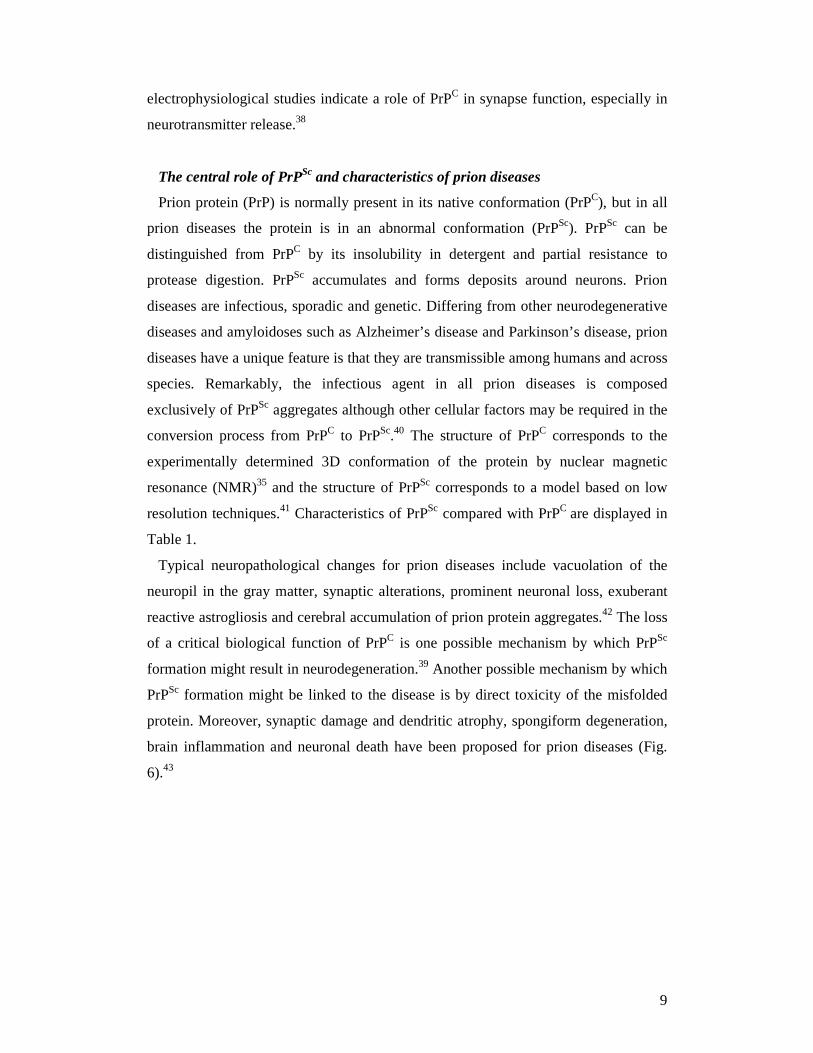
The central role of PrPSc and characteristics of prion diseases
Prion protein (PrP) is normally present in its native conformation (PrPC), but in all
prion diseases the protein is in an abnormal conformation (PrPSc). PrPSc can be
distinguished from PrPC by its insolubility in detergent and partial resistance to
protease digestion. PrPSc accumulates and forms deposits around neurons. Prion
diseases are infectious, sporadic and genetic. Differing from other neurodegenerative
diseases and amyloidoses such as Alzheimer’s disease and Parkinson’s disease, prion
diseases have a unique feature is that they are transmissible among humans and across
species. Remarkably, the infectious agent in all prion diseases is composed
exclusively of PrPSc aggregates although other cellular factors may be required in the
conversion process from PrPC to PrPSc.40 The structure of PrPC corresponds to the
experimentally determined 3D conformation of the protein by nuclear magnetic
resonance (NMR)35 and the structure of PrPSc corresponds to a model based on low
resolution techniques.41 Characteristics of PrPSc compared with PrPC are displayed in
Table 1.
Typical neuropathological changes for prion diseases include vacuolation of the
neuropil in the gray matter, synaptic alterations, prominent neuronal loss, exuberant
reactive astrogliosis and cerebral accumulation of prion protein aggregates.42 The loss
of a critical biological function of PrPC is one possible mechanism by which PrPSc
formation might result in neurodegeneration.39 Another possible mechanism by which
PrPSc formation might be linked to the disease is by direct toxicity of the misfolded
protein. Moreover, synaptic damage and dendritic atrophy, spongiform degeneration,
brain inflammation and neuronal death have been proposed for prion diseases (Fig.
6).43
10
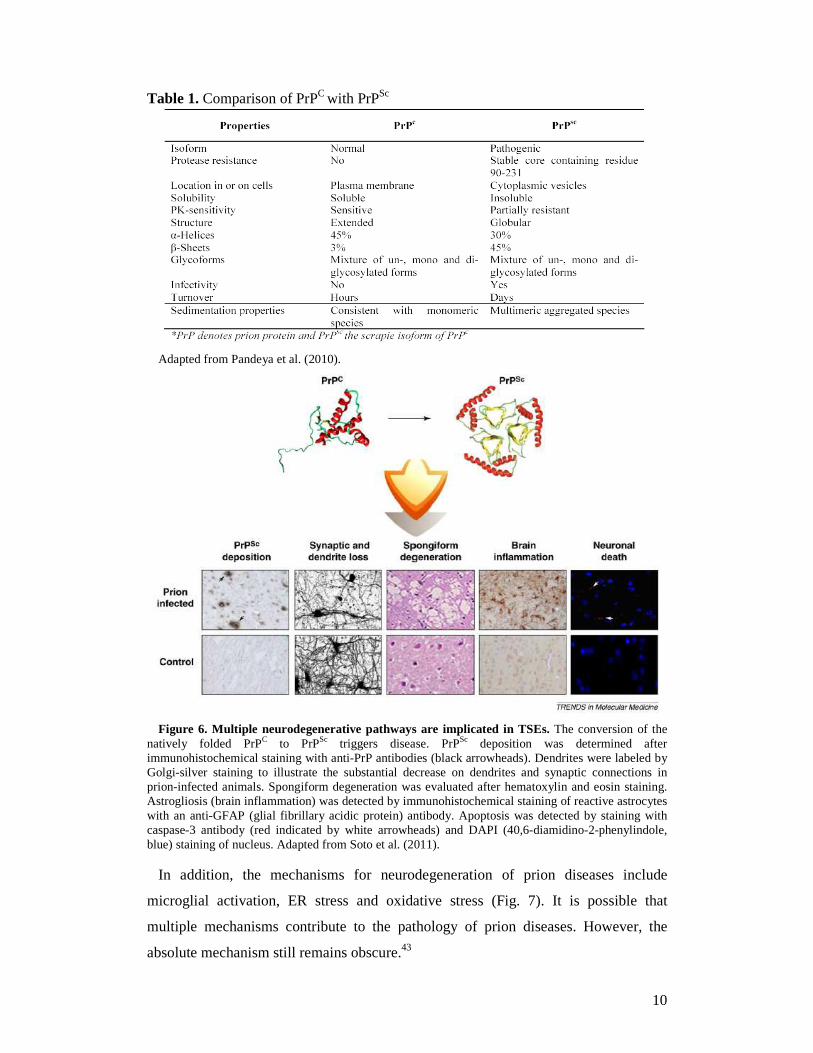
Table 1. Comparison of PrPC with PrPSc
Adapted from Pandeya et al. (2010).
Figure 6. Multiple neurodegenerative pathways are implicated in TSEs. The conversion of the
natively folded PrPC to PrPSc triggers disease. PrPSc deposition was determined after immunohistochemical staining with anti-PrP antibodies (black arrowheads). Dendrites were labeled by Golgi-silver staining to illustrate the substantial decrease on dendrites and synaptic connections in prion-infected animals. Spongiform degeneration was evaluated after hematoxylin and eosin staining. Astrogliosis (brain inflammation) was detected by immunohistochemical staining of reactive astrocytes with an anti-GFAP (glial fibrillary acidic protein) antibody. Apoptosis was detected by staining with caspase-3 antibody (red indicated by white arrowheads) and DAPI (40,6-diamidino-2-phenylindole, blue) staining of nucleus. Adapted from Soto et al. (2011).
In addition, the mechanisms for neurodegeneration of prion diseases include
microglial activation, ER stress and oxidative stress (Fig. 7). It is possible that
multiple mechanisms contribute to the pathology of prion diseases. However, the
absolute mechanism still remains obscure.43
11
Figure 7. Putative signaling pathways for PrPSc-induced neurodegeneration in prion diseases.
Several mechanisms have been proposed by which PrPC to PrPSc conversion results in neurodegeneration. PrPSc might produce mitochondrial stress, leading to apoptosis. An alternative model implicates sustained ER stress. Adapted from Soto et al. (2011).
Recently, Soto and Satani proposed a model in which the primary abnormality is
PrPSc formation and accumulation, from in peripheral tissues to in the brain. The
disease process starts with the formation of PrPSc, beginning a long and clinically
silent presymptomatic phase, in which PrPSc slowly but gradually accumulates in the
brain. PrPSc accumulation triggers ER stress and activation of the unfolded protein
response, which represents the first line of defense against protein misfolding. Other
early consequences of PrPSc accumulation are brain inflammation (in the form of
astrocytosis and microglial activation) and autophagy. Both inflammation and
autophagy might initially be defensive mechanisms, but later could also contribute to
neuronal death and perhaps brain vacuolation. The first damage leading to noticeable
clinical consequences is probably synaptic disruption, ending the presymtomatic
phase and beginning the early clinical phase of the disease. Synaptic dysfunction
produces loss of dendrites and finally neuronal death. The end and irreversible stages
of the disease are characterized by massive spongiform degeneration and neuronal
death, which are probably triggered by a variety of interconnecting cellular
pathways.43
1.4 Prion replication
Prion replication involves the direct interaction between the PrPSc template and
the endogenous cellular PrPC driving the formation of nascent infectious prions.44
12
The central feature of prion pathogenesis is the conversion of PrPC to PrPSc which
is thought to proceed via formation of a complex between PrP isoforms and an
unknown molecular chaperone "X" (Fig. 8).45 This conversion occurs post-
translationally and thought to involve conformational change rather than covalent
modification. The mechanism by which the conversion of PrPC to PrPSc takes
place and results in the distinct pathogenesis of prion diseases remains unknown.
Figure 8. The conformational conversion of the PrPC to PrPSc, which is thought to involve an unknown molecular chaperone "X". Adapted from Telling et al. (1995).
Models for the conformational conversion of PrPC to PrPSc
There are two models to explain the conversion of PrPC to PrPSc as the
“refolding” and the “seeding” models. In the first model, there is an interaction
between exogenously introduced PrPSc and endogenous PrPC, which is induced to
transform itself into further PrPSc. A high energy barrier may prevent spontaneous
conversion of PrPC to PrPSc (Fig. 9A). In the latter, by nucleation-polymerization,
PrPC and PrPSc are in a reversible thermodynamic equilibrium. The seed formation
begins very slowly, then monomeric PrPSc can be recruited and eventually
aggregate to amyloid. Fragmentation of PrPSc aggregates increases the number of
nuclei, which can recruit further PrPSc and thus results in apparent replication of
the agent (Fig. 9B).46
PrPSc
PrPSc
PrPSc multimers
PrP*
PrP*
Wild-type PrPC
X
X
X
X
13
Figure 9. Models for the conformational conversion of PrPC to PrPSc. (A) the “refolding” or template assistance model. (B) the “seeding” or nucleation-polymerization model. Adapted from Aguzzi et al. (2009).
Recently, the protein misfolding cyclic amplification (PMCA) technique which is
designed to mimic PrPSc autocatalytic replication has been developed.47-49 In the
PMCA, PrPSc is amplified in a cyclic manner by incubating small amounts of
PrPSc-containing brain homogenate with PrPC-containing brain homogenate.
Hence, PrPC is recruited into growing aggregates of PrPSc and it undergoes
conformational conversion and becomes PrPSc. The growing PrPSc species are
disrupted by repeated sonication in the presence of detergents to generate multiple
smaller units functioning as a seed for the continued formation of new PrPSc
aggregates. The whole procedure is repeated several times (Fig. 10).46
In addition, an important mechanism of prion replication process is the
propagation of prions through fragmentation of existing fibrils verified for yeast
prions,50 mammalian prion51 and non-prion related amyloid fibrils.52
14
Figure 10. Schematic representation of the protein misfolding cyclic amplification (PMCA) reaction. PrPC is shown as light gray spheres. PrPSc is shown as trapezium. The original seed is in dark gray, and the newly formed PrPSc is in light gray. Adapted from Aguzzi et al. (2009).
Cell biology of PrPC and PrPSc with potential sites of conversion
PrPC is usually associated with detergent-resistant membrane domains known as
rafts. The scrapie-associated conversion site for membrane-anchored wild-type PrPC
seems to be on the cell surface and/or in endosomes. However, PrPC released from the
cell due to lack of a GPI anchor may be converted to extracellular deposits such as
amyloid fibrils and plaques.37 In N2a cells, PrPSc mainly accumulates in late
endosomes and lysosomes53,54 and only very small amounts of PrPSc are located at
plasma membrane.55 In addition, some PrPSc was also found in the Golgi apparatus
detected in N2a cells persistently infected with RML/Chandler scrapie, but not in
hamster cells infected with a hamster scrapie strain by immuno-EM study.56 Also in
this cell line infected with RML or 22L scrapie strain, increased PrPSc levels are along
with increased retrograde transport to Golgi and ER.57 Furthermore, misfolded PrPC
can be subject to the ER-associated degradation pathway (ERAD). Under conditions
of proteosome inhibition, cytoplasmic forms of PrP aggregates are associated with
neurotoxicity such as aggresomes.58 Excessive levels of misfolded proteins in the
cytosol might impair proteosome function, either directly or after incorporation into
aggresomes.37
Regarding the presence of co-factors in conformational convesion of PrP, the
laminin receptor or its precusor as crucial co-factors is important for PrPSc
formation in GT1 cells infected with Chandler scrapie strain. Although it is
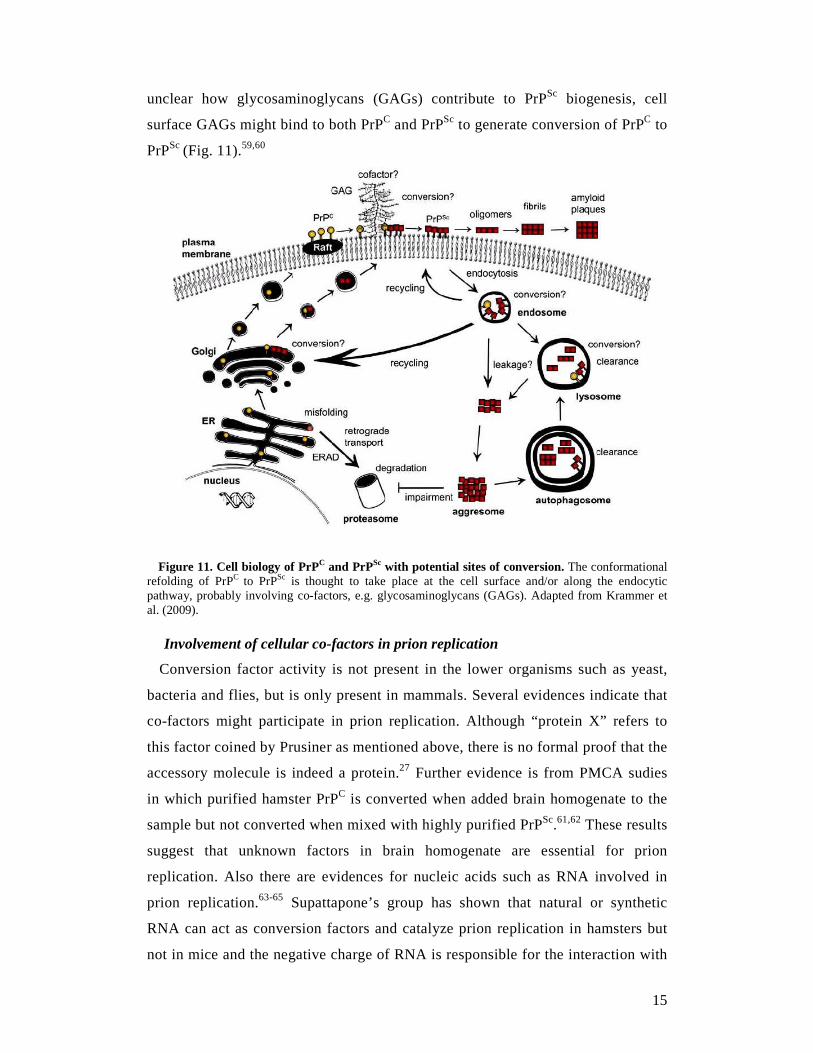
15
unclear how glycosaminoglycans (GAGs) contribute to PrPSc biogenesis, cell
surface GAGs might bind to both PrPC and PrPSc to generate conversion of PrPC to
PrPSc (Fig. 11).59,60
Figure 11. Cell biology of PrPC and PrPSc with potential sites of conversion. The conformational refolding of PrPC to PrPSc is thought to take place at the cell surface and/or along the endocytic pathway, probably involving co-factors, e.g. glycosaminoglycans (GAGs). Adapted from Krammer et al. (2009).
Involvement of cellular co-factors in prion replication
Conversion factor activity is not present in the lower organisms such as yeast,
bacteria and flies, but is only present in mammals. Several evidences indicate that
co-factors might participate in prion replication. Although “protein X” refers to
this factor coined by Prusiner as mentioned above, there is no formal proof that the
accessory molecule is indeed a protein.27 Further evidence is from PMCA sudies
in which purified hamster PrPC is converted when added brain homogenate to the
sample but not converted when mixed with highly purified PrPSc.61,62 These results
suggest that unknown factors in brain homogenate are essential for prion
replication. Also there are evidences for nucleic acids such as RNA involved in
prion replication.63-65 Supattapone’s group has shown that natural or synthetic
RNA can act as conversion factors and catalyze prion replication in hamsters but
not in mice and the negative charge of RNA is responsible for the interaction with
16
PrP.66 In addition, synthetic anionic phospholipids are required for PrPSc
replication67 and higher infectivity was reported with lipid membrane-associated
PrPSc.68 Surprisingly, treatments that eliminate nucleic acids, lipids, or proteins do
not prevent prion replication in vitro. Indeed, the addition of various classes of
molecules (synthetic nucleic acids, heparin, albumin or fatty acids) produces a
small but detectable effect on enhancing prion replication in vitro. These findings
suggest that various different compounds might act as a conversion factor in vitro,
that elimination of only some of them does not prevent prion replication.27,62
At least five different scenarios can be proposed for the involvement of cellular
co-factors in prion propagation. (i) The co-factor might integrate into the
infectious agent, alter PrPSc folding, and provide biological information to the
infectivity process, perhaps by determining strain characteristics. (ii) The co-factor
might act as an essential catalyst for prion replication, perhaps by interacting with
PrPC, altering its folding, and permitting its interaction with PrPSc. (iii) Through
binding and integration into the PrPSc polymer, the co-factor might help to
stabilize the conformation of PrPSc. (iv) The co-factor might participate in the key
process of fragmenting PrPSc polymers to produce smaller structures, and
multiplying the number of seeds to allow the continuation of prion replication. (v)
The co-factor might bind to PrPSc, thus increasing its biological stability, reducing
its clearance in vivo, and increasing its chances to reach target organs. It is
important to highlight that these possibilities are not mutually exclusive, and
indeed, it is likely that a co-factor could be involved in several of these processes
simultaneously.27
Therefore, in prion therapy, the molecules binding to either PrPC or PrPSc
conformers at the binding interface may inhibit the interaction of PrPC with PrPSc,
thus interrupting prion production. Additionally, the compounds that bind to the
molecules supporting and participating in prion replication, such as chaperones or
other ligands, may also be good candidates for blocking prion replication.
1.5 Prion infectivity
In peripheral infection, prions silently accumulate and replicate in peripheral organs
or reservoirs and transit through at least one PrP-positive (PrP+) tissue before reaching
the CNS.69 Prions replicate in lymphoid organs during the early stages of infection.70
Within the lymphoreticular system, follicular dendritic cells (FDCs) are a prominent
17
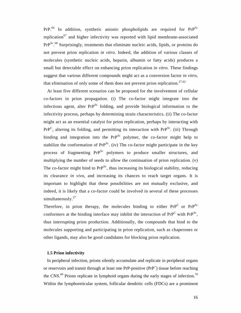
site of PrPSc deposition.71 In rodent scrapie models, prion replication is typically
detected first in the spleen and reaches plateau levels before detectable neuroinvasion
after peripheral inoculation was performed. Then brain levels rise exponentially to
100-fold or more above splenic levels before clinical disease occurs. Infectivity can
be recovered from the spleen very early during the incubation period (Fig. 12a).72
Infectivity is detectable on hamster and mouse bioassay following inoculation of
wild-type CD-1 mice with 263K hamster prions. On hamster bioassay, infectivity can
be recovered early in incubation period and at low level of the original 263K
inoculum or new infectivity can be accumulated slowly. On mouse bioassay,
infectivity appears after a lengthy period and the animal dies of natural causes
following a normal lifespan. At this stage, on both hamster and mouse bioassay,
infectivity can be recovered at high levels from these clinically normal animals (Fig.
12b).72
Figure 12. Prion infection in vivo. Adapted from Hill et al. (2003).
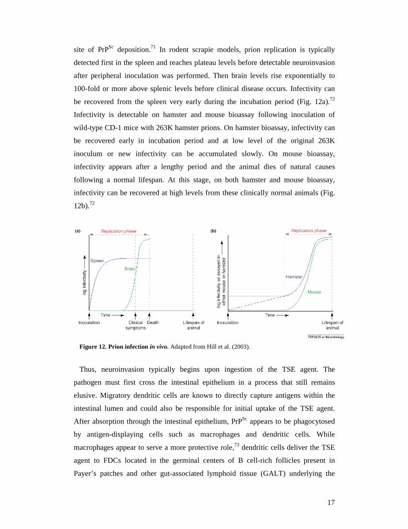
Thus, neuroinvasion typically begins upon ingestion of the TSE agent. The
pathogen must first cross the intestinal epithelium in a process that still remains
elusive. Migratory dendritic cells are known to directly capture antigens within the
intestinal lumen and could also be responsible for initial uptake of the TSE agent.
After absorption through the intestinal epithelium, PrPSc appears to be phagocytosed
by antigen-displaying cells such as macrophages and dendritic cells. While
macrophages appear to serve a more protective role,73 dendritic cells deliver the TSE
agent to FDCs located in the germinal centers of B cell-rich follicles present in
Payer’s patches and other gut-associated lymphoid tissue (GALT) underlying the
18
intestinal epithelium. After incubation in lymphoid tissue such as the GALT and
spleen, the TSE agent spreads to CNS via the enteric nervous system (Fig. 13).69
Routes for neuroinvasion including tunneling nanotubes, exosomes and blood have
been studied. Tunneling nanotubes are important for intracellular transfer of prion
during neuroinvasion.74 Prions gain access into and between neurons by hijacking
tunneling nanotubes (for 12 hours of co-culture) which is more effective than
transportation by exosomes (for 5 days of co-culture).75 Recently, for removing TSE
infectivity from whole blood, the removal of all white cells reduced infectivity by
only 42%, suggesting that other blood components, cells or plasma, could be
infectious.69,76
Figure 13. The route of prion neuroinvasion. After absorption through the intestinal epithelium, prion reach the peyer’s patches, via blood constituents (Plasminogen that bind to PrPSc). FDCs are infected in the patches and in other lymphoid organs, including the spleen. The prions reach the spleen by a B-cell independent route involving complement factors. Other factors that are required for spreading infection to the CNS are lymphotoxin (stimulus for FDCs), and at least one interposed PrP+ tissue. Adapted from Pandeya et al. (2010).
1.6 Therapies for prion diseases
Early treatment regimes, including various prophylactic compounds and
immunotherapies, have sought efficacy through neutralization of infectious sources,
blockade of infection via the most common peripheral routes, and/or blockade of
neuroinvasion. Effective therapies targeting later disease, which are initiated after the
appearance of clinical signs, will most likely involve some combination of inhibiting
pathogenic PrP formation, destabilizing or enhancing clearance of existing pathogenic
19
PrP, blocking neurotoxic effects of the infection, and/or promoting the recovery of
lost functions in the CNS.77
1.6.1 Chemotherapy for prion diseases
Many compounds have been proposed for the treatment of prion diseases,
including polysulfated anions, dextranes, and cyclic tetrapyrroles.78-81 Recently,
some success has been achieved using pentosan polysulfate82 although this
compound seems to be unsuccessful in the treatment of human prion diseases.83 In
addition, molecules targeting the different molecular steps involved in
pathological prion replication have also been investigated.84-90 However, to date,
the use of these compounds in clinical applications is limited, due to their high
toxicity and poor crossing of the blood-brain barrier (BBB). Thus, there is an
urgent need to develop systematic pharmacological and mechanistic studies for the
identification of a new class of compounds as therapeutic agents capable of
inhibiting several pathways in prion conversion and replication.
Strategies for developing new drugs
Three strategies are usually developed to identified new drugs against a well-
characterized disease. (i) A rational approach will specifically target the key
molecules responsible for the disease. The limiting factor here is to possess the
structure of one or more proteins implicated in the pathogenesis of the disease. (ii) A
blind screening on a large panel of drug already synthesized and commercially
available. The idea is to identify new molecules with a chemical structure different
from those already existing and which can serve as lead molecule for the
pharmaceutical chemistry. (iii) Synthesis of chemical derivatives of recently identified
lead molecules that showed promising therapeutic properties. The idea here is to
modify some chemical characteristics of the drug in order to make it more potent or to
facilitate its delivery, especially in the brain for instance. Various combinations
between these three strategies are possible in the drug development field.91
A common task in pharmaceutical research is the search for new lead compounds
against diseases that show a greater specificity and/or fewer side effects than already-
known agents. Therefore, a widely used search method as high throughput screening
(HTS) of large compound collections was established. However, since this approach
is expensive and time consuming and futher on only can be used once a suitable test
20
assay is developed, the silico design and proposal of new lead structures becomes
more important.92 Moreover, the nature of the prion agent as well as its replication
cycle which are not yet completely understood, does not facilitate HTS.91 A central
origin of the strategy of in silico screening of drug database is the experience that
similarities in structures are indicative of similarities in activities of drugs. Thus, a
structural search of large compound databases is of great interest. Today, about two
million chemical compounds are available commercially.93 The use of SuperDrug94
(http://bioinf.charite.de/superdrug), a new data base of essential WHO approved drugs
(2003), for 2D and 3D search for new lead structures starting from compounds against
prion diseases was performed by Lorenzen and co-workers.92 In this method, the
authors started with known lead compounds, a data base is searched to create a pool
of putative drugs. These compounds are compared to known inhibitors and non-
inhibitors, and drugs with similarities to inactive structures are removed from the list
of proposed inhibitors. Combining structural features of ineffective substances with
property filtering rules allows the exclusion of further candidates. Drugs surpassing
this sieve are proposed as new TSE inhibitors. Furthermore, the first PrP(90-231)
NMR structure solved in 1996 provided some structural information to monomeric
structure of the PrP protein and opened a new area of drug research since PrP
constitutes an attractive therapeutic target within the replication cycle of prions.
However, the surface of the PrP(90-231) NMR structure presented no crevasses, so
the classical docking program used for the structure-based drug design was not
adapted in this case.91 For the rational approach, Perrier and co-workers (2000) found
two drugs Cp60 and Cp62 that could mimic a small region at the PrP surface to inhibit
prion replication.90
In addition, Korth and co-workers (2001) have performed a blind screening on drugs
known to cross the BBB. They found that quinacrine and chlorpromazine, both
tricyclic compounds with an aliphatic side chain in their middle ring, exhibited an
anti-prion activity with EC50 of 0.3 and 3 µM respectively. Hence, Korth and co-
workers synthesized nine derivatives of quinacrine in order to establish a structure-
activity relationship study.95 Their results have revealed the importance of the
aliphatic side chain for the inhibition of PrPSc formation.91
21
Models for studying chemotherapeutic candidates
In vivo tests provide the most rigorous evaluations of anti-TSE treatments, but are
slow, costly, and impractical for screening purposes. A variety of relatively high
throughput, low cost, cell culture models,96,97 and some yeast models,98 have enabled
the identification of a number of different classes of anti-prion compounds which then
have shown efficacy in animal models. Also, in vitro assays have allowed
investigation of the mechanisms of prion inhibition. In many cases, anti-prion
compounds which bind to PrPC cause it to cluster and internalize, thus rendering PrPC
inaccessible or incompatible for conversion to protease-resistant prion protein
(PrPres).37,97,99 Non-cellular in vitro assays have also been developed to assess a wide
range of potentially effective compounds. These methods generally assess the
competitive binding of PrPC and PrPres or the prevention of PrP amyloid fibril
formation. Recent techniques include surface plasmon resonance,100 fluorescence
correlation spectroscopy,101 semiautomated cell-free conversion,102 and a fluorescence
polarization-based competitive binding assay.103 Computer “in silico” modeling is
also being used to predict binding molecules.90,104
Ultimately, promising treatments discovered in vitro require testing in animals and
humans. Of the many compounds studied in rodent models, few have made their way
into human trials or case reports, and the effectiveness of treatment administered at
the onset of clinical symptoms, or when there is significant neuropathology, is low.
However, many compounds show some prophylactic or early treatment effect in TSE-
infected animals, and are therefore relevant to decontamination and early therapy
efforts. These drugs need not be permeable to the BBB since they can target the
peripheral replication of the agent, before neuroinvasion that arise following oral or
other peripheral exposures. The option for early treatment has been hindered by a lack
of early diagnosis, but with the recent development of new sensitive detection
assays,66,105-108 there is hope for early preclinical TSE diagnostics, and more effective
screening and testing of at risk individuals. This, coupled with the rising concern of
blood transmission of variant Creutzfeldt Jakob disease (vCJD), the occurrence of
BSE in livestock, and the spread of CWD, lends tremendous relevance to such
chemoprophylaxis compounds and potential decontaminants in the management of
prion diseases.77
22
Identification of prion drug targets
A major focus of drug screening efforts has been the PrP conversion reaction. Many
inhibitors prevent conversion by directly binding and blocking interactions between
PrPC and PrPSc. Others affect conversion by interfering with important accessory
molecules, or by altering PrPC expression and distribution.37 Many different chemical
classes of compounds have been screened and tested in vitro, and additional in vivo
data are available for some (Table 2), including early or prophylactic treatments and
later stage therapies.77
Targeting PrP conversion of the compounds include binding PrPC and/or PrPres,
redistribution or sequestration of PrPC (e.g. cholesterol-depleting agents such as the
statin drug simvastatin109 and polyene antibiotics such as amphotericin B110),
suppressing PrPC expression by using small interfering RNA (siRNA) to the PrP
gene,111 targeting accessory molecules and pathways to conversion with Laminin
receptor precursor protein (LRP/LR) antibodies112 or tyrosine kinase inhibitor STI571
(known as imatinib mesylate),113,114 enhanced PrPres clearance with polycations115-117
and other unknown mechanisms by using copper chelators118,119 or dimethylsulfoxide
(DMSO)120 or antivirals such as vidarabine (adenine arabinoside).121
For binding PrPC and/or PrPres, the compounds include polyanions (RNA,63,122,123
sulfated glycosaminoglycans (GAGs),124,125 pentosan polysulfate (PSS),82 fucoidan,126
phosphorothioate oligonucleotides,127 copaxone128), sulphonated dyes and similar
compounds (Congo red,84,129 suramin,130,131 curcumin132-134), cyclic tetrapyrroles
(porphyrins, phthalocyanines),81,135,136 lysosomotropic factors (quinacrine, quinoline,
acridines, phenothiazines),95,137-140 tetracyclic compounds (tetracycline,
doxycycline),141-143 other amyloidophilic compounds,144 pyridine dicarnitrile
compounds,145 peptide aptamers and β-sheet breaker.13,89,146
In addition, compounds without direct effects on PrPC, PrPres or conversion may
have therapeutic potential as neuroprotective agents or symptomatic treatment
including analgesics such as flupirtine maleate,147 cannabidiol (a nonpsychoactive
constituent of cannabis)148 and antioxidants such as pyrazolone derivatives149 (Table
2).
23
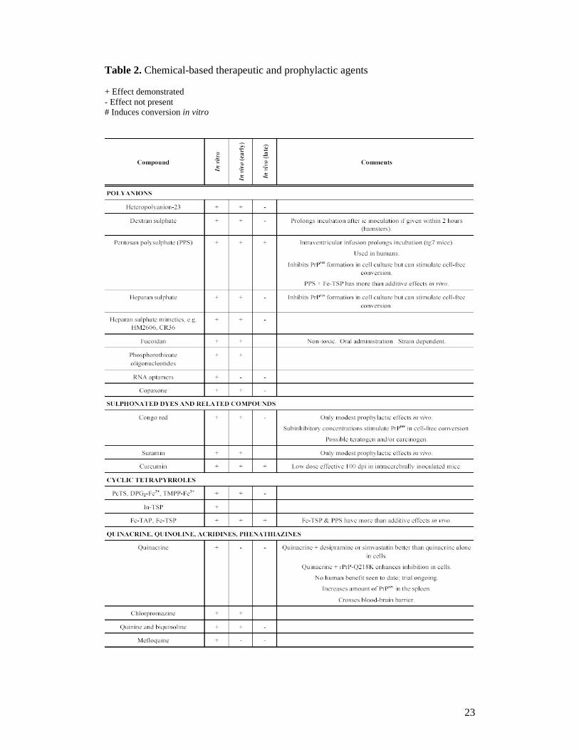
Table 2. Chemical-based therapeutic and prophylactic agents + Effect demonstrated - Effect not present # Induces conversion in vitro
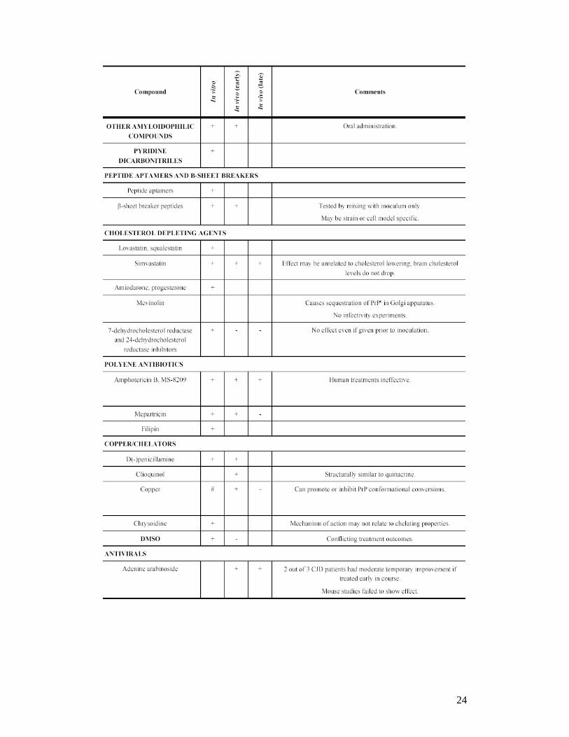
24
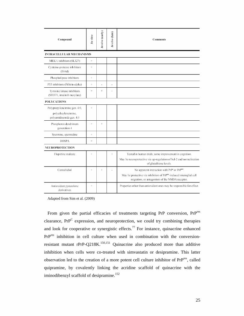
25
Adapted from Sim et al. (2009)
From given the partial efficacies of treatments targeting PrP conversion, PrPres
clearance, PrPC expression, and neuroprotection, we could try combining therapies
and look for cooperative or synergistic effects.77 For instance, quinacrine enhanced
PrPres inhibition in cell culture when used in combination with the conversion-
resistant mutant rPrP-Q218K.150,151 Quinacrine also produced more than additive
inhibition when cells were co-treated with simvastatin or desipramine. This latter
observation led to the creation of a more potent cell culture inhibitor of PrPres, called
quipramine, by covalently linking the acridine scaffold of quinacrine with the
iminodibenzyl scaffold of desipramine.152
26
1.6.2 Immunotherapy for prion diseases
Rational drug design strategies which are the basis of most modern drug discoveries
are difficult to set up for prion diseases, due to the absence of a well-defined tertiary
and/or quaternary structure of both PrP isoforms and lack of knowledge of the
replication cycle of the prion agent. Only antibodies directed against the prion protein
can set free from these barriers as they specifically recognized PrP isoforms and bind
to their target with a high affinity.153 Treatment of cells with Fab fragments D18 and
D13, which recognized epitope 132-156 and 97-106 of PrP protein respectively, has
been proven effective in clearing pre-existing PrPSc in ScN2a cells.88 Monoclonal
anti-PrP antibody 6H4 which recognized epitope 144-152 of PrP protein, also
prevents infection of susceptible N2a.154 Moreover, transgenic mice expressing anti-
PrP antibody 6H4 in their spleen, prevent scrapie pathogenesis in vivo, sustaining the
development of vaccination strategy.155
For the roles of the lymphoid system and immune cells in prion pathogenesis,
immunotherapeutic approaches to prion diseases have been studied. At least four
strategies including removal of functional FDCs and therefore ablation of
lymphoid prion replication sites; stimulation of the innate immune system;
enhancement of elimination of PrPSc using PrP-specific antibodies; or binding of
available PrPC or PrPSc so that they are unavailable for conversion have been
studied (Fig. 14). All of these approaches, which include both suppression and
stimulation of the immune system, are now being tested in suitable in vivo systems
using mice experimentally infected with mouse-adapted scrapie. However,
because the lymphoid system has been found to be involved in almost all forms of
TSE, it is reasonable to presume that mouse-adapted scrapie provides a realistic
generic model for TSE therapy.46
27
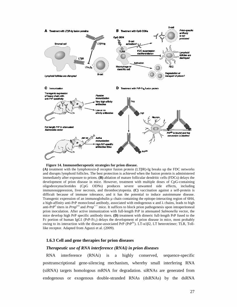
Figure 14. Immunotherapeutic strategies for prion disease.
(A) treatment with the lymphotoxin-β receptor fusion protein (LTβR)-Ig breaks up the FDC networks and disrupts lymphoid follicles. The best protection is achieved when the fusion protein is administered immediately after exposure to prions. (B) ablation of mature follicular dendritic cells (FDCs) delays the development of prion disease in mice. However, treatment with multiple doses of CpG-containing oligodeoxynucleotides (CpG ODNs) produces severe unwanted side effects, including immunosuppression, liver necrosis, and thrombocytopenia. (C) vaccination against a self-protein is difficult because of immune tolerance, and it has the potential to induce autoimmune disease. Transgenic expression of an immunoglobulin µ chain containing the epitope-interacting region of 6H4, a high-affinity anti-PrP monoclonal antibody, associated with endogenous κ and λ chains, leads to high anti-PrPC titers in Prnpo/o and Prnp+/+ mice. It suffices to block prion pathogenesis upon intraperitoneal prion inoculation. After active immunization with full-length PrP in attenuated Salmonella vector, the mice develop high PrP specific antibody titers. (D) treatment with dimeric full-length PrP fused to the Fc portion of human IgG1 (PrP-Fc2) delays the development of prion disease in mice, most probably owing to its interaction with the disease-associated PrP (PrPSc). LT-α1β2, LT heterotrimer; TLR, Toll-like receptor. Adapted from Aguzzi et al. (2009).
1.6.3 Cell and gene therapies for prion diseases
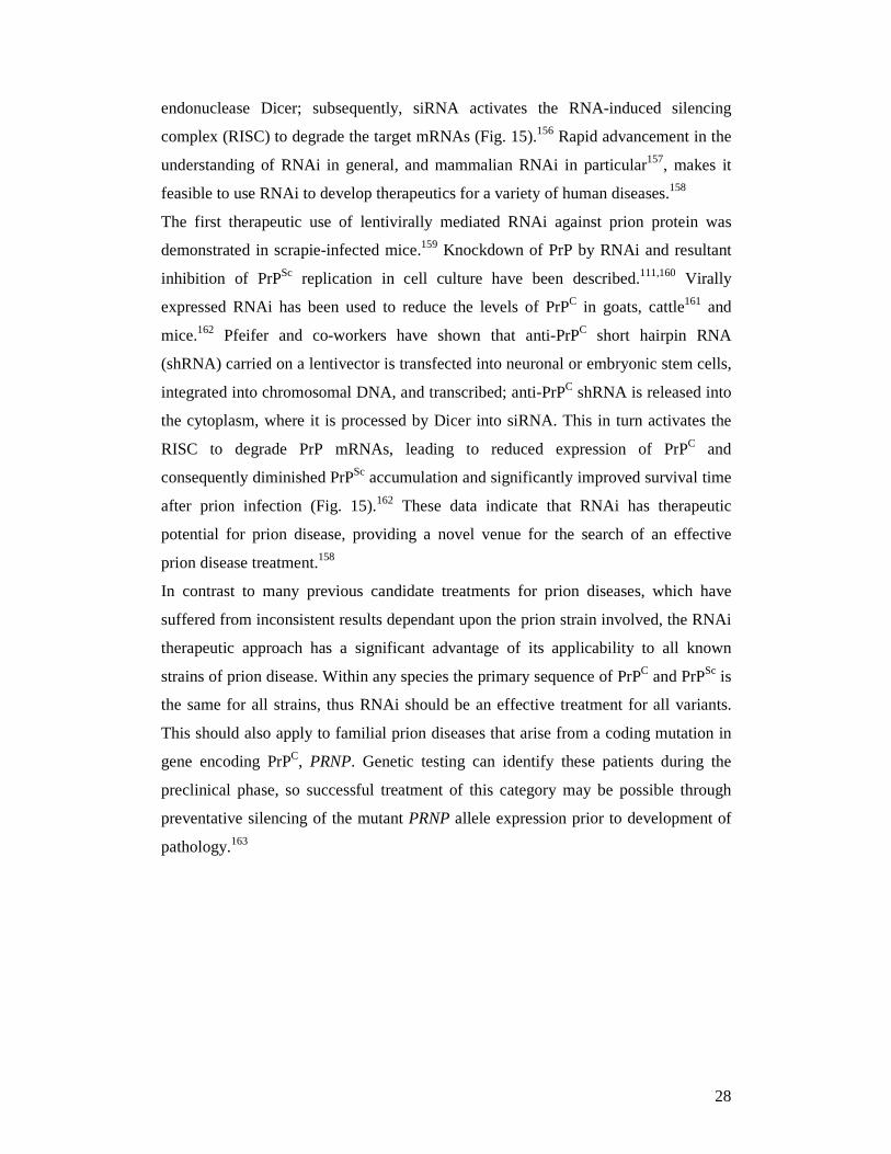
Therapeutic use of RNA interference (RNAi) in prion diseases
RNA interference (RNAi) is a highly conserved, sequence-specific
posttranscriptional gene-silencing mechanism, whereby small interfering RNA
(siRNA) targets homologous mRNA for degradation. siRNAs are generated from
endogenous or exogenous double-stranded RNAs (dsRNAs) by the dsRNA
28
endonuclease Dicer; subsequently, siRNA activates the RNA-induced silencing
complex (RISC) to degrade the target mRNAs (Fig. 15).156 Rapid advancement in the
understanding of RNAi in general, and mammalian RNAi in particular157, makes it
feasible to use RNAi to develop therapeutics for a variety of human diseases.158
The first therapeutic use of lentivirally mediated RNAi against prion protein was
demonstrated in scrapie-infected mice.159 Knockdown of PrP by RNAi and resultant
inhibition of PrPSc replication in cell culture have been described.111,160 Virally
expressed RNAi has been used to reduce the levels of PrPC in goats, cattle161 and
mice.162 Pfeifer and co-workers have shown that anti-PrPC short hairpin RNA
(shRNA) carried on a lentivector is transfected into neuronal or embryonic stem cells,
integrated into chromosomal DNA, and transcribed; anti-PrPC shRNA is released into
the cytoplasm, where it is processed by Dicer into siRNA. This in turn activates the
RISC to degrade PrP mRNAs, leading to reduced expression of PrPC and
consequently diminished PrPSc accumulation and significantly improved survival time
after prion infection (Fig. 15).162 These data indicate that RNAi has therapeutic
potential for prion disease, providing a novel venue for the search of an effective
prion disease treatment.158
In contrast to many previous candidate treatments for prion diseases, which have
suffered from inconsistent results dependant upon the prion strain involved, the RNAi
therapeutic approach has a significant advantage of its applicability to all known
strains of prion disease. Within any species the primary sequence of PrPC and PrPSc is
the same for all strains, thus RNAi should be an effective treatment for all variants.
This should also apply to familial prion diseases that arise from a coding mutation in
gene encoding PrPC, PRNP. Genetic testing can identify these patients during the
preclinical phase, so successful treatment of this category may be possible through
preventative silencing of the mutant PRNP allele expression prior to development of
pathology.163
29
Figure 15. RNAi and other strategies for prion disease treatment. Monomeric PrPC (yellow
ovals) converts into multimeric PrPSc (yellow rectangles) in the process of prion replication and prion pathogenesis. Reagents or strategies that effectively reduce the PrPC level or interfere with the PrPC-to-PrPSc conversion process have shown therapeutic potential for prion disease. Adapted from Kong (2006).
Cell grafting therapy for prion diseases
The transplantation of embryonic cells or tissue to protect against neuronal loss
could be a promising strategy for late stage treatment of prion diseases. Since PrP is
essential for prion replication, grafting of PrP knock-out cells would prevent prion
replication in the transplanted cells. Therefore, Brown and co-workers have used the
fetal cells from PrP knock-out mice (PrPo/o) to inject in hippocampal area 150 days
after scrapie infection in C57bl/6//VM mice and observed a neuron survival of 54%
greater than the control group despite no delay in the incubation time of the
disease.91,164
1.7 Therapies for other neurodegenerative diseases
1.7.1 Therapies for Alzheimer’s disease
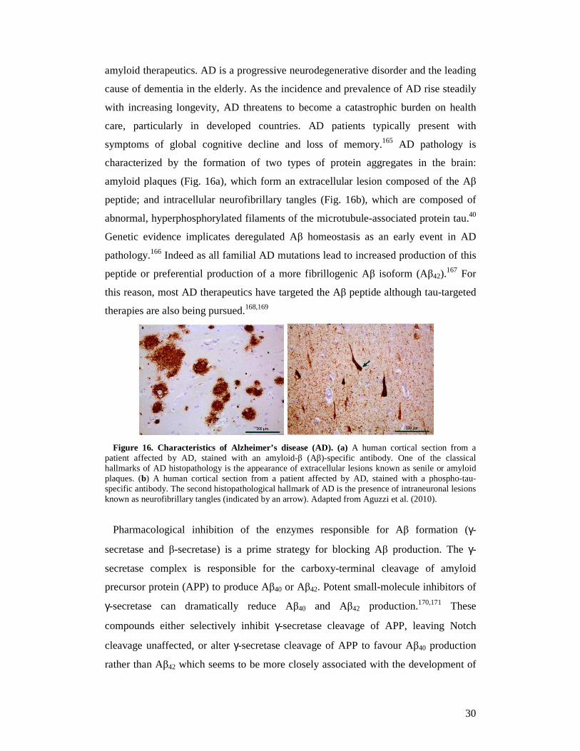
Alzheimer’s disease (AD), the most common neurodegenerative disorder, is
currently the focus of some of the most exciting and rapidly progressing research on
30
amyloid therapeutics. AD is a progressive neurodegenerative disorder and the leading
cause of dementia in the elderly. As the incidence and prevalence of AD rise steadily
with increasing longevity, AD threatens to become a catastrophic burden on health
care, particularly in developed countries. AD patients typically present with
symptoms of global cognitive decline and loss of memory.165 AD pathology is
characterized by the formation of two types of protein aggregates in the brain:
amyloid plaques (Fig. 16a), which form an extracellular lesion composed of the Aβ
peptide; and intracellular neurofibrillary tangles (Fig. 16b), which are composed of
abnormal, hyperphosphorylated filaments of the microtubule-associated protein tau.40
Genetic evidence implicates deregulated Aβ homeostasis as an early event in AD
pathology.166 Indeed as all familial AD mutations lead to increased production of this
peptide or preferential production of a more fibrillogenic Aβ isoform (Aβ42).167 For
this reason, most AD therapeutics have targeted the Aβ peptide although tau-targeted
therapies are also being pursued.168,169
Figure 16. Characteristics of Alzheimer’s disease (AD). (a) A human cortical section from a
patient affected by AD, stained with an amyloid-β (Aβ)-specific antibody. One of the classical hallmarks of AD histopathology is the appearance of extracellular lesions known as senile or amyloid plaques. (b) A human cortical section from a patient affected by AD, stained with a phospho-tau-specific antibody. The second histopathological hallmark of AD is the presence of intraneuronal lesions known as neurofibrillary tangles (indicated by an arrow). Adapted from Aguzzi et al. (2010).
Pharmacological inhibition of the enzymes responsible for Aβ formation (γ-
secretase and β-secretase) is a prime strategy for blocking Aβ production. The γ-
secretase complex is responsible for the carboxy-terminal cleavage of amyloid
precursor protein (APP) to produce Aβ40 or Aβ42. Potent small-molecule inhibitors of
γ-secretase can dramatically reduce Aβ40 and Aβ42 production.170,171 These
compounds either selectively inhibit γ-secretase cleavage of APP, leaving Notch
cleavage unaffected, or alter γ-secretase cleavage of APP to favour Aβ40 production
rather than Aβ42 which seems to be more closely associated with the development of
31
amyloid pathology than Aβ40.40 Drugs that modulate γ-secretase activity in this
manner include non-steroidal anti-inflammatory drugs (NSAIDs). The NSAID (R)-
flurbiprofen (also known as tarenflurbil), effectively reduced amyloid plaque
formation172 and rescued memory deficits173 in APP-transgenic mice. It also yielded
encouraging results in early human trials.174,175
The amino-terminal cleavage of APP to form both Aβ40 and Aβ42 results from β-
secretase activity. After the discovery that β-secretase cleavage of APP seemed to be
due to the activity of a single aspartic protease, β-secretase 1 (BACE1; also known as
memapsin 2 and ASP2), there was much interest in the possibility of targeting β-
secretase for the treatment of AD.176-180 Inhibition of BACE1 activity can block the
production of Aβ, prevent the development of amyloid pathology in the brain and
rescue AD-related memory deficits in mice.181-184 The large BACE1 active site
requires the identification of large compounds for potent BACE1 inhibition that also
readily penetrate the BBB185 and are reasonably stable. Unfortunately, the slow
progress of the BACE1 inhibitor field is a testament to the fact that such molecules
are relatively rare.40 Nevertheless, some BACE1 inhibitors have progressed to early
clinical trials.186
An alternative approach to protein aggregation therapeutics is to enhance the
degradation of the aggregating protein or the aggregates themselves. Manipulating the
immune system for the purpose of enhancing Aβ clearance has been pursued as a
therapeutic approach for AD.40 Several studies reported dramatically reduced Aβ
levels and plaque pathology and/or cognitive improvements upon active
immunization of APP-transgenic mice with full-length Aβ peptide,187,188 Aβ peptide
fragments189 and passive transfer of Aβ-specific antibodies.190-192 Based on these
studies and encouraging results from Phase I trials, active Aβ immunotherapy in
humans subsequently progressed to a widely publicized Phase II clinical trial in 2001.
Unfortunately, this trial was halted in January 2002 owing to the development of
sterile meningoencephalitis in some patients.40,193
In addition, several compounds, such as Congo red,194 anthracycline,195
rifampicin,196 anionic sulphonates197 or melatonin,198 can interact with Aβ and prevent
its aggregation into fibrils in vitro, thereby reducing toxicity. Moreover, certain non-
fibrillogenic, Aβ homologous peptides can bind to Aβ and break the formation of β-
sheet structure.12,199
32
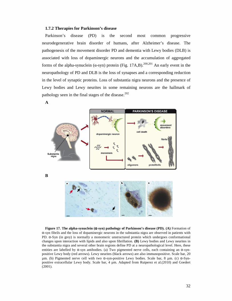
1.7.2 Therapies for Parkinson’s disease
Parkinson’s disease (PD) is the second most common progressive
neurodegenerative brain disorder of humans, after Alzheimer’s disease. The
pathogenesis of the movement disorder PD and dementia with Lewy bodies (DLB) is
associated with loss of dopaminergic neurons and the accumulation of aggregated
forms of the alpha-synuclein (α-syn) protein (Fig. 17A,B).200,201 An early event in the
neuropathology of PD and DLB is the loss of synapses and a corresponding reduction
in the level of synaptic proteins. Loss of substantia nigra neurons and the presence of
Lewy bodies and Lewy neurites in some remaining neurons are the hallmark of
pathology seen in the final stages of the disease.202
A
B
Figure 17. The alpha-synuclein (αααα-syn) pathology of Parkinson’s disease (PD). (A) Formation of α-syn fibrils and the loss of dopaminergic neurons in the substantia nigra are observed in patients with PD. α-Syn (in grey) is normally a monomeric unstructured protein which undergoes conformational changes upon interaction with lipids and also upon fibrillation. (B) Lewy bodies and Lewy neurites in the substantia nigra and several other brain regions define PD at a neuropathological level. Here, these entities are labelled by α-syn antibodies. (a) Two pigmented nerve cells, each containing an α-syn-positive Lewy body (red arrows). Lewy neurites (black arrows) are also immunopositive. Scale bar, 20 µm. (b) Pigmented nerve cell with two α-syn-positive Lewy bodies. Scale bar, 8 µm. (c) α-Syn-positive extracellular Lewy body. Scale bar, 4 µm. Adapted from Ruiperez et al.(2010) and Goedert (2001).
33
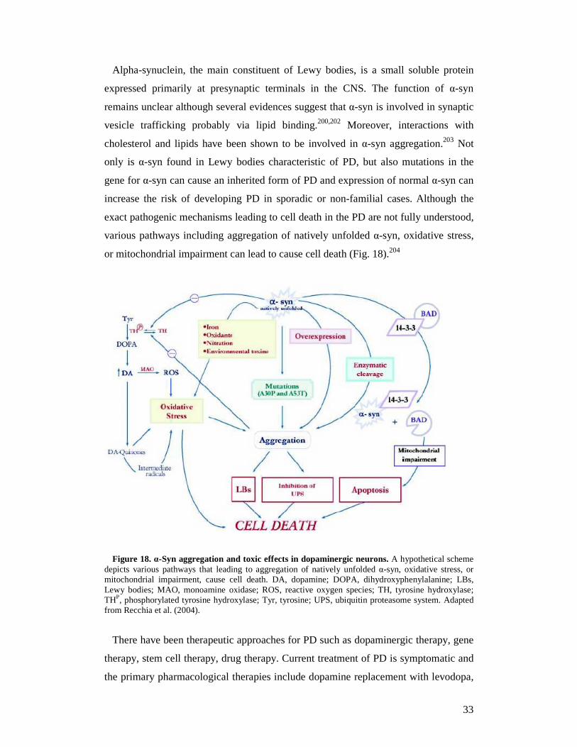
Alpha-synuclein, the main constituent of Lewy bodies, is a small soluble protein
expressed primarily at presynaptic terminals in the CNS. The function of α-syn
remains unclear although several evidences suggest that α-syn is involved in synaptic
vesicle trafficking probably via lipid binding.200,202 Moreover, interactions with
cholesterol and lipids have been shown to be involved in α-syn aggregation.203 Not
only is α-syn found in Lewy bodies characteristic of PD, but also mutations in the
gene for α-syn can cause an inherited form of PD and expression of normal α-syn can
increase the risk of developing PD in sporadic or non-familial cases. Although the
exact pathogenic mechanisms leading to cell death in the PD are not fully understood,
various pathways including aggregation of natively unfolded α-syn, oxidative stress,
or mitochondrial impairment can lead to cause cell death (Fig. 18).204
Figure 18. α-Syn aggregation and toxic effects in dopaminergic neurons. A hypothetical scheme depicts various pathways that leading to aggregation of natively unfolded α-syn, oxidative stress, or mitochondrial impairment, cause cell death. DA, dopamine; DOPA, dihydroxyphenylalanine; LBs, Lewy bodies; MAO, monoamine oxidase; ROS, reactive oxygen species; TH, tyrosine hydroxylase; THP, phosphorylated tyrosine hydroxylase; Tyr, tyrosine; UPS, ubiquitin proteasome system. Adapted from Recchia et al. (2004).
There have been therapeutic approaches for PD such as dopaminergic therapy, gene
therapy, stem cell therapy, drug therapy. Current treatment of PD is symptomatic and
the primary pharmacological therapies include dopamine replacement with levodopa,
34
synthetic dopamine agonists, and drugs which increase dopamine supply by inhibiting
its metabolism (catechol-O-methyltransferase inhibitors and monoamine oxidase B
inhibitors).205 Though pharmacological treatment and brain stimulation have been
shown to reduce symptoms of PD, they are not cures. The controlled production of
dopaminergic cells in large amounts for cell replacement (neurotransplantation) in PD
is technically possible. Embryonic stem cells might have a great potential for cell
replacement strategies in PD, either concerning their proliferative or their
differentiative capacity. The use of fetal tissue-specific neural stem cells seems to be
the safest and most likely the fastest way to establish a transplantation protocol in
PD.206
To determine if statins, cholesterol synthesis inhibitors, might interfere with α-syn
accumulation in cellular models, Bar-on and co-workers studied the effects of
lovastatin, simvastatin, and pravastatin on the accumulation of α-syn in a stably
transfected neuronal cell line (B103 neuroblastoma cells) and in primary fetal human
neurons. Their results revealed that statins reduced the levels of α-syn accumulation in
the detergent insoluble fraction of the transfected cells and enhanced neurite
outgrowth while the contrastive results were obtained if the media supplemented with
cholesterol. These results suggest that regulation of cholesterol levels with cholesterol
inhibitors might be a novel approach for the treatment of PD.203
In addition, several compounds, such as dopamine analogs207,208 can interact with α-
syn and prevent its aggregation into fibrils in vitro.
1.8 Aim of the present work
One of the causes for neurodegenerative diseases including Alzheimer’s disease,
Parkinson’s disease and prion diseases is protein misfolding and aggregation. The
protein misfolding may be associated to the diseases by either gain of a toxic activity
by the misfolded protein or by the lack of biological function of the natively folded
protein. Therefore, designed drugs aim to inhibit or reverse the conformational
changes as a therapy to protein conformational diseases. However, the effective
pharmacological tools for the diseases are not yet available. Moreover, the recently
established interplay between prion and Alzheimer’s diseases have led to an urgent
demand to develop systematic pharmacological and mechanistic studies for the
identification of new classes of compounds as therapeutic agents capable of inhibiting
several pathways in prion conversion and replication. Thereby, the molecules binding
35
to either PrPC or PrPSc conformers at the binding interface may interrupt prion
production by inhibiting the interaction between the PrPSc template and the
endogenous cellular PrPC. Additionally, the compounds that bind molecules
supporting and participating in prion replication, such as chaperones or other ligands,
may also be good candidates for blocking prion replication. Most of the lead
compounds identified so far are derived from screening approaches in established
cellular models.
Based on these considerations, it emerges that rational design of anti-prion
compounds is still a big challenge for medicinal chemists. However, a favorable point
that could further motivate rational drug discovery in prion diseases, is that the
lessons we can learn from their investigation with small molecules might have an
impact on other conformational diseases, characterized by a similar pathological
aggregation and accumulation of misfolded proteins.209 In this connection, it is
relevant to note the recent discovery that PrPC is a mediator of Aβ oligomer-induced
synaptic dysfunction, and hence PrPC-specific compounds may have therapeutic
potential for Alzheimer’s disease.210,211
In the thesis, we discuss about therapeutic approaches for prion diseases and
other neurodegenerative diseases from biological evaluation of four new libraries
of the compounds designed and synthesized on nanotechnology, computational
study and chemistry as well as study their mechanism of action in inhibiting
prion replication. The most active anti-prion compounds may be therapeutic
agents for the diseases.
36
2. Polyelectrolyte multilayer-coated gold nanoparticles as multi-target compounds for treatment of prion diseases and related neurodegenerative disorders
2.1 Introduction
The field of nanoparticle technology is rapidly expanding and promises
revolutionary advances in the diagnosis and treatment of many devastating human
diseases. Nanoparticles have been developed to allow targeted delivery and
sustained release of therapeutics. Such nanoparticle-based drug formulations
interact with biological systems both at molecular and supra-molecular levels.
Nanoparticles can be tailored to respond to specific cell environments, and even to
induce desired physiological responses in cells, whilst minimizing unwanted side
effects. Compared to conventional drugs, nanoparticles-bearing therapeutics
possess higher intrinsic pharmacological activity and their main advantage is their
small dosage that would not require the administration of large amounts of
potentially toxic therapeutics.212
Here, we report the preparation of coated gold nanoparticles (AuNP) exposing,
on their surface, functional groups that can selectively bind, inhibit or prevent the
formation of misfolded protein aggregates such as prions. The build-up of
nanoparticles was carried out with gold nanoparticles as core and a layer-wise
deposition of oppositely charged polyelectrolytes, such as polycation
polyallylamine hydrochloride (PAH) and polyanion polystyrenesulfonate (PSS).
To examine the structure-activity relationship, we tested different numbers of
layers, as well as the nanoparticles’ outermost layer surface charge, for their
possible role in inhibiting scrapie prion formation in vitro and in vivo. Our
findings represent the first report of functionalized nanoparticles as novel potent
anti-prion drugs.
2.2 Materials and methods
2.2.1 Synthesis of gold nanoparticles
Monodisperse AuNPs were prepared as described by Turkevich and co-
workers.213 For particles with a size of 15 ± 1 nm, 5.3 mg of NaAuCl4.2 H2O in 25
mL of Milli-Q grade water were boiled under reflux. One milliliter of a 1%
trisodium citrate solution was rapidly added to the boiling pale yellow solution,
37
which resulted in a color change to deep red. After boiling for additional 20 min,
the solution was cooled down to room temperature and stored protected from light
at room temperature. All experiments described here were performed with the
above colloidal gold nanoparticle stock solution.
The 46 nm gold particles were prepared using the same procedure but with 10.6
mg of NaAuCl4 in 25 mL water and the fast addition of 750 µL 1% trisodium
citrate solution.
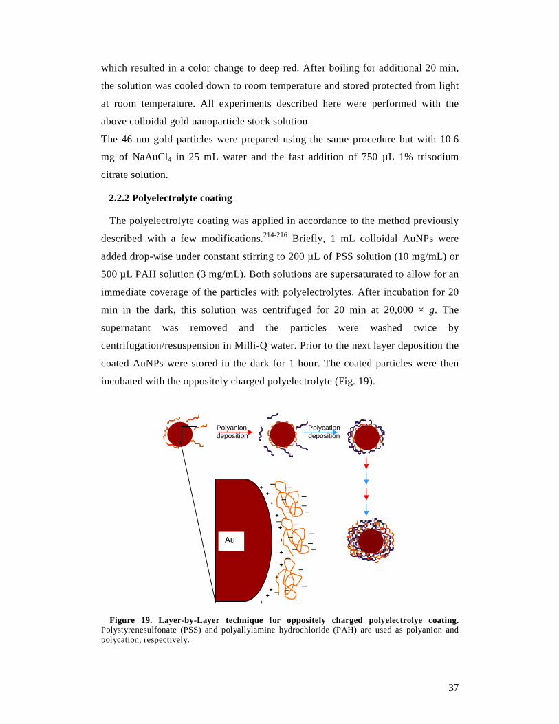
2.2.2 Polyelectrolyte coating
The polyelectrolyte coating was applied in accordance to the method previously
described with a few modifications.214-216 Briefly, 1 mL colloidal AuNPs were
added drop-wise under constant stirring to 200 µL of PSS solution (10 mg/mL) or
500 µL PAH solution (3 mg/mL). Both solutions are supersaturated to allow for an
immediate coverage of the particles with polyelectrolytes. After incubation for 20
min in the dark, this solution was centrifuged for 20 min at 20,000 × g. The
supernatant was removed and the particles were washed twice by
centrifugation/resuspension in Milli-Q water. Prior to the next layer deposition the
coated AuNPs were stored in the dark for 1 hour. The coated particles were then
incubated with the oppositely charged polyelectrolyte (Fig. 19).
Figure 19. Layer-by-Layer technique for oppositely charged polyelectrolye coating. Polystyrenesulfonate (PSS) and polyallylamine hydrochloride (PAH) are used as polyanion and polycation, respectively.
+ +
+
+
+
+
+
+
+
+
+
+
Au
_ _ _ _ _
_ _
_ _ _ _
_ _
_ _
_ _
_
_
_
_
Polycation deposition
Polyanion deposition
38
Each coating step was proved on a NS Zetasizer (Malvern, Milan, Italy) with
dynamic light scattering (DLS) for size and polydispersity index (PDI) and zeta-
potential measurements for changes in the surface charge. The concentration of the
AuNPs was determined at 580 nm in a UV/Vis spectrometer applying the Beer-
Lambert law (λabs = 518 nm, ε = 5.14×10⁷ M-1 cm-1).217
2.2.3 Transmission electron microscopy of coated gold nanoparticles
High-resolution transmission electron microscopy (HRTEM) measurements
were performed by diluting the coated AuNP solution with Milli-Q water to a ratio
of 1:100. Then the solution was deposited on a carbon-covered 200-mesh copper
grid and dried in air at room temperature. The images were acquired with
acceleration voltage ranging from 18.5 to 150 kV. The non-digital images were
digitized and the data analyses of the images were performed using ImageJ
software.
2.2.4 Cell culture, drug treatment and cell viability
Cell culture, drug treatment and cell viability were performed in accordance to
protocols described previously.218
Cell culture
ScGT1 cells were seeded in 10-cm plates containing 10 mL of Dulbecco’s modified
Eagle’s medium (DMEM) culture media, supplemented with 10% fetal bovine serum
(FBS) and 1% penicillin-streptomycin. ScN2a cells were cultivated in 10-cm plates,
containing 10 mL of minimal essential medium with Earle’s salt (EMEM) culture
media, supplemented with 10% FBS, 1% non-essential amino acids, 1% L-glutamax,
1% penicillin-streptomycin. The cells were grown at 37°C in 5% CO2 to 95%
confluence for 1 week before splitting at 1:10 for further cultivation.
Drug treatment
Quinacrine was dissolved in PBS at 1 mM. Imipramine was dissolved in 100%
dimethyl sulfoxide (DMSO) to a solution with a concentration of 100 mM. This
solution was then further diluted into the final stock solution of 10 mM with 10%
(v/v) DMSO/PBS. The final concentration of DMSO in the cell medium was never
above 0.1%. The nanoparticles were diluted in PBS. The media were refreshed and
drugs were added to the cultures 2 days after splitting of the cells and incubated for 5
days. Each experiment was performed using triplicate cultures.
39
Cell viability
ScGT1 and ScN2a cells were maintained in DMEM and EMEM, respectively, and
supplemented with 10% FBS. After 1 day of incubation, media were aspirated from a
confluent 10-cm plate of cells, and cells were detached by addition of 1 mL of 1X
trypsin-EDTA solution. Media were added, and cell density determined by cell
counting using a haemacytometer. The cell density was adjusted to 2.5 × 10⁵ cells/mL
with DMEM for ScGT1cells and 3.0 × 10⁵ cells/mL with EMEM for ScN2a cells. A
96-well, tissue culture-treated, clear bottom, black plate (Costar) wetted with 90 µL of
DMEM or EMEM, was incubated at 37°C, prior to use. One hundred µL of the cell
suspension were added to each well and the cells were allowed to settle for 2 hours,
prior to the addition of the test compound. Compound library stocks were prepared as
described above and diluted 1/20 with sterile PBS prior to use at the required
concentrations in 96-well plates. Ten µL of the compounds were added to each well,
and the plates were incubated at 37°C in 5% CO2. Final DMSO concentration was
never above 0.1% (v/v). Media were aspirated after incubation of 5 days and cells
were washed twice with 200 µL of PBS. One hundred µL of 2.5 µM calcein-AM were
added, and the plates were incubated at 37°C for 30 min. Fluorescence emission
intensity was quantified using a SpectraMax Gemini EM or SpectraMax M5
fluorescence plate reader, excitation/emission ratio equal to 492/525 nm.
2.2.5 PrPSc detection in cell lysates by Western blot
After 5 days of drug treatment, the accumulation of PrPSc was detected by
proteinase K (PK) digestion followed by immunoblotting of lysed cells as
described previously.88 One mL of lysis buffer (10 mM Tris-HCl pH 8.0, 150 mM
NaCl, 0.5 % nonidet P-40, 0.5 % deoxycholic acid sodium salt) was added to cell
plates and the cell lysates were collected after centrifugation at 2,000 rpm for 5
min in a bench microfuge (Eppendorf). The total protein amount of the samples
was measured by the bicinchoninic acid assay (BCA) (Pierce). Five hundred µL of
1 mg/mL ScGT1 or 100 µL of 1 mg/mL ScN2a cell lysates were digested by 20
µg/mL of PK for 1 hour at 37°C. The reaction was stopped with 2 mM
phenylmethylsulphonylfluoride (PMSF) and the PK-digested cell lysates
centrifuged at 48,000 rpm for 1 hour at 4°C in an ultracentrifuge (Beckman
Coulter). The pellets were resuspended in 1X sample loading buffer. For the non-
PK digested sample, 50 µg of cell lysates for ScGT1 or 25 µg of cell lysates for
40
ScN2a were used and 2X loading buffer (125 mM Tris HCl, pH 6.8, 10% 2-
mercapethanol, 4 % SDS, 0.2 % bromophenol blue, 20 % glycerol) was added in a
1:1 ratio. The samples were boiled for 5 min at 100°C, loaded onto either a 12% or
a 15% Tris-Glycine SDS- PAGE gel, and transferred overnight onto Immobilon P
PVDF membranes (Millipore). Membranes were blocked by 5% nonfat milk,
incubated with 1 µg/mL anti-PrP Fab D18 followed by incubation with goat anti-
human IgG F(ab)2 fragment conjugated with horseradish peroxidase. Blots were
developed with the enhanced chemiluminescent system (ECL, Amersham
Biosciences) and visualized on Hyperfilm (Amersham Biosciences).
2.2.6 PrPSc quantification by ELISA
The quantification of PrPSc by ELISA followed a protocol described
previously.137 Briefly, PK digestion of cell lysates was as described above. PK-
digested PrPSc was selectively precipitated by the addition of 0.5% aqueous
phosphotungstic acid (PTA, Sigma-Aldrich) solution with continuous shaking at
37°C, 350 rpm for 1 hour, and centrifuged at room temperature, 14,000 x g for 30
min. Pellets were dissolved and denatured in 50 µL of 8M guanidine
hydrochloride (GdnHCl) in coating buffer (0.1 M sodium bicarbonate, pH 8.2) for
1 hour and diluted into 500 µL of coating buffer. Twenty µL of the suspension
were transferred to 96-well MaxiSorp ELISA plates (Nunc), with each well
containing 180 µL coating buffer and the plates were sealed and incubated
overnight at 4°C. To increase the immunoreactivity of PrPSc, coated proteins were
denatured in situ. Fifty µL of 8M GdnHCl were added to each well and incubated
for 10 min at room temperature. The ELISA plates were washed three times with
TBST (20 mM Tris-HCl, 137 mM NaCl, 0.05% Tween-20, pH 7.5) and blocked
with 200 µL of 3% BSA, made up in TBS (20 mM Tris-HCl, 137 mM NaCl, pH
7.5) sealed and incubated at 37°C. After 1 hour, the plates were washed three
times with TBST, and incubated with 100 µL of anti-PrP antibody D18 (2 µg/mL)
in 1% BSA/TBS, at 37°C for 2 hours. They were then washed seven times with
TBST. One hundred µL of goat anti-human IgG Fab conjugated to HRP and
diluted 1:1000 with 1% BSA/TBS were added to the plates and incubated at 37°C
for 1 hour. Again, plates were washed seven times with TBST, and then developed
with 100 µL of 1-step TMB (3,3’,5,5’- tetramethylbenzidine) Turbo ELISA HRP
substrate (Pierce). The reaction was stopped by the addition of 100 µL of 2 M
sulfuric acid to the plates. Absorbance at 450 nm was measured using a microplate
41
reader (VersaMax, Molecular Devices). Dose-response curves and EC50 values
were computed using GraphPad Prism (version 4.0).
2.2.7 Coated nanogold uptake studies
GT1 and N2a neuronal cell lines (1.0 × 10⁵ cells) were seeded on 24 x 24 mm cover
slips in 35 mm-plates with 2 mL of DMEM supplemented with 10% FBS, 1%
penicillin-streptomycin and EMEM with 10% FBS, 1% non-essential amino acids,
1% L-glutamax, 1% penicillin-streptomycin, respectively, and were cultured for 24
hours at 37°C in 5% CO2. For the uptake 50 µL of a solution containing either
nanogold particles coated with (PSS/FITC-PAH) tagged as 2A, or coated with
(PSS/FITC-PAH)2/PSS called 5S were added, along with 30 µM of DiA [4-di-16-
ASP, (4-(4-dihexadecylamino)styryl)-N-methylpyridinium iodide, Molecular Probes]
(labeling intracellular membranes) and incubated for 2, 6, 12, 24 or 48 hours at 37°C
in 5% CO2. After incubation, the culture media were aspirated and the adherent cells
were washed 2 times with 2 mL of respective media, without antibiotics and sera.
Then, the cells were studied by confocal fluorescence microscopy. The experiments
were repeated at least 3 times for each cell line and time.
Imaging acquisition was performed with Nikon C1 laser scanning confocal unit
(Nikon D-eclipse C1, Japan) attached to an inverse fluorescence microscope
(Nikon D-eclipse C1Si, Japan) with 100 X/1.49 oil Apo TIRF objective (Nikon,
Japan). Excitation was performed with an air-cooled Argon laser emitting at 488
nm for FITC (λex = 488 nm, λem = 520 nm) and a diode laser at 561nm exciting
DiA (λex = 570 nm, λem = 630 nm), with appropriate filter sets to collect the
fluorescence emission. Images were acquired and processed using the operation
software EZ-C1.
2.2.8 Detection of in vitro effect of the nanoparticles on prion fibril formation
and amyloid seeding assay (ASA)
Fibril formation was performed in accordance to the method previously
described with a few modifications.219 Briefly, 500 µL of 2 mg/mL ScN2a or
ScGT1 cell lysates was used for PTA precipitation by adding 500 µL of PBS/4%
Sarkosyl/protease inhibitor and 0.5% PTA with continuous shaking at 37°C, 350
rpm for 1 hour, and centrifuged at room temperature, 14,000 x g for 30 min. The
pellets were washed with 500 µL of PBS/2% Sarkosyl/protease inhibitor,
centrifuged and resuspended in 150 µL of water and then stored at -80oC until use.
42
In ASA, 4 µL of resuspended PTA pellets was diluted into 400 µL of water and 20
µL of the diluted sample was added to each well containing 180 µL of reaction
solution (50 µg/mL MoPrP(23-230), 0.4 M GdnHCl, 10 µM ThT in PBS 1X
buffer) in a 96-well black plate (BD Falcon). The nanoparticles added in different
concentrations to each well. Each sample was performed in four replicates. Each
well contained one 3-mm glass bead (Sigma). The plate was covered with sealing
tape (Fisher Scientific), incubated at 37°C with continuous shaking and read on
SpectraMax M5 or Gemini EM fluorescence plate reader (Molecular Devices) by
top fluorescence reading every 5 min at excitation of 444 nm and emission of 485
nm.
2.2.9 Detection of in vitro effect of the nanoparticles on β-amyloid and α-
synuclein fibril formation
Twenty µL of the nanoparticles added in different concentrations to each well
containing 180 µL of reaction solution (10 µg/mL Aβ1-40 wild-type, 10 µM ThT in
50 mM Tris-HCl pH 7.4 for β-amyloid fibril formation; or 1 mg/mL recombinant
human α-synuclein, 100 mM NaCl, 10 µM ThT in 20 mM Tris-HCl pH 7.4 for α-
synuclein fibril formation) in a 96-well black plate (BD Falcon). Each sample was
performed in four replicates. Each well contained one 3-mm glass bead (Sigma).
The plate was covered with sealing tape (Fisher Scientific), incubated at 37°C with
continuous shaking and read on SpectraMax M5 fluorescence plate reader
(Molecular Devices) by top fluorescence reading every 5 min at excitation of 444
nm and emission of 485 nm.
2.2.10 Binding activity of the nanoparticles to prion proteins in cultured
cells
N2a, GT1, ScN2a, ScGT1 were cultured for 5 days, refreshed media and added
100 pM of nanogold particles coated with (PSS/FITC-PAH) tagged as 2A, or
coated with (PSS/FITC-PAH)2/PSS called 5S, and then incubated for 2 more days.
The cells were washed by media without antibiotics and sera and the cell proteins
were extracted. Five hundred µg protein was used with or without 20 µg/mL of
PK, PTA-precipitated and pellets dissolved in 200 µL lysis buffer. The samples
were added in each well of 96-well black plate and relative fluorescence unit
(RFU) values were measured by Gemini EM fluorescence plate reader (Molecular
Devices) at 488/525 nm.
43
2.2.11 Preparation of the nanoparticles pre-treated brain homogenates
Ten % RML (Rocky Mountain Laboratories) infected brain homogenate (wt/vol)
was prepared from pools of brains of terminally RML-sick CD-1® IGS mice
(Charles River Laboratories), while 10% mock homogenate (wt/vol) was obtained
from pools of brains of healthy CD-1 mice. 2A and 5S coated AuNPs were
separately pre-incubated with both RML and mock brain homogenates at 4°C for
24 hours. Even RML and mock brain homogenates untreated with nanoparticles
were subjected at the same pre-treatment. The final 2A coated AuNPs
concentration in both RML and mock homogenates was 25.35 nM while that of
AuNPs 5S was 26.65 nM.
2.2.12 Analysis of the homogenates by transmission electron microscopy
Mouse brain homogenates (both RML and mock) treated with 2A or 5S particles
were dissolved in distilled water (1:2) then 5 µL of the final suspension were
applied to Formvar-carbon-200-mesh nickel grids for 6 min, negatively stained
with uranyl acetate and observed with an electron microscope (EM109 Zeiss,
Oberkoken, Germany) operated at 80 kV at a standard magnification (X 30,000),
calibrated with an appropriate grid. The samples were evaluated for the presence
and amount of AuNP aggregates.
2.2.13 Animal inoculation
All mice were divided in six different groups, housed in ventilated cages and
identified by ear-tags. Each group was intracerebrally inoculated with 30 µL of a
precise solution: i.e. (i) RML or (ii) mock brain homogenate pre-incubated with
AuNPs 2A; (iii) RML or (iv) mock brain homogenate pre-incubated with AuNPs
5S; (v) RML and (vi) mock brain homogenates untreated. 10-15 mice for each
group were anesthetized with sevofluorane and inoculated into the right caudatus
nucleus by using Hamilton syringes with 26G needle. Both preparations of the
inocula and their injection were carried out using sterile instrumentation and
disposable equipment for each animal and each inoculum. Groups of mock-
inoculated and untreated mice were included as controls.
2.2.14 Behavioral monitoring
Behavioral monitoring was carried out weekly, beginning at 16 weeks post-
inoculation, and included spontaneous locomotor activity in the open field, nest
44
construction test, reactivity to external stimuli and inverted screen test.220,221 The
incubation time was calculated as the period between the day of inoculation and
the appearance of clinical signs of disease, confirmed by a subsequent assessment
at 3 days interval. At the terminal stage of disease (characterized by ataxia,
hunched dorsal kyphosis, and suppressed righting reflex) clinically affected mice
were sacrificed, while the other mice were monitored for the entire predicted life
span and then culled and subjected to necroscopy.
2.2.15 Histological examinations
All mouse tissues were collected for the study. Regarding CNS, the left
hemisphere of each mouse brain was fixed in Carnoy solution at 4°C for 24
hours,222 while the right hemisphere was frozen at -80°C for Western blot analysis.
The same procedure was followed for the other organs (i.e. brain stem, muscle,
spleen, liver, kidney, Peyer’s patch, etc.). Fixed brain samples were cut in four
standard coronal levels,223 dehydrated, and embedded in paraplast. 7 µm thick
serial sections from paraffin embedded tissues were stained with hematoxylin-
eosin (HE) or probed with different antibodies (i.e. 6H4, GFAP, Caspase-3, etc.).
Spongiform profiles were determined on HE-stained sections, by scoring the
vacuolar changes in nine standard grey matter area as described.223
2.2.16 Immunohistochemical staining
Sections were immunostained with monoclonal antibody to PrP (6H4 1:1000,
Prionics), monoclonal antibody to myelin protein (CNPase 1:500, Sigma-Aldrich),
polyclonal antibody to glial fibrillary acidic protein (GFAP 1:1000, Dako),
monoclonal antibody to T-lymphocyte (CD3ε 1:500, Millipore) and a polyclonal
antibody to apoptotic cells (Caspase-3 1:100, Millipore). Before PrP
immunostaining, the sections were sequentially subjected to PK digestion (10
µg/mL, room temperature, 5 min) and guanidine isothiocyanate treatment (3M,
room temperature, 20 min), and non-specific binding was prevented using Animal
Research Kit Peroxidase (Dako). Immunoreactions were visualized using 3-3’-
diaminobenzidine (DAB, Dako) chromogen.
2.2.17 PK immunoblot analysis of mouse brain homogenates
Ten % (wt/vol) brain homogenates from frozen tissues were prepared in lysis
buffer (100 mM sodium chloride, 10 mM EDTA, 0.5% Nonidet P40, 0.5% sodium
45
deoxycholate in 10 mM Tris-HCl, pH 7.4). Aliquots of cleared lysate equivalent to
100 µg were digested with 50 µg/mL of PK for 1 hour at 37°C. Reactions were
terminated by the addition of phenylmethanesulfonyl fluoride (PMSF, 5mM).
Treated homogenates were loaded on 12,5% polyacrylamide gels, transferred to
polyvinylidene fluoride membranes and probed with anti-PrP antibody 6H4
(1:10000, Prionics), anti-CNPase antibody (1:1000, Sigma-Aldrich) and anti-
Caspase-3 antibody (1:100, Millipore). The immunoreactions were visualized by
enhanced chemiluminescence system (Amersham).
2.2.18 Magnetic resonance imaging (MRI)
MRI was performed in clinically symptomatic CD-1 mice challenged with RML
and in non-infected control mice on a Brucker BioSpec 70/30 USR Tesla scanner.
Mice were anesthetized with isofluorane at a dose approximately of 2.5 L/min,
modulated according to the breathing frequency. The animals were positioned on
apposite bed inside the magnet and were monitored throughout the procedure for
breathing frequency and body temperature with specific probes. Twenty-six axial
slices were acquired for each mouse with T2 High Resolution Turbo Spin Echo
sequences. The following parameters were employed: thickness 0.60 mm without
gap, TR 3000 ms, TE 27.1 ms, FOV 2.20/2.20 cm, and 256 matrix.
2.2.19 Statistical analysis
Statistical analyses were performed using the GraphPad-Prism 4.0 software.
Kaplan-Meier survival curves were plotted, and differences in survival between
groups of mice inoculated with RML (positive control) and RML pre-incubated
with AuNPs 2A or AuNPs 5S were compared using the log-rank test.
2.3 Results
2.3.1 Physical characteristics of polyelectrolyte multilayer-coated gold
nanoparticles
The physical characteristics of the coated nanoparticles are given in Table 3. For
the experiments to inhibit PrPSc replication in vitro, particles from 1 to 5 layers
were prepared, finishing with either a positive (PAH) or negative (PSS) charged
layer. All particle preparations finishing with PSS were tagged as nS (n = 1-5) and
each one with PAH was labeled mA (m = 1-5). The layers were deposited onto
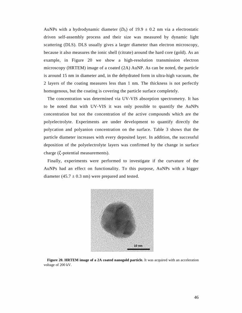
46
AuNPs with a hydrodynamic diameter (Dh) of 19.9 ± 0.2 nm via a electrostatic
driven self-assembly process and their size was measured by dynamic light
scattering (DLS). DLS usually gives a larger diameter than electron microscopy,
because it also measures the ionic shell (citrate) around the hard core (gold). As an
example, in Figure 20 we show a high-resolution transmission electron
microscopy (HRTEM) image of a coated (2A) AuNP. As can be noted, the particle
is around 15 nm in diameter and, in the dehydrated form in ultra-high vacuum, the
2 layers of the coating measures less than 1 nm. The thickness is not perfectly
homogenous, but the coating is covering the particle surface completely.
The concentration was determined via UV-VIS absorption spectrometry. It has
to be noted that with UV-VIS it was only possible to quantify the AuNPs
concentration but not the concentration of the active compounds which are the
polyelectrolyte. Experiments are under development to quantify directly the
polycation and polyanion concentration on the surface. Table 3 shows that the
particle diameter increases with every deposited layer. In addition, the successful
deposition of the polyelectrolyte layers was confirmed by the change in surface
charge (ζ-potential measurements).
Finally, experiments were performed to investigate if the curvature of the
AuNPs had an effect on functionality. To this purpose, AuNPs with a bigger
diameter (45.7 ± 0.3 nm) were prepared and tested.
Figure 20. HRTEM image of a 2A coated nanogold particle. It was acquired with an acceleration voltage of 200 kV.
10 nm
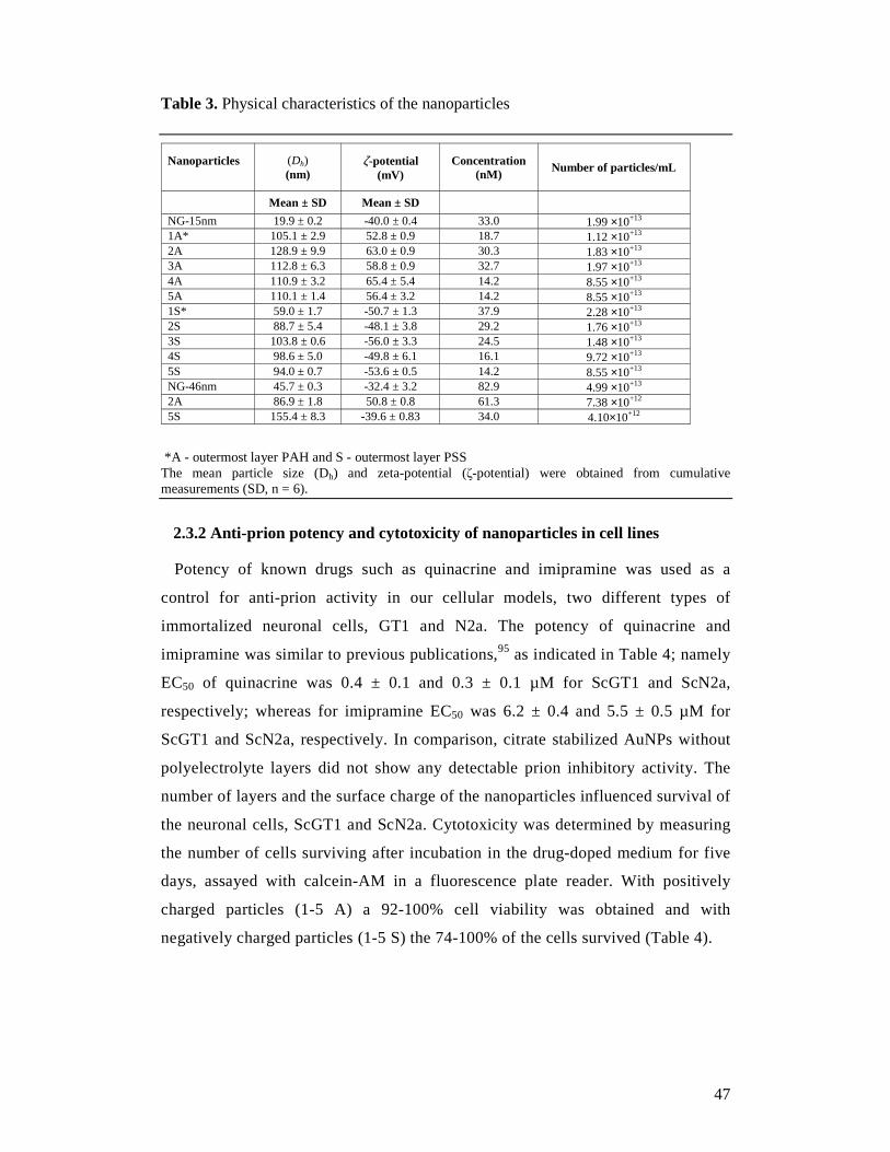
47
Table 3. Physical characteristics of the nanoparticles
Nanoparticles
(Dh) (nm)
ζ-potential (mV)
Concentration (nM)
Number of particles/mL
Mean ± SD Mean ± SD
NG-15nm 19.9 ± 0.2 -40.0 ± 0.4 33.0 1.99 ×10+13 1A* 105.1 ± 2.9 52.8 ± 0.9 18.7 1.12 ×10+13 2A 128.9 ± 9.9 63.0 ± 0.9 30.3 1.83 ×10+13 3A 112.8 ± 6.3 58.8 ± 0.9 32.7 1.97 ×10+13 4A 110.9 ± 3.2 65.4 ± 5.4 14.2 8.55 ×10+13 5A 110.1 ± 1.4 56.4 ± 3.2 14.2 8.55 ×10+13 1S* 59.0 ± 1.7 -50.7 ± 1.3 37.9 2.28 ×10+13 2S 88.7 ± 5.4 -48.1 ± 3.8 29.2 1.76 ×10+13 3S 103.8 ± 0.6 -56.0 ± 3.3 24.5 1.48 ×10+13 4S 98.6 ± 5.0 -49.8 ± 6.1 16.1 9.72 ×10+13 5S 94.0 ± 0.7 -53.6 ± 0.5 14.2 8.55 ×10+13 NG-46nm 45.7 ± 0.3 -32.4 ± 3.2 82.9 4.99 ×10+13 2A 86.9 ± 1.8 50.8 ± 0.8 61.3 7.38 ×10+12 5S 155.4 ± 8.3 -39.6 ± 0.83 34.0 4.10×10+12
*A - outermost layer PAH and S - outermost layer PSS The mean particle size (Dh) and zeta-potential (ζ-potential) were obtained from cumulative measurements (SD, n = 6).
2.3.2 Anti-prion potency and cytotoxicity of nanoparticles in cell lines
Potency of known drugs such as quinacrine and imipramine was used as a
control for anti-prion activity in our cellular models, two different types of
immortalized neuronal cells, GT1 and N2a. The potency of quinacrine and
imipramine was similar to previous publications,95 as indicated in Table 4; namely
EC50 of quinacrine was 0.4 ± 0.1 and 0.3 ± 0.1 µM for ScGT1 and ScN2a,
respectively; whereas for imipramine EC50 was 6.2 ± 0.4 and 5.5 ± 0.5 µM for
ScGT1 and ScN2a, respectively. In comparison, citrate stabilized AuNPs without
polyelectrolyte layers did not show any detectable prion inhibitory activity. The
number of layers and the surface charge of the nanoparticles influenced survival of
the neuronal cells, ScGT1 and ScN2a. Cytotoxicity was determined by measuring
the number of cells surviving after incubation in the drug-doped medium for five
days, assayed with calcein-AM in a fluorescence plate reader. With positively
charged particles (1-5 A) a 92-100% cell viability was obtained and with
negatively charged particles (1-5 S) the 74-100% of the cells survived (Table 4).
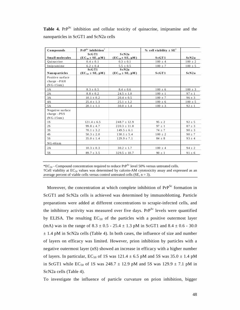
48
Table 4. PrPSc inhibition and cellular toxicity of quinacrine, imipramine and the
nanoparticles in ScGT1 and ScN2a cells
Compounds PrPSc inhibi tion * % cell v iability ± SE †
Small mo lecules ScG T1
(E C 50 ± SE , µ M ) S cN2a
(EC 5 0 ± S E, µM ) S cGT 1 ScN2a Qui nacrine 0.4 ± 0.1 0.3 ± 0.1 100 ± 4 100 ± 2
Imip ramime 6.2 ± 0.4 5.5 ± 0.5 100 ± 7 100 ± 5
Nan op art icles ScG T1
(EC 5 0 ± SE, pM ) S cN2a
(EC 5 0 ± SE, pM ) S cGT 1 ScN2a
Positive su rface charge –PAH
(N G-15nm )
1A 8.3 ± 0.5 8.4 ± 0.6 100 ± 6 100 ± 3 2A 8.8 ± 0.2 24.5 ± 1.0 100 ± 1 97 ± 1 3A 10 .1 ± 0 .2 20.4 ± 0.5 100 ± 7 96 ± 3 4A 25 .4 ± 1 .3 25.1 ± 1.2 100 ± 6 100 ± 5 5A 20 .1 ± 1 .1 30.0 ± 1.4 100 ± 3 92 ± 1
Negati ve surface charge –PS S
(N G-15nm ) 1S 121 .4 ± 6 .5 248.7 ± 12 .9 95 ± 2 92 ± 5 2S 99 .8 ± 4 .7 220.3 ± 11 .8 97 ± 1 87 ± 3 3S 70 .1 ± 3 .2 149 .5 ± 6 .1 74 ± 7 90 ± 3 4S 50 .3 ± 2 .0 130 .1 ± 5 .4 100 ± 2 90 ± 7 5S 35 .0 ± 1 .4 129 .9 ± 7 .1 84 ± 8 93 ± 4
NG-46nm
2A 10 .3 ± 0 .3 30.2 ± 1.7 100 ± 4 94 ± 2
5S 89 .7 ± 3 .5 329.5 ± 10 .7 90 ± 1 91 ± 6
*EC50 - Compound concentration required to reduce PrPSc level 50% versus untreated cells. †Cell viability at EC50 values was determined by calcein-AM cytotoxicity assay and expressed as an average percent of viable cells versus control untreated cells (SE, n = 3).
Moreover, the concentration at which complete inhibition of PrPSc formation in
ScGT1 and ScN2a cells is achieved was determined by immunoblotting. Particle
preparations were added at different concentrations to scrapie-infected cells, and
the inhibitory activity was measured over five days. PrPSc levels were quantified
by ELISA. The resulting EC50 of the particles with a positive outermost layer
(mA) was in the range of 8.3 ± 0.5 - 25.4 ± 1.3 pM in ScGT1 and 8.4 ± 0.6 - 30.0
± 1.4 pM in ScN2a cells (Table 4). In both cases, the influence of size and number
of layers on efficacy was limited. However, prion inhibition by particles with a
negative outermost layer (nS) showed an increase in efficacy with a higher number
of layers. In particular, EC50 of 1S was 121.4 ± 6.5 pM and 5S was 35.0 ± 1.4 pM
in ScGT1 while EC50 of 1S was 248.7 ± 12.9 pM and 5S was 129.9 ± 7.1 pM in
ScN2a cells (Table 4).
To investigate the influence of particle curvature on prion inhibition, bigger
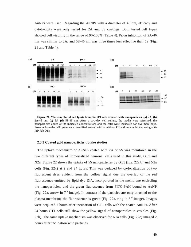
49
AuNPs were used. Regarding the AuNPs with a diameter of 46 nm, efficacy and
cytotoxicity were only tested for 2A and 5S coatings. Both tested cell types
showed cell viability in the range of 90-100% (Table 4). Prion inhibition of 2A-46
nm was similar to 2A, and 5S-46 nm was three times less effective than 5S (Fig.
21 and Table 4).
Figure 21. Western blot of cell lysate from ScGT1 cells treated with nanoparticles. (a) 2A, (b) 2A-46 nm, (c) 5S, (d) 5S-46 nm. After a two-day cell culture, the media were refreshed, the nanoparticles added at the indicated concentrations and the cells were incubated for five more days. Proteins from the cell lysate were quantified, treated with or without PK and immunoblotted using anti-PrP Fab D18.
2.3.3 Coated gold nanoparticles uptake studies
The uptake mechanism of AuNPs coated with 2A or 5S was monitored in the
two different types of immortalized neuronal cells used in this study, GT1 and
N2a. Figure 22 shows the uptake of 5S nanoparticles by GT1 (Fig. 22a,b) and N2a
cells (Fig. 22c) at 2 and 24 hours. This was deduced by co-localization of two
fluorescent dyes evident from the yellow signal due the overlap of the red
fluorescence emitted by lipid dye DiA, incorporated in the membrane encircling
the nanoparticles, and the green fluorescence from FITC-PAH bound to AuNP
(Fig. 22a, arrow in 7th image). In contrast if the particles are only attached to the
plasma membrane the fluorescence is green (Fig. 22a, ring in 3rd image). Images
were acquired 2 hours after incubation of GT1 cells with the coated AuNPs. After
24 hours GT1 cells still show the yellow signal of nanoparticles in vesicles (Fig.
22b). The same uptake mechanism was observed for N2a cells (Fig. 22c) imaged 2
hours after incubation with particles.
50 37
25
PK - PK +
pM 0 1 4 16 64 256 0 1 4 16 64 256
(a)
(c) PK +
0 1 4 16 64 pM 0 1 4 16 64
PK -
50
37
25
50 37
25
pM 0 5 20 40 60 120
(b)
50
25
0 5 20 40 60 120
37
(d)
pM
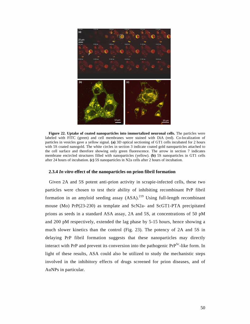
50
20 µm
20 µm
A
B C
20 µm10 µm
20 µm
(a)
(b) (c)
Figure 22. Uptake of coated nanoparticles into immortalized neuronal cells. The particles were labeled with FITC (green) and cell membranes were stained with DiA (red). Co-localization of particles in vesicles gave a yellow signal. (a) 3D optical sectioning of GT1 cells incubated for 2 hours with 5S coated nanogold. The white circles in section 3 indicate coated gold nanoparticles attached to the cell surface and therefore showing only green fluorescence. The arrow in section 7 indicates membrane encircled structures filled with nanoparticles (yellow). (b) 5S nanoparticles in GT1 cells after 24 hours of incubation. (c) 5S nanoparticles in N2a cells after 2 hours of incubation.
2.3.4 In vitro effect of the nanoparticles on prion fibril formation
Given 2A and 5S potent anti-prion activity in scrapie-infected cells, these two
particles were chosen to test their ability of inhibiting recombinant PrP fibril
formation in an amyloid seeding assay (ASA).219 Using full-length recombinant
mouse (Mo) PrP(23-230) as template and ScN2a- and ScGT1-PTA precipitated
prions as seeds in a standard ASA assay, 2A and 5S, at concentrations of 50 pM
and 200 pM respectively, extended the lag phase by 5-15 hours, hence showing a
much slower kinetics than the control (Fig. 23). The potency of 2A and 5S in
delaying PrP fibril formation suggests that these nanoparticles may directly
interact with PrP and prevent its conversion into the pathogenic PrPSc-like form. In
light of these results, ASA could also be utilized to study the mechanistic steps
involved in the inhibitory effects of drugs screened for prion diseases, and of
AuNPs in particular.
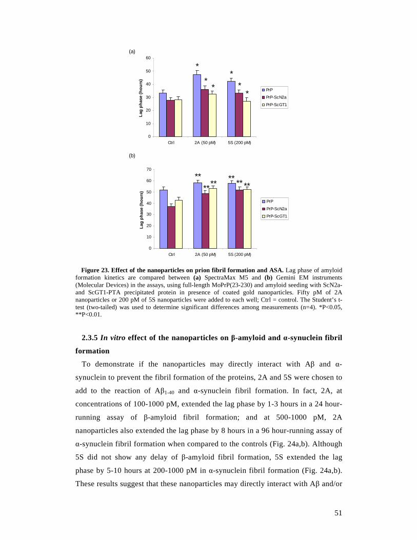
51
0
10
20
30
40
50
60
Ctrl 2A (50 pM) 5S (200 pM)La
g ph
ase
(hou
rs)
PrP
PrP-ScN2a
PrP-ScGT1
*
**
**
*
(a)
0
10
20
30
40
50
60
70
Ctrl 2A (50 pM) 5S (200 pM)
Lag
phas
e (h
ours
)
PrP
PrP-ScN2a
PrP-ScGT1
(b)
******
** ** **
Figure 23. Effect of the nanoparticles on prion fibril formation and ASA. Lag phase of amyloid
formation kinetics are compared between (a) SpectraMax M5 and (b) Gemini EM instruments (Molecular Devices) in the assays, using full-length MoPrP(23-230) and amyloid seeding with ScN2a- and ScGT1-PTA precipitated protein in presence of coated gold nanoparticles. Fifty pM of 2A nanoparticles or 200 pM of 5S nanoparticles were added to each well; Ctrl = control. The Student’s t-test (two-tailed) was used to determine significant differences among measurements (n=4). *P<0.05, **P<0.01. 2.3.5 In vitro effect of the nanoparticles on β-amyloid and α-synuclein fibril
formation
To demonstrate if the nanoparticles may directly interact with Aβ and α-
synuclein to prevent the fibril formation of the proteins, 2A and 5S were chosen to
add to the reaction of Aβ1-40 and α-synuclein fibril formation. In fact, 2A, at
concentrations of 100-1000 pM, extended the lag phase by 1-3 hours in a 24 hour-
running assay of β-amyloid fibril formation; and at 500-1000 pM, 2A
nanoparticles also extended the lag phase by 8 hours in a 96 hour-running assay of
α-synuclein fibril formation when compared to the controls (Fig. 24a,b). Although
5S did not show any delay of β-amyloid fibril formation, 5S extended the lag
phase by 5-10 hours at 200-1000 pM in α-synuclein fibril formation (Fig. 24a,b).
These results suggest that these nanoparticles may directly interact with Aβ and/or
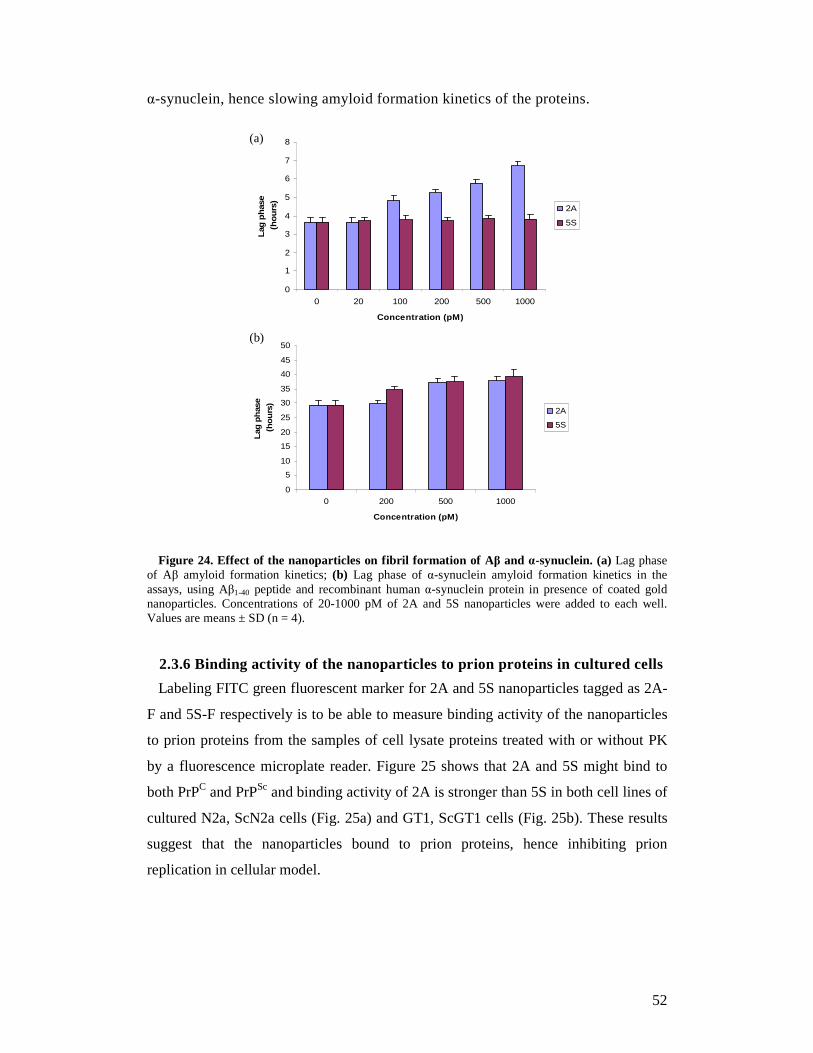
52
α-synuclein, hence slowing amyloid formation kinetics of the proteins.
Figure 24. Effect of the nanoparticles on fibril formation of Aβ and α-synuclein. (a) Lag phase of Aβ amyloid formation kinetics; (b) Lag phase of α-synuclein amyloid formation kinetics in the assays, using Aβ1-40 peptide and recombinant human α-synuclein protein in presence of coated gold nanoparticles. Concentrations of 20-1000 pM of 2A and 5S nanoparticles were added to each well. Values are means ± SD (n = 4).
2.3.6 Binding activity of the nanoparticles to prion proteins in cultured cells
Labeling FITC green fluorescent marker for 2A and 5S nanoparticles tagged as 2A-
F and 5S-F respectively is to be able to measure binding activity of the nanoparticles
to prion proteins from the samples of cell lysate proteins treated with or without PK
by a fluorescence microplate reader. Figure 25 shows that 2A and 5S might bind to
both PrPC and PrPSc and binding activity of 2A is stronger than 5S in both cell lines of
cultured N2a, ScN2a cells (Fig. 25a) and GT1, ScGT1 cells (Fig. 25b). These results
suggest that the nanoparticles bound to prion proteins, hence inhibiting prion
replication in cellular model.
0
1
2
3
4
5
6
7
8
0 20 100 200 500 1000
Concentration (pM)
Lag
phas
e
(ho
urs) 2A
5S
0
5
10
15
20
25
30
35
40
45
50
0 200 500 1000
Concentration (pM)
Lag
phas
e
(ho
urs) 2A
5S
(a)
(b)
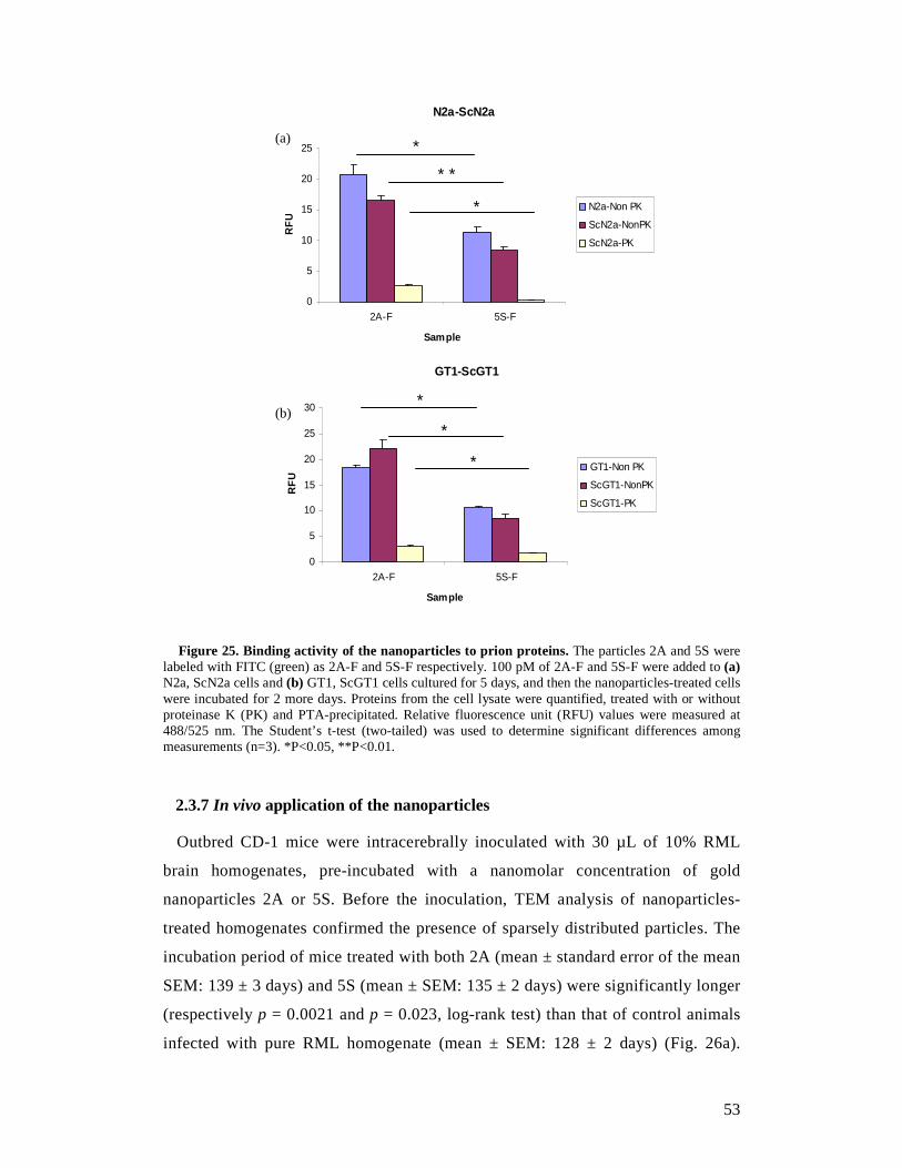
53
Figure 25. Binding activity of the nanoparticles to prion proteins. The particles 2A and 5S were labeled with FITC (green) as 2A-F and 5S-F respectively. 100 pM of 2A-F and 5S-F were added to (a) N2a, ScN2a cells and (b) GT1, ScGT1 cells cultured for 5 days, and then the nanoparticles-treated cells were incubated for 2 more days. Proteins from the cell lysate were quantified, treated with or without proteinase K (PK) and PTA-precipitated. Relative fluorescence unit (RFU) values were measured at 488/525 nm. The Student’s t-test (two-tailed) was used to determine significant differences among measurements (n=3). *P<0.05, **P<0.01.
2.3.7 In vivo application of the nanoparticles
Outbred CD-1 mice were intracerebrally inoculated with 30 µL of 10% RML
brain homogenates, pre-incubated with a nanomolar concentration of gold
nanoparticles 2A or 5S. Before the inoculation, TEM analysis of nanoparticles-
treated homogenates confirmed the presence of sparsely distributed particles. The
incubation period of mice treated with both 2A (mean ± standard error of the mean
SEM: 139 ± 3 days) and 5S (mean ± SEM: 135 ± 2 days) were significantly longer
(respectively p = 0.0021 and p = 0.023, log-rank test) than that of control animals
infected with pure RML homogenate (mean ± SEM: 128 ± 2 days) (Fig. 26a).
N2a-ScN2a
0
5
10
15
20
25
2A-F 5S-F
Sample
RF
U
N2a-Non PK
ScN2a-NonPK
ScN2a-PK
(a) *
* *
*
GT1-ScGT1
0
5
10
15
20
25
30
2A-F 5S-F
Sample
RF
U
GT1-Non PK
ScGT1-NonPK
ScGT1-PK
(b) *
*
*
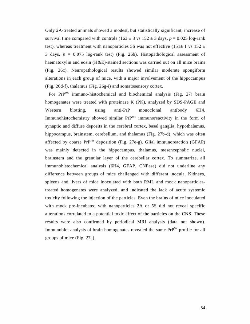
54
Only 2A-treated animals showed a modest, but statistically significant, increase of
survival time compared with controls (163 ± 3 vs 152 ± 3 days, p = 0.025 log-rank
test), whereas treatment with nanoparticles 5S was not effective (151± 1 vs 152 ±
3 days, p = 0.075 log-rank test) (Fig. 26b). Histopathological assessment of
haematoxylin and eosin (H&E)-stained sections was carried out on all mice brains
(Fig. 26c). Neuropathological results showed similar moderate spongiform
alterations in each group of mice, with a major involvement of the hippocampus
(Fig. 26d-f), thalamus (Fig. 26g-i) and somatosensory cortex.
For PrPres immuno-histochemical and biochemical analysis (Fig. 27) brain
homogenates were treated with proteinase K (PK), analyzed by SDS-PAGE and
Western blotting, using anti-PrP monoclonal antibody 6H4.
Immunohistochemistry showed similar PrPres immunoreactivity in the form of
synaptic and diffuse deposits in the cerebral cortex, basal ganglia, hypothalamus,
hippocampus, brainstem, cerebellum, and thalamus (Fig. 27b-d), which was often
affected by coarse PrPres deposition (Fig. 27e-g). Glial immunoreaction (GFAP)
was mainly detected in the hippocampus, thalamus, mesencephalic nuclei,
brainstem and the granular layer of the cerebellar cortex. To summarize, all
immunohistochemical analysis (6H4, GFAP, CNPase) did not underline any
difference between groups of mice challenged with different inocula. Kidneys,
spleens and livers of mice inoculated with both RML and mock nanoparticles-
treated homogenates were analyzed, and indicated the lack of acute systemic
toxicity following the injection of the particles. Even the brains of mice inoculated
with mock pre-incubated with nanoparticles 2A or 5S did not reveal specific
alterations correlated to a potential toxic effect of the particles on the CNS. These
results were also confirmed by periodical MRI analysis (data not shown).
Immunoblot analysis of brain homogenates revealed the same PrPSc profile for all
groups of mice (Fig. 27a).
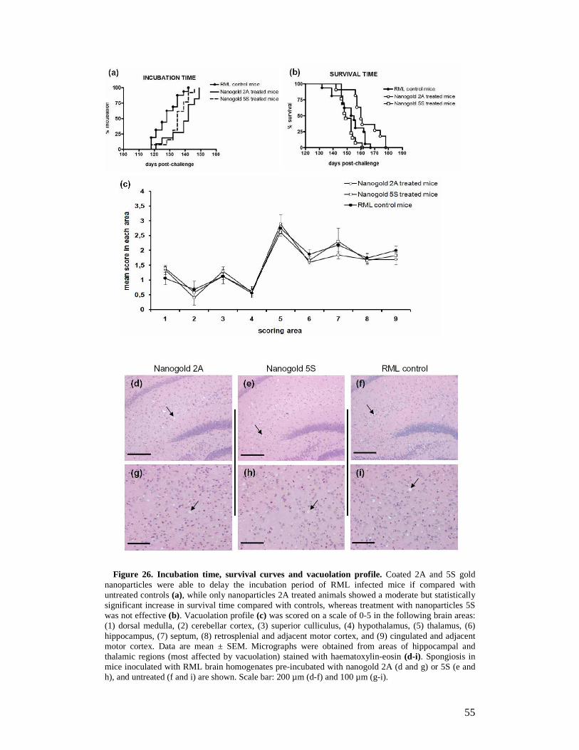
55
Figure 26. Incubation time, survival curves and vacuolation profile. Coated 2A and 5S gold nanoparticles were able to delay the incubation period of RML infected mice if compared with untreated controls (a), while only nanoparticles 2A treated animals showed a moderate but statistically significant increase in survival time compared with controls, whereas treatment with nanoparticles 5S was not effective (b). Vacuolation profile (c) was scored on a scale of 0-5 in the following brain areas: (1) dorsal medulla, (2) cerebellar cortex, (3) superior culliculus, (4) hypothalamus, (5) thalamus, (6) hippocampus, (7) septum, (8) retrosplenial and adjacent motor cortex, and (9) cingulated and adjacent motor cortex. Data are mean ± SEM. Micrographs were obtained from areas of hippocampal and thalamic regions (most affected by vacuolation) stained with haematoxylin-eosin (d-i). Spongiosis in mice inoculated with RML brain homogenates pre-incubated with nanogold 2A (d and g) or 5S (e and h), and untreated (f and i) are shown. Scale bar: 200 µm (d-f) and 100 µm (g-i).
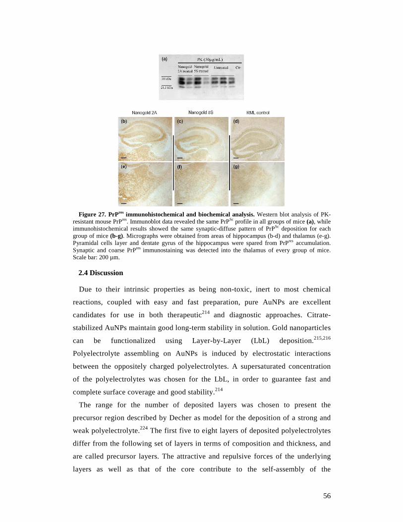
56
Figure 27. PrPres immunohistochemical and biochemical analysis. Western blot analysis of PK-
resistant mouse PrPres. Immunoblot data revealed the same PrPSc profile in all groups of mice (a), while immunohistochemical results showed the same synaptic-diffuse pattern of PrPSc deposition for each group of mice (b-g). Micrographs were obtained from areas of hippocampus (b-d) and thalamus (e-g). Pyramidal cells layer and dentate gyrus of the hippocampus were spared from PrPres accumulation. Synaptic and coarse PrPres immunostaining was detected into the thalamus of every group of mice. Scale bar: 200 µm.
2.4 Discussion
Due to their intrinsic properties as being non-toxic, inert to most chemical
reactions, coupled with easy and fast preparation, pure AuNPs are excellent
candidates for use in both therapeutic214 and diagnostic approaches. Citrate-
stabilized AuNPs maintain good long-term stability in solution. Gold nanoparticles
can be functionalized using Layer-by-Layer (LbL) deposition.215,216
Polyelectrolyte assembling on AuNPs is induced by electrostatic interactions
between the oppositely charged polyelectrolytes. A supersaturated concentration
of the polyelectrolytes was chosen for the LbL, in order to guarantee fast and
complete surface coverage and good stability.214
The range for the number of deposited layers was chosen to present the
precursor region described by Decher as model for the deposition of a strong and
weak polyelectrolyte.224 The first five to eight layers of deposited polyelectrolytes
differ from the following set of layers in terms of composition and thickness, and
are called precursor layers. The attractive and repulsive forces of the underlying
layers as well as that of the core contribute to the self-assembly of the
57
polyelectrolyte. If the core is negatively charged, the first positively charged
polyelectrolyte layer will be self-assembled exclusively by attractive forces, and
by binding they will overcompensate the charge of the core. For the next layer, the
opposite charge of the first polyelectrolyte layer is present, but due to the vicinity
to the core also the repulsive forces by the like-wise charged core. This leads to a
less tight binding and a decreased amount of bound polyelectrolytes. Moreover,
the polyelectrolyte layers penetrate each other in the precursor layer, and are
thinner than the following set of layers, which are more distant from the core.224
We used the intercalation of polycations and polyanions, containing either
sulfonate or primary amine groups, to selectively bind and inhibit prion formation,
creating a surface exposing both moieties randomly and in varying ratios.
There is some reported evidence that polyamines can have an effect on PrPSc.
Indeed, Supattapone and co-workers225 found that branched polyamines are able to
disintegrate aggregates of PrPSc to undetectable levels. For polysulfates, the
influence of their chemical structure on selective binding to either PrPC or PrPSc
leading to prion inhibitory activity was also demonstrated.226 In our work, both
functional groups were combined on the surface of the AuNPs in varying ratios, to
obtain a platform of possible interactive sites for the misfolded PrP.
Two studies on the bio-distribution of citrate stabilized AuNPs should also be
considered, though the results were somehow contradictory: De Jong and co-
workers227 found that only 10 nm particles were crossing the BBB, while Sonavane
and co-workers228 found 50 nm particles in the brain. Our particles were found to
have a hydrodynamic diameter 90 to 130 nm, and in electron microscopy the fully
dehydrated polyelectrolyte matrix was condensed until it was only 1-2 nm thick.
We assume that the polyelectrolyte shell loses some of its water molecules while
crossing the BBB.
From previous experiments on cells of the BBB, it is known that AuNPs coated
with different numbers of polyelectrolyte layers are cytotoxic (for example,
porcine brain capillary endothelial cells).215 Moreover, a strong dependency on the
number of layers and surface charge was observed. Polycations were more
cytotoxic than polyanions and with lower number of layers were more cytotoxic
than a higher number of layers. An additional binding of albumin to the final
polyelectrolyte layer should diminish the toxicity, and animal experiments
confirmed that after intracranial inoculation no toxicity leading to morphological
58
changes in the brain was present.229
Preliminary in vitro experiments showed the efficacy of both nanoparticles (2A
and 5S) to interfere with prion propagation. Because of the limited information
available about the toxic effects of these particles when injected in animals, we
started our experiments by using a dosage of compound lower than those generally
reported in the literature for other therapeutic approaches.227 Whereas 5S
nanoparticles significantly increased only the incubation time of treated mice, 2A
nanoparticles showed higher anti-prion activity. Indeed, even when 2A were used
at nanomolar concentrations (25.35 nM), a moderate but statistically significant
increase in both incubation and survival time was observed, thus indicating a
possible interaction between PrPSc and the nanoparticles. Even though the animals
were treated with just a single dose of nanoparticles the increase of incubation and
survival time was statistically significant. Several studies are currently underway
to determine the best 2A concentration, able to inhibit or perhaps completely block
disease progression. To this end, several mice were intracerebrally infected with
RML brain homogenate pre-incubated with higher dosages of 2A particles (53 nM
vs 25.35 nM). Groups of control mice were also included to monitor the onset of
general toxic effects. Since the gold 2A, modified with the addition of albumin in
the outermost layer, were able to cross the BBB,229 new therapeutic approaches
based on their injection into the tail vein of mice have already been scheduled.
Coated nanogold is not only a potential drug delivery shuttle through the BBB but
by itself it can be used as a drug for the treatment of prion disease and perhaps
other neurodegenerative diseases. With different microscopic techniques like near
infrared time domain (NIR-TD) imaging, X-ray tomography, confocal
fluorescence microscopy and standard cell stains visualized by wide field light
microscopy combined with fluorescence staining, it was possible to show that the
particles accumulate in regions of the brain close to possible target cells of prion,
Alzheimer’s and Parkinson’s diseases.229
2.5 Conclusion
The polyelectrolyte multilayer-coated gold nanoparticles are a novel class of
potential anti-prion drugs. While they showed in vitro very high efficacy at very
low concentration, their potential in vivo to inhibit completely prion aggregation
needs further improvement. A slight increase in incubation time and survival time

59
was observed for the 2A coated particles and at the tested concentration. Further
concentrations as well as other coatings need to be studied in vivo as the
correlation between the in vitro experiments and the in vivo inoculation is not so
striking. Especially the extremely low concentration required to suppress complete
the prion aggregation raise the expectations that this small amount of particles will
pass the blood-brain barrier and will enter the brain parenchyma.
60
3. Discovery of a class of diketopiperazines as anti-prion
compounds
3.1 Introduction
Prion diseases, also known as transmissible spongiform encephalopathies (TSEs),
are neurodegenerative and infectious disorders that affect both humans and animals,
and are not curable with drugs. Although reliable proof of principle was demonstrated
in a variety of experimental models, and several small molecules have been identified
as active against TSE, the mode of action and targets for most of these molecules
remain largely unexplored. As a result, drugs effective against this process are still
years away from approval.230 There are two main reasons why drug discovery for the
treatment of prion diseases has not progressed as rapidly as in other pharmaceutical
fields. First, human prion diseases are very rare. Each year, only approximately 300
people in the USA and approximately 100 people in the UK succumb to various forms
of prion disease.231 Nevertheless, these disorders have come to public and scientific
attention due to the fact that they can be transmissible among humans and, in
particular conditions, from animals to humans. The emergence of a CJD variant in the
1990s demonstrated the transmissibility of BSE to humans and set the scene for a
hypothetical epidemic scenario.231 Second, TSE is a conformational disease,232 where
the cellular form of the prion protein (PrPC) is converted to a misfolded variant (PrPSc)
through a nucleated polymerization process.37 From a medicinal chemistry
perspective, all conformational diseases are ‘black boxes’ because the knowledge of
the 3D structure and mechanistic properties of the target, fundamental prerequisites in
modern drug discovery, are mostly unknown. In general, high-resolution structural
information on amyloid fibrils is very scarce and is currently almost exclusively
restricted to amyloid fibrils formed by small peptides. This is mostly due to the
inability of X-ray crystallography and NMR spectroscopy to address insoluble,
filamentous specimens.233 In the case of PrP, the partial structure of PrP has been
solved to high resolution,234 whereas for PrPSc only models have been proposed,
including the β-helix fold41 and the spiral model.235
As a consequence, if we exclude the discovery of aptamers236 and RNA
interference,163 most of the lead compounds identified so far are derived from
screening approaches in established cellular models. Based on these considerations, it
61
emerges that rational design of anti-prion compounds is still a big challenge for
medicinal chemists.
3.2 Materials and methods
3.2.1 Design rationale
In an effort to identify anti-prion lead compounds with unprecedented molecular
frameworks, we started from the following basic assumption. We noticed that most of
the anti-prion molecules possessed a symmetrical bifunctional structure consisting of
two moieties joined via an appropriate spacer. This is the case for the dye Congo red84
and its analogues,237 the polysulfonated aromatic urea derivative suramin,131 the
natural product curcumin,132 the bis-acridine218 and bis-quinoline analogues,238 and
the diphenylmethane derivative GN8239 (see Fig. 28 for structures). Notably, among
them, planar molecules with aromatic end groups share common anti-aggregating
properties.240 For all of these compounds, and for bivalent compounds in general, the
role of the spacer has been demonstrated to be very critical.241
Therefore, in our search of novel bifunctional molecules, we looked for a spacer that
might have an active role in the molecular recognition process. In inhibiting protein-
protein interactions (PPIs), peptides mimicking the interacting zone have been
considered as relevant starting points in the rational design of effective molecules.242
Similarly, peptidomimetics have become effective modulators of a range of
biologically significant PPIs243 by orienting their side-chain substituents in a spatially
defined manner. Furthermore, these compounds are stable to proteolysis and
consequently possess better drug-like properties than peptides.
Among possible peptidomimetic fragments, we focused on the 2,5-diketopiperazine
(DKP) scaffold for several reasons: (i) it is synthetically readily accessible and
amenable to compound library generation; (ii) DKP derivatives have been shown to
modulate PPIs,244 to possess neuroprotective activities,245 and also to cross the blood-
brain barrier (BBB);246 (iii) it has been extensively explored in medicinal
chemistry.247 Therefore, it seemed conceivable that DKPs carrying (Z)-alkene units of
general structure I (Fig. 28) could serve as a template for a diverse array of
pharmacophores towards the identification of novel bifunctional structures for the
treatment of prion diseases. Building on this, a compound library was generated by
appending several aromatic and heteroaromatic rings in positions 3 and 6 of the 3,6-
dimethylenepiperazine-2,5-dione scaffold. The role of aromatic residues in molecular
recognition and self-assembly processes leading to various fibrillar aggregates has
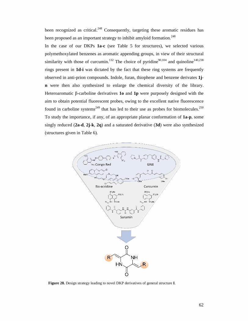
62
been recognized as critical.248 Consequently, targeting these aromatic residues has
been proposed as an important strategy to inhibit amyloid formation.248
In the case of our DKPs 1a-c (see Table 5 for structures), we selected various
polymethoxylated benzenes as aromatic appending groups, in view of their structural
similarity with those of curcumin.132 The choice of pyridine90,104 and quinoline140,238
rings present in 1d-i was dictated by the fact that these ring systems are frequently
observed in anti-prion compounds. Indole, furan, thiophene and benzene derivates 1j-
n were then also synthesized to enlarge the chemical diversity of the library.
Heteroaromatic β-carboline derivatives 1o and 1p were purposely designed with the
aim to obtain potential fluorescent probes, owing to the excellent native fluorescence
found in carboline systems249 that has led to their use as probes for biomolecules.250
To study the importance, if any, of an appropriate planar conformation of 1a-p, some
singly reduced (2a-d, 2j-k, 2q) and a saturated derivative (3d) were also synthesized
(structures given in Table 6).
Figure 28. Design strategy leading to novel DKP derivatives of general structure I.
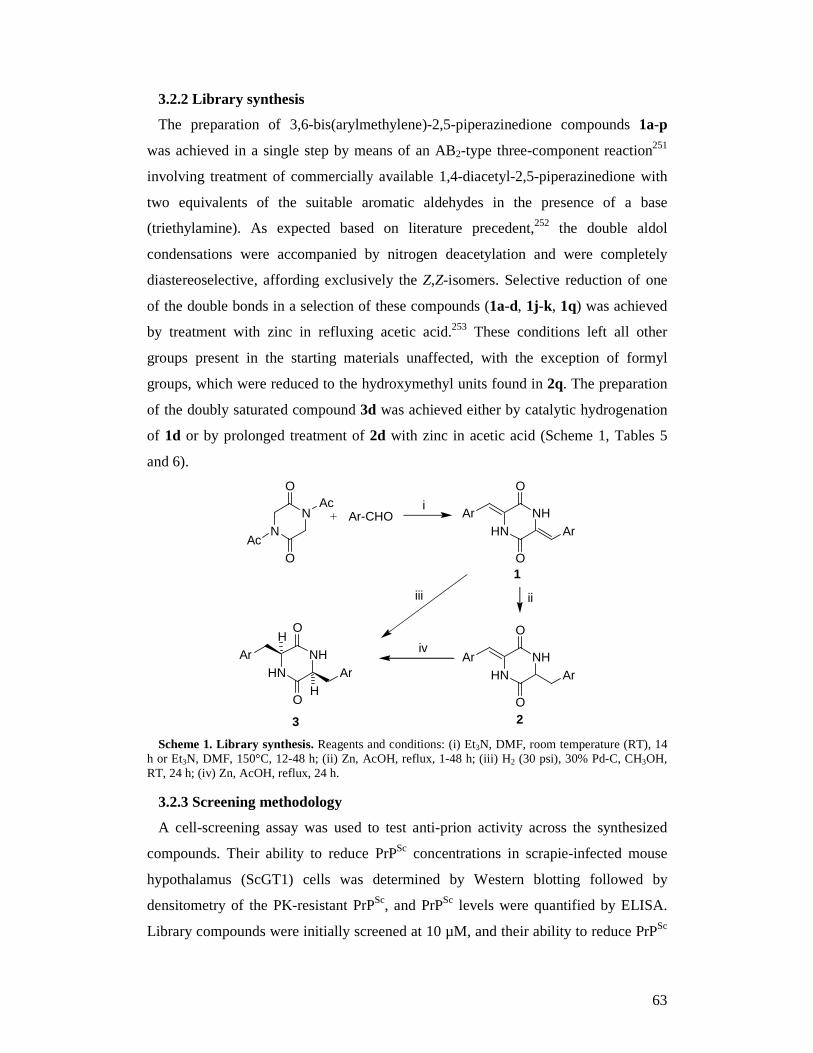
63
3.2.2 Library synthesis
The preparation of 3,6-bis(arylmethylene)-2,5-piperazinedione compounds 1a-p
was achieved in a single step by means of an AB2-type three-component reaction251
involving treatment of commercially available 1,4-diacetyl-2,5-piperazinedione with
two equivalents of the suitable aromatic aldehydes in the presence of a base
(triethylamine). As expected based on literature precedent,252 the double aldol
condensations were accompanied by nitrogen deacetylation and were completely
diastereoselective, affording exclusively the Z,Z-isomers. Selective reduction of one
of the double bonds in a selection of these compounds (1a-d, 1j-k, 1q) was achieved
by treatment with zinc in refluxing acetic acid.253 These conditions left all other
groups present in the starting materials unaffected, with the exception of formyl
groups, which were reduced to the hydroxymethyl units found in 2q. The preparation
of the doubly saturated compound 3d was achieved either by catalytic hydrogenation
of 1d or by prolonged treatment of 2d with zinc in acetic acid (Scheme 1, Tables 5
and 6).
NN
Ac
AcO
O
Ar-CHOHN
NH
O
O
ArAr
HNNH
O
O
ArArHN
NH
O
O
ArAr
H
H
i
ii
iv
iii
1
23 Scheme 1. Library synthesis. Reagents and conditions: (i) Et3N, DMF, room temperature (RT), 14
h or Et3N, DMF, 150°C, 12-48 h; (ii) Zn, AcOH, reflux, 1-48 h; (iii) H2 (30 psi), 30% Pd-C, CH3OH, RT, 24 h; (iv) Zn, AcOH, reflux, 24 h.
3.2.3 Screening methodology
A cell-screening assay was used to test anti-prion activity across the synthesized
compounds. Their ability to reduce PrPSc concentrations in scrapie-infected mouse
hypothalamus (ScGT1) cells was determined by Western blotting followed by
densitometry of the PK-resistant PrPSc, and PrPSc levels were quantified by ELISA.
Library compounds were initially screened at 10 µM, and their ability to reduce PrPSc
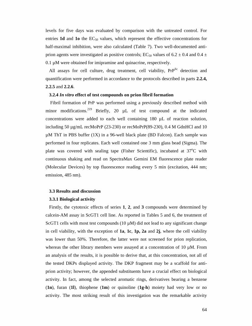
64
levels for five days was evaluated by comparison with the untreated control. For
entries 1d and 1o the EC50 values, which represent the effective concentrations for
half-maximal inhibition, were also calculated (Table 7). Two well-documented anti-
prion agents were investigated as positive controls; EC50 values of 6.2 ± 0.4 and 0.4 ±
0.1 µM were obtained for imipramine and quinacrine, respectively.
All assays for cell culture, drug treatment, cell viability, PrPSc detection and
quantification were performed in accordance to the protocols described in parts 2.2.4,
2.2.5 and 2.2.6.
3.2.4 In vitro effect of test compounds on prion fibril formation
Fibril formation of PrP was performed using a previously described method with
minor modifications.219 Briefly, 20 µL of test compound at the indicated
concentrations were added to each well containing 180 µL of reaction solution,
including 50 µg/mL recMoPrP (23-230) or recMoPrP(89-230), 0.4 M GdnHCl and 10
µM ThT in PBS buffer (1X) in a 96-well black plate (BD Falcon). Each sample was
performed in four replicates. Each well contained one 3 mm glass bead (Sigma). The
plate was covered with sealing tape (Fisher Scientific), incubated at 37oC with
continuous shaking and read on SpectraMax Gemini EM fluorescence plate reader
(Molecular Devices) by top fluorescence reading every 5 min (excitation, 444 nm;
emission, 485 nm).
3.3 Results and discussion
3.3.1 Biological activity
Firstly, the cytotoxic effects of series 1, 2, and 3 compounds were determined by
calcein-AM assay in ScGT1 cell line. As reported in Tables 5 and 6, the treatment of
ScGT1 cells with most test compounds (10 µM) did not lead to any significant change
in cell viability, with the exception of 1a, 1c, 1p, 2a and 2j, where the cell viability
was lower than 50%. Therefore, the latter were not screened for prion replication,
whereas the other library members were assayed at a concentration of 10 µM. From
an analysis of the results, it is possible to derive that, at this concentration, not all of
the tested DKPs displayed activity. The DKP fragment may be a scaffold for anti-
prion activity; however, the appended substituents have a crucial effect on biological
activity. In fact, among the selected aromatic rings, derivatives bearing a benzene
(1n), furan (1l), thiophene (1m) or quinoline (1g-h) moiety had very low or no
activity. The most striking result of this investigation was the remarkable activity
65
displayed by the 2-pyridyl derivative 1d, which completely inhibits prion replication
at 10 µM (100.8 ± 2.5 % inhibition). Intriguingly, when the pyridine nitrogen was
moved to positions 3 (1e) or 4 (1f), the activity decreased to 12.4% and 11.5%,
respectively. Similarly, the potent activity of 1d was reduced by fusing additional
aromatic rings, as in the 2-quinolinyl 1g and β-carbolin-2-yl 1o.
Significant preliminary structure-activity relationships can be gathered from this first
set of tested compounds. DKP bearing a 2-pyridine substituent was revealed as a strict
requirement for potency in the cellular assay. Intriguingly, this result is in line with
that obtained in a recently reported series of anti-prion compounds, where a 2-
substituted pyridine conferred optimal activity.254 The only exception to this trend was
the 4-quinolyl derivative 1i, which was the only compound lacking the key molecular
feature and displaying an activity higher than 20%. Notably, several 4-substituted
quinolines have shown anti-prion activity.140,238 As a general comment, it should be
mentioned that in cellular assays changes in activity also correlate to physicochemical
parameters, such as solubility and permeability. However, the results obtained could
imply that a planar conformation, as in 1d, is as a basic requirement for activity.
Starting from this assumption, we were keen to examine the effects of reduction on
the double bonds at positions 3 and 6 of the DKP system. To this end, a second library
was synthesized (2a-d, 2j-k , 2q), generated from the singly reduced DKP scaffold in
combination with a subset of the aromatic substituents reported in Table 5 (Scheme 1,
Table 6). As expected, even in this series the most active compound was the 2-pyridyl
derivative 2d. Again, this can be rationalized by assumption that at least one half of
the molecule is able to assume the planar conformation critical for activity. In line
with this speculation, the doubly saturated compound 3d showed lower potency. In
this second library, an interesting profile was shown by 1-tosylindole 2k, which
exhibited 37.7% inhibition.
Given the high inhibitory activity of 1d and the possible use of 1o as a fluorescent
probe, their anti-prion potential was studied in more detail by calculating their EC50
values. For 1d, a remarkable single-digit micromolar EC50 value (4.1 ± 0.2 µM; Table
7) was observed, comparable to that of imipramine (6.2 ± 0.4 µM), a reference anti-
prion compound. Notably, 1o (EC50 = 15.8 ± 0.9 µM) was only fourfold less active
than 1d, emerging as a promising fluorescent probe.

66
Table 5. Reaction conditions, yields and screening results for the DKP Series 1
1a-p
Cpd R Conditions Yield [%] Viable cells
[%] b
PrPSc
inhibition
[%] c
1a
RT, 16 h 97 32.2 ± 1.0a NDd
1b
RT, 16 h 99 72.7 ± 3.3 1.4 ± 0.1a
1c
RT, 16 h 74 20.6 ± 1.7 ND
1d
reflux, 48 h 64 50.9 ± 2.1 100.8 ± 2.5
1e
reflux, 16 h 41 102.7 ± 3.7 12.4 ± 0.5
1f
reflux, 24 h 68 108.5 ± 4.2 11.5 ± 0.5
1g
reflux, 20 h 80 71.7 ± 3.1 5.1 ± 0.1
1h
reflux, 16 h 65 63.7 ± 2.8 3.5 ± 0.1
1i
reflux, 16 h 84 94.8 ± 4.1 20.6 ± 0.8
1j
reflux, 16 h 94 81.3 ± 2.5 7.4 ± 0.5
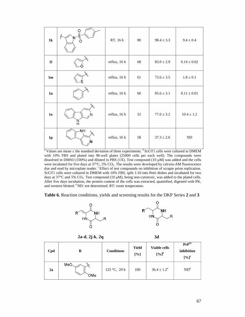
67
1k
RT, 16 h 80 98.4 ± 3.3 9.4 ± 0.4
1l
reflux, 16 h 68 83.0 ± 2.9 0.14 ± 0.02
1m
reflux, 16 h 61 73.6 ± 3.5 1.8 ± 0.1
1n
reflux, 16 h 60 85.6 ± 3.1 0.11 ± 0.01
1o
reflux, 16 h 52 77.0 ± 3.2 10.4 ± 1.2
1p
reflux, 16 h 58 37.3 ± 2.6 ND
a Values are mean ± the standard deviation of three experiments. b ScGT1 cells were cultured in DMEM with 10% FBS and plated into 96-well plates (25000 cells per each well). The compounds were dissolved in DMSO (100%) and diluted in PBS (1X). Test compound (10 µM) was added and the cells were incubated for five days at 37°C, 5% CO2. The results were developed by calcein-AM fluorescence dye and read by microplate reader. c Effect of test compounds on inhibition of scrapie prion replication. ScGT1 cells were cultured in DMEM with 10% FBS, split 1:10 into Petri dishes and incubated for two days at 37°C and 5% CO2. Test compound (10 µM), being non-cytotoxic, was added to the plated cells. After five days incubation, the protein content of the cells was extracted, quantified, digested with PK, and western blotted. d ND: not determined. RT: room temperature. Table 6. Reaction conditions, yields and screening results for the DKP Series 2 and 3
2a-d, 2j-k, 2q 3d
Cpd R Conditions Yield
[%]
Viable cells
[%] b
PrPSc
inhibition
[%] c
2a
125 °C, 20 h 100 36.4 ± 1.2a NDd
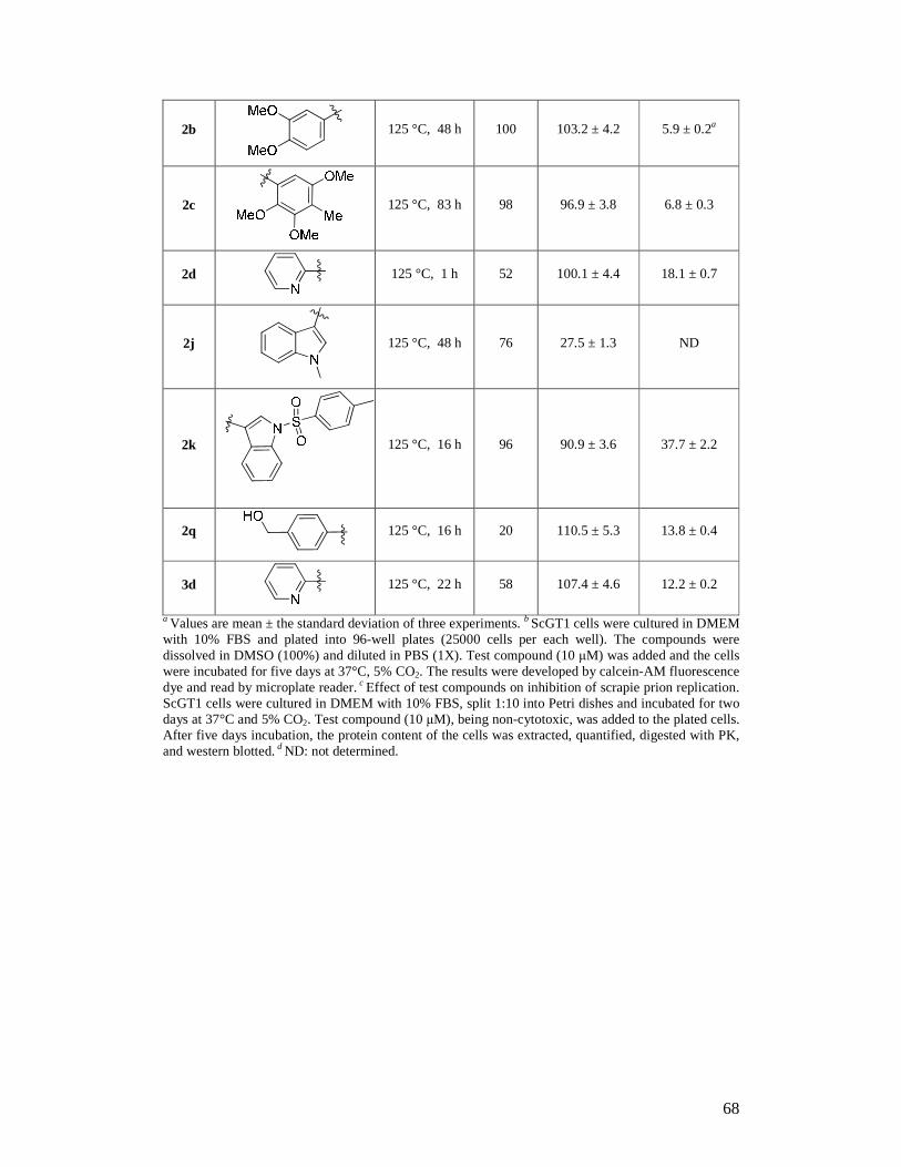
68
2b
125 °C, 48 h 100 103.2 ± 4.2 5.9 ± 0.2a
2c
125 °C, 83 h 98 96.9 ± 3.8 6.8 ± 0.3
2d
125 °C, 1 h 52 100.1 ± 4.4 18.1 ± 0.7
2j
125 °C, 48 h 76 27.5 ± 1.3 ND
2k
125 °C, 16 h 96 90.9 ± 3.6 37.7 ± 2.2
2q
125 °C, 16 h 20 110.5 ± 5.3 13.8 ± 0.4
3d
125 °C, 22 h 58 107.4 ± 4.6 12.2 ± 0.2
a Values are mean ± the standard deviation of three experiments. b ScGT1 cells were cultured in DMEM with 10% FBS and plated into 96-well plates (25000 cells per each well). The compounds were dissolved in DMSO (100%) and diluted in PBS (1X). Test compound (10 µM) was added and the cells were incubated for five days at 37°C, 5% CO2. The results were developed by calcein-AM fluorescence dye and read by microplate reader. c Effect of test compounds on inhibition of scrapie prion replication. ScGT1 cells were cultured in DMEM with 10% FBS, split 1:10 into Petri dishes and incubated for two days at 37°C and 5% CO2. Test compound (10 µM), being non-cytotoxic, was added to the plated cells. After five days incubation, the protein content of the cells was extracted, quantified, digested with PK, and western blotted. d ND: not determined.
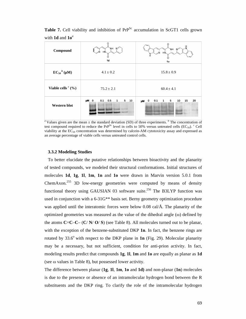
69
Table 7. Cell viability and inhibition of PrPSc accumulation in ScGT1 cells grown
with 1d and 1oa
a Values given are the mean ± the standard deviation (SD) of three experiments. b The concentration of test compound required to reduce the PrPSc level in cells to 50% versus untreated cells (EC50).
c Cell viability at the EC50 concentration was determined by calcein-AM cytotoxicity assay and expressed as an average percentage of viable cells versus untreated control cells.
3.3.2 Modeling Studies
To better elucidate the putative relationships between bioactivity and the planarity
of tested compounds, we modeled their structural conformations. Initial structures of
molecules 1d, 1g, 1l, 1m, 1n and 1o were drawn in Marvin version 5.0.1 from
ChemAxon.255 3D low-energy geometries were computed by means of density
functional theory using GAUSIAN 03 software suite.256 The B3LYP function was
used in conjunction with a 6-31G** basis set. Berny geometry optimization procedure
was applied until the interatomic forces were below 0.08 cal/Å. The planarity of the
optimized geometries was measured as the value of the dihedral angle (ω) defined by
the atoms C=C–C– (C/ N/ O/ S) (see Table 8). All molecules turned out to be planar,
with the exception of the benzene-substituted DKP 1n. In fact, the benzene rings are
rotated by 33.6o with respect to the DKP plane in 1n (Fig. 29). Molecular planarity
may be a necessary, but not sufficient, condition for anti-prion activity. In fact,
modeling results predict that compounds 1g, 1l, 1m and 1o are equally as planar as 1d
(see ω values in Table 8), but possessed lower activity.
The difference between planar (1g, 1l, 1m, 1o and 1d) and non-planar (1n) molecules
is due to the presence or absence of an intramolecular hydrogen bond between the R
substituents and the DKP ring. To clarify the role of the intramolecular hydrogen
0 0.1 1 5 10 15 20µM 0 0.1 1 5 10 15 20µM
0 0.1 0.5 1 5 10µM 0 0.1 0.5 1 5 10µM
Western blot
60.4 ± 4.1
75.2 ± 2.1
Viable cells c (%)
15.8 ± 0.9
4.1 ± 0.2
EC50
b (µM)
Compound
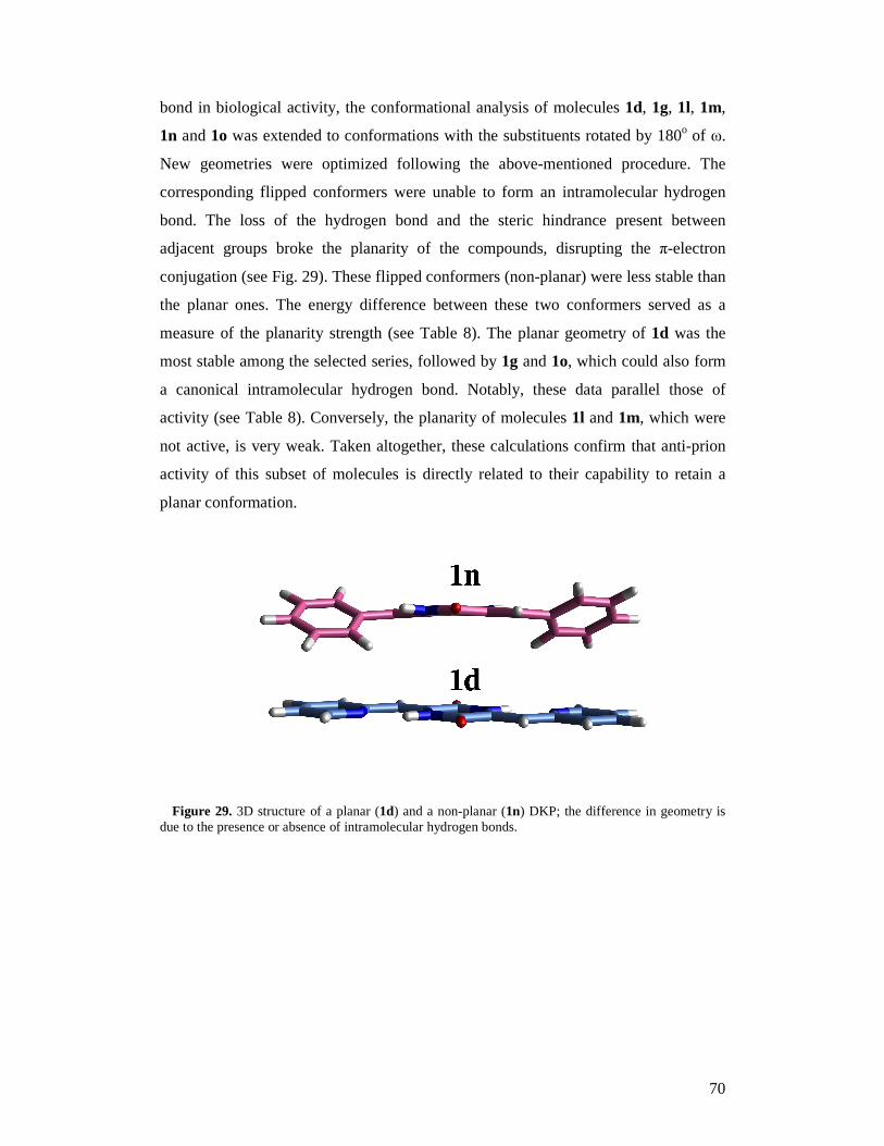
70
bond in biological activity, the conformational analysis of molecules 1d, 1g, 1l, 1m,
1n and 1o was extended to conformations with the substituents rotated by 180o of ω.
New geometries were optimized following the above-mentioned procedure. The
corresponding flipped conformers were unable to form an intramolecular hydrogen
bond. The loss of the hydrogen bond and the steric hindrance present between
adjacent groups broke the planarity of the compounds, disrupting the π-electron
conjugation (see Fig. 29). These flipped conformers (non-planar) were less stable than
the planar ones. The energy difference between these two conformers served as a
measure of the planarity strength (see Table 8). The planar geometry of 1d was the
most stable among the selected series, followed by 1g and 1o, which could also form
a canonical intramolecular hydrogen bond. Notably, these data parallel those of
activity (see Table 8). Conversely, the planarity of molecules 1l and 1m, which were
not active, is very weak. Taken altogether, these calculations confirm that anti-prion
activity of this subset of molecules is directly related to their capability to retain a
planar conformation.
Figure 29. 3D structure of a planar (1d) and a non-planar (1n) DKP; the difference in geometry is
due to the presence or absence of intramolecular hydrogen bonds.
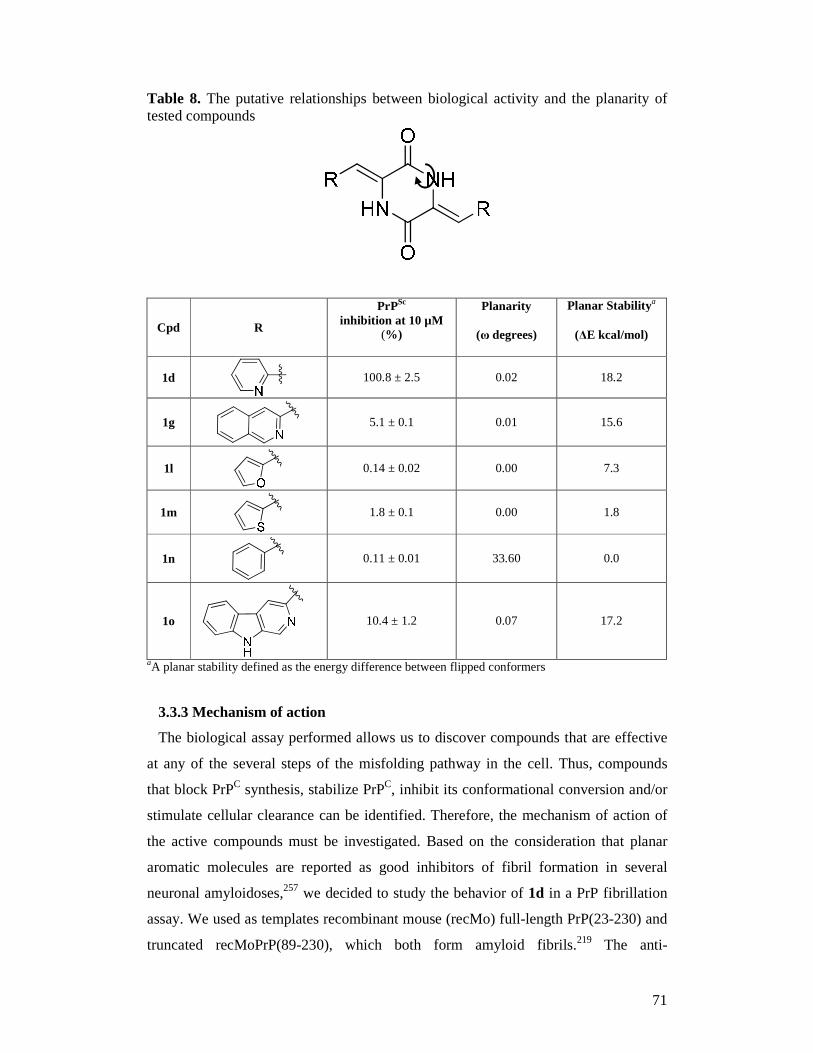
71
Table 8. The putative relationships between biological activity and the planarity of tested compounds
Cpd R
PrPSc inhibition at 10 µM
(%)
Planarity
(ω degrees)
Planar Stabilitya
(∆E kcal/mol)
1d
100.8 ± 2.5 0.02 18.2
1g
5.1 ± 0.1 0.01 15.6
1l
0.14 ± 0.02 0.00 7.3
1m
1.8 ± 0.1 0.00 1.8
1n
0.11 ± 0.01 33.60 0.0
1o
10.4 ± 1.2 0.07 17.2
aA planar stability defined as the energy difference between flipped conformers
3.3.3 Mechanism of action
The biological assay performed allows us to discover compounds that are effective
at any of the several steps of the misfolding pathway in the cell. Thus, compounds
that block PrPC synthesis, stabilize PrPC, inhibit its conformational conversion and/or
stimulate cellular clearance can be identified. Therefore, the mechanism of action of
the active compounds must be investigated. Based on the consideration that planar
aromatic molecules are reported as good inhibitors of fibril formation in several
neuronal amyloidoses,257 we decided to study the behavior of 1d in a PrP fibrillation
assay. We used as templates recombinant mouse (recMo) full-length PrP(23-230) and
truncated recMoPrP(89-230), which both form amyloid fibrils.219 The anti-

72
aggregation potential of 1d (50 µM) was quantitatively detected by observing the
increase in the mean lag phase of the fibrillation reaction, compared with control
samples. Compound 1d (50 µM) exhibited PrP amyloid fibril formation inhibitory
activity, showing a much slower kinetics than the control, in which the lag phase was
57 hours and 15 hours for recMoPrP(23-230) and recMoPrP(89-230), respectively. In
particular, 1d significantly extended the lag phase to 72 hours in recMoPrP(23-230)
and 48 hours in recMoPrP(89-230) fibrillation assays. Given the potency of 1d at
inhibiting recPrP fibril formation, we suggest that the compound might interact
directly with recPrP to prevent its conversion to the pathogenic PrPSc-like form.
3.4 Conclusion
In conclusion, we have identified DKP as a novel scaffold in anti-prion drug design.
Compound 1d, thanks to its planar conformation, is able to inhibit PrP amyloid fibril
formation in vitro. Moreover, it inhibits prion replication in the low micromolar range
in a cellular context. For these reasons, 1d is a lead candidate for further optimization
studies. While preliminary, these results represent a first step toward the discovery of
novel DKPs with therapeutic potential against prion diseases. Clearly, proof of
concept will involve in vivo investigation of the profile of 1d.
A second major outcome of this work is the identification of the β-carboline
derivative 1o. Fluorescent probes that specifically target amyloid aggregates are of
great relevance to advance our understanding of the molecular pathogenesis
underlying cerebral amyloidoses.257,258 Derivative 1o might represent a valuable
starting point to design novel probes for the optical imaging of amyloid plaques in
prion disease. Properly addressed studies aimed at the investigation of the fluorescent
profile of 1o and its labeling of amyloid fibrils are in progress and will be reported in
due course.
73
4. Anti-prion activity and preliminary structure-ac tivity
relationship of benzoquinones
4.1 Introduction
Despite the numerous efforts aimed at identifying compounds useful against prion
diseases, there are still no therapies on the market. Therefore, it is of continued
importance to identify chemical scaffolds to be exploited for the design of novel
drugs.259 Most of the anti-prion molecules that have been identified so far are derived
from screening approaches. Structurally, diverse chemical compounds covering a
broad range of the chemical space have been identified.226 Intriguingly, most of them
share a common bivalent structure. This is the case of the natural product curcumin,132
the bis-acridine analogues,218,260 the diphenyl-methane derivative (GN8),239
bebeerines,96 bisepigallocatechin digallate,96 2,2’-bisquinolines,140 4,5-
dianilinophthalimide,261 analogues of Congo red237 and diketopiperazines (DKP)
derivatives.262
Although a structure-activity relationship is not easy to discern from such chemically
unrelated compounds, we envisaged that bivalent ligands bearing lipophilic bi- or tri-
(hetero)-cyclic scaffolds connected by a central core might possess anti-prion activity.
Bivalency, and multivalency in general, is a well-known and efficient strategy widely
used by medicinal chemists to enhance binding efficacy in molecular recognition
processes.263 Multivalent chemical probes, featuring multiple copies of an amyloid
binding motif connected by a spacer, have been developed with the aim to
simultaneously bind to several binding sites or several amyloid peptides, thus
achieving higher potency.232,264
In prion research, by joining two quinacrine moieties through a piperazine spacer,
May and co-workers afforded the first bivalent anti-prion ligand BiCappa (BC; Fig.
30), which was 10 times more potent than monomeric quinacrine.218 Heterodimers
incorporating recognition elements taken from quinacrine itself and imipramine with a
piperazine unit were shown to improve the anti-prion efficacy of quinacrine up to a
low nanomolar range.265 Furthermore, assembling multiple acridine or curcumin
moieties to a cyclopeptide scaffold has emerged as a promising strategy for the
development of inhibitors against amyloid formation.266,267
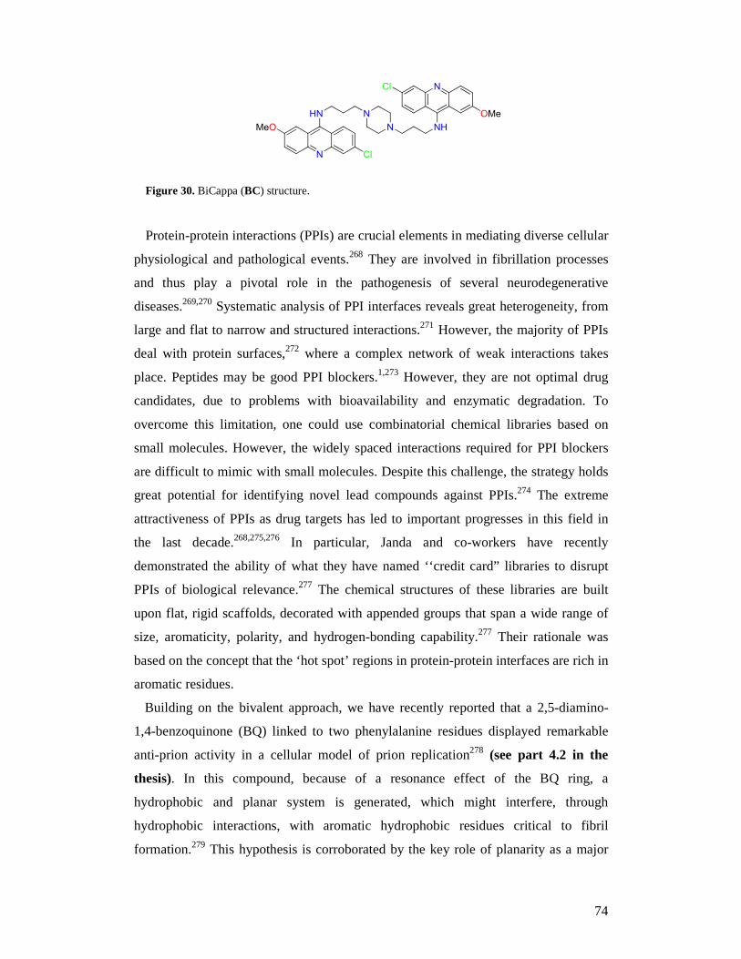
74
N
HN
Cl
MeO
N
N NH
NCl
OMe
Figure 30. BiCappa (BC) structure.
Protein-protein interactions (PPIs) are crucial elements in mediating diverse cellular
physiological and pathological events.268 They are involved in fibrillation processes
and thus play a pivotal role in the pathogenesis of several neurodegenerative
diseases.269,270 Systematic analysis of PPI interfaces reveals great heterogeneity, from
large and flat to narrow and structured interactions.271 However, the majority of PPIs
deal with protein surfaces,272 where a complex network of weak interactions takes
place. Peptides may be good PPI blockers.1,273 However, they are not optimal drug
candidates, due to problems with bioavailability and enzymatic degradation. To
overcome this limitation, one could use combinatorial chemical libraries based on
small molecules. However, the widely spaced interactions required for PPI blockers
are difficult to mimic with small molecules. Despite this challenge, the strategy holds
great potential for identifying novel lead compounds against PPIs.274 The extreme
attractiveness of PPIs as drug targets has led to important progresses in this field in
the last decade.268,275,276 In particular, Janda and co-workers have recently
demonstrated the ability of what they have named ‘‘credit card” libraries to disrupt
PPIs of biological relevance.277 The chemical structures of these libraries are built
upon flat, rigid scaffolds, decorated with appended groups that span a wide range of
size, aromaticity, polarity, and hydrogen-bonding capability.277 Their rationale was
based on the concept that the ‘hot spot’ regions in protein-protein interfaces are rich in
aromatic residues.
Building on the bivalent approach, we have recently reported that a 2,5-diamino-
1,4-benzoquinone (BQ) linked to two phenylalanine residues displayed remarkable
anti-prion activity in a cellular model of prion replication278 (see part 4.2 in the
thesis). In this compound, because of a resonance effect of the BQ ring, a
hydrophobic and planar system is generated, which might interfere, through
hydrophobic interactions, with aromatic hydrophobic residues critical to fibril
formation.279 This hypothesis is corroborated by the key role of planarity as a major
75
determinant for anti-prion activity in a recently synthesized series of bivalent DKP262
(see part 3 in the thesis). Altogether these results allowed us to propose that the
planar BQ scaffold might be considered as a privileged motif in modulating PPIs and
as a promising spacer in the search for more effective bivalent anti-prion chemical
probes280 (see part 4.3 in the thesis).
4.2 Synthesis and evaluation of a library of 2,5-bisdiamino-benzoquinone
derivatives as probes to modulate protein-protein interactions in prions
So far, several peptides have been developed with the specific aim of blocking PPIs
and reversing the aberrant conformational changes. A short synthetic peptide (iPrP13,
DAPAAPAGPAVPV), designed by Soto and co-workers on the basis of sequence
homology with PrPC, acted as a β-sheet breaker, inducing unfolding of β-pleated sheet
structure.13 More recently, Gilbert and co-workers281 reported on a series of small
peptides active at levels of 100 µM in two prion disease models and in an in vitro
anti-aggregation polymerization assay. Prompted by the advantages of using small
molecules as PPI inhibitors as opposed to peptides, here we propose the planar 2,5-
bisdiamino-benzoquinone scaffold as a privileged motif in modulating PPIs. This is
based on (i) Janda’s criteria for credit card libraries;277 (ii) the finding that a 2,5-
bisdiamino-benzoquinone derivative binds to β-amyloid (Aβ), and interferes with the
native ability of Aβ to self-assemble, by disrupting PPIs.282 Due to a resonance effect,
a hydrophobic and planar system is generated in 2,5-bisdiamino-benzoquinones. This
should, in principle, perturb PPIs in the fibrillogenesis processes.283
Therefore, in our search for novel anti-prion compounds, we decided to attach seven
amino acid methyl esters to two different benzoquinone cores, generating a small
combinatorial library of fourteen 2,5-bisdiamino-benzoquinones (1-7a and 1-7b),
reported in Figure 31. The selected amino acid esters (AlaOMe (1), Nω-Nitro-
ArgOMe (2), Nε-BOC-LysOMe (3), IleOMe (4), MetOMe (5), PheOMe (6), TrpOMe
(7)) act as capping groups, allowing us to enlarge the library’s chemical diversity by
exploiting differences in size, aromaticity, polarity, and hydrogen-bonding capability.
Analysis of natural amino acids involved in PPIs revealed that Trp, Phe, Tyr, and Ile
are the most important in driving aggregation.284 Consequently, it is highly
conceivable that the novel derivatives bearing these motifs might compete for binding
and, therefore, efficiently disrupt the assembly of prion protein.
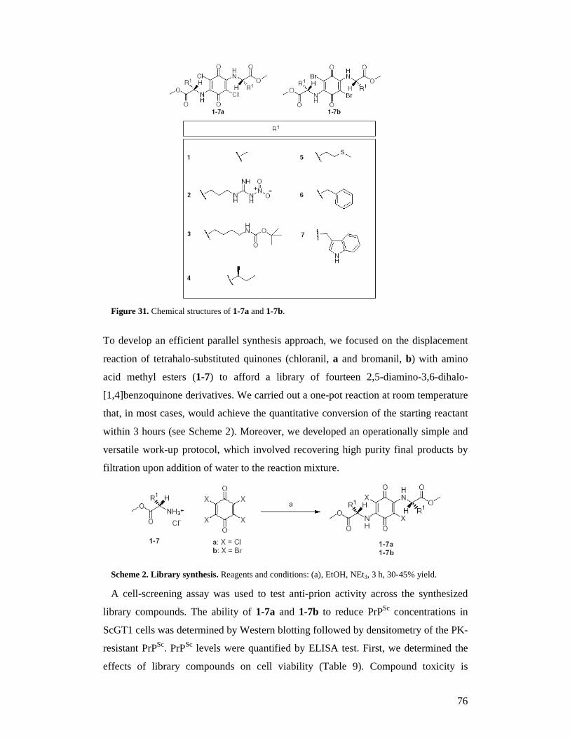
76
Figure 31. Chemical structures of 1-7a and 1-7b.
To develop an efficient parallel synthesis approach, we focused on the displacement
reaction of tetrahalo-substituted quinones (chloranil, a and bromanil, b) with amino
acid methyl esters (1-7) to afford a library of fourteen 2,5-diamino-3,6-dihalo-
[1,4]benzoquinone derivatives. We carried out a one-pot reaction at room temperature
that, in most cases, would achieve the quantitative conversion of the starting reactant
within 3 hours (see Scheme 2). Moreover, we developed an operationally simple and
versatile work-up protocol, which involved recovering high purity final products by
filtration upon addition of water to the reaction mixture.
Scheme 2. Library synthesis. Reagents and conditions: (a), EtOH, NEt3, 3 h, 30-45% yield.
A cell-screening assay was used to test anti-prion activity across the synthesized
library compounds. The ability of 1-7a and 1-7b to reduce PrPSc concentrations in
ScGT1 cells was determined by Western blotting followed by densitometry of the PK-
resistant PrPSc. PrPSc levels were quantified by ELISA test. First, we determined the
effects of library compounds on cell viability (Table 9). Compound toxicity is
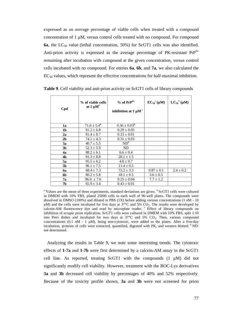
77
expressed as an average percentage of viable cells when treated with a compound
concentration of 1 µM, versus control cells treated with no compound. For compound
6a, the LC50 value (lethal concentration, 50%) for ScGT1 cells was also identified.
Anti-prion activity is expressed as the average percentage of PK-resistant PrPSc
remaining after incubation with compound at the given concentration, versus control
cells incubated with no compound. For entries 6a, 6b, and 7a, we also calculated the
EC50 values, which represent the effective concentrations for half-maximal inhibition.
Table 9. Cell viability and anti-prion activity on ScGT1 cells of library compounds
Cpd
% of viable cells at 1 µMb
% of PrPSc
inhibition at 1 µM c
EC50c (µM) LC 50
b (µM)
1a 71.6 ± 5.4a 0.36 ± 0.03a 1b 91.2 ± 6.8 0.29 ± 0.05 2a 81.4 ± 8.7 0.23 ± 0.01 2b 74.1 ± 4.3 0.31 ± 0.03 3a 40.7 ± 5.5 NDd 3b 52.3 ± 5.9 ND 4a 88.2 ± 6.1 6.6 ± 0.4 4b 91.3 ± 8.8 28.1 ± 1.5 5a 95.5 ± 6.2 4.8 ± 0.7 5b 96.1 ± 7.5 11.4 ± 0.5 6a 68.4 ± 7.3 73.2 ± 3.3 0.87 ± 0.1 2.4 ± 0.2 6b 80.2 ± 5.8 18.1 ± 0.5 3.6 ± 0.5 7a 96.0 ± 7.6 0.25 ± 0.04 7.7 ± 1.2 7b 65.9 ± 3.4 0.43 ± 0.01
a Values are the mean of three experiments, standard deviations are given. b ScGT1 cells were cultured in DMEM with 10% FBS, plated 25000 cells in each well of 96-well plates. The compounds were dissolved in DMSO (100%) and diluted in PBS (1X) before adding various concentrations (1 nM - 10 µM) and the cells were incubated for five days at 37°C and 5% CO2. The results were developed by calcein-AM fluorescence dye and read by microplate reader. c Effect of library compounds on inhibition of scrapie prion replication. ScGT1 cells were cultured in DMEM with 10% FBS, split 1:10 into Petri dishes and incubated for two days at 37°C and 5% CO2. Then, various compound concentrations (0.1 nM - 1 µM), being non-cytotoxic, were added to the plates. After a five-day incubation, proteins of cells were extracted, quantified, digested with PK, and western blotted. d ND: not determined.
Analyzing the results in Table 9, we note some interesting trends. The cytotoxic
effects of 1-7a and 1-7b were first determined by a calcein-AM assay in the ScGT1
cell line. As reported, treating ScGT1 with the compounds (1 µM) did not
significantly modify cell viability. However, treatment with the BOC-Lys derivatives
3a and 3b decreased cell viability by percentages of 40% and 52% respectively.
Because of the toxicity profile shown, 3a and 3b were not screened for prion
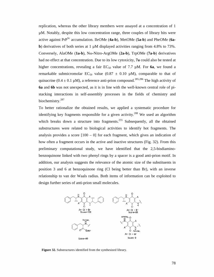
78
replication, whereas the other library members were assayed at a concentration of 1
µM. Notably, despite this low concentration range, three couples of library hits were
active against PrPSc accumulation. IleOMe (4a-b), MetOMe (5a-b) and PheOMe (6a-
b) derivatives of both series at 1 µM displayed activities ranging from 4.8% to 73%.
Conversely, AlaOMe (1a-b), Nω-Nitro-ArgOMe (2a-b), TrpOMe (7a-b) derivatives
had no effect at that concentration. Due to its low cytoxicity, 7a could also be tested at
higher concentrations, revealing a fair EC50 value of 7.7 µM. For 6a, we found a
remarkable submicromolar EC50 value (0.87 ± 0.10 µM), comparable to that of
quinacrine (0.4 ± 0.1 µM), a reference anti-prion compound.285,286 The high activity of
6a and 6b was not unexpected, as it is in line with the well-known central role of pi-
stacking interactions in self-assembly processes in the fields of chemistry and
biochemistry.287
To better rationalize the obtained results, we applied a systematic procedure for
identifying key fragments responsible for a given activity.288 We used an algorithm
which breaks down a structure into fragments.255 Subsequently, all the obtained
substructures were related to biological activities to identify hot fragments. The
analysis provides a score [100 – 0] for each fragment, which gives an indication of
how often a fragment occurs in the active and inactive structures (Fig. 32). From this
preliminary computational study, we have identified that the 2,5-bisdiamino-
benzoquinone linked with two phenyl rings by a spacer is a good anti-prion motif. In
addition, our analysis suggests the relevance of the atomic size of the substituents in
position 3 and 6 at benzoquinone ring (Cl being better than Br), with an inverse
relationship to van der Waals radius. Both items of information can be exploited to
design further series of anti-prion small molecules.
Figure 32. Substructures identified from the synthesized library.
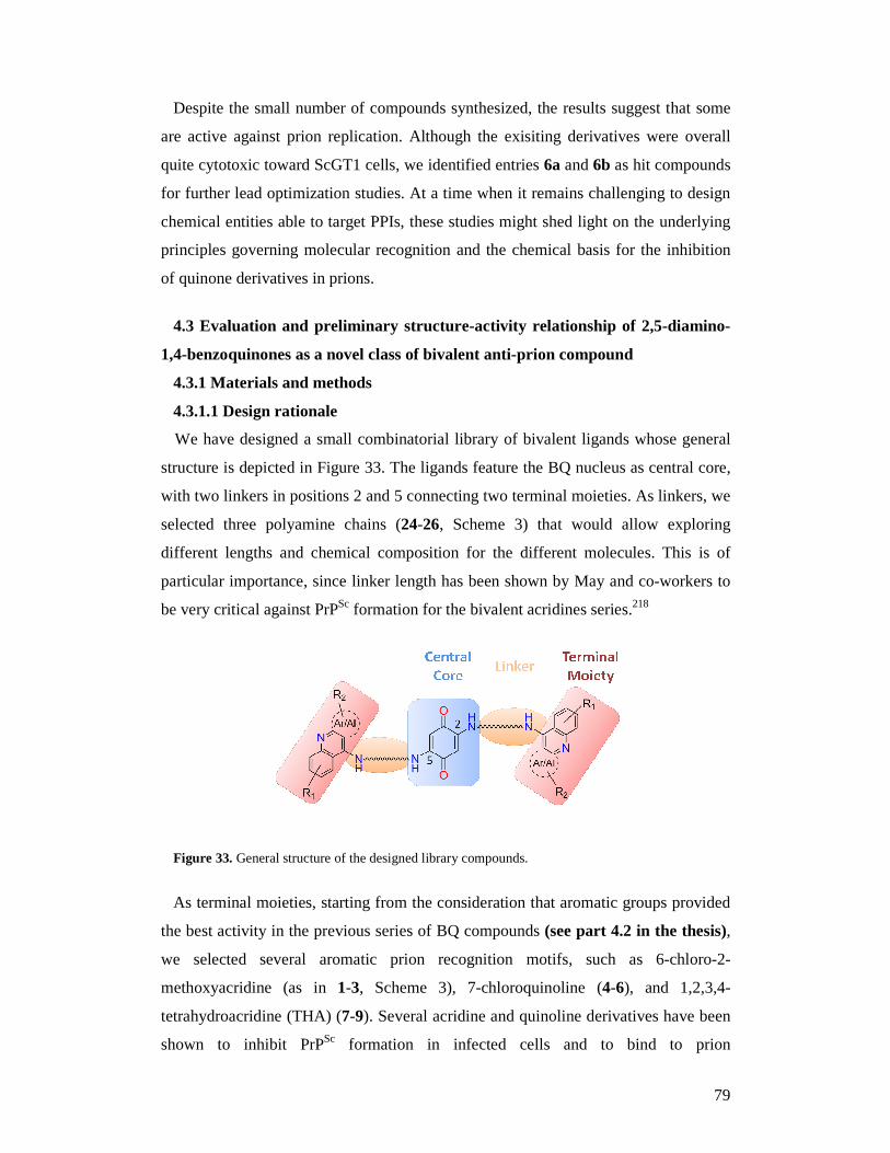
79
Despite the small number of compounds synthesized, the results suggest that some
are active against prion replication. Although the exisiting derivatives were overall
quite cytotoxic toward ScGT1 cells, we identified entries 6a and 6b as hit compounds
for further lead optimization studies. At a time when it remains challenging to design
chemical entities able to target PPIs, these studies might shed light on the underlying
principles governing molecular recognition and the chemical basis for the inhibition
of quinone derivatives in prions.
4.3 Evaluation and preliminary structure-activity r elationship of 2,5-diamino-
1,4-benzoquinones as a novel class of bivalent anti-prion compound
4.3.1 Materials and methods
4.3.1.1 Design rationale
We have designed a small combinatorial library of bivalent ligands whose general
structure is depicted in Figure 33. The ligands feature the BQ nucleus as central core,
with two linkers in positions 2 and 5 connecting two terminal moieties. As linkers, we
selected three polyamine chains (24-26, Scheme 3) that would allow exploring
different lengths and chemical composition for the different molecules. This is of
particular importance, since linker length has been shown by May and co-workers to
be very critical against PrPSc formation for the bivalent acridines series.218
Figure 33. General structure of the designed library compounds.
As terminal moieties, starting from the consideration that aromatic groups provided
the best activity in the previous series of BQ compounds (see part 4.2 in the thesis),
we selected several aromatic prion recognition motifs, such as 6-chloro-2-
methoxyacridine (as in 1-3, Scheme 3), 7-chloroquinoline (4-6), and 1,2,3,4-
tetrahydroacridine (THA) (7-9). Several acridine and quinoline derivatives have been
shown to inhibit PrPSc formation in infected cells and to bind to prion
80
proteins.140,238,289 Given that the analogous THA-9-amine is active against yeast
prion,290 derivatives 7-9 were also designed. As a second step, on the basis of the
remarkable profile shown by 5, three other derivatives (10-12) were designed with the
aim of further optimizing activity in the existing series of compounds. Herein, we
present a solution-phase parallel synthesis of a library of bivalent BQ derivatives,
which were chosen for their anti-prion activity in ScGT1 cells, together with their
capability of inhibiting PrPSc aggregation and of reducing oxidative stress (OS).
4.3.1.2 Library synthesis
Disubstitution reaction of diamines with 2,5-dimethoxy-1,4-benzoquinone provides
an easy access to a variety of 2,5-diamino-1,4-benzoquinones.283,291 Encouraged by
the good yields and the straightforward work-up associated with this reaction, we
decided to synthesize the designed bivalent compounds 1-11 using a solution phase
parallel synthesis approach. Eleven N-substituted polyamines (13-23, Scheme 3) were
loaded with 2,5-dimethoxy-1,4-benzoquinone into different vessels of a carousel
workstation. After heating at 50°C for 5 hours, the desired products formed in
moderate to good yields (38-88%). Monovalent 12 was obtained by Michael reaction
starting from naftoquinone and amine 17 (40%).
The preparation of intermediates 13-23 was easily achieved treating in parallel
fashion commercially available polyamines 24-26 with heteroaryl halides 27-31.
Compounds 13-23 were obtained in 25-67% yield by reacting a large excess of the
polyamine with the corresponding heteroaryl halide (27:1) in phenol and using NaI as
a catalyst (Scheme 3). In these conditions we were able to obtain selective mono-
substitution at the terminal primary amino group of the polyamine, obviating the need
for protection/deprotection of the other amino functionalities.292 Furthermore, we
overcame the low-yield of common SNAr reactions and the use of costly reagents of
Pd-catalyzed amination methodologies.293
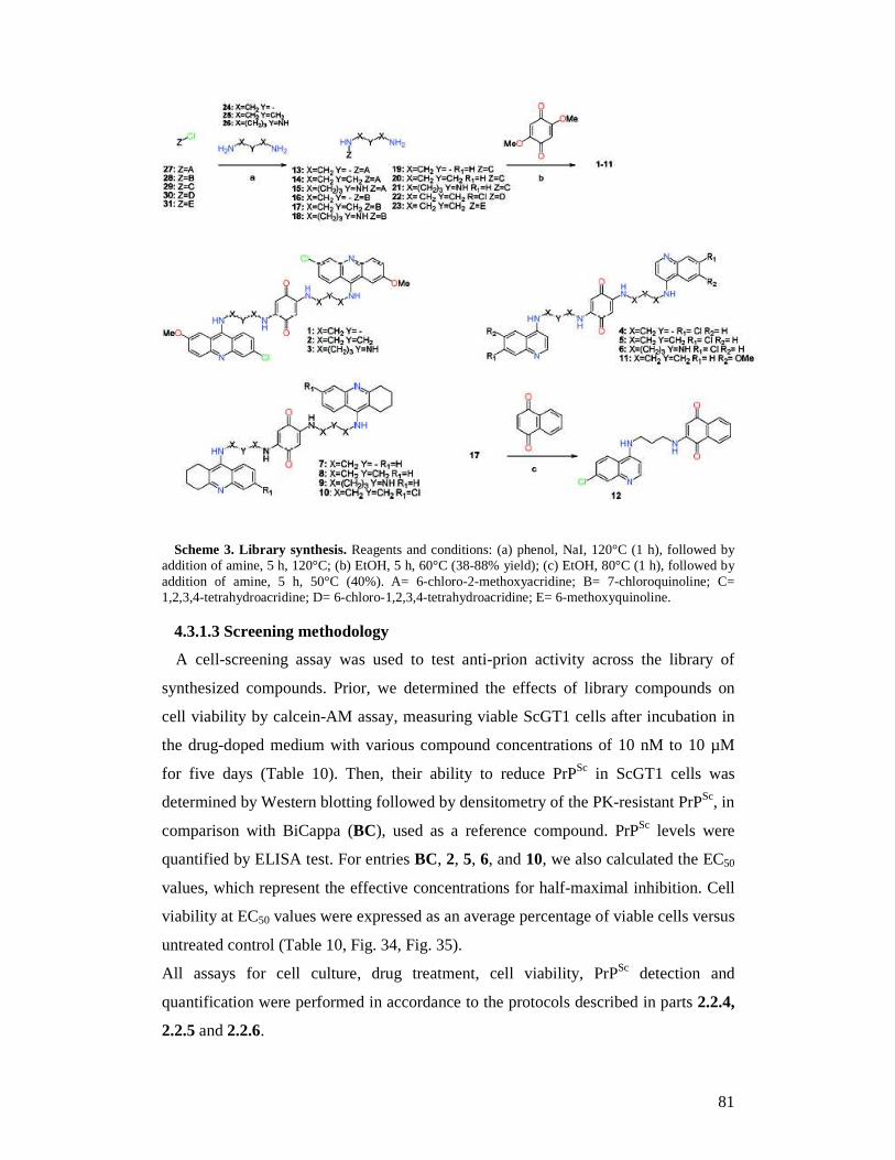
81
Scheme 3. Library synthesis. Reagents and conditions: (a) phenol, NaI, 120°C (1 h), followed by addition of amine, 5 h, 120°C; (b) EtOH, 5 h, 60°C (38-88% yield); (c) EtOH, 80°C (1 h), followed by addition of amine, 5 h, 50°C (40%). A= 6-chloro-2-methoxyacridine; B= 7-chloroquinoline; C= 1,2,3,4-tetrahydroacridine; D= 6-chloro-1,2,3,4-tetrahydroacridine; E= 6-methoxyquinoline.
4.3.1.3 Screening methodology
A cell-screening assay was used to test anti-prion activity across the library of
synthesized compounds. Prior, we determined the effects of library compounds on
cell viability by calcein-AM assay, measuring viable ScGT1 cells after incubation in
the drug-doped medium with various compound concentrations of 10 nM to 10 µM
for five days (Table 10). Then, their ability to reduce PrPSc in ScGT1 cells was
determined by Western blotting followed by densitometry of the PK-resistant PrPSc, in
comparison with BiCappa (BC), used as a reference compound. PrPSc levels were
quantified by ELISA test. For entries BC, 2, 5, 6, and 10, we also calculated the EC50
values, which represent the effective concentrations for half-maximal inhibition. Cell
viability at EC50 values were expressed as an average percentage of viable cells versus
untreated control (Table 10, Fig. 34, Fig. 35).
All assays for cell culture, drug treatment, cell viability, PrPSc detection and
quantification were performed in accordance to the protocols described in parts 2.2.4,
2.2.5 and 2.2.6.
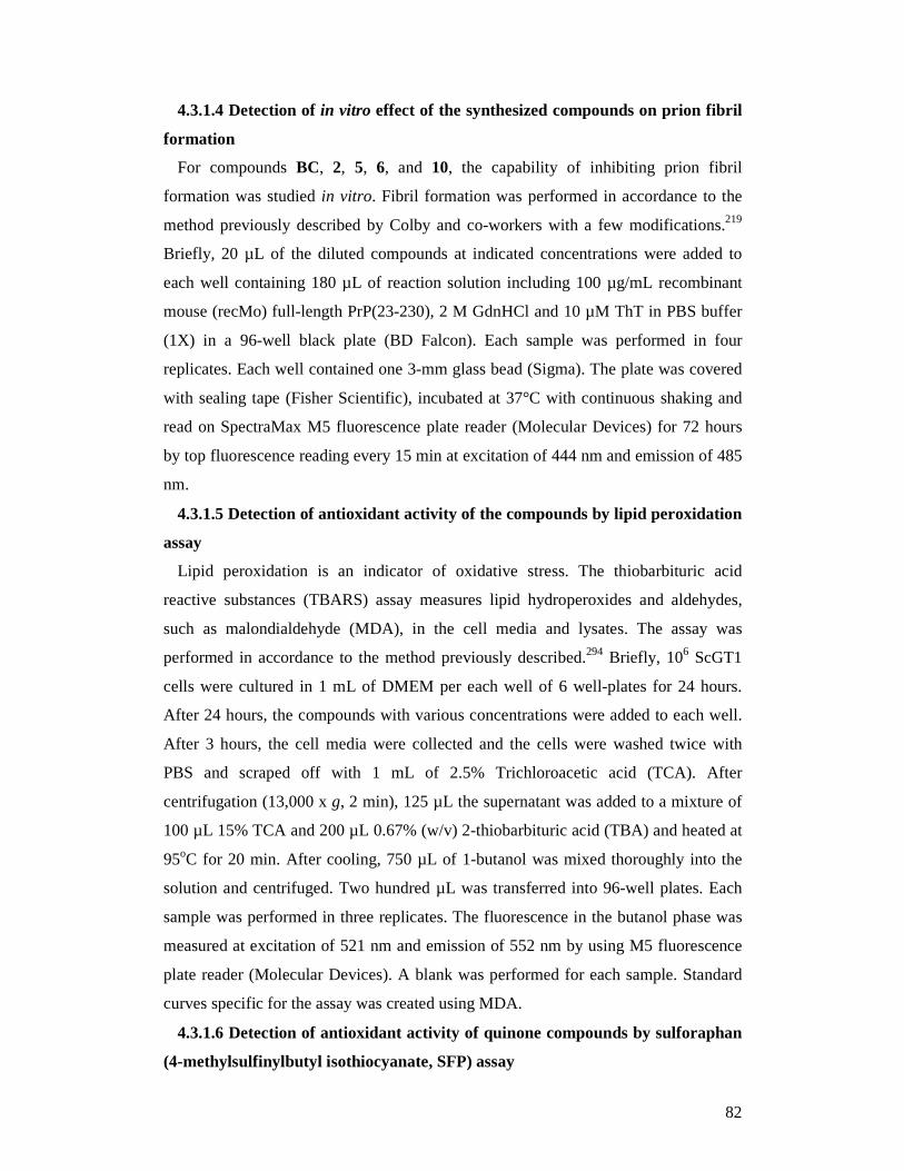
82
4.3.1.4 Detection of in vitro effect of the synthesized compounds on prion fibril
formation
For compounds BC, 2, 5, 6, and 10, the capability of inhibiting prion fibril
formation was studied in vitro. Fibril formation was performed in accordance to the
method previously described by Colby and co-workers with a few modifications.219
Briefly, 20 µL of the diluted compounds at indicated concentrations were added to
each well containing 180 µL of reaction solution including 100 µg/mL recombinant
mouse (recMo) full-length PrP(23-230), 2 M GdnHCl and 10 µM ThT in PBS buffer
(1X) in a 96-well black plate (BD Falcon). Each sample was performed in four
replicates. Each well contained one 3-mm glass bead (Sigma). The plate was covered
with sealing tape (Fisher Scientific), incubated at 37°C with continuous shaking and
read on SpectraMax M5 fluorescence plate reader (Molecular Devices) for 72 hours
by top fluorescence reading every 15 min at excitation of 444 nm and emission of 485
nm.
4.3.1.5 Detection of antioxidant activity of the compounds by lipid peroxidation
assay
Lipid peroxidation is an indicator of oxidative stress. The thiobarbituric acid
reactive substances (TBARS) assay measures lipid hydroperoxides and aldehydes,
such as malondialdehyde (MDA), in the cell media and lysates. The assay was
performed in accordance to the method previously described.294 Briefly, 106 ScGT1
cells were cultured in 1 mL of DMEM per each well of 6 well-plates for 24 hours.
After 24 hours, the compounds with various concentrations were added to each well.
After 3 hours, the cell media were collected and the cells were washed twice with
PBS and scraped off with 1 mL of 2.5% Trichloroacetic acid (TCA). After
centrifugation (13,000 x g, 2 min), 125 µL the supernatant was added to a mixture of
100 µL 15% TCA and 200 µL 0.67% (w/v) 2-thiobarbituric acid (TBA) and heated at
95oC for 20 min. After cooling, 750 µL of 1-butanol was mixed thoroughly into the
solution and centrifuged. Two hundred µL was transferred into 96-well plates. Each
sample was performed in three replicates. The fluorescence in the butanol phase was
measured at excitation of 521 nm and emission of 552 nm by using M5 fluorescence
plate reader (Molecular Devices). A blank was performed for each sample. Standard
curves specific for the assay was created using MDA.
4.3.1.6 Detection of antioxidant activity of quinone compounds by sulforaphan
(4-methylsulfinylbutyl isothiocyanate, SFP) assay
83
The assay was performed in accordance to the method previously described.291
Briefly, ScGT1 cells were seeded in 96-well plates at 3 x 10⁴ cells/well in DMEM.
Experiments were performed after 24 hours of incubation at 37oC in 5% CO2 with
SFP (2.5 µM), a potent inducer of cytosolic NQO1. After 24 hours, the cells were
washed and treated for 24 hours with 1 µM of compounds. The antioxidant activity of
compounds was evaluated after 30 minutes of incubation with 10 µM fluorescent
probe (2’,7’-dichlorofluorescein diacetate, DCFH-DA) in PBS. After removal of
DCFH-DA, the cells were incubated with 0.1 mM tert-butyl hydroperoxide (t-
BuOOH) in PBS for 30 min. The fluorescence values of the cells from each well were
measured at excitation of 485 nm and emission of 535 nm by using M5 fluorescence
plate reader (Molecular Devices). Each sample was performed in three replicates.
4.3.1.7 Docking studies
In Autodock,295 a Lamarckian genetic search algorithm was used to identify low
energy binding sites and orientations of 10. Binding modes were ranked by a scoring
function implemented in the Autodock. A point grid with a spacing 0.386 Å and 106
× 82 × 66 points were used. A point grid was centered to the center of mass of the
cellular prion protein (PDBentry: 1HJM). Gasteiger atom charges were assigned to
the protein atoms using AutoDock tools. Water molecules were excluded from the
protein before docking. One hundred randomly seeded runs were performed. The
binding poses were identified by the AClAP 1.0 clustering procedure.296
4.3.2 Results and discussion
Preliminarily, the possible toxicity of 1-9 was assessed in ScGT1 cells. At 1 µM,
the toxicity profiles among the library members varied from 1.5% to 114.8% (Table
10). Treatment with 1 and 3 decreased cell viability to 18.2% and 1.5%, respectively.
Because of the toxicity shown, 1 and 3 were studied for their anti-prion activity at a
lower concentration (0.2 µM), whereas the other library members were assayed at 1
µM. The synthesized 2, 4-9 were found to cover a broad range of activity against
PrPSc formation, with inhibition spanning from 3.4% to 89.7% (Table 10).
84
Table 10. Cell viability and anti-prion activity on ScGT1 cells of library compounds
Cpd % of viable
cells at 1 µMb
% of PrPSc
inhibition at 1 µMc
% of viable cells at 0.2
µMb
% of PrPSc inhibition at 0.2 µMc
EC50 (µM)c % of viable
cells at EC50b
BC 75.6 ± 7.1 a 102.1 ± 2.7 a 0.32 ± 0.03a 92.4 ± 6.2a
1 18.2 ± 1.2 NDd 80.1 ± 6.3 a 3.1 ± 0.3 a
2 65.5 ± 5.6 89.7 ± 5.1 0.68 ± 0.05 75.2 ± 8.4
3 1.5 ± 0.2 ND 65.8 ± 4.6 5.4 ± 0.4
4 114.8 ± 7.9 6.2 ± 0.6
5 100.4 ± 3.6 85.5 ± 3.9 0.73 ± 0.03 99.6 ± 2.7
6 105.0 ± 7.4 49.1 ± 2.2 1.2 ± 0.1 91.3 ± 4.2
7 108.0 ± 8.4 7.1 ± 0.9
8 104.4 ± 5.6 3.6 ± 0.4
9 95.4 ± 7.4 3.4 ± 0.2
10 78.6 ± 5.2 105.3 ± 5.5 0.17 ± 0.01 101.5 ± 3.6
11 87.2 ± 5.8 4.7 ± 0.3
12 94.3 ± 3.8 2.9 ± 0.1
a Values are the mean of three experiments, standard deviations are given. b ScGT1 cells were cultured in DMEM with 10% FBS, plated 25000 cells in each well of 96-well plates. The compounds were dissolved in DMSO (100%) and diluted in PBS (1X) before adding various concentrations (10 nM - 10 µM) and the cells were incubated for five days at 37°C and 5% CO2. The results were developed by calcein-AM fluorescence dye and read by microplate reader. c Effect of library compounds on inhibition of scrapie prion replication. ScGT1 cells were cultured in DMEM with 10% FBS, split 1:10 into Petri dishes and incubated for two days at 37°C and 5% CO2. Then, various compound concentrations (10 nM - 2 µM), being non-cytotoxic, were added to the plates. After a five-day incubation, proteins of cells were extracted, quantified, digested with PK, and western blotted. d ND: not determined.
Compounds 1-3, bearing an acridine moiety, displayed a general higher toxicity in the
cell viability assay. 2 turned out to be the most active compound, with a
submicromolar EC50 (0.68 ± 0.05 µM) and a percentage of viable cells at EC50 of
75.2% (Table 10, Fig. 34 A,B). A different toxicity profile was observed for quinoline
derivatives 4-6, which were not toxic to ScGT1 cells (cell viability of > 100% at 1
µM). Intriguingly, 5 and 6 showed also remarkable submicromolar EC50 values (0.73
± 0.03 µM, and 1.2 ± 0.1 µM, respectively; Fig. 34A) comparable to that of BC (0.32
± 0.03 µM). To note, a series of bisquinolines with a polyamine linker have been
already designed and tested in ScN2a cell line but showed a lower activity against
prion infection (in the one-digit micromolar range).238 This might confirm the design
rationale, indicating that the presence of a BQ core is critical for activity. The
replacement of the 2,6-disubstituted acridine ring of 1-3 with the unsubstituted THA,
85
as in 7-9, resulted in a complete loss of activity, pointing out to a possible role for the
aromatic substituents in the recognition process. Interestingly, these latter compounds
did not show toxicity. For all the three series (1-3, 4-6, and 7-9), data from the cell-
screening assay suggest that a linker length of three methylenes is important for
optimal activity. Intriguingly, a similar trend was observed by May and co-workers in
their series of analogous bivalent ligands.218 Altogether, these preliminary results
suggest that a specific length of the linker and the presence of a chlorine substituent
on the prion recognition motifs might contribute to activity against PrPSc formation.
Regarding toxicity, the presence of the acridine ring seems to be a major determinant,
in line with the reported DNA intercalation properties of this heterocycle.218
Conversely, quinoline and THA moieties do not confer cytotoxicity. Building on
these considerations, we decided to synthesize a second set of compounds in which
the effect of the substituents on the heteroaromatic ring was investigated by
synthesizing the 6-chloro-THA (10) and the 6-methoxyquinoline (11) derivatives.
Furthermore, to probe the bivalent mechanism of action of 5, its corresponding
monomeric derivative 12 was designed.
From the biological studies (Table 10), as expected, quinoline 11, lacking the
chlorine atom, was not toxic against ScGT1 cells while displaying negligible activity
against prion replication (inhibition of 4.7%). These results again point out the critical
role played by the chlorine substituent of the aromatic ring. This speculation was
further confirmed by the outstanding activity shown by 10. In contrast to 8, which
does not carry the chlorine atom and is devoid of anti-prion activity, 10 showed a
remarkable EC50 of 0.17 µM, which is the lowest among the present series of
derivatives, even better than that of BC. Remarkably, 10 showed a concomitant low
toxicity (101.5% of viable cells at EC50 value) (Fig. 34A,B).
By comparing the dramatically different profiles shown by monovalent 12 and
bivalent 5 (2.9% vs 85.5% of inhibition), we were able to provide the definitive proof
of principle that two proper substituted aromatic prion recognition motifs connected
by a BQ spacer are critical for activity.
To study the mechanism of action of the most active compounds (2, 5, 6, and 10) at
a molecular level, a prion fibrillation assay was used. Only 5, 10, and BC, at 2 µM,
exhibited significant PrP amyloid fibril formation inhibitory activity. In fact, they
extended the lag phase to ≥ 53 hours, showing significantly slower kinetics than the
control (45 hours, Fig. 35). These results, although preliminary, are in agreement with
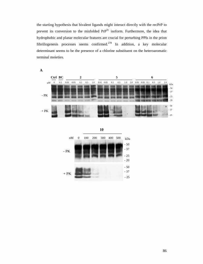
86
the starting hypothesis that bivalent ligands might interact directly with the recPrP to
prevent its conversion to the misfolded PrPSc isoform. Furthermore, the idea that
hydrophobic and planar molecular features are crucial for perturbing PPIs in the prion
fibrillogenesis processes seems confirmed.278 In addition, a key molecular
determinant seems to be the presence of a chlorine substituent on the heteroaromatic
terminal moieties.
A
0 100 nM
10
200 300 400 500
- PK
+ PK
- 50
- 37
- 25
- 20
kDa
- 50
- 37
- 25
+ PK - 50 - 37 - 25
µM 0 0.5 0.01 0.05 0.1 0.5 1.0 0.01 0.05 0.1 0.5 1.0 2.0 0.01 0.05 0.1 0.5 1.0 2.0 Ctrl BC 2 5 6
- PK
- 50 - 37 - 25 - 20
kDa
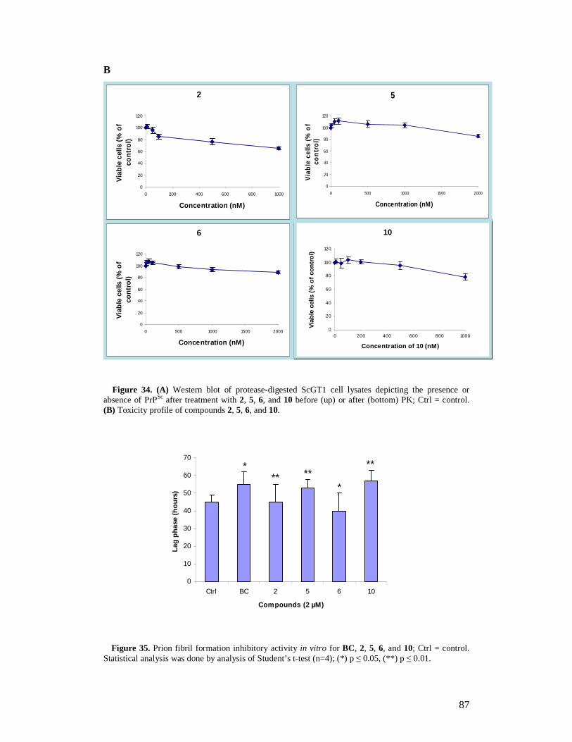
87
B
Figure 34. (A) Western blot of protease-digested ScGT1 cell lysates depicting the presence or absence of PrPSc after treatment with 2, 5, 6, and 10 before (up) or after (bottom) PK; Ctrl = control. (B) Toxicity profile of compounds 2, 5, 6, and 10.
Figure 35. Prion fibril formation inhibitory activity in vitro for BC, 2, 5, 6, and 10; Ctrl = control. Statistical analysis was done by analysis of Student’s t-test (n=4); (*) p ≤ 0.05, (**) p ≤ 0.01.
0
20
40
60
80
100
120
0 200 400 600 800 1000
Concentration of 10 (nM)
Via
ble
cells
(%
of c
ontr
ol)
2
0
20
40
60
80
100
120
0 200 400 600 800 1000
Concentration (nM)
Via
ble
cells
(%
of
cont
rol)
5
0
20
40
60
80
100
120
0 500 1000 1500 2000
Concentration (nM)
Via
ble
cells
(%
of
con
trol
)
6
0
20
40
60
80
100
120
0 500 1000 1500 2000
Concentration (nM)
Via
ble
cells
(%
of
cont
rol)
10
0
10
20
30
40
50
60
70
Ctrl BC 2 5 6 10
Compounds (2 µM)
Lag
phas
e (h
ours
)
* **
*
** **
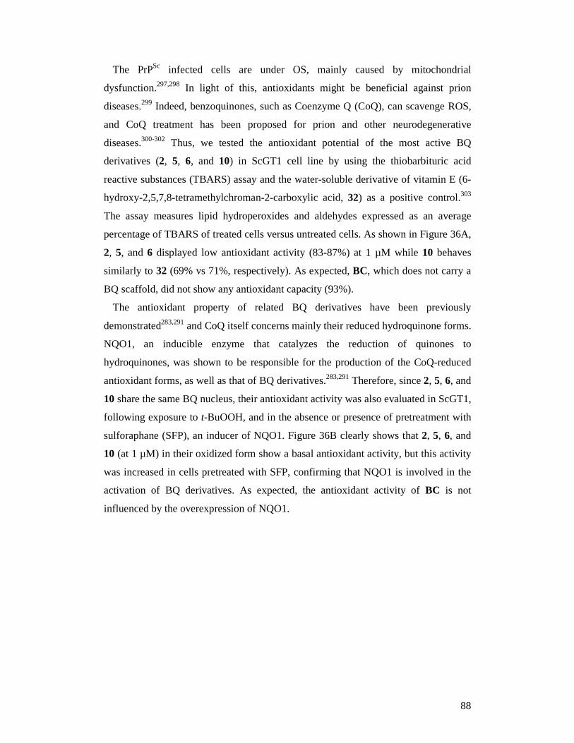
88
The PrPSc infected cells are under OS, mainly caused by mitochondrial
dysfunction.297,298 In light of this, antioxidants might be beneficial against prion
diseases.299 Indeed, benzoquinones, such as Coenzyme Q (CoQ), can scavenge ROS,
and CoQ treatment has been proposed for prion and other neurodegenerative
diseases.300-302 Thus, we tested the antioxidant potential of the most active BQ
derivatives (2, 5, 6, and 10) in ScGT1 cell line by using the thiobarbituric acid
reactive substances (TBARS) assay and the water-soluble derivative of vitamin E (6-
hydroxy-2,5,7,8-tetramethylchroman-2-carboxylic acid, 32) as a positive control.303
The assay measures lipid hydroperoxides and aldehydes expressed as an average
percentage of TBARS of treated cells versus untreated cells. As shown in Figure 36A,
2, 5, and 6 displayed low antioxidant activity (83-87%) at 1 µM while 10 behaves
similarly to 32 (69% vs 71%, respectively). As expected, BC, which does not carry a
BQ scaffold, did not show any antioxidant capacity (93%).
The antioxidant property of related BQ derivatives have been previously
demonstrated283,291 and CoQ itself concerns mainly their reduced hydroquinone forms.
NQO1, an inducible enzyme that catalyzes the reduction of quinones to
hydroquinones, was shown to be responsible for the production of the CoQ-reduced
antioxidant forms, as well as that of BQ derivatives.283,291 Therefore, since 2, 5, 6, and
10 share the same BQ nucleus, their antioxidant activity was also evaluated in ScGT1,
following exposure to t-BuOOH, and in the absence or presence of pretreatment with
sulforaphane (SFP), an inducer of NQO1. Figure 36B clearly shows that 2, 5, 6, and
10 (at 1 µM) in their oxidized form show a basal antioxidant activity, but this activity
was increased in cells pretreated with SFP, confirming that NQO1 is involved in the
activation of BQ derivatives. As expected, the antioxidant activity of BC is not
influenced by the overexpression of NQO1.
89
Figure 36. (A) Effect of 32, BiCappa, 2, 5, 6, and 10 (1 µM) on ScGT1, evaluated by TBARS formation. Values are the means ± SD (n = 3); (B) Antioxidant activity of BiCappa, 2, 5, 6, and 10 in ScGT1 cells against ROS formation induced by t-BuOOH. Experiments were performed with ScGT1 cells treated or not with 2.5 µM SFP; (*) p ≤ 0.05 with respect to t-BuOOH treated samples, (#) p ≤ 0.05 with respect to t-BuOOH + SFP treated samples.
With an aim to further unraveling the molecular mechanism whereby the present
series of compounds can tackle prion-mediated infections, we carried out docking
simulations by means of the AutoDock software at the putative binding region defined
by the H2 helix and the loop connecting β-sheet S2 and helix H1 (Fig. 37).239,304,305
Such computations did not provide a unique binding mode for compound 10, in line
with our previous findings clearly demonstrating that only a combination of several
different techniques would allow to propose a reasonable binding mode for the
molecule GN8 at the prion protein.272 Furthermore, different molecular mechanisms,
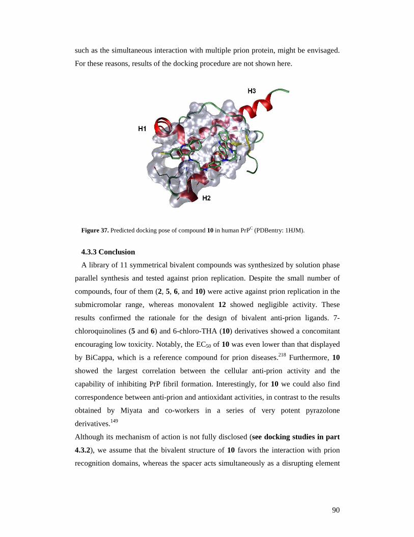
90
such as the simultaneous interaction with multiple prion protein, might be envisaged.
For these reasons, results of the docking procedure are not shown here.
Figure 37. Predicted docking pose of compound 10 in human PrPC (PDBentry: 1HJM).
4.3.3 Conclusion
A library of 11 symmetrical bivalent compounds was synthesized by solution phase
parallel synthesis and tested against prion replication. Despite the small number of
compounds, four of them (2, 5, 6, and 10) were active against prion replication in the
submicromolar range, whereas monovalent 12 showed negligible activity. These
results confirmed the rationale for the design of bivalent anti-prion ligands. 7-
chloroquinolines (5 and 6) and 6-chloro-THA (10) derivatives showed a concomitant
encouraging low toxicity. Notably, the EC50 of 10 was even lower than that displayed
by BiCappa, which is a reference compound for prion diseases.218 Furthermore, 10
showed the largest correlation between the cellular anti-prion activity and the
capability of inhibiting PrP fibril formation. Interestingly, for 10 we could also find
correspondence between anti-prion and antioxidant activities, in contrast to the results
obtained by Miyata and co-workers in a series of very potent pyrazolone
derivatives.149
Although its mechanism of action is not fully disclosed (see docking studies in part
4.3.2), we assume that the bivalent structure of 10 favors the interaction with prion
recognition domains, whereas the spacer acts simultaneously as a disrupting element
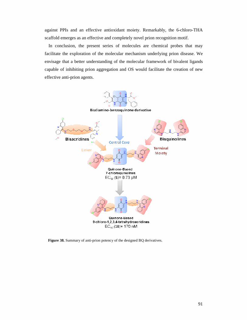
91
against PPIs and an effective antioxidant moiety. Remarkably, the 6-chloro-THA
scaffold emerges as an effective and completely novel prion recognition motif.
In conclusion, the present series of molecules are chemical probes that may
facilitate the exploration of the molecular mechanism underlying prion disease. We
envisage that a better understanding of the molecular framework of bivalent ligands
capable of inhibiting prion aggregation and OS would facilitate the creation of new
effective anti-prion agents.
Figure 38. Summary of anti-prion potency of the designed BQ derivatives.
92
5. Synthesis and biological evaluation of lipoic acid hybrids
as novel compounds against prion diseases
5.1 Introduction
Prion diseases pathogenesis involves a complex array of processes operating
simultaneously and synergistically. These include: (i) protein aggregation,46,51 (ii)
oxidative stress accompanied by lipid and protein oxidation,298,300,303,306,307 (iii)
reduced levels of potent free-radical scavenger such as polyunsaturated fatty acids, α-
tocopherol, and glutathione,299,306 (iv) unbalance of metal ions,308 and (v) brain
inflammation with activation of astrocytes and microglia.43 Thus, the failures of drug
candidates developed accordingly to the traditional drug discovery paradigm “one
molecule - one target” and the current challenge of discovering an efficacious therapy
are likely related to such a multifactorial nature of these diseases. Against this
backdrop, a polypharmacological approach, which, rather than consisting of a single
compound that interacts with a single target, is a concerted pharmacological
intervention of compound(s) that interact with multiple targets, offers promise for the
effective treatment of prion diseases. Although this approach is still in its infancy, two
different strategies have been already addressed to achieve polypharmacology: drug
combination (DC) and multi-target directed ligand (MTDL) approaches. In DC
approach multiple drugs (drug cocktail) are combined in the therapeutic regimen. A
major drawback of DC therapy is the drug-drug interactions. Conversely, the MTDL
approach, where two pharmacophores with distinct mechanism of action are
chemically merged in a single structure with a single ADMET (being an acronym for
absorption, distribution, metabolism, elimination, and toxicology) profile, offers
advantages over DC therapy. Notably, this approach, already used in other complex
diseases,309-313 might be envisaged as being optimal also in prion diseases.
For prion diseases, DC strategy has been applied in numerous in vitro and in vivo
approaches with the aim of exploiting synergistic effects. The several examples
reported in Table 11 suggest that inhibition of prion replication can be effectively
potentiated by DC treatment. As for MTDLs, the literature contains cases where the
deliberate aim of creating an MTDL has not always been explicitly stated. Instead, the
molecular hybridization strategy has been followed, leading to chimeric molecules
capable, in principle, of modulating multiple targets.140,218,238,289,314 The first anti-prion
chimeric ligand, Quinpramine, was designed on the basis of in vitro synergistic anti-
93
prion effects of drugs Quinacrine and Imipramine. Quinpramine, obtained by linking
Quinacrine and Imipramine moieties through a piperazine ring, showed an improved
anti-prion activity as much as 15-fold over quinacrine and 250-fold over
imipramine.152,265 Recently, we have reported a new class of anti-prion compounds
obtained by linking the antioxidant nucleus of 2,5-diamino-1,4-benzoquinone to
several heterocyclic scaffolds potentially able to perturb protein-protein interactions
in prion (6-chloro-2-methoxyacridin-9-amine, or 4-amino-7-chloroquinoline or 6-
chloro-1,2,3,4-tetrahydroacridin-9-amine). These compounds displayed a multitarget
profile, effectively contrasting both prion fibril formation and oxidative stress in a cell
culture model of prion replication.280
Table 11. Drug combination strategy in anti-prion compounds Two-drug
combination
cholesterol ester modulators (everolimus, pioglitazone, progesterone, and verapamil) and an antipsychotic drug (chlorpromazine) or an antimalarial drug (quinacrine)
Ref.315
anti-inflammatory drug (pentosan polysulfate) and an anticancer compound (Fe(III)meso-tetra(4-sulfonatophenyl)porphine)
Ref.316
polyphenolic antioxidant (epigallocatechin-3-gallate) and 4,5-bis-(4-methoxyanilino)phthalimide
Ref.317
Three-drug
combination
antimalarial drug (quinacrine), a tricyclic antidepressants (desipramine) and an inhibitor of cholesterol biosynthesis (simvastatin)
Ref.152
These encouraging results together with a solid background of MTDLs in the field of
Alzheimer’s disease led us to pursue in that direction. We hypothesized that the
presence of a so-called prion-recognition motif (PRM), key element for anti-prion
activity, along with a moiety endowed with an alternative mechanism of action
against prion diseases might lead to discover more effective compounds.280
5.2 Materials and methods
5.2.1 Design rationale
As a first step, a 4-amino-7-chloroquinoline nucleus was linked by an alkylamino
chain to diverse antioxidant moieties. The choice of a N1-(7-chloroquinolin-4-
yl)propane-1,3-diamine (13) as PRM was supported by our previous results280 and the
studies of Cordeiro and co-workers that demonstrated the direct binding of 13 to
94
PrP.314 As a further strength, a 4-amino-7-chloroquinoline motif recurs in a broad
variety of biologically active compounds that hit different targets and pathways. Thus,
it can be classified as a privileged sub-structure, with optimal pharmacokinetic
properties and a high degree of drug-likeness, extremely favorable features when
starting a drug discovery program.140,238,289
Building on 13, we selected different antioxidant fragments, such as caffeic acid (9,
Scheme 4), trans-ferulic acid (10), and lipoic acid (12), for several reasons: (i) all of
them are natural antioxidant; (ii) well tolerated in vivo; (iii) effective against fibril
formation; (iv) chemically linkable to the amine group of 13 by amide bond
formation. Several lines of evidence have shown that 9 and 10 act as a potent
scavenger of reactive species, including ROS and reactive nitrogen species, and
thereby reduce the chance of free radical attack on proteins and DNA and hence
preventing their oxidative modification.318-320 Furthermore, both appeared to be
neuroprotective against β-amyloid-induced cytotoxicity.321-323 9 also inhibits 5-
lipoxygenase (5-LOX) enzyme, which is involved in the early events of prion
disease.324 12 has been proposed as a lead structure for designing multi-target directed
drugs for neurodegeneration.325 More importantly, it was administered together with
other antioxidants to a patient affected by prion disease, showing moderate
therapeutic effects,326 and shown to inhibit the formation of β-amyloid fibrils.327,328 In
particular, 12 and its derivatives have been shown to have a variety of properties
which interfere with several pathological events of Alzhemer’s disease (AD).325,327,329
As a further application of the MTDL approach, we aimed at combining 13 with the
molecular features of guanidinium chloride. Guanidinium chloride causes curing of
yeast prions by perturbing Hsp104, a molecular chaperone essential for both
dissolving protein aggregates and inhibiting prion propagation.330,331 This choice was
driven by previous results that guanidinium chloride and Guanabenz, a guanidine
derivative, are anti-prion compounds.290 However, the guanidine group is nearly
quantitatively protonated under physiological conditions, which might result in a lack
of CNS penetration. To overcome this limitation, in 3 (Scheme 4) as guanidinium
motif we selected L-arginine (11). The availability of a transport system for arginine
in humans means that a portal of entry to the brain for circulating drugs is potentially
available.
In a second round of optimization, based on the promising anti-prion profile of 4
(Scheme 4), we decided to expand the alpha-lipoic acid hybrids series. Four additional
95
derivatives (5-8) were hence designed and tested. Derivative 5 is a structural analogue
of 4, whereas in 6-8 other PRMs were selected: 6-chloro-2-methoxy-acridin-9-amine
and 8-chloro-1,2,3,4-tetrahydroacridin-9-amine.
5.2.2 Library synthesis
The synthesis of 1-2 was obtained by using dicyclohexyl carbodiimide (DCC) as a
coupling reagent. We have carried out the reaction of the caffeic acid (9) or trans-
ferulic acid (10) with the polyamine 13 (Scheme 4) under reflux THF to give the
corresponding amide products in good yield (42-53%). In these conditions we were
able to obtain selective amide bond formation, obviating the need for
protection/deprotection of the hydroxyl functionalities.
The synthesis of quinoline-arginine hybrid requests the protection of the guanidino
and amine groups of the arginine. In our case, all groups were protected by tert-
butoxycarbonyl groups. Using this protected arginine derivative (19, Scheme 4),
compound 20 was prepared via standard peptide coupling procedure. Removal of the
protecting groups of 20 with a solution of HCl/dioxane afforded 3 in high yield.
A reaction of diamines with lipoic acid (12) provides easy access to a variety of
derivatives.291,332 Thus, we decided to synthesize the designed compounds 4-7
following the procedure described by Rosini and co-workers.332 12 was loaded with
EDCI, HOBt and NEt3, then N-substituted polyamine (13-16, Scheme 4) was added at
0°C for 2 hours. After stirring over night at room temperature, the desired product was
obtained in good yields (67-96%).
The preparation of intermediate 13-16 was easily achieved by parallel synthesis as
described by Bongarzone and co-workers.280
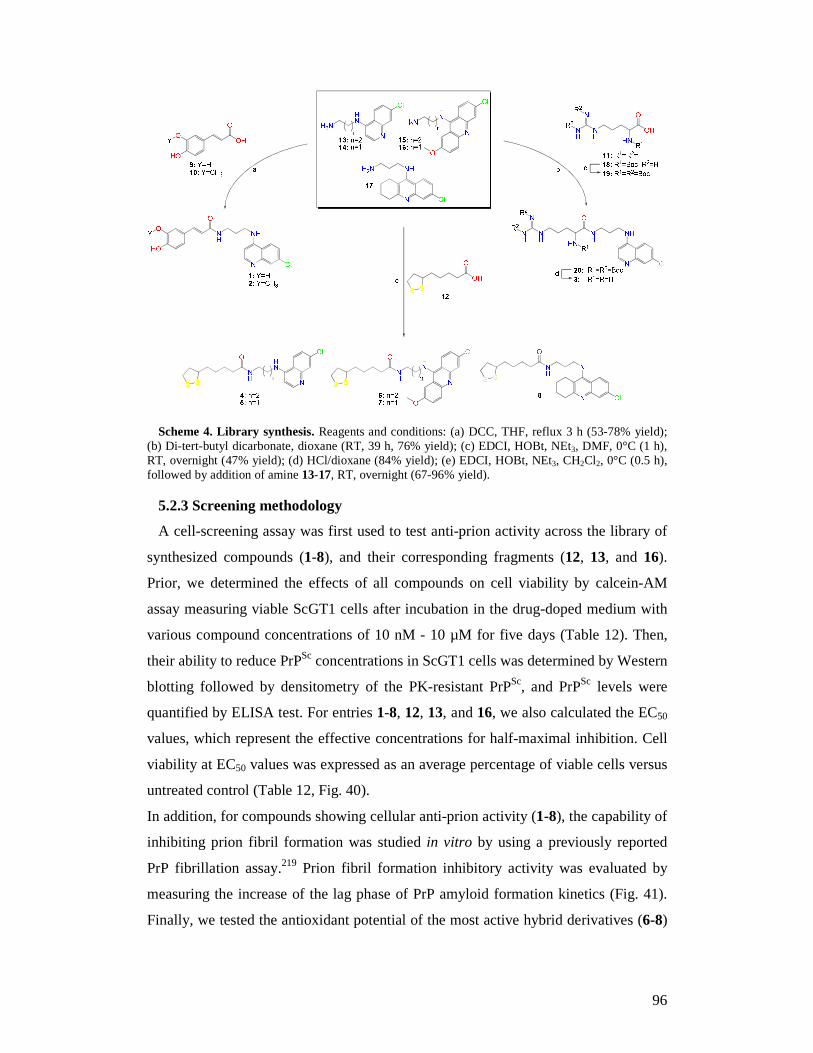
96
Scheme 4. Library synthesis. Reagents and conditions: (a) DCC, THF, reflux 3 h (53-78% yield); (b) Di-tert-butyl dicarbonate, dioxane (RT, 39 h, 76% yield); (c) EDCI, HOBt, NEt3, DMF, 0°C (1 h), RT, overnight (47% yield); (d) HCl/dioxane (84% yield); (e) EDCI, HOBt, NEt3, CH2Cl2, 0°C (0.5 h), followed by addition of amine 13-17, RT, overnight (67-96% yield).
5.2.3 Screening methodology
A cell-screening assay was first used to test anti-prion activity across the library of
synthesized compounds (1-8), and their corresponding fragments (12, 13, and 16).
Prior, we determined the effects of all compounds on cell viability by calcein-AM
assay measuring viable ScGT1 cells after incubation in the drug-doped medium with
various compound concentrations of 10 nM - 10 µM for five days (Table 12). Then,
their ability to reduce PrPSc concentrations in ScGT1 cells was determined by Western
blotting followed by densitometry of the PK-resistant PrPSc, and PrPSc levels were
quantified by ELISA test. For entries 1-8, 12, 13, and 16, we also calculated the EC50
values, which represent the effective concentrations for half-maximal inhibition. Cell
viability at EC50 values was expressed as an average percentage of viable cells versus
untreated control (Table 12, Fig. 40).
In addition, for compounds showing cellular anti-prion activity (1-8), the capability of
inhibiting prion fibril formation was studied in vitro by using a previously reported
PrP fibrillation assay.219 Prion fibril formation inhibitory activity was evaluated by
measuring the increase of the lag phase of PrP amyloid formation kinetics (Fig. 41).
Finally, we tested the antioxidant potential of the most active hybrid derivatives (6-8)
97
in ScGT1 cell line by using the thiobarbituric acid reactive substances (TBARS) assay
and the antioxidant Trolox as a positive control (Fig. 42).
All assays for cell culture, drug treatment, cell viability, PrPSc detection and
quantification were performed in accordance to the protocols described in parts 2.2.4,
2.2.5 and 2.2.6.
Detection of in vitro effect of the synthesized compounds on prion fibril formation
was performed in accordance to the protocol described in part 4.3.1.4.
Detection of antioxidant activity of the compounds by lipid peroxidation assay was
performed in accordance to the protocol described in part 4.3.1.5.
5.3 Results and discussion
Preliminarily, the possible toxicity of the hybrid compounds 1-4 was assessed in
ScGT1 cells. At 1 µM, the toxicity profile among quinoline-based hybrid compounds
(1-4) was very low and therefore they were studied for their anti-prion activity at
same concentration (Table 12). All hybrid derivatives 1-4 turned out to have mild
anti-prion activity, with similar micromolar EC50 values (2.7-9.5 µM), despite the
potential different mechanism of action. However, 2 and 4, carrying the ferulic and
lipoic acids moieties respectively, were slightly more active than 1 and 3 (Table 12).
To better investigate the multi-target activity of the designed compounds, we tested
the anti-prion activity of their starting fragments (9-13). As expected, the PRM 13
showed a micromolar EC50 value (7.8 ± 0.3 µM) with no toxicity in ScGT1 cells up to
10 µM. Regarding the three antioxidant fragments (9, 10, and 12) and guanidinium
derivative (11), only lipoic acid (12) possessed an interesting activity against PrPSc
formation (EC50 = 5.3 ± 0.4 µM), together with a suitable toxicity profile (no toxic
effects up to 100 µM). To our knowledge, this is the first time that the proposed anti-
prion potential of 12 has been demonstrated in a cellular model. Notably, 9-12 did not
show toxicity at 10 µM (see Fig. 39). Altogether, these preliminary results suggested
us to link 12 with other PRMs (6-chloro-2-methoxy-acridin-9-amine and 8-chloro-
1,2,3,4-tetrahydroacridin-9-amine), already exploited by us and others in the search
for anti-prion ligands and drug-likeness similar to that of 13.218,265,280,289 Thus, we
decided to synthesize and test a further subset of alpha-lipoic acid hybrids (5-8).
From the biological studies (Table 12), this rational design was confirmed by the
outstanding activity shown by 6 and 7. In fact, both compounds did not show toxicity
against ScGT1 cells at 1 µM, while they displayed high activity against prion
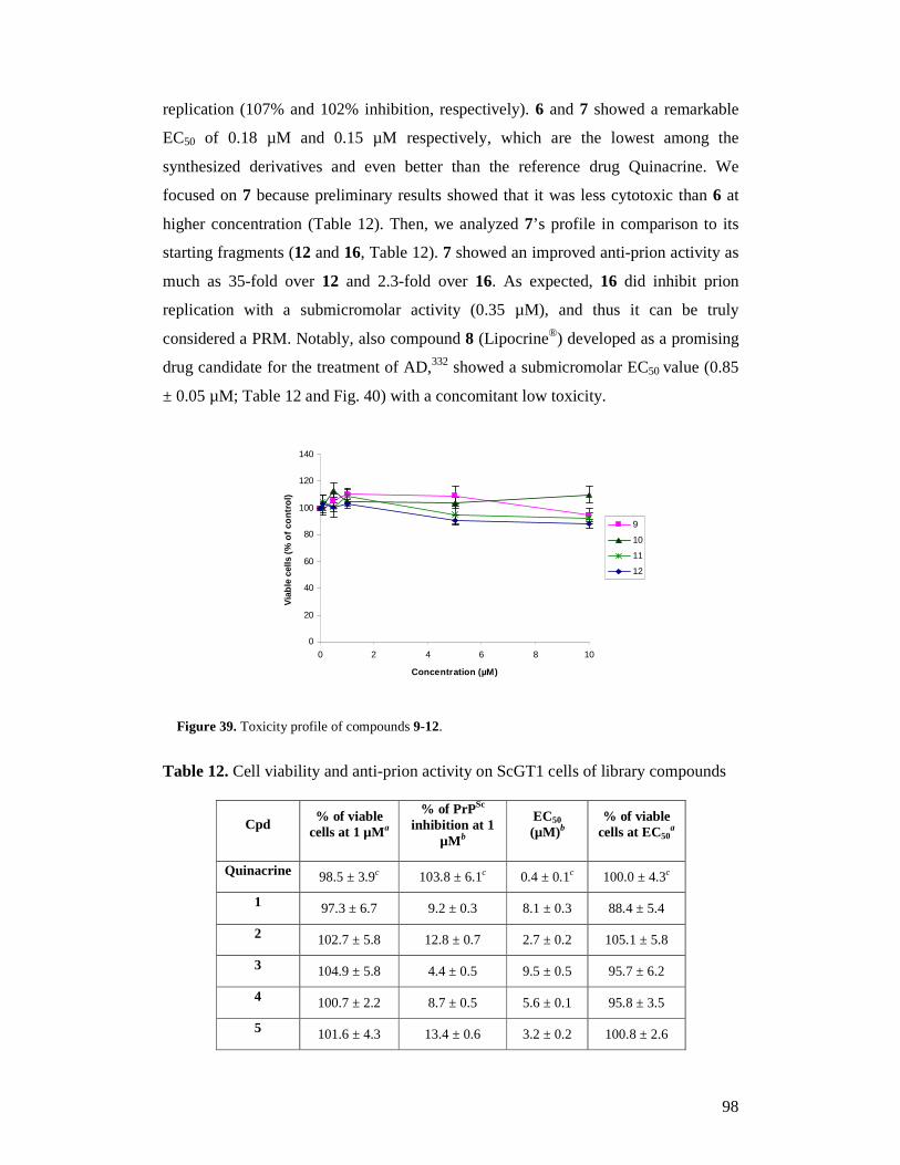
98
replication (107% and 102% inhibition, respectively). 6 and 7 showed a remarkable
EC50 of 0.18 µM and 0.15 µM respectively, which are the lowest among the
synthesized derivatives and even better than the reference drug Quinacrine. We
focused on 7 because preliminary results showed that it was less cytotoxic than 6 at
higher concentration (Table 12). Then, we analyzed 7’s profile in comparison to its
starting fragments (12 and 16, Table 12). 7 showed an improved anti-prion activity as
much as 35-fold over 12 and 2.3-fold over 16. As expected, 16 did inhibit prion
replication with a submicromolar activity (0.35 µM), and thus it can be truly
considered a PRM. Notably, also compound 8 (Lipocrine®) developed as a promising
drug candidate for the treatment of AD,332 showed a submicromolar EC50 value (0.85
± 0.05 µM; Table 12 and Fig. 40) with a concomitant low toxicity.
0
20
40
60
80
100
120
140
0 2 4 6 8 10
Concentration (µM)
Via
ble
cells
(%
of c
ontr
ol)
9
10
11
12
Figure 39. Toxicity profile of compounds 9-12.
Table 12. Cell viability and anti-prion activity on ScGT1 cells of library compounds
Cpd % of viable
cells at 1 µMa
% of PrPSc
inhibition at 1 µM b
EC50 (µM)b
% of viable cells at EC50
a
Quinacrine 98.5 ± 3.9c 103.8 ± 6.1c 0.4 ± 0.1c 100.0 ± 4.3c
1 97.3 ± 6.7 9.2 ± 0.3 8.1 ± 0.3 88.4 ± 5.4
2 102.7 ± 5.8 12.8 ± 0.7 2.7 ± 0.2 105.1 ± 5.8
3 104.9 ± 5.8 4.4 ± 0.5 9.5 ± 0.5 95.7 ± 6.2
4 100.7 ± 2.2 8.7 ± 0.5 5.6 ± 0.1 95.8 ± 3.5
5 101.6 ± 4.3 13.4 ± 0.6 3.2 ± 0.2 100.8 ± 2.6
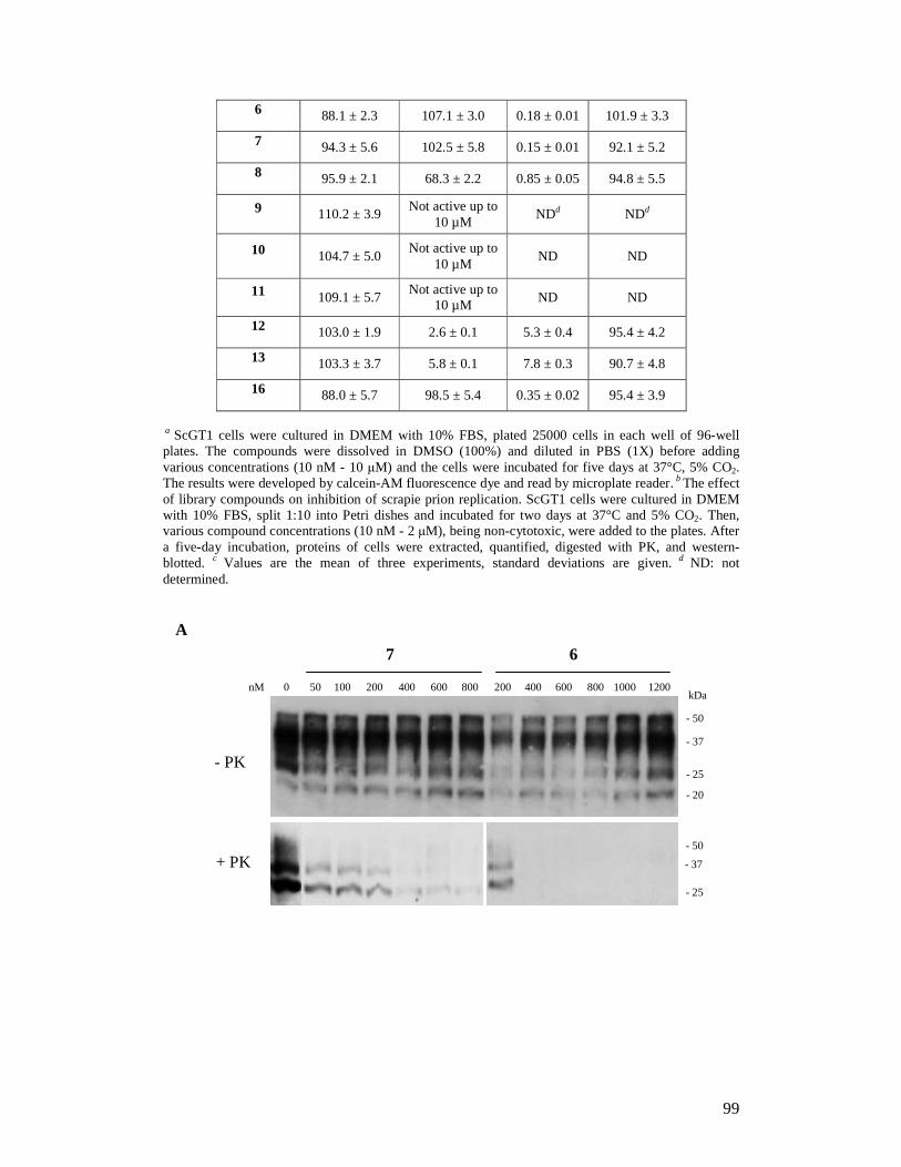
99
6 88.1 ± 2.3 107.1 ± 3.0 0.18 ± 0.01 101.9 ± 3.3
7 94.3 ± 5.6 102.5 ± 5.8 0.15 ± 0.01 92.1 ± 5.2
8 95.9 ± 2.1 68.3 ± 2.2 0.85 ± 0.05 94.8 ± 5.5
9 110.2 ± 3.9 Not active up to
10 µM NDd NDd
10 104.7 ± 5.0 Not active up to
10 µM ND ND
11 109.1 ± 5.7 Not active up to
10 µM ND ND
12 103.0 ± 1.9 2.6 ± 0.1 5.3 ± 0.4 95.4 ± 4.2
13 103.3 ± 3.7 5.8 ± 0.1 7.8 ± 0.3 90.7 ± 4.8
16 88.0 ± 5.7 98.5 ± 5.4 0.35 ± 0.02 95.4 ± 3.9
a ScGT1 cells were cultured in DMEM with 10% FBS, plated 25000 cells in each well of 96-well plates. The compounds were dissolved in DMSO (100%) and diluted in PBS (1X) before adding various concentrations (10 nM - 10 µM) and the cells were incubated for five days at 37°C, 5% CO2. The results were developed by calcein-AM fluorescence dye and read by microplate reader. b The effect of library compounds on inhibition of scrapie prion replication. ScGT1 cells were cultured in DMEM with 10% FBS, split 1:10 into Petri dishes and incubated for two days at 37°C and 5% CO2. Then, various compound concentrations (10 nM - 2 µM), being non-cytotoxic, were added to the plates. After a five-day incubation, proteins of cells were extracted, quantified, digested with PK, and western-blotted. c Values are the mean of three experiments, standard deviations are given. d ND: not determined.
A
0 50 100 200 400 600 800 200 400 600 800 1000 1200
7 6
nM
+ PK
- PK
- 50 - 37 - 25
- 50 - 37 - 25 - 20
kDa
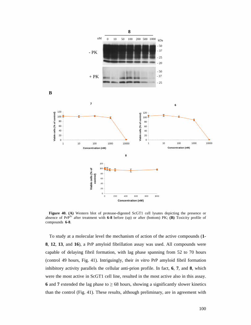
100
B
Figure 40. (A) Western blot of protease-digested ScGT1 cell lysates depicting the presence or absence of PrPSc after treatment with 6-8 before (up) or after (bottom) PK; (B) Toxicity profile of compounds 6-8.
To study at a molecular level the mechanism of action of the active compounds (1-
8, 12, 13, and 16), a PrP amyloid fibrillation assay was used. All compounds were
capable of delaying fibril formation, with lag phase spanning from 52 to 70 hours
(control 49 hours, Fig. 41). Intriguingly, their in vitro PrP amyloid fibril formation
inhibitory activity parallels the cellular anti-prion profile. In fact, 6, 7, and 8, which
were the most active in ScGT1 cell line, resulted in the most active also in this assay.
6 and 7 extended the lag phase to ≥ 68 hours, showing a significantly slower kinetics
than the control (Fig. 41). These results, although preliminary, are in agreement with
- 50 - 37 - 25
10 50 100 200 500 1000 8
- 50 - 37 - 25 - 20
kDa nM 0
+ PK
- PK
7
0
20
40
60
80
100
120
1 10 100 1000 10000
Concentration (nM)
Via
ble
cells
(%
of c
ontr
ol)
6
0
20
40
60
80
100
120
1 10 100 1000 10000
Concentration (nM)
Via
ble
cells
(%
of c
ontr
ol)
8
0
20
40
60
80
100
120
0 200 400 600 800 1000
Concentration (nM)
Via
ble
cells
(%
of
cont
rol)

101
the starting hypothesis that PRM might interact directly with PrP to prevent its
conversion to the misfolded PrPSc isoform. Furthermore, the low anti-amyloid activity
of 12 versus those of hybrids 4-8 suggests that marked aggregation inhibition may be
achieved only when 12 and a suitable PRM are combined into the same structure, as
in 6 and 7.
Figure 41. Prion fibril formation inhibitory activity in vitro for 1-8, 12, 13, and 16; Ctrl = control. Statistical analysis was done by analysis of Student’s t-test (n=4); (*) p ≤ 0.05, (**) p ≤ 0.01.
The PrPSc infected cells are under OS, mainly caused by mitochondrial
dysfunction.297,298 In light of this, antioxidant fragments might be beneficial against
prion diseases.299 Indeed, lipoic acid hybrids scavenge ROS, and they have been
proposed for the treatment of other multifactorial neurodegenerative diseases.300,302,312
Thus, we tested the antioxidant potential of the most active derivatives (6-8) in ScGT1
cell line by using TBARS assay and the antioxidant Trolox as a positive control.303
The assay measures lipid hydroperoxides and aldehydes expressed as an average
percentage of TBARS of treated cells versus untreated cells. Figure 42 clearly shows
that 6 and 7 displayed antioxidant activity (78-82% of control) at 1 µM, while 8 was
even better than Trolox (58% vs 71%, respectively). Although a perfect match with
cellular data is not evident, these results confirm the design rationale, indicating that
the presence of a PRM (a lipophilic bi- or tri-(hetero)-cyclic scaffold) and an
antioxidant fragment (lipoic acid) is suitable to the discovery of anti-prion hybrids.
0
10
20
30
40
50
60
70
80
Ctrl 1 2 3 4 5 6 7 8 12 13 16
Compounds (10 µM)
Lag
phas
e (h
ours
) * *
* ** *
** ** *
* *
**
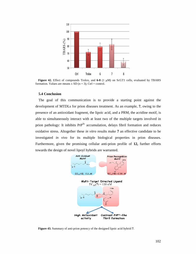
102
Figure 42. Effect of compounds Trolox, and 6-8 (1 µM) on ScGT1 cells, evaluated by TBARS formation. Values are means ± SD (n = 3); Ctrl = control.
5.4 Conclusion
The goal of this communication is to provide a starting point against the
development of MTDLs for prion diseases treatment. As an example, 7, owing to the
presence of an antioxidant fragment, the lipoic acid, and a PRM, the acridine motif, is
able to simultaneously interact with at least two of the multiple targets involved in
prion pathology: It inhibits PrPSc accumulation, delays fibril formation and reduces
oxidative stress. Altogether these in vitro results make 7 an effective candidate to be
investigated in vivo for its multiple biological properties in prion diseases.
Furthermore, given the promising cellular anti-prion profile of 12, further efforts
towards the design of novel lipoyl hybrids are warranted.
Figure 43. Summary of anti-prion potency of the designed lipoic acid hybrid 7.
103
6. Concluding remarks
In the thesis, we discuss about biological evaluation of four new libraries of
anti-prion compounds designed and synthesized on nanotechnology,
computational study and chemistry.
Firstly, in the first library, we discovered a novel class of anti-prion compounds
as polyelectrolyte multilayer-coated gold nanoparticles for their ability to interact
and reduce the accumulation of the disease-causing prion protein (PrPSc) in
scrapie prion-infected cell lines and mice. Gold nanoparticles coated with
oppositely charged polyelectrolytes, such as polyallylamine hydrochloride and
polystyrenesulfonate, were tested for potential inhibition of prion protein
aggregation. Different coatings, finishing with a positive or negative layer, were
tested, and different numbers of layers were investigated for their ability to
interact and reduce the accumulation of PrPSc in scrapie prion-infected ScGT1
and ScN2a cells. The particles efficiently hampered the accumulation of PrPSc in
ScN2a cells and showed curing effects on ScGT1 cells with a nanoparticle
concentration in the picomolar range. Moreover, incubation periods of prion-
infected mice treated with nanomolar concentrations of the nanoparticles were
significantly longer compared to untreated controls. In addition, the coated gold
nanoparticles may interact with Aβ and α-synuclein to inhibit β-amyloid and α-
synuclein fibril formation implicated in Alzheimer’s and Parkinson’s diseases,
respectively.
Secondly, we have also developed possible peptidomimetic fragments in
inhibiting protein-protein interactions including diketopiperazines and 2,5-
diamino-1,4-benzoquinones for search of bivalent anti-prion compounds. In the
second library, we report the identification of a novel bifunctional
diketopiperazine (DKP) derivative 1d, which exhibited activity in the low
micromolar range against prion replication in ScGT1 cells, while showing low
toxicity. By properly addressed molecular modeling studies we hypothesized that
a planar conformation is the major determinant for activity in this class of
compounds. Moreover, studies aimed to assess the mechanism of action at a
molecular level showed that 1d might interact directly with recPrP to prevent its
conversion to the pathogenic PrPSc-like form. This investigation suggests that
104
DKP based anti-prion compounds can serve as a promising lead scaffold in
developing new drugs to combat prion diseases. In the third library, the
compounds combining two different benzoquinone cores with seven (L) amino
acid methyl esters (alanine, Nω-nitro-arginine, Nε-BOC-lysine, isoleucine,
methionine, phenylalanine and tryptophan) were prepared and tested for prion
replication inhibition in ScGT1 cells. The most potent hit, 6a, displayed an EC50
value of 0.87 µM, which is very close to that of quinacrine (0.4 µM). From this
finding, a small library of twelve entries, featuring a 2,5-diamino-1,4-
benzoquinones nucleus as spacer connecting two aromatic prion recognition
motifs (PRMs): 6-chloro-2-methoxyacridin-9-amine, or 4-amino-7-
chloroquinoline or 6-chloro-1,2,3,4-tetrahydroacridin-9-amine, was designed and
evaluated against prion infection. Notably, 6-chloro-1,2,3,4-tetrahydroacridine 10
showed an EC50 of 0.17 µM, which was lower than that displayed by reference
compound BiCappa. More importantly, 10 possessed the capability of inhibiting
prion fibril formation and oxidative stress, together with a low cytotoxicity. This
study further corroborates the bivalent strategy as a viable approach to the
rational design of anti-prion chemical probes.
Finally, in the fourth library, we have designed a small library of multi-target
directed ligands (MTDLs) as the alpha-lipoic acid hybrids. Hybrid compounds 6-8 are
based on PRMs: 6-chloro-2-methoxyacridin-9-amine and 8-chloro-1,2,3,4-
tetrahydroacridin-9-amine, possessed anti-prion activity and the capability of
inhibiting prion fibril formation and oxidative stress. 7 showed a remarkable EC50 of
0.15 µM, which is the lowest among lipoic acid derivatives. 7, owing to the presence
of an antioxidant fragment, the lipoic acid, and a PRM, the acridine motif, is able to
simultaneously interact with at least two of the multiple targets involved in prion
disease pathology. The development of MTDLs might lead to new pharmaceutical
treatments for the multifactorial pathology of prion diseases.
105
List of publications
1. Bongarzone S,* Tran HN ,* Cavalli A, Roberti M, Carloni P, Legname G,
Bolognesi ML. Parallel synthesis, evaluation, and preliminary structure-activity
relationship of 2,5-diamino-1,4-benzoquinones as a novel class of bivalent anti-prion
compound. J Med Chem. 2010; 53(22):8197-201. ( * co-first authors)
2. Ai Tran HN , Sousa F, Moda F, Mandal S, Chanana M, Vimercati C, Morbin M,
Krol S, Tagliavini F, Legname G. A novel class of potential prion drugs: preliminary
in vitro and in vivo data for multilayer coated gold nanoparticles. Nanoscale. 2010;
2(12):2724-32.
3. Bolognesi ML, Ai Tran HN , Staderini M, Monaco A, López-Cobeñas A,
Bongarzone S, Biarnés X, López-Alvarado P, Cabezas N, Caramelli M, Carloni P,
Menéndez JC, Legname G. Discovery of a class of diketopiperazines as antiprion
compounds. ChemMedChem. 2010; 5(8):1324-34.
4. Tran HN , Bongarzone S, Carloni P, Legname G, Bolognesi ML. Synthesis and
evaluation of a library of 2,5-bisdiamino-benzoquinone derivatives as probes to
modulate protein-protein interactions in prions. Bioorg Med Chem Lett. 2010;
20(6):1866-8.
5. Bongarzone S,* Tran HN ,* Cavalli A, Roberti M, Rosini M, Carloni P, Legname
G, Bolognesi ML. Hybrid lipoic acid derivatives to attack prion disease on multiple
fronts. submitted. ( * co-first authors)

106
Acknowledgements
I would like to thank my supervisor Prof. Giuseppe Legname for giving me the
opportunity to work in his group and for instructing me during my PhD study. He
always helped and gave me kindly and useful advice in the research and social life.
I am grateful to my external collaborators Prof. Fabrizio Tagliavini, Dr. Silke Krol
from Carlo Besta Neurological Institute (Milan, Italy) for scientific discussions and
my PhD project. A special thank goes to Salvatore Bongarzone, Matteo Staderini, Dr.
Fabio Moda and Dr. Fernanda Sousa who carried out experiments that supported our
work, for all their discussions and friendship. Thanks go to all collaborators in the
project of drug discovery, especially to Prof. Maria Laura Bolognesi, Prof. Andrea
Cavalli and Prof. Marinella Roberti from University of Bologna (Italy), to Prof. Paolo
Carloni from German Research School for Simulation Sciences (Aachen, Germany)
and to Prof. Carlos Mennendez from University of Madrid (Spain) for giving me all
fruitful discussions.
I would also like to thank all lab members of the Prion Biology Laboratory. We were
always friendly in helping together as well as sharing happiness and difficulties in my
lab life.
Many thanks to my husband Minh Tuan, my parents and to my brothers and sisters in
Vietnam for all their love, belief and support. That was an encouragement and
promotion to help me to finish my PhD.
107
References
1 Soto C. Protein misfolding and disease; protein refolding and therapy. FEBS Lett. 2001; 498: 204-207.
2 Kelly JW. Alternative conformations of amyloidogenic proteins govern their behavior. Curr Opin Struct Biol. 1996; 6: 11-17.
3 Carrell RW, Gooptu B. Conformational changes and disease-serpins, prions and Alzheimer's. Curr Opin Struct Biol. 1998; 8: 799-809.
4 Johnson WG. Late-onset neurodegenerative diseases-the role of protein insolubility. J Anat. 2000; 196: 609-616.
5 Buxbaum JN, Tagoe CE. The genetics of the amyloidoses. Annu Rev Med. 2000; 51: 543-569.
6 Prusiner SB, Scott MR. Genetics of prions. Annu Rev Genet. 1997; 31: 139-175.
7 Harper JD, Lansbury PT Jr. Models of amyloid seeding in Alzheimer's disease and scrapie: mechanistic truths and physiological consequences of the time-dependent solubility of amyloid proteins. Annu Rev Biochem. 1997; 66: 385-407.
8 Cohen FE, Prusiner SB. Pathologic conformations of prion proteins. Annu Rev Biochem. 1998; 67: 793-819.
9 Prusiner SB. Prions. Proc Natl Acad Sci U S A. 1998; 95: 13363-13383.
10 Serpell LC et al. Examination of the structure of the transthyretin amyloid fibril by image reconstruction from electron micrographs. J Mol Biol. 1995; 254: 113-118.
11 Soto C. Alzheimer's and prion disease as disorders of protein conformation: implications for the design of novel therapeutic approaches. J Mol Med. 1999; 77: 412-418.
12 Soto C et al. Beta-sheet breaker peptides inhibit fibrillogenesis in a rat brain model of amyloidosis: implications for Alzheimer's therapy. Nat Med. 1998; 4: 822-826.
13 Soto C et al. Reversion of prion protein conformational changes by synthetic beta-sheet breaker peptides. Lancet. 2000; 355: 192-197.
14 Stefani M. Protein misfolding and aggregation: new examples in medicine and biology of the dark side of the protein world. Biochim Biophys Acta. 2004; 1739: 5-25.

108
15 Chiti F et al. Rationalization of the effects of mutations on peptide and protein aggregation rates. Nature. 2003; 424: 805-808.
16 Sitia R, Braakman I. Quality control in the endoplasmic reticulum protein factory. Nature. 2003; 426: 891-894.
17 Goldberg AL. Protein degradation and protection against misfolded or damaged proteins. Nature. 2003; 426: 895-899.
18 Sherman MY, Goldberg AL. Cellular defenses against unfolded proteins: a cell biologist thinks about neurodegenerative diseases. Neuron. 2001; 29: 15-32.
19 Serpell LC et al. The protofilament substructure of amyloid fibrils. J Mol Biol. 2000; 300: 1033-1039.
20 Sunde M, Blake C. The structure of amyloid fibrils by electron microscopy and X-ray diffraction. Adv Protein Chem. 1997; 50: 123-159.
21 Lashuel HA et al. Neurodegenerative disease: amyloid pores from pathogenic mutations. Nature. 2002; 418: 291.
22 Lashuel HA et al. Mixtures of wild-type and a pathogenic (E22G) form of Abeta40 in vitro accumulate protofibrils, including amyloid pores. J Mol Biol. 2003; 332: 795-808.
23 Prusiner SB, DeArmond SJ. Prion diseases and neurodegeneration. Annu Rev Neurosci. 1994; 17: 311-339.
24 Alper T, Cramp WA, Haig DA, Clarke MC. Does the agent of scrapie replicate without nucleic acid? Nature. 1967; 214: 764-766.
25 Griffith JS. Self-replication and scrapie. Nature. 1967; 215: 1043-1044.
26 Prusiner SB. Novel proteinaceous infectious particles cause scrapie. Science. 1982; 216: 136-144.
27 Soto C. Prion hypothesis: the end of the controversy? Trends Biochem Sci. 2010: 1-8 (doi:10.1016/j.tibs.2010.1011.1001).
28 Simonic T et al. cDNA cloning of turtle prion protein. FEBS Lett. 2000; 469: 33-38.
29 Strumbo B, Ronchi S, Bolis LC, Simonic T. Molecular cloning of the cDNA coding for Xenopus laevis prion protein. FEBS Lett. 2001; 508: 170-174.
30 Rivera-Milla E, Stuermer CA, Málaga-Trillo E. An evolutionary basis for scrapie disease: identification of a fish prion mRNA. Trends Genet. 2003; 19: 72-75.
31 Ford MJ, Burton LJ, Morris RJ, Hall SM. Selective expression of prion protein in peripheral tissues of the adult mouse. Neuroscience. 2002; 113: 177-192.
109
32 Aguzzi A, Polymenidou M. Mammalian prion biology: one century of evolving concepts. Cell. 2004; 116: 313-327.
33 Moser M, Colello RJ, Pott U, Oesch B. Developmental expression of the prion protein gene in glial cells. Neuron. 1995; 14: 509-517.
34 Heikenwalder M, Julius C, Aguzzi A. Prions and peripheral nerves: a deadly rendezvous. J Neurosci Res. 2007; 85: 2714-2725.
35 Riek R et al. NMR characterization of the full-length recombinant murine prion protein, mPrP(23-231). FEBS Lett. 1997; 413: 282-288.
36 Riek R et al. NMR structure of the mouse prion protein domain PrP(121-321). Nature. 1996; 382: 180-182.
37 Caughey B, Baron GS. Prions and their partners in crime. Nature. 2006; 443: 803-810.
38 Aguzzi A, Baumann F, Bremer J. The prion's elusive reason for being. Annu Rev Neurosci. 2008; 31: 439-477.
39 Hetz C, Maundrell K, Soto C. Is loss of function of the prion protein the cause of prion disorders? Trends Mol Med. 2003; 9: 237-243.
40 Aguzzi A, O'Connor T. Protein aggregation diseases: pathogenicity and therapeutic perspectives. Nat Rev Drug Discov. 2010; 9: 237-248.
41 Govaerts C, Wille H, Prusiner SB, Cohen FE. Evidence for assembly of prions with left-handed beta-helices into trimers. Proc Natl Acad Sci U S A. 2004; 101: 8342-8347.
42 Budka H. Neuropathology of prion diseases. Br Med Bull. 2003; 66: 121-130.
43 Soto C, Satani N. The intricate mechanisms of neurodegeneration in prion diseases. Trends Mol Med. 2011; 17: 14-24.
44 Prusiner SB, Scott MR, DeArmond SJ, Cohen FE. Prion protein biology. Cell. 1998; 93: 337-348.
45 Telling GC et al. Prion propagation in mice expressing human and chimeric PrP transgenes implicates the interaction of cellular PrP with another protein. Cell. 1995; 83: 79-90.
46 Aguzzi A, Calella AM. Prions: protein aggregation and infectious diseases. Physiol Rev. 2009; 89: 1105-1152.
47 Castilla J et al. Crossing the species barrier by PrP(Sc) replication in vitro generates unique infectious prions. Cell. 2008; 134: 757-768.
48 Green KM et al. The elk PRNP codon 132 polymorphism controls cervid and scrapie prion propagation. J Gen Virol. 2008; 89: 598-608.
110
49 Saborio GP, Permanne B, Soto C. Sensitive detection of pathological prion protein by cyclic amplification of protein misfolding. Nature. 2001; 411: 810-813.
50 Brachmann A, Baxa U, Wickner RB. Prion generation in vitro: amyloid of Ure2p is infectious. EMBO J. 2005; 24: 3082-3092.
51 Legname G et al. Synthetic mammalian prions. Science. 2004; 305: 673-676.
52 Dobson CM. Protein folding and misfolding. Nature. 2003; 426: 884-890.
53 Arnold JE et al. The abnormal isoform of the prion protein accumulates in late-endosome-like organelles in scrapie-infected mouse brain. J Pathol. 1995; 176: 403-411.
54 Mironov A Jr et al. Cytosolic prion protein in neurons. J Neurosci. 2003; 23: 7183-7193.
55 Vey M et al. Subcellular colocalization of the cellular and scrapie prion proteins in caveolae-like membranous domains. Proc Natl Acad Sci U S A. 1996; 93: 14945-14949.
56 Taraboulos A, Serban D, Prusiner SB. Scrapie prion proteins accumulate in the cytoplasm of persistently infected cultured cells. J Cell Biol. 1990; 110: 2117-2132.
57 Béranger F, Mangé A, Goud B, Lehmann S. Stimulation of PrP(C) retrograde transport toward the endoplasmic reticulum increases accumulation of PrP(Sc) in prion-infected cells. J Biol Chem. 2002; 277: 38972-38977.
58 Kristiansen M et al. Disease-related prion protein forms aggresomes in neuronal cells leading to caspase activation and apoptosis. J Biol Chem. 2005; 280: 38851-38861.
59 Lee KS et al. Hemin interactions and alterations of the subcellular localization of prion protein. J Biol Chem. 2007; 282: 36525-36533.
60 Krammer C, Vorberg I, Schätzl HM, Gilch S. Therapy in prion diseases: from molecular and cellular biology to therapeutic targets. Infect Disord Drug Targets. 2009; 9: 3-14.
61 Saborio GP et al. Cell-lysate conversion of prion protein into its protease-resistant isoform suggests the participation of a cellular chaperone. Biochem Biophys Res Commun. 1999; 258: 470-475.
62 Abid K, Morales R, Soto C. Cellular factors implicated in prion replication. FEBS Lett. 2010; 584: 2409-2414.
63 Deleault NR, Lucassen RW, Supattapone S. RNA molecules stimulate prion protein conversion. Nature. 2003; 425: 717-720.
111
64 Deleault NR, Kascsak R, Geoghegan JC, Supattapone S. Species-dependent differences in cofactor utilization for formation of the protease-resistant prion protein in vitro. Biochemistry. 2010; 49: 3928-3934.
65 Geoghegan JC et al. Selective incorporation of polyanionic molecules into hamster prions. J Biol Chem. 2007; 282: 36341-36353.
66 Deleault NR et al. Protease-resistant prion protein amplification reconstituted with partially purified substrates and synthetic polyanions. J Biol Chem. 2005; 280: 26873-26879.
67 Wang F, Wang X, Yuan CG, Ma J. Generating a prion with bacterially expressed recombinant prion protein. Science. 2010; 327: 1132-1135.
68 Baron GS, Magalhães AC, Prado MA, Caughey B. Mouse-adapted scrapie infection of SN56 cells: greater efficiency with microsome-associated versus purified PrP-res. J Virol. 2006; 80: 2106-2117.
69 Pandeya DR, Acharya NK, Hong ST. The prion and its potentiality. Biomed Res. 2010; 21: 111-125.
70 Hill AF, Zeidler M, Ironside J, Collinge J. Diagnosis of new variant Creutzfeldt-Jakob disease by tonsil biopsy. Lancet. 1997; 349: 99-100.
71 Kitamoto T et al. Abnormal isoform of prion protein accumulates in follicular dendritic cells in mice with Creutzfeldt-Jakob disease. J Virol. 1991; 65: 6292-6295.
72 Hill AF, Collinge J. Subclinical prion infection. Trends Microbiol. 2003; 11: 578-584.
73 Mabbott NA, MacPherson GG. Prions and their lethal journey to the brain. Nat Rev Microbiol. 2006; 4: 201-211.
74 Gerdes HH. Prions tunnel between cells. Nat Cell Biol. 2009; 11: 235-236.
75 Gousset K et al. Prions hijack tunnelling nanotubes for intercellular spread. Nat Cell Biol. 2009; 11: 328-336.
76 Gregori L et al. Partitioning of TSE infectivity during ethanol fractionation of human plasma. Biologicals. 2004; 32: 1-10.
77 Sim VL, Caughey B. Recent advances in prion chemotherapeutics. Infect Disord Drug Targets. 2009; 9: 81-91.
78 Kimberlin RH et al. Disinfection studies with two strains of mouse-passaged scrapie agent. Guidelines for Creutzfeldt-Jakob and related agents. J Neurol Sci. 1983; 59: 355-369.
79 Ehlers B, Diringer H. Dextran sulphate 500 delays and prevents mouse scrapie by impairment of agent replication in spleen. J Gen Virol. 1984; 65: 1325-1330.
112
80 Diringer H, Ehlers B. Chemoprophylaxis of scrapie in mice. J Gen Virol. 1991; 72: 457-460.
81 Priola SA, Raines A, Caughey WS. Porphyrin and phthalocyanine antiscrapie compounds. Science. 2000; 287: 1503-1506.
82 Doh-ura K et al. Treatment of transmissible spongiform encephalopathy by intraventricular drug infusion in animal models. J Virol. 2004; 78: 4999-5006.
83 Tsuboi Y, Doh-Ura K, Yamada T. Continuous intraventricular infusion of pentosan polysulfate: clinical trial against prion diseases. Neuropathology. 2009; 29: 632-636.
84 Caughey B, Race RE. Potent inhibition of scrapie-associated PrP accumulation by congo red. J Neurochem. 1992; 59: 768-771.
85 Meier P et al. Soluble dimeric prion protein binds PrP(Sc) in vivo and antagonizes prion disease. Cell. 2003; 113: 49-60.
86 Rhie A et al. Characterization of 2'-fluoro-RNA aptamers that bind preferentially to disease-associated conformations of prion protein and inhibit conversion. J Biol Chem. 2003; 278: 39697-39705.
87 Proske D et al. Prion protein-specific aptamer reduces PrPSc formation. Chembiochem. 2002; 3: 717-725.
88 Peretz D et al. Antibodies inhibit prion propagation and clear cell cultures of prion infectivity. Nature. 2001; 412: 739-743.
89 Horiuchi M, Baron GS, Xiong LW, Caughey B. Inhibition of interactions and interconversions of prion protein isoforms by peptide fragments from the C-terminal folded domain. J Biol Chem. 2001; 276: 15489-15497.
90 Perrier V et al. Mimicking dominant negative inhibition of prion replication through structure-based drug design. Proc Natl Acad Sci U S A. 2000; 97: 6073-6078.
91 Perrier V, Crozet C, Solassol J, Lehmann S. From chemical drug to immunotherapy: new approaches for the treatment of prion diseases. Curr Med Chem - Immun, Endoc & Metab Agents. 2003; 3: 199-205.
92 Lorenzen S, Dunkel M, Preissner R. In silico screening of drug databases for TSE inhibitors. Biosystems. 2005; 80: 117-122.
93 Bradley MP. An overview of the diversity represented in commercially-available databases. Mol Divers. 2002; 5: 175-183.
94 Goede A et al. SuperDrug: a conformational drug database. Bioinformatics. 2005; 21: 1751-1753.
113
95 Korth C, May BC, Cohen FE, Prusiner SB. Acridine and phenothiazine derivatives as pharmacotherapeutics for prion disease. Proc Natl Acad Sci U S A. 2001; 98: 9836-9841.
96 Kocisko DA et al. New inhibitors of scrapie-associated prion protein formation in a library of 2000 drugs and natural products. J Virol. 2003; 77: 10288-10294.
97 Kocisko DA, Caughey B. Searching for anti-prion compounds: cell-based high-throughput in vitro assays and animal testing strategies. Methods Enzymol. 2006; 412: 223-234.
98 Bach S et al. Isolation of drugs active against mammalian prions using a yeast-based screening assay. Nat Biotechnol. 2003; 21: 1075-1081.
99 Kiachopoulos S, Heske J, Tatzelt J, Winklhofer KF. Misfolding of the prion protein at the plasma membrane induces endocytosis, intracellular retention and degradation. Traffic. 2004; 5: 426-436.
100 Kawatake S et al. Surface plasmon resonance analysis for the screening of anti-prion compounds. Biol Pharm Bull. 2006; 29: 927-932.
101 Bertsch U et al. Systematic identification of antiprion drugs by high-throughput screening based on scanning for intensely fluorescent targets. J Virol. 2005; 79: 7785-7791.
102 Breydo L, Bocharova OV, Baskakov IV. Semiautomated cell-free conversion of prion protein: applications for high-throughput screening of potential antiprion drugs. Anal Biochem. 2005; 339: 165-173.
103 Kocisko DA et al. Identification of prion inhibitors by a fluorescence polarization-based competitive binding assay. Anal Biochem. 2007; 363: 154-156.
104 Reddy TR et al. Library design, synthesis, and screening: pyridine dicarbonitriles as potential prion disease therapeutics. J Med Chem. 2006; 49: 607-615.
105 Castilla J et al. Protein misfolding cyclic amplification for diagnosis and prion propagation studies. Methods Enzymol. 2006; 412: 3-21.
106 Atarashi R et al. Ultrasensitive detection of scrapie prion protein using seeded conversion of recombinant prion protein. Nat Methods. 2007; 4: 645-650.
107 Saá P, Castilla J, Soto C. Ultra-efficient replication of infectious prions by automated protein misfolding cyclic amplification. J Biol Chem. 2006; 281: 35245-35252.
108 Atarashi R et al. Simplified ultrasensitive prion detection by recombinant PrP conversion with shaking. Nat Methods. 2008; 5: 211-212.
114
109 Kempster S, Bate C, Williams A. Simvastatin treatment prolongs the survival of scrapie-infected mice. Neuroreport. 2007; 18: 479-482.
110 Mangé A et al. Amphotericin B inhibits the generation of the scrapie isoform of the prion protein in infected cultures. J Virol. 2000; 74: 3135-3140.
111 Tilly G et al. Efficient and specific down-regulation of prion protein expression by RNAi. Biochem Biophys Res Commun. 2003; 305: 548-551.
112 Leucht C et al. The 37 kDa/67 kDa laminin receptor is required for PrP(Sc) propagation in scrapie-infected neuronal cells. EMBO Rep. 2003; 4: 290-295.
113 Ertmer A et al. The tyrosine kinase inhibitor STI571 induces cellular clearance of PrPSc in prion-infected cells. J Biol Chem. 2004; 279: 41918-41927.
114 Yun SW et al. The tyrosine kinase inhibitor imatinib mesylate delays prion neuroinvasion by inhibiting prion propagation in the periphery. J Neurovirol. 2007; 13: 328-337.
115 Supattapone S et al. Elimination of prions by branched polyamines and implications for therapeutics. Proc Natl Acad Sci U S A. 1999; 96: 14529-14534.
116 Bera A, Nandi PK. Biological polyamines inhibit nucleic-acid-induced polymerisation of prion protein. Arch Virol. 2007; 152: 655-668.
117 Winklhofer KF, Tatzelt J. Cationic lipopolyamines induce degradation of PrPSc in scrapie-infected mouse neuroblastoma cells. Biol Chem. 2000; 381: 463-469.
118 Sigurdsson EM et al. Copper chelation delays the onset of prion disease. J Biol Chem. 2003; 278: 46199-46202.
119 Pollera C, Carcassola G, Ponti W, Poli G. Development of in vitro cell cultures for the evaluation of molecules with antiprionic activity. Vet Res Commun. 2003; 27: 719-721.
120 Tatzelt J, Prusiner SB, Welch WJ. Chemical chaperones interfere with the formation of scrapie prion protein. EMBO J. 1996; 15: 6363-6373.
121 Furlow TW Jr, Whitley RJ, Wilmes FJ. Repeated suppression of Creutzfeldt-Jakob disease with vidarabine. Lancet. 1982; 2: 564-565.
122 Deleault NR, Harris BT, Rees JR, Supattapone S. Formation of native prions from minimal components in vitro. Proc Natl Acad Sci U S A. 2007; 104: 9741-9746.
123 Caughey B, Kocisko DA. Prion diseases: a nucleic-acid accomplice? Nature. 2003; 425: 673-674.
115
124 Wong C et al. Sulfated glycans and elevated temperature stimulate PrP(Sc)-dependent cell-free formation of protease-resistant prion protein. EMBO J. 2001; 20: 377-386.
125 Ben-Zaken O et al. Cellular heparan sulfate participates in the metabolism of prions. J Biol Chem. 2003; 278: 40041-40049.
126 Doh-Ura K et al. Prophylactic effect of dietary seaweed Fucoidan against enteral prion infection. Antimicrob Agents Chemother. 2007; 51: 2274-2277.
127 Kocisko DA et al. Potent antiscrapie activities of degenerate phosphorothioate oligonucleotides. Antimicrob Agents Chemother. 2006; 50: 1034-1044.
128 Engelstein R, Ovadia H, Gabizon R. Copaxone interferes with the PrP Sc-GAG interaction. Eur J Neurol. 2007; 14: 877-884.
129 Ingrosso L, Ladogana A, Pocchiari M. Congo red prolongs the incubation period in scrapie-infected hamsters. J Virol. 1995; 69: 506-508.
130 Gilch S et al. Intracellular re-routing of prion protein prevents propagation of PrP(Sc) and delays onset of prion disease. EMBO J. 2001; 20: 3957-3966.
131 Nunziante M et al. Charged bipolar suramin derivatives induce aggregation of the prion protein at the cell surface and inhibit PrPSc replication. J Cell Sci. 2005; 118: 4959-4973.
132 Caughey B et al. Inhibition of protease-resistant prion protein accumulation in vitro by curcumin. J Virol. 2003; 77: 5499-5502.
133 Hafner-Bratkovic I et al. Curcumin binds to the alpha-helical intermediate and to the amyloid form of prion protein - a new mechanism for the inhibition of PrP(Sc) accumulation. J Neurochem. 2008; 104: 1553-1564.
134 Riemer C et al. Evaluation of drugs for treatment of prion infections of the central nervous system. J Gen Virol. 2008; 89: 594-597.
135 Caughey WS, Raymond LD, Horiuchi M, Caughey B. Inhibition of protease-resistant prion protein formation by porphyrins and phthalocyanines. Proc Natl Acad Sci U S A. 1998; 95: 12117-12122.
136 Priola SA, Raines A, Caughey WS. Prophylactic and therapeutic effects of phthalocyanine tetrasulfonate in scrapie-infected mice. J Infect Dis. 2003; 188: 699-705.
137 Ryou C et al. Differential inhibition of prion propagation by enantiomers of quinacrine. Lab Invest. 2003; 83: 837-843.
138 Collins SJ et al. Quinacrine does not prolong survival in a murine Creutzfeldt-Jakob disease model. Ann Neurol. 2002; 52: 503-506.
139 Barret A et al. Evaluation of quinacrine treatment for prion diseases. J Virol. 2003; 77: 8462-8469.
116
140 Murakami-Kubo I et al. Quinoline derivatives are therapeutic candidates for transmissible spongiform encephalopathies. J Virol. 2004; 78: 1281-1288.
141 Forloni G et al. Tetracyclines affect prion infectivity. Proc Natl Acad Sci U S A. 2002; 99: 10849-10854.
142 Guo YJ et al. Treatment of scrapie pathogen 263K with tetracycline partially abolishes protease-resistant activity in vitro and reduces infectivity in vivo. Biomed Environ Sci. 2007; 20: 198-202.
143 Tagliavini F et al. Tetracycline affects abnormal properties of synthetic PrP peptides and PrP(Sc) in vitro. J Mol Biol. 2000; 300: 1309-1322.
144 Kawasaki Y et al. Orally administered amyloidophilic compound is effective in prolonging the incubation periods of animals cerebrally infected with prion diseases in a prion strain-dependent manner. J Virol. 2007; 81: 12889-12898.
145 Guo K et al. Synthesis and evaluation of a focused library of pyridine dicarbonitriles against prion disease. Eur J Med Chem. 2008; 43: 93-106.
146 Gilch S, Kehler C, Schätzl HM. Peptide aptamers expressed in the secretory pathway interfere with cellular PrPSc formation. J Mol Biol. 2007; 371: 362-373.
147 Perovic S et al. Effect of flupirtine on Bcl-2 and glutathione level in neuronal cells treated in vitro with the prion protein fragment (PrP106-126). Exp Neurol. 1997; 147: 518-524.
148 Dirikoc S et al. Nonpsychoactive cannabidiol prevents prion accumulation and protects neurons against prion toxicity. J Neurosci. 2007; 27: 9537-9544.
149 Kimata A et al. New series of antiprion compounds: pyrazolone derivatives have the potent activity of inhibiting protease-resistant prion protein accumulation. J Med Chem. 2007; 50: 5053-5056.
150 Kishida H et al. Non-glycosylphosphatidylinositol (GPI)-anchored recombinant prion protein with dominant-negative mutation inhibits PrPSc replication in vitro. Amyloid. 2004; 11: 14-20.
151 Crozet C et al. Inhibition of PrPSc formation by lentiviral gene transfer of PrP containing dominant negative mutations. J Cell Sci. 2004; 117: 5591-5597.
152 Klingenstein R et al. Tricyclic antidepressants, quinacrine and a novel, synthetic chimera thereof clear prions by destabilizing detergent-resistant membrane compartments. J Neurochem. 2006; 98: 748-759.
153 Perrier V et al. Anti-PrP antibodies block PrPSc replication in prion-infected cell cultures by accelerating PrPC degradation. J Neurochem. 2004; 89: 454-463.
117
154 Enari M, Flechsig E, Weissmann C. Scrapie prion protein accumulation by scrapie-infected neuroblastoma cells abrogated by exposure to a prion protein antibody. Proc Natl Acad Sci U S A. 2001; 98: 9295-9299.
155 Heppner FL et al. Prevention of scrapie pathogenesis by transgenic expression of anti-prion protein antibodies. Science. 2001; 294: 178-182.
156 Sen GL, Blau HM. A brief history of RNAi: the silence of the genes. FASEB J. 2006; 20: 1293-1299.
157 Collins RE, Cheng X. Structural and biochemical advances in mammalian RNAi. J Cell Biochem. 2006; 99: 1251-1266.
158 Kong Q. RNAi: a novel strategy for the treatment of prion diseases. J Clin Invest. 2006; 116: 3101-3103.
159 White MD et al. Single treatment with RNAi against prion protein rescues early neuronal dysfunction and prolongs survival in mice with prion disease. Proc Natl Acad Sci U S A. 2008; 105: 10238-10243.
160 Daude N, Marella M, Chabry J. Specific inhibition of pathological prion protein accumulation by small interfering RNAs. J Cell Sci. 2003; 116: 2775-2779.
161 Golding MC et al. Suppression of prion protein in livestock by RNA interference. Proc Natl Acad Sci U S A. 2006; 103: 5285-5290.
162 Pfeifer A et al. Lentivector-mediated RNAi efficiently suppresses prion protein and prolongs survival of scrapie-infected mice. J Clin Invest. 2006; 116: 3204-3210.
163 White MD, Mallucci GR. Therapy for prion diseases: Insights from the use of RNA interference. Prion. 2009; 3: 121-128.
164 Brown KL et al. Fetal cell grafts provide long-term protection against scrapie induced neuronal loss. Neuroreport. 2001; 12: 77-82.
165 Hooli BV, Tanzi RE. A current view of Alzheimer's disease. F1000 Biol Rep. 2009; 1: pii. 54.
166 Masters CL et al. Neuronal origin of a cerebral amyloid: neurofibrillary tangles of Alzheimer's disease contain the same protein as the amyloid of plaque cores and blood vessels. EMBO J. 1985; 4: 2757-2763.
167 Tanzi RE, Bertram L. Twenty years of the Alzheimer's disease amyloid hypothesis: a genetic perspective. Cell. 2005; 120: 545-555.
168 Barten DM, Albright CF. Therapeutic strategies for Alzheimer's disease. Mol Neurobiol. 2008; 37: 171-186.
169 Gura T. Hope in Alzheimer's fight emerges from unexpected places. Nat Med. 2008; 14: 894.
118
170 Dovey HF et al. Functional gamma-secretase inhibitors reduce beta-amyloid peptide levels in brain. J Neurochem. 2001; 76: 173-181.
171 Siemers ER et al. Effects of a gamma-secretase inhibitor in a randomized study of patients with Alzheimer disease. Neurology. 2006; 66: 602-604.
172 Imbimbo BP et al. In vitro and in vivo profiling of CHF5022 and CHF5074 Two beta-amyloid1-42 lowering agents. Pharmacol Res. 2007; 55: 318-328.
173 Kukar T et al. Chronic administration of R-flurbiprofen attenuates learning impairments in transgenic amyloid precursor protein mice. BMC Neurosci. 2007; 8: 54.
174 Galasko DR et al. Safety, tolerability, pharmacokinetics, and Abeta levels after short-term administration of R-flurbiprofen in healthy elderly individuals. Alzheimer Dis Assoc Disord. 2007; 21: 292-299.
175 Wilcock GK et al. Tarenflurbil Phase II Study investigators. Efficacy and safety of tarenflurbil in mild to moderate Alzheimer's disease: a randomised phase II trial. Lancet Neurol. 2008; 7: 483-493.
176 Vassar R et al. Beta-secretase cleavage of Alzheimer's amyloid precursor protein by the transmembrane aspartic protease BACE. Science. 1999; 286: 735-741.
177 Hussain I et al. Identification of a novel aspartic protease (Asp 2) as beta-secretase. Mol Cell Neurosci. 1999; 14: 419-427.
178 Lin X et al. Human aspartic protease memapsin 2 cleaves the beta-secretase site of beta-amyloid precursor protein. Proc Natl Acad Sci U S A. 2000; 97: 1456-1460.
179 Sinha S et al. Recent advances in the understanding of the processing of APP to beta amyloid peptide. Ann N Y Acad Sci. 2000; 920: 206-208.
180 Yan R et al. Membrane-anchored aspartyl protease with Alzheimer's disease beta-secretase activity. Nature. 1999; 402: 533-537.
181 Ohno M et al. BACE1 deficiency rescues memory deficits and cholinergic dysfunction in a mouse model of Alzheimer's disease. Neuron. 2004; 41: 27-33.
182 Ohno M et al. BACE1 gene deletion prevents neuron loss and memory deficits in 5XFAD APP/PS1 transgenic mice. Neurobiol Dis. 2007; 26: 134-145.
183 Laird FM et al. BACE1, a major determinant of selective vulnerability of the brain to amyloid-beta amyloidogenesis, is essential for cognitive, emotional, and synaptic functions. J Neurosci. 2005; 25: 11693-11709.
184 McConlogue L et al. Partial reduction of BACE1 has dramatic effects on Alzheimer plaque and synaptic pathology in APP transgenic mice. J Biol Chem. 2007; 282: 26326-26334.
119
185 Ghosh AK, Gemma S, Tang J. Beta-secretase as a therapeutic target for Alzheimer's disease. Neurotherapeutics. 2008; 5: 399-408.
186 Neugroschl J, Sano M. An update on treatment and prevention strategies for Alzheimer's disease. Curr Neurol Neurosci Rep. 2009; 9: 368-376.
187 Schenk D et al. Immunization with amyloid-beta attenuates Alzheimer-disease-like pathology in the PDAPP mouse. Nature. 1999; 400: 173-177.
188 Janus C, Chishti MA, Westaway D. Transgenic mouse models of Alzheimer's disease. Biochim Biophys Acta. 2000; 1502: 63-75.
189 Sigurdsson EM et al. Immunization with a nontoxic/nonfibrillar amyloid-beta homologous peptide reduces Alzheimer's disease-associated pathology in transgenic mice. Am J Pathol. 2001; 159: 439-447.
190 Bard F et al. Peripherally administered antibodies against amyloid beta-peptide enter the central nervous system and reduce pathology in a mouse model of Alzheimer disease. Nat Med. 2000; 6: 916-919.
191 DeMattos RB et al. Peripheral anti-A beta antibody alters CNS and plasma A beta clearance and decreases brain A beta burden in a mouse model of Alzheimer's disease. Proc Natl Acad Sci U S A. 2001; 98: 8850-8855.
192 DeMattos RB et al. Brain to plasma amyloid-beta efflux: a measure of brain amyloid burden in a mouse model of Alzheimer's disease. Science. 2002; 295: 2264-2267.
193 Senior K. Dosing in phase II trial of Alzheimer's vaccine suspended. Lancet Neurol. 2002; 1: 3.
194 Lorenzo A, Yankner BA. Beta-amyloid neurotoxicity requires fibril formation and is inhibited by congo red. Proc Natl Acad Sci U S A. 1994; 91: 12243-12247.
195 Merlini G et al. Interaction of the anthracycline 4'-iodo-4'-deoxydoxorubicin with amyloid fibrils: inhibition of amyloidogenesis. Proc Natl Acad Sci U S A. 1995; 92: 2959-2963.
196 Tomiyama T et al. Inhibition of amyloid beta protein aggregation and neurotoxicity by rifampicin. Its possible function as a hydroxyl radical scavenger. J Biol Chem. 1996; 271: 6839-6844.
197 Kisilevsky R et al. Arresting amyloidosis in vivo using small-molecule anionic sulphonates or sulphates: implications for Alzheimer's disease. Nat Med. 1995; 1: 143-148.
198 Pappolla M et al. Inhibition of Alzheimer beta-fibrillogenesis by melatonin. J Biol Chem. 1998; 273: 7185-7188.
120
199 Soto C, Kindy MS, Baumann M, Frangione B. Inhibition of Alzheimer's amyloidosis by peptides that prevent beta-sheet conformation. Biochem Biophys Res Commun. 1996; 226: 672-680.
200 Ruipérez V, Darios F, Davletov B. Alpha-synuclein, lipids and Parkinson's disease. Prog Lipid Res. 2010; 49: 420-428.
201 Goedert M. Alpha-synuclein and neurodegenerative diseases. Nat Rev Neurosci. 2001; 2: 492-501.
202 Schulz-Schaeffer WJ. The synaptic pathology of alpha-synuclein aggregation in dementia with Lewy bodies, Parkinson's disease and Parkinson's disease dementia. Acta Neuropathol. 2010; 120: 131-143.
203 Bar-On P et al. Statins reduce neuronal alpha-synuclein aggregation in in vitro models of Parkinson's disease. J Neurochem. 2008; 105: 1656-1667.
204 Recchia A et al. Alpha-synuclein and Parkinson's disease. FASEB J. 2004; 18: 617-626.
205 Leegwater-Kim J, Bortan E. The role of rasagiline in the treatment of Parkinson's disease. Clin Interv Aging. 2010; 5: 149-156.
206 Meyer AK et al. Restorative approaches in Parkinson's Disease: which cell type wins the race? J Neurol Sci. 2010; 289: 93-103.
207 Li HT et al. Inhibition of alpha-synuclein fibrillization by dopamine analogs via reaction with the amino groups of alpha-synuclein. Implication for dopaminergic neurodegeneration. FEBS J. 2005; 272: 3661-3672.
208 Latawiec D et al. Modulation of alpha-synuclein aggregation by dopamine analogs. PLoS One. 2010; 5: e9234.
209 May BC, Govaerts C, Cohen FE. Developing therapeutics for the diseases of protein misfolding. Neurology. 2006; 66: S118-122.
210 Cisse M, Mucke L. Alzheimer's disease: A prion protein connection. Nature. 2009; 457: 1090-1091.
211 Laurén J et al. Cellular prion protein mediates impairment of synaptic plasticity by amyloid-beta oligomers. Nature. 2009; 457: 1128-1132.
212 Sahoo SK, Labhasetwar V. Nanotech approaches to drug delivery and imaging. Drug Discov Today. 2003; 8: 1112-1120.
213 Turkevich J, Stevenson PC, Hillier J. A study of the nucleation and growth processes in the synthesis of colloidal gold. Disc Farad Soc. 1951; 11: 55–75.
214 Schneider G, Decher G. Functional core/shell nanoparticles via layer-by-layer assembly. Investigation of the experimental parameters for controlling particle aggregation and for enhancing dispersion stability. Langmuir. 2008; 24: 1778-1789.
121
215 Chanana M et al. Interaction of polyelectrolytes and their composites with living cells. NanoLetters. 2005; 5: 2605-2612.
216 Schneider G, Decher G. From functional core/shell nanoparticles prepared via layer-by-layer deposition to empty nanospheres. NanoLetters. 2004; 4: 1833-1839.
217 Liu X, Atwater M, Wang J, Huo Q. Extinction coefficients of gold nanoparticles with different sizes and different capping ligands. Coll Surf B. Biointerfaces. 2007; 58: 3-7.
218 May BC et al. Potent inhibition of scrapie prion replication in cultured cells by bis-acridines. Proc Natl Acad Sci U S A. 2003; 100: 3416-3421.
219 Colby DW et al. Prion detection by an amyloid seeding assay. Proc Natl Acad Sci U S A. 2007; 104: 20914-20919.
220 Guenther K, Deacon RJ, Perry VH, Rawlins JN. Early behavioral changes in scrapie-affected mice and the influence of dapsone. Eur J Neurosci. 2001; 14: 401-409.
221 Dell'Olmo G et al. Early behavioural changes in mice infected with BSE and scrapie: automated home cage monitoring reveals prion strain differences. Eur J Neurosci. 2002; 16: 735-742.
222 Giaccone G et al. Creutzfeldt-Jakob disease: Carnoy's fixative improves the immunohistochemistry of the proteinase K-resistant prion protein. Brain Pathol. 2000; 10: 31-37.
223 Fraser H, Dickinson AG. The sequential development of the brain lesions of scrapie in three strains of mice. J Comp Path. 1968; 78: 301-311.
224 Decher G. Polyelectrolyte multilayers, an Overview. In Multilayer thin films, edited by G. Decher, J. Schlenoff. Wiley-VCH, Weinheim. 2003: pp. 1-17.
225 Supattapone S et al. Branched polyamines cure prion-infected neuroblastoma cells. J Virol. 2001; 75: 3453–3461.
226 Trevitt CR, Collinge J. A systematic review of prion therapeutics in experimental models. Brain. 2006; 129: 2241-2265.
227 De Jong WH et al. Particle size-dependent organ distribution of gold nanoparticles after intravenous administration. Biomaterials. 2008; 29: 1912-1919.
228 Sonavane G, Tomoda K, Makino K. Biodistribution of colloidal gold nanoparticles after intravenous administration: Effect of particle size. Coll Surf B: Biointerfaces. 2008; 66: 274-280.
229 Sousa F et al. Functionalized gold nanoparticles: a detailed in vivo multimodal microscopic brain distribution study. Nanoscale. 2010; 2: 2826-2834.
122
230 Soto C, Martin Z. Therapeutic strategies against protein misfolding in neurodegenerative diseases. Expert Opin Drug Discov. 2009; 4: 71-84.
231 Beekes M. Prions and prion diseases. FEBS J. 2007; 274: 575.
232 Carrell RW, Lomas DA. Conformational disease. Lancet. 1997; 350: 134-138.
233 Baxa U. Structural basis of infectious and non-infectious amyloids. Curr Alzheimer Res. 2008; 5: 308-318.
234 Zahn R et al. NMR solution structure of the human prion protein. Curr Pharm Design. 2000; 97: 145-150.
235 DeMarco ML, Daggett V. From conversion to aggregation: protofibril formation of the prion protein. Proc Natl Acad Sci U S A. 2004; 101: 2293-2298.
236 King DJ, Safar JG, Legname G, Prusiner SB. Thioaptamer interactions with prion proteins: sequence-specific and non-specific binding sites. J Mol Biol. 2007; 369: 1001-1014.
237 Sellarajah S et al. Synthesis of analogues of Congo red and evaluation of their anti-prion activity. J Med Chem. 2004; 47: 5515-5534.
238 Klingenstein R et al. Similar structure-activity relationships of quinoline derivatives for antiprion and antimalarial effects. J Med Chem. 2006; 49: 5300-5308.
239 Kuwata K et al. Hot spots in prion protein for pathogenic conversion. Proc Natl Acad Sci U S A. 2007; 104: 11921-11926.
240 Reinke AA, Gestwicki JE. Structure-activity relationships of amyloid beta-aggregation inhibitors based on curcumin: influence of linker length and flexibility. Chem Biol Drug Des. 2007; 70: 206-215.
241 Mammen M, Choi SK, Whitesides GM. Polyvalent interactions in biological systems: Implications for design and use of multivalent ligands and inhibitors. Ang Chem Int Ed. 1998; 37: 2755-2794.
242 Nieddu E, Pasa S. Interfering with protein-protein contact: molecular interaction maps and peptide modulators. Curr Top Med Chem. 2007; 7: 21-32.
243 De Vega MJ, Martin-Martinez M, Gonzalez-Muniz R. Modulation of protein-protein interactions by stabilizing/mimicking protein secondary structure elements. Curr Top Med Chem. 2007; 7: 33-62.
244 Niida A et al. Stereoselective synthesis of 3,6-disubstituted-3,6-dihydropyridin-2-ones as potential diketopiperazine mimetics using organocopper-mediated anti-SN2' reactions and their use in the preparation of low-molecule CXCR4 antagonists. J Org Chem. 2006; 71: 3942-3951.
123
245 Faden AI, Knoblach SM, Movsesyan VA, Cernak I. Novel small peptides with neuroprotective and nootropic properties. J Alzheimers Dis. 2004; 6: S93-97.
246 Teixido M et al. Diketopiperazines as a tool for the study of transport across the blood-brain barrier (BBB) and their potential use as BBB-shuttles. J Am Chem Soc. 2007; 129: 11802-11813.
247 O'Neill JC, Blackwell HE. Solid-phase and microwave-assisted syntheses of 2,5-diketopiperazines: small molecules with great potential. Comb Chem High Throughput Screen. 2007; 10: 857-876.
248 Frydman-Marom A et al. Cognitive-performance recovery of Alzheimer's disease model mice by modulation of early soluble amyloidal assemblies. Ang Chem Int Ed Engl. 2009; 48: 1981-1986.
249 Martin L, Olives AI, del Castillo B, Martin MA. Fluorescence quenching of beta-carboline alkaloids in micellar media. A study to select the adequate surfactant to use in analytical techniques. Luminescence. 2005; 20: 152-161.
250 Garcia-Zubiri IX et al. A spectroscopic study of the interaction of the fluorescent beta-carboline-3-carboxylic acid N-methylamide with DNA constituents: nucleobases, nucleosides and nucleotides. J Fluoresc. 2008; 18: 961-972.
251 Tejedor D, Garcia-Tellado F. Chemo-differentiating ABB' multicomponent reactions. Privileged building blocks. Chem Soc Rev. 2007; 36: 484-491.
252 Katritzky AR et al. Conjugated systems derived from piperazine-2,5-dione. J Het Chem. 1988; 25: 591-597.
253 Marcuccio JM, Elix JA. Pyrazine chemistry. II. Reduction of 3,6-dibenzylidenepiperazine-2,5-diones. Austr J Chem. 1984; 37: 1791-1794.
254 Ghaemmaghami S, May BC, Renslo AR, Prusiner SB. Discovery of 2-aminothiazoles as potent antiprion compounds. J Virol. 2010; 84: 3408-3412.
255 Chemaxon Marvin 5.0.1. 2008. (http://www.chemaxon.com).
256 Frisch MJ et al. Gaussian 03, Revision B.01. Gaussian, Inc., Wallingford CT. 2008.
257 Nesterov EE et al. In vivo optical imaging of amyloid aggregates in brain: Design of fluorescent markers. Ang Chem Int Ed. 2005; 44: 5452-5456.
258 Aslund A et al. Novel pentameric thiophene derivatives for in vitro and in vivo optical imaging of a plethora of protein aggregates in cerebral amyloidoses. ACS Chem Biol. 2009; 4: 673-684.
259 Cashman NR, Caughey B. Prion diseases-close to effective therapy? Nat Rev Drug Discov. 2004; 3: 874-884.

124
260 Csuk R et al. Synthesis of monomeric and dimeric acridine compounds as potential therapeutics in Alzheimer and prion diseases. Arch Pharm. 2009; 342: 699-709.
261 Wang H et al. Direct and selective elimination of specific prions and amyloids by 4,5-dianilinophthalimide and analogs. Proc Natl Acad Sci U S A. 2008; 105: 7159-7164.
262 Bolognesi ML et al. Discovery of a class of diketopiperazines as antiprion compounds. ChemMedChem. 2010; 5: 1324-1334.
263 Corson TW, Aberle N, Crews CM. Design and applications of bifunctional small molecules: why two heads are better than one. ACS Chem Biol. 2008; 3: 677-692.
264 Kim YS, Lee JH, Ryu J, Kim DJ. Multivalent & multifunctional ligands to beta-amyloid. Curr Pharm Des. 2009; 15: 637-658.
265 Dollinger S et al. A chimeric ligand approach leading to potent antiprion active acridine derivatives: design, synthesis, and biological investigations. J Med Chem. 2006; 49: 6591-6595.
266 Dolphin GT et al. A multimeric quinacrine conjugate as a potential inhibitor of Alzheimer's beta-amyloid fibril formation. Chembiochem. 2008; 9: 952-963.
267 Ouberai M, Dumy P, Chierici S, Garcia J. Synthesis and biological evaluation of clicked curcumin and clicked KLVFFA Conjugates as inhibitors beta-amyloid fibril formation. Bioconjugate Chem. 2009; 20: 2123-2132.
268 Arkin MR, Wells JA. Small-molecule inhibitors of protein-protein interactions: progressing towards the dream. Nat Rev Drug Discov. 2004; 3: 301-317.
269 Forman MS, Trojanowski JQ, Lee VM. Neurodegenerative diseases: a decade of discoveries paves the way for therapeutic breakthroughs. Nat Med. 2004; 10: 1055-1063.
270 Trojanowski JQ, Lee VM. "Fatal attractions" of proteins. A comprehensive hypothetical mechanism underlying Alzheimer's disease and other neurodegenerative disorders. Ann N Y Acad Sci. 2000; 924: 62-67.
271 Nooren IM, Thornton JM. Diversity of protein-protein interactions. EMBO J. 2003; 22: 3486-3492.
272 Kranjc A et al. Docking ligands on protein surfaces: The case study of prion protein. J Chem Theory Comput. 2009; 5: 2565-2573.
273 Soto C. Unfolding the role of protein misfolding in neurodegenerative diseases. Nat Rev Neurosci. 2003; 4: 49-60.
125
274 Gestwicki JE, Marinec PS. Chemical control over protein-protein interactions: beyond inhibitors. Comb Chem High Throughput Screen. 2007; 10: 667-675.
275 Haydar SN, Yun HE, Staal RG, Hirst WD. Small-Molecule Protein–Protein interaction inhibitors as therapeutic agents for neurodegenerative diseases: Recent progress and future directions. In annual reports in medicinal chemistry. Elsevier Academic: San Diego. 2009; 44: 51.
276 Blazer LL, Neubig RR. Small molecule protein-protein interaction inhibitors as CNS therapeutic agents: current progress and future hurdles. Neuropsychopharmacology. 2009; 34: 126-141.
277 Xu Y et al. A credit-card library approach for disrupting protein-protein interactions. Bioorg Med Chem. 2006; 14: 2660-2673.
278 Tran HN et al. Synthesis and evaluation of a library of 2,5-bisdiamino-benzoquinone derivatives as probes to modulate protein-protein interactions in prions. Bioorg Med Chem Lett. 2010; 20: 1866-1868.
279 Talaga P. Beta-amyloid aggregation inhibitors for the treatment of Alzheimer disease: Dream or reality? Mini Rev Med Chem. 2001; 1: 175-186.
280 Bongarzone S et al. Parallel synthesis, evaluation, and preliminary structure activity relationship of 2,5-diamino-1,4-benzoquinones as a novel class of bivalent anti-prion compound. J Med Chem. 2010; 53: 8197-8201.
281 Sellarajah S et al. Synthesis and testing of peptides for anti-prion activity. Eur J Med Chem. 2008; 43: 2418-2427.
282 Bartolini M et al. Insight into the kinetic of amyloid beta (1-42) peptide self-aggregation: elucidation of inhibitors' mechanism of action. Chembiochem. 2007; 8: 2152-2161.
283 Bolognesi ML et al. Novel class of quinone-bearing polyamines as multi-target-directed ligands to combat Alzheimer's disease. J Med Chem. 2007; 50: 4882-4897.
284 Pawar AP et al. Prediction of "aggregation-prone" and "aggregation-susceptible" regions in proteins associated with neurodegenerative diseases. J Mol Biol. 2005; 350: 379-392.
285 Yung L et al. Pharmacokinetics of quinacrine in the treatment of prion disease. BMC Infect Dis. 2004; 4: 53.
286 Ghaemmaghami S et al. Continuous quinacrine treatment results in the formation of drug-resistant prions. PLoS Pathog. 2009; 5: e1000673.
287 Gazit E. A possible role for pi-stacking in the self-assembly of amyloid fibrils. FASEB J. 2002; 16: 77-83.
288 Lewell XQ, Judd DB, Watson SP, Hann MM. RECAP--retrosynthetic combinatorial analysis procedure: a powerful new technique for identifying
126
privileged molecular fragments with useful applications in combinatorial chemistry. J Chem Inf Comput Sci. 1998; 38: 511-522.
289 Cope H et al. Synthesis and SAR study of acridine, 2-methylquinoline and 2-phenylquinazoline analogues as anti-prion agents. Eur J Med Chem. 2006; 41: 1124-1143.
290 Tribouillard-Tanvier D et al. Antihypertensive drug Guanabenz is active in vivo against both yeast and mammalian prions. Plos One. 2008; 3: e1981.
291 Bolognesi ML et al. Toward a rational design of multitarget-directed antioxidants: Merging memoquin and lipoic acid molecular frameworks. J Med Chem. 2009; 52: 7883-7886.
292 Mamos P et al. Simple total syntheses of N-substituted polyamine derivatives using N-tritylamino acids. Tetrahedron Lett. 1995; 36: 5187-5190.
293 Margolis BJ et al. Assembly of 4-aminoquinolines via palladium catalysis: A mild and convenient alternative to SNAr methodology. J Org Chem. 2007; 72: 2232-2235.
294 Dubuisson ML et al. Antioxidative properties of natural coelenterazine and synthetic methyl coelenterazine in rat hepatocytes subjected to tert-butyl hydroperoxide-induced oxidative stress. Biochem Pharmacol. 2000; 60: 471-478.
295 Morris GM et al. Automated docking using a Lamarckian genetic algorithm and an empirical binding free energy function. J Comput Chem. 1998; 19: 1639-1662.
296 Bottegoni G, Rocchia W, Recanatini M, Cavalli A. ACIAP, autonomous hierarchical agglomerative cluster analysis based protocol to partition conformational datasets. Bioinformatics. 2006; 22: E58-E65.
297 Choi SI et al. Mitochondrial dysfunction induced by oxidative stress in the brains of hamsters infected with the 263 K scrapie agent. Acta Neuropathol. 1998; 96: 279-286.
298 Milhavet O et al. Prion infection impairs the cellular response to oxidative stress. Proc Nat Acad Sci U S A. 2000; 97: 13937-13942.
299 Singh N, Singh A, Das D, Mohan ML. Redox control of prion and disease pathogenesis. Antiox Redox Signal. 2010; 12: 1271-1294.
300 Pamplona R et al. Increased oxidation, glycoxidation, and lipoxidation of brain proteins in prion disease. Free Radical Biol Med. 2008; 45: 1159-1166.
301 Martin SF et al. Coenzyme Q and protein/lipid oxidation in a BSE-infected transgenic mouse model. Free Radical Biol Med. 2007; 42: 1723-1729.
127
302 Bolognesi ML, Cavalli A, Melchiorre C. Memoquin: A multi-target-directed ligand as an innovative therapeutic opportunity for Alzheimer's disease. Neurotherapeutics. 2009; 6: 152-162.
303 Klamt F et al. Imbalance of antioxidant defense in mice lacking cellular prion protein. Free Radical Biol Med. 2001; 30: 1137-1144.
304 Yamamoto N, Kuwata K. Regulating the conformation of prion protein through ligand binding. J Phys Chem B. 2009; 113: 12853-12856.
305 Ji HF, Zhang HY. Beta-sheet constitution of prion proteins. Trends Biochem Sci. 2010; 35: 129-134.
306 Arlt S et al. Increased lipid peroxidation in cerebrospinal fluid and plasma from patients with Creutzfeldt-Jakob disease. Neurobiol Dis. 2002; 10: 150-156.
307 Freixes M, Rodríguez A, Dalfó E, Ferrer I. Oxidation, glycoxidation, lipoxidation, nitration, and responses to oxidative stress in the cerebral cortex in Creutzfeldt-Jakob disease. Neurobiol Aging. 2006; 27: 1807-1815.
308 Lehmann S. Metal ions and prion diseases. Curr Opin Chem Biol. 2002; 6: 187-192.
309 Vramovich-Tirosh Y et al. Therapeutic targets and potential of the novel brain- permeable multifunctional iron chelator-monoamine oxidase inhibitor drug, M-30, for the treatment of Alzheimer's disease. J Neurochem. 2007; 100: 490-502.
310 Zhan P, Liu XY. Designed multiple ligands: An emerging anti-HIV drug discovery paradigm. Curr Pharm Design. 2009; 15: 1893-1917.
311 Pokrovskaya V, Baasov T. Dual-acting hybrid antibiotics: a promising strategy to combat bacterial resistance. Expert Opin Drug Discov. 2010; 5: 883-902.
312 Cavalli A, Bolognesi ML. Neglected tropical diseases: Multi-target-directed ligands in the search for novel lead candidates against Trypanosoma and Leishmania. J Med Chem. 2009; 52: 7339-7359.
313 Petrelli A, Giordano S. From single- to multi-target drugs in cancer therapy: When aspecificity becomes an advantage. Curr Med Chem. 2008; 15: 422-432.
314 Macedo B et al. Synthesis and anti-prion activity evaluation of aminoquinoline analogues. Eur J Med Chem. 2010; 45: 5468-5473.
315 Orru CD et al. In vitro synergistic anti-prion effect of cholesterol ester modulators in combination with chlorpromazine and quinacrine. Central Eur J Biol. 2010; 5: 151-165.
128
316 Kocisko DA, Caughey B, Morrey JD, Race RE. Enhanced antiscrapie effect using combination drug treatment. Antimicrob Agents Chemother. 2006; 50: 3447-3449.
317 Roberts BE et al. A synergistic small-molecule combination directly eradicates diverse prion strain structures. Nat Chem Biol. 2009; 5: 936-946.
318 Gulcin I. Antioxidant activity of caffeic acid (3,4-dihydroxycinnamic acid). Toxicology. 2006; 217: 213-220.
319 Kikuzaki H et al. Antioxidant properties of ferulic acid and its related compounds. J Agri Food Chem. 2002; 50: 2161-2168.
320 Srinivasan M, Sudheer AR, Menon VP. Ferulic acid: therapeutic potential through its antioxidant property. J Clin Biochem Nutr. 2007; 40: 92-100.
321 Kim YC. Neuroprotective phenolics in medicinal plants. Arch Pharm Res. 2010; 33: 1611-1632.
322 Sul D et al. Protective effect of caffeic acid against beta-amyloid-induced neurotoxicity by the inhibition of calcium influx and tau phosphorylation. Life Sci. 2009; 84: 257-262.
323 Kim HS et al. Inhibitory effects of long-term administration of ferulic acid on microglial activation induced by intracerebroventricular injection of beta-amyloid peptide (1-42) in mice. Biol Pharm Bull. 2004; 27: 120-121.
324 Stewart LR et al. Involvement of the 5-lipoxygenase pathway in the neurotoxicity of the prion peptide PrP106-126. J Neurosci Res. 2001; 65: 565-572.
325 Bolognesi ML, Minarini A, Tumiatti V, Melchiorre C. Lipoic acid, a lead structure for multi-target-directed drugs for neurodegeneration. Mini Rev Med Chem. 2006; 6: 1269-1274.
326 Drisko JA. The use of antioxidants in transmissible spongiform encephalopathies: A case report. J Am Coll Nutr. 2002; 21: 22-25.
327 Ono K, Hirohata M, Yamada M. Alpha-lipoic acid exhibits anti-amyloidogenicity for beta-amyloid fibrils in vitro. Biochem Biophys Res Commun. 2006; 341: 1046-1052.
328 Packer L, Witt EH, Tritschler HJ. Alpha-lipoic acid as a biological antioxidant. Free Radical Biol Med. 1995; 19: 227-250.
329 Holmquist L et al. Lipoic acid as a novel treatment for Alzheimer's disease and related dementias. Pharmacol Ther. 2007; 113: 154-164.
330 White CR et al. L-Arginine inhibits xanthine oxidase-dependent endothelial dysfunction in hypercholesterolemia. FEBS Lett. 2004; 561: 94-98.
129
331 Dasgupta T, Hebbel RP, Kaul DK. Protective effect of arginine on oxidative stress in transgenic sickle mouse models. Free Radical Biol Med. 2006; 41: 1771-1780.
332 Rosini M et al. Rational approach to discover multipotent anti-Alzheimer drugs. J Med Chem. 2005; 48: 360-363.