Quarterly Survey on Certain Stainless Steel and Alloy Tool ...
The Propagation of Mammalian Cells in a 20-Liter
Stainless Steel Fermentor'2
D. W. ZIEGLER,3 E. V. DAVIS, W. J. THOMAS, AND W. F. MCLIMANS
Wistar Institute of Anatomy and Biology, and the University of Pennsylvania School of Veterinary Medicine and the Medical School,
Philadelphia, Pennsylvania
Received for publication January 29, 1958
The propagation of mammalian cells in stainlesssteel fermentors of a type used in antibiotic fermenta-tions represents a marked departure from the techniquesemployed in classical tissue culture studies. Since thepioneer work of Harrison (1907), in which he demon-strated the outgrowth of cells from nerve tissue main-tained in vitro, many techniques for the in vitro cultureof cells have been developed.An older method of cell culture, which is still used,
coinsists of imbedding small fragments of excised tissuein a drop of nutrient medium on the glass surface of atest tube or flask. The medium generally includesembryo extracts and blood plasma which form a co-agulum about the tissue mass. The imbedded tissuemay be bathed in a nutrient solution, composed ofserum, carbohydrate, amino acids, or protein hy-drolyzates, and salts. The small amount of tissueproduced as well as the nature of environmental con-ditions make application of this system to viral,nutritional, and physiological studies most difficult(Parker, 1950).
Aniother technique, the cultivation of cells as a mono-layer on glass surfaces, was described by Carrel andEIbeling (1922). Today the culture of cells as mono-layers in flasks and bottles is the most widely usedmethod (Hanks et al., 1955). The preparation of cellsfor monolayer culture requires their dispersion fromclumps and tissue masses by mechanical means or bythe use of agents such as trypsin and Versene. Thedispersed cells are suspended in a nutrient medium andallowed to settle onto the bottom of the glast vesselwhere they adhere to the glass surface and multiply.This method makes possible investigations concernedwith the nutrition of the cells as well as the kinetics ofvirus-host cell reactions. However, the amount of cellswhich can be produced is necessarily limited and(1uantitation of cell responses to imposed conditionslacks precision.
Recent investigations have established the feasi-
1 Contribution No. 29 from Microbiology in Medicine,Wistar Institute.
2 The investigations herein reported were conducted under
a contract with the U. S. Army Chemical Corps, Fort, Detrick,Frederick, Mlaryland.
3Submitted in partial fulfillment of the requirements forthe cand(lidate's Ph.1). degree.
bility of propagating several mammalian cell lines asdiscrete units in agitated fluid suspension (submergedculture). A variety of techniques and environmentalconditions have been proposed for the cultivation oftissue cells in the submerged state. These include therotary shaker of Earle et al. (1956), the tumble tube ofOwens et al. (1953), the roller tube of Graham andSiminovitch (1955), the suspended stirrer in Erlenmeyerflasks of Cherry and Hull (1956), Powell's (1954)hexagonal roller tube, Hardy and Brown's (1957)wrist shaker, and the glass stirrer of Danes (1957).Investigations conducted in our laboratories led to thedevelopment of a culture system designated by us asthe spinner culture (McLimans et al., 1957a). Usingthe spinner culture system, the following stable celllines were propagated successfully in submerged cultureas single discrete cells; HeLa (Gey et al., 1952), L cell(Earle et al., 1943), human conjunctiva (Chang, 1954),and human amnion (Fogh et al., 1957).The spinner culture consists of a stationary flask
in which a Teflon-covered magnet is suspended by aswivel. Agitation of cell suspensions is obtained byplacing the flask with the suspended Teflon magnet inthe field of a magnetic stirrer. This system permits thecells to proliferate, in most instances, as suspensionsof single discrete cells, without clumping or stickingto the walls of the flask. The spinner culture has servedas the prototype for scale-up of the submerged culturesystem to successful propagation of several cell lines ina conventional 5-L New Brunswick Fermentor4(McLimans et al., 1957b).The successful adaptation of these techniques to
larger size equipment is the subject of this paper. Thepropagation of mammalian cells, by the methods de-scribed, permits one to contemplate the production ofviral vaccines, hormones, and other physiologicalagents by methods analogous to techniques employedin microbiological fermentations.
MATERIALS AND METHODS
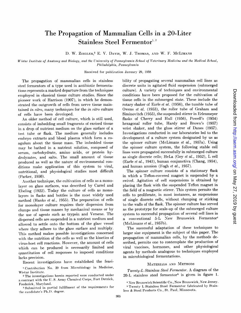
Twenty-L Stainless Steel Fermentor. A diagram of the20-L stainless steel fermentor5 is given in figure 1.
4New Brtunswick Scientific Co., New Bruinswick, New Jersey.Twentv L Stainless Steel Fermentor fabricated by Stain-
less & Steel Products Co., St. Paul, Minnesota.
305
on May 27, 2019 by guest
http://aem.asm
.org/D
ownloaded from
ZIEGLER, DAVIS, THOMAS, AND McLIMANS
The fermentor shell of this equipment was fabricatedfrom type 316 stainless steel alloy, an alloy known tobe nontoxic to certain mammalian cells cultured invitro (Giardinello et al., 1958). All lines and serviceconnections which are in direct contact with the culturechamber are constructed of stainless steel of a suitablealloy. Valves, including those in the air lines, steamlines, and drain lines, are diaphragm-type valves withstainless steel bodies. Neoprene6 diaphragms have beenfound to be satisfactory in the operation of these valves.However, direct toxicity tests of the Neoprenediaphragm material have not been carried out.A 6-bladed, 4-in. impeller driven by a Reeves Vari-
Speed Motodrive7 is used to agitate the cell suspensions.Cell culture in this equipment has been carried out withagitation speeds varying from 140 rpm to 190 rpm.
The speed of agitation of the culture has not appearedto be a critical factor as evidenced by the similar ratesof growth observed under different rates of agitation.Generally, the speed of agitation was adjusted to a
point at which vigorous movement of the suspensionwas attained without foaming. Volumes of 6 L, thesmallest volume which can be handled in this equip-ment, were agitated at 140 to 160 rpm. Volumes of 12 Lwere agitated at 170 to 190 rpm. Under these conditions,there was no evidence that the cells settled out or
adhered to the walls of the fermentation vessel.Sterilization of all components of the fermentor was
accomplished by in-place steam sterilization. A steriliza-
6Grinnell Company, Inc., Providence, Rhode Island.I Reeves Pulley Co., Columbus, Indiana.
I I
II4
I-Is lo
Figure 1. Twenty L stainless steel fermentor
tion period of 2 hr at 18 lb per in.2 with steam beingbled through the filters and outlets has given com-
pletely satisfactory results, both in actual operation andin sterility tests.
Aeration of submerged tissue cultures by sparginggas into the bottom of the fermentation vessels hasbeen found unnecessary. Hence, tank cultures were
maintained with a gas overlay. Further simplificationof the aeration system was afforded by the use in thegrowth medium of an increased phosphate buffer con-
centration, which eliminated the necessity of the use
of the conventional bicarbonate-gaseous carbon dioxidebuffer. Since the need for carbon dioxide gassing was
circumvented, the submerged cultures were aeratedonly with air from a small air compressor at a flow-rateof 0.5 to 2.0 L per min. The air was sterilized by passage
through a glass wool-packed column. A positive pressure
of 5 to 8 lb per in.2 within the fermentors appeared toaid in the exclusion of contaminants from the tissueculture system.An incubation temperature of 36 to 37 C was main-
tained by the circulation of water from a constanttemperature water bath through the water jacket ofthe fermentor.Medium. The culture medium employed in these
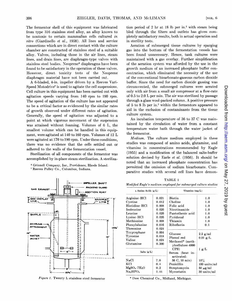
studies was composed of amino acids, glutamine, andvitamins in concentrations recommended by Eagle(1955) and a modification of the balanced salts-buffersolution devised by Earle et al. (1956). It should benoted that an increased phosphate concentration haspermitted the omission of sodium bicarbonate. Com-parative studies with several cell lines have demon-
TABLE 1Modified Eagle's medium emnployed for submerged culture studies
L Amino Acids (g/L) Vitamins (mg/L)
Arginine*HCl 0.021 Biotin 1.0Cystine 0.012 Choline 1.0Histidine* HCl 0.008 Folic acid 1.0Isoleucine 0.026 Nicotinamide 1 .0Leucine 0.026 Pantothenic acid 1 .0Lysine * HCl 0.026 Pyridoxal 1.0Methionine 0.008 Thiamin 1 .0Phenylalanine 0.016 Riboflavin 0.1Threonine 0.024Tryptophane 0.004 Glucose 2.5 g/mlTyrosine 0.018 Phenol red 0.01 g/LValine 0.024 Methocel* (meth-Glutamine 0.300 ylcellulose-4000
Salts_____-g/L CPS) 1 g/LSalts (g/L) Serum (heat in-
activated:NaCl 7.0 56 C, 10 min) 10%KCl 0.4 Penicillin 100 units/mlMgSO4 *7H20 0.2 Streptomycin 50 ,ug/mnlNa2HPO4 1.44 Mycostatin 30 units/ml
* Dow Chemical Co., Midland, Michigan.
[VOL. 6'306
on May 27, 2019 by guest
http://aem.asm
.org/D
ownloaded from
SUBMERGED CULTURE OF MAMMALIAN CELLS
strated that medium buffered with M/100 phosphatepermits cell proliferation equal to the growth obtainedin a C02: bicarbonate buffered medium. The completeingredients of the medium are listed in table 1.The necessity for complete medium changes was
eliminated by the periodic addition of the amino acid,arginine (Thomas et al., 1958), to the actively pro-liferating cell suspension. The use of this technique,together with the addition of fresh medium, allowedthe cultures to be maintained for periods of 10 to 20days without medium changes.
Cell lines. Propagation of three cell lines was studiedin the 20-L fermentation equipment. These are strainL, a mouse fibroblast; strain HeLa, an epithelial-typecell isolated from a human cervical carcinoma; andstrain KD of the ERK cell line (Westwrood et al., 1957),isolated from embryonic rabbit kidney. The latter cellline is of special interest because it supports the pro-liferation of the virus of poliomyelitis (Sheffield andChurcher, 1957).
Cell stocks of each of these cell lines were maintainedroutinely both in spinner culture (suspended agitatedculture) and in bottle culture (stationary culture).IInoculum for the initiation of the stainless steelfermentors was produced in 3 L volumes in New Bruns-wick fermentors.
Inocula for cultures initiated in the New Brunswickfermentors were obtained either from a spinner cultureor from glass-grown cells. Either source of inoculumhas proved to be satisfactory. Tissue cultures in theNew Brunswick equipment were started with 1.5 L ofcell suspension containing 1.5 to 3.0 X 10⁵ cells per ml.As cell multiplication was noted, fresh medium wasadded until a total volume of 3 L, of cell suspension wasobtained. The maximum cell population usually at-tained was 1.0 to 1.5 X 10⁶ cells per ml. The detailedoperation of tissue culture in this equipment has beenreported elsewhere (McLimans et al., 1957b). A culturewas initiated in the larger 20 L fermentor by introduc-tionl of 2 to 3 L of cell suspension inoculum plus suffi-cient fresh medium to give a total volume of 6 L, witha resultant cell population of 1.7 to 3.0 X 10⁵ cellsper ml.
Sampling and cell counting. Cell multiplication wasdetermined by counting the cells in a hemocytometer.The general state of a culture was reflected by theviability of the cells which was determined by a stainingmethod employing trypan blue ('appenheimer, 1917;Hewitt, 1953). It has been observed that degeneratingcells have an affinity for the stain, trypan blue, whereasactively proliferating cells are not stained. The viabili-ties of cell cultures were determined by makingdifferential counts of stained and unstained cells. Thepublication of McLimans et al. (1957a), describing thedevelopment of the spinner culture system, includes adescription of the trypan blue method. Additionally,
Gwatkin et al. (1957) have demonstrated a good correla-tion between the total cell count as obtained by adirect counting procedure and actual isolated cellculture using a feeder layer (Puck and Fisher, 1956).
RESULTS AND DISCUSSIONGrowth of strain L. The techniques employed in cul-
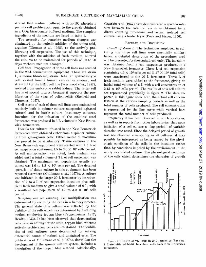
turing the three cell lines were essentially similar;hence, a detailed description of the procedures usedwill be presented for the strain L cell only. The inoculumwas obtained from a cell suspension produced in aNew Brunswick fermentor. Three L of cell suspensioncontaining 4.9 X 10⁵ cells per ml (1.47 X 10⁹ total cells)were transferred to the 20 L fermentor. Three L offresh medium were added to the fermentor, giving aninitial total volume of 6 L with a cell concentration of2.45 X 105 cells per ml. The results of this cell cultureare represented graphically in figure 2. The data re-ported in this figure show both the actual cell concen-tration at the various sampling periods as well as thetotal number of cells produced. The cell concentrationis represented by the line curve while vertical barsrepresent the total number of cells produced.
Frequently it has been observed in our laboratories,as well as in reports from other laboratories, that uponinitiation of a cell culture a "lag period" of variableduration was noted. Since the delayed period of growthwas not observed consistently in all cultures, it maypossibly be interpreted as being caused by the physi-ologic condition of the cells in the inoculum ratherthan by conditions imposed by the environment in theniewly established culture. The physiological conditionof the cells which determines the character of growth
9.6.7'-
6.
Time (Days)
Figutre 2. Growth of "L" cells in 20 L fermentor. Tank no.
1. Date initiated 3-8-58. Inoculum: cells from New Brunswickfermentor.
195a8] 307
Isxa6
1a415IU
4;00u
0u
10'
x0
1-
on May 27, 2019 by guest
http://aem.asm
.org/D
ownloaded from
ZIEGLER, DAVIS, THOMAS, AND McLIMANS
upon subculture may reflect the nutritional state ofthe cells or the growth phase of the cells at the time ofsubculture. Accordingly, an inoculum of cells from aculture in a stationary growth phase would produce a"lag" upon subculture, while cells from a culture inlog-growth phase would not exhibit an initial "lagperiod."The "lag period" shown in figure 2 was minimal and
was followed by an increase in the cell population. After4 days of growth, the cell suspension contained ap-proximately 4.0 X 10⁵ cells per ml and at this pointan additional 6 L of medium was added to the tank.A constant increase in the cell population resulted andwas maintained over the 7-day period by three additionsof Eagle's amino acid concentrates. The amount ofeach amino acid addition was equal to the originalconcentration of the amino acid in fresh medium. Workby Thomas et al. (1958) has shown that arginine presentin the amino acid mixture is the vital factor whichmay be used to supplement medium additions duringthe first 10 days in this type of culture. The addition ofarginine alone in subsequent cell cultures was equallyas effective as the addition of the complete amino acidsmixture.
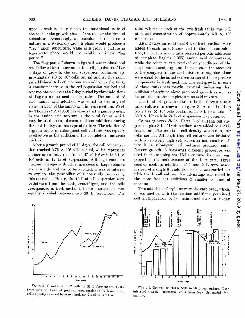
After a growth period of 11 days, the cell concentra-tion reached 6.75 X 10⁵ cells per ml, which representsan increase in total cells from 1.47 X 10⁹ cells to 8.1 X10⁹ cells in 12 L of suspension. Although completemedium changes with cell suspensions in large volumesare unwieldy and are to be avoided, it was of interestto explore the possibility of successfully performingthis operation. Hence, the 12 L of cell suspension werewithdrawn from the tank, centrifuged, and the cellsresuspended in fresh medium. The cell suspension wasequally divided between two 20 L fermentors. The
total volume in each of the two fresh tanks was 6 Lat a cell concentration of approximately 6.0 X 10⁵cells per ml.
After 5 days an additional 6 L of fresh medium ereieadded to each tank. Subsequent to the medium addi-tion, the culture in one tank received periodic additionsof complete Eagle's (1955) amino acid concentrate,while the other culture received only additions of thesingle amino acid, arginine. In each case, the amouintsof the complete amino acid mixture or arginine alonewere equal to the initial concentration of the respectivecomponents in fresh medium. The cell growth in eachof these tanks was nearly identical, indicating thataddition of arginine alone promoted growth as well asthe addition of the complete amino acid mixture.The total cell growth obtained in the three separate
tank cultures is shown in figure 3. A cell build-upfrom 1.47 X 10⁹ cells contained in 6 L to a total of30.0 X 109 cells in 24 L of suspension was obtained.Growth of strain HeLa. Three L of a HeLa cell sus-
pension plus 3 L of fresh medium were added to a 20 Lfermentor. The resultant cell density was 4.6 X 10⁵cells per ml. Although this cell culture was initiatedwith a relatively high cell concentration, smaller cellinocula in subsequent cell cultures produced satis-factory growth. A somewhat different procedure wasused in maintaining the HeLa culture than was em-ployed in the maintenance of the L culture. Threesmaller medium additions of 1 and 2 L were madeinstead of a single 6 L addition such as was carried outwith the L cell culture. No advantage was noted inthe more frequent additions of smaller volumes ofmedium.Two additions of arginine were also employed, which,
in conjunction with the medium additions, permittedcell multiplication to be maintained over an 11-day
20
* 109
8
.7
6
.5I
Time (Days)
Figure 4. Growth of HeLa cells in 20 L fermentors. Date
initiated 4-15-57. Inoculum: cells from New Brunswick fer-
mentor.
Time (Days)
Figure 3. Growth of "L" cells in 20 L fermentors. Cellsfrom tank no. 1 centrifuged and resuspended in fresh medium;cells equally divided between tank no. 2 and tank no. 3.
308 [VOL. 6
s
Izu11
1.
sx
AI;.n1;u
on May 27, 2019 by guest
http://aem.asm
.org/D
ownloaded from

SUBMERGED CULTURE OF MAMMALIAN CELLS
period. During this period, the total cell populationincreased from 2.8 X 10⁹ cells suspended in 6 L to10.6 X 109 cells contained in 11 L. The results obtainedfrom this cell culture are shown in figure 4.The possibility of producing large amounts of viral
agents in submerged culture-produced cells is indicatedby the demonstration that strain HeLa, as propagatedin spinner culture, supported the proliferation of allthree types of poliovirus (Davis, 1957).Growth of strain KD. Figure 5 presents data describ-
ing the growth obtained with a third cell line, strainERK/KD cultivated in the stainless steel equipment.The inoculum was a cell suspension produced in a NewBrunswick fermentor. The cell inoculum, plus sufficientmedium to make a total initial volume of 6 L containing5.6 X 105 cells per ml, was introduced into a tank. Inthis culture, as in the other cell cultures, growth wasmaintained by a medium addition and additions ofarginine. On the third day following the initiation ofthe culture, the addition of fresh medium appeared tocause a slight lag in the rate of cell multiplication. Thereason for the "lag" has not been determined, but inthis culture and in studies with other cell lines, it seemsthat the lag may be caused by an endogenous depletionof essential factors in the cells prior to the addition offresh medium. Although the addition of arginine per-mits continued growth without medium changes for aperiod of 10 days, the question relating to the depletionof other metabolites remains to be elucidated.
I * n 10r 1 C<|i! a 4.
14 ..I = I
12
10 Cells/miT (NO xlO5)
7_0
T Total Cell
41 ~~~~~~~(nox 109)
3
During the 6-day growvth period, the use of arginineand fresh medium promoted a continuous increase inthe total cell population from 3.4 X 10⁹ cells to 11.1 X10⁹ cells with a final cell density of 1.3 X 10⁶ cells perml. Although satisfactory growth of the ERK/KDcell, as well as with strain HeLa and strain L, wasobtained with these procedures, future nutritionalstudies undoubtedly will permit both more rapid ratesof multiplication and the attainment of higher celldensities. This particular nonprimate cell line seemsto be of interest since it supports the proliferation ofpoliovirus when cultured in the submerged state(McLimans et al., 1957c) in volumes up to 10 to 15 L.
ACKNOWLEDGMENTS
We wish to acknowledge the cooperation and in-valuable assistance rendered during many discussionsby Doctors William Hinshaw and Arthur Brown ofFort Detrick as well as Mr. Fletcher L. Glover andMr. Carrell J. Kucera of the Wistar Staff. We arefurther indebted to E. R. Squibb & Sons for their gen-erosity in making available the stainless steel fermentorsused in this investigation.
SUMMARYTwenty-liter stainless steel fermentors, designed for
use in the fermentation industry, were adapted for usein the propagation of mammalian cells in an agitatedfluid suspension system. The medium and culturalconditions employed in the investigations are discussed.
Three unrelated cell lines (strain L, a fibroblasticcell of mouse origin; strain HeLa, aln epithelial-type cellisolated from a human cervical carcinoma; and strainERK/KD, isolated from embryonic rabbit kidney)were successfully cultivated in the large stainless steelequipment. It was possible to obtain 3- to 6-fold in-creases with each of the cell lines during a 6- to 12-daygrowth period.
Since cells of three different origins were readilycultured under the conditions described, these resultsoffer promise that many stable cell lines may be propa-gated in mass in equipment designed for large scalefermentations. The possibility of using submergedculture cells to produce viral vaccines, hormones, anidother physiologically active cell products is indicated.
REFERENCES
CARREL, A. AND EBELING, A. H. 1922 Pure cultures of lar gemononuclear leucocytes. J. Exptl. Med., 36, 365.
CHANG, R. S. 1954 Continuous subcultivation of epithelial-like cells from normal human tissues. Proc. Soc. Exptl.Biol. Med., 87, 440-443.
CHERRY, W. R. AND HULL, R. N. 1956 Studies on the growthof mammalian cells in agitated fluid media. Abstract,Tissue Culture Association meeting, Milwaukee, Wiscon-sin, April 3 and 4, 1956.
D)ANES, S. 1957 Suspension cultures of strain L mousefibroblasts. I. A glass stirrer apparatus for the cultivationof cell suspension. Exptl. Cell Research, 12, 169-179.
Time (Days)
Figure 5. The growth of strain KI) cells in the 20 L fermentor
1958] 309
on May 27, 2019 by guest
http://aem.asm
.org/D
ownloaded from

ZIEGLER, DAVIS, THOMAS, AND McLIMANS
DAVIS, E. V. 1957 Multiplication of cells in spinner cultureand the effect of viral parasitism on these cells. Ph.D.Dissertation, University of Pennsylvania (DissertationAbstracts VXVII No. 11, 2379, 1957).
EAGLE, H. 1955 Nutrition needs of mammalian cells intissue culture. Science, 122, 501-504.
EARLE, W. R., SCHILLING, E. L., STARK, T. H., STRAUS, N. P.,BROWN, M. F., AND SHELTON, E. 1943 Production ofmalignancy in vitro. IV. The mouse fibroblast cultures andchanges seen in living cells. J. Natl. Cancer Inst., 4,165-212.
EARLE, W. R., BRYANT, J. C., SCHILLING, E. L., AND EVANS,V. J. 1956 Growth of cell suspensions in tissue culture.Annals N. Y. Acad. Sci., 63, 666-682.
FOGH, J. AND LUND, R. 0. 1957 Continuous cultivation ofepithelial cell strain (FL) from human amniotic mem-brane. Proc. Soc. Exptl. Biol. Med., 94, 532-537.
GEY, G. 0., COFFMAN, W. D., AND KUBICEK, M. T. 1952Tissue culture studies of the proliferation capacity ofcervical carcinoma and normal epithelium. Cancer Re-search, 12, 264-265.
GIARDINELLO, F. E., McLIMANS, W. F., AND RAKE, G. W.1958 The apparent toxicity of metallic materials of con-struction and antifoam agents for mammalian cell lines.Appl. Microbiol., 6, 30-35.
GRAHAM, A. F. AND SIMINOVITCH, L. 1955 Proliferation ofmonkey kidney cells in rotating cultures. Proc. Soc.Exptl. Biol. Med., 89, 326-327.
GWATKIN, J. E., TILL, G. E., WHITMORE, G. F., SIMONOVITCH,L., AND GRAHAM, A. F. 1957 Multiplication of animalcells in suspension measured by colony counts. Proc.NatI. Acad. Sci., 43, 451-456.
HANKS, J. H., SCHERER, W. F., FAWCETT, D. W., LEIGHTON,J., AND PORTER, K. R. 1955 An introduction to cell andtissue culture. 1st ed. Burgess Publishing Co., Minneap-olis, Minnesota.
HARDY, F. M. AND BROWN, A. 1957 Growth of VenezuelanEquine Encephalomyelitis virus in monolayer and fluidsuspension cultures of L cells. Presented at the Springmeeting of the Maryland Society of American Bacteriolo-gists, Ft. Detrick, Frederick, Maryland, April 6, 1957.
HARRISON, R. G. 1907 Observations on the living nervefiber. Proc. Soc. Exptl. Biol. Med., 4, 140-143.
HEWITT, H. B. 1953 Quantitative transplantation of mousesarcoma. Brit. J. Cancer, 7, 367-383.
McLIMANS, W. F., DAVIS, E. V., GLOVER, F. L., AND RAKE,G. W. 1957a The submerged culture of mammaliancells: the spinner culture. J. Immunol., 79, 428-433.
McLIMANS, W. F., GIARDINELLO, F. E., DAVIS, E. V., KUCERA,C. J., AND RAKE, G. W. 1957b Submerged culture ofmammalian cells: 5 L fermentor. J. Bacteriol., 74, 768-774.
McLiMANS, W. F., ZIEGLER, D. W., DAVIS, E. V., THOMAS,W. J., AND KUCERA, C. J. 1957c Proliferation of polio-virus in a nonprimate host cell in submerged culture.Presented at the Fourth International Poliomyelitis Con-ference. Geneva, Switzerland, July 12, 1957.
OWENS, H., GEY, M. K., AND GEY, G. 0. 1953 A new methodfor the cultivation of mammalian cells suspended in agi-tated fluid medium. Proc. Am. Assoc. Cancer Research(Abstract 1), 1953.
PAPPENHEIMER, A. M. 1917 Experimental studies uponlymphocytes. I. The reactions of lymphocytes under vari-ous experimental conditions. J. Exptl. Med., 25, 633-650.
PARKER, R. C. 1950 Methods of tissue culture, 2nd ed. PaulB. Hoeber, Inc., New York, New York.
POWELL, A. F. 1954 Culture of ascites tumor cells in vitro.British Empire Cancer Campaign. 32nd Annual Report,125-227.
PUCK, T. T. AND FISHER, H. W. 1956 On the function ofx-irradiated "feeder" cells in supporting growth of singlemammalian cells. Proc. Natl. Acad. Sci., 42, 900-906.
SHEFFIELD, F. W. AND CHURCHER, G. M. 1957 The serialpropagation of poliomyelitis viruses in cells derived fromrabbit embryo kidney. Brit. J. Exptl. Pathol., 38, 155-159.
THOMAS, W. J., ZIEGLER, D. W., SCHEPARTZ, S. A., AND Mc-LIMANS, W. F. 1958 The use of arginine to eliminatemedium changes during the propagation of mammaliancells in submerged culture. Science, 127, 591-592.
WESTWOOD, J. C. N., MACPHERSON, I. A., AND TITMUS, D.H. J. 1957 Transformation of normal cells in tissueculture: its significance relative to malignancy and virusvaccine production. Brit. J. Exptl. Pathol., 38, 138-154.
310 [VOL. 6
on May 27, 2019 by guest
http://aem.asm
.org/D
ownloaded from