Thenaturalphosphoinositidederivative ... ·...
15
The natural phosphoinositide derivative glycerophosphoinositol inhibits the lipopolysaccharide- induced inflammatory and thrombotic responses Received for publication, December 29, 2016, and in revised form, May 22, 2017 Published, Papers in Press, June 9, 2017, DOI 10.1074/jbc.M116.773861 Mariangela Vessichelli ‡1,2 , Stefania Mariggiò ‡1 , Alessia Varone ‡1,2 , Pasquale Zizza ‡1,2,3 , Angelomaria Di Santo § , Concetta Amore § , Giuseppe Dell’Elba § , Adele Cutignano ¶ , Angelo Fontana ¶ , Carmela Cacciapuoti , Gaetano Di Costanzo , Mariastella Zannini**, Tiziana de Cristofaro**, Virgilio Evangelista §4 , and Daniela Corda ‡5 From the ‡ Institute of Protein Biochemistry, National Research Council, Via P. Castellino 111, 80131 Naples, Italy, § Laboratory of Vascular Biology and Pharmacology, Consorzio and Fondazione Mario Negri Sud, Via Nazionale 8/A, 66030 Santa Maria Imbaro, Chieti, Italy, ¶ Institute of Biomolecular Chemistry, National Research Council, Via Campi Flegrei 34, 80078 Pozzuoli, Naples, Italy, Transfusion Service, Department of Hematology-Oncology and Stem Cell Transplantation Unit, National Cancer Institute G. Pascale Foundation, Istituto di Ricovero e Cura a Carattere Scientifico, Via M. Semmola 52, 80131 Naples, Italy, and **Institute of Experimental Endocrinology and Oncology, National Research Council, Via S. Pansini 5, 80131 Naples, Italy Edited by Dennis R. Voelker Inflammatory responses are elicited through lipid products of phospholipase A 2 activity that acts on the membrane phos- pholipids, including the phosphoinositides, to form the proinflammatory arachidonic acid and, in parallel, the glyc- erophosphoinositols. Here, we investigate the role of the glycerophosphoinositol in the inflammatory response. We show that it is part of a negative feedback loop that limits proinflammatory and prothrombotic responses in human monocytes stimulated with lipopolysaccharide. This inhibi- tion is exerted both on the signaling cascade initiated by the lipopolysaccharide with the glycerophosphoinositol-depen- dent decrease in IB kinase /, p38, JNK, and Erk1/2 kinase phosphorylation and at the nuclear level with decreased NF-B translocation and binding to inflammatory gene promoters. In a model of endotoxemia in the mouse, treat- ment with glycerophosphoinositol reduced TNF- synthesis, which supports the concept that glycerophosphoinositol inhibits the de novo synthesis of proinflammatory and pro- thrombotic compounds and might thus have a role as an endogenous mediator in the resolution of inflammation. As indicated, this effect of glycerophosphoinositol can also be exploited in the treatment of manifestations of severe inflam- mation by exogenous administration of the compound. Inflammation is a beneficial host response to foreign patho- gens and tissue injury that ultimately leads to bacterial clear- ance and restoration of tissue structure and function (1). The host response involves different cell types and signaling mole- cules, such as cytokines, chemokines, and bioactive lipids (2). These bioactive lipids include prostaglandins, leukotrienes, and endoperoxides, which originate enzymatically from the phos- pholipase A 2 /arachidonic acid cascade and are formed and act in a cell-specific fashion (3). More recently, other compounds that intervene in the resolution of inflammatory responses have been characterized; these include lipoxins, resolvins, protec- tins, and maresins (all of which are biosynthesized from essen- tial polyunsaturated fatty acids), the receptors for the bioactive lipids mentioned above, and various microRNAs (4). It is now evident that the control of the initiation of inflammation and its natural resolution involve several essential components that need to be finely modulated in space and time to restore tissue homeostasis. Indeed, prolonged or uncontrolled inflammation represents the pathogenetic basis of chronic inflammatory dis- eases (5). As a prolonged activation of the cells of the immune system is the driving force behind inflammatory diseases, the identification of anti-inflammatory compounds that can switch off proinflammatory responses at one of the crucial steps and thus restore immunological homeostasis remains of great interest. Recent data obtained in our laboratory have addressed some aspects of this need as they show the poten- tial of glycerophosphoinositol (GroPIns) 6 as an anti-inflam- This work was supported in part by Italian Association for Cancer Research (Milan, Italy) Grants IG10341 and IG14675, the Ministry for Education, Uni- versities and Research Project “FaReBio di Qualità,” Programma Operativo Nazionale Project 01-00862, Research Projects of National Interest Project 2012CK5RPF_05, National Research Plan-National Research Council Aging Program 2012–2014, Progetto Bandiera “Epigen,” and the Programma Operativo Regionale Project OcKey. D. Corda, P. Zizza, and S. Mariggiò are authors of United States Patent 9351983, Use of Glycerophosphoinositols for the Treatment of Septic Shock. This article contains supplemental Figs. S1–S4. 1 These authors contributed equally to this work. 2 Recipients of fellowships from the Italian Foundation for Cancer Research (Milan, Italy). 3 Present address: Oncogenomic and Epigenetic Unit, Regina Elena National Cancer Inst., Via E. Chianesi, 53, 00144 Rome, Italy. 4 To whom correspondence may be addressed. E-mail: evangelista59gino@ gmail.com. 5 To whom correspondence may be addressed: Institute of Protein Biochem- istry, National Research Council, Via P. Castellino 111, 80131 Naples, Italy. Tel.: 39-081-6132536; Fax: 39-081-6132277; E-mail: [email protected]. 6 The abbreviations used are: GroPIns, glycerophosphoinositol; GroPIns4P, glycerophosphoinositol 4-phosphate; TLR, toll-like receptor; Mac-1, M2 integrin; TF, tissue factor; COX, cyclooxygenase; IB, inhibitor of B; IKK, IB kinase; p38, p38 mitogen-activated protein kinase; MyD88, myeloid differentiation primary response gene 88; PLA 2 , phospholipase A 2 ; Tx, thromboxane. cros ARTICLE 12828 J. Biol. Chem. (2017) 292(31) 12828 –12841 © 2017 by The American Society for Biochemistry and Molecular Biology, Inc. Published in the U.S.A. by guest on February 23, 2019 http://www.jbc.org/ Downloaded from
Transcript of Thenaturalphosphoinositidederivative ... ·...

The natural phosphoinositide derivativeglycerophosphoinositol inhibits the lipopolysaccharide-induced inflammatory and thrombotic responsesReceived for publication, December 29, 2016, and in revised form, May 22, 2017 Published, Papers in Press, June 9, 2017, DOI 10.1074/jbc.M116.773861
Mariangela Vessichelli‡1,2, Stefania Mariggiò‡1, Alessia Varone‡1,2, Pasquale Zizza‡1,2,3, Angelomaria Di Santo§,Concetta Amore§, Giuseppe Dell’Elba§, Adele Cutignano¶, Angelo Fontana¶, Carmela Cacciapuoti�,Gaetano Di Costanzo�, Mariastella Zannini**, Tiziana de Cristofaro**, Virgilio Evangelista§4,and Daniela Corda‡5
From the ‡Institute of Protein Biochemistry, National Research Council, Via P. Castellino 111, 80131 Naples, Italy, §Laboratory ofVascular Biology and Pharmacology, Consorzio and Fondazione Mario Negri Sud, Via Nazionale 8/A, 66030 Santa Maria Imbaro,Chieti, Italy, ¶Institute of Biomolecular Chemistry, National Research Council, Via Campi Flegrei 34, 80078 Pozzuoli, Naples, Italy,�Transfusion Service, Department of Hematology-Oncology and Stem Cell Transplantation Unit, National Cancer Institute G.Pascale Foundation, Istituto di Ricovero e Cura a Carattere Scientifico, Via M. Semmola 52, 80131 Naples, Italy, and **Institute ofExperimental Endocrinology and Oncology, National Research Council, Via S. Pansini 5, 80131 Naples, Italy
Edited by Dennis R. Voelker
Inflammatory responses are elicited through lipid productsof phospholipase A2 activity that acts on the membrane phos-pholipids, including the phosphoinositides, to form theproinflammatory arachidonic acid and, in parallel, the glyc-erophosphoinositols. Here, we investigate the role of theglycerophosphoinositol in the inflammatory response. Weshow that it is part of a negative feedback loop that limitsproinflammatory and prothrombotic responses in humanmonocytes stimulated with lipopolysaccharide. This inhibi-tion is exerted both on the signaling cascade initiated by thelipopolysaccharide with the glycerophosphoinositol-depen-dent decrease in I�B kinase �/�, p38, JNK, and Erk1/2 kinasephosphorylation and at the nuclear level with decreasedNF-�B translocation and binding to inflammatory genepromoters. In a model of endotoxemia in the mouse, treat-ment with glycerophosphoinositol reduced TNF-� synthesis,which supports the concept that glycerophosphoinositolinhibits the de novo synthesis of proinflammatory and pro-thrombotic compounds and might thus have a role as anendogenous mediator in the resolution of inflammation. Asindicated, this effect of glycerophosphoinositol can also be
exploited in the treatment of manifestations of severe inflam-mation by exogenous administration of the compound.
Inflammation is a beneficial host response to foreign patho-gens and tissue injury that ultimately leads to bacterial clear-ance and restoration of tissue structure and function (1). Thehost response involves different cell types and signaling mole-cules, such as cytokines, chemokines, and bioactive lipids (2).These bioactive lipids include prostaglandins, leukotrienes, andendoperoxides, which originate enzymatically from the phos-pholipase A2/arachidonic acid cascade and are formed and actin a cell-specific fashion (3). More recently, other compoundsthat intervene in the resolution of inflammatory responses havebeen characterized; these include lipoxins, resolvins, protec-tins, and maresins (all of which are biosynthesized from essen-tial polyunsaturated fatty acids), the receptors for the bioactivelipids mentioned above, and various microRNAs (4). It is nowevident that the control of the initiation of inflammation and itsnatural resolution involve several essential components thatneed to be finely modulated in space and time to restore tissuehomeostasis. Indeed, prolonged or uncontrolled inflammationrepresents the pathogenetic basis of chronic inflammatory dis-eases (5). As a prolonged activation of the cells of the immunesystem is the driving force behind inflammatory diseases, theidentification of anti-inflammatory compounds that can switchoff proinflammatory responses at one of the crucial stepsand thus restore immunological homeostasis remains ofgreat interest. Recent data obtained in our laboratory haveaddressed some aspects of this need as they show the poten-tial of glycerophosphoinositol (GroPIns)6 as an anti-inflam-
This work was supported in part by Italian Association for Cancer Research(Milan, Italy) Grants IG10341 and IG14675, the Ministry for Education, Uni-versities and Research Project “FaReBio di Qualità,” Programma OperativoNazionale Project 01-00862, Research Projects of National Interest Project2012CK5RPF_05, National Research Plan-National Research Council AgingProgram 2012–2014, Progetto Bandiera “Epigen,” and the ProgrammaOperativo Regionale Project OcKey. D. Corda, P. Zizza, and S. Mariggiò areauthors of United States Patent 9351983, Use of Glycerophosphoinositolsfor the Treatment of Septic Shock.
This article contains supplemental Figs. S1–S4.1 These authors contributed equally to this work.2 Recipients of fellowships from the Italian Foundation for Cancer Research
(Milan, Italy).3 Present address: Oncogenomic and Epigenetic Unit, Regina Elena National
Cancer Inst., Via E. Chianesi, 53, 00144 Rome, Italy.4 To whom correspondence may be addressed. E-mail: evangelista59gino@
gmail.com.5 To whom correspondence may be addressed: Institute of Protein Biochem-
istry, National Research Council, Via P. Castellino 111, 80131 Naples, Italy.Tel.: 39-081-6132536; Fax: 39-081-6132277; E-mail: [email protected].
6 The abbreviations used are: GroPIns, glycerophosphoinositol; GroPIns4P,glycerophosphoinositol 4-phosphate; TLR, toll-like receptor; Mac-1, �M�2integrin; TF, tissue factor; COX, cyclooxygenase; I�B�, inhibitor of �B; IKK,I�B kinase; p38, p38 mitogen-activated protein kinase; MyD88, myeloiddifferentiation primary response gene 88; PLA2, phospholipase A2; Tx,thromboxane.
crosARTICLE
12828 J. Biol. Chem. (2017) 292(31) 12828 –12841
© 2017 by The American Society for Biochemistry and Molecular Biology, Inc. Published in the U.S.A.
by guest on February 23, 2019http://w
ww
.jbc.org/D
ownloaded from

matory compound that can prevent endotoxin shock in themouse.
GroPIns is one of the naturally occurring phosphoinositidemetabolites, the glycerophosphoinositols, that are producedthrough the activity of phospholipase A2IV� (PLA2IV�), anenzyme that preferentially hydrolyzes phospholipids carryingarachidonic acid in position sn-2 of the glycerol backbone (6, 7).The formation of GroPIns requires two deacylation steps, bothcarried out by PLA2IV�. PLA2IV� has intrinsic phospholipaseA2 and lysolipase activities; thus, it hydrolyzes the membranephosphoinositide (carrying the arachidonic acid in the sn-2position), producing in parallel arachidonic acid and thelysoderivative lysophosphatidylinositol, and then in sequence,PLA2IV� forms GroPIns by deacylating lysophosphatidylinosi-tol (6, 8). Noticeably, this PLA2IV�-dependent metabolismgives rise to three biologically active metabolites that start inde-pendent cascades, i.e. G-protein– coupled receptor-dependentsignaling (by the GPR55 ligand lysophosphatidylinositol),cyclooxygenase/lipoxygenase metabolism (by producing theirsubstrate, arachidonic acid), and glycerophosphoinositol-mod-ulated processes (which include cell proliferation and actincytoskeleton organization) (6, 8 –10).
The glycerophosphoinositols are ubiquitous, freely diffusiblemolecules, and they can be detected both within cells and in theextracellular space (11, 12). Their intracellular concentrationsare cell type-dependent, and their production can vary upononcogenic transformation, cell differentiation, and hormonalstimulation (6, 11). They are present in different forms that canbe either unphosphorylated (GroPIns) or phosphorylated (e.g.glycerophosphoinositol 4-phosphate (GroPIns4P), glycero-phosphoinositol 4,5-phosphate, and glycerophosphoinositol3-phosphate) with different cellular activities (6, 13). Hemato-poietic cells, such as monocytes and macrophages, have astrictly regulated PLA2IV� activity that provides a fine modu-lation of the intracellular glycerophosphoinositol levels inresponse to environmental stimuli, including proinflammatoryagents such as bacterial lipopolysaccharide (LPS) (7). To fur-ther clarify the potential roles of these lipid-derived mediatorsin inflammation, we have investigated the effect of GroPIns in awell characterized in vitro model of inflammatory and proco-agulant responses mimicked by human blood monocytes chal-lenged with LPS.
Here we show that GroPIns can inhibit the expression ofprothrombotic and proinflammatory genes that are induced byEscherichia coli LPS through inhibition of the signaling cascadedownstream of the toll-like receptor 4 (TLR4) and of thenuclear activity of nuclear factor-�B (NF-�B). Of note, the mea-surement of inflammatory markers in mice subjected to LPS-induced endotoxemia showed that treatment with GroPIns isassociated with reduced plasma levels of tumor necrosis fac-tor-� (TNF-�) and decreased expression of �M�2 integrin(Mac-1) on the surface of circulating neutrophils, whichstrengthens the potential pharmacological relevance of this lip-id-derived mediator. Thus, the present study provides evidencethat GroPIns, which is produced endogenously by activatedinflammatory cells, can act as a resolution signal to control theproinflammatory and prothrombotic responses associated withparticularly exuberant inflammatory states (14).
Results
GroPIns inhibits endotoxin-induced prothrombotic andinflammatory responses in human monocytes
Increases of the intracellular GroPIns levels have beenobserved in response to inflammatory stimuli, such as LPS, inmacrophages (7). Based on this, we investigated the possibilitythat GroPIns can regulate the inflammatory reactions in a sim-ple and well characterized in vitro model system: the produc-tion of inflammatory cytokines in human blood monocytesupon challenge with endotoxin.
Human monocytes have a major role in the innate immuneresponse to bacterial infection. Bacterial LPS binds TLR4 on themonocyte membrane and triggers the expression of a variety ofinflammatory mediators, which include cytokines such asTNF-� and interleukin (IL)-1�, and enzymes, such as cyclooxy-genase (COX)-2. These in turn produce inflammatory prostan-oids, such as prostaglandin E2 and thromboxane A2 (TxA2)(15), which act locally to elicit the cardinal signs of acute inflam-mation (16).
We first tested the effects of GroPIns on mRNA levels ofsome of the major inflammatory mediators in freshly isolatedhuman monocytes challenged with LPS (0.1 �g/ml). Asexpected, LPS induced a pronounced increase in the expres-sion of IL-1�, TNF-�, COX-2, and the anti-inflammatory medi-ator IL-10 (Fig. 1, A–D). Preincubation with 300 �M GroPInsreduced the levels of IL-1�, TNF-�, and COX-2 mRNAsalthough to different extents (by 50 –75% at 24 h; Fig. 1, A–C).Of note, treatment with GroPIns considerably reduced theexpression of the inflammatory genes at both early (1.5 h) andlong times (24 h) of stimulation with very consistent effects atlong times (24 h; Fig. 1, A–C). However, GroPIns did not affectthe expression of the anti-inflammatory cytokine IL-10 (Fig.1D) or the suppressor of cytokine signaling 3, a transcriptionaltarget of the signal transducer and activator of transcription 3(STAT3) that is downstream of IL-10 signaling (Ref. 17 andsupplemental Fig. S1). In parallel experiments, monocytespreincubated with 300 �M GroPIns were challenged withTNF-� (50 ng/ml) for 1.5 h; no effect on gene expression wasobserved under these conditions (Fig. 1, E and F), indicatingthat GroPIns specifically interferes with the LPS signalingcascade. Accordingly, GroPIns also affected the expressionof cytokines when monocytes were challenged with agonistsof TLR7/8 and TLR9 that share a common downstreampathway with TLR4 (i.e. the myeloid differentiation primaryresponse gene 88 (MyD88)-dependent pathway (Ref. 18 andsupplemental Fig. S1).
In line with the effects on gene expression, 300 �M Gro-PIns reduced the LPS-stimulated release of both TNF-� andIL-1� cytokines at 24 h (with up to 60% inhibition) withnegligible effects at earlier times (Fig. 1G). Moreover, Gro-PIns reduced the accumulation of TxB2 by 60%. TxB2 is thestable metabolite of TxA2, which is a major product of themetabolic activity of COX-2 in monocytes (Fig. 1G), thusconfirming that these decreased mRNA levels result inreduced metabolic activity.
Monocytes also have a major role in thrombotic eventsthrough the de novo synthesis of tissue factor (TF), a transmem-
Glycerophosphoinositol cooperates in resolving inflammation
J. Biol. Chem. (2017) 292(31) 12828 –12841 12829
by guest on February 23, 2019http://w
ww
.jbc.org/D
ownloaded from

brane glycoprotein that initiates blood coagulation through itsbinding to factors VII and VIIa, which leads to fibrin clot for-mation (19). Treatment with 300 �M GroPIns reduced the LPS-induced expression of TF gene by 50% at 24 h of stimulation(Fig. 2A). Following these observations, we investigatedwhether modulation of TF gene expression also translates intoa reduced procoagulant activity of LPS-challenged monocytes.Procoagulant activity is practically undetectable in freshly iso-lated monocytes, and it is strongly up-regulated by exposure toLPS (Fig. 2B). Treatment of monocytes with 50 –300 �M Gro-PIns before LPS stimulation resulted in a dose-dependentreduction in TF activity with up to 60% inhibition (Fig. 2B).Similar to what was observed with the cytokines, the efficacy ofGroPIns in reducing TF activity became significant after 24 h ofstimulation (Fig. 2B). Moreover, a series of experiments wereconducted at 24 h of GroPIns stimulation where the cells werechallenged with 0.1–1000 ng/ml LPS after pretreatment with300 �M GroPIns; the inhibitory effect of GroPIns on the proco-agulant activity of TF was evident across this whole range ofLPS concentrations (Fig. 2C).
Thus, GroPIns can suppress proinflammatory and pro-thrombotic responses in human monocytes stimulated with LPS.This activity was consistently observed in numerous assays withsamples obtained from different donors.
The action of GroPIns involves the inhibition of theextracellular signal-regulated kinase 1/2 (Erk1/2), c-Jun N-terminal kinase (JNK), and p38 mitogen-activated proteinkinase (p38) kinase activities
To investigate the mechanism through which GroPInsaffects LPS-induced monocyte responses, we analyzed theireffects on the phosphorylation of p38, JNK, and Erk1/2, threekinases that are known to transduce the LPS signal downstreamof TLR4 (20). In these cells, LPS (0.1 �g/ml) activated Erk1/2,JNK, and p38, and treatment with GroPIns (300 �M) for differ-ent times reduced their phosphorylation by about 60% (Fig.3A), indicating that GroPIns acts by modulating the activity ofthese kinases. Similar experiments were carried out in mono-cytes pretreated with 300 �M GroPIns4P, and no difference wasobserved between control and treated cells (Fig. 3A). This phos-phorylated derivative, besides the already reported immuno-modulatory activity in T lymphocytes (21), has also been shownto affect the LPS-induced transcription of proinflammatorygenes in monocytes.7 Therefore, GroPIns and GroPIns4P areable to exert similar anti-inflammatory activity acting at differ-ent levels of the signaling cascade. These results are in line withcurrent knowledge that both GroPIns and GroPIns4P act intra-
7 P. Zizza and D. Corda, unpublished observations.
Figure 1. GroPIns reduces the LPS-induced proinflammatory response in human monocytes. A–C, GroPIns inhibits the LPS-mediated transcriptionof TNF-�, IL-1�, and COX-2 at 1.5 and 24 h. D, GroPIns does not inhibit the LPS-mediated expression of IL-10. E and F, GroPIns does not inhibit theTNF-�-mediated expression of proinflammatory genes (TNF-� and IL-1�). Human monocytes were purified from peripheral blood of healthy donors andincubated at 37 °C for 20 min without or with 300 �M GroPIns and for an additional 1.5 or 24 h in the absence or presence of 0.1 �g/ml LPS or 50 ng/mlTNF-�. At each time point, the RNA was extracted and converted to cDNA for real-time PCR analysis. Data were calculated using the �� method (2���CT).Transcription of the housekeeping gene GAPDH was used to normalize the data. Measurements of mRNA levels are expressed as -fold increase over therespective control (unstimulated) cells, and are means � S.D. of triplicates (n � 3) from two experiments performed with cells from two different donors(A-C) and means � S.D. of three independent experiments (n � 3) performed in duplicate (D–F). Asterisks indicate statistically significant differences (*,p � 0.05; **, p � 0.01). ns, not significant. G, GroPIns inhibits LPS-induced protein expression levels of TNF-� and IL-1� and reduces COX-derived TxB2.Monocytes were preincubated for 20 min at 37 °C in the absence or presence of 300 �M GroPIns and then incubated for a further 5 and 24 h without orwith 0.1 �g/ml LPS. At the end of this incubation, the supernatants were prepared, and the levels of TNF-�, IL-1�, and TxB2 in the medium were assessedusing ELISA. In untreated monocytes, the protein concentrations were under the assay detection limits at both times. Data show the percentage ofinhibition induced by GroPIns treatment on LPS-induced protein release. Data represent three independent experiments performed with cells fromthree different donors. Error bars represent S.D.
Glycerophosphoinositol cooperates in resolving inflammation
12830 J. Biol. Chem. (2017) 292(31) 12828 –12841
by guest on February 23, 2019http://w
ww
.jbc.org/D
ownloaded from

cellularly as there is so far no evidence of a membrane receptoror of specific activity at the membrane level (6, 11).
GroPIns inhibits the LPS-dependent signaling leading to thenuclear translocation of NF-�B
To evaluate the consequence of the reduced phosphorylationof Erk1/2, JNK, and p38, we have analyzed the well documentedLPS-dependent control of transcription. LPS-responsive, cis-acting DNA promoter elements have been characterized in the5�-flanking regions of several prothrombotic and inflammatorygenes, including TF, COX-2, IL-1�, and TNF-� (22). The tran-scription factors that bind to these LPS-responsive elementsinclude NF-�B, activator protein-1, and cAMP response ele-ment-binding protein, which all cooperate to produce variouscytokines, the levels of which are barely detectable in restingcells. NF-�B, however, is the only transcription factor that isrequired for the induction of all of the LPS-inducible genes sofar analyzed (23); therefore, it was selected for our investigation.The activation signaling to NF-�B involves its release from thecytosolic inhibitor I�B� followed by its translocation into thenucleus. This event is tightly dependent on the phosphorylationand proteasomal degradation of I�B�, which relies directly onthe phosphorylation-dependent activation of the upstream I�Bkinase (IKK) complex (24). We therefore investigated the acti-vation of the IKK kinases and the phosphorylation and totallevels of I�B�. In monocytes, LPS (0.1 �g/ml) elicited a rapidphosphorylation of the IKK�–� complex that was completelyprevented when cells were exposed to 300 �M GroPIns (10-minLPS treatment). Consistent with this observation, the LPS-in-duced degradation kinetics of I�B� was delayed by GroPIns,reaching a maximum at 10 min (Fig. 3B). The inhibitory effectof GroPIns was not observed when monocytes were treatedwith TNF-� (50 ng/ml) (Fig. 3C). These results support theobservation that GroPIns action is exerted on the signaling
events triggered by LPS treatment and that its inhibitory effectis specific for the LPS signaling.
GroPIns reduces LPS-induced nuclear translocation of NF-�Band the binding to the promoters of target genes
As indicated above, I�B� determines the cell localization,and thus activity, of NF-�B. Because GroPIns was able to affectboth the activation and degradation of I�B�, we evaluated thenuclear translocation of NF-�B in the presence of GroPIns byusing antibodies specific for p65 and p50 proteins (the subunitsforming the heterodimer mainly involved in transcriptionalregulation; see also Ref. 24 and “Experimental procedures”).The LPS-induced nuclear translocation of NF-�B became evi-dent after 15 min and maximal after 30 min (Fig. 4). Within 30min of the GroPIns addition, the nuclear translocation of bothp65 and p50 was reduced by 45 and 50%, respectively (Fig. 4B).This impairment in the nuclear translocation of NF-�B afterLPS stimulation is in line with, and corroborates, the rapideffect of the GroPIns on IKK�–� complex phosphorylation (seeabove).
Following these observations, we performed an electropho-retic mobility shift assay (EMSA) on nuclear extracts of humanmonocytes treated with LPS (0.1 �g/ml) in the absence andpresence of GroPIns (50 and 300 �M). Because the LPS-inducednuclear translocation of NF-�B becomes evident after 15 min oftreatment and maximal after 30 min and the GroPIns inhibitionwas detected within this time frame, we analyzed these nuclearextracts after 30 min of stimulation. As shown in Fig. 5A, Gro-PIns consistently reduced the LPS-induced binding of NF-�B toa radiolabeled oligo probe containing the NF-�B-binding sitesequence (see “Experimental procedures”); of note, GroPInswas effective at both 50 and 300 �M. Under the same experi-mental conditions, GroPIns did not affect the binding to DNAof either cAMP response element-binding protein or activator
Figure 2. GroPIns inhibits LPS-induced tissue factor expression and activity in human monocytes. A, human monocytes were purified from peripheralblood of healthy donors and incubated at 37 °C for 20 min without or with GroPIns 300 �M and for an additional 1.5 or 24 h in the absence or presence of 0.1�g/ml LPS. The RNA was extracted and converted to cDNA for real-time PCR analysis. Each sample was measured in triplicate, and the data were calculatedusing the �� method (2���CT). Transcription of the housekeeping gene GAPDH was used to normalize the data. Measurements of mRNA levels are expressedas -fold increase over the respective control (unstimulated) cells and are means � S.D. of triplicates from two experiments performed with cells from twodifferent donors. Asterisks indicate statistically significant differences (*, p � 0.05). B, human monocytes were purified from peripheral blood of healthy donorsand incubated at 37 °C for 20 min without or with increasing concentrations (50 –300 �M) of GroPIns and for a further 5 or 24 h in the presence of 0.1 �g/ml LPS.At these time points, the cells were lysed, and the procoagulant activity of tissue factor was assessed using one-stage clotting time (see “Experimentalprocedures”). For each experiment, monocytes that were not activated with LPS were analyzed as the negative control. The data are means � S.D. of triplicatesfrom two experiments performed on cells from two independent donors. Asterisks indicate statistically significant differences (**, p � 0.01; ***, p � 0.001). C,monocytes pretreated with 300 �M GroPIns or left untreated were stimulated for 24 h with increasing concentrations of LPS. At the end of incubation, the cellswere lysed, and procoagulant activity of tissue factor was assessed using one-stage clotting time (see “Experimental procedures”). The data are representativeof more than 10 experiments performed with cells from different donors. Error bars represent S.D.
Glycerophosphoinositol cooperates in resolving inflammation
J. Biol. Chem. (2017) 292(31) 12828 –12841 12831
by guest on February 23, 2019http://w
ww
.jbc.org/D
ownloaded from

Glycerophosphoinositol cooperates in resolving inflammation
12832 J. Biol. Chem. (2017) 292(31) 12828 –12841
by guest on February 23, 2019http://w
ww
.jbc.org/D
ownloaded from

Figure 3. GroPIns affects the LPS-induced activation of Erk1/2, JNK, p38, and the IKK�–� complex. A, GroPIns affects the phosphorylation of Erk1/2, JNK,and p38. B, GroPIns affects both the phosphorylation of the IKK complex and the degradation of I�B�. C, GroPIns does not affect the TNF-�-induced phos-phorylation of the IKK complex and degradation of I�B�. A–C, blots, left side of the panel, monocytes were treated without or with either GroPIns or GroPIns4P(300 �M) for 20 min and for an additional 5, 10, 20, and 30 min in the presence of 0.1 �g/ml LPS (A and B) or 50 ng/ml TNF-� (C). Representative Western blotsresolved using 10% SDS-PAGE and revealing phosphorylated (p-) Erk1/2 (Thr-202/Tyr-204), phosphorylated JNK (Thr-183/Tyr-185), phosphorylated p38 (Thr-180/Tyr-182), and total levels of Erk1/2, JNK, and p38 are shown. The phosphorylated IKK�–� complex (Ser-176/180 on IKK� and Ser-177/181 on IKK�), thephosphorylated I�B� (Ser-32/36), and the total levels of I�B� are also shown. These blots are representative of three independent experiments performed withcells purified from three independent donors. A–C, bar graphs, right side of the panel, densitometric analysis of the bands was performed with ImageJ software.The protein abundance has been expressed relative to the housekeeping signal (vinculin or GAPDH).
Figure 4. GroPIns reduces the LPS-induced nuclear translocation of NF-�B. Representative confocal microscopy images of NF-�B intracellular localizationin human peripheral blood monocytes are shown. A, cells were incubated at 37 °C for 20 min in the presence or absence of 300 �M GroPIns and then treatedfor 15 and 30 min (�) with 0.1 �g/ml LPS. Cells were then fixed and processed for immunofluorescence (see “Experimental procedures”). The intracellularlocalization of NF-�B was detected using antibodies specific for p50 and p65 proteins revealed with Alexa Fluor 488-labeled (green) and Alexa Fluor 568-lebeled(gray) secondary antibodies, respectively. Nuclei were stained with DAPI (blue). The samples were analyzed on a laser scanning confocal microscope (LSM710)equipped with a 63� objective. B, the nuclear translocation of NF-� on cells left untreated or treated with LPS in the presence or absence of GroPIns has beenquantified by randomly counting 100 cells/sample and expressing the percentage of cells with nuclear staining of p50 or p65. Quantification of nuclear eventsis presented as the mean � S.D. of three independent experiments. Statistical analysis was performed using Student’s t test (*, p � 0.05; **, p � 0.01). Error barsrepresent S.D. Scale bars, 5 �m.
Glycerophosphoinositol cooperates in resolving inflammation
J. Biol. Chem. (2017) 292(31) 12828 –12841 12833
by guest on February 23, 2019http://w
ww
.jbc.org/D
ownloaded from

protein-1, implying that the GroPIns modulation is specific forthe LPS-induced activation of NF-�B (supplemental Fig. S3).
To better clarify whether GroPIns could interfere withNF-�B activity by directly displacing it from the promoter, weperformed in vitro competition assays. Nuclear extracts of LPS-treated monocytes were incubated with the radiolabeled probeand increasing concentrations of GroPIns; these were able todisplace NF-�B from DNA (Fig. 5B).
Next, we examined which of the NF-�B subunits wasinvolved in the binding to the radiolabeled probe by supershift
assays (see “Experimental procedures”). The preincubation ofnuclear lysates with an antibody specific either for p65 or for p50induced a supershift that was more pronounced in the case of thep50 antibody (Fig. 5C). This observation is in line with the reportedhigh expression of p50 homodimer in fresh monocytes (25).
The specificity of the antibodies used was validated innuclear extracts of HeLa cells treated with TNF-� (20 ng/ml)for 20 min. Both antibodies supershifted the same complex,indicating that in this cellular model the heterodimer p50 –p65is mainly involved in the binding.
Figure 5. GroPIns reduces the LPS-induced binding of NF-�B to promoters of target genes. A, GroPIns reduces the LPS-triggered binding of NF-�B to DNA.For the EMSA, monocytes were incubated at 37 °C for 20 min without or with the indicated concentrations of GroPIns and then incubated for a further 30 minin the presence of 0.1 �g/ml LPS. Unstimulated monocytes were incubated in parallel as a control (T0). At the end of the incubation, the cells were lysed, andthe nuclear extracts were incubated with radiolabeled oligonucleotide probes that contained the NF-�B-binding site (see “Experimental procedures”).Protein–DNA complexes were separated using 5% non-denaturing acrylamide gels and visualized by autoradiography. Data are representative of two inde-pendent experiments. B, GroPIns directly displaces NF-�B from DNA. For the EMSA competition assay, GroPIns (at the indicated concentrations) was added tothe binding mixture containing the radiolabeled probe and the nuclear lysates from monocytes treated with 0.1 �g/ml LPS for 60 min. The EMSA wasperformed as reported above. Data are representative of three independent experiments. C, left side of the blot, both p65–p50 heterodimer and p50 –p50homodimer participate in the binding to the radiolabeled probe. For the EMSA supershift, antibody specific for p65 or p50 protein was added to the bindingmixture containing the radiolabeled probe and the nuclear lysates from monocytes treated with 0.1 �g/ml LPS for 60 min; an antibody specific for tubulin wasused as a negative control. C, right side of the blot, HeLa cells treated with 20 ng/ml TNF-� for 20 min were used as a positive control. The EMSA was performedas reported above. D, GroPIns reduces the recruitment of p65 to promoters of tissue factor and TNF-� genes. For the ChIP assay, the binding of p65 onchromatin was evaluated by a specific rabbit polyclonal antibody to p65 protein; a normal rabbit IgG was used as a background control. The enrichedpromoters were quantified by real-time PCR using primers that specifically amplify a region containing the �B site. The intensity of the PCR signal is propor-tional to the occupancy on the binding site. The amount of p65-immunoprecipitated chromatin is represented as signal relative to the total amount of inputchromatin (percentage of binding relative to input). Data are mean � S.D. of three independent experiments with cells from three independent donors.Significance was tested by analysis of variance with Dunnett post hoc analysis (*, p � 0.05; **, p � 0.01; ***, p � 0.001). Error bars represent S.D. UNTD, untreated.
Glycerophosphoinositol cooperates in resolving inflammation
12834 J. Biol. Chem. (2017) 292(31) 12828 –12841
by guest on February 23, 2019http://w
ww
.jbc.org/D
ownloaded from

According to the data obtained, GroPIns may participate inthe regulation of transcription by modulating the nuclear-cyto-plasmic trafficking of NF-�B as well as by affecting the bindingto the promoters of target genes. Therefore, we investigated therecruitment of NF-�B to the promoters of those genes. To thisend, we performed a chromatin immunoprecipitation (ChIP)assay (see “Experimental procedures”) using a p65-specificantibody. The binding of p65 to promoters was quantified byreal-time PCR performed on immunoprecipitated chromatinusing primers that specifically amplify a genomic region con-taining the sequence bound by NF-�B (�B site) (26 –28). Asshown in Fig. 5D, treatment of monocytes with LPS (0.1 �g/ml;60 min) elicited a significant binding of p65 on the promoters ofboth TF and TNF-� genes, whereas the concomitant addition ofGroPIns (300 �M) decreased the LPS-induced binding by about60 and 40%, respectively. In summary, the GroPIns inhibitoryactivity may be exerted both at the cytosolic level by regulatingthe kinase activities necessary for NF-�B translocation and atthe nuclear level by reducing the binding of the NF-�B subunitsto the promoter region.
GroPIns reduces LPS-induced endotoxin shock in the mouse
The actions of GroPIns as anti-inflammatory agent indicatedby the in vitro data reported above prompted us to analyze itspotential efficacy in an in vivo model of endotoxin shock in mice(29). The efficacy of GroPIns on inflammatory responses trig-gered by LPS in vivo was evaluated by measuring the levels ofthe TNF-� cytokine in the plasma of placebo-treated and Gro-PIns (10 mg/kg)-treated mice 2 h after challenge with LPS (1mg/kg). TNF-� was practically undetectable in healthy, place-bo-treated mice (Fig. 6A). In contrast, TNF-� significantlyincreased in the plasma of the LPS-treated mice, and it wassignificantly reduced by GroPIns treatment (Fig. 6A).
As an additional marker of inflammatory cell activation, weevaluated the up-regulation of Mac-1 in circulating neutrophilsusing flow cytometry. LPS induces the delivery of the intracel-lularly stored Mac-1 to the plasma membrane of neutrophils.The increased cell surface expression favors leukocyte adhesionto the activated endothelium and crawling into blood vessels,contributing to transmigration toward inflamed tissues (30).Once there, Mac-1 also mediates inflammatory effector func-tions, such as phagocytosis and release of bactericidal products,that are responsible for tissue damage during acute inflamma-tion (31). As shown in Fig. 6B, LPS significantly up-regulatedMac-1 expression, and this was significantly affected by thetreatment with GroPIns.
Discussion
The present study provides new information on the physio-logical role and pharmacological potential of the PLA2IV�metabolite GroPIns as a novel mediator in the resolution ofinflammation. The data thus pertain to the line of studies thathave addressed the possibility of using lipid derivatives for thetreatment of immune/inflammatory diseases (4).
Although lipid mediators that originate from PLA2/arachi-donic acid metabolism, such as the prostaglandins and leukot-rienes, are essential for mounting an inflammatory reaction,other lipid metabolites derived from arachidonic, eicosapen-
taenoic, and docosahexaenoic acids, such as lipoxins, resolvins,protectins, and maresins, mediate the resolution phase ofinflammation (32). From a physiological point of view, GroPInscan be considered as the counterpart of the arachidonic acidcascade as it is formed in the same enzymatic pathway when thePLA2 acts preferentially on the membrane phosphoinositides(7, 8), and importantly, it parallels some of the anti-inflamma-tory effects of the arachidonic acid metabolites (see above).GroPIns can therefore be thought of as an adjuvant/cooperatorin anti-inflammatory strategies.
The GroPIns anti-inflammatory potential is also involved inthe ability of this compound to counteract blood-brain barrierfailure, replicating in this way the effects of dexamethasone asanalyzed in an in vitro model based on a co-culture of endothe-lial cells and glia (33). Indeed, GroPIns improved blood-brainbarrier functions and repair in a dose-dependent manner with-out the cytotoxic effect that was observed with high doses ofdexamethasone (33). The authors have thus proposed this nat-ural compound as a powerful alternative to steroidal drugs to befurther validated in in vivo studies (33).
As indicated, GroPIns counteracts the LPS-induced proin-flammatory and prothrombotic responses in human bloodmonocytes, inhibiting the signaling of TLR4 and leading to adecrease in the nuclear translocation and binding of the tran-scription factor NF-�B to promoters. This reduces the tran-scription of inflammatory genes.
The activity of GroPIns is specific because it was ineffectivewhen cells were treated with TNF-�. It is worth noting thatboth TNF-� and LPS share a common pathway downstream ofthe IKK complex, but they differ in regard to the componentslinking the receptor to the activation of the IKK complex (24).Thus, GroPIns appears to act on this early part of the LPS path-
Figure 6. GroPIns reduces the endotoxin shock in LPS-treated mice.Effects on plasma levels of TNF-� cytokine and on Mac-1 expression in circu-lating neutrophils in LPS-treated mice are shown. The treatment protocolswere described under “Experimental procedures.” Citrated whole blood fromvehicle- or GroPIns-treated mice (nine mice per group) was collected 2 h afterLPS challenge. A, aliquots of whole blood were immediately centrifuged, andthe platelet-poor plasma was stored at �80 °C for measurements of TNF-�cytokine using specific immunoassays. B, aliquots were incubated for 15 minwith phycoerythrin-conjugated rat anti-mouse Mac-1 antibody (cloneM1/70). Nonspecific phycoerythrin-conjugated rat IgG was used as a negativecontrol staining. After staining, the red blood cells were lysed by addition oflysing-fixing solution according to the manufacturer’s instructions. Flowcytometric analysis was performed with a FACStar flow cytometer as follows.Events showing side light scatter (granularity) and forward light scatter(dimension) characteristic of polymorphonuclear leukocytes were analyzedfor Mac-1 expression levels. A and B, data are for single animals, and thehorizontal lines indicate the median values. **, p � 0.01; ***, p � 0.001. MFI,mean fluorescence intensity.
Glycerophosphoinositol cooperates in resolving inflammation
J. Biol. Chem. (2017) 292(31) 12828 –12841 12835
by guest on February 23, 2019http://w
ww
.jbc.org/D
ownloaded from
way, which is independent of the TNF-� signaling. Accord-ingly, we observed a specific inhibitory effect of GroPIns in theTLR-activated pathway involving MyD88 but not in thatinvolving toll/interleukin-1 receptor (TIR) domain-containingadapter inducing interferon-� (supplemental Figs. S1 and S2).This is also in line with our observation that GroPIns directlybinds to a component of the TLR pathway, the protein-tyrosinephosphatase Shp1 (34), as indicated by our proteomic analysisof coimmunoprecipitated complexes, with consequences on itsactivity.8
The reported competition assays also suggest a direct nuclearactivity of GroPIns. NF-�B binding to promoters and targetgene expression rely on a number of nuclear events, such as thecorrect recruitment of remodeling complexes that act on thechromatin architecture (35), balance among the regulators ofNF-�B stability (36), and relocalization of the monomer p65from �B sites to specific nuclear compartments (37).
We have analyzed canonical and validated �B sites within thepromoters (26) because it is common knowledge that func-tional NF-�B-binding sites are present inside enhancer/in-tronic regions (38). Indeed, NF-�B is a well recognized masterregulator of the inflammatory response and a valuable thera-peutic target. However, we cannot rule out an indirect effect ofGroPIns involving its binding and/or action on other cofactorsthat cooperate with NF-�B in the transcriptional output (39).This possibility is supported by the observation that under theconditions tested there is no evidence of a direct binding ofGroPIns to members belonging to the NF-�B family. At thesame time, more than 50 different GroPIns potential targetswith different activities have been identified in a proteomicstudy.9 These include several nuclear proteins that could play arole in the process reported, but their characterization and val-idation are still in progress.9
These actions of GroPIns have been studied using a pharma-cological approach (i.e. exogenous addition); under physiolog-ical conditions, this process starts from the production andrelease of GroPIns upon activation of PLA2IV� in monocytes(7). Indeed, the stimulated intracellular levels of GroPIns fallwell within the range of concentrations that can elicit the effectsdescribed (40); however, a direct demonstration that these phe-nomena can take place in co-culture of cells or tissue is ham-pered by the lack of a means to fully inhibit GroPIns produc-tion. Actually, GroPIns is still produced in PLA2IV��/� cells,potentially by a rescuing activity of other cytosolic PLA2s in thecells (11, 41).
A possible physiological role of endogenously formed Gro-PIns can be envisioned in the context of transcription. Thenucleus is considered a functionally distinct compartment forinositol lipid metabolism (42), and because PLA2IV� is able totranslocate to the nuclear envelope and locally hydrolyze phos-phoinositides (43), it could form GroPIns from the nuclearmembrane phosphoinositides and be responsible for itsreported nuclear activity. Alternatively, the GroPIns formed in
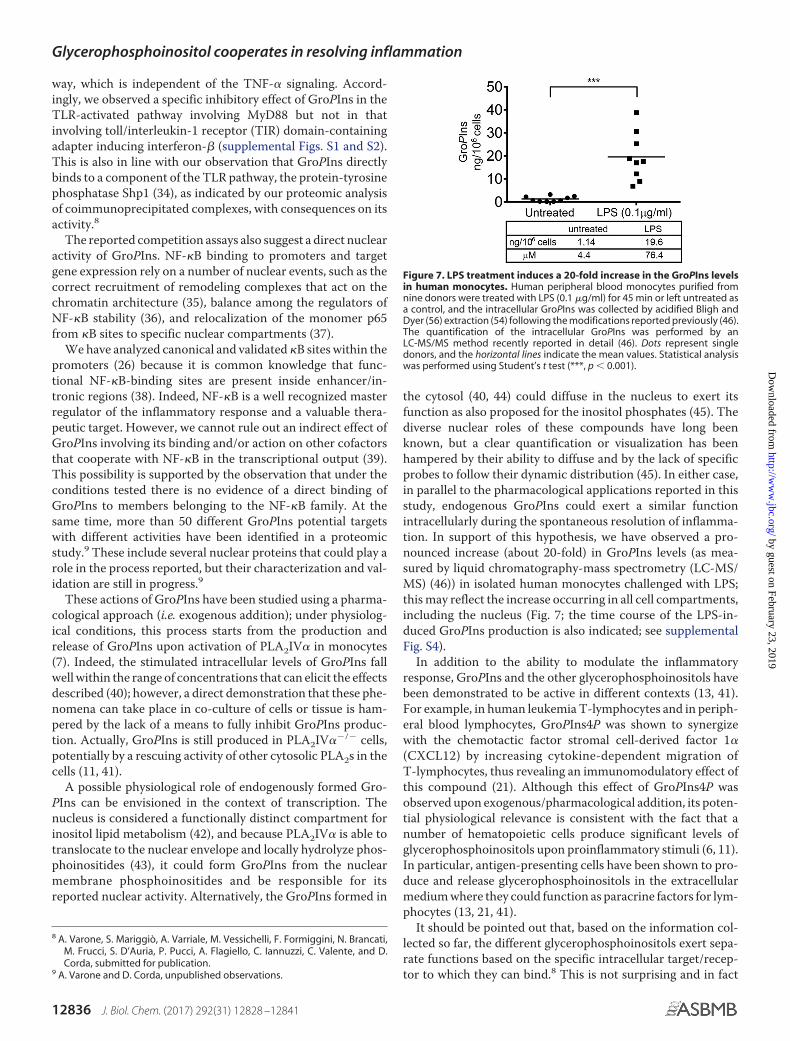
the cytosol (40, 44) could diffuse in the nucleus to exert itsfunction as also proposed for the inositol phosphates (45). Thediverse nuclear roles of these compounds have long beenknown, but a clear quantification or visualization has beenhampered by their ability to diffuse and by the lack of specificprobes to follow their dynamic distribution (45). In either case,in parallel to the pharmacological applications reported in thisstudy, endogenous GroPIns could exert a similar functionintracellularly during the spontaneous resolution of inflamma-tion. In support of this hypothesis, we have observed a pro-nounced increase (about 20-fold) in GroPIns levels (as mea-sured by liquid chromatography-mass spectrometry (LC-MS/MS) (46)) in isolated human monocytes challenged with LPS;this may reflect the increase occurring in all cell compartments,including the nucleus (Fig. 7; the time course of the LPS-in-duced GroPIns production is also indicated; see supplementalFig. S4).
In addition to the ability to modulate the inflammatoryresponse, GroPIns and the other glycerophosphoinositols havebeen demonstrated to be active in different contexts (13, 41).For example, in human leukemia T-lymphocytes and in periph-eral blood lymphocytes, GroPIns4P was shown to synergizewith the chemotactic factor stromal cell-derived factor 1�(CXCL12) by increasing cytokine-dependent migration ofT-lymphocytes, thus revealing an immunomodulatory effect ofthis compound (21). Although this effect of GroPIns4P wasobserved upon exogenous/pharmacological addition, its poten-tial physiological relevance is consistent with the fact that anumber of hematopoietic cells produce significant levels ofglycerophosphoinositols upon proinflammatory stimuli (6, 11).In particular, antigen-presenting cells have been shown to pro-duce and release glycerophosphoinositols in the extracellularmedium where they could function as paracrine factors for lym-phocytes (13, 21, 41).
It should be pointed out that, based on the information col-lected so far, the different glycerophosphoinositols exert sepa-rate functions based on the specific intracellular target/recep-tor to which they can bind.8 This is not surprising and in fact
8 A. Varone, S. Mariggiò, A. Varriale, M. Vessichelli, F. Formiggini, N. Brancati,M. Frucci, S. D’Auria, P. Pucci, A. Flagiello, C. Iannuzzi, C. Valente, and D.Corda, submitted for publication.
9 A. Varone and D. Corda, unpublished observations.
Figure 7. LPS treatment induces a 20-fold increase in the GroPIns levelsin human monocytes. Human peripheral blood monocytes purified fromnine donors were treated with LPS (0.1 �g/ml) for 45 min or left untreated asa control, and the intracellular GroPIns was collected by acidified Bligh andDyer (56) extraction (54) following the modifications reported previously (46).The quantification of the intracellular GroPIns was performed by anLC-MS/MS method recently reported in detail (46). Dots represent singledonors, and the horizontal lines indicate the mean values. Statistical analysiswas performed using Student’s t test (***, p � 0.001).
Glycerophosphoinositol cooperates in resolving inflammation
12836 J. Biol. Chem. (2017) 292(31) 12828 –12841
by guest on February 23, 2019http://w
ww
.jbc.org/D
ownloaded from

parallels the behavior of the other inositol-containing mole-cules, such as the phosphoinositide and inositol phosphatefamilies, which participate in different mechanisms accordingto the number of phosphate residues bound to the inositol ring(6). In addition, the metabolism of the inositol-containing mol-ecules needs to be thought of as a whole in the context of cellhomeostasis, and it depends on the available inositol pool in thedifferent cell systems and conditions (resting versus receptorstimulation and transformation (40)).
Here we have focused on the pharmacological exploitation ofGroPIns. The administration of GroPIns might act on mono-cytes to decrease the expression of cytokines and TF gene. Thiswould favor the resolution of inflammation and control poten-tially harmful prothrombotic activities. The same mechanismmight be involved in the GroPIns-mediated blood-brain barrierrepair mentioned above (33).
It is conceivable that the capability of GroPIns to regulate TFexpression might be exploited to control disseminated intra-vascular coagulation, the most devastating complication of sep-sis (14, 47), which remains untouched by specific therapeutictreatments. The advantages of using this compound in the res-olution of inflammation are severalfold. Because of its chemicalnature, GroPIns is easily manageable: it is water-soluble, and itcrosses the plasma membrane, rapidly reaching its intracellulartargets (48). As it is a natural compound that is present in vir-tually all cell types, it could be administered (e.g. intravenously)in cases of disseminated intravascular coagulation that do notrespond to therapy (i.e. septic shock) (47). These are proposalsthat require further evaluation in in vivo models of inflamma-tory disease. Nevertheless, GroPIns is a very promising com-pound as indicated by the reduced plasma levels of TNF-� itinduces in a model of endotoxin shock in mice (see “Results”).Moreover, GroPIns reduces the surface expression of Mac-1,which is involved in both chemokine-guided leukocyte infiltra-tion and detrimental inflammatory effector functions; indeed,the inhibition of Mac-1 has been shown to reduce both neutro-phil transmigration and tissue injury in vivo (49). Although it iswidely accepted to consider leukocyte infiltration as a commonmarker of inflammation, it is worth mentioning that neutro-phils can also perform active healing functions during resolu-tion (50); indeed, the inhibition of their trafficking towardinjured sites is a common and effective mechanism shared byseveral proresolving lipid mediators (4).
We conclude that GroPIns has the potential to regulate theinflammatory response. When produced by monocytes/macro-phages, GroPIns can act in a potential negative feedback loopthat signals the switching off of the inflammatory response and,in parallel, the inhibition of both expression10 and activity (51)of PLA2. In summary, by targeting proinflammatory and pro-thrombotic mediators and thereby affecting inflammatory-re-lated transcription factors and deactivating inflammatory cells,GroPIns would shape an anti-inflammatory microenvironmentfavoring the restoration of tissue homeostasis. In view of thisscenario, our data supportive the pharmacological exploitationof this compound (55).
Experimental procedures
Cell culture
Monocytes were isolated from whole blood obtained fromhealthy donors who gave their informed consent to participatein the study and who did not take any form of medication for atleast 10 days before blood donation. Approval was obtainedfrom the independent ethics committee, and buffy coats wereprovided by Istituto Nazionale per lo Studio e il Trattamentodei Tumori, Fondazione Giovanni Pascale (Naples, Italy). Initialexperiments were carried out on human monocytes obtainedwith approval from the Consorzio Mario Negri Sud ReviewBoard for these studies. The human monocytes were taken tomore than 95% purity through two centrifugation steps onLymphoprep (Axis-Shield, Oslo, Norway) followed by a Percoll(GE Healthcare) gradient as described previously (52) andfinally resuspended in serum-free RPMI 1640 medium (Gibco,Life Technologies). These monocytes were left unstimulated orwere challenged with 0.1 �g/ml bacterial LPS (E. coli serotype055:B5; Sigma-Aldrich) or TNF-� (210-TA-010/CF, R&D Sys-tems, Minneapolis, MN) with incubations for different times at37 °C in 5% CO2. GroPIns and GroPIns4P were provided byEchelon Biosciences Inc. HeLa cells were from American Tis-sue Type Collection (ATTC) and were grown in minimumEagle’s medium supplemented with 10% fetal calf serum (Bio-chrom, Cambridge, UK). Cells were grown under a controlledatmosphere in the presence of 5% CO2 at 37 °C.
Procoagulant activity
The procoagulant activity of TF was measured in cell lysatesaccording to one-stage clotting time. Briefly, 100 �l of cell lysatewas added to a tube containing 100 �l of prewarmed, poolednormal human plasma. After addition of 100 �l (20 mM) CaCl2,the clotting time was determined using a KC4A Amellungcoagulometer (Mascia Brunelli, Milan, Italy). The clottingtimes were converted to arbitrary units by interpolation with astandard curve generated with serial dilutions of human recom-binant thromboplastin (Hemoliance RecombiPlasTin, Instru-mentation Laboratory Co., Pleasantville, NY). As the concen-tration of recombinant TF (Hemoliance RecombiPlasTin) usedto produce the standard curve of procoagulant activity is notreported by the manufacturer, we measured the TF concentra-tions in the serial dilutions used to produce the standard activ-ity curve using an IMUBIND enzyme-linked immunosorbentassay (ELISA) (International Laboratory Co.). The correlationsbetween TF activity and the concentration of the TF antigen inthe recombinant TF preparations have been reported (52).
Western blotting
The levels of phosphorylation of p38, Erk1/2, and JNK wereanalyzed by Western blotting using the following phosphospe-cific antibodies: phospho-p38 MAPK (Thr-180/Tyr-182, cata-log number 9211, rabbit polyclonal, Cell Signaling Technology,Beverly, MA; 1:3000), phospho-JNK (Thr-183/Tyr-185, catalognumber 4668, rabbit polyclonal, Cell Signaling Technology;1:3000), and phospho-p44/42 MAPK (Erk1/2) (Thr-202/Tyr-204, catalog number 4370, rabbit monoclonal, Cell SignalingTechnology; 1:3000). The level of phosphorylated I�B� was10 M. Vessichelli, A. Varone, and D. Corda, unpublished observations.
Glycerophosphoinositol cooperates in resolving inflammation
J. Biol. Chem. (2017) 292(31) 12828 –12841 12837
by guest on February 23, 2019http://w
ww
.jbc.org/D
ownloaded from

analyzed using an antibody that specifically recognizes phos-phoserine 32/36 on I�B� (phospho-I�B�, catalog number9246, mouse monoclonal, Cell Signaling Technology; 1:3000).The levels of IKK�–� phosphorylation were analyzed using anantibody that recognizes phosphoserine 176/180 on IKK� andphosphoserine 177/181 on IKK� (phospho-IKK�–�, catalognumber 2697, rabbit monoclonal, Cell Signaling Technology;1:3000). In parallel, total proteins were monitored with the fol-lowing specific antibodies: p38 (p38� mitogen-activated pro-tein kinase, catalog number 9218, rabbit polyclonal, Cell Signal-ing Technology; 1:3000), JNK (SAPK/JNK, catalog number9258, rabbit monoclonal, Cell Signaling Technology; 1:3000),Erk1/2 (Erk1, sc-94, rabbit polyclonal, Santa Cruz Biotechnol-ogy, Inc., San Diego, CA; 1:3000), IKK� (catalog number 11930,mouse monoclonal, Cell Signaling Technology; 1:2000), andI�B� (catalog number 4812, rabbit polyclonal, Cell SignalingTechnology; 1:3000). The levels of glyceraldehyde-3-phosphatedehydrogenase (GAPDH) (catalog number 4699-9555, mousemonoclonal, AbD Serotec, Kidlington, Oxford, UK; 1:10,000)and vinculin (hVin-1, catalog number v9131, Sigma-Aldrich)were used as loading controls.
Real-time reverse transcription-polymerase chain reaction(RT-PCR)
Total RNA was extracted from the cells using the thiocya-nate/cesium chloride method. One microgram of total RNAwas converted to cDNA using Moloney murine leukemia virusreverse transcriptase (Applied Biosystems). Real-time PCRwere performed using 10 ng of cDNA, 50 nM concentration ofeach primer, and SYBR Green Master Mix (Applied Biosys-tems) in 20-�l reactions using an Applied Biosystems PRISM7500 Fast Real-time PCR System. The reverse transcriptionwas primed using random hexamers. The following TF genesequences were used as validated primers: cag tga ttc cct ccc gaaca (forward) and tgc ctt tct aca act gtg tag ag (reverse). Prelim-inary validation assays showed a band on agarose gel electro-phoresis of the expected size for amplification of TF cDNA insamples of monocytes/platelets co-incubated for 24 h. The TFcDNA band was present in samples treated with DNase I butnot in the samples in which Moloney murine leukemia virusreverse transcriptase was omitted, indicating that the signal isnot from contaminated genomic DNA (52). Similarly, real-timePCR measurements of mRNA expression of COX-2, TNF-�,IL-1�, and IL-10 were performed using the following validatedprimers: COX-2 forward, ttc cag atc cag agc tca tta aa; COX-2reverse, ccg gag cgg gaa gaa ct; TNF-� forward, cct gta gcc catgtt gta g; TNF-� reverse, tgg tta tct ctc agc tcc ac; IL-1� forward,gat gca cct gta cga tca ct; IL-1� reverse, gac atg gag aac aac act t;IL-10 forward, ttc ttc cct gtg aaa aca ag; IL-10 reverse, tca aac tcactc atg gct tt; GAPDH forward, caa ctt tgg tat cgt gga agg ac; andGAPDH reverse, aca gtc ttc ttg gtg gca gtg (GAPDH served asthe housekeeping gene). Each sample was measured in tripli-cate, and the data generated were analyzed with SDS 2.0 soft-ware (Applied Biosystems) using the �� method (2���CT) forcomparison of relative expression results. Resting monocytesincubated alone were considered as the reference sample.
ELISA
Conditioned medium from monocytes was sedimented bycentrifugation at 300 � g at room temperature for 10 min, andthe supernatant was recovered. The concentrations of TNF-�,IL-1�, and TxB2 were assessed according to the manufacturer’sprotocol (Amersham Biosciences).
NF-�B DNA-binding assay
Nuclear extracts for EMSA were prepared as reported previ-ously (53) with modifications. Monocytes, pretreated with Gro-PIns (300 �M; 20 min) and challenged with LPS (0.1 �g/ml),were harvested in PBS and centrifuged 5 min at 300 � g. Pelletswere lysed in buffer A (10 mM Hepes, pH 7.9, 1.5 mM MgCl2, 10mM KCl, 0.5 mM DTT) supplemented with protease and phos-phatase inhibitors (Roche Applied Science) and incubated for15 min at 4 °C. 10% Triton X-100 was added, and extracts werecentrifuged for 1 min at 12,000 � g. The supernatants wererecovered as the cytosolic fraction, and the pellets containingintact nuclei were lysed in buffer C (20 mM Hepes, pH 7.9, 25%(v/v %) glycerol, 0.4 mM NaCl, 1.5 mM MgCl2, 0.2 mM EDTA, 0.5mM DTT, 0.5 mM PMSF) supplemented with protease andphosphatase inhibitors and incubated for 30 min at 4 °C on ashaker. Lysates were centrifuged for 10 min at maximum speed,and the supernatants were recovered as the nuclear fraction.Nuclear extracts were incubated with 32P-radiolabeled (Amer-sham Biosciences) double-stranded oligonucleotide probescontaining the NF-�B-binding site, 5�-AgTTgAggggATTTC-CCAggC-3� (Sigma-Aldrich). Protein–DNA complexes wereseparated on a 5% non-denaturing acrylamide gel in 0.5� Trisborate-EDTA. Autoradiography was performed on Kodak XARfilm. For competition assays, monocytes were treated with LPS(0.1 �g/ml) or left untreated as a control, and nuclear lysateswere obtained as reported above. Different concentrations ofGroPIns were added to the nuclear lysates together with theradiolabeled probe. For supershift assays, p65 antibody (NF-�Bp65, catalog number 8242, rabbit monoclonal, Cell SignalingTechnology), p50 antibody (NF-�B1 p105/p50, catalog number12540, rabbit monoclonal, Cell Signaling Technology), or tubulin(sc-5286, mouse monoclonal, Santa Cruz Biotechnology, Inc.) wasadded together with the radiolabeled probe to the nuclear lysatesobtained from untreated or LPS-treated monocytes.
ChIP assay
Monocytes were treated with GroPIns (300 �M; 20 min) andthen challenged with LPS (0.1 �g/ml) for 60 min. Chromatin forthe immunoprecipitation was prepared by fixing the cells byadding 37% formaldehyde (Sigma-Aldrich) into the medium ata final concentration of 1% for 10 min and quenching with 125mM glycine (Sigma-Aldrich) for 5 min. Cells were collected andlysed in SDS buffer (1.1% SDS, 10 mM EDTA, pH 8, 50 mM Tris,pH 8) with protease inhibitors. Chromatin was sonicated forthree cycles of 10-s pulse at 30% of maximum power (BransonSonifier) to obtain fragments 500 –1000 kb in size. Sampleswere then centrifuged and diluted by 10-fold with dilutionbuffer (1% Triton X-100, 1.2 mM EDTA, pH 8, 16.7 mM Tris, pH8, 167 mM NaCl) with protease inhibitors and precleared with25 �l of 50% salmon sperm/protein A-agarose slurry (EMDMillipore) for 30 min at 4 °C. Precleared chromatin was incu-
Glycerophosphoinositol cooperates in resolving inflammation
12838 J. Biol. Chem. (2017) 292(31) 12828 –12841
by guest on February 23, 2019http://w
ww
.jbc.org/D
ownloaded from

bated overnight with 2 �g of p65 polyclonal antibody (7970,chip grade, Abcam, Cambridge, UK) or normal rabbit IgG(Santa Cruz Biotechnology, Inc.). A portion of the preclearedchromatin (10% of the sample) was stored at 4 °C as input tofurther quantitate the amount of immunoprecipitated DNA.The antibody–protein– chromatin complex was isolated with25 �l of 50% salmon sperm/protein A-agarose slurry (Milli-pore) for 2 h at 4 °C. After washing, pellets were eluted (1% SDS,0.1 M NaHCO3), reverse cross-linked with 5 M NaCl at 65 °Covernight, and treated with RNase A (Thermo Fisher Scientific,Waltham, MA) and Proteinase K (Thermo Fisher Scientific). Sam-ples were purified with a commercially available purification kit(Qiagen, Hilden, Germany) and used as templates in real-timePCR analysis. The intensity of the PCR signal is proportional to theoccupancy on the binding site. Normalization of data has beenperformed by using the percent input method: signals obtained byChIP/PCR have been divided by signals obtained from input/PCR.The validated primers used were: TNF-� forward, GCGATGGA-GAAGAAACCGAG; TNF-� reverse, GAGGGCGGGGAAAG-AATCA; TF forward, GCAACTAGACCCGCCTGC; and TFreverse, CTCCTCCCGGTAGGAAACTC.
Immunofluorescence
For nuclear trafficking of NF-�B, immunofluorescenceexperiments were performed as follows. Cells (3 � 105) wereseeded on glass coverslips in culture medium and treatedthe day after according to the experimental protocol. Cells wererinsed with PBS and fixed with 4% (w/v) paraformaldehyde for10 min at room temperature and permeabilized with blockingsolution containing 0.05% (w/v) saponin for 20 min. The intra-cellular localization of NF-�B was revealed using antibodies spe-cific to p65 (catalog number 7970, Abcam; dilution, 1:30) or p50(catalog number 12540, Cell Signaling Technology; dilution, 1:50)for 1 h at room temperature. After washes, cells were stained withAlexa Fluor 568-labeled (A10042, Molecular Probes) or AlexaFluor 488-labeled (A21206, Molecular Probes) secondary anti-body and 4,6-diamidino-2-phenylindole (DAPI) dye for 1 h atroom temperature. Coverslips were mounted in Mowiol (Calbi-ochem) and analyzed on a laser scanning confocal microscope(LSM710, Carl Zeiss) equipped with a 63� objective. The nucleartranslocation of NF-�B on monocytes treated with LPS in the pres-ence or absence of GroPIns was quantified by randomly counting100 cells/sample and expressing the percentage of cells labeledwith p50 or p65 nuclear staining for each sample.
Analysis of the intracellular GroPIns levels
Peripheral blood monocytes purified from nine healthydonors were treated with LPS (0.1 �g/ml) for 45 min or leftuntreated, and intracellular GroPIns was recovered by Blighand Dyer (56) extraction (54) with minor modifications (asdetailed under “Sample preparation” in Ref.46). The quantita-tive analysis of GroPIns was performed by an LC-MS/MSmethod as reported previously (46).
Mouse experiments
Mouse treatments—C57BL/6J male mice (9 –12 weeks old)were obtained from Charles River Laboratories (Calco, Lecco,Italy), fed ad libitum, and housed in a temperature-controlled
and light-controlled room. Treatments were approved by theEthics Committee of the Consorzio Mario Negri Sud. GroPIns(Euticals S.p.A, Lodi, Italy) or saline (vehicle) was given intra-peritoneally (i.p.). The mice were treated with three doses ofGroPIns (10 mg/kg) administered 15 min before, at the sametime as, and 60 min after LPS administration (1 mg/kg; E. coliserotype 055:B5). Two hours after LPS administration, the micewere anesthetized with a mixture of ketamine and xylazine (100and 20 mg/kg, respectively), and their blood was collectedin 0.38% trisodium citrate (final concentration) by cardiacpuncture.
Measurement of TNF-� cytokine—Blood was centrifuged for10 min to obtain platelet-poor plasma. This plasma from eachmouse was divided in aliquots and stored at �80 °C before anal-ysis. Mouse TNF-� was measured using ELISA kits (PierceBiotechnology).
Flow cytometric analysis of Mac-1 expression in neutrophilsfrom mouse whole blood—Citrated whole blood was diluted 1:1with citrated saline and incubated for 15 min with phycoeryth-rin-conjugated rat anti-mouse Mac-1 (clone M1/70, BDPharmingenTM). Nonspecific phycoerythrin-conjugated ratIgG was used as the staining negative control. After staining, thered blood cells were lysed by addition of lysing-fixing solution(BD Biosciences) according to the manufacturer’s instructions.Flow cytometry analysis was performed with a FACStar flowcytometer as follows. Events showing side light scatter (granu-larity) and forward light scatter (dimension) characteristic ofpolymorphonuclear leukocytes were analyzed for Mac-1expression levels. The data are shown as mean fluorescenceintensities for each of the nine mice analyzed.
Statistical analysis
The data are expressed as means � S.E. Statistical differencesbetween two groups were determined by paired t tests. To testfordifferencesacrossdifferenttreatmentgroups,repeated-mea-sures analysis of variance and Dunnett tests were used. Statis-tical significance was defined as p � 0.05.
Author contributions—M. V. and P. Z. designed, performed, andanalyzed all the experiments and co-wrote the manuscript. A. V.contributed to the in vitro experiments and to the discussion. S. M.and V. E. designed and analyzed the initial experiments; V. E. alsoco-wrote the manuscript. M. Z and T. d. C. performed and discussedthe EMSA. A. D. S., C. A, and G. D.’ E. performed all the in vivoexperiments and part of the in vitro experiments. A. C. and A. F.performed and discussed LC-MS/MS analysis. C. C. and G. D. C.provided buffy coats. D. C. conceived and supervised the project,discussed and analyzed the data, and co-wrote the manuscript.
Acknowledgments—We thank C. Valente (Institute of Protein Bio-chemistry (IBP), National Research Council (CNR)) for assistancewith the immunofluorescence analyses and discussion, D. Acampora(Institute of Genetics and Biophysics, CNR) for assistance in ChIPanalysis, D. Boraschi and A. Luini (IBP, CNR) for discussion andcritical reading of the manuscript, L. Grauso for help in theLC-MS/MS analysis, N. Martelli for FACS analysis, and A. Capasso(IBP, CNR) for help with the biochemical assays.
Glycerophosphoinositol cooperates in resolving inflammation
J. Biol. Chem. (2017) 292(31) 12828 –12841 12839
by guest on February 23, 2019http://w
ww
.jbc.org/D
ownloaded from

References1. Medzhitov, R. (2008) Origin and physiological roles of inflammation. Na-
ture 454, 428 – 4352. Dinarello, C. (2007) Historical insights into cytokines. Eur. J. Immunol 37,
S34 –S453. Dennis, E. A., and Norris, P. C. (2015) Eicosanoid storm in infection and
inflammation. Nat. Rev. Immunol. 15, 511–5234. Serhan, C. N., Chiang, N., Dalli, J., and Levy, B. D. (2014) Lipid mediators
in the resolution of inflammation. Cold Spring Harb. Perspect. Biol. 7,a016311
5. Nathan, C., and Ding, A. (2010) Nonresolving inflammation. Cell 140,871– 882
6. Corda, D., Iurisci, C., and Berrie, C. P. (2002) Biological activities andmetabolism of the lysophosphoinositides and glycerophosphoinositols.Biochim. Biophys. Acta 1582, 52– 69
7. Zizza, P., Iurisci, C., Bonazzi, M., Cossart, P., Leslie, C. C., Corda, D., andMariggiò, S. (2012) Phospholipase A2IV� regulates phagocytosis indepen-dent of its enzymatic activity. J. Biol. Chem. 287, 16849 –16859
8. Mariggiò, S., Sebastià, J., Filippi, B. M., Iurisci, C., Volonté, C., Amadio, S.,De Falco, V., Santoro, M., and Corda, D. (2006) A novel pathway of cellgrowth regulation mediated by a PLA2�-derived phosphoinositide me-tabolite. FASEB J. 20, 2567–2569
9. Filippi, B. M., Mariggiò, S., Pulvirenti, T., and Corda, D. (2008) SRC-de-pendent signalling regulates actin ruffle formation induced by glycero-phosphoinositol 4-phosphate. Biochim. Biophys. Acta 1783, 2311–2322
10. Mancini, R., Piccolo, E., Mariggiò, S., Filippi, B. M., Iurisci, C., Pertile, P.,Berrie, C. P., and Corda, D. (2003) Reorganization of actin cytoskeleton bythe phosphoinositide metabolite glycerophosphoinositol 4-phosphate.Mol. Biol. Cell 14, 503–515
11. Corda, D., Zizza, P., Varone, A., Filippi, B. M., and Mariggiò, S. (2009) Theglycerophosphoinositols: cellular metabolism and biological functions.Cell. Mol. Life Sci. 66, 3449 –3467
12. Falasca, M., Iurisci, C., Carvelli, A., Sacchetti, A., and Corda, D. (1998)Release of the mitogen lysophosphatidylinositol from H-Ras-transformedfibroblasts; a possible mechanism of autocrine control of cell proliferation.Oncogene 16, 2357–2365
13. Corda, D., Zizza, P., Varone, A., Bruzik, K. S., and Mariggiò, S. (2012) Theglycerophosphoinositols and their cellular functions. Biochem. Soc. Trans.40, 101–107
14. Levi, M. (2014) Diagnosis and treatment of disseminated intravascularcoagulation. Int. J. Lab. Hematol. 36, 228 –236
15. Suzuki, T., Hashimoto, S., Toyoda, N., Nagai, S., Yamazaki, N., Dong,H. Y., Sakai, J., Yamashita, T., Nukiwa, T., and Matsushima, K. (2000)Comprehensive gene expression profile of LPS-stimulated human mono-cytes by SAGE. Blood 96, 2584 –2591
16. Lawrence, T., Willoughby, D. A., and Gilroy, D. W. (2002) Anti-inflam-matory lipid mediators and insights into the resolution of inflammation.Nat. Rev. Immunol. 2, 787–795
17. Murray, P. J. (2006) Understanding and exploiting the endogenous inter-leukin-10/STAT3-mediated anti-inflammatory response. Curr. Opin.Pharmacol. 6, 379 –386
18. O’Neill, L. A., Golenbock, D., and Bowie, A. G. (2013) The history ofToll-like receptors—redefining innate immunity. Nat. Rev. Immunol. 13,453– 460
19. Nemerson, Y. (1988) Tissue factor and hemostasis. Blood 71, 1– 820. Kim, J. H., Na, H. K., Pak, Y. K., Lee, Y. S., Lee, S. J., Moon, A., and Surh, Y. J.
(2008) Roles of ERK and p38 mitogen-activated protein kinases in phorbolester-induced NF-�B activation and COX-2 expression in human breastepithelial cells. Chem. Biol. Interact. 171, 133–141
21. Patrussi, L., Mariggiò, S., Paccani, S. R., Capitani, N., Zizza, P., Corda, D.,and Baldari, C. T. (2007) Glycerophosphoinositol-4-phosphate enhancesSDF-1�-stimulated T-cell chemotaxis through PTK-dependent activa-tion of Vav. Cell. Signal. 19, 2351–2360
22. Sweet, M. J., and Hume, D. A. (1996) Endotoxin signal transduction inmacrophages. J. Leukoc. Biol. 60, 8 –26
23. Müller, J. M., Ziegler-Heitbrock, H. W., and Baeuerle, P. A. (1993) Nuclearfactor �B, a mediator of lipopolysaccharide effects. Immunobiology 187,233–256
24. Karin, M. (1999) How NF-�B is activated: the role of the I�B kinase (IKK)complex. Oncogene 18, 6867– 6874
25. Lewin, S. R., Lambert, P., Deacon, N. J., Mills, J., and Crowe, S. M. (1997)Constitutive expression of p50 homodimer in freshly isolated humanmonocytes decreases with in vitro and in vivo differentiation: a possiblemechanism influencing human immunodeficiency virus replication inmonocytes and mature macrophages. J. Virol. 71, 2114 –2119
26. Lim, C. A., Yao, F., Wong, J. J., George, J., Xu, H., Chiu, K. P., Sung, W. K.,Lipovich, L., Vega, V. B., Chen, J., Shahab, A., Zhao, X. D., Hibberd, M.,Wei, C. L., Lim, B., et al. (2007) Genome-wide mapping of RELA(p65)binding identifies E2F1 as a transcriptional activator recruited by NF-�Bupon TLR4 activation. Mol. Cell 27, 622– 635
27. Oeth, P. A., Parry, G. C., Kunsch, C., Nantermet, P., Rosen, C. A., andMackman, N. (1994) Lipopolysaccharide induction of tissue factor geneexpression in monocytic cells is mediated by binding of c-Rel/p65 het-erodimers to a �B-like site. Mol. Cell. Biol. 14, 3772–3781
28. Udalova, I. A., Knight, J. C., Vidal, V., Nedospasov, S. A., and Kwiatkowski,D. (1998) Complex NF-�B interactions at the distal tumor necrosis factorpromoter region in human monocytes. J. Biol. Chem. 273, 21178 –21186
29. Lowell, C. A., and Berton, G. (1998) Resistance to endotoxic shock andreduced neutrophil migration in mice deficient for the Src-family kinasesHck and Fgr. Proc. Natl. Acad. Sci. U.S.A. 95, 7580 –7584
30. Kolaczkowska, E., and Kubes, P. (2013) Neutrophil recruitment and func-tion in health and inflammation. Nat. Rev. Immunol. 13, 159 –175
31. Wright, H. L., Moots, R. J., Bucknall, R. C., and Edwards, S. W. (2010)Neutrophil function in inflammation and inflammatory diseases. Rheu-matology 49, 1618 –1631
32. Serhan, C. N. (2011) The resolution of inflammation: the devil in the flaskand in the details. FASEB J. 25, 1441–1448
33. Cucullo, L., Hallene, K., Dini, G., Dal Toso, R., and Janigro, D. (2004)Glycerophosphoinositol and dexamethasone improve transendothelialelectrical resistance in an in vitro study of the blood-brain barrier. BrainRes. 997, 147–151
34. An, H., Hou, J., Zhou, J., Zhao, W., Xu, H., Zheng, Y., Yu, Y., Liu, S., andCao, X. (2008) Phosphatase SHP-1 promotes TLR- and RIG-I-activatedproduction of type I interferon by inhibiting the kinase IRAK1. Nat. Im-munol. 9, 542–550
35. Natoli, G. (2009) Control of NF-�B-dependent transcriptional responsesby chromatin organization. Cold Spring Harb. Perspect. Biol.[ 1, a000224
36. Neumann, M., and Naumann, M. (2007) Beyond I�Bs: alternative regula-tion of NF-�B activity. FASEB J. 21, 2642–2654
37. Ghosh, S., and Hayden, M. S. (2008) New regulators of NF-�B in inflam-mation. Nat. Rev. Immunol. 8, 837– 848
38. Martone, R., Euskirchen, G., Bertone, P., Hartman, S., Royce, T. E.,Luscombe, N. M., Rinn, J. L., Nelson, F. K., Miller, P., Gerstein, M., Weiss-man, S., and Snyder, M. (2003) Distribution of NF-�B-binding sites acrosshuman chromosome 22. Proc. Natl. Acad. Sci. U.S.A. 100, 12247–12252
39. Krappmann, D., Wegener, E., Sunami, Y., Esen, M., Thiel, A., Mordmuller,B., and Scheidereit, C. (2004) The I�B kinase complex and NF-�B act asmaster regulators of lipopolysaccharide-induced gene expression andcontrol subordinate activation of AP-1. Mol. Cell. Biol. 24, 6488 – 6500
40. Berrie, C. P., Dragani, L. K., van der Kaay, J., Iurisci, C., Brancaccio, A.,Rotilio, D., and Corda, D. (2002) Maintenance of PtdIns45P2 pools underlimiting inositol conditions, as assessed by liquid chromatography-tan-dem mass spectrometry and PtdIns45P2 mass evaluation in Ras-trans-formed cells. Eur. J. Cancer 38, 2463–2475
41. Patrussi, L., Mariggiò, S., Corda, D., and Baldari, C. T. (2013) The glycero-phosphoinositols: from lipid metabolites to modulators of T-cell signal-ing. Front. Immunol. 4, 213
42. Faenza, I., Fiume, R., Piazzi, M., Colantoni, A., and Cocco, L. (2013) Nu-clear inositide specific phospholipase C signalling—interactions and ac-tivity. FEBS J. 280, 6311– 6321
43. Schievella, A. R., Regier, M. K., Smith, W. L., and Lin, L. L. (1995) Calcium-mediated translocation of cytosolic phospholipase A2 to the nuclear en-velope and endoplasmic reticulum. J. Biol. Chem. 270, 30749 –30754
Glycerophosphoinositol cooperates in resolving inflammation
12840 J. Biol. Chem. (2017) 292(31) 12828 –12841
by guest on February 23, 2019http://w
ww
.jbc.org/D
ownloaded from

44. Dragani, L. K., Berrie, C. P., Corda, D., and Rotilio, D. (2004) Analysis ofglycerophosphoinositol by liquid chromatography-electrospray ioni-sation tandem mass spectrometry using a �-cyclodextrin-bondedcolumn. J. Chromatogr. B Analyt. Technol. Biomed. Life Sci. 802,283–289
45. Fisher, S. K., Novak, J. E., and Agranoff, B. W. (2002) Inositol and higherinositol phosphates in neural tissues: homeostasis, metabolism and func-tional significance. J. Neurochem. 82, 736 –754
46. Grauso, L., Mariggiò, S., Corda, D., Fontana, A., and Cutignano, A. (2015)An improved UPLC-MS/MS platform for quantitative analysis of glycero-phosphoinositol in mammalian cells. PLoS One 10, e0123198
47. Angus, D. C., and van der Poll, T. (2013) Severe sepsis and septic shock.N. Engl. J. Med. 369, 2063
48. Mariggiò, S., Iurisci, C., Sebastià, J., Patton-Vogt, J., and Corda, D. (2006)Molecular characterization of a glycerophosphoinositol transporter inmammalian cells. FEBS Lett. 580, 6789 – 6796
49. Hirahashi, J., Hishikawa, K., Kaname, S., Tsuboi, N., Wang, Y., Simon, D. I.,Stavrakis, G., Shimosawa, T., Xiao, L., Nagahama, Y., Suzuki, K., Fujita, T.,and Mayadas, T. N. (2009) Mac-1 (CD11b/CD18) links inflammation andthrombosis after glomerular injury. Circulation 120, 1255–1265
50. Colgan, S. P. (2015) Neutrophils and inflammatory resolution in the mu-cosa. Semin. Immunol. 27, 177–183
51. Corda, D., and Falasca, M. (1996) Glycerophosphoinositols as potentialmarkers of ras-induced transformation and novel second messengers. An-ticancer Res. 16, 1341–1350
52. Di Santo, A., Amore, C., Dell’Elba, G., Manarini, S., and Evangelista, V.(2011) Glycogen synthase kinase-3 negatively regulates tissue factor ex-pression in monocytes interacting with activated platelets. J. Thromb.Haemost. 9, 1029 –1039
53. Dignam, J. D., Lebovitz, R. M., and Roeder, R. G. (1983) Accurate tran-scription initiation by RNA polymerase II in a soluble extract from isolatedmammalian nuclei. Nucleic Acids Res. 11, 1475–1489
54. Berrie, C. P., Iurisci, C., Piccolo, E., Bagnati, R., and Corda, D. (2007)Analysis of phosphoinositides and their aqueous metabolites. MethodsEnzymol. 434, 187–232
55. Corda, D., Zizza, P., Luini, A., and Mariggiò, S. (May 31, 2016) Use ofGlycerophosphoinositols for the Treatment of Septic Shock, U. S. Patent9351983
56. Bligh, E. G., and Dyer, W. J. (1959) A rapid method of total lipid extractionand purification. Can. J. Biochem. Physiol. 37, 911–917
Glycerophosphoinositol cooperates in resolving inflammation
J. Biol. Chem. (2017) 292(31) 12828 –12841 12841
by guest on February 23, 2019http://w
ww
.jbc.org/D
ownloaded from

Cristofaro, Virgilio Evangelista and Daniela CordaFontana, Carmela Cacciapuoti, Gaetano Di Costanzo, Mariastella Zannini, Tiziana de
Angelomaria Di Santo, Concetta Amore, Giuseppe Dell'Elba, Adele Cutignano, Angelo Mariangela Vessichelli, Stefania Mariggiò, Alessia Varone, Pasquale Zizza,
lipopolysaccharide-induced inflammatory and thrombotic responsesThe natural phosphoinositide derivative glycerophosphoinositol inhibits the
doi: 10.1074/jbc.M116.773861 originally published online June 9, 20172017, 292:12828-12841.J. Biol. Chem.
10.1074/jbc.M116.773861Access the most updated version of this article at doi:
Alerts:
When a correction for this article is posted•
When this article is cited•
to choose from all of JBC's e-mail alertsClick here
Supplemental material:
http://www.jbc.org/content/suppl/2017/06/09/M116.773861.DC1
http://www.jbc.org/content/292/31/12828.full.html#ref-list-1
This article cites 55 references, 14 of which can be accessed free at
by guest on February 23, 2019http://w
ww
.jbc.org/D
ownloaded from