The Wilson 3D Quad-Helix and Maxillary Expansion · The Wilson 3D Quad-Helix and Maxillary...
23
The Wilson 3D Quad-Helix and Maxillary Expansion A narrow maxilla is a common problem in orthodontics. Approximately 80% of orthodontic patients need some type of arch expansion. 1 The incidence of posterior cross-bite is high and is present in more than 50% of the orthodontic cases, with the upper molars being affected in more than 80% of the cases, and the lower molars affected in more than 19% of those cases. 2 A narrow upper arch can produce undesired transverse growth changes. In order to intercept abnormal development and properly guide the patient’s growth into a physiological pattern, it is necessary to expand the maxilla. Maxillary expansion will avoid occlusion problems that can produce oclusal and facial disharmony (asymmetries). The cross-bite can not be corrected without treatment, regardless of the etiology and modality of clinical occurrence. 3 Early cross-bite corrections lead to a stable and normal occlusion pattern, and contribute to symmetrical condile growth, harmonious TMJ, and overall growth in the mandible. 4-7 Young patients should start visiting the orthodontist around 4 years of age. Thus, the orthodontist can identify and intercept a narrow maxilla early, avoiding late treatment and the risk of create a symmetrical occlusion in an asymmetrical skeletal system. Waiting until after 9 years old can lead to TMJ problems and future relapse. 8 Correcting the narrow maxilla fostered an increase in the Mandibular Width measurement and released the mandible to a normal transverse growth. When considering arch expansion, the practitioner should always consider proper diagnosis and planning procedures in the three planes of the space, converting information from the models, comprehensive cephalometrics analysis (lateral and frontal) and divine proportions analysis. 29 The postero-anterior radiograph is a very important tool to be used when analyzing the transverse plane. Maxillary expansion procedures can be divided in two major categories, according to previous literature. The first, Rapid Maxillary Expansion or RME, is a procedure that is generally accomplished by using an appliance that incorporates a screw, for example a Haas or Hyrax. These appliances tend to disrupt the midpalatal suture. The second category for maxillary expansion is the slow maxillary expansion group. These appliances apply slow and continuous forces which do not attempt, as a main objective, to open the midpalatal suture. These appliances include: removable expansion plates, Porter W arch, and Quad-Helix. The Quad-Helix was developed in 1975 by Robert Murray Ricketts from Porter’s “W” arch, adding four loops to the appliance, increasing the wire length on 40 to
Transcript of The Wilson 3D Quad-Helix and Maxillary Expansion · The Wilson 3D Quad-Helix and Maxillary...

The Wilson 3D Quad-Helix and Maxillary Expansion
A narrow maxilla is a common problem in orthodontics. Approximately 80% of orthodontic patients need
some type of arch expansion.1 The incidence of posterior cross-bite is high and is present in more than 50% of the
orthodontic cases, with the upper molars being affected in more than 80% of the cases, and the lower molars affected
in more than 19% of those cases.2
A narrow upper arch can produce undesired transverse growth changes. In order to intercept abnormal
development and properly guide the patient’s growth into a physiological pattern, it is necessary to expand the
maxilla. Maxillary expansion will avoid occlusion problems that can produce oclusal and facial disharmony
(asymmetries). The cross-bite can not be corrected without treatment, regardless of the etiology and modality of
clinical occurrence.3 Early cross-bite corrections lead to a stable and normal occlusion pattern, and contribute to
symmetrical condile growth, harmonious TMJ, and overall growth in the mandible.4-7 Young patients should start
visiting the orthodontist around 4 years of age. Thus, the orthodontist can identify and intercept a narrow maxilla
early, avoiding late treatment and the risk of create a symmetrical occlusion in an asymmetrical skeletal system.
Waiting until after 9 years old can lead to TMJ problems and future relapse.8
Correcting the narrow maxilla fostered an increase in the Mandibular Width measurement and released the
mandible to a normal transverse growth.
When considering arch expansion, the practitioner should always consider proper diagnosis and planning
procedures in the three planes of the space, converting information from the models, comprehensive cephalometrics
analysis (lateral and frontal) and divine proportions analysis.29 The postero-anterior radiograph is a very important
tool to be used when analyzing the transverse plane.
Maxillary expansion procedures can be divided in two major categories, according to previous literature.
The first, Rapid Maxillary Expansion or RME, is a procedure that is generally accomplished by using an appliance
that incorporates a screw, for example a Haas or Hyrax. These appliances tend to disrupt the midpalatal suture. The
second category for maxillary expansion is the slow maxillary expansion group. These appliances apply slow and
continuous forces which do not attempt, as a main objective, to open the midpalatal suture. These appliances include:
removable expansion plates, Porter W arch, and Quad-Helix. The Quad-Helix was developed in 1975 by Robert
Murray Ricketts from Porter’s “W” arch, adding four loops to the appliance, increasing the wire length on 40 to
50mm. The objective was softening the forces and better control molar rotations.32 Many authors have written that the
Quad-Helix appliance can deliver sufficient forces to promote skeletal changes on maxillary bone in younger patients
(during deciduous and mixed dentitions phases). 2, 7, 10-13, 15, 17-19
Slow maxillary expansion, using the Quad-Helix appliance, is a recommended choice and it is widely
accepted and applied by orthodontists. Many practitioners prefer the Quad-Helix as an expansion device because it is
a very versatile appliance, with applications such as: molar rotation control, torque and tipping control. It can also
produce advancement in the incisor region and create greater anterior expansion, resulting in an improved arch form
(taking advantage of the anterior arms that deliver a “sweeping action”). Furthermore the practitioners don’t need the
patient’s or parent’s cooperation to reach the set objectives.7, 19-21
Transverse maxillary expansion is achieved using a combination of movements, such as: buccal tooth
version (A), alveolar bone and molar buccal translation combined with molar torque control (B), midpalatal suture
opening and buccal molar translation (C), midpalatal suture disrupting (D), and a combination of two or more of
those factors (Figure 1).3
It is possible, when the treatment plan demands, to open the mid palatal suture on a young growing patient
from 400g of transverse pressure applied. 10,22
The amount of force delivered by the Quad-helix depends on two major factors: Quad- Helix construction
and amount of activation. Basically the Quad-Helix is constructed by 4 helicoids on .036 round wire. Dr. Ricketts
recommends the use of blue Elgiloy wire to deliver softer amount of forces and easier bending.
FIGURE 1. Expansion Movement Possibilities for the Maxilla and Upper Molars.

In general, using the Quad-Helix for treatment leads to skeletal changes in maxillary bone, when
desired by the practitioner and indicated in the treatment objectives. Adjustments are made by simply changing the
amount and frequency of the activations. The Quad-Helix can provide a force range from 221 grams to 1149 grams.
The Quad-Helix can rotate the supporting molars and it can be adjusted to expand the molars and anterior teeth
differentially.17 It can also be used to control molar torquing. These features make the Quad-Helix a very versatile
appliance.
It is observed that when correctly employed, the Quad-Helix can produce similar results to the RMEs and
also correct all transverse problems in growing patients. 8 These findings also coincide with what Cotton concluded
after his work with monkeys.26 Hicks reported substantial skeletal changes with slow expansion, especially in
younger children.11 Additionally, slow expansion is related to a more physiological reorganization of the maxilla in
the three planes of the space, providing more stability and less relapse possibilities than RMEs. We can observe these
findings in the works produced by Ohshima26 and Storey. 27
Usually the conventional Quad-Helix is cemented pre activated with certain amount of expansion. When the
case being treated needs extra amounts of activations, normally the clinicians can due it using of a three jaw plier
inside the mount. This modality of activation strongly depends of the practicer experience to control the amount of
force and movements delivered. Due to this situation, it is found on the literature some authors that recommends
remove the Quad-Helix out from the mouth to place new actions and recement it after these changes. To avoid
removing and recementing the bands, many practicers usually construct the Quad-Helix to be inserted on lingual
sheaths tubes for horizontal insertion and removing. Also, it is find this kind of Quad-Helix pre fabricated from many
ortho manufacturers.
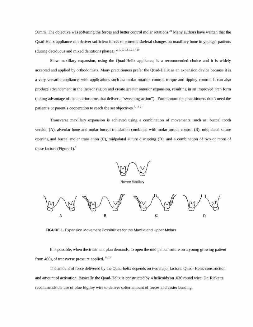
On 1983 Wilson & Wilson30 presented to orthodontics community an inserting/removing system called as
3D fixed/removable appliances. This kind of insertion brought to the practicers versatility and easier
inserting/removing procedures due to an innovative vertical inserction. Using Wilson’s 3D Quad-Helix it is possible,
very easier; control the molars on the three planes of space during all expansion movements. Its fitting system is
composed by stamped two posts laser soldered to the Blue Elgiloy .038” Quad-Helix (Fig. 2) and a vertical inserting
tubes (Fig. 3). The 3D Quad-Helix very precisely allow the orthodontists control the amount of forces employed and
control molars on the three planes of the space, strongly increasing movements control. Dr Wilson recommends
installing the appliance, at first patient visit, absolute passive to malocclusion and starting to activate the 3D Quad-

Helix on a second visit. New activations should be posted on 40 to 40 days period; on majority of cases the activation
can not exceed 1 to 2mm in order to keep case under control (see Figs 4, 5 and 6). Also, as it is pre fabricated on 6
different sizes, the orthodontists can save time and money, avoiding laboratorial steps and install it on chair side with
quite of few adaptations. No doubts, the launch of the Wilson 3D Quad-Helix in the market boosts the control of
expansion forces, the inserting/removing system, keeping the molars properly torqued, tipped and rotated during all
the expansion moments.
The clinical case 1 shown on Figs. 7 and 8 exemplifies the expansion and molars 3D control using the 3D
Quad-Helix. It is noticeable how the upper molars were expanded with complete torque control.
On case 2 it is easy to see the features and possibilities of the 3D Quad-Helix during an expansion treatment.
Note the severe transverse problem on the beginning and the high amount of expansion obtained after treatment,
noticeable on models measurement, an 8mm of total molar expansion. The P.A. tracings showed 2.3mm increasing of
J-J width, 8.6mm on upper molars width and 3.1mm enlargement on Nasal Cavity width. Similarly to Case 1 the
upper molar had the upper molars torque properly controlled.
The full 3D system kit also contents other appliances and the orthodontist can choose according to the needs
the right appliance for each case, and/or exchange the appliance during the treatment without remove the molar
bands. Dr Wilson calls this full kit as Wilson 3D Tool Box.
I strongly recommend to the orthodontists to use the vertical inserting system developed by Dr Wilson, we
can keep expansions and upper molars fully 3D controlled due to the inventive fitting system, save our precious time
cutting off lab steps and also it is cost effective. No doubt a great upgrade on Dr Ricketts invention!
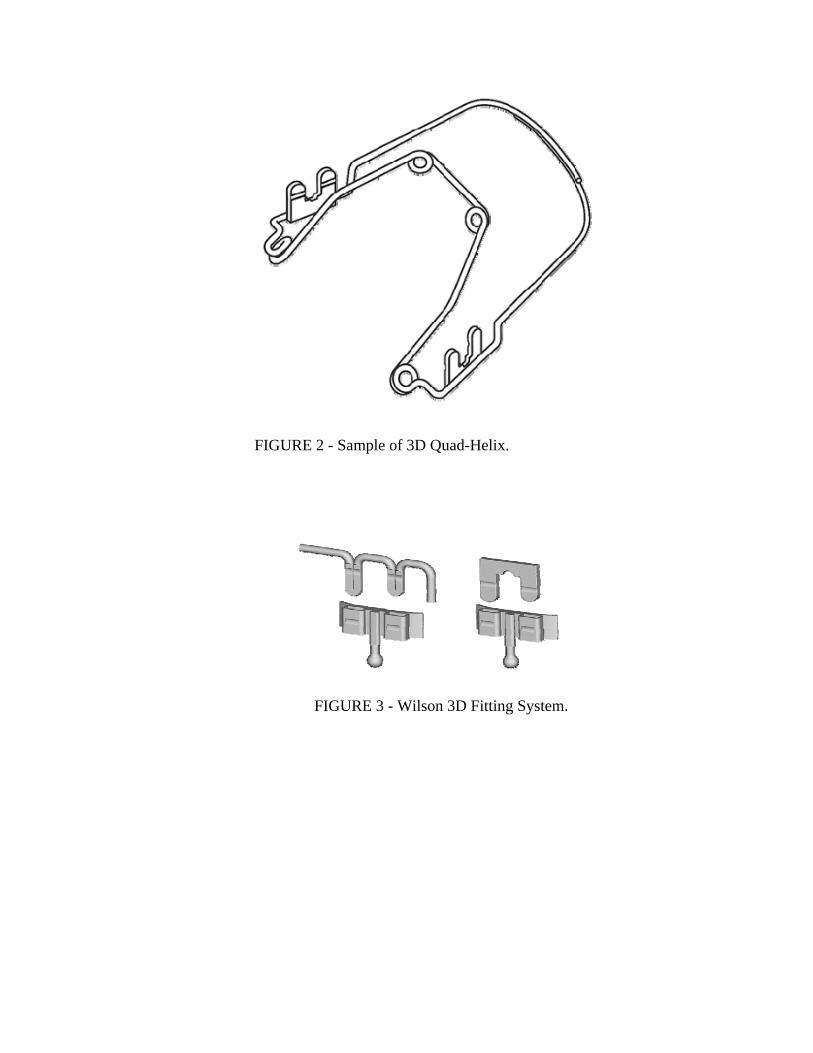
FIGURE 2 - Sample of 3D Quad-Helix.
FIGURE 3 - Wilson 3D Fitting System.
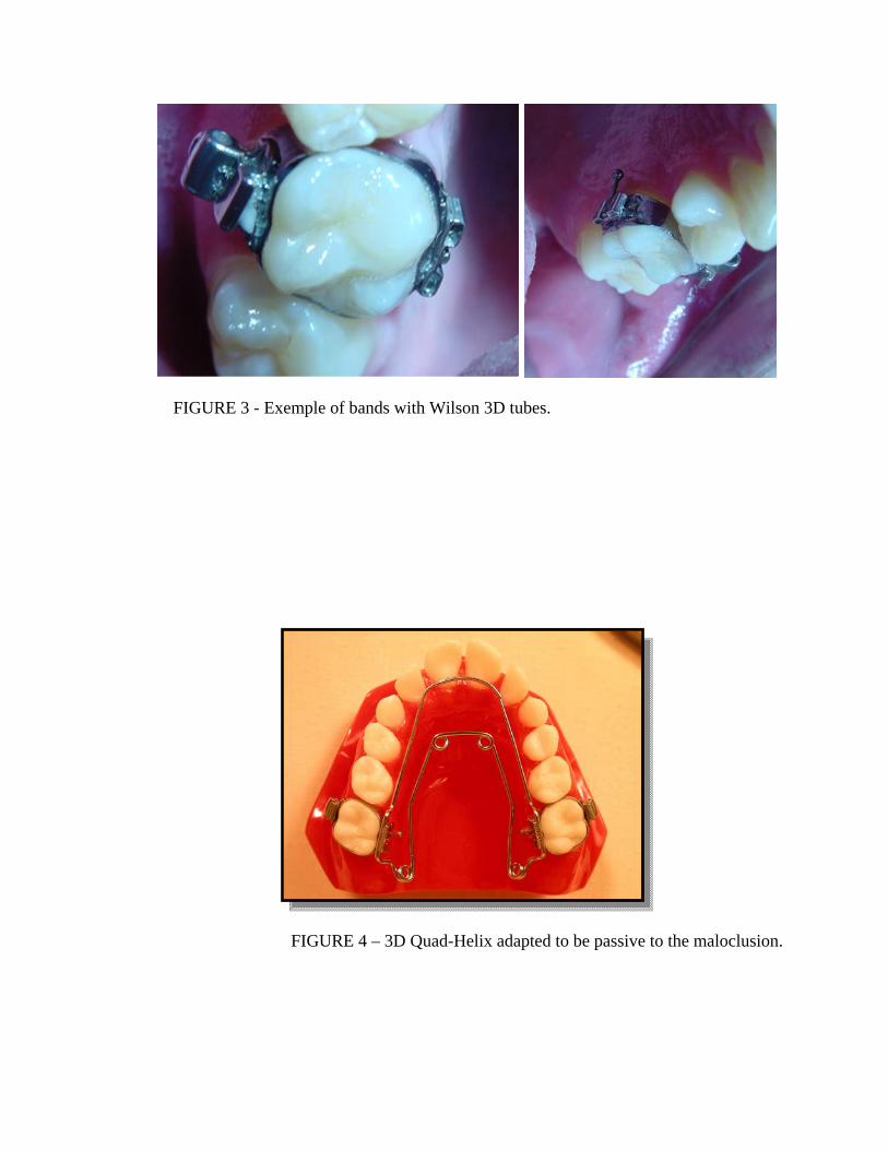
FIGURE 3 - Exemple of bands with Wilson 3D tubes.
FIGURE 4 – 3D Quad-Helix adapted to be passive to the maloclusion.

FIGURE 5 – Checking the amount of expansion forces.
FIGURE 6 – Checking the amount of rotation forces.

FIGURE 7 – Expansion case 1 sample after 4 months – note molar rotation.
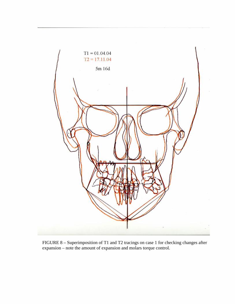
FIGURE 8 – Superimposition of T1 and T2 tracings on case 1 for checking changes after expansion – note the amount of expansion and molars torque control.
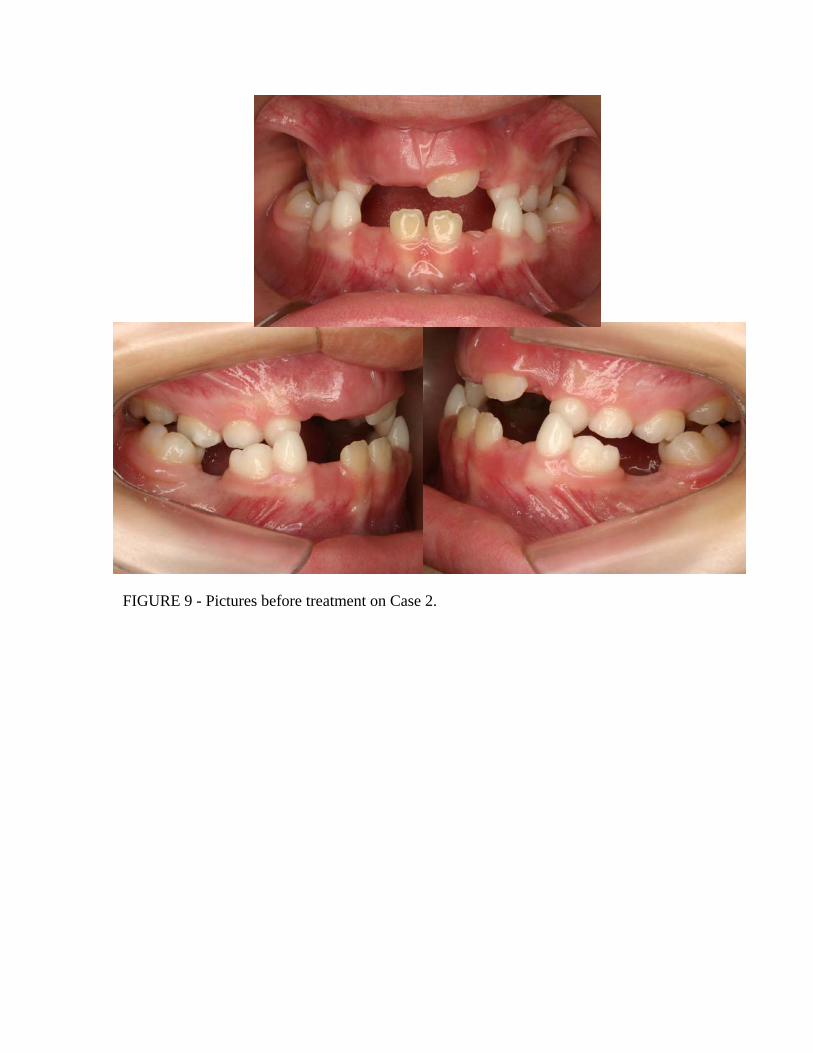
FIGURE 9 - Pictures before treatment on Case 2.

FIGURE 10 - Oclusal view before treatment on Case 2.
FIGURE 11 - Models pictures before treatment on Case 2.
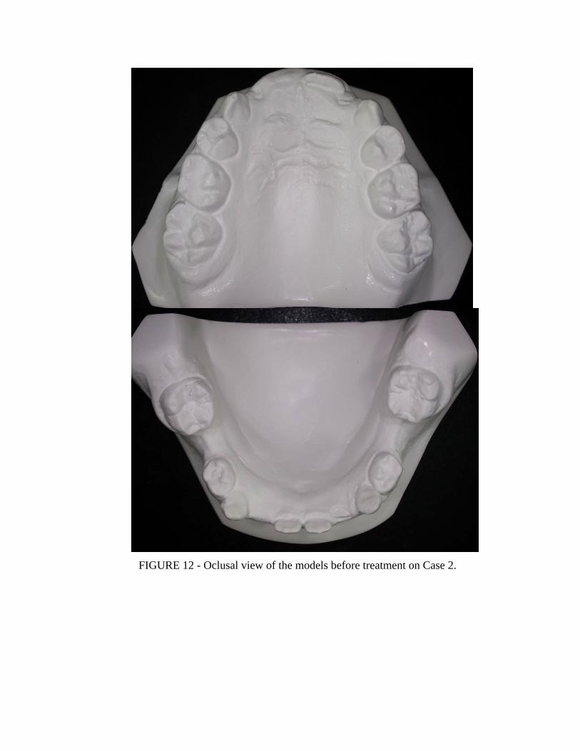
FIGURE 12 - Oclusal view of the models before treatment on Case 2.

FIGURE 13 - Transverse dimension (49mm molar width) of the maxila before treatment on Case 2.

FIGURE 14 - Postero Anterior X Rays Image – T1.

FIGURE 15 - Tracing and finds on T1 before expansion on Case 2.

FIGURE 17 - After 2 months.
FIGURE 16 - Beggining of Treatment.

FIGURE 19 - Before and After Quad-Helix 3D expansion – 5 months total time.
FIGURE 18 - After 4 months.

FIGURE 20 - Transverse dimension (57mm molar width) of the maxila after expansion on Case 2.

FIGURE 21 - Postero Anterior X Rays Image – T2.
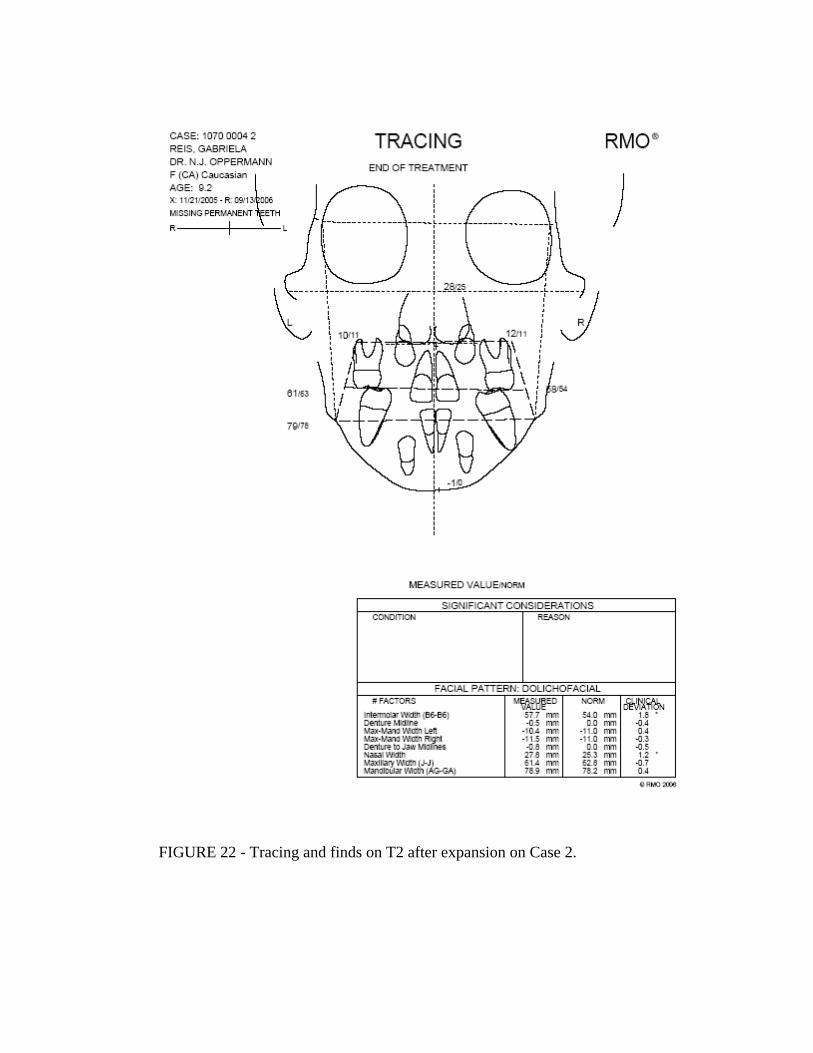
FIGURE 22 - Tracing and finds on T2 after expansion on Case 2.

FIGURE 23 - Facial changes before and after 3D Quad-Helix expansion treatment.
REFERENCES
1. Bench RW et al. Terapia Bioprogressiva. 3ª Edição. São Paulo: Editora Santos; 1996.
2. Bench RW, Gugino CF, Hilgers JJ. Bioprogressive Therapy. J Clin Orthod. 1978; 12: 279-298.
3. Langlade M. Otimização terapêutica da incidência transversal das oclusões cruzadas posteriores. 1a
Edição. São Paulo: Editora Santos; 1998.
4. Kutin G & Hawes RR. Posterior crossbites in the deciduous and mixed dentition. Am J Orthod. 1969; 56:
491-504.
5. Harberson VA, Myers DR. Midpalatal suture opening during functional posterior cross-bite correction. Am J
Orthod. 1978; 74 (3): 310-3.
6. Vadiakas GP, Roberts MW. Primary posterior crossbite: diagnosis and treatment. J Clin Pediatr Dent. 1991;
16(1): 1-4.
7. Myers DR, Barenie JT, Bell RA, et al. Condylar position in children with functional posterior crossbites:
before and after crossbite correction. Pediatr Dent. 1980; 2(3): 190-4.
8. Bell, RA, LeCompte EJ. The effects of maxillary expansion using a Quad-Helix appliance during the
deciduous and mixed dentitions. Am J Orthod. 1981; 2 (79): 152-161.
9. Slavicek R. Dr. Rudolf Slavicek on clinical and instrumental functional analysis for diagnosys and treatment
planning part 1. J Clin Orthod Interviews. 1988; 358-370.
10. Chaconas SJ, Albay Levy JA. Orthopedic and orthodontic applications of the quad-helix appliance. Am J
Orthod. 1977; 72 (4): 422-8.
11. Hicks EP. Slow maxillary expansion: a clinical study of the skeletal versus dental response to low magnitude
force. Am J Orthod. 1978; 73(2): 121-41.
12. Frank SW, Engel GA. The effects of maxillary on cephalometric measurements in growing orthodontic
patients. Am J Orthod. 1982; 5 (81): 378-389.
13. Chaconas SJ, Caputo AA. Observation of orthopedic force distribution produced by maxillary orthodontic
appliances. Am J Orthod. 1982; 82(6): 492-501.
14. Silva Filho OG, Alves RM, Capelozza Filho L. Alterações cefalométricas ocorridas na dentadura mista após
o uso de um expansor fixo tipo Quadrihélice. Ortodontia. 1986; 19(1-2): 22-33.
15. Proffit WR, Ortodontia Contemporânea. Pancast Editora Com e Repes Ltda. 1991; 589p.
16. Ladner PT, Muhl ZF. Changes concurrent with orthodontic treatment when maxillary expansion is a primary
goal. Am J Orthod Dentofac Orthop. 1995; 108(2): 184-93.
17. Brin I, Ben-Bassat Y, Blustein Y. Skeletal and functional effects of treatment for unilateral posterior
crossbite. Am J Orthod Dentofac Orthop. 1996; 109 (2): 173-9.
18. Sandikçioglu M, Hazar S. Skeletal and dental changes after maxillary expansion in the mixed dentition. Am
J Orthod Dentofac Orthop. 1997; 111(3): 321-7.
19. Boysen B, La Cour K, Athanasiou AE, et al. Three-dimensional evaluation of dentoskeletal changes after
posterior cross-bite correction by quad-helix or removable appliances. Br J Orthod. 1992; 19 (2): 97-107.
20. Erdinç AE, Ugur T, Erbay EA. Comparision of different treatment techniques for posterior crossbite in
mixed dentition. Am J Dentofac Orthop. 1999; 116: 287-300.

21. Gugino CF. An Orthodontic Philosophy. Denver, CO: RM/Comunicators; 1977.
22. Ranta R. Treatment of unilateral posterior crossbite: comparison of the quad-helix and removable plate. J
Dent Child. 1988; 55(2): 102-104.
23. Mazzieiro ET, Henriques JFC, Freitas MR de. Estudo cefalométrico, em norma frontal, das alterações dento-
esqueléticas após a expansão rápida da maxila. Ortodontia. 1996; 29(1).
24. Siqueira DF, de Almeida RR, Henriques JFC. Estudo comparativo, por meio de analise cefalométrica em
norma frontal, dos efeitos dentoesqueléticos produzidos por três tipos de expansores palatinos. R Dental
Press Ortodon Ortop Facial. 2002; 7(6): 27-47.
25. Cotton LA. Slow maxillary expansion: skeletal versus dental response to low magnitude force in Macaca
mulatta. Am J Orthod. 1978; 73(1): 1-23.
26. Ohshima, O. Effects of lateral expansion force on the maxillary structure in cynomolgus monkey. J Osaka
Dent Univ. 1972; 6(1): 11-50.
27. Storey A. Tissue response to the movement of bones. Am J Orthod. 1973; 3(64): 229-47.
28. Urbaniak JA, Brantley WA, Pruhs RJ, et al. Effects of appliance size, arch wire diameter and alloy
composition on the in vitro force delivery of the quad-helix appliance. Am J Dentofac Orthop. 1988; 94(4):
311-6.
29. Ricketts, R M. Logic and Keys to Bio Philosophy and Treatment Mechanics. 1986; American Institute for
Bioprogresive education, Scottsdale – AZ, 98p.
30. Wilson W, Wilson Wilson R. Modular 3D lingual appliance. Part I – Quad –Helix. J Clin Orthod. 1983 Nov;
761-766
31. Wilson W, Wilson R. Force systems mechanotherapy manual – book 2, Denver: RMO; 1989, 173p.
32. Duarte, M.S. O aparelho quadrihélice (qua-helix) e suas variações. R Dental Press Ortodon Ortop Facial.
2006; 11(2): 128-156.


![Design of Ionofree Micro Strip Quad Helix Antenna for ... · antenna, bifilar helices antenna, microstrip antenna, quadrafilar helix antenna. ... Helical antenna [1],[2] is broadband](https://static.fdocuments.in/doc/165x107/5b9506e809d3f2ea5c8b5a04/design-of-ionofree-micro-strip-quad-helix-antenna-for-antenna-bifilar-helices.jpg)