The value of the reprocessing method of paraffin-embedded … · The value of the reprocessing...
5
Romanian Journal of Morphology and Embryology 2009, 50(4):613–617 ORIGINAL PAPER The value of the reprocessing method of paraffin-embedded biopsies for transmission electron microscopy RODICA LIGHEZAN 1) , FLAVIA BADERCA 1) , AURORA ALEXA 1) , M. IACOVLIEV 1) , DIANA BONŢE 2) , ELENA DOINA MURĂRESCU 3) , ADRIANA NEBUNU 1) 1) Department of Histology 2) Department of Biochemistry “Victor Babeş” University of Medicine and Pharmacy, Timisoara 3) Department of Pathology, “Grigore T. Popa” University of Medicine and Pharmacy, Iassy Abstract Transmission electron microscopy (TEM) implies an elaborate preparation protocol that includes: fixation in glutaraldehyde followed by osmium tetraoxide postfixation, specimen dehydration, infiltration, resin embedding, ultrathin sectioning and staining with heavy metal salts. The aim of TEM is to examine the ultrastructure of specimens in ways that cannot be examined using other equipments or techniques. In some cases, when the requirement for TEM were made after tissue collection, useful information can be obtained from reprocessing the formalin-fixed, wax-embedded tissue used for light microscopy. Keywords: reprocessing method, transmission electron microscopy. Introduction The use of transmission electron microscopy (TEM) is a complementary diagnostic tool. The requirement for TEM is usually predetermined so that the tissues are collected and processed specifically in order to obtain good ultrastructural details [1]. When the requirement for TEM may not be anticipated or when the tissue collected for TEM may be inadequate, useful information can be obtained from reprocessing the formalin-fixed, wax-embedded tissue, usually used for light microscopy. Material and Methods There have been investigated 36 biopsies from patients with upper urinary tract tumors (n=26), glomerular diseases (n=9) and a histiocytic sarcoma. In each case, the tissue was fixed in 4% neutral buffered formalin and embedded in paraffin. Sections (5 μm) were cut from the paraffin blocks and stained with the Hematoxylin–Eosin morphological stain. Transmission electron microscopy (TEM) was performed retrospectively on specific areas of these formalin-fixed, paraffin-embedded blocks. For the electrono-microscopical examination, tissue fragments of 1 mm³ were cut with a sharp razor blade from distinct, well chosen areas of the paraffin block and reprocessed after a modified protocol described by Craciun C and Horobin R [2]. The reprocessing protocol included: • xylene dewaxing at 60 0 C, 2 × 30 minutes; • xylene dewaxing, 12 hours; • specimen rehydration: o xylene–ethanol (1:1), 5 minutes; o ethanol 100%, 2 minutes; o ethanol 95%, 3 × 5 minutes; o ethanol 80%, 3 × 5 minutes; o ethanol 70%, 3 × 5 minutes. • washing in phosphate buffer 0.1 M, 10 minutes; • post-fixing in aqueous OsO 4 1%, at 4 0 C, 60 minutes; • washing in distilled water, 2 × 10 minutes; • specimen dehydration: o ethanol 70%, 2 × 15 minutes; o ethanol 80%, 2 × 15 minutes; o ethanol 96%, 2 × 15 minutes; o ethanol 100%, 2 × 15 minutes. • Infiltration with propylene oxide, 2 × 15 minutes; • Infiltration with Epon 812 overnight; • Epon 812 polimerisation at 60 0 C, 24 hours. The Epon blocks were trimmed and than sectioned with glass knives attached to an ultramicrotome (LKB Ultrotome) obtaining semithin and ultrathin sections. Semithin sections (1000 nm) of resin embedded samples were stained with Richardson’s stain, an aqueous solution of 1% Methylene Blue made up in 1% borax, mixed in equal parts with 1% Azure II [3]. The semithin sections were examined with the light microscope in order to identify the specific diagnostic areas and images were picked up with a Coolpix camera. In order to visualize the specimen in the TEM,
Transcript of The value of the reprocessing method of paraffin-embedded … · The value of the reprocessing...

Romanian Journal of Morphology and Embryology 2009, 50(4):613–617
OORRIIGGIINNAALL PPAAPPEERR
The value of the reprocessing method of paraffin-embedded biopsies for transmission electron microscopy RODICA LIGHEZAN1), FLAVIA BADERCA1), AURORA ALEXA1),
M. IACOVLIEV1), DIANA BONŢE2), ELENA DOINA MURĂRESCU3), ADRIANA NEBUNU1)
1)Department of Histology 2)Department of Biochemistry
“Victor Babeş” University of Medicine and Pharmacy, Timisoara 3)Department of Pathology,
“Grigore T. Popa” University of Medicine and Pharmacy, Iassy
Abstract Transmission electron microscopy (TEM) implies an elaborate preparation protocol that includes: fixation in glutaraldehyde followed by osmium tetraoxide postfixation, specimen dehydration, infiltration, resin embedding, ultrathin sectioning and staining with heavy metal salts. The aim of TEM is to examine the ultrastructure of specimens in ways that cannot be examined using other equipments or techniques. In some cases, when the requirement for TEM were made after tissue collection, useful information can be obtained from reprocessing the formalin-fixed, wax-embedded tissue used for light microscopy. Keywords: reprocessing method, transmission electron microscopy.
Introduction
The use of transmission electron microscopy (TEM) is a complementary diagnostic tool. The requirement for TEM is usually predetermined so that the tissues are collected and processed specifically in order to obtain good ultrastructural details [1].
When the requirement for TEM may not be anticipated or when the tissue collected for TEM may be inadequate, useful information can be obtained from reprocessing the formalin-fixed, wax-embedded tissue, usually used for light microscopy.
Material and Methods
There have been investigated 36 biopsies from patients with upper urinary tract tumors (n=26), glomerular diseases (n=9) and a histiocytic sarcoma.
In each case, the tissue was fixed in 4% neutral buffered formalin and embedded in paraffin. Sections (5 µm) were cut from the paraffin blocks and stained with the Hematoxylin–Eosin morphological stain.
Transmission electron microscopy (TEM) was performed retrospectively on specific areas of these formalin-fixed, paraffin-embedded blocks.
For the electrono-microscopical examination, tissue fragments of 1 mm³ were cut with a sharp razor blade from distinct, well chosen areas of the paraffin block and reprocessed after a modified protocol described by Craciun C and Horobin R [2].
The reprocessing protocol included: • xylene dewaxing at 600C, 2 × 30 minutes;
• xylene dewaxing, 12 hours; • specimen rehydration:
o xylene–ethanol (1:1), 5 minutes; o ethanol 100%, 2 minutes; o ethanol 95%, 3 × 5 minutes; o ethanol 80%, 3 × 5 minutes; o ethanol 70%, 3 × 5 minutes.
• washing in phosphate buffer 0.1 M, 10 minutes; • post-fixing in aqueous OsO4 1%, at 40C, 60 minutes; • washing in distilled water, 2 × 10 minutes; • specimen dehydration:
o ethanol 70%, 2 × 15 minutes; o ethanol 80%, 2 × 15 minutes; o ethanol 96%, 2 × 15 minutes; o ethanol 100%, 2 × 15 minutes.
• Infiltration with propylene oxide, 2 × 15 minutes; • Infiltration with Epon 812 overnight; • Epon 812 polimerisation at 600C, 24 hours.
The Epon blocks were trimmed and than sectioned with glass knives attached to an ultramicrotome (LKB Ultrotome) obtaining semithin and ultrathin sections.
Semithin sections (1000 nm) of resin embedded samples were stained with Richardson’s stain, an aqueous solution of 1% Methylene Blue made up in 1% borax, mixed in equal parts with 1% Azure II [3].
The semithin sections were examined with the light microscope in order to identify the specific diagnostic areas and images were picked up with a Coolpix camera.
In order to visualize the specimen in the TEM,

Rodica Lighezan et al.
614
the specific areas of the Epon block was trimmed again and sectioned, obtaining ultrathin sections (60–100 nm). These were placed on copper grids (200 mesh) contrasted with 2% uranyl acetate and lead citrate in the standard manner and subsequently examined with a Leo 906 transmission electron microscope (Zeiss, Oberkochen, Germany).
Results
The reason for using a transmission electron microscope is to increase in capabilities, due to the wavelength of the illumination that is produced by the energized beam of electrons. The main role of the TEM is to reveal the ultrastructural details of the specimen in a way that cannot be done using other technique or equipment.
In renal pathology, the diagnostic electron microscopy is usually performed on glutaraldehyde-fixed, resin-embedded biopsies. Sometimes, when the glutaraldehyde fixed biopsy is missing, the formalin-fixed, paraffin-embedded tissue had to be used for electron microscopy following the re-embedding protocol of paraffin sections into epoxy resin.
Although this reprocessing technique can produce artifactual distortion of cellular and extracellular matrix architecture, in our case we found it adequate for
detection of many renal ultrastructural alterations such as the presence and location of electron-dense deposits at the glomerular basement membrane level and the degree of podocyte foot process effacement. TEM demonstrated the presence of vascular, tubular and interstitial lesions, and revealed the presence of epithelial degeneration, necrosis or fibrin deposits in the vascular wall. It had limited diagnostic importance in thin basement membrane nephropathy (TBMN) where the formalin-fixation affects GBM thickness, so that the GBM appears thinner.
In renal tumor biopsies, the aim of the reprocessing was the demonstration of various aspects of tumor angiogenesis. The tumoral area with the richest vascularisation was identified on Hematoxylin–Eosin stained slides, and then sliced from the paraffin block and embedded in resin.
After semithin sectioning, specific areas were identified on Richardson’s stained sections and the resin block was retrieved, and ultrathin sectioned (Figure 1).
The TEM identified in the mature, normal vessels, flat endothelial cells with oval, heterochromatic nuclei surrounded by rich cytoplasms, that beared the specific Weibel–Palade bodies recognizable due to their specific shape and content. They formed a uniform monolayer, with overlapping cell borders (Figure 2).
Figure 1 – Semithin section of a glomerulus and the adjacent tubules, light microscopy (Richardson’s stain, ×100).
Figure 2 – Ultrathin section of a glomerular capillary showing pedicels effacement (TEM, ×6000).
The semithin sections permitted the identification of new formed blood vessels, in different stages of angiogenesis. They were lined by activated endothelial cells lying on a continuous, well-developed basal membrane that sometimes displayed reduplications. (Figures 3 and 4).
The lining cells of the intratumoral vessels exhibited some abnormalities: they were tall, round or irregular shaped due to luminal protrusions into the lumen, had large nuclei and overlapped other cells. In most vessels, tumor cells were located close to the wall, some coming in contact with the vessel lumen (Figure 3).
On some sections, the angiogenesis progression front was present, consisting of sprouts budding from preexisting vessels that assembled into solid cords, which underwent tubulogenesis to form vessels with a central lumen.
The case with suspicion of histiocytic sarcoma represents a good illustration of the utility of paraffin embedded material for TEM. Histiocytic sarcoma is a rare malignant neoplasm, with unknown etiology that occurs in lymph nodes, skin and gastrointestinal tract. In our case, the young patient diagnosed two years earlier with Hodgkin’s disease, was referred to Thoracic Surgery Department with an ulcerated nodular sternal mass, which was surgically removed.
The light microscopy of the surgical piece revealed a cell population consisting of histiocytes, inflammatory cells, mixed with eosinophilic abscesses and necrosis (Figure 5). The Sternberg–Reed cells were absent. The numerous histiocytes had large nucleolated nuclei and abundant eosinophilic cytoplasm. Binucleated giant cells were also present.
Immunohistochemistry demonstrated that the
The value of the reprocessing method of paraffin-embedded biopsies for transmission electron microscopy
615
tumoral histiocytes were positive for S-100, CD68 and vimentin and negative for CD34, CD15, CD30, NFAP and desmin.
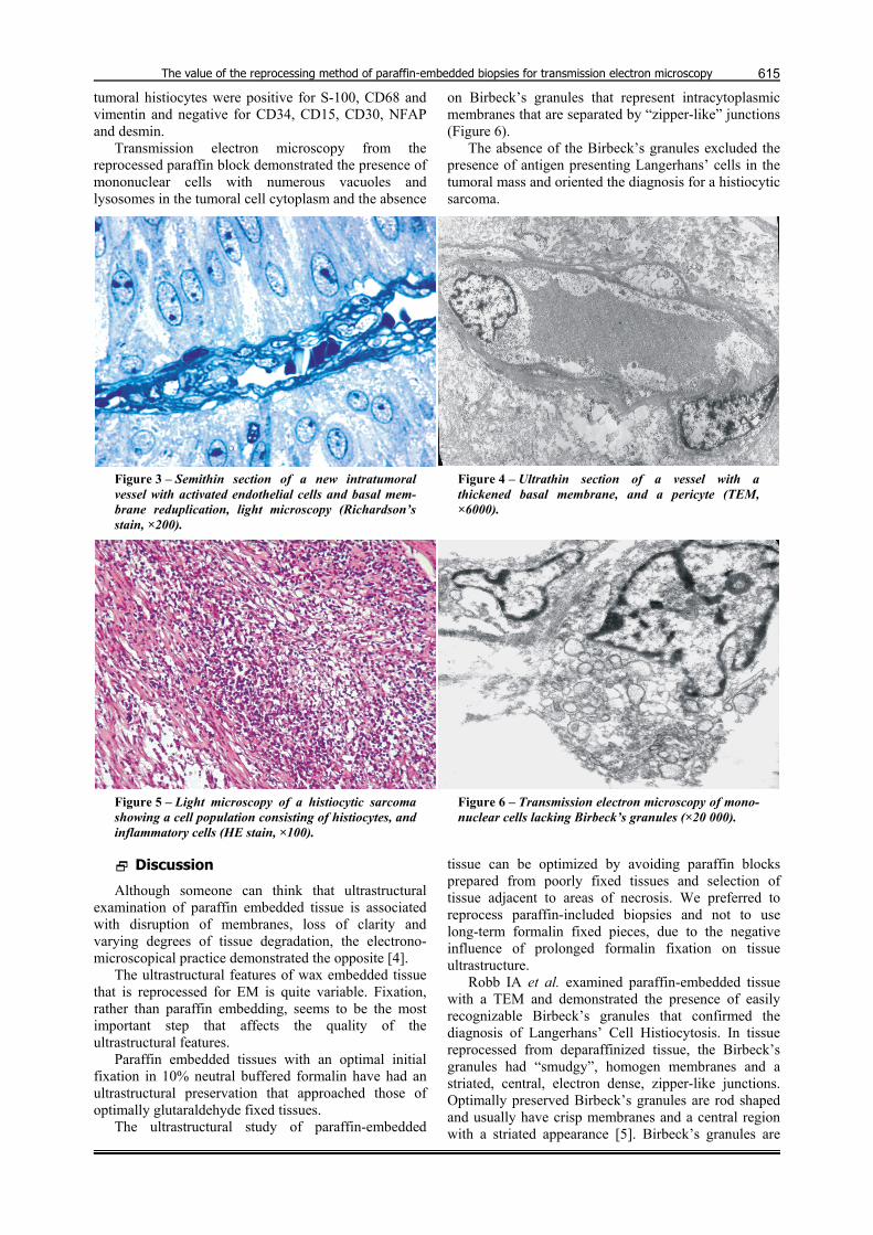
Transmission electron microscopy from the reprocessed paraffin block demonstrated the presence of mononuclear cells with numerous vacuoles and lysosomes in the tumoral cell cytoplasm and the absence
on Birbeck’s granules that represent intracytoplasmic membranes that are separated by “zipper-like” junctions (Figure 6).
The absence of the Birbeck’s granules excluded the presence of antigen presenting Langerhans’ cells in the tumoral mass and oriented the diagnosis for a histiocytic sarcoma.
Figure 3 – Semithin section of a new intratumoral vessel with activated endothelial cells and basal mem-brane reduplication, light microscopy (Richardson’s stain, ×200).
Figure 4 – Ultrathin section of a vessel with a thickened basal membrane, and a pericyte (TEM, ×6000).
Figure 5 – Light microscopy of a histiocytic sarcoma showing a cell population consisting of histiocytes, and inflammatory cells (HE stain, ×100).
Figure 6 – Transmission electron microscopy of mono-nuclear cells lacking Birbeck’s granules (×20 000).
Discussion
Although someone can think that ultrastructural examination of paraffin embedded tissue is associated with disruption of membranes, loss of clarity and varying degrees of tissue degradation, the electrono-microscopical practice demonstrated the opposite [4].
The ultrastructural features of wax embedded tissue that is reprocessed for EM is quite variable. Fixation, rather than paraffin embedding, seems to be the most important step that affects the quality of the ultrastructural features.
Paraffin embedded tissues with an optimal initial fixation in 10% neutral buffered formalin have had an ultrastructural preservation that approached those of optimally glutaraldehyde fixed tissues.
The ultrastructural study of paraffin-embedded
tissue can be optimized by avoiding paraffin blocks prepared from poorly fixed tissues and selection of tissue adjacent to areas of necrosis. We preferred to reprocess paraffin-included biopsies and not to use long-term formalin fixed pieces, due to the negative influence of prolonged formalin fixation on tissue ultrastructure.
Robb IA et al. examined paraffin-embedded tissue with a TEM and demonstrated the presence of easily recognizable Birbeck’s granules that confirmed the diagnosis of Langerhans’ Cell Histiocytosis. In tissue reprocessed from deparaffinized tissue, the Birbeck’s granules had “smudgy”, homogen membranes and a striated, central, electron dense, zipper-like junctions. Optimally preserved Birbeck’s granules are rod shaped and usually have crisp membranes and a central region with a striated appearance [5]. Birbeck’s granules are

Rodica Lighezan et al.
616
one of the many organelles that can be recognized in the TEM of paraffin embedded material [6–8].
Other organelles that are recognizable in tissues reprocessed for EM, are represented by nuclei, desmosomes, sarcomeres, intermediate filaments, tonofilaments and electron dense granules.
Mitochondria, polyribosomes, microtubules, lipids, glycogen and Weibel–Palade bodies can exhibit various degradation degrees in reprocessed paraffin embedded tissue.
In thin basement membrane nephropathy, where diagnosis can only be established by electron micro-scopy, the glomerular basal membrane appears thinner on reprocessed, formalin fixed tissue than on glutaral-dehyde fixed tissue [9].
There have been described two reprocessing proce-dures: one with the melting of the whole paraffin block, followed by resin re-embedding of parts from the entire piece and the second that implies the cutting of a well established area from the paraffin block that is later embedded in Epon. We preferred the second method because it preserved the paraffin block and permitted the examination of the small, distinct, and well-chosen areas.
In addition to the processing artifact that is present in the reprocessed paraffin embedded tissue, the duration of the reprocessing method is also important. The initial methods that were used for preparation of paraffin-embedded tissue for EM had lasted up to one week. The majority of this time was spent in paraffin removing from the tissue sample. These lengthy processing methods have been replaced by simpler methods that can have a processing time as short as thirty minutes [7].
Alternative rapid processing methods for fresh renal biopsies implied solely a direct LRWhite embedding, and a polymerization at 00C. Morphological, immuno-histochemical and electrono-microscopical procedures were later applied on semi- and ultrathin sections [10].
Other methods that excluded post-fixation in 1% buffered aqueous osmium tetroxide and dehydration lasted less than half hour and demonstrated similar results [8, 11].
Widáhn S and Kindblom LG described a rapid and simple re-embedding method for electron microscopy: the paraffin-embedded tissue was dewaxed in xylene, stained in a 0.01% Toluidine Blue/absolute ethanol solution, infiltrated in a propylene oxide/resin mixture, and embedded in Epon [8].
This quick method excluded rehydration, post-osmification, and dehydration, lasted about three hours and produced results similar to previously described more laborious re-embedding techniques.
Regarding the fine preservation of nuclei, tonofibrils and desmosomes of keratinocytes in normal skin, Ogiyama Y et al. demonstrated that there was no difference between the quick re-embedding method and traditional re-embedding methods [12]. Myofilaments, dense bodies, Weibel–Palade bodies, neurosecretory granules of Merkel’s cell and melanosomes were also recognized by the quick method. Mitochondria was an exception, because this cytoplasmatic organelle had a
constant poor preservation, in all the used methods [12]. The advantage of the reprocessing method resides in
the fact that it is simple, not expensive and provides useful ultrastructural informations due to the preser-vation of many cytoplasmic organelles. The conser-vation of the ultrastructural aspects, although far from ideal, provides additional information, thus facilitating the diagnosis in various inflammatory and proliferative disorders.
The reprocessing method has some limitations. One of them is the destruction of antigen structure due to the negative influence of osmification on antigenic structure combined with the embedding in epoxi resin, not allowing the colloidal-gold immunolabeling used for immuno-electrono-microscopical studies. Immuno-electron microscopy detects, at high resolutions, the intracellular location of specific proteins with the use of colloidal – gold labeling [6, 13].
There is also a limitation in thin basement membrane nephropathy, where TEM on re-embedded paraffin sections can be used only to exclude, but not to establish the diagnosis, due to basal glomerular membrane shrinkage under formalin action.
Paraffin embedded tissue that is reprocessed for EM will never have the optimal ultrastructural preservation of glutaraldehyde fixed tissues. However, with careful attention to tissue fixation, the processing of paraffin embedded tissue for EM needs to become a good choice providing useful diagnostic information.
Conclusions
The reprocessing method is simple, not expensive, not time consuming and can provide useful, diagnostic information.
The diagnostic importance of semithin and ultrathin sections in renal diseases cannot be denied. In inflammatory renal diseases it highline the glomerular changes, the vascular and interstitial lesions.
In the last decades, electron microscopy has become a complementary method in tumor diagnosis. TEM of the reprocessed tissue made possible the exclusion of the Langerhans’ cell histiocytoma diagnosis and revealed various angiogenesis aspects in carcinomas of the upper urinary tract.
The reprocessing method represents a useful diagnostic tool and has an immense potential in retrospective studies.
References [1] STIRLING J. W, WOODS A., Transmission electron
microscopy: diagnostic applications. In: BANCROFT J. D., GAMBLE M. (eds), Theory and practice of histological techniques, 5th edition, Churchill Livingstone, London, 2002, 701–728.
[2] CRACIUN C., HOROBIN R., Tissue processing schedule for parallel light and electron microscopy, Proc Royal Microsc Soc (London), 1989, 24(4):223–225.
[3] RICHARDSON K. C., JARRETT L., FINKE E. H., Embedding in epoxy resins for ultrathin sectioning in electron microscopy, Stain Technol, 1960, 35:313–323.
[4] ERLANDSON R. A., Preparing cells and tissues for electron microscopy. In: ERLANDSON R. A. (ed), Diagnostic transmi-ssion electron microscopy of tumors: with clinicopatho-logical, immmunohistochemical, and cytogenetic correla-tions, Raven Press, New York, 1994, 7–13.

The value of the reprocessing method of paraffin-embedded biopsies for transmission electron microscopy
617[5] ROBB I. A., JIMENEZ C. L., CARPENTER B. F., Birbeck
granules or Birbeck junctions? Intercellular “zipperlike” lattice junctions in eosinophilic granuloma of bone, Ultrastruct Pathol, 1992, 16(4):423–428.
[6] WANG N.-S., MINASSIAN H., The formaldehyde-fixed and paraffin-embedded tissues for diagnostic transmission electron microscopy: a retrospective and prospective study, Hum Pathol, 1987, 18(7):715–727.
[7] VAN DEN BERGH WEERMAN M. A., DINGEMANS K. P., New techniques rapid deparaffinization for electron microscopy, Ultrastruct Pathol, 1984, 7(1):55–57.
[8] WIDÁHN S., KINDBLOM L.-G., A rapid simple method for electron microscopy of paraffin-embedded tissue, Ultrastruct Pathol, 1988, 12(1):131–136.
[9] NASR S. H., MARKOWITZ G. S., VALERI A. M., YU Z., CHEN L., D’AGATI V. D., Thin basement membrane nephropathy cannot be diagnosed reliably in deparaffinized, formalin-fixed tissue, Nephrol Dial Transplant, 2007, 22(4):1228–1232.
[10] BOWDLER A. L., GRIFFITHS D. F., NEWMAN G. R., The mor-phological and immunohistochemical analysis of renal biopsies by light and electron microscopy using a single processing method, Histochem J, 1989, 21(7):393–402.
[11] TOLIVIA J., NAVARRO A., TOLIVIA D., Polychromatic staining of epoxy semithin sections: a new and simple method, Histochemistry, 1994, 101(1):51–55.
[12] OGIYAMA Y., OHASHI M., Electron microscopic examination of cutaneous lesions by the quick re-embedding method from paraffin-embedded blocks, J Cutan Pathol, 1994, 21(3):239–246.
[13] DINGEMANS K. P., RAMKEMA M., Immunoelectron micro-scopy on material retrieved from paraffin: accurate sampling on the basis of stained paraffin sections, Ultrastruct Pathol, 2001, 25(3):201–206.
Corresponding author Rodica Lighezan, Lecturer, MD, PhD, Department of Histology, “Victor Babeş” University of Medicine and Pharmacy, 2 Eftimie Murgu Square, 300041 Timişoara, Romania; Phone +40743–871600, e-mail: [email protected] Received: September 10th, 2009
Accepted: October 25th, 2009