The Use of Point-of-Care Ultrasonography in Trauma...
14
The Use of Point-of-Care Ultrasonography in Trauma Anesthesia Davinder Ramsingh, MD a, *, Venkat Reddy Mangunta, MD b,c INTRODUCTION Caring for the trauma patient is one of the most complicated and high-risk patient care situations encountered by anesthesiologists. Around the world, trauma is the third leading cause of death overall, with more than 5 million deaths per year. 1 These Disclosure Statement: Dr D. Ramsingh is a consultant for Edwards Life Sciences; Fujifilm Sono- site; and receives funds for research from General Electric on point-of-care ultrasound and anesthesia delivery systems, Merck Pharmaceuticals, Pacira Pharmaceuticals, Masimo Corpora- tion, and Edwards Lifesciences. Dr V.R. Mangunta has no disclosures. a Department of Anesthesiology, Loma Linda University School of Medicine, Loma Linda Uni- versity Medical Center, 11234 Anderson Street, MC-2532-D, Loma Linda, CA 92354, USA; b Department of Anesthesiology, Division of Cardiovascular Anesthesia, Saint Luke’s Mid America Heart Institute, University of Missouri-Kansas City School of Medicine, 4401 Wornall Road, Room 3103, Kansas City, MO 64111, USA; c Department of Anesthesiology, Division of Critical Care Medicine, Saint Luke’s Mid America Heart Institute, University of Missouri-Kansas City School of Medicine, 4401 Wornall Road, Room 3103, Kansas City, MO 64111, USA * Corresponding author. E-mail address: [email protected] KEYWORDS Perioperative point-of-care ultrasound Point-of-care ultrasonography in trauma Trauma anesthesia Ultrasound education in anesthesia Ultrasound applications for trauma KEY POINTS Management of the trauma patient requires rapid, coordinated care by anesthesiologists using various diagnostic or therapeutic modalities. Trauma anesthesiologists must become facile with the use of point-of-care ultrasound (POCUS) to maximize diagnosis and treatment of patients with traumatic injury. POCUS can assist in quickly diagnosing a multitude of traumatic injuries and differenti- ating diagnoses. Comprehensive POCUS educational curricula can assist anesthesiology residency pro- grams in teaching residents this vital skillset. It is essential to the evolution of the specialty that anesthesiologists and anesthesiology training programs make POCUS education a priority. Anesthesiology Clin 37 (2019) 93–106 https://doi.org/10.1016/j.anclin.2018.09.011 anesthesiology.theclinics.com 1932-2275/19/ª 2018 Elsevier Inc. All rights reserved. Downloaded for Anonymous User (n/a) at Kaweah Delta Health Care District from ClinicalKey.com by Elsevier on March 20, 2019. For personal use only. No other uses without permission. Copyright ©2019. Elsevier Inc. All rights reserved.
Transcript of The Use of Point-of-Care Ultrasonography in Trauma...

The Use of Point-of-CareUltrasonography in TraumaAnesthesia
Davinder Ramsingh, MDa,*, Venkat Reddy Mangunta, MDb,c
INTRODUCTION
Caring for the trauma patient is one of the most complicated and high-risk patient caresituations encountered by anesthesiologists. Around the world, trauma is thethird leading cause of death overall, with more than 5 million deaths per year.1 These
Disclosure Statement: Dr D. Ramsingh is a consultant for Edwards Life Sciences; Fujifilm Sono-site; and receives funds for research from General Electric on point-of-care ultrasound andanesthesia delivery systems, Merck Pharmaceuticals, Pacira Pharmaceuticals, Masimo Corpora-tion, and Edwards Lifesciences. Dr V.R. Mangunta has no disclosures.a Department of Anesthesiology, Loma Linda University School of Medicine, Loma Linda Uni-versity Medical Center, 11234 Anderson Street, MC-2532-D, Loma Linda, CA 92354, USA;b Department of Anesthesiology, Division of Cardiovascular Anesthesia, Saint Luke’s MidAmerica Heart Institute, University of Missouri-Kansas City School of Medicine, 4401 WornallRoad, Room 3103, Kansas City, MO 64111, USA; c Department of Anesthesiology, Division ofCritical Care Medicine, Saint Luke’s Mid America Heart Institute, University of Missouri-KansasCity School of Medicine, 4401 Wornall Road, Room 3103, Kansas City, MO 64111, USA* Corresponding author.E-mail address: [email protected]
KEYWORDS
! Perioperative point-of-care ultrasound ! Point-of-care ultrasonography in trauma! Trauma anesthesia ! Ultrasound education in anesthesia! Ultrasound applications for trauma
KEY POINTS
! Management of the trauma patient requires rapid, coordinated care by anesthesiologistsusing various diagnostic or therapeutic modalities.
! Trauma anesthesiologists must become facile with the use of point-of-care ultrasound(POCUS) to maximize diagnosis and treatment of patients with traumatic injury.
! POCUS can assist in quickly diagnosing a multitude of traumatic injuries and differenti-ating diagnoses.
! Comprehensive POCUS educational curricula can assist anesthesiology residency pro-grams in teaching residents this vital skillset.
! It is essential to the evolution of the specialty that anesthesiologists and anesthesiologytraining programs make POCUS education a priority.
Anesthesiology Clin 37 (2019) 93–106https://doi.org/10.1016/j.anclin.2018.09.011 anesthesiology.theclinics.com1932-2275/19/ª 2018 Elsevier Inc. All rights reserved.
Downloaded for Anonymous User (n/a) at Kaweah Delta Health Care District from ClinicalKey.com by Elsevier on March 20, 2019.For personal use only. No other uses without permission. Copyright ©2019. Elsevier Inc. All rights reserved.

patients often present in the middle of the night or on weekends when comprehensiveresources and immediate consultation are not available. Therapy is often required forthese patients for rapidly developing acute pathologic complications with little to noinformation on their past medical history. Furthermore, these patients may have rapidalterations to their cardiovascular and pulmonary status, and have a predisposition tooccult injuries.The management of the trauma patient requires rapid and coordinated care by
trained physicians. Specialized training in trauma has undergone significant evolutionover the past few decades. In 1990, Congress passed the Trauma Care Systems Plan-ning and Development Act that led to the development of organized statewide traumasystems.2 The American College of Surgeons (ACS) Committee on Trauma identifiedtrauma centers as level I to level V, with the goal of ensuring that adequate resourcesand trauma-trained physicians are available to treat patients based on their severity ofinjury.2 Additionally, the Advanced Trauma Life Support (ATLS) program has beenadopted by the ACS and provides organized algorithms and approaches to managingtrauma patients.3
Parallel to the development of highly reproducible algorithms for the treatment oftrauma, ultrasound (US) technology has seen rapid evolution and expansion intoemergency medicine (EM) training programs across the nation. EM physicianscurrently use bedside US for diagnostic and therapeutic purposes (eg, diagnosinghemothorax, retinal detachment). Point-of-care US (POCUS) has revolutionized thepractice of emergency physicians.4 The focused assessment with sonography fortrauma (FAST)5 examination has historically dominated the role of US in trauma man-agement and remains the most studied example of focused clinical US.6 However,over recent years, academic EM departments have greatly expanded the use of thistechnology from the periphery of trauma management to now include cardiopulmo-nary assessment, regional nerve blocks, and transesophageal echocardiography(TEE).As much as EM has embraced this technology and developed focused applications
for US use over the past decade, anesthesiology has lagged behind with regard tobroadly adopting US technology. Althoughmost anesthesiologists graduate residencywith proficiency in performing US-guided regional anesthesia and vascular access, itsuse in the perioperative setting to quickly diagnose and aid in the treatment of emer-gent conditions (eg, pneumothorax, hemothorax, abdominal hemorrhage, hypovole-mic or cardiogenic shock) has traditionally not been a focus of anesthesiologyresidency programs. By and large, anesthesia residents are graduating from residencywith no formal education in the use of US for the acute diagnosis of adverse conditionsin the perioperative setting.The growing push for anesthesiologists to advance beyond their role in the intrao-
perative setting and realize their role as true perioperative physicians has led to thedevelopment of the subspecialty of trauma anesthesiology.2 Since 2013, trauma anes-thesiology has been recognized as a distinct subspecialty of anesthesiology by theAmerican Society of Anesthesiologists (ASA).2 In 2017, the ASA Committee on Traumaand Emergency Preparedness (COTEP) published the results of a survey demon-strating that there was significant disconnect between trauma surgeons and anesthe-siologists regarding whether the anesthesiologists at their respective institutions wereappropriately trained to manage trauma patients.7 Trauma anesthesiologists must beprepared to emergently care for a patient with any severity of injury and must be adeptat using the samemanagement approaches described in the ATLS program. Develop-ment of POCUS curricula within anesthesiology residency programs is of paramountimportance in the training of trauma anesthesiologists. This article reviews the topics
Ramsingh & Mangunta94
Downloaded for Anonymous User (n/a) at Kaweah Delta Health Care District from ClinicalKey.com by Elsevier on March 20, 2019.For personal use only. No other uses without permission. Copyright ©2019. Elsevier Inc. All rights reserved.
of POCUS that are relevant to the perioperative trauma patient by reviewing the utilityof POCUS for each category of the ATLS algorithm.
AIRWAY MANAGEMENT
The utility of POCUS for airway management has been demonstrated for the identifi-cation of difficult laryngoscopy, appropriate location of the endotracheal tube, and tofacilitate cricothyrotomy or tracheostomy procedures.
Difficult Laryngoscopy and Endotracheal Tube Localization
POCUS has been demonstrated to improve the airway examination. Recently,Reddy and colleagues8 found that anterior neck soft tissue thickness at the levelof the vocal cords is a predictor of difficult airway. In addition to use of US for visu-alization of the hyoid bone, decreased temporomandibular joint mobility, measure-ment of the hyomental distance with neck extension, and the measurement ofanterior soft tissue thickness at the thyrohyoid membrane have all been used topredict difficult airways.9
Endotracheal Tube Localization
US has demonstrated utility for verification of successful endotracheal intubation,reporting sensitivity and specificity of 100% versus esophageal intubation.10 Becausethis is likely less of a concern in the perioperative setting, US has also demonstratedthe ability to detect tracheal versus endobronchial intubation. Studies have demon-strated a higher degree of sensitivity and specificity (>93%) with POCUS than withauscultation.11
Cricothyrotomy or Tracheostomy
The use of surface landmarks to identify the cricothyroid membrane may be difficult,particularly for obese and female patients.12,13 Bedside US is a reliable modality forrapid identification of anatomy for emergent cricothyrotomy.14,15 Similarly, US hasdemonstrated improved success in accessing the trachea with more than 90% first-pass attempts.16 The use of POCUS for percutaneous tracheostomies has demon-strated improved accuracy and has also been suggested to decrease complicationrates.17,18
BREATHING OR PULMONARY
The ability of US to provide insight into pulmonary disease was previously thoughtto be impossible secondary to the acoustic impedance difference with aerated tis-sue. However, recent evidence has demonstrated POCUS to be extremely useful inestablishing a differential diagnosis for acute respiratory failure in the postoperativeperiod.19 POCUS has proven to be faster and superior to chest radiograph (CXR)in diagnosing pneumothorax, pleural effusion, and alveolar interstitial diseases.20,21
Ford and colleagues22 recently demonstrated the ability of POCUS to detectperioperative pulmonary disease (atelectasis, consolidation, alveolar-interstitialsyndrome, pleural effusion, and pneumothorax) in patients undergoing cardiac sur-gery with a high degree of specificity to CXR and physical examination findings.However, it is important to stress that a significant portion of pulmonary US dealswith the detection and recognition of artifact generated by pathologic complica-tions. This key point makes pulmonary US a challenging topic for the novicePOCUS user.
Use of POCUS in Trauma Anesthesia 95
Downloaded for Anonymous User (n/a) at Kaweah Delta Health Care District from ClinicalKey.com by Elsevier on March 20, 2019.For personal use only. No other uses without permission. Copyright ©2019. Elsevier Inc. All rights reserved.

Evaluation of Pneumothorax
US is highly accurate at detecting pneumothorax.20,21 The primary US feature is theabolition of lung sliding, which is defined as the motion of visceral pleura againstthe parietal pleura during respiration. Importantly, this nonspecific finding is seen inseveral other conditions, such as malignancy, chronic obstructive pulmonary disease,and pneumonia. Additionally, it only detects pneumothoraces at the location at whichthe operator is scanning, thus it has the potential to miss pneumothoraces present atdifferent scan locations. To this effect, it is important to scan the lung fields in morethan one location. More specifically, it is the visualization of a lung point that is patho-gnomonic for pneumothorax. The lung point is the point at which the visceral pleuraand parietal pleura separate. In a pneumothorax, both pleural layers separate. Tracingthe pleural layer separation back to a lung point (point of separation) confirms that thelack of lung sliding is due to pneumothorax and not another disease process.
Parenchymal Lung Diseases
POCUS has demonstrated the same high degree of sensitivity for the detection ofairspace disease within the lung parenchyma. Common practice in POCUS involvesdetecting artifacts generated from the disease developing within the lung paren-chyma. Specifically, parenchyma diseases (edema, pneumonia, inflammation) willcause the interlobular septa to thicken, producing long vertical lines through thelung parenchyma on US imaging. This is commonly referred to as an US lung comet(ULC) (Fig. 1). Recent studies have demonstrated that the presence of greater than9 ULCs per lung field is associated with 100% specificity for cardiogenic dyspnea.23
Multiple protocols have been developed regarding pulmonary POCUS, mostly in thecritical care setting. Of these, the bedside lung US in emergency (BLUE) protocolstands out as a comprehensive approach to rapidly facilitate the diagnosis of a patientin acute respiratory failure.24
Assessment for Pleural Effusion
US is more sensitive and specific than auscultation or CXR and is, therefore, themethod of choice in detecting pleural effusion.25,26 Effusions greater than 1 cm areeasily detected and have a greater than 90% sensitivity and specificity for pleural effu-sion.26 The utility for POCUS to guide thoracentesis has also been suggested.27
Fig. 1. ULC of pulmonary airspace disease.
Ramsingh & Mangunta96
Downloaded for Anonymous User (n/a) at Kaweah Delta Health Care District from ClinicalKey.com by Elsevier on March 20, 2019.For personal use only. No other uses without permission. Copyright ©2019. Elsevier Inc. All rights reserved.

CIRCULATION
POCUS has proven to be critical in the assessment of causes of hemodynamic insta-bility and shock. Beyond assessing the patient’s volume status, POCUS allows theanesthesiologist to differentiate between causes of shock and assess for injuriesthat may be leading to a shock state.The following sections review the methods commonly used in POCUS that address
these topics.
MECHANISMS OF HYPOTENSION
The FAST examination is the most widely used POCUS examination currently prac-ticed in the acute care setting. This examination has been shown to very reliablydetect greater than 200 mL of blood or fluid in body cavities (abdomen, pleuralspace, and pericardium) and it is a highly effective tool in the detection of clinicallysignificant hemoperitoneum and hemopericardium in unstable patients.5,28–30 Bothtrauma patients and patients in the postoperative care unit may have injuries thatcan cause significant blood loss and remain undetected by physical examination.The application of this examination allows the perioperative physician to determineif hemodynamic instability is secondary to pericardial and/or peritoneal injury,resulting in free fluid that can occur before or after surgery. Trauma patients canalso sustain direct injury to their thoracic structures, such as the aorta or myocar-dium. A penetrating injury to the chest, for instance, may demonstrate an injury tothe RV free wall, leading to a large pericardial effusion and tamponade. Additionally,elderly trauma patients with underlying ischemic heart disease are at risk formyocardial injury. POCUS can reveal new regional wall motion abnormalities in pa-tients with previously normal cardiac function. In this way, obstructive (tamponade),hemorrhagic or hypovolemic, and cardiogenic causes of shock can quickly beelucidated by the perioperative physician.
Assessment of Cardiac Function
Transthoracic echocardiography (TTE) examination of the cardiopulmonary systemusing bedside POCUS technology has proven to be a reliable tool when comparedwith formal echocardiography.31 Indeed, assessments of global left ventricular (LV)function, have shown a strong correlation (r"0.92) between POCUS and formal echo-cardiography examinations.31 Similarly, good correlation between POCUS and formalechocardiography was also shown when assessing right ventricular (RV) function andvalvular function (excluding aortic stenosis) (r >0.81).31 Additional support for bedsideTTE has been demonstrated in patients with shock in which adequate image qualitywas obtained in 99% of cases with a sensitivity and specificity approaching 100%and 95%, respectively, for identifying a cardiogenic cause for shock. Finally, it hasbeen demonstrated that noncardiologists can be trained to perform and interpret alimited transthoracic examination focused on assessment of LV function.32,33 Rela-tively straightforward measures of LV function can be obtained by obtaining paraster-nal short-axis views of the LV. By obtaining a parasternal short-axis view of the LV atthe level of the papillary muscles, the operator can record a 2 to 4 beat video clip. TheLV end-diastolic area (LVEDA) (Fig. 2A) and LV end-systolic area (LVESA) (Fig. 2B) canboth be obtained by tracing the endocardial border using the trace function on the US.From this point, using the area measurements obtained, the fractional area change(FAC) can be calculated as follows:
FAC 5 [LVEDA – LVESA]/LVEDA.
Use of POCUS in Trauma Anesthesia 97
Downloaded for Anonymous User (n/a) at Kaweah Delta Health Care District from ClinicalKey.com by Elsevier on March 20, 2019.For personal use only. No other uses without permission. Copyright ©2019. Elsevier Inc. All rights reserved.

FAC is a two-dimensional assessment of LV function. A normal value ("50%) cor-relates to a normal LV ejection fraction ("55%). Alternatively, another basic measureof LV function, fractional shortening (FS), can also quickly be obtained from the sameparasternal short-axis view, or the parasternal long-axis view just beyond the mitralvalve at the level of the chordae tendineae. After this view is obtained, the Motion-mode beam is placed through the LV (Fig. 2C), taking care not to include the papillarymuscles. Using the caliper tool on the US, the LV end-diastolic diameter (LVEDd)(5.72 cm in Fig. 2C) and LV end-systolic diameter (LVESd) (3.18 cm in Fig. 2C) aremeasured and the following equation is used:
FS 5 [LVEDd – LVESd]/LVEDd.
FS values greater than 30% are considered normal LV function. It is important toremember that both these measurements, FAC and FS, are affected by regionalwall motion abnormalities. Additionally, FAC is essentially a 2-dimensional measure-ment and FS is a 1-dimensional measurement. Therefore, although they are useful in-sofar as providing a quick generalized assessment of LV systolic function, they do notprovide a complete picture. The previous assessments are basic measures of systolicfunction and can be done by perioperative physicians with basic US training. Recently,guidelines have been published for cardiac POCUS by noncardiologists for the inten-sive care setting.33 These guidelines can also be extended to the perioperative arena.
Volume Status
The concept of goal-directed fluid therapy is based on the evidence that either too littleor too much fluid administration during the perioperative period can worsen a patient’s
Fig. 2. Parasternal short-axis imaging. (A, B) Tracing of LVEDA and LVESA to calculate frac-tional area change and (C) M-mode to calculate fractional shortening. The red circle is atracing of endocardial border in diastole (A) and systole (B). The green line is a tracing ofthe endocardial diameter in Motion Mode (C).
Ramsingh & Mangunta98
Downloaded for Anonymous User (n/a) at Kaweah Delta Health Care District from ClinicalKey.com by Elsevier on March 20, 2019.For personal use only. No other uses without permission. Copyright ©2019. Elsevier Inc. All rights reserved.

clinical picture and/or organ function.34 POCUS provides several techniques to assessstatic and dynamic indices of volume status. Regarding static indices, the diameter ofthe inferior vena cava (IVC) and its percent collapsibility from amaximal negative inspi-ratory breath has been shown to correlate to central venous pressures.35,36 Anothermodality that can elucidate the volume status of a patient involves the direct measure-ment of LVEDA from a parasternal short-axis view (see Fig. 2A). Several studies haveshown its utility in helping predict preload.37–39
Although these static parameters may be more reliable than urine output or othertraditional identifiers of hypovolemia, they may not always predict fluid responsive-ness. The Frank-Starling curve is an excellent depiction of the relationship betweenpreload and cardiac output (Fig. 3). For increasing preload, cardiac output will in-crease to a point, after which no measurable increases in cardiac output will occur.Dynamic flow parameters are used to identify where on the Frank-Starling curve a pa-tient exists. A patient on the steep portion (point A; see Fig. 3) of the Frank-Starlingcurve will respond to fluid by generating greater cardiac output (fluid-responsive).Alternatively, if the patient is on the flat portion of the curve (point B; see Fig. 3) therewill be minimal increases in cardiac output for any further increases in preload. Toassess a patient’s location on the Frank-Starling curve and volume responsiveness,measurements must be made over several cardiac cycles. Patients subject topositive-pressure ventilation undergo regular changes in intrathoracic pressure thatchange loading conditions in the cardiovascular system. The greater the degree ofintravascular volume depletion, the greater the effect positive-pressure ventilationhas on RV (thereby LV) preload. Ideally, measurements are made before and after avolume challenge to assess for degree of response.POCUS affords us several modalities to evaluate fluid responsiveness. Assessment
of the IVC diameter change secondary to the mechanical ventilatory cycle has shownto predict fluid responsiveness. Specifically, the IVC diameter at end-expiration (Dmin)and the IVC diameter at end-inspiration (Dmax) can be measured to calculate thedistensibility index of the IVC (dIVC) (Fig. 4A). Using a threshold dIVC of 18%, re-sponders and nonresponders were discriminated with 90% sensitivity and 90% spec-ificity in trauma patients presenting in shock.40 More intricate dynamic methods ofusing POCUS to determine volume status include the use of Doppler ultrasonography(pulse-wave Doppler) to measure the stroke distance, which is termed velocity time
Fig. 3. Frank-Starling curve. Relationship between preload and cardiac output.
Use of POCUS in Trauma Anesthesia 99
Downloaded for Anonymous User (n/a) at Kaweah Delta Health Care District from ClinicalKey.com by Elsevier on March 20, 2019.For personal use only. No other uses without permission. Copyright ©2019. Elsevier Inc. All rights reserved.
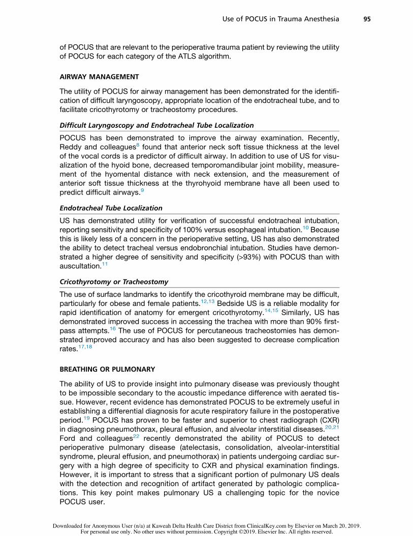
integral (VTI), for the LV outflow tract (LVOT) or aorta. The stroke distance is essentiallythe measurement of the distance a volume of blood is moved, from point A to point B,with each ventricular contraction. By multiplying the stroke distance or VTI by thecross-sectional area of the tube through which the blood is flowing (LVOT in thisexample), the stroke volume (SV) is determined. Assuming no acute changes in LVcontractility occur, the SV or LVOT VTIs can be compared during the inspiratoryand expiratory phases of mechanical positive-pressure ventilation. A change in SVor VTI across LVOT during mechanical ventilation has been shown to indicate fluidresponsiveness (Fig. 4B).41,42 Likewise, the perioperative physician can ascertainwhere on the Frank-Starling curve their patient exists (volume-responsive steepsegment or volume-nonresponsive flat segment) by measuring the SV or VTI beforeand after a fluid challenge. If a fluid challenge is given and the change in SV or VTIacross the LVOT before and after is minimal, the patient is unlikely to respond to esca-lating amounts of fluid therapy.
DISABILITY OR NEUROLOGIC ASSESSMENT
There are many emerging areas in which POCUS can assist in the neurologic assess-ment of trauma patients. Probably among themost validated topics is using POCUS toestimate intracranial pressure (ICP) values. Visualizing the optic nerve sheath (ONS)and then measuring the ONS diameter has been shown to provide a rapid and accu-rate assessment of whether ICP is elevated. The ONS is contiguous with the duramater and has a trabeculated arachnoid space through which cerebrospinal fluid cir-culates. The relationship between the ONS diameter (ONSD) and ICP has been well
Fig. 4. US methods of volume status. (A) IVC collapsibility. (B) Doppler ultrasonography: ve-locity time integral variation.
Ramsingh & Mangunta100
Downloaded for Anonymous User (n/a) at Kaweah Delta Health Care District from ClinicalKey.com by Elsevier on March 20, 2019.For personal use only. No other uses without permission. Copyright ©2019. Elsevier Inc. All rights reserved.

established.43,44 The sensitivity for US in detecting elevated ICP was 100% (95% CI68%–100%) and specificity was 63% (95% CI 50%–76%). An ONSD of greaterthan 5 mm, at a point approximately 2 mm from the retina, suggests elevated ICP.Additionally, rapid assessment of the retina and vitreous can reveal any obvious retinaldetachment or vitreous hemorrhage that may have occurred due to trauma.
EXPOSURE OR ENVIRONMENT CONTROLGastric Content
The ability of POCUS to determine the volume of gastric content has emerged as avalidated technique.45 Perlas and colleagues45 suggested that the presence of fluidin the gastric antrum identified on US in both the supine and right lateral decubitus po-sitions may help identify patients at increased risk of aspiration. US can differentiatebetween solid and clear liquid stomach contents, with more particulate contents beingassociated with worsened outcomes if aspirated. Although healthy fasting patientsmay have gastric volumes of up to 1.5 mL/kg with minimal risk of aspiration, the visu-alization of gastric antrum fluid volumes of greater than 180 mL suggests patients maybe at an increased risk for aspiration. The ability to detect gastric volume via POCUSmay prove to be a valid method to aid in assessing aspiration risk, as well as deter-mining if elective surgeries should be postponed based on the US examinationfindings.
Advanced Vascular Access
The use of US to assist with vascular access has advanced beyond its now wide-spread use for central venous access. Specifically, US has proven to reliably aid inthe placement of difficult intravenous46,47 and intraarterial catheters.48,49 Use of USfor peripheral venous access has also been shown to significantly increase successrates.50 At the Mid America Heart Institute, US-guided access of the basilic veinand introduction of a guidewire followed by threading of a 4F vascular access sheathhas reliably allowed preservation of the central veins for future access in patients withlimited vascular access. A recent meta-analysis was conducted to compareUS-guided and anatomic landmark-guided techniques for central venous catheterplacement. The results demonstrated a decreased risk of cannulation failure, arterialpuncture, hematoma, and hemothorax with the US-guided placement technique.51
In addition to US-guided vascular catheter placement, the Mid America Heart Insti-tute’s critical care and cardiovascular anesthesia providers use both POCUS and TEEto assist in the management of cardiac arrest and refractory acute respiratory distresssyndrome (ARDS). TEE-guided insertion of a bicaval dual-lumen catheter into the rightinternal jugular vein by cardiac critical care anesthesiologists facilitates rapid transitionto venovenous extracorporeal membrane oxygenation (ECMO) in severe ARDS. Simi-larly, during cardiac arrest and early cardiopulmonary resuscitation refractory to stan-dard ACLS protocols, POCUS is used to guide the rapid placement of guidewires intothe right femoral vein and left femoral artery for potential venoarterial ECMO cannula-tion by cardiothoracic surgeons in the operating room, if patients are candidates.
Evaluation of Deep Vein Thrombosis and Pulmonary Embolus
The current standard for evaluation of patients with suspected deep vein thrombosisor pulmonary embolus often involves lower compression ultrasonography andcomputed tomography pulmonary angiography. These tests are often performeddespite low pretest probability and the long time to perform and obtain the resultsof these tests may contribute to diagnostic delays. Recent evidence supports that
Use of POCUS in Trauma Anesthesia 101
Downloaded for Anonymous User (n/a) at Kaweah Delta Health Care District from ClinicalKey.com by Elsevier on March 20, 2019.For personal use only. No other uses without permission. Copyright ©2019. Elsevier Inc. All rights reserved.

the use of a focused POCUS examination, performed by intensivists, involving lung USfor subpleural infarcts, assessment for RV dilatation by cardiac US, and assessmentfor pulmonary embolus by leg vein US, can provide a high degree of sensitivity(90%) and specificity (86.2%) for the detection of pulmonary embolus.52
BRINGING THESE MODALITIES TO TRAUMA ANESTHESIOLOGISTS
Although it is encouraging to see the expansion of perioperative POCUS (P-POCUS),there remains the potential for tremendous growth with respect to the application ofPOCUS in trauma patient care. Other specialties have developed formalized educa-tional and certification pathways.53 EM has adopted POCUS training as a core com-petency for residency training and provides a year of fellowship training in clinical US.Of note, a similar interest in P-POCUS education has been reported by anesthesiologyresidents.54
Integration of comprehensive P-POCUS curricula has been developed for anes-thesia residency training but is currently not widely adopted. The 2018 AccreditationCouncil for Graduate Medical Education program requirements for anesthesiology,however, now require “competency in using surface ultrasound and transesophagealand transthoracic echocardiography to guide the performance of invasive proceduresand to evaluate organ function and pathology as related to anesthesia, criticalcare and resuscitation.”55 This change in educational requirements will encourageresidency programs to adopt existing P-POCUS curricula. However, the effectivenessand efficacy of implementing P-POCUS into anesthesiology training program curricularemains to be determined. The implementation of POCUS training into anesthesiologyresidency programs still faces many obstacles, especially without standardizedtraining protocols. It may take years for POCUS curricula to be effectively carriedout and demonstrate proficient clinical skill levels and knowledge among trainees.One study evaluated the utility of implementing a comprehensive POCUS educa-
tional curriculum for anesthesia residency training called Focused PerioperativeRisk Evaluation Sonography Involving Gastro-Abdominal Hemodynamic and Trans-thoracic US (FORESIGHT). This curriculum incorporated the topics of
1. Cardiac2. Pulmonary3. Hemodynamic4. Gastro-abdominal5. Airway6. Vascular access7. ICP assessment
In this single-center study, implementation of the curriculum into residency trainingdemonstrated statistical significance for positive participant satisfaction, improvedknowledge, and acquisition of hands-on skills as evaluated via the Kirkpatrick assess-ment tool.54 Additionally, a positive clinical impact was also suggested after 1 year oftraining. To further P-POCUS education, this curriculum is now online and providesopen access, under a Creative Commons license (www.foresightultrasound.com).This is among several initiatives to further the development of P-POCUS. Other ini-
tiatives include the focus-assessed TTE (FATE) protocol (http://usabcd.org), whichwas developed by an anesthesiologist and is among the most widely referencedPOCUS examination protocols. Additional online resources available for educationon P-POCUS include those from the Society of Critical Care Medicine (http://www.sccm.org/Education Center/Ultrasound/Pages/default.aspx), the American Institute
Ramsingh & Mangunta102
Downloaded for Anonymous User (n/a) at Kaweah Delta Health Care District from ClinicalKey.com by Elsevier on March 20, 2019.For personal use only. No other uses without permission. Copyright ©2019. Elsevier Inc. All rights reserved.

of Ultrasound (http://www.aium.org), and various Continuing Medical Educationtraining programs.53
Although these resources are important, the onus is on anesthesiologists to developstructured guidelines, endorsed educational pathways, and certified credentialingprocesses to incorporate P-POCUS into everyday practice. With the evolution ofanesthesiology from a specialty relegated to the operative theater and intensivecare units to a comprehensive perioperative role, anesthesiologists must embraceUS technology and become leaders and educators in the development of new appli-cations of this technology.
REFERENCES
1. American Association for the Surgery of Trauma; 2018. Available at: http://www.aast.org/trauma-facts. Accessed November 2, 2018.
2. Delegates ASoAHo. Statement of principles: trauma anesthesiology. AmericanSociety of Anesthesiologists House of Delegates; 2013. Available at: https://www.asahq.org/resources/resources-from-asa-committees/committee-on-trauma-and-emergency-preparedness/trauma-anesthesiology. Accessed November 2,2018.
3. The Advanced Trauma Life Support. 2008. Available at: https://www.facs.org/quality-programs/trauma/atls.
4. Kendall JL, Hoffenberg SR, Smith RS. History of emergency and critical care ul-trasound: the evolution of a new imaging paradigm. Crit Care Med 2007;35:S126–30.
5. Rozycki GS, Ochsner MG, Schmidt JA, et al. A prospective study of surgeon-performed ultrasound as the primary adjuvant modality for injured patient assess-ment. J Trauma 1995;39:492–8 [discussion: 498–500].
6. Gillman LM, Ball CG, Panebianco N, et al. Clinician performed resuscitative ultra-sonography for the initial evaluation and resuscitation of trauma. Scand J TraumaResusc Emerg Med 2009;17:34.
7. Kaslow O, Kuza CM, McCunn M, et al. Trauma anesthesiology as part of the coreanesthesiology residency program training: expert opinion of the American Soci-ety of Anesthesiologists Committee on Trauma and emergency preparedness(ASA COTEP). Anesth Analg 2017;125:1060–5.
8. Reddy PB, Punetha P, Chalam KS. Ultrasonography - a viable tool for airwayassessment. Indian J Anaesth 2016;60:807–13.
9. Fulkerson JS, Moore HM, Anderson TS, et al. Ultrasonography in the preoperativedifficult airway assessment. J Clin Monit Comput 2017;31:513–30.
10. Muslu B, Sert H, Kaya A, et al. Use of sonography for rapid identification ofesophageal and tracheal intubations in adult patients. J Ultrasound Med 2011;30:671–6.
11. Ramsingh D, Frank E, Haughton E, et al. Auscultation versus point of care ultra-sound to determine endotracheal versus bronchial intubation: a diagnostic accu-racy study. Anesthesiology 2016;124(5):1012–20.
12. Elliott DS, Baker PA, Scott MR, et al. Accuracy of surface landmark identificationfor cannula cricothyroidotomy. Anaesthesia 2010;65:889–94.
13. Aslani A, Ng SC, Hurley M, et al. Accuracy of identification of the cricothyroidmembrane in female subjects using palpation: an observational study. AnesthAnalg 2012;114:987–92.
Use of POCUS in Trauma Anesthesia 103
Downloaded for Anonymous User (n/a) at Kaweah Delta Health Care District from ClinicalKey.com by Elsevier on March 20, 2019.For personal use only. No other uses without permission. Copyright ©2019. Elsevier Inc. All rights reserved.

14. Nicholls SE, Sweeney TW, Ferre RM, et al. Bedside sonography by emergencyphysicians for the rapid identification of landmarks relevant to cricothyrotomy.Am J Emerg Med 2008;26:852–6.
15. Osman A, Sum KM. Role of upper airway ultrasound in airway management.J Intensive Care 2016;4:52.
16. Kleine-Brueggeney M, Greif R, Ross S, et al. Ultrasound-guided percutaneoustracheal puncture: a computer-tomographic controlled study in cadavers. Br JAnaesth 2011;106:738–42.
17. Rudas M, Seppelt I, Herkes R, et al. Traditional landmark versus ultrasoundguided tracheal puncture during percutaneous dilatational tracheostomy in adultintensive care patients: a randomised controlled trial. Crit Care 2014;18:514.
18. Yavuz A, Yilmaz M, Goya C, et al. Advantages of US in percutaneous dilatationaltracheostomy: randomized controlled trial and review of the literature. Radiology2014;273:927–36.
19. Lee FC. Lung ultrasound-a primary survey of the acutely dyspneic patient.J Intensive Care 2016;4:57.
20. Xirouchaki N, Magkanas E, Vaporidi K, et al. Lung ultrasound in critically ill pa-tients: comparison with bedside chest radiography. Intensive Care Med 2011;37:1488–93.
21. Blaivas M, Lyon M, Duggal S. A prospective comparison of supine chest radiog-raphy and bedside ultrasound for the diagnosis of traumatic pneumothorax.Acad Emerg Med 2005;12:844–9.
22. Ford JW, Heiberg J, Brennan AP, et al. A pilot assessment of 3 point-of-care stra-tegies for diagnosis of perioperative lung pathology. Anesth Analg 2017;124(3):734–42.
23. Gargani L, Frassi F, Soldati G, et al. Ultrasound lung comets for the differentialdiagnosis of acute cardiogenic dyspnoea: a comparison with natriuretic pep-tides. Eur J Heart Fail 2008;10:70–7.
24. Lichtenstein DA, Meziere GA. Relevance of lung ultrasound in the diagnosis ofacute respiratory failure: the BLUE protocol. Chest 2008;134:117–25.
25. Doust BD, Baum JK, Maklad NF, et al. Ultrasonic evaluation of pleural opacities.Radiology 1975;114:135–40.
26. Lichtenstein D, Goldstein I, Mourgeon E, et al. Comparative diagnostic perfor-mances of auscultation, chest radiography, and lung ultrasonography in acuterespiratory distress syndrome. Anesthesiology 2004;100:9–15.
27. Lichtenstein D, Hulot JS, Rabiller A, et al. Feasibility and safety of ultrasound-aided thoracentesis in mechanically ventilated patients. Intensive Care Med1999;25:955–8.
28. Scalea TM, Rodriguez A, Chiu WC, et al. Focused assessment with sonographyfor trauma (FAST): results from an international consensus conference. J Trauma1999;46:466–72.
29. Rose JS. Ultrasound in abdominal trauma. Emerg Med Clin North Am 2004;22:581–99, vii.
30. Kirkpatrick AW, Sirois M, Laupland KB, et al. Prospective evaluation of hand-heldfocused abdominal sonography for trauma (FAST) in blunt abdominal trauma.Can J Surg 2005;48:453–60.
31. Andersen GN, Haugen BO, Graven T, et al. Feasibility and reliability of point-of-care pocket-sized echocardiography. Eur J Echocardiogr 2011;12:665–70.
32. Manasia AR, Nagaraj HM, Kodali RB, et al. Feasibility and potential clinical utilityof goal-directed transthoracic echocardiography performed by noncardiologist
Ramsingh & Mangunta104
Downloaded for Anonymous User (n/a) at Kaweah Delta Health Care District from ClinicalKey.com by Elsevier on March 20, 2019.For personal use only. No other uses without permission. Copyright ©2019. Elsevier Inc. All rights reserved.

intensivists using a small hand-carried device (SonoHeart) in critically ill patients.J Cardiothorac Vasc Anesth 2005;19:155–9.
33. Mazraeshahi RM, Farmer JC, Porembka DT. A suggested curriculum in echocar-diography for critical care physicians. Crit Care Med 2007;35:S431–3.
34. Bundgaard-Nielsen M, Holte K, Secher NH, et al. Monitoring of peri-operativefluid administration by individualized goal-directed therapy. Acta AnaesthesiolScand 2007;51:331–40.
35. Ommen SR, Nishimura RA, Hurrell DG, et al. Assessment of right atrial pressurewith 2-dimensional and Doppler echocardiography: a simultaneous catheteriza-tion and echocardiographic study. Mayo Clin Proc 2000;75:24–9.
36. Prekker ME, Scott NL, Hart D, et al. Point-of-care ultrasound to estimate centralvenous pressure: a comparison of three techniques. Crit Care Med 2013;41:833–41.
37. Cannesson M, Slieker J, Desebbe O, et al. Prediction of fluid responsiveness us-ing respiratory variations in left ventricular stroke area by transoesophageal echo-cardiographic automated border detection in mechanically ventilated patients.Crit Care 2006;10:R171.
38. Scheuren K, Wente MN, Hainer C, et al. Left ventricular end-diastolic area is ameasure of cardiac preload in patients with early septic shock. Eur J Anaesthesiol2009;26:759–65.
39. Subramaniam B, Talmor D. Echocardiography for management of hypotension inthe intensive care unit. Crit Care Med 2007;35:S401–7.
40. Sefidbakht S, Assadsangabi R, Abbasi HR, et al. Sonographic measurement ofthe inferior vena cava as a predictor of shock in trauma patients. Emerg Radiol2007;14:181–5.
41. Broch O, Renner J, Gruenewald M, et al. Variation of left ventricular outflow tractvelocity and global end-diastolic volume index reliably predict fluid responsive-ness in cardiac surgery patients. J Crit Care 2012;27:325.e7-13.
42. Charron C, Caille V, Jardin F, et al. Echocardiographic measurement of fluidresponsiveness. Curr Opin Crit Care 2006;12:249–54.
43. Hansen HC, Helmke K. Validation of the optic nerve sheath response to changingcerebrospinal fluid pressure: ultrasound findings during intrathecal infusion tests.J Neurosurg 1997;87:34–40.
44. Tayal VS, Neulander M, Norton HJ, et al. Emergency department sonographicmeasurement of optic nerve sheath diameter to detect findings of increased intra-cranial pressure in adult head injury patients. Ann Emerg Med 2007;49:508–14.
45. Perlas A, Van de Putte P, Van Houwe P, et al. I-AIM framework for point-of-caregastric ultrasound. Br J Anaesth 2016;116:7–11.
46. Costantino TG, Parikh AK, Satz WA, et al. Ultrasonography-guided peripheralintravenous access versus traditional approaches in patients with difficult intrave-nous access. Ann Emerg Med 2005;46:456–61.
47. Keyes LE, Frazee BW, Snoey ER, et al. Ultrasound-guided brachial and basilicvein cannulation in emergency department patients with difficult intravenous ac-cess. Ann Emerg Med 1999;34:711–4.
48. Ashworth A, Arrowsmith JE. Ultrasound-guided arterial cannulation. Eur J Anaes-thesiol 2010;27:307.
49. Shiver S, Blaivas M, Lyon M. A prospective comparison of ultrasound-guided andblindly placed radial arterial catheters. Acad Emerg Med 2006;13:1275–9.
50. Stolz LA, Stolz U, Howe C, et al. Ultrasound-guided peripheral venous access: ameta-analysis and systematic review. J Vasc Access 2015;16(4):321–6.
Use of POCUS in Trauma Anesthesia 105
Downloaded for Anonymous User (n/a) at Kaweah Delta Health Care District from ClinicalKey.com by Elsevier on March 20, 2019.For personal use only. No other uses without permission. Copyright ©2019. Elsevier Inc. All rights reserved.

51. Wu SY, Ling Q, Cao LH, et al. Real-time two-dimensional ultrasound guidance forcentral venous cannulation: a meta-analysis. Anesthesiology 2013;118:361–75.
52. Nazerian P, Vanni S, Volpicelli G, et al. Accuracy of point-of-care multiorgan ultra-sonography for the diagnosis of pulmonary embolism. Chest 2014;145:950–7.
53. Mahmood F, Matyal R, Skubas N, et al. Perioperative ultrasound training in anes-thesiology: a call to action. Anesth Analg 2016;122:1794–804.
54. Ramsingh D, Rinehart J, Kain Z, et al. Impact assessment of perioperative point-of-care ultrasound training on anesthesiology residents. Anesthesiology 2015;123:670–82.
55. ACGME program requirements for graduate medical education in anesthesiology.Suite 2000, 401 North Michigan Avenue, Chicago, IL 60611. 2018. p. 17–8.Available at: https://www.acgme.org/Portals/0/PFAssets/ProgramRequirements/040Anesthesiology2018.pdf?ver52018-06-14-142529-527. Accessed July 24,2018.
Ramsingh & Mangunta106
Downloaded for Anonymous User (n/a) at Kaweah Delta Health Care District from ClinicalKey.com by Elsevier on March 20, 2019.For personal use only. No other uses without permission. Copyright ©2019. Elsevier Inc. All rights reserved.