The Use of Extended Wear Contact Lenses in the Aviation ... the Aviation Environment: An Armywide...
195
1o1•3..j.......-:c: USAARL Report No. 92-35 lw...... DTIC ISM - 01k ELECTE AD-A260 938 z I" The Use of Extended Wear Contact Lenses in the Aviation Environment: An Armywide Study By Morris R. Lattimore Sensory Research Division and Rhonda L. S. Cornum Biomedical Applications Research Division September 1992 93-02003 Approved for public release; distributoio unlimited. 93 2 3 United States Army Aeromedical Research Laboratory Fort Rucker, Alabama 36362-5292
Transcript of The Use of Extended Wear Contact Lenses in the Aviation ... the Aviation Environment: An Armywide...
1o1•3..j.......-:c: USAARL Report No. 92-35
lw......
DTICISM - 01k ELECTE
AD-A260 938z I"
The Use of Extended Wear Contact Lensesin the Aviation Environment:
An Armywide Study
ByMorris R. Lattimore
Sensory Research Division
and
Rhonda L. S. CornumBiomedical Applications Research Division
September 1992
93-02003
Approved for public release; distributoio unlimited.
93 2 3United States Army Aeromedical Research Laboratory
Fort Rucker, Alabama 36362-5292


Notice
Qualified requesters
Qualified requesters may obtain copies from the DefenseTechnical Information Center (DTIC), Cameron Station, Alexandria,Virginia 22314. Orders will be expedited if placed through thelibrarian or other person designated to request documents fromDTIC.
Change of address
Organizations receiving reports from the U.S. ArmyAeromedical Research Laboratory on automatic mailing lists shouldconfirm correct address when corresponding about laboratoryreports.
Disposition
Destroy this report when it is no longer needed. Do not returnto the originator.
Disclaimer
The views, opinions, and/or findings contained in this reportare those of the authors and should not be construed as anofficial Department of the Army position, policy, or decision,unless so designated by other official documentation. Citationof trade names in this report does not constitute an officialDepartment of the Army endorsement or approval of the use of suchcommercial items.
Human use
Human subjects participated in these studies after givingtheir free and informed voluntary consent. Investigators adheredto AR 70-25 and USAMRDC Reg 70-25 on Use of volunteers inResearch.
Reviewed:
IARD R. 6VCE4ý ý
Dfrector, Sensory ResearchDi sion Released for publication:
ROM W. WTqLEY,-OD. 0h.D. DAVID H. KAEYCh irman, Scientific Colonel, MC, SFS
Review Committee Commanding
N

DISCLAIMEI NOTICE
THIS DOCUMENT IS BEST
QUALITY AVAILABLE. THE COPY
FURNISHED TO DTIC CONTAINED
A SIGNIFICANT NUMBER OF
COLOR PAGES WHICH DO NOT
REPRODUCE LEGIBLY ON BLACK
AND WHITE MICROFICHE.
UnclassifiedSECURITY CLASSIFICATION OF THIS PAGE
Form ApprovedREPORT DOCUMENTATION PAGE OMB No. 0704-0188
Ia. REPORT SECURITY CLASSIFICATION ilb RESTRICTIVE MARKINGS
Unclassified _2a. SECURITY CLASSIFICATION AUTHORITY 3. DISTRIBUTION /AVAILABILITY OF REPORT
Approved for public release; distribution2b. DECLASSIFICATION / DOWNGRADING SCHEDULE unlimited
4. PERFORMING ORGANIZATION REPORT NUMBER(S) 5. MONITORING ORGANIZATION REPORT NUMBER(S)
USAARL Report No. 92-35
6a. NAME OF PERFORMING ORGANIZATION 6b. OFFICE SYMBOL 7a. NAME OF MONITORING ORGANIZATIONU.S. Army Aeromedical Research (If applicable) U.S. Army Medical Research and DevelopmentLaboratory I SGRD-UAS-VD Command6c. ADDRESS (City, State, and ZIP Code) 7b. ADDRESS (City, State, and ZIP Code)P.O. Box 577 Fort DetrickFort Rucker, AL 36362-5292 Frederick, f1D 21702-501?
8,. NAME OF FUNDING/SPONSORING 8b. OFFICE SYMBOL 9. PROCUREMENT INSTRUMENT IDENTIFICATION NUMBERORGANIZATION (If applicable)
8c. ADDRESS(City, State, and ZIP Code) 10. SOURCE OF FUNDING NUMBERSPROGRAM PROJECT TASK WORK UNITELEMENT NO. NO. 3M162 NO. ACCESSION NO.
0602787A 1787A879 I BG 16811. TITLE (Include Security Classification) The Use of Extended Wear Contact Lenses in the Aviation
Environment: An Army-wide Study
11. PERSONAL AUTHOR(S)
Lattimore, tMorris R., and Cornum, Rhonda L.S.13a. TYPE OF REPORT 13b. TIME COVERED 114. DATE OF REPORT (Year, Month, Day) 15. PAGE COUNT
Einal FROM TO _ 1992 September 16816. SUPPLEMENTARY NOTATION
17. COSATI CODES 18. SUBJECT TERMS (Continue on reverse if necessary and identify by block number)FIELD GROUP SUB-GROUP contact lenses, Army aviators, flight safety, cornealUW 04 physiology, tear production, water content, central cornealub _ 07 thickening, sterile ulcers, endothelial analyses
19. ABSTRACT (Continue on reverse it necessary and identify by block number)
Standard refractive error correction options for the M-43 protectivemask have proven to be incompatible wi..h the Helmet Display Unit (HDU)component of the AH-64 "Apache" Integrated Helmet and Display SightingSystem (IHADSS). Glue-on and outsert packages push the HDU, a Maxwellian-
)view virtual imaging system, far enough from the spectacle-wearingqviator's eye to significantly reduce the available field-c'f-view.Consequently, portions of critical peripheral instrumentatLon and weapon
%system overlays cannot be visualized. In November 1988, the U.S. ArmyAeromedical Research Laboratory (USAARL) initiated the AH-64 contact lensresearch protocol to provide both an interim readiness fix and to develop acomprehensive database on contact lens wear in a variety of environments.
Continued
20. DISTRIBUTION /AVAILABILITY OF ABSTRACT 21. ABSTRACT SECURITY CLASSIFICATION91UNCLASSIFIED/UNLIMITED 0 SAME AS RPT. 0 DTIC USERS Unclassified
22a. NAME OF RESPONSIBLE INDIVIDUAL 15' 22b TELEPHONE (Include Area Code) 22c. OFFICE SYMBOLChif.Scenifc nfrmtion Center (205) 255-6907 5G-RD-UAX-SI
D0 Form 1473, JUN 86 Previous editions are obsolete. SECURITY CLASSIFICATION OF THIS PAGEUnclassified

19. ABSTRACT (Continued)
The protocol was organized from three perspectives withconcerns dii -cted toward operational and flight safety, ocularhealth, and corneal physiology issues, and concluded at the endof September 1991. Fundamental operational and safety data werechronicled, along with written questionnaires, to assesssubjective effectiveness of routine contact lens use. Ocularhealth complications were collated from the aviation medicine,optometric, and ophthalmological communities. Clinical andphysiological data were gathered by one USAARL optometrist, andtwo contract civilian optometrists and their supportingtechnicians.
In September 1990, a general aviation version of the M-43protective mask was identified for early fielding without itsspectacle outsert. All spectacle-wearing aircrew (pilots,crewchiefs, door gunners, flight medics, and flight surgeons)deploying to Southwest Asia were examined for possible contactlens wear under the auspices of the existing Armywide contactlens research protocol. Ten Army optometrists and 10 techniciansperformed the additional examinations at over a dozen U.S.locations ai;u 3 locations in Europe. Four of the optometristspermanently deployed to Saudi Arabia in direct support, and forthe duration, of Operations Desert Shield/Storm. The originalprotocol included 238 subjects, while the Desert Storm portion(general aviation) added 344 subjects. Roughly 450 of the total582 contact lens-wearing subjects served in Southwest Asia onOperation Desert Storm. Overall initial fitting success was 72percent (624 fitted out of 868 volunteers). Unsuccessful fittingattempts fell into four general groupings: presbyopes, highastigmats, extremely steep or flat corneal curvatures, andpreexisting medical conditions. Wearing success was 67 percent;42 subjects withdrew or were discontinued from lens wear over thecourse of the study. The primary dissatisfiers related to thesame groupings affecting fitting success.
The original protocol used a three-tier contact lens fittingsystem, with the initial lens of choice being a moderate to highwater content disposable extended wear soft lens. Backup lensesconsisted of a low water content standard extended wear soft lensutilized on a disposable basis, and a rigid gas permeable (RGP)lens used with a chemical disinfection system. Both types ofsoft lenses had analogous diameters and base curves (14.0 mm and8.8 mm, respectively). The RGP lenses were not fielded onOperation Desert Storm because of concerns for possible foreignbody intrusion from blowing dust and dirt. Limited RGP use byDesert Storm participants from the original protocol confirmedthis concern. The original protocol wearing schedule was set ata maximum of 7 days/6 nights of extended lens wear. Desert Storm
(2)

19. ABSTRACT (Continued)
subjects were advised to follow a more conservat've 3-day/2-nightschedule. The subjects were instructed that the night of lensremoval was to be passed without any new lens wear; worn softlenses were to be discarded, and RGP lenses cleaned, disinfected,and stored overnight. After at least one full night of lens-freesleep. the subjects were instructed that they could apply a newsoft iens, or resume wear of the cleaned and disinfected RGPlenses.
Over the 33 months of the study, there were minimal changesrnoted in clinical appraisal of the tarsal conjunctiva, possiblecorneal edema, tear BUT, and tear production for all subjects.Mild changes were evident in evaluation of the bulbarconjunctiva, limbal injection, corneal vascularization, rosebengal staining, and fluorescein staining for the originalprotocol subjects. Mild to moderate changes were seen in many ofthe Desert Storm subjects. However, despite the harsh desertenvironment, contact lens wear in Southwest Asia, as assessed byslit lamp evaluation, was much less stressful than expected.
There have been six cases of bacterial ulcerative keratitis;two during the AH-64 portion of the protocol, one associated withdeployment on Operation Desert Shield, none documented during theDesert Storm combat phase of the deployment, and thr'Ze occurringduring or shortly after redeployment from Southwest Asia. Whileaffected aircrew were temporarily grounded from flight duty
* during the course of the infection, ali returned to full flight* duties after resolution of the acute infection. Visual acuity
recovered to 20/20 or better in all six subjects. Tie resultantcalculated risk for ulcerative keratitis was 1 per 112 subjectsper year of contact lens wear (8.9/1000/year). Civilianestimates have placed the risk of infective ulcerative keratitisfrom 2.1/1000/year to 15/1000/year to 48/1000/year. Althoughthis study had a relatively low number of subjects compared tomany civilian studies, the occurrence of this severe infectionfell within the range established in the published literature.
The subjective evaluation of routine contact lens wear washigh in garrison, field, and combat conditions, as wereperformance assessments. Combat missions included: attack,troop transport, equipment transport, surveillance, intelligence,special operations, and medical evacuation. The Apache radarinter'diction mission into Iraq on 16 January 1991, w.iLchinitL;ted Operation Desert Storm, consisted of several contactlens wearers, including the mission commander. By questionnaire,subjects overwhelmingly approved contact lens use in allsettings; 95 percent expressed greater combat readiness andeffectiveness with contact lenses; 98 percent felt contact lens
(3)
19. ABSTRACT (Continued)
use (and maintenance) in the cockpit had no adverse impact onsafety of flight; and 98 percent endorsed the routine use ofcontact lenses in the performance of flight duties.
Based on the clinical, ocular health and subjectivequestionnaire data, contact lens wear by Army aircrew is a viablealternative to spectacle wear. However, because of uniquedifficultiefs encountered in attempting to fit presbyopes, highastigmats, and those with extreme corneal curvatures (either veryflat or very steep), one-third of spectacle-wearing aircrew maynot be able to successfully wear contact lenses. This fractionlikely will decrease in a routine clinical program if lensparameter availability is expanded beyond those used in thisstudy. Nevertheless, routine contact lens wear will representonly a partial solution to Army aviation's spectacleincompatibility problem. Therefore, developmental hardwarealternatives must be included in future system programming ormany air crewmembers will be prevented from performing certainflight duties.
(4)

Acknowledgments
The iuthors wish to thank the volunteer subjects and theircommanders for their enthusiastic support of this exhaustiveresearch effort. Sincere appreciation is also extended to SGTVincent Reynoso who provided technical support and to Ms. CarolynJohnson who provided clerical support.
Acee"1.a yor
amm
D'MC QUALITY INSPIY'TD S Dintrtb ir,/
tDist

This page intentionally left blank.
ii

THIS
PAGEIs
MISSING
IN
ORIGINAL
DOCUMENTi 1 1+ 1 .0
Table of contents (Continued)
Discus,: ion ...................................... 62Conclusions ..................................... 63
Hydrogel lens water content ......................... 63Introduction .................................... 63Methods and materials .......................... 64Results and discussion ......................... 64Conclusions .................................... 70
pH and water content correlational analysis ........ 70Introduction .................................... 70Methods and materials .......................... 70Results and discussion ......................... 73Conclusions .................................... 74
Corneal thickness measurement differences .......... 74Introduction .................................... 74Methods ........................................ 74Results and discussion ......................... 75Conclusions ..................................... 78
Visual acuity testing .............................. 79Introduction .................................... 79Methods and materials .......................... 80Results and discussion ......................... 80Conclusions .................................... 82
Case report: endothelial guttata .................. 82Introduction .................................... 82Methods ....................................... 85Results ....................................... 86Discussion ...................................... 88
Schirmer tear test ................................... 88Introduction .................................... 88Methods and materials .......................... 89Results ........................................ 89Discussion ...................................... 90
Operations Desert Shield and Desert Storm .......... 92Introduction .................................... 92Methods and materials .......................... 93Results ......................................... 95Discussion ...................................... 96Conclusions ..................................... 98
Assessment of subjective questionnaire data ........ 99Introduction .................................. 99Results and discussion ........................ 100
Handling of lenses and training .......... 101Comfort/vision ........................... 106Environmental conditions ................. 107Compliance to wearing schedule ........... 109General physical lens wear ............... 110Night vision goggles ..................... I11M-43 protective mask ..................... 112
V

Table of contents (Continued)
Safety of flight ......................... 114Final assessments ........................ 115
Conclusions ................................... 116Overall conclusions .......................................... 117References .................................................... 121
List of figures
Figure 1. Proposed hydrogel matrix pH gradient ............ 20Figure 2. Contact lens study refractive error
distribution ................................... 32Figure 3. A comparative distribution of subject
spherical refractive error .................... 33Figure 4. Concact lens study distribution by power
and lens type .................................. 34Figure 5. Contact lens study age distribution ............. 35Figure 6. Aviation population age distribution ............ 36Figure 7. Corneal limbal vascular development ............. 37Figure 8. Rose bengal and fluorescein assessments ......... 38Figure 9. Influence of wearing time on rose bengal
stain severity ................................. 39Figure 10. Influence of wearing time on fluorescein
stain severity ................................. 40Figure 11. Influence of wearing time on stain
severity assessments .......................... 41Figure 1?. Tear BUT assessment comparison ................... 42Figure 13. Anterior optical surface curvature during
spherical soft lens wear (38 percent watercontent lenses) ................................ 46
Figure 14. Anterior optical surface curvature duringspherical soft lens wear (58 percent watercontent lenses) ................................ 47
Figure 15. Anterior optical surface cylinder duringspherical soft lens wear (38 percent watercontent lenses) ................................ 48
Figure 16. Anterior optical surface cylinder duringspherical soft lens wear (58 percent watercontent lenses) ................................ 49
Figure 17. Corneal and anterior lens surfacetopographies ................................... 50
Figure 18. Aesthesiometer calibration plot .................. 51Figure 19. Corneal sensation thresholds ..................... 52Figure 20. Peripheral, sterile ulcer ....................... 53Figure 21. Paracentral, bacterial ulcer ..................... 55Figure 22. Influence of wearing time on translens
oxygen flux (grouped data) .................... 58
vi
List of figures (Continued)
Figure 23. Influence of wearing time on translensoxygen flux (by lens type) .................... 59
Figure 24. Influence of wearing time on hydrogel lensanterior surface pH (grouped data) ............ 61
Figure 25. Influence of wearing time on hydrogel lensanterior surface pH (by lens type) ............ 62
Figure 26. Influence of wearing time on hydrogel lenswater content (two measurement methods; 58percent water content lenses only) ............ 65
Figure 27. Hydrogel lens dehydration under extendedwear conditions (Fort Rucker and DesertShield data) ................................... 66
Figure 28. Hydrogel lens dehydration under extended-wear conditions (38 percent water contentlenses) ........................................ 67
Figure 29. Hydrogel lens dehydration under extended-wear conditions (by lens type) ................ 68
Figure 30. Normalized hydration levels in two typesof soft lenses ................................. 69
Figure 31. Influence of wearing time on hydrogel lensanterior surface pH and water content(grouped data) ................................. 71
Figure 32. Influence of wearing time on hydrogel lensanterior surface pH and water content(by lens type) ................................. 72
Figure 33. Influence of anterior lens surface pH onhydrogel lens water content (a linearcorrelation of two nonlinear variables) ....... 73
Figure 34. Paired measurement pachometer comparison ......... 76Figure 35. Pachometry frequency histogram ................... 77Figure 36. Corneal thickness measurement: long
term lens wear/technique differences .......... 78Figure 37. Corneal thickness measurement: duration
of wear/technique differences ................. 79Figure 38. Matched acuity response frequency
distribution ................................... 81Figure 39. Low illuminance - low contrast acuity
as a function of subject age(contact lens and spectacle-wearingconditions) .................................... 83
Figure 40. Endothelial photomicrograph, right eye ........... 84Figure 41. Endothelial photomicrograph, left eye ............ 85Figure 42. Endothelial analysis examples from
initial and final examinations ................ 86Figure 43. Endothelial cell size distribution ............... 87Figure 44. Schirmer tear test (without anesthetic) .......... 91Figure 45. Schirmer tear test (with anesthetic) ............. 91Figure 46. Desert Storm primary aircraft
distribution ................................... 93
vii

List of figures (Continued)
Figure 47. Desert Storm aircrew job distribution ............ 94Figure 48. Desert Storm aircrew age distribution ............ 95Figure 49. Aviation population age distribution ............. 96Figure 50. Desert Storm unsuccessful fit distribution ....... 97Figure 51. Desert Storm refractive error distribution ....... 98Figure 52. Tarsal conjunctival irritation ................... 99Figure 53. Bulbar injection ................................. 100Figure 54. Limbal injection ................................. 101Figure 55. Corneal edema .................................... 102
Figure 56. Fluorescein staining ............................. 103Figure 57. Rose bengal staining ............................. 103Figure 58. Tear breakup time (with lenses) .................. 104Figure 59. Tear breakup time (without lenses) ............... 104Figure 60. Lens blink movement .............................. 105
List of tables
Table 1. Recent tear pH studies ............................. 19Table 2. Visual acuity correlations ......................... 84Table 3. Endothelial analysis data .......................... 87Table 4. Schirmer tear test .................................. 90
Appendixes
Appendix A: Initial examination............................. 133Appendix B: Followup examination ............................ 140Appendix C: List of manufacturers ........................... 147Appendix D: Volunteer agreement affidavit ................... 148Appendix E. Volunteer registry data sheet ................... 155Appendix Y. Medical identification card ..................... 158Appendix G. Qualitative data ................................ 159
viii
Introduction
Purpose
The purpose of this study was to determine the compatibilityof extended wear contact lenses for the U.S. Army aviationenvironment on a worldwide basis. The specific projectobjectives were to:
1. Determine success rates in the fitting and thewearing of contact lenses in a volunteer sample ofametropic aircrewmembers on a worldwide basis.
2. Document the descriptive characteristics of thevolunteer sample of Army aviation and makeprojections concerning the Army aviation populationas a whole.
3. Characterize the physiological/biochemical responseof the cornea to contact lens wear, and quantifypotentially altered contact lens characteristicsafter application onto the eye.
4. Document incidence of corneal complications andattempt to identify underlying mechanisms ofpathology.
5. Obtain a quantifiable assessment of contact lensacceptability and identify the operational impactof contact lens wear associated with theoccupational tasks and conditions unique to Armyaviation.
Approach
Volunteer ametropic aircrewmembers were fitted with extendedwear contact lenses. It should be stressed that this was acontact lens research program, not a field test of ccntact lensuse; as such, only volunteer participants were used. Thevoluntary nature of this program was repeatedly stressed toparticipants so that they fully understood their right to notparticipate or to withdraw at any time. Periodic clinicalmonitoring was accomplished to safeguard the ocular health of thesubjects and their continued safety of flight. Combined with theclinical monitoring process were a number of tests designed toassess the physiological and biochemical response of the corneato contact lens wear. Operational effectiveness was assessed byway of volunteer subjective responses concerning positive andnegative effects of CL wear as reported to their unit flightsurgeon, and as reported on a quarterly questionnaire.
1

It should be noted that Army Regulations (AR) 40-5, AR 40-8,AR 40-63, and AR 40-501 express certain prohibitions associatedwith the use of contact lenses by Army aircrewmembers. A waiverof policy relating to the use of contact lenses was approved byThe office of The Surgeon General for the wear of contact lensesunder controlled investigational conditions, and more recently,in conjunction with Operation(s) Desert Shield and Storm. Anindividual waiver for each subject participating in the study wasinitiated by the subject's unit flight surgeon via an abbreviatedaeromedical summary, which stipulated that only one contact lenswearer be allowed on any one individual flight. The waivez.• wereprocessed through the Aeromedical Center at Fort Rucker, and thenthrough the Total Army Personnel Command (PERSCOM) for finalwaiver approval.
Military significance
The role of vision in aviation has always been an importantone. Now, with the ever-increasing technological complexity ofthe man-machine interface, optimal visual performance has becomeabsolutely essential to aircraft operation. However,sophisticated electro-optical display devices often can present acompatibility problem with spectacles; the use of specializedenvironmental protective systems can further confound thisproblem. As a result, spectacle wearing aviators, many withadvanced aviation skills, could be deselected from duty incertain aircraft. The use of extended wear contact lenses couldhelp maintain the pool of qualified aviators available fordeployment in these sophisticated aviation systems.
Background
Statement of the specific problem
Traditionally, prospective aviators have had to meetstringent vision standards for acceptance into a trainingprogram. Over the years some standards have been subject towaiver, the requirement for relative emmetropia being one ofthem. This, in conjunction with the reduction of otherstandards, and with the development of late-onset, maturationalmyopia in some individuals, has led to the development of asizeable ametropic population in the Army aviation community.Currently, approximately 23 percent of all Army aviators wear aspectacle correction (Schrimsher and Lattimore, 1991).
Specific problems have emerged involving the integration ofspectacle wear with certain avionics systems. The standard issueaviator spectacle is not compatible with the Integrated HelmetDisplay and Sighting System (IHADSS) of the Advanced AttackHelicopter, AH-64. As a result, a modified right eye-piece wasdeveloped for the aviator spectacle frame. This modification,
2

however, still does not prevent IHADSS combiner lens positioningfrom being a difficult process. Unauthorized individualmodifications, designed to move the right spectacle lens closerto the face, have forced some individuals to trim their eyelashesso that they won't rub against the repositioned lens.Yet, even this extreme measure fails to solve the positioningproblem.
The optical relay tube (ORT), found in both the AH-I and AH-64, presents another spectacle compatibility problem. Theseinstrumentation interfacing difficulties have a detrimentaleffect on operational efficiency. The reduction or eliminationof these difficulties are essential to effective combat flightoperations.
The M-43 protective mask for AH-64 aviators, and the generalaviation version of that mask, present yet another problem.Initially, it was anticipated that ametropic aviators would havetheir correction incorporated onto the protective mask eyepiece.This system adaptation has been shown to induce a number ofvisual problems which lead to an incompatibility with theIntegrated Helmet and Display Sighting Subsystem (IHADSS) orHelmet Display Unit (HDU) on the Apache. As a result, ametropicaviators are unable to optimally operate the AH-64 in achemically contaminated environment with the glue-on correction.In addition, there has been a recent question concerning nightvision goggle (NVG) compatibility with the optical outsertdesigned for the new general aviation version of the M-43 mask.
The use of contact lenses by ametropic aircrewmembers offersa potential solution to these problems. However, because Armyaviation's combat mission requires an immediate response, theonly feasible type of contact lens would be extended wear innature. The Army aviator's immediate combat responsivenessrequirement would not permit adequate time for lens preparationand insertion prior to the mission. Furthermore, disposablelenses are desirable in order to minimize cleaning anddisinfection problems, particularly those associated with anoperational field setting. It should be stressed that whiledisposable lenses may help minimize the potential for ocularinfection, other problems can be induced by the use of contactlenses on an extended wear basis. It is the ultimate objectiveof this study to document both the benefits and deficiencies ofcontact lens wear so that the Army can determine the overallacceptability of this visual correction option.
Literature review introduction
Recent technological advances have had a major impact onmilitary aviation. While modern methods of providing visualinformation via electro-optics/visionics systems have extended
3
the aviator's operational envelope, these devices are becomingincreasingly incompatible with spectacle wear. Due to uniquestringent regulations, the Navy and Marine Corps do not allowservice members with high refractive errors (i.e., uncorrectedvisual acuity worse than 20/70) to pilot aircraft equipped withthese advanced visionics systems; if an aviator develops anexcessive refractive error, administrative reassignment as aflight officer (bombardier/navigator, radar intercept officer)ensues (Markovits, 1988). Alternatively, Navy/Marine Corpsaviators with uncorrected visual acuity from 20/25 to 20/70,correctable to 20/20 or better, are permitted to operate thesehigh performance aircraft. This type of partial deselectionprocess has, for the moment, been rejected by the Army and AirForce. Since close to 23 percent of Army aviators (Schrimsherand Lattimore, 1991) and 27 percent of Air Force aviators(Dennis, 1988) are ametropic (spectacle wearing), and since anincreasing percentage of training applicants are ametropic,alternative means of providing a refractive error correction needto be investigated.
One alternative being considered is the utilization of acontact lens correction. Current and past armed forcesregulations have prohibited the wearing of contact lenses byaviators while flying. However, waivers to these regulationshave been approved at certain locations where controlledscientific investigations are being conducted. Because ofdifferences in i.issions and operational scenarios, researchefforts are being directed along somewhat divergent paths. AirForce concerns concentrate on low atmospheric pressure/lowambient oxygen issues, low relative humidity, and high g-forceeffects on daily lens wear. Army concerns center on theoperational field environment, its impact on proper lens hygiene(cleaning and disinfection), and the physiological/biochemicalresponse of the cornea to extended contact lens wear. Since thequestion of contact lens use by aviation personnel is a matter ofcurrent interest throughout the aviation and aeromedicalcommunities, this review provides a general overview of salientissues and considerations.
Aviation literature review
A number of types of contact lenses have been evaluatedwithin the aviation environment. The first Army aviation studywas in 1974 (Crosley, Braun, and Bailey). Of concern at the timewas the fact that "hard" polymethylmethacrylate (PMMA) contactlenses were prone to dust particle interference between thecornea and the contact lens when worn by ground troops in anoperational environment (Rengstorff, 1965, and 1972; LaPiana,1980). Since Army aviators routinely were exposed to dustyenvironments, the PMMA lenses had been ruled out as an Armyaviator optical correction. The Bausch and Lomb (B&L) "Soflens"
4
was found to be independent of dust-induced foreign bodyproblems. However, an unacceptable variability in visual acuitydid result. A parallel study (Polishuk and Raz, 1975) obtainedsimilar results concerning both absence of dust and dirt problemsand variable visual acuity in a population of Israeli militaryand civilian pilots. Acuity variation was not attributed to anyspecific origin.
Since soft contact lenses have a moderate to high watercontent, other studies have been concerned with the effects ofboth low atmospheric pressure and low relative humidity on lensdehydration and corneal health. A number of clinical casereports concerning extended passenger travel difficulties withcontact lenses surfaced in the literature (Jagerman, 1973;Casebeer, 1973; Corboy and Tannehill, 1973) serving to stimulatespecific laboratory investigations. A hypobaric chamber study,simulating altitudes up to 30,000 feet on the B&L "Soflens,"failed to demonstrate an effect on contact lens wearability (Eng,Rasco, and Marano, 1978). However, in a study by Forgie (1981)with simulations at 25,000 feet for 2.5 hours and at 9,000 feetfor 6 hours, subjects demonstrated some tear film debris andexperienced minor discomfort. Despite these findings, aircraftcontrol was not significantly degraded, and visual acuities weresaid not to be affected. Forgie's findings were in agreementwith those of Hapnes (1980), whereby subjects were kept at 1/2atmosphere for 4 hours. All subjects exhibited minor objectivecorneal changes that appeared to be epithelial in origin. Morerecently, the U.S. Air Force conducted a series of hypobaricchamber "flights" in order to assess soft contact lens wear ataltitude (Flynn et al., 1988). Indicators of physiologicalstress to the cornea (by slit lamp examination) showed heightenedresponses at altitude with contact lenses. However, thesechanges occurred without measurable degradation in vision and didnot preclude the normal wear of soft contact lenses.
Another recent study (Flynn et al., 1987) has documentedsubcontact lens bubble formation in a hypobaric chamber protocol.Soft contact lens bubble formation was limited to the lensperiphery, and was without sequelae to vision or cornealepithelium integrity. Rigid, gas permeable lens bubble formationwas primarily central in location, with potentially adverseeffects on vision and the corneal epithelium. It should be notedthat similar bubble formation has been documented in hyperbaricdecompression studies for the Navy (Simon and Bradley, 1980;Molinari and Socks, 1987).
Since PMMA lenses had a propensity for accidentaldisplacement from the central cornea, centrifuge studies alsohave been performed on soft contact lens-wearing subjects(Forgie, 1981). A +5.1 G2 force at eye level induced a subject-variable displacement, but never enough to leave the pupil
5
uncovered by the optical zone of the lens. An anecdotal reportconcerning one fighter pilot (Nilsson and Rengstorff, 1979)stated the individual, over a 3-year period, encountered noproblems with gravity forces up to +6 G,. In U.S. Air Forcecentrifuge studies, forces of up to +8 G, failed to significantlyinterfere with the visual acuity and physical fit of soft contactlens wearing subjects (Flynn et al., 1985). Similar work withrigid gas permeable lenses has been recently completed'.
Draeger, in the Federal Republic of Germany (1981),addressed all three of the above areas of interest in one study.His results indicated: (1) low atmospheric pressure does notinduce a problem in modern, well-fitted lenses; (2) low humiditydoes not cause significant corneal or conjunctival irritation;and (3) high g loads do not significantly affect lens positioningon the cornea. Braithwaite (1983) described the experiences ofseven British Army aviators wearing several different types ofcontact lenses; among the conclusions was the statement that softlenses were generally better tolerated than hard lenses. Inanother study from the United Kingdom, 17 officer aircrew werefitted with medium (50 percent) and high (75 percent) watercontent extended-wear soft contact lenses (Brennan and Girvin,1985). The subjEcts were exposed to hypoxia, rapiddecompression, pressure breathing, vibration, extremes inclimate, g forces, and the prolonged wearing of an aircrewrespirator during the course of the flight-simulation study. Theauthors reported that visual performance of soft contact lens-wearing subjects, under the flight simulation ground-testingconditions, did not differ significantly from the control group.It was concluded that soft contact lenses are acceptable foraircrew use. Reportedly, the Royal Air Force currently isauthorizing contact lens use on a limited basis (Crosley andBachman, 1985).
In contrast to the above conclusion, two retrospective,epidemiological studies have suggested that civilian contactlens-wearing aviators may be more likely to be involved in amishap than the spectacle-wearing and visually "normal" civilianaviation populations (Dille and Booze, 1980 and 1982). Despitethe apparent controversy, Air Force researchers have statedcontact lenses appear to be a viable alternative for their ownspectacle compatibility problems. However, they did expressconcerns regarding implementation of widespread usage (Trediciand Flynn, 1987).
Poster presentation by Dennis, R., and Miller, B. at theAmerican Academy of Optometry Annual Meeting, December 1989,New Orleans.
6

The U.S. Air Force recently concluded a field test ofcontact lens use by Tactical Air Command (TAC) aviators (Dennis,1988). The joint operational test was conducted by the U.S. AirForce School of Aerospace Medicine (USAFSAM) and the Tactical AirWarfare Center (TAWC). Eighty-five aircrewmembers from five TACbases participated in this test of two different water contentsoft contact lenses. Although divided into three separate phaseswith interim completion dates, the conclusion of the study andthe final report will be published soon. Based on preliminaryresults, the Air Force has approved the use of soft contactlenses for all ametropic aviators2 .
Several U.S. Army organizations have addressed a variety ofaspects of contact lens wear in military aviation. In order todevelop relative safety patterns in established Army rotary-wingsystems, an initial feasibility study of contact lens wearinvolved volunteer National Guard aviators at Fort IndiantownGap, Pennsylvania (Halliday, 1985). Plano powered, FDA approvedextended-wear contact lenses were fitted to the nondominant eyeof volunteer aviators. Of 35 volunteers, 34 were adequatelyfitted with a 55 percent water content soft lens. Administrative(scheduling) losses totalled 5, so that the actual subject samplesize was 29. During the 63-day course of the 30-day lens wearprotocol, six subjects were unsuccessful in the program (four asa result of mild conjunctivitis believed to be seasonal innature, one as a result of a corneal abrasion and secondarywithdrawal, one resulting from lost lenses with no access toreplacement lenses). No incidents of operational significancewere reported, and the author summarized that this monocularfitting methodology could be applied to large scale researchefforts in the future.
Following that preliminary report, another investigationconducted by the U.S. Army Aeromedical Research Laboratory(USAARL) utilized Army ametropic aviators qualified in a numberof different aircraft as volunteer contact lens wearers, in orderto further document aviation safety and flight operations issues(Bachman, 1988). Forty-four aviators were fit with extended-wearcontact lenses, both soft and rigid gas permeable; the lenseswere worn on a 7-day/6-night schedule. That is, after theinitial fitting, the lenses were worn continuously for 7 days and6 nights. The lenses were then removed prior to retiring for the7th night, and were reapplied the following morning afterutilizing an appropriate disinfection and lens-care regimen.Postfitting followup examinations were provided on day 1, day 8,
2 USAF Contact Lens Implementation Plan (89-73) dated 21 June1989.
7

and every 30 days thereafter. The study ran for 6 months with an86 percent wearing success rate.
Prior to the initial contact lens fitting, the mean flyingtime for the subject sample was 2,136 hours; over the 6-monthperiod of the study, the mean flying time for the contact lenswearing subjects was 294 hours. During the course of the study,there were no groundings for contact lens related reasons, andthere were no aircraft accidents involving the test subjects.Subjective performance assessments rated the contact lensesutilized as being superior to spectacle wear for a vast majorityof the aviators for: preflight (68 percent), takeoffs (83percent), routine flight (83 percent), nap-of-the-earth (NOE)flight (89 percent), night vision goggle (NVG) flight (88percent), instrument flight (83 percent), and mission orientedprotective posture (MOPP 4; i.e., in full protective clothinqwith protective mask in place) conditions (100 percent).
Temporary discontinuance of contact lens wear occurred forsix pilots a total of nine times. The affected aviators merelywore their spectacles in lieu of the contact lenses. A total of6 of the original 44 subjects were unable to complete the study.Reasons for withdrawal from this voluntary study were: acuity(two) and discomfort (four). In summary, the initial feasibilitystudy demonstrated the safe short-term use, both in medical andflight terms, of extended-wear contact lenses by Army aviators.
A number of reports have documented the use of contactlenses in a military field environment other than aviation.Gauvreau (1976) fitted soft lenses to freefall parachutists. Ifprotective goggles remained on the eye throughout the course ofthe jump, no untoward effects of soft lens wear were encountered.However, if the protective goggles and/or the soft lenses wereblown off the face, the postjump slit lamp evaluation revealedcorneal epithelial punctate staining and temporarily reducedvisual acuity. The staining was interpreted as an indicator oflens adherence to the superficial aspect of the cornealepithelium.
Van Norren (1984) submitted a questionnaire to 100 DutchArmy contact lens wearers immediately after a large-scale fieldexercise. Sixty percent of the contact lens wearers were able towear their lenses throughout the duration of the exercise.Twenty percent of the respondents did not wear their lenses atall on the exercise, while 20 percent had started the exercisewearing their lenses, but were forced to discontinue wear for onereason or another. In effect, of those respondents attempting towear their lenses during the course of the exercise, 75 percentwere successfully able to do so (i.e., 60 of the 80 subjectsattempting to wear their lenses during the field trainingexercise were successful).
8

Another Dutch Army study (Rouwen and Rosenbrand, 1986)evaluated soft contact lens wear by 28 soldiers over a 3-monthperiod. During that time, 29 percent of the subjects were forcedto discontinue lens wear, yielding a success rate of 71 percent.Similarly, a combined U.S. Army study (TCATA Test Report, 1986;Bachman et al., 1987) of 215 armor troops over a 6-month periodestablished a success rate for contact lens wear in garrison andfield training environments at 74 percent.
Related anatomy and physioloqy
Precorneal tearfilm
The primary refracting surface of the eye is the frontsurface of the precorneal tearfilm. The largest refractive indexdifference along the ocular light path, from the externalenvironment to the retinal photoreceptors, is found at this mediatransition point from air to fluid. Therefore, the precornealtearfilm plays a critical role in vision.
While the tearfilm is referred to as a single refractingsurface it is actually a complex laminate of three layers. Theanterior-most layer is composed of a thin film of oil or lipidproduced by the meibomian glands lining the margins of the upperand lower eyelids or tarsus. This ultrathin film of oil istheoretically responsible for preventing abnormal evaporation ofunderlying fluids. However, it may also play a significant rolein the stabilization of tearfilm thickness and refracting powerby way of surface tension dynamics. While little information isavailable concerning excess lipid production, a deficiency inthis layer can lead to difficulties in contact lens wear (thiswill be discussed later).
The second layer of the tearfilm is an aqueous or watermedia produced primarily by the lacrimal gland located at thesuperior temporal aspect of the anterior orbit, situatedpartially under the orbital rim. Additional components arecontributed by the accessory lacrimal glands of Krause andWolfring. The watery product is a complex dialysate containing avast array of ionic solutes, proteins and protein fragments, andassociative immunoreactive components. The total protein contentof tears is about 0.5 percent. The water layer is the thickest,accounting for a major portion of the 7 micrometer (um) deeptearfilm. It is in this layer that tearfilm-contact lens-corneainteraction takes place. Total estimated tear volume is 7microliters of which 1.1 microliters is within the precornealtearfilm and the rest is within the marginal strips and thefornices (Mishima et al., 1966).
The last layer is mucoid in nature, directly abuttingagainst the corneal epithelium. The mucous is secreted by goblet
9
cells located within the bulbar and palpebral conjunctiva. Theuniquely structured mucous orients itself so that a highly polarportion of the molecule faces the water layer, while at the sametime a nonpolar portion faces the cornea. Such a molecularorientation allows the irregular anterior surface of the cornealepithelium to be transformed into a smooth optical surface thatalso provides a uniformly charged field enhancing distribution ofthe overlying watery layer. In isolation, and in concert, eachof the three tearfilm layers plays an important role in theestablishment of a congruous refracting surface.
Cornea
The cornea is an optically transparent layer of tissueserving as an optical window for the passage of light into theeye. As such, the physical structure and shape of the cornea hasconsiderable influence on the quality of vision. It is composedof five discrete strata: the epithelium, anterior limitinglamina (classically referred to as Bowman's layer), the stroma,the posterior limiting lamina (classically referred to asDescemet's layer), and the endothelium. Each can be influencedby contact lens wear.
The epithelium is the anterior-most layer ='nd traditionallythought to be the most influenced by the presence of a contactlens. The epithelium can be 50 to 90 micrometers in thicknessand consists of several sublaminae, each with characteristic cellgroups: superficial or squamous cells, wing cells, and basal orcolumnar cells.
The deepest layer of the epithelium is the basal layer. Thebasal cells are tightly attached to their own basement membraneby hemidesmosomes, which in turn juxtaposes with Bowmen's layeror the anterior limiting lamina. These cells are the mostmetabolically active, serving as the source for all otherepithelial cells by way of mitosis. The mitotic process servesto push older cells forward into the midepithelium.
These matured cells, within the intermediate areas of theepithelium are termed wing cells. The wing cell layer is roughlytwo cells thick; the cell shape is slightly verticallycompressed compared to basal cells. Moderately activemetabolically, wing cells are closely yoked by both adherentareas (zonula adherens) and by gap or ionic junctions.
The outermost layer is the squamous or superficial celllayer. Cells making up this layer are closely connectedphysically by zonula occludens, which serve as a barrier topassive fluid passage. Superficial cells are rough on thesurface, possessing many microvilli. As the superficial cellsmigrate toward the corneal exterior these microvilli increase in
10

size; their presence allows for an enhanced spreading andsmoothing of mucous, which translates to a stable tearfilm.Surface cells eventually are sloughed into the tearfilm. Theepithelial cell lifespan from basal cell formation todesquamation is approximately 7 days.
The anterior limiting lamina is histologically distinct fromthe basement membrane of the epithelium. However, at the limbus,the lamina appears to merge with the basement membrane of thebulbar conjunctiva. This suggests a possible barrier role toenhance the function of the epithelial basal cell layer and itsbasement membrane. Totally acellular, this layer consists ofsmall diameter (25 nm) collagen fibrils that are arrangedrandomly in all three dimensions. These physical characteristicsclearly differentiate this lamina from the stroma.
The stroma makes up 90 percent of the total cornealthickness. Stromal collagen fibers are close to 50 nm indiameter and arranged in a periodic pattern. The stroma issubdivided into 250 laminae of fibers. Each lamina has itscollagen fibers running in parallel from limbus to limbus. Yetthe orientation of these laminae, c-ompared to one another, isscattered randomly across the cornea. The result is a verystrong layer capable of effectively resisting any stretch orshear forces. This regular placement of fibers permits opticalclarity. If excessive fluid accumulation occurs, disrupting thisperiodicity, clarity can be lost. Within the collagen fibermatrix are cellular and ground substance components. Keratocytesare the cells responsible for the manufacture of both thecollagen fibers and ground substance, thereby playing the overallengineering role in the construction and maintenance of thestroma. Other cellular components fall within the generalcategory of defense cells, including lymphocytes, macrophages,monocytes, and neutrophils. Corneal sensory nerves also can befound within the stroma. The nerves enter the cornea at thelimbus and proceed radially toward the apex. Anterior branchingoccurs across the cornea, with nerve penetration reaching forwardto the level of the basal layer of the epithelium. Althoughsuperficial corneal sensitivity suggests nerve fiber presenceclose to or at the corneal surface, no such innervation has beenshown anatomically.
The posterior limiting lamina, yet another layer of regularlyarranged collagen fibrils, is produced by the underlying cornealendothelium. Continually increasing in thickness throughoutlife, this layer may also function ds a basement membrane, sincethe endothelium has no classic, anatomical basement membrane.However, the posterior limiting lamina is not a good barrier topassive fluid and electrolyte movement and also has no solidattachment to the endothelium, both characteristics of a truebasement membrane.
11

The endothelium is a single cell layer thick sheet ofhexagonal shaped cells lining the posterior surface of thecornea, interfacing with the anterior chamber's aqueous humor.As previously mentioned, tight attachment to the posteriorlimiting lamina is lacking; local attachments to adjacent cellsare maintained closely by way of adherens occludens complexes.At birth, the endothelial cells are very nearly alike in apparentsize and shape. Increasing variations in cell size and shapeoccur with aging. Other factors have been known to stimulatethese changes: ultraviolet radiation, contact lens wear, andtoxic exposures. The apparent function of the endothelium is tomaintain corneal clarity by keeping the stroma in a relativelydehydrated state. Membrane-localized enzymes are responsible forcellular pumping of fluid out of the cornea into the aqueous.Changes in endothelial morphology have been linked to deficits incell pump function. Therefore, contact lens wear could have afunctional effect on endothelial physiology.
Introduction to soft contact lenses
So called soft contact lenses are composed of polymers thathave had hydrophilic subgroups inrkc.rporated into the polymericchain. The resulting material is capable of absorbing water.Soft contact lenses can be manufactured by two different basicmethods; lathe cutting and spin Lasting. The unhydrated polymeris cut into a generic "button," which then can be lathe cut orshaved to specific parameters. Once made to specification, thelens is hydrated by chemical means. Lathe cut lenses usually aresuperior in optics, but have a limited, circumscribed opticalzone which can be distracting to the discriminating wearer onperipheral gaze or at night. The spin cast method involves theinjection of forming hydrated polymer into molds spinning atadjustable rates. At higher rates of spin more plastic isdisplaced toward the periphery of the mold, thereby permitting aminus powered lens to be obtained. Computer regulatedmanufacturing has reduced the cost of spin cast lenses. Theoptical quality of spin cast lenses has been suspect at times, aswell as the quality of the lens edge. However, a spin cast lenshas an optical zone very nearly equal to that of its overalldiameter and subjectively translates to a superior field-of-viewby discriminating wearers. Actual testing has not yet beenconducted to verify these partisan reports.
A successful contact lens fitting provides clear stablevision combined with physical comfort, without undue risk ofsecondary complications. Hydrogel lenses are so "soft" that theywill conform to any distortion in the shape of a cornea so thatmoderate amounts of astigmatism are not correctable optically.Therefore, visual acuity in some cases may not be correctable to20/20 or better with soft contact lenses. Excessive discomfortrelated to the mere physical presence of a soft lens is very
12
rare; any reports of discomfort should be closely investigatedby slit lamp evaluation. Secondary complications can represent awide range of conditions from a normal physiological variant tophysical ocular degeneration. A detailed slit lamp examinationcan help differentiate the benign physiological entity from thepathological disease state.
The extent of soft contact lens availability has increasedwith the development of high water content lenses. The firsthydrophilic material was 40 percent water, but to make itmanageable for handling as a contact lens, cross-links wereinduced which cut the water content to 38.6 percent. The newerlenses have a copolymer added to the matrix; the copolymers haveadditional hydrophilic binding sites which attract water. Thus,the water content of the lens can be increased to as much as 78percent, but it should be noted that further lens handlingmanageability is lost as a tradeoff. In an effort to improveease of handling, the lens manufacturers often increase the lensthickness. This is important to remember, because waterpercentage of a lens material only indirectly reflects oxygentransmissibility (T). Factors directly affecting oxygentransmissibility are: oxygen diffusivity (D), oxygen solubility(K), and thickness (L) of the material. The mathematicalrepresentation is: T = DK/L. As a result of this mathematicalrelationship, a high water content material does not necessarilysignify greater oxygen transmissibility, since high water contentlenses generally are thicker than other lenses (Sarver et al.,1981).
In the case of moderate to high water content soft lenses(greater than or equal to 55 percent water) that are manufacturedas disposable lenses, improved transmissibility can be achievedby using a thinner lens profile. Such a lens is very comfortableto dear, and since it is disposable, fragility in handling is nota pertinent issue. However, such disposable lenses recently havebeen tested on a 2-week daily wear basis with routine cleaningand disinfection. Initial trials led to many torn lenses.Reportedly, with practice, daily cleaning of such lenses cansuccessfully be accomplished after an introductory learning curveis established.
Review of clinical issues
Superficial punctate keratitis
Some soft contact lens complications can occur that aredirectly related to lens wear and are observed often enough to beconsidered common in nature (Friend, 1983; Rao, 1984; Efron andHolden, 1986; Holden et al., 1986; McNally et al., 1987; Osbornand Zantos, 1988). These include breaks in epithelial integrity,
13
which can be identified through use of a water-soluble sodium-fluorescein stain. Certain patterns of corneal fluoresceinstaining can be diagnostic of nocturnal corneal exposure,preservative toxicity, subcontact lens foreign body presence, andhydrogel lens desiccation. These conditions will readily resolvewithin hours to days after removal of the offending contact lens.In addition, physical characteristics or parameters of thecontact lens may be modified selectively to minimize/eliminatethese states.
Neovascularization
Contact lens wear can stimulate blood vessel growth from thecorneal limbus or perimeter into the cornea proper. While thisphenomenon commonly is associated with extended soft lens wear,it also can be present in a daily lens wearer, as well (Weinberg,1977; Rao, 1984). Since the cornea normally is avascular andoptically clear, the corneal haze that accompanies thisneovascularization potentially can interfere with visual acuityif this ingrowth is not arrested. Vessel ingrowth from thelimbus proper can normally be anywhere from 0.5 to 1.5 mm,depending on the individual; it is very important to record thelevel of ingrowth at the time of the initial contact lens fittingin order to provide a baseline measurement. Abnormal ingrowthcan exceed 3 mm of vascular development; clinical concern shouldbe manifested by the 2.0 to 2.5 mm point. Steps to slow down orstop this process can include: fitting a new lens of increasedDk/L, refitting with a different category of lens material,changing the lens-care regimen, or simply reducing wearing time(Efron and Holden, 1986). However, recovery from thiscomplication can be illusory. If contact lens wear isdiscontinued, the vessels will empty of blood, but the vesselwalls remain intact, ready to immediately refill with blood ifthe initiating condition returns (i.e., reintroduction of theoriginal contact lens conditions). Therefore, the importance ofmaking the necessary corrective measures cannot be overstated.
Giant papillary conjunctivitis
Giant papillary conjunctivitis (GPC), a specific immunologicresponse of the posterior portion of the eyelids (the upper lidin particular) to the physical presence of a contact lens, hasbeen known to interfere with the successful wearing of contactlenses on a long-term basis. The epithelial cells comprising theconjunctiva on the back surface of the upper eyelid tend toswell, causing the appearance of discrete bumps or papillae.Symptoms can include increased contact lens awareness, itching,and blurred vision. It has been stated that "the four key
14

etiological factors appear to be chronic hypoxia under the upperlid, mechanical irritation of the conjunctival surface due tolid-lens interaction, reaction to preservatives in solution, andan immunological reaction facilitated by environmental antigensharbored in lens deposits on the anterior surface of the lens"(Efron and Holden, 1986). A parallel between GPC and allergic orvernal conjunctivitis has been drawn (Allansmith, Baird, andGreiner, 1979) in that both conditions involve a basophil-richdelayed hypersensitivity. The prognosis for a full-blown case ofGPC in the resumption of contact lens wear can be poor in thatsigns and symptoms can persist after years without lens wear.This will serve to be the greatest obstacle to any routinelongterm contact lens wear program. Some studies have suggestedthat a change in contact lens material may help alleviatesymptoms, but the clinical signs can still remain; in time thesymptoms reappear, necessitating another change in lens material.
Superior limbic keratoconjunctivitis
A pattern of tissue disruption similar to that seen in GPCalso can be detected on the superior bulbar conjunctiva andsuperior corneal epithelium underlying the upper lid. Thisclinical entity has been labeled contact lens induced superiorlimbic keratoconjunctivitis (SLK), and more often is associatedwith soft lenses than with rigid lenses. Causative factors arepurportedly the same ones delineated for GPC (Rao, 1984; Efronand Holden, 1986). Although SLK appears to be an immunologicresponse, signs and symptoms will be alleviated with thediscontinuation of lens wear, and follow the same pattern as GPCpatients on resumption of lens wear.
Bacterial infection
A major ocular consideration related to contact lens wear isthat of increased susceptibility to corneal infection. Estimatesof the contact lens contribution to overall bacterial cornealinfection rates range from 20 percent (Alfonso et al., 1986) to70 percent (Omerod and Smith, 1986). Contact lens relatedbacterial corneal infections often are caused by gram-negativebacteria (Alfonso et al., 1986), which tend to be moredestructive of corneal tissue. Methods to aggressively intervenewith this potentially devastating sequela to contact lens wearmust be pursued. The primary suspect in bacterial infection hasbeen patient noncompliance with directed lens care procedures(Mondino et al., 1986); 82 percent of one sample (9 of 11) ofcorneal ulcer patients had not been properly caring for theirlenses. However, 41 percent of an associated corneal ulcersample (12 of 29) reportedly were caring for their lenses usingappropriate procedures and materials, highlighting the idea thatnoncompliance may not be solely responsible for contact lens-associated keratitis. Consequently, efforts to fully understand
15

this condition's underlying mechanism should be pursued in order
to improve intervention techniques.
Acanthamoeba infection
Corneal infection associated with contact lens wear also hasbeen linked to organisms of the amoebae genus Acanthamoeba.While 83 percent of the Acanthamoeba-infected individuals in arecent retrospective clinical study were contact lens wearers,there was a strong indication that nonsterile water (home-madesaline/swimming with lenses in place) had a key role in theinfection process (Stehr-Green et al., 1987). Through June of1988, 287 cases have been reported in the United States; based onthis database, the three primary risk factors for Acanthamoebakeratitis are: history of corneal trauma, exposure tocontaminated water, and contact lens wear (Stehr-Green, Bailey,and Visvesvara, 1989). While the trophozoite form ofAcanthamoeba can be susceptible to several forms of disinfection,the encysted form is susceptible primarily to heat disinfectiontechniques only. However, the roles of the cyst and trophozoiteforms within the infection acquisition and progression processeshave not been delineated.
Detailed review of ocular considerations
Oxygen
The maintenance of normal corneal function is dependent onsufficient amounts of oxygen reaching the tissue (Fatt and St.Helen, 1971; Fatt and Linn, 1976; Polse, 1979). The clinicalobservation for compromised corneal function is corneal edema orswelling, and the thrust of many contact lens investigations hasbeen toward determining the minimal level of oxygen necessary toavoid excessive corneal swelling. The underlying assumption isthat a contact lens-wearing cornea exhibiting minimal to no edemais a normal cornea.
Oxygen is supplied to the anterior cornea via passivediffusion from the atmosphere by way of the precorneal tear filmwhen the eye is open. Therefore, the normal amount of oxygenavailable to the tissue has a partial pressure of 159 mm Hg,since oxygen makes up 20.9 percent of the atmosphere and thenormal atmospheric pressure is 760 mm Hg. When a contact lens isplaced upon the cornea, the availability of oxygen can bereduced, causing corneal edema or swelling. The percent increasein corneal thickness can serve as an indicator of relativecorneal health; a greater degree of corneal swelling would beindicative of a more severe physical insult. In addition, thelonger the cornea takes to recover to a normal thickness afterlens removal can be indicative of secondary underlyingendothelial dysfunction.
16
Polymethylmethacrylate (PMMA) "hard" contact lenses, for allpractical purposes, do not transmit oxygen. As a result, specialfitting techniques are required to stimulate tear flow betweenthe cornea and the contact lens, and therefore oxygen exchange,under the contact lens. Optimum fitting designs allow for a 10percent tear exchange under the PMMA lens per blink (Fatt, 1969;Cuklanz and Hill, 1969; Fatt and Hill, 1970). Even with optimumdesign, the oxygen concentration under a PMMA lens after severalhours wear can drop to about 3 percent, a level that is 1/7th thenormal condition. The inescapable result is a certain degree ofcorneal swelling. It is this swelling that is responsible forrebound corneal parameter changes (e.g., spectacle blur, inducedastigmia) when PMMA lens wear is discontinued.
In an effort to get more oxygen to the cornea, oxygen trans-mitting plastics have been utilized for contact lens applica-tions. The most extensively fit lenses have been the hydro-philic, "soft" contact lenses. These lenses are fit larger thanthe diameter of the cornea, so that there is little lens movementon a blink. This provides both lens stability and lens securityfrom foreign body intrusion (properties PMMA lenses lack). Whilethe oxygen supply to the cornea occurs by direct transmissionthrough the contact lens, there is still a reduction in oxygenavailability. There is also little rejuvenatior of the postsoftcontact lens tear layer, with only a 1 percent tear exchange rateper blink (Polse, 1979; Wagner, Polse, and Mandell, 1980).Whether this tear stasis or stagnation has any major importanceis unclear and is only recently being specifically investigated.However, the presence of minute epithelial defects, termed micro-cysts, has been suggested to represent an extracellular accumula-tion of metabolic byproducts trapped within the deeper aspects ofthe epithelium, a chronic metabolic stress result (Zantos, 1984).
New polymerization processes have led to the development ofrigid, gas permeable (RGP) contact lenses. Oxygen permeabilityin RGP lenses is obtained by the chemical polymerization ofsilicone and/or fluorine with PMMA. Oxygen transmissibility ofthese materials can exceed that of soft lenses. Silicone lensesthus far have the highest oxygen permeability, and apparentlycause less corneal swelling when worn during sleep than with nolens on the eye (Sweeney and Holden, 1987). A recent paperprovides a physical explanation for this phenomenon (Refojo,Koch, and Leong, 1989). If an individual experiences incompleteeye closure during sleep with silicone lenses in place, then thesuperior oxygen transmissibility properties of these lenses canpermit oxygen to be readily dispensed through the lens and acrossthe cornea to a degree beyond that available in an incompletelyclosed nonsilicon lens-wearing eye.
Even with advanced lens technology, corneal hypoxia is stillan issue of concern. Numerous studies have sought to establish
17

the minimum amount of oxygen availability required to avoid cor-neal insult. The more the question has been studied over theyears, the greater have been the estimates. Early estimates oftolerable hypoxia used gas infused goggles to create an exposureto artificially low oxygen levels for 1.5 hours (Polse andMandell, 1970); below a critical level of 2.5 percent oxygen(partial pressure of 19 mm Hg) the corneas of experimental sub-jects reacted with increased hydration or edema. A similar study(Mandell and Farrell, 1980) established the minimum oxygen re-quirement for the avoidance of corneal swelling to be at least3.02 percent (equivalent to a partial pressure of 23 mm Hg). Alater study (1984) by Holden, Sweeney, and Sanderson suggestedthe above values were insufficient for the maintenance of normalcorneal function. Their results indicated the minimum precornealoxygen tension to avoid corneal edema to be at least 10.1 percent(an oxygen tension of 74 mm Hg). Holden and Mertz (1984) morespecifically stated that while a daily lens wear regimen requires10.1 percent oxygen, an extended lens wear regimen carries a min-imum oxygen requirement of 17.1 percent in order to avoid cornealswelling beyond normally encountered physiological levels.
More recent work, monitoring corneal oxygen uptake rates(Benjamin, 1986), has suggested that 18 percent oxygen (137 mmHg) represents the minimum value for normal corneal function,although corneal swelling is not evident at 18 percent oxygen.This last finding suggests the clinical method of assessing lossof normal corneal function (i.e., corneal thickness) may be aninadequate measure. Finally, the authors of yet another article(Efron and Brennan, 1987) have suggested the critical oxygenrequirement of the cornea is that which is normally availablefrom the natural environment (20.9 percent).
Ocular surface pH issues
Attempts at quantifying the normal tear pH value haveyielded varying cesults. Although one cause of variation appearsto be due to instrumentation differences, the primary cause ofvariation appears to be the location or source of the tear sam-ple. In the past, the tearfilm has been approached as a unitaryentity independent of whether a sample or pH reading was obtainedfrom the fornix, cul-de-sac, inferior meniscus, or limbus. Ef-forts at documenting the pH of the precorneal tearfilm (i.e.,that canopy of mucin, aqueous, and oil directly anterior to thecornea) have obtained a mean value range of 7.45 to 7.83, shownin Table 1 (Carney and Hill, 1976; Abelson, Udell, and Weston,1981; Fischer and Wiederholt, 1982; Coles and Jaros, 1984; Norn,1988; Andres et al., 1988; Chen and Maurice, 1990). Since mea-surements of precorneal tearfilm pH under the extended open-eyecondition have been shown to match that predicted by CO2 equili-bration calculations (Fischer and Wiederholt, 1982), it is likelythese values are very close to the true precorneal tearfilm pH.
18

Accepting previous reports of pH decrease/CO2 trapping orbuildup under hydrogel lenses (Holden, Sweeney, and Vannas, 1985,and Holden, Ross, and Jenkins, 1987; Chen and Maurice, 1990),combined with reports of normal tear pH at the anterior lens sur-face, it is possible a pH gradient is obtained within the matrixof a hydrogel lens. Moreover, this internal gradient, borderedby distinctly different pH environments at each hydrogel lenssurface, would preclude a lens from being considered as simply aunitary piece of plastic. It previously has been shown soft lenshydration is directly influenced by the pH of its solution(McCarey and Wilson, 1982). Therefore, a lens in close approxi-mation with a cornea, with differing pH solutions at each surfacecould have a transitional water content from one surface to theother. Consequently, there would be a varying index of refrac-tion, as well. This varying pH gradient then would create alayer of 'lenses" within the physical confines of the physicalanterior and posterior lens surfaces (Figure 1). This laminararrangement of varying water content and refractive indixes couldbe responsible for the optical issues linked to certain contrastsensitivity deficits of hydrogel lens wear (Woo and Hess, 1979;Bernstein and Brodrick, 1981; Grey, 1986).
Table 1.Recent tear pH studies.
Author(s) Year Location Instrument N (subjects) Mean+/- SEMNorn 1988 Inferior Microglass 41 6.93+/-0.24
fornix electrode
Coles and 1984 Lateral Direct contact 133 7.11+/-1.50Jaros fornix microelectrode
Fischer 1982 Limbus Micro-pH electrode 4 7.60+/-0.09and Wiederholt (1 o'clock)
Limbus Micro-pH electrode 4 7.50+/-0.08(5 o'clock)
Abelson 1981 Inferior Microcombination 44 7.00+/-0.20et al. cul-de-sac glass pH probe
Andres 1988 Precorneal Micro-pH electrode 71 7.51+/-0.18et al.
Carney 1976 Meniscus Microelectrode 16 7.45+/-0.16and Hill
Chen and 1990 Precorneal Fluorescent probe 6 7.83+/-0.10Maurice
Lattimore 1990 Precorneal Self-referenced 28 7.43+/-0.06pH electrode
19
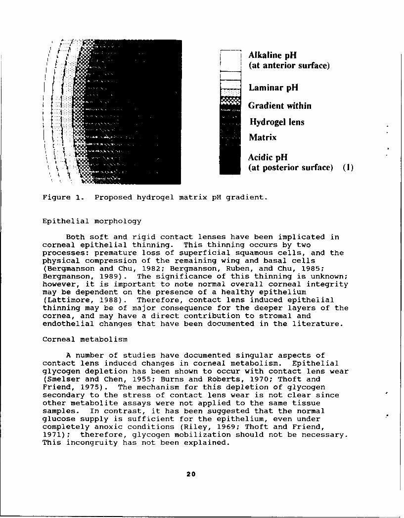
III iAlkaline pH(at anterior surface)
Laminar pH
Gradient within
Hydrogel lens
Matrix
w. Acidic pH(at posterior surface) (1)
Figure 1. Proposed hydrogel matrix pH gradient.
Epithelial morphology
Both soft and rigid contact lenses have been implicated incorneal epithelial thinning. This thinning occurs by twoprocesses: premature loss of superficial squamous cells, and thephysical compression of the remaining wing and basal cells(Bergmanson and Chu, 1982; Bergmanson, Ruben, and Chu, 1985;Bergmanson, 1989). The significance of this thinning is unknown;however, it is important to note normal overall corneal integritymay be dependent on the presence of a healthy epithelium(Lattimore, 1988). Therefore, contact lens induced epithelialthinning may be of major consequence for the deeper layers of thecornea, and may have a direct contribution to stromal andendothelial changes that have been documented in the literature.
Corneal metabolism
A number of studies have documented singular aspects ofcontact lens induced changes in corneal metabolism. Epithelialglycogen depletion has been shown to occur with contact lens wear(Smelser and Chen, 1955; Burns and Roberts, 1970; Thoft andFriend, 1975). The mechanism for this depletion of glycogensecondary to the stress of contact lens wear is not clear sinceother metabolite assays were not applied to the same tissuesamples. In contrast, it has been suggested that the normalglucose supply is sufficient for the epithelium, even undercompletely anoxic conditions (Riley, 1969; Thoft and Friend,1971); therefore, glycogen mobilization should not be necessary.This incongruity has not been explained.
20
Another metabolite study has shown increased lactate in thecorneal stroma accompanying contact lens wear (Klyce, 1981). Itwas theorized this lactate accumulation was responsible forcontact lens induced stromal edema by osmotic pressure. However,if the source of increased lactate is the corneal epithelium,this theory does not explain how the accumulated lactate, andedema, build in a posterior to anterior fashion in the stroma.Again, paired metabolite assays were not performed, so acomprehensive picture of the contact lens-simulated metabolicshifts was not obtained.
Endothelial morphology
The typical corneal endothelial layer mosaic, consisting ofcells of similar shape and equal size, may be altered so themonolayer is transformed into a variety of cell shapes(pleomorphism) and a variety of cell sizes (polymegethism).Although only a relatively recently documented phenomenon,polymegethism has been reported in wearers of nearly 6-l types ofcontact lenses except those wearing the silicon elastoj, •r(Schoessler, 1983; Snyder, 1982; Schoessler, Barr, and Freson,1984; Stocker and Schoessler, 1985). It should be pointed outvariations in cell shape and size can exist without a decrease incell density (Schoessler and Woloschak, 1981). An endothelialassessment is performed using specular microscopy, combined withcomputer analysis of photographs of the central endothelium todetermine mean cell area and density, standard deviation of thecell area, and maximum cell area/minimum cell area ratio.Holden, Sweeney, and Vannas, (1985) monitored the cornealendothelium of subjects fitted with extended wear contact lenses,and found an increase in the cell size variability within 2 weeksof the start of lens wear, with little to no recovery of the cellsize distribution after discontinuance of lens wear.
Endothelial function
Initial studies of the corneal endothelium examined physicalchanges only; subsequent investigations have attempted to linkphysical changes with functional alterations. In Holden'slaboratory (Sweeney, 1985) thick hydrogel lenses, combined witheye closure for 2 hours, were used to induce moderate cornealedema in subjects with polymegethism. The level of inducedcorneal edema correlated with the degree of polymegethism, plusthe rate of deswelling had an inverse correlation with the degreeof polymegethism. Other studies also have suggested thefunctional capacity of the endothelial pump mechanism might becorrelated with morphological appearance; patients who displayedcorneal endothelial polymegethism prior to cataract removal andintraocular lens implantation had significantly greaterpostsurgical corneal edema when compared to their homomegethouscounterparts (Rao et al., 1979; Rao, 1984, and Rao et al., 1984).
21

O'Neal and Polse (1985) found a significant correlation betweenthe degree of polymegethism and the rate of recovery from aninduced corneal edema; along with this they found age-relatedchanges in both endothelial morphology and function.
It has been proposed that corneal hypoxia represents theunderlying cause of contact lens induced polymegethism(Schoessler and Woloschak, 1981; Schoessler, 1983; Hirst et al.,1984; Stocker and Schoessler, 1985). Contact lenses have beenfound to decrease the level of oxygen dissolved in the aqueous inanimals (Barr and Silver, 1973; Stefansson, Wolbarsht, andLanders, 1983; Stefansson, Foulkes, and Hamilton, 1987). Thiscondition presumably places the endothelium in a hypoxicenvironment. The implication of these studies is that a hypoxicendothelium is subject to polymegethous morphological changes, aswell as the functional changes paired with it. The precisephysiological mechanism of how corneal hypoxia inducespolymegethous changes has not been determined. Possiblemechanisms include: an accumulation of carbon dioxide, anaccumulation of 'a-tate, a pH change, and/or a change in ATP andcalcium ion cohucrntrations (Barr and Schoessler, 1980; Schoesslerand Woloschak, !981; Caldwell et al., 1982; Schoessler, 1983;Schoessler, Barr, and Freson, 1984; Zagrod and Connor, 1988).
Generally, it has been assumed that alteration of the cellshape serves to inhibit the endothelial pump function. However,it may be possible that inhibition of the endothelial pumpfunction is what alters cell shape. Indeed, ionic flux changes,combined with the increased presence of water, might reasonablybe expected to alter cell shape. Transient corneal endothelialmosaic changes have been recorded within minutes after placingcontact lenses on the eyes of unadapted patients (Zantos andHolden, 1977; Barr and Schoessler, 1980; Kamiya, 1982). Animmediate response is indicative of a metabolic shift of somesort. As a result, it's certainly possible specific metaboliteassays could be used to determine the mechanism responsible forpolymegethous changes.
Summary statement
Based on the volume and detail of available operationalevidence, contact lenses appear to have a valid place in themilitary aviation environment. However, factors not consideredin this review must be appraised. Not everyone can obtain clearand comfortable vision while wearing contact lenses. Also, aconsistent and reliable bifocal contact lens is not yetavailable, although some promising concepts are under civilianstudy. Since the most accomplished aviators are often timesmatured into presbyopia, a significant portion of the military'smost highly skilled pilot population would not be correctablewith contact lenses. Last, a number of physiological,
22

biochemical, and clinical issues associated with contact lenswear have yet to be resolved. Consequently, contact lenseslikely represent only a partial solution to spectacleincompatibility problems. Only a coordinated, multidisciplinaryapproach to systems development will provide the finalcombination of elements necessary for long-term success indealing with optical compatibility issues.
In conclusion, regardless of the type of lens, a contactlens wearing cornea either exhibits, or has the potential fordeveloping any number of physiological, biochemical, and/orpathological changes. These apparently have been considered tobe of minor significance by the general civilian health carecommunity, as evidenced by the proliferation of the contact lensindustry. However, if the military is to become activelyinvolved in the provision of contact lens care to its aviators,then it is imperative that a comprehensive clinical andscientific database be established. The objectives of futureinvestigations should be to document both the corneal changesthat can be induced by contact lens wear and the operationalaspects of contact lens wear by military aviators. These arerequired in order to permit the military leadership to make afully informed decision based on a complete understanding of thebenefits and deficiencies of this form of visual correction.
Materials and methods
Subjects
Ametropic aircrewmembers were identified by their brigadeflight surgeon for possible inclusion in the study. It wasinitially estimated that a total sample of 400 ametropic aircrewwould be obtained. Potential subjects were provided with aninformed consent briefing by a representative from USAARL; amedical history was obtained immediately following the informedconsent briefing to ensure medical acceptability. Exclusionaryconditions included a history of:
1. chronic/acute inflammations of the anteriorsegment of the eye.
2. disease processes affecting sclera, conjunctiva,or cornea.
3. systemic disease affecting the anterior segmentof the eye.
Those found eligible were further briefed on the study;salient risks and potential benefits were carefully explained.It was stated that lens wear could not be continued by thesubject after the study had been terminated, unless contact lenswear was adopted as a routine clinical program following the
23

conclusion of the study. Each eligible individual was given anopportunity to volunteer for or to reject participation in theresearch project. Those individuals volunteering were requestedto read and sign a Volunteer Agreement Affidavit (Appendix D),and a Volunteer Registry Data Sheet (Appendix E). Detailed plansfor scheduled followups were coordinated with the subject, thesubject's unit, and USAARL/contract investigation teams.
Medical personnel and facilities
Professional requirements
A contact lens fitting team, comprised of two experiencedoptometrists and one technician, under task order contract toUSAARL were responsible for most examinations. A USAARL researchoptometrist and two technical assistants were also involved atthe specific locations of Fort Rucker, Fort Bragg, Fort Riley,and Fort Campbell. The USAARL personnel were additionally usedas a temporary backup, when the contract team suffered atransient personnel shortage. Ten two-man teams of active dutyArmy optometrists and eye technicians were involved in examsassociated with Desert Shield/Storm deployments. Four of thoseteams deployed to Saudi Arabia to provide on-site care,consultation, and evaluation of deployed subjects.
Brigade and other designated flight surgeons responsible forthe routine health care of aircrewmembers were actively involvedin the operational aspect of the study as field medical monitorsin seeking individual assessments of contact lens effectivenesson a daily basis. Selected flight surgeons received 1 month oftraining, at the Army Medical Center level, in order to provide ageneral background necessary for the recognition of commoncontact lens-related complications. Detailed briefings at annualflight surgeon continuing education meetings (USAREUR, OAP, ASMA)were provided by USAARL personnel as an additional means ofkeeping the "field" up to date.
The optometrist(s) located at regional health carefacilities were briefed on the investigation in the event primarymedical support was required. This was coordinated through OTSG,HSC, and 7th Medical Command (MEDCOM) Headquarters. Detailedbriefings at annual optometric continuing education meetings wereprovided by USAARL personnel as an additional means of keepingthe "field" up to date.
The ophthalmologist(s) located at regional health carefacilities were briefed on the investigation in the eventtertiary medical support was required. This was coordinatedthrough OTSG, HSC, and 7th MEDCOM Headquarters.
24

Technical support
As part of the contract team, a qualified technician wasutilized in the initial fitting of the lenses, as well as duringall other examinations. Two USAARL technicians also wereavailable for additional assistance in certain instances.
Facilities
Regional eye care facilities were used for all examinations,scheduled followups and unscheduled follow-ups. Regional eyecarepersonnel were not involved in scheduled fittings and followupexaminations. However, they were available for individualsubject consultation between scheduled visits, as desired.
Medical equipment
The following equipment was used in scientific control ofdata collection:
Phoropter with stand, examination chair, andprojector.
Automated keratometer with automated refractorBiomicroscopeObjective automated refractorAesthesiometerUltrasound PachometerEndothelial cameraNanoliter osmometerpH meterMicropolarographic electrode
Lens materials
Extended wear contact lenses were utilized in this study.Ir general, soft lenses can be of either low (25-35 percent),medium (35-60 percent), or high (60-85 percent) water content,and can be made of either an ionic copolymer or a nonionicpolymer. Two types of soft lenses were used in this study:a medium/high water content lens and a low water content lens.Rigid gas permeable (RGP) lenses were used [as backups] forhyperopic and highly astigmatic subjects. All soft lenses weredisposed of on a weekly basis. RGP lenses were worn on a weeklybasis. However, these lenses were cleaned and disinfectedweekly, and reinserted after at least one night without lenswear. RGP lenses were replaced at the quarterly followups. Anew soft lens supply was furnished at each quarterly followup, aswell. All materials (i.e., lenses and solutions) werecommercially available and approved by the U.S. Food and DrugAdministration (FDA). The protocol was determined not to be an
25

original use of contact lenses, since the Federal AviationAdministration (FAA) does not restrict the use of contact lensesby civilian pilots.
Since the soft lens system was disposable, soft lensdisinfection and cleaning solutions were not needed. Comfortsolutions, available through an independent manufacturer, wereprovided to the subjects, as were weekly replacement lenses. Thecomfort solution of choice was "Lens Plus Rewetting Drops" byAllergan Pharmaceuticals*. The advantages of this lens lubricantwere its being preservative-free and packaged in boxes of 30single-use 0.01 ounce vials; the container was discarded aftereach use. Employment of this product bypassed potential problemsrelated to reusable bottle contamination issues and preservative-induced toxic and hypersensitivity reactions. RGP lensesrequired the use of cleaning and conditioning solutions that werespecifically formulated for the RGP lenses being used. Allsolutions were provided to each subject on a quarterly basis.
Procedures
Initial examination and fitting
Ametropic aircrewmembers who volunteered and were selectedfor the study were seen individually on an appointment basis.Once an acceptable initial fit was achieved, subjects were giveninstructions by a technician concerning lens insertion, removal,and care. At that time, the subject also was instructed onsymptoms which might necessitate lens removal and/or unscheduledprofessional care. A written summary of essential informationwas provided for each participant to keep. The aviation brigadeflight surgeon served as a point of contact for followupappointments and additional consultation, if needed. Eachsubject was given a wallet-sized medical identification cardspecially designed for contact lens wearers. The subjects wereinstructed that this card (Appendix F) should be prominentlydisplayed in their wallet and would serve to alert medicalpersonnel that the individual was wearing contact lenses shouldhe be incapable of relating such information. Additional medicalalert methods (dog-tag identifiers, bracelets, etc.) could beapplied at the individual discretion of each subject and/or localflight surgeon.
Followup examinations
Followup examinations were performed at 24 hours, 7 days,and 3 months postfitting. Thereafter, all followup examinationswere on a quarterly basis. Additional eyecare visits and/or
* See list of manufacturers.
26

consultations could be initiated by the subjects through theirflight surgeon to the regional eyecare facility shouldunscheduled followup be required.
Wearing schedule
Upon completion of the initial fitting session participantswere instructed to begin continuous wear immediately. Thesubject were required to return for a 24-hour evaluation; duringthe initial 24-hour period the subject was restricted to duty notto include flying (DNIF). After the 24-hour exam, the newlydispensed lenses were to be worn without removal for 6 more days.The flight surgeon issued an "up-slip" once the 24-hour exam wascompleted and after confirming that visual acuity met AR 40-501standards (i.e., 20/20 or better Snellen visual acuity in eacheye with contact lenses in place). On the seventh day, thesubject returned to the exam facility for additional evaluation.At the successful conclusion of this evaluation, a 3-month supplyof lenses was issued. Appropriate amounts of comfort dropsolutions were dispensed at each session, as well. The lenseswere worn continuously for a maximum of 7 days. On the eveningof the last day of lens wear, the soft lenses were removed anddiscarded. The RGP lens wearers removed their lenses andperformed the required lens cleaning and disinfection procedures;the lenses then were stored overnight in a provided storage case.The subject then would sleep that night without any lenses; onrising the next day, the subject applied either a new pair ofsoft lenses or the cleaned RGP lenses. The subjects wereinstructed that if they must remove a soft lens for any reason(i.e.: foreign body sensation, mild irritation), the lens shouldnot be reapplied. Instead, a new lens should be applied, and theold one disposed of. It was permissible for RGP lenses to beremoved, cleaned, and reinserted. Subjects were advised thatshould the irritation/discomfort continue, the local flightsurgeon should immediately be consulted. It was stressed that inno case should a subject sleep with a contact lens on anirritated eye; when in doubt of ocular health, the subjects wereadvised to immediately seek medical assistance.
Data collection
A complete data record was chronicled during each exam(initial, followups) for all subjects. Standardized datacollection forms were used throughout the course of the study.The contractor developed a database, and provided all data toUSAARL investigators in both hard copy and organized floppy diskformat. Ultimately, three separate databases were maintained:local Fort Rucker subjects, worldwide protocol subjects, andDesert Storm subjects. There was some overlap in the databases
27
since a number of people fell in all three groups over the courseof the overall investigation. Quanlitative data can be found inAppendix G and quantitative data can be found in Appendixes A andB.
Eye-related svmptoms and medical emergencies
When a subject developed an adverse ocular sign or symptom,or sustained an eye injury, he was instructed to contact the unitflight surgeon immediately. Special instructions were providedto these flight surgeons regarding identification, treatment, anddisposition of these subjects. This consisted of a supervised4-week clinical experience in a MEDCEN Ophthalmology Clinic,coordinated through the Ophthalmology Consultant to The SurgeonGeneral of the Army.
Subjective assessment procedures
A self-administered questionnaire was used to obtainsubjective information from the participants at the quarterlyfollowups; some questions were asked only at the first quarterlyfollowup, others were repeated at each followup. Thequestionnaire addressed user acceptability, job performanceimpact, problems encountered, medical services, and training. Inaddition, there was some objective observation time in AH-64simulators for performance assessment under contact lens/spectacle-wear conditions by unit flight surgeons and USAARLinvestigators. These data, in addition to the flight surgeonobtained interview/debriefing data, were used in assessing theoperational impact of contact lens wear.
Lens physical assessment
Sample used lenses, obtained at scheduled followup exams,were returned to USAARL for examination for any physical defects,transmittance/absorbance analysis, and for protein depositdetermination.
Medical support data
Information related to medical resources and logisticalrequirements to support contact lenses in the field weredocumented throughout the course of the study. Records of allmaterials consumed were maintained as an embedded part of thedata collection form, and were therefore extractable from thecomputerized database for interim and final report purposes.Incidence of health facility visits, diseases, and injuries werelogged as well.
28
Experimental design/data analysis
This study was designed to assess the feasibility of contactlens wear as an option to spectacle wear for ametropic Armyaircrewmembers. The primary objectives were to characterize thephysiological/biochemical response of the cornea to contact lenswear, and to identify the strengths and weaknesses in contactlens wear associated with the occupational tasks andenvironmental conditions unique to the Army aviation environment.Experimental data were obtained through the use of objectiveclinical scientific evaluation techniques, subjectivequestionnaires, and informal interviews concerning operationalflight performance. Data analysis consisted of simpledescriptive statistics and analysis of variance procedures.Specific factors of interest were type of contact lenses worn,geographical location of unit, and type of aircraft flown. Thedata sheets provided in Appendixes A and B indicate the dependentvariables of interest. Civilian studies served to providecontrol data for interpreting clinical findings. It wasanticipated that with the acquisition and analysis of thisinformation, the military community would be able to make aninformed decision regarding the long-term feasibility ofutilizing ametropic aviators in advanced attack helicopter
systems.
Human use justification
The experimental question related to the practicalapplication of contact lenses to a specific military environment.As such, an animal model simulation was not able to providepractical information regarding the subjective strengths andweaknesses of contact lens wear.
Regulation compliance
This protocol was in compliance with:- AR 70-25, Use of volunteers as subjects of research.- USAMRDC Regulation 70-25, Use of human subjects in
research, development, testing and evaluation.- USAARL Policy 70-3.
Health and safety of volunteers
Risks
The salient risks of wearing extended wear contact lenses(both soft and rigid) were presented in the volunteer briefing,and are detailed in the next paragraph. The ophthalmicexamination procedures were standard, accepted techniquescommonly utilized in the field of contact lens practice andresearch. Performance evaluation was not intended to interfere
29

with the individual's or unit's normal training activities.However, it was necessary for each unit to give followup examappointments top priority for this program to succeed.
The chief source of risk to participants in this study wasthe possibility of an accident resulting from the actual wearingof contact lenses in the performance of military flight and otherduties. The wearing of extended wear soft contact lenses has inthe past been associated with the following effects:
a. Minor, temporary risks that are usually notserious and do not last very long.
a. Mild watering of the eyes.b. Mild sensitivity to light.c. Temporarily blurred vision.d. Slight redness of the eyes.e. Faint sensation of dryness of the eyes.f. Mild feeling of irritation to the eyes.g. Mild eye pain.h. Slight swelling of the cornea or eyelids.i. Discomfort/reduced vision due to a
foreign body, displaced lens, or lensdrying.
b. Serious and possibly permanent risks.
a. Abnormal growth of blood vessels intothe cornea.
b. Scarring of the cornea.c. Subtle changes in the cornea which
reduce vision.d. Eye infections, possibly leading to
surgical replacement of the cornea,or loss of an eye.
e. Decreased corneal capacity to cope withfluid buildup, which can lead tosurgical replacement of the cornea,or loss of the eye.
c. Based on preliminary studies, in-flight risksfor a standard flight profile were judged tobe minimal. However, as a safeguard, primaryaircraft operators were instructed that theywere not to fly with another contact lens wearingsubject or copilot; this requirement wasalso documented on the formal waiver, whichrequired both commander and individualsignature. Backup spectacles were carriedat all times, in case lenses had to beremoved while performing actual flight duties.
30
Medical safeguards
This project was been planned for maximum safety and thevolunteer subjects were closely monitored by eyecareprofessionals. Many procedures were built in to ensure thesafety of study participants. These safeguards included:
1. Thorough eye examinations on a regular schedule.2. Contact lenses were not prescribed if deemed medically
unsuitable.3. Training was provided in the safe use and care of the
lenses.4. Lenses were replaced on a periodic basis.5. Contact lens wear was temporarily suspended if
medically indicated.6. Participation was discontinued if medically indicated.7. The local flight surgeon was an integral part of the
investigational team, and was appropriately trained inthe recognition of common contact lens relatedcomplications, and was the initial point of contact inthe event of an adverse incident.
8. Regional medical personnel (optometrists,ophthalmologists, and emergency room personnel werebriefed on the project in case of a medical emergency.
Medical monitor
The protocol medical monitor was board certified inophthalmology. The field medical monitors for this study werethe unit flight surgeons normally responsible for the health careof tha subjects at their regular aviation medicine clinic or TMC.
Individual privacy and handling of data
Other than the flight waiver, only information arising fromserious medical incidents was placed in the individual's medicalrecord. All research data files were kept in the strictestconfidence in accordance with regulations. Raw data forms andcomputer files have had limited access and are used for researchpurposes only. No individual information was released withoutexpressed written consent from the subject or other recognizedauthority.
Results and discussion
Introductory description
General technical report
A total of 582 aircrew members participated as contact lens-wearing subjects. The Armywide AH-64 portion had 238 subjects,
31

while the Desert Shield/Storm portion had 344 subjects. Overall,there were 868 volunteers for both portions of the study.Therefore, the comprehensive fitting success rate was 72 percent(582/868). Forty-two subjects withdrew or were discontinuedbecause of poor physical fit and/or unsatisfactory visual acuity.All unsuccessful fits and withdrawals were essentially a functionof presbyopia, moderate to high astigmatism, and extreme cornealcurvatures. This left a 67 percent wearing success rate of theoriginal 868 volunteers; and of those successfully fitted withlenses this represents a 93 percent wearing success rate.
Since Army aircrew are subject to a form of screening viathe initial entry flight physical standards, many of theametropes exhibited low visual corrections compared to thegeneral spectacle-wearing population. A display of sphericalrefractive errors for all 582 subjects can be seen in Figure 2.The refractive error distribution of these subjects peaked at -0.75 diopters (D), which is considerably less than the civilianspectacle-wearing population distribution peak near -3.50 D. Acomparison histogram between the Desert Storm and the AH-64subjects also was established (Figure 3). Using a nonparametric,
Contact lens studyrefractive error distribution
200- 200N =1161
180 180
160 -160
S140 140
S 120 120S100 100
S80 8
S 60 60
40 40
20 20
0 0-6 -5 -4 -3 -2 -1 0 +1 + 2 + 3
Contact lens power (diopters)
Figure 2. Contact lens study refractive error distribution.
32

discrete variable analysis for subject ages, the two samples werefound to not be statistically different (p=0.54). Therefore,this refractive error distribution pattern likely describes thatof the overall ametropic Army aviation population.
A comparative distributionof subject spherical refractive error
240 .2402 4 Desert Storm subjects On AH-64 subjects
220 N = 685eyes N =467 eyes 220
200 200
180 180
S160 160
W 140 140
O120 120
100 100
80 80
80. 6040
40
20 20
0 - 0-5 -4 -3 -2 -1 0 +1 +2 +3
Spherical refractive error (diopters)
Figure 3. A comparative distribution of subject sphericalrefractive error.
Slit lamp examination
Clinical evaluations were done on a quarterly basis.Specific physical and physiological issues are addressedindependently in other sections of this report. However,selected slit lamp exam results are presented here as a part ofdiscussions on general clinical issues. There were nosignificant changes over time in terms of gross physical
33

appearance of the eyes. Qualitatively, the bulbar and palpebralconjunctiva could be described as slightly chemotic and hyperemicin the typical subject (more so in the soft lens wearers than theRGP wearers). However, this was not statistically demonstrable.
The relative distribution of lens types used reflected thefitting methodology. Since the 58 percent water content lens wasthe lens of initial choice, that lens was worn by the majority ofsubjects. The 38 percent lens and RGP lens were backups in theevent the initial lens did not provide an adequate fit orsatisfactory vision. Consequently, the 38 percent and RGP lenseswere used less frequently (Figure 4). The ages of the subjectsranged from 18 to 46 (Figure 5). The bimodal distributionpattern was of some concern, so the Aviation Epidemiology DataRegister (AEDR) was probed for the age of all Army dviators. A
Contact lens studydistribution by power and lens type
160- 160N = 1161
M 58% water content lens140 2M 38% water content lens 140
CM Rigid gas permeable lens
120 120
W ~100- -100
"s80 80
~6076
40 40
20 20O- L 0
-6 -5 -4 -3 -2 -1 0 +1 + 2 + 3
Contact lens power(diopters)
Figure 4. Contact lens study: distribution by power and lenstype.
34

similar bimodal pattern was evident (Figure 6); therefore, oursample is shown to be representative of not just the ametropicpopulation, but also of the aviation population in general.
Lens costs were a reflection of manufacturer materials andtechniques. The 58 percent lenses cost $320/person/year; the 38percent lenses cost $740/person/year; the RGP lenses cost$215/person/year. RGP care kits were $200/person/year andwetting solution for soft lenses cost $95/person/year. Meanwearing time was 4.4 days for soft lens wearers by questionnaire.Verbal discussion confirmed that, prior to Operations DesertShield and Storm, subjects typically wore their soft lensesduring the 5-day garrison work week, taking weekends off. TheRGP lens wearers never got beyond a 2-day/l-night scheduleprimarily because of difficulties adapting to lens edges andthickness. During the Southwest Asia deployment, subjects wereprovided with enough lenses to adopt a 3-day wearing schedule.
Contact lens studyage distribution
40 40N N=582
36 36
32 32
S28 28
S24 24
20 20
S16 16
= 12 12
8 8
4 4
0 02025 3035 40 45
Age (years)
Figure 5. Contact lens study: age distribution.
35

Most reported adhering to that regimen. However, during thecombat phase wearing time increased as a function of fieldconditions and operational intensity.
Limbal evaluations were negative in terms of inflammationand associated clinical significance, but assessments of limbalvascular development over the course of the study werenevertheless statistically significant (p<0.001). Limbalvascularization scores, as a function of quarterly examination,increased throughout the time course of the study (Figure 7).Regional comparisons of limbal vascularization revealed no changein the nasal and temporal quadrants (p=0.17 and 0.31,respectively); vascular progress in the superior and inferiorquadrants was statistically significant (p<0.0001 for both).Since those limbal areas already are impinged upon by theeyelids, hydrogel effects would compound those already hypoxic
Aviation populationage distribution
1000 1000N 1.3973 10
900 900
800 1S0
S700• 700
8 00 600
500. 500
400 400
SE 300 300
200 200
100 100
20 25 so 35 40 45
Age (years)
Figure 6. Aviation population age distribution.
36

areas. These areas of the limbus should therefore serve as themost sensitive indicators of hydrogel lens-induced cornealstress. A breakout of scores by lens type revealed the RGPlenses to have been stable over time, while the soft lenses werenot. Therefore, the overall effect was a reflection of soft lenswear only.
Evaluations of conjunctival and corneal epithelial surfaceintegrity were done with both rose bengal and fluorescein stains(Figure 8). The rose bengal failed to detect significantdevitalization of corneal tissue, but a significant amount ofbulbar conjunctival devitalization was manifested. This wasparticularly so in the inferior region, but could be seen in allquadrants. The subjective impression was that the soft lensacted somewhat as a sponge, drawing moisture from adjacentconjunctival tissue. Over the course of the study, mean rose
Corneal limbal vascular development2.00
R=0.98 * Acuvue grouped data1N=2578 0 B&L grouped datao 1.75 Mn scor E Equalens grouped data
1.50
1.25
S1.00
e 0.75 T
S' 0.50
S0.25
U 0.00Initial 2nd qtr 4th qtr 6th qtr 8th qtr
1st qtr 3rd qt 5th qtr 7th qtr
ExaminationFigure 7. Corneal limbal vascular development.
37

bengal stain severity score did not change. Howeve7, it didinitially change as a function of duration of extended wear ofthe pair of lenses currently being worn (Figure 9).
While fluorescein uptake was seen conjunctivally, most oftenit was associated with a break in the corneal epithelial barrierseen as punctate staining (Figure 8). The vast majority of theseobservations were associated with fine, scattered punctatestaining that was graded at level 1 or 2 on a 0-4 scale. Again,the subjective impression was of a contact lens-induced tissuewater or moisture loss. As with rose bengal, mean fluoresceinseverity score did not change over the course of the study.However, it did change as a function of duration of extended wearof the lenses currently being worn (Figure 10).
Figure 11 emphasizes the differences between the two stainmean score curves. While rose bengal-documented cell
Rose bengal and fluorescein assessments700 700
Rose bengal N=397 Fluorescein N=685
1600 600
500 500
S400 400
0300 300
S200 200
100 100
0 0Bulbar Both Abrasion 3/9 o'clock
Corneal Foreign body Punctate
Rose bengal Fluorescein
Type of stain assessment and location
Figure 8. Rose bengal and fluorescein assessments.
38
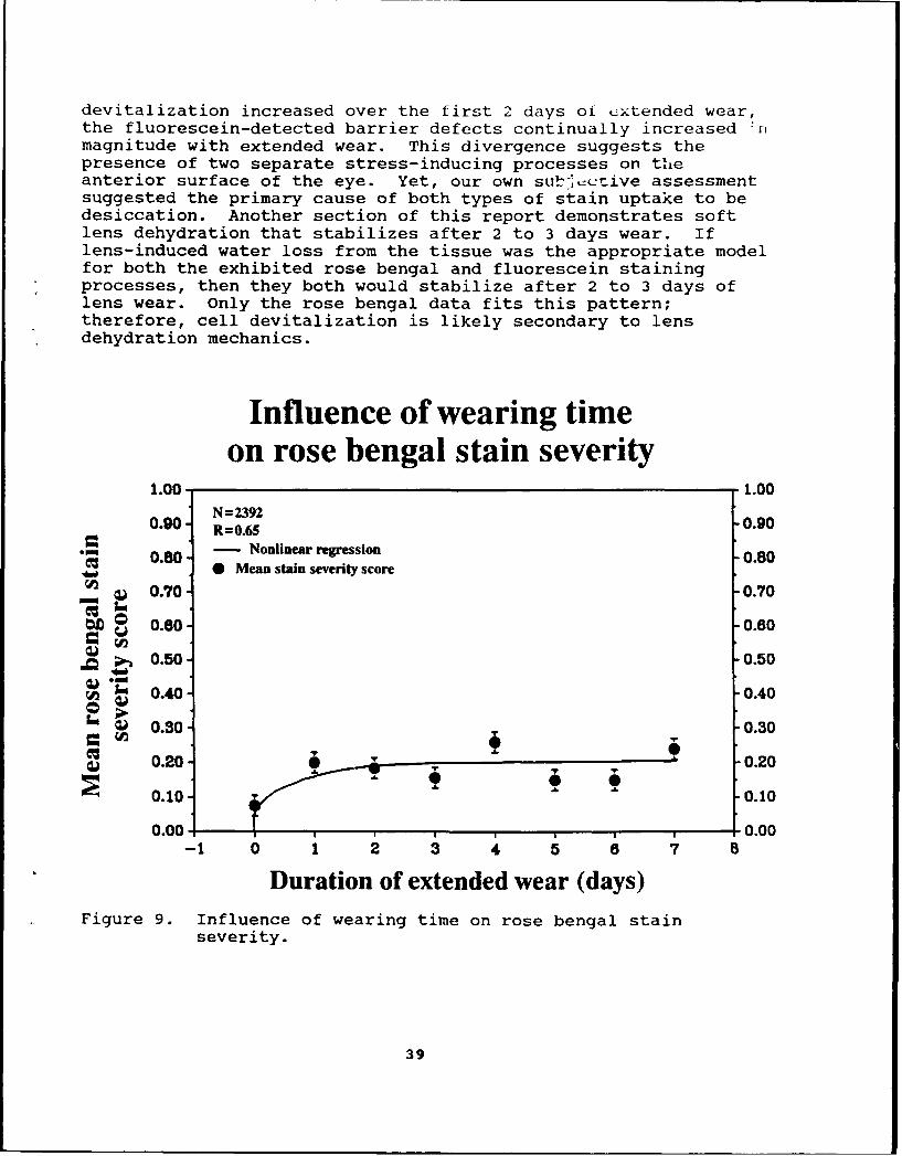
devitalization increased over the first 2 days of cxtended wear,the fluorescein-detected barrier defects continually increased 'r1magnitude with extended wear. This divergence suggests thepresence of two separate stress-inducing processes on theanterior surface of the eye. Yet, our own subjective assessmentsuggested the primary cause of both types of stain uptake to bedesiccation. Another section of this report demonstrates softlens dehydration that stabilizes after 2 to 3 days wear. Iflens-induced water loss from the tissue was the appropriate modelfor both the exhibited rose bengal and fluorescein stainingprocesses, then they both would stabilize after 2 to 3 days oflens wear. Only the rose bengal data fits this pattern;therefore, cell devitalization is likely secondary to lensdehydration mechanics.
Influence of wearing timeon rose bengal stain severity
1.00 1.00N = 2392
0.90 R=0.65 0.900.80 - Nonlinear regression 0.80
S 0.04 Mean stain severity score
__ 0.70 -0.70
tm 0.80- 0.80
S 0.50- 0.50
"0.40 0.40
0.30 0.30
0.20- 0.20
0.10 T " 0.10
0.00 , , ,0.00-1 0 1 2 3 4 5 S 7 8
Duration of extended wear (days)Figure 9. Influence of wearing time on rose bengal stain
severity.
39

Clearly then, the extended soft wear-stimulated deficit inthe corneal epithelium's barrier function is not secondary to adesiccation process. An alternative hypothesis involves cornealmetabolic waste accumulation under the soft lens that eventuallyproves toxic to the various layers of the cornea. Epithelialinfluence is seen early via punctate fluorescein staining;endothelial influence is seen later via morphological changes.It is likely the epithelial barrier function loss is linked tobacterial invasion of the cornea in ulcerative keratitis.
Tear break up time (BUT) was assessed using, as the measure,the duration or maintenance of a smooth tear film after adeliberate blink. Timed measurements were documented with highmolecular weight fluorescein while lenses were worn, and withstandard fluorescein strips when lenses were removed. Acorrelation of BUT methods was not significant (p=0.29; Figure12). While the two methods have been shown to clinicallycorrelate (Patel, Farrell, and Bevan, 1989), it is obvious from
Influence of wearing timeon fluorescein stain severity assessments1.00 1.00
0.90- N = 2524 ,0.90R=0.95
0.80 - Nonlinear regression -0.80__j 0 Mean stain severity score
S0.70 •0.70
0.80- 0.80
• 0.50 -0.50
0.40 -0.40
= 0.30 0.30
0.20 T 0.20002
0.10/ 0.100.00-- ,0.00
-1 0 1 2 3 4 5 8 7 8
Duration of extended wear (days)
Figure 10. Influence of wearing time on fluorescein stainseverity.
40

these data that the presence of an unstable precorneal tearfilmdoes not mean that a contact lens will not be covered by a stabletear film. The obverse also is true, in that a stable precornealtearfilm does not guarantee maintenance of an optically smoothfront contact lens surface. Because of this lack of mutualexclusion, both methods of tear film assessment are recommendedin routine patient evaluation.
Precorneal tearfilm stability in normal individuals has beenpositively related to tear production as measured by the Schirmertear test (Patel, Farrell, and Bevan, 1989). Yet, dry eyepatients (keratoconjunctivitis sicca) have been shown to exhibitno relationship between tearfilm stability and tear production(Rolando, Refojo, and Kenyon, 1983). These findings conflictwith a basic understanding of tearfilm behavior. Logic dictates
Influence of wearing timeon stain severity assessments
1.00 1.000 Fluorescein, N=2524, R=0.95O Rose bengal, N=2392, R=0.65
0.80 -0.80
0.70 0.70
S U 0.60 -0.60
0.50 T 0.50
0.40 T 0.40
S0.30 -0.30
0.20 T 0 0.20T 0 -0 9 6 .20
0.10 0.10
0.00 L , ,0.00
-1 0 1 2 3 4 5 e 7 8
Duration of extended wear (days)
Figure 11. Influence of wearing time on stain severityassessments.
41

that a stable precorneal tearfilm will have little need forplentiful tear production, and an unstable tearfilm willsecondarily lead to reflex tearing. Therefore, the referencedfindings don't make clinical sense.
In an effort to test at least a part of these disparatefindings, tear-film stability assessments and tear productionmeasurements were correlated in the study subjects. Thecorrelation was very poor (R=0.07) and the ANOVA was notstatistically significant (p=0.12). Therefore, contrary toinferences drawn by Patel et al. (1989), the present dataindicate that there is no relationship between tear BUT and tearproduction in clinically normal contact lens wearers. Additionalassessments of BUT (both high molecular weight fluorescein andstandard fluorescein strips) did not correlate well with numberof days lenses had been worn prior to the followup exam.Therefore, even though protein buildup is acknowledged to occurover wearing time, it apparently does not influence this type of
Tear BUT assessment comparison30 0 0 0 0 - 30
28- . .o • .- *.o o 0 0 * • 28- 264 .* * so . •.*• 26
0 0 0 0 2-244 . . . oooo, ./ .ooo .2424. 0 0 0 0 0 0 0 0 2
oo22o • ooooo: oo :• 22S20 *• o• * ••••% @ 0o0 0o0 20
@0000 S 000 0o 18 0 0000000 0 0 18
0 000 0000 0 0 0 00.1S16 ,@ .oooo oooo:o:ooo 16
14 oo o o0 * oooo ooo • 1412 o oo •oo 00o 0 0 1210 000 0 00000 000 0o 00 10-• _- o oo00 0 00000 00 0o
8 00000 0 0 001 60 0 0 0 00 0 0 0 0 0
410 0 0 60 R=0.29 4~2- N N=2477 2
is 000 2 4 6 8 10 12 14 16 18 20 22 24 26 28 30
Tear break up time (seconds)(fluorescein strips without contact lenses)
Figure 12. Tear BUT assessment comparison.
42

tear BUT assessment. As a result, both tear stability andproduction assessments should routinely be performed on theinitial contact lens evaluation, but are unlikely to offer anynew information on followup examination.
Practically speaking, none of the subjects had insur-mountable objective or subjective problems wearing the lensesused in this study, even those with less than 5 mm of tear stripwetting. Subjects had been given an ample supply of unit-dose,sterile, unpreserved wetting solution, and were encouraged to useas much as they needed. Extra supplies of wetting solution werereadily available at the subjects request, if required.
Safety concerns
Safety issues were of the primary concern at the start ofthe study. The main worry centered on the possibility of acontact lens-induced sudden incapacitation causing loss ofaircraft control. As the initial local phase of the studyunfolded, it became obvious that sudden incapacitation would notbe an issue, prompting expansion of the protocol to the Armywideeffort, and later the Desert Shield/Storm inclusion. Again, theinitial worry proved to be unsubstantiated. Aircrew wore theirsoft lenses in a doors-open or -off scenario with no adverseeffects on lens comfort. RGP lens wearers had a lot of trouble,however, with foreign bodies in the same flight profiles.
There were three occurrences involving contact lens-wearingsubjects that prompt mention in the safety arena. 1.) There wasone equipment failure in flight that resulted in a "crash"landing that was controlled with minimal damage to the aircraftand no damage to the subject. 2.) There was a mishap involvingtwo AH-64s hovering in an LZ. The main rotor blades impacted,resulting in a crash, with moderate to severe damage to bothaircraft and one case of severe burns secondary to a postcrashfire. Both frontseat aviators were contact lens subjects, butneither was at the control of their aircraft at the time of theincident. In both these episodes, Army Safety Centerinvestigations ruled out contact lens wear as a contributingfactor. 3.) The last safety-related episode involved anoperationally-stimulated ejection (bad weather and low on fuel)from an intelligence fixed-wing aircraft (RV-l Mohawk). Thepilot stated that his contact lenses stayed in place during theejection, whereas glasses would have been lost as a result of thewindblast on ejection. At the start of this program, the majorsafety fear was that of sudden pilot incapacitation due to lensloss or foreign body sensation. These concerns were needless.Based on questionnaire responses recorded later in this report,it's entirely possible contact lens wear may be safer than flyingwith glasses.
43

Subject sentiment
Specific subjective assessments by formal questionnaire areaddressed elsewhere in this document. Verbal feedback during thecourse of the study was always very positive. Subjects wereespecially appreciative of the increased apparent field-of-viewwhile on NVG operations. They also commented on the lack otdistracting reflections that can be a problem with glasses.Apache student pilots were particularly full of praise forcontact lenses after finishing their 2 weeks of training "in thebag" when their only view of the outside world is via the HDUbecause the windscreens are completely obscured. The overallconsensus supported the idea of routine contact lens use whileperforming flight duties.
Ocular health
General complications of contact lens wear also were trackedin order to determine possible impact on medical resources andunit readiness. The major complication associated with extendedsoft lens wear, ulcerative keratitis, will be discussed fully ina separate section. Overall, few ocular complications wereencountered. This despite that a detailed monitoring system hadbeen established through flight surgeon, optometry, andophthalmology channels at the Army installations involved in thestudy. There were six cases of acute, ulcerative keratitis; sixcases of peripheral corneal infiltrates noted on followup exams;three cases of bacterial conjunctivitis; two cases of viralconjunctivitis; two cases of anterior uveitis/iritis; and onecase of a severe allergic reaction. Many of the above conditionswere probably not contact lens related, although the anterioruveitis cases may have been related to a temporary tight lenscondition. In order to rule out a tight lens fit, both subjectswere put back in lenses of the same parameters as before; therewere no recurrences of the uveitis. In the case of the severeallergic reaction, the contact lenses may have been protective innature. After flying through a localized, dense "fog" in adoors-off condition while wearing his soft lenses, a specialoperations pilot experienced severe conjunctival redness anditching accompanied by external eyelid erythema. On slit lampexamination, his corneas were found to be uninvolved in theapparent allergic or hypersensitivity process.
Synopsis
In summary, results were very positive compared to initialexpectations. Success rates were acceptable in light of thelimited lens types and parameters used. Clinical evaluationswere essentially normal with no remarkable contraindications tolens wear noted. Subjective evaluations by the contact lenswearers were highly positive. Safety of flight concerns were
44

essentially unfounded. Finally, the ocular health impact wasminimal compared to the number of subjects, the duration of thestudy, and the varied and stressful environments encountered.Therefore, the basic general conclusion is that contact lens wearshould be adopted as a routine means as correcting the refractiveerrors of Army aircrew. Specific conclusions and recommendationsare outlined in another section after the detailed clinical,physical, and physiological analyses.
Detailed analyses
Corneal curvature assessment
Introduction
The purpose of this portion of the investigation was toassess possible water content-related differences in how softcontact lenses drape across the corneal surface. Related to thisinitial assessment was an interest in documenting possiblecorneal cylinder masking differences, as well. Anecdotal reportshave indicated water content differences can affect physical lensbehavior in situ. However, a literature review failed to provideany data on this topic.
Methods
Over a 3-year study, the 223 subjects assigned to AH-64 andspecial operations units were fitted with either a 38 percent or58 percent water content soft lens of analogous base curve (8.8mm) and diameter (14.0 mm) under disposable wearing conditions.Subjects received followup exams at 24 hours and 7 dayspostfitting. Quarterly followup exams were conducted thereafte-.An autokeratometer was used to measure both corneal and anteriorlens surface curvatures on all exams; similar corneal and contactlens surface cylinder assessments also were recorded.
Results
Grouped data plots of anterior soft lens curvature (flattestK-reading) as a function of underlying corneal curvature for eachlens type indicate a collective linear relationship that does notstatistically vary by water content in the two types of lensesstudied (F ratio approaches 0.000 and p approaches 1.000; Figures13 and 14). Paired corneal cylinder plots reinforce the aboveresult by revealing no significant masking differences betweenthe two lens types (p = 0.201; Figures 15 and 16).
4S

Discussion
Based on these data, it is concluded that physical lens fitis independent of water content when the base curve and diameterare kept constant. This contradicts the notion of greatercorneal cylinder masking ability of low water content softlenses. However, it is recognized that the measurement systemsused to gather these data record central corneal parameters only.Therefore, this investigation is conclusive only for lensbehavior over a restricted area of the central cornea.
Using a corneal topographical modeling system and adigitizing board, a preliminary analysis of overall anterior lenssurface topographic areas reveals a marginal statisticaldifference between the two types of soft lenses on one testsubject (Figure 17; p = 0.048). However, the isolated central
Anterior optical surface curvatureduring spherical soft lens wear
47 4738% water content lens
46 R=0.87 4645 N=524 45
• 44 44
43 43
S.S 42 42
&W 41 - 41L 40 40
W 39
3M 0 38V 38 3
37 37
36 . - 3639 40 41 42 43 44 45 46 47 48
Corneal curvature(diopters)
Figure 13. Anterior optical surface curvature during sphericalsoft lens wear (38 percent water content lenses).
46
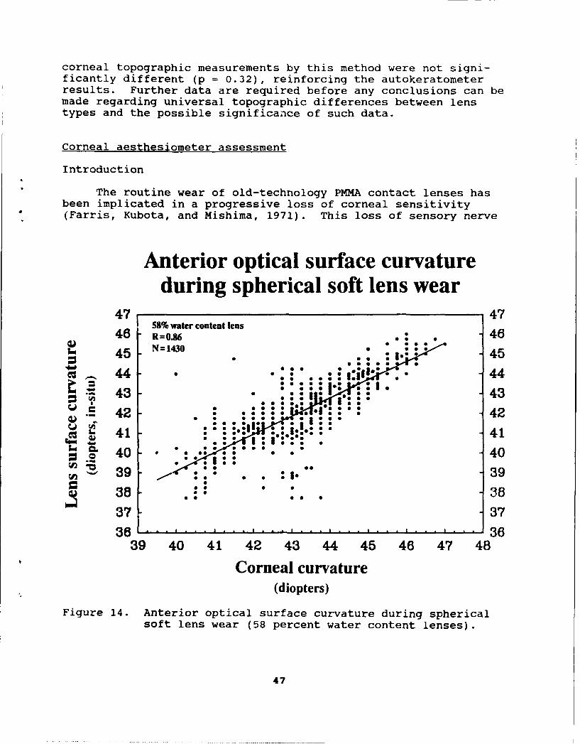
corneal topographic measurements by this method were not signi-ficantly different (p = 0.32), reinforcing the autokeratometerresults. Further data are required before any conclusions can bemade regarding universal topographic differences between lenstypes and the possible significance of such data.
Corneal aesthesiometer assessment
Introduction
The routine wear of old-technology PMMA contact lenses hasbeen implicated in a progressive loss of corneal sensitivity(Farris, Kubota, and Mishima, 1971). This loss of sensory nerve
Anterior optical surface curvatureduring spherical soft lens wear
47 4758% water content lens
46 R=OM8• 4645 Nffi1430
44 - 44
"43 - 43
42 - 42S41 - 41
=C 40 -. 403O9 3938 * 38
37 3736 36
39 40 41 42 43 44 45 46 47 48
Corneal curvature(diopters)
Figure 14. Anterior optical surface curvature during sphericalsoft lens wear (58 percent water content lenses).
47
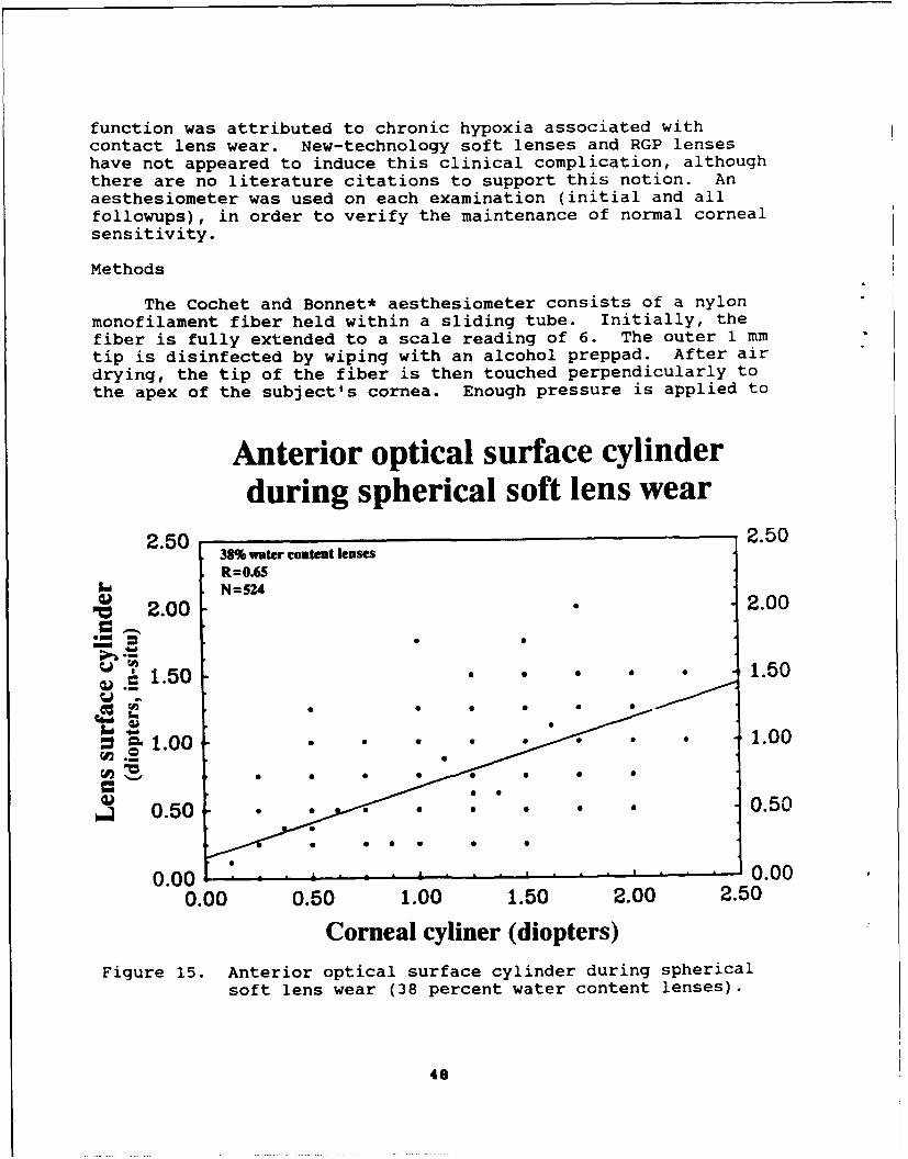
function was attributed to chronic hypoxia associated withcontact lens wear. New-technology soft lenses and RGP lenseshave not appeared to induce this clinical complication, althoughthere are no literature citations to support this notion. Anaesthesiometer was used on each examination (initial and allfollowups), in order to verify the maintenance of normal cornealsensitivity.
Methods
The Cochet and Bonnet* aesthesiometer consists of a nylonmonofilament fiber held within a sliding tube. Initially, thefiber is fully extended to a scale reading of 6. The outer 1 mmtip is disinfected by wiping with an alcohol preppad. After airdrying, the tip of the fiber is then touched perpendicularly tothe apex of the subject's cornea. Enough pressure is applied to
Anterior optical surface cylinderduring spherical soft lens wear
2.50 ,2.5038% water content lenses
R=OASN=524
"PC 2.00 2.00a I
. 1.50 . 1.50
1.0 1.00
0.50 .0.50
0.00 0.000.00 0.50 1.00 1.50 2.00 2.50
Corneal cyliner (diopters)
Figure 15. Anterior optical surface cylinder during sphericalsoft lens wear (38 percent water content lenses).
48

obtain a 4 percent deflection of the fiber (first visiblebending). If the subject does not feel or react to the fiber'spresence, the fiber is withdrawn, shortened to the next settingdemarcation (5.5), and reapplied. This series is repeated untilthe subject senses or reacts to the fiber. The fiber settingscan be translated into pressure values in grams per squaremillimeter, by way of a calibration plot provided by themanufacturer (Figure 18).
Results and discussion
Corneal sensitivity measurements were not statisticallysignificant as a function of time in the protocol (p=0.37), as afunction of number of days of lens wear (p=0.42), or as afunction of type of contact lens (p=O.18). Corneal sensationthresholds ranged between approximately 1.3 and 1.5 grams/mm2
(Figure 19). Although overall lens type classification was not a
Anterior optical surface cylinderduring spherical soft lens wear
2.50 58% water content lenses 2.50R=0.59N= 1430
2.00 2.00Op" "=i"a " . . .'M 1.5 1.5
C .1.00 1.00. S 0
0.00 0 1.000.00 0.50 1.00 1.50 2.00 2.50
"Corneal cyliner (diopters)Figure 16. Anterior optical surface cylinder during spherical
soft lens wear (58 percent water content lenses).
49
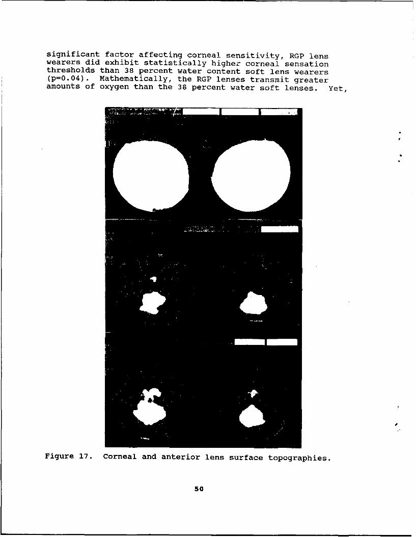
significant factor affecting corneal sensitivity, RGP lenswearers did exhibit statistically higher corneal sensationthresholds than 38 percent water content soft lens wearers(p=0.04). Mathematically, the RGP lenses transmit greateramounts of oxygen than the 38 percent water soft lenses. Yet,
Figure 17. Corneal and anterior lens surface topographies.
50
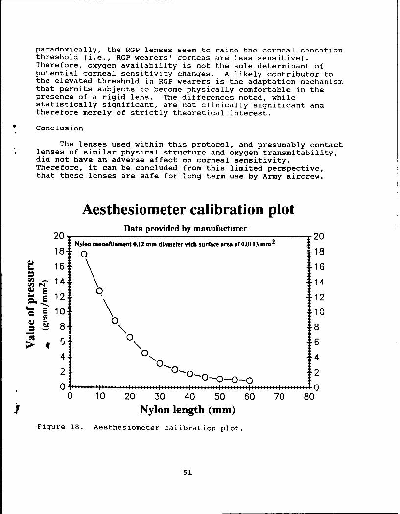
paradoxically, the RGP lenses seem to raise the corneal sensationthreshold (i.e., RGP wearers' corneas are less sensitive).Therefore, oxygen availability is not the sole determinant ofpotential corneal sensitivity changes. A likely contributor tothe elevated threshold in RGP wearers is the adaptation mechanismthat permits subjects to become physically comfortable in thepresence of a rigid lens. The differences noted, whilestatistically significant, are not clinically significant andtherefore merely of strictly theoretical interest.
Conclusion
The lenses used within this protocol, and presumably contactlenses of similar physical structure and oxygen transmitability,did not have an adverse effect on corneal sensitivity.Therefore, it can be concluded from this limited perspective,that these lenses are safe for long term use by Army aircrew.
Aesthesiometer calibration plotData provided by manufacturer20 2
Nyon monoilaiment 0.12 mm diameter with surface area of 0.0113 mm 2
18 0 18
16 1614 14
E 10 10
01
> 64 0 4\ 42 0 1010-0-_0___0 2
2 2
0 10 20 30 40 50 60 70 80
Nylon length (mm)Figure 18. Aesthesiometer calibration plot.
51
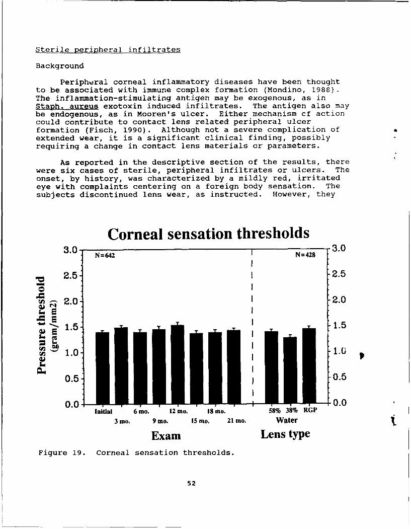
Sterile peripheral infiltrates
Background
Peripheral corneal inflammatory diseases have been thoughtto be associated with immune complex formation (Mondino, 1988).The inflammation-stimulating antigen may be exogenous, as inStaph. aureus exotoxin induced infiltrates. The antigen also maybe endogenous, as in Mooren's ulcer. Either mechanism cf actioncould contribute to contact lens related peripheral ulcerformation (Fisch, 1990). Although not a severe complication of aextended wear, it is a significant clinical finding, possiblyrequiring a change in contact lens materials or parameters.
As reported in the descriptive section of the results, therewere six cases of sterile, peripheral infiltrates or ulcers. Theonset, by history, was characterized by a mildly red, irritatedeye with complaints centering on a foreign body sensation. Thesubjects discontinued lens wear, as instructed. However, they
Corneal sensation thresholds3.0. N=642 N=428 3.0
2.5 2.5
W 2.0 2.0
157 1.5
1.0 1.6
0.5 I 0.5
0.0 0.0Initial 6 mo. 12 mo. 18 mo. 58% 38% RGP
3 mo. 9 mo. 15 Mo. 21 mo. Water
Exam Lens typeFigure 19. Corneal sensation thresholds.
52

did not seek objective evaluation also as instructed, sincesymptoms abated on lens removal and disappeared entirely within24 to 72 hours. At the time of symptom abatement, normal lenswear was reinstituted by all affected subjects. On the requiredquarterly followup exam, the existence of a residual scar wasobserved, and the supporting history obtained.
Objectively, there was indication of a localized infiltrateconcentration at the level of the anterior stroma near thelimbus; location in all cases was restricted to the inferiorhemisphere of the limbus (between 3 and 9 o'clock). While thegeneral shape of the lesions could be described as circular, manywere slightly oblong with the central axis appearing to parallelthe limbus (Figure 20). Subsequent follow up examinationsdocumented a gradual resolution or elimination of theprecipitated immune complexes.
Figure 20. Peripheral, sterile ulcer.
Discussion
The location and appearance of the peripheral corneallesions noted above are reminiscent of another phenomenonassociated with soft contact lens wear: subcontact lens bubbleformation at altitude (Flynn et al., 1987). Described soft lensbubbles occurred in 24 percent of the study eyes (22/92), andwere always formed at the limbus. None were ever detected overthe central cornea. Once formed, these peripheral bubblesincreased in size and coalesced with increasing altitude. Thebubbles did not disappear with blinking, but once descent wasinitiated, the dissipation process only took several minutes.The authors suggested that observed bubbles were the result of aninduced expansion of previously existing bubble nuclei trapped bythe soft contact lens acting as a semipermeable membrane.
53

While bubble composition was never identified, the mechanismof bubble nuclei formation was suggested to be via the negativehydrostatic pressure "produced from the contact lens tear pump."An analysis of research in the areas of tear film pH andassociated s ft contact lens wear underscores an alternativeexplanation indicating a major subcontact lens bubble nucleisource to be corneal-vented carbon dioxide. If this hypothesisis correct, then CO2 bubbles, as a metabolic byproduct, could beused to model subcontact lens metabolite trapping. The patternof epithelial punctate staining described earlier in this reportcould conceivably be a direct result of isolated or uncoalescedcarbon dioxide bubble-nuclei trapping across the surface of thecornea.
A gross comparison of the six sterile peripheral infiltratesto published soft lens bubbles highlights a correlation in bothlimbal position and relative shape. This relationship coulddescribe the underlying process of sterile peripheral infiltrateformation as a localized inflammatory or hypersensitivity re-sponse to peripherally coalesced and concentrated endogenousmetabolic byproducts trapped under the soft lens. The proposedCO2 bubble formation model then could directly be applied to theassessment of pertinent soft contact lens fitting parameters(i.e., base curve, diameter, and corneal curvatures) governing atight vs. an appropriate fit. Such a model would be extremelyuseful in providing a quantitative nomogram for soft contact lensfitting in lieu of the subjectively qualitative methods incurrent use.
Bacterial ulcers
Background
The primary drawback to the routine use of contact lenses byArmy aircrew is the risk of bacterial ulcerative keratitis. Thisserious ocular complication has been closely linked to the use ofextended wear soft lenses (Galentine et al., 1984; Weissman etal., 1984; Chalupa et al., 1987). Alternatively, poor lens hy-giene compliance has been suggested to be the primary cause ofdaily wear soft lens-induced corneal infection (Mondino et al.,1986). Since field conditions for Army aircrew would place amajor hygiene challenge upon contact lens wearers, it was decidedto use a disposable, extended wear soft lens system. It washoped the decreased risk of hygiene and compliance problems bygoing disposable would offset the increased risk posed byextended wear.
There were a total of six cases of bacterial ulcerativekeratitis. Although corneal scrapings and cultures werenegative, all cases were presumed to be bacterial based on:
54

physical location and appearance of the lesion, severity ofassociated signs and symptoms, and effectiveness of antibacterialtherapy. All cases were initially seen by general medicalpractitioners and treated with topical antibiotics prior tospecialty referral, so it isn't surprising that scrapings andcultures were negative. All subjects recovered from the acutephase of the infection within 4 to 8 days, primarily as a resultof the subject promptly seeking medical attention. All recoveredvisual acuity of 20/20 or better within the noted time. Anexample ulcer is shown in Figure 21 (Lattimore and Varr, 1991).While affected aircrew were temporarily grounded during thecourse of the infection, all were eventually returned to fullflight duty, and all voluntarily returned to contact lens wear,as well. To date, there have been no recurrences of ulcerationin affected subjects. Since fungal dermatitis was a majorproblem for troops deployed to Southwest Asia, there also wasconcern for possible fungal keratitis in the deployed portion ofour study group. However, no cases were documented.
Figure 21. Paracentral, bacterial ulcer.
Discussion
The combined ulcerative keratitis risk of all study subjects(all original AH-64 protocol subjects plus Desert Shield/Stormsubjects) from November 1988 through September 1991, equated to1/112/year, or 8.93/1000/year. This combined risk falls withinthe wide range of risk estimates (2.1/1000/year by Poggio et al.,1989; 4-6/1000/year by Schein et al., 1989; 15/1000/year byGrant, Kotow, and Holden, 1987; 48/1000/year by Efron et al.,
5S
1991) proposed for nonaphakic extended soft lens wear in thecivilian literature. The less than 1 percent level of risk forulcerative keratitis has been judged to be acceptable within thecontext of other risks faced by Army aircrew members in theperformance of their flight duties. However, the public healthissue of placing healthy eyes at increased risk of severeinfection should not be ignored. Ways of minimizing thiscomplication should still aggressively be pursued. Possibleoptions to be considered include planned replacement, flexiblewear of soft lenses or even a daily disposable system underadverse conditions.
Hvdrogel translens oxvyen flux
Introduction
Many studies have examined oxygen transmittance and oxygenavailability issues related to hydrogel contact lens wear. Onemethodology is to place a test lens on the eye, and after aspecified period of wear the lens is removed. Immediately afterlens removal, the oxygen uptake rate of the cornea is obtained.This postlens rate is compared to an uptake rate obtained priorto application of the test lens. Since the contact lensinhibited oxygen flow while it was in place, the newly lens-freecornea typically exhibits an increase in the oxygen uptake ratecompared to the prelens baseline. This increase can bemathematically converted into an equivalent oxygen percentage(EOP) that had been available to the cornea under the lens.
Another methodology uses corneal thickness as an index foroxygen availability. Induced hypoxia can be achieved by varioushydrogel lenses or by goggles that have a complete or partialnitrogen atmosphere retained between the eye and the goggle.Various levels of decreased oxygen availability will inducesecondary corneal swelling. By scaling the swelling response toavailable oxygen levels, the investigators estimate the minimallevel of oxygen necessary for normal corneal function. However,as discussed in the review portion of the report, the results areas varied as the methodologies.
Despite the wealth of oxygen data, there are no data formodeling behavior of the cornea under the hydrogel lens while itis in situ. Corneal behavior after lens removal simply examinesthe poststress response, not the concurrent stress response.Using corneal thickness as an index for the hypoxic stress-response is not a completely appropriate because other factors(bicarbonate ion, Na-K ATPase, tissue pH) have been shown toaffect corneal thickness concurrent with, and independent ofhydrogel lens wear (Zagrod and Connor, 1988).
56
Methods
The micropolarographic oxygen probe consisted of a platinumcathode (25 Am diameter) and a silver anode embedded in a plasticcarrier. A potassium chloride (KCl) solution served as anelectrolytic bridge between the cathode and the anode. An oxygenpermeable polyethylene membrane, 25 Am thick, effectively sealedthe entire electrode-KCl assembly into one operating unit. Themicropolarographic system was similar to that used by Benjaminand Hill, 1986.
The experimental procedure involved applying the probe tothe anterior surface of the corneal epithelium of the subject'shydrogel lens covered cornea. The sensor, when applied to theanterior surface of the in situ contact lens, provided a limitedreservoir of oxygen for the underlying tissue. The average rateof oxygen depletion from the sensor reservoir, between recordingsof 140 mm Hg and 40 mm Hg, became the measure of the cornealoxygen uptake rate. This, in turn, represents only a relativemeasure of the aerobic requirement of the cornea, since theextent that the epithelium, stroma, and endothelium eachcontribute to the corneal oxygen uptake rate has not adequatelybeen established. Published estimates for the epithelium rangefrom 55 percent to 70 percent, with an unpublished estimateranging as high as 93 percent.
The oxygen uptake rate recordings were obtained within 5minutes after lens application on the initial exam. Subsequenttranslens oxygen flux recordings were obtained at each followupexamination. Data reported here were from 212 hydrogel lenswearing subjects.
Results and discussion
Translens oxygen flux as a function of number of daysextended lens wear is represented in Figure 22. Although ananalysis of variance of the data was not statisticallysignificant (p=0.25), there is a clear pattern visible inclinical terms. The ability for oxygen to flow through a lens isshown to increase with wearing time over the first 3 or 4 days ofextended wear, followed by a flux falloff after 5,6, and 7 daysof wear. This non-monotonic curve is counter to the notion thata contact lens' oxygen transmittance is a fixed figure.Intuitively, a linear relationship was expected.
Obviously the ability of oxygen to flow through a hydrogellens varies as the precorneal environment in which it findsitself varies. An undeniable component of this day 1 through day4 flux increase, compared to the initial exam base, would beincreased tissue respiratory demand. However, other factors such
57

as tearfilm and hydrogel fluid pH, level of lens hydration, andlens temperature can all be interrelated in governing oxygenflux.
The falloff in translens oxygen flux at day 5 and beyond maybe an indicator of mucus and protein deposition. As the hydrogelsurface becomes microscopically coated, the ability for oxygen toflow across the membrane may be affected adversely. If this isso, then extended wear beyond 4 days may be contraindicated. Abreakdown of the data by type of hydrogel lens worn (Figure 23)shows that the process is similar for both types of hydrogellenses used. However, it should be pointed out thatparadoxically, the lower water content lens possessed a greaterflux ability after 5 days of wear. Since the lenses used comefrom two different FDA categories, the flux differences mayhighlight protein deposition differences of ionic materials vs.nonionic materials.
Influence of wearing timeon translens oxygen flux
8.00 8.007.75 a Mean data points; N=1213 measurements 7.75
.750 __ -Nonlinear regression; r=0.94S 7.50 -7.50
, 7.25 7.25SC 7.00 7.00
6.75 6.758~.50 6.50
C 625 6.256.00 6.005.75 5.75
C 5.50 5.505.25 5.255.00 ,5.00
0 1 2 3 4 5 6 7
Duration of extended wear (days)
Figure 22. Influence of wearing time on translens oxygen flux(grouped data).
58

Conclusions
The ability of oxygen to cross the in situ hydrogel membraneis not a simplistic linear function with a fixed oxygentransmittance ceiling. Decreased oxygen flux after 4+ days ofwear suggests a practical 4-day limit on the extended wear ofhydrogel lenses, particularly for ionic lens polymers.
Influence of wearing timeon translens oxygen flux
8.00 8.007.75 3 58% water content lenses A 38% water content lenses 7.75
7 Nonlinear regression; r=0.94 N=1213 measurements
7.50 7.50
7.25 7.25
7.00 7.00
6.75• 6.75
6.50 6.50
E6.25- 6.25I 6.00 6.00
0 5.75 5.75
5.50 5.50
5.25 5.255.00 , I 1 , 1 1 1 1 5.00
0 1 2 3 4 5 6 7
Duration of extended wear (days)Figure 23. Influence of wearing time on translens oxygen flux
(by lens type).
Anterior lens surface pH
Introduction
The anterior corneal surface is associated closely with anoverlying canopy of moisture known as the precorneal tearfilm.Traditionally, clinicians have been concerned with how certaincharacteristics of the tears can influence corneal integrity;tearfilm formation problems and tear osmolarity issues represent
59

two examples of purported tearfilm influence upon the cornea.However, the tearfilm can be susceptible to influence by thecornea, as evidenced by the presence of both glycolytic and tri-carboxylic acid cycle enzymes within the tear layer. The sourceof these enzymes has been shown not to be the lacrimal gland, butrather the underlying corneal tissue. Therefore, tear chemistryis affected directly by the cornea. Consequently, cliniciansshould be reminded that although anatomically distinct, thecornea and its tear film are functionally interactive.
Attempts at quantifying the normal tear pH value haveyielded varying results. Although one cause of variation appearsto be due to instrumentation differences, the primary cause ofvariation appears to be the location or source of the tear sam-ple. Efforts at documenting the pH of the precorneal tearfilm(i.e., that canopy of mucin, aqueous, and oil directly anteriorto the cornea) have obtained a mean value range of 7.45 to 7.83.Since measurements of precorneal tearfilm pH under the extendedopen-eye condition have been shown to match that predicted by CO2equilibration calculations, it is likely these values are veryclose to the true precorneal tearfilm pH.
Initial hydrogel lens research indicated that these contactlenses may provide a barrier to carbon dioxide (C02 ) efflux fromthe cornea, although at the time this was considered to be insig-nificant in terms of corneal physiology. However, recent mea-surements of tear CO2 accumulation under hydrogel lenses, pairedwith the detection of a decrease in both subcontact lens andstromal pH following contact lens wear, indicates yet anotherfunctional link between the precorneal tearfilm and cornealphysiLology.
Materials and methods
A self-referenced pH electrode, designed for pH recordingfrom semisolid materials was used to assess the in situ anteriorcont-,ct lens surface pH response to continuous wear of 38 percentand S8 percent water content hydrogel lenses worn on a disposablebasis. The recorded pH reading was the peak value of a transientresponse. Upon initial probe application, the measured pH valuewas within 0.2 of the final or peak value. However, a gradualdrift in the alkaline direction led to stabilization of the read-ing, presumably due to temperature changes at the probe surface.If the probe was kept in contact with the lens beyond the stabi-lization period, a gradual shift in the acidic direction wasnoted. This has been attributed to CO2 accumulation under theprobe (Fatt, personal communication).
Subjects were on a 1-week wearing cycle, after which timethe lenses were removed, disposed of, and replaced after at leastone night of lens-free sleep. Use of this pH electrode method-
60

ology assumes the anterior contact lens surface pH measurementaccurately represents both the prelens tearfilm pH and the pH ofthe anterior water component of the hydrogel contact lens. How-ever, it is possible these two entities could have slightlydifferent pH values. The pH electrode was calibrated with a 7.00and a 10.00 pH standard solution at 35° C and disinfected byalcohol swab and surface drying between each assessment. Mea-surements were recorded from the contact lens in its storagepacket immediately after opening, then 5 minutes after initiallens application onto the volunteer subject's eye, 24 hours afterinitial lens application, 7 days after initial lens application,and on subsequent quarterly followup examination. Each measure-ment for any one individual was taken at the same time of day inorder to minimize error from individual diurnal variations. How-ever, pH assessments across individuals occurred at varying timesof day, thereby eliminating any group diurnal effect.
Results
Figure 24 provides a graphical representation of the groupeddata for all lenses; lens differences were not statistically
Influence on wearing timeon hydrogel lens anterior surface pH
7.70 7.70
S7.60 7.60S7.50 7.50
S7.40 7.40
7.30 7.30S7.20 7.20
7.10 Mean packaged baseline 7.10• " 7 00 . . ............................. ..•7 00 .0 7.00
S6.90 Total N=517 6.906.50 - 0 pH data and regression; r=0.91 6.80
General equation: y= (a+b-x)
6.70 , 6.700 1 2 3 4 5 6 7Duration of extended wear (days)
Figure 24. Influence of wearing time on hydrogel lens anteriorsurface pH (grouped data).
61

significant (p=0.43; Figure 25). The contact lenses in solutionwere at or very near a neutral pH of 7.00 (38 percent lenses =7.05; 58 percent lenses = 7.00). Within the first 5 minutes ofcontact lens wear, the pH reading started to rise into thealkaline region (7.23); this increase in pH continued over thecourse of the first 2 days of wear to asymptote near theestablished norm (7.45). Statistical analysis (ANOVA) of pH byduration of extended wear of currently worn lenses wassignificant (p<0.001).
Influence on wearing timeon hydrogel lens anterior surface pH
7.70 7.70
7.60 7.60
7•750 7.50
S7.40 7.40
M 7.30 7.30
M 7.20 7.20
S7.10 Packaged baseline for 38% water content lenses 7.10
S7.00 ... .................................... 00"" 7=.. .Paaged baseline for 58% water content lenses
6.90 Total N=517 6.90'. 38% water content lenses
6.80 A 58% water content lenses 6.80General equation: y= (a+b-x) for grouped data, r=0.91
6.70 1 , , , 1 1 1 1 6.700 1 2 3 4 5 6 7
Duration of extended wear (days)Figure 25. Influence of wearing time on hydrogel lens anterior
surface pH (by lens type).
Discussion
The initial in situ pH reading of 7.23, taken just 5 minutesafter lens application, suggests that a fluid exchange betweenthe anterior tearfilm and the contact lens occurs very quickly.Indeed, by the point of 2 days wear the pH has stabilized nearthe norm of 7.45, and is not much different from the acceptedpublished norms for the precorneal tearfilm. The initial data(days 0 and 1) are less alkaline compared to precorneal tearfilm
62

norms due to the starting lens pH of a nominal neutral pH value;if the lenses were packaged in a storage solution of a morealkaline nature near 7.45, this pattern of pH adjustment wouldnot be exhibited. In any event, the obtained data do not supportthe use of this system as a useful indirect monitor of corneal pHchanges related to hydrogel lens wear. However, the methodologydoes support data suggesting that the pH of the precorneal tearfilm is very near 7.45.
The fact that both types of lenses chosen for use in theprotocol were packaged in neutral, or near neutral, solutions maybe significant. It is possible that hydrogel stability andphysical performance parameters are optimum near a pH of 7.Therefore, lenses may be packaged at that pH in order to maximizeshelf life. If that is the case, and the inference of decreasedstability and physical performance at pHs other than neutral alsois correct. Then future material research should perhaps becentered on finding polymers possessing optimal qualities at theprecorneal tearfilm pH of 7.45.
Conclusions
Simple pH measurement of the anterior lens surface does notappear to provide clinically useful information, although it doesconfirm normal precorneal tear pH values established by others.A question can be raised concerning the susceptibility of lensphysical performance to external influence by the physical stateof the hydrogel material after 2 days or more extended wear.Future polymer research may isolate materials less influenced bythe precorneal environment.
Hydrogel lens water content
Introduction
Soft lens behavior, after initial placement on the surfaceof the eye, has been shown to change over the first 24 hours ofwear. Clinically, hydrogel lenses appear to "tighten" over thefirst day or two of wear, necessitating a 24- to 48-hour followupexamination to ensure an adequate fit is obtained by the patient.This process has been attributed to a drying or dehydration ofthe soft lenses as a function of initial wearing time. Since themajor proportion of hydration change in those studies occurredwithin the first several hours, it was assumed that water contentstabilization developed by the end of one day of continuous lenswear. All of these reports used only one means of water contentdetermination without method cross-comparison, although thespecific method used in each study varied. Therefore, thepurpose of this portion of the overall research protocol was two-fold: to describe the dehydration course over 7 days of wear forboth lens types used, and to compare two methods of water contentdetermination in the 58 percent lens.
63
Methods and materials
Forty-'ne myopic subjects, Wearing corrections that varied "from :-0;505diopters to -3.50 diopters Were followed on aquaxterly-basis tor approximately 1 year. Subjects wore theirsoft lehses on an extended wear, disposable paradigm, with amaximum wearing schedule of 7 days/6 nights. After thisschedule, lenses were removed and discarded, and at least onesleep period was spent Iens-.ree. One hundred and ninety-sixdata entrits were obtained on periodic followup examination overa 1-yea -period.
The gravimetric determination of water content was obtainedby way f a analytical scale enclosed in a controlledenvironmental chamber kept at 35°C. The change in lens weightfrom the fresh, wet state to the completely dehydrated statecould be used to calculate the beginning water cdntent oncereference standards were established. All 38 percent lenses weremeasured by this method of appraisal of in situ hydration level.Half of the 58 percent lenses were measured via this methodologyby using one lens from each subject.
The refractive method used a refractometer (Marco*) with afixed scale (based on one specific index of refraction) tomeasure the lens against. The refractive scale ranged from 35 to75 percent water. The 38 percent lenses could not be measuredwith this method because they tended to drift just below 35percent-water with any amount of wearing time. Half of the 58percent lenses were assessed by the refractive method by usingone lens from each subject. In this manner, a correlationbetween the two methods could be established for the 58 percentwater content lenses.
Results and discussion
A logarithmic water content decline occurred over the timeperiod tested by both measurement systems (Figure 26). Anindependent analysis of variance for water content factored bydays wear was highly significant (p<0.001). The largest decreasewas from day 0 to day 1, with a slower decrease occurring to day4. Specific statistical groupings at the 0.05 level ofsignificance were: day 0; days 1 and 2; and days 3-7. Figure 26highlights the apparent stabilization developed by day 4extending through day 7. Contrary to suggestions of 24-hourhydration stabilization made by other investigators, these datademonstrate long-term dehydration occurring over the first 3 to 4days of wear. In addition, the two methods of water contentdetermination agreed remarkably well, correlating with each otherat a Pearson coefficient of 0.96.
64

Influence of wearing timeon hydrogel lens water content
60 60TT Packaged baseline
,55 55
0 50 50
S45 Total N =148 45
- 0 Grmetoa c data and regression; r=0.86Refractometer data and regression; r= 0.86General equation: y= (a+ bx
40 , ' 400 1 2 3 4 5 6 7
Duration of extended wear (days)Figure 26. Influence of wearing time on hydrogel lens water
content (two measurement methods; 58 percent watercontent lenses only).
Refractometer data on 58 percent water content lenses wereinitially being gathered in the summer of 1990. A number ofsubjects seen at Fort Rucker were deployed to Saudi Arabia onOperation Desert Shield. In order to meet the requirements ofthe research protocol, the subjects were given a quarterlyfollowup exam in Saudi Arabia in November 1990. As a part of theexam, water content data were obtained using the refractivemethod (Figure 27). After 6 to 11 weeks "in-country," thesubjects exhibited the same pattern of lens hydration, as afunction of duration of lens wear, as they had at Fort Rucker.Data obtained were for 1, 2, 6, and 7 days of wear. In andaround Saudi Arabia, it seemed that soft lens wearers fell intotwo groups: those who maintained their habitual wearing scheduleof 6 or 7 days with no problems, and those who decreased theirwearing time to 2 or 3 days of wear because of changes in comfortor clarity. The notion of soft lenses excessively dehydrating inthe desert were shown to be erroneous by the data, but the
65
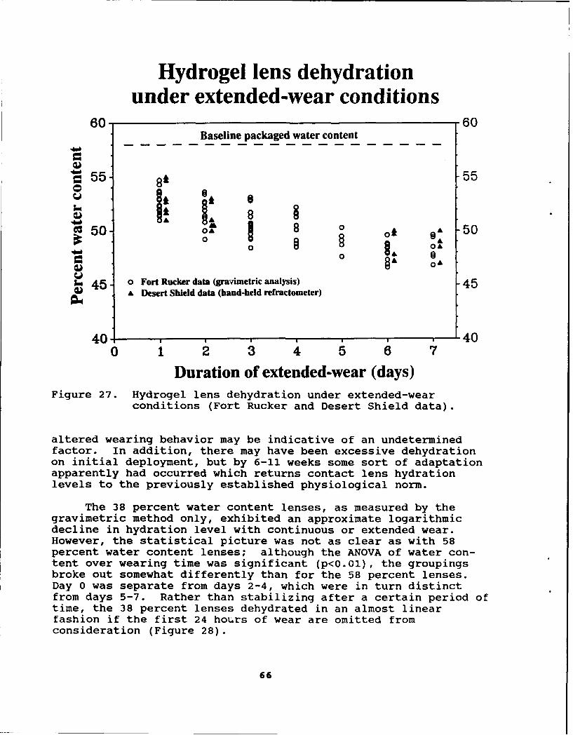
Hydrogel lens dehydrationunder extended-wear conditions
60 60Baseline packaged water content
S55 81 55
0i
C50 Q1A 0 oA A 50§ OA
-"o Li 84, O
0 L 0A A
3.~ 45 0 Fort Rucker data (gravinietric analysis) 4& Desert Shield data (hand-held ref~ractometer)
40 400 1 2 3 4 5 6 7
Duration of extended-wear (days)Figure 27. Hydrogel lens dehydration under extended-wear
conditions (Fort Rucker and Desert Shield data).
altered wearing behavior may be indicative of an undeterminedfactor. In addition, there may have been excessive dehydrationon initial deployment, but by 6-11 weeks some sort of adaptationapparently had occurred which returns contact lens hydrationlevels to the previously established physiological norm.
The 38 percent water content lenses, as measured by thegravimetric method only, exhibited an approximate logarithmicdecline in hydration level with continuous or extended wear.However, the statistical picture was not as clear as with 58percent water content lenses; although the ANOVA of water con-tent over wearing time was significant (p<0.01), the groupingsbroke out somewhat differently than for the 58 percent lenses.Day 0 was separate from days 2-4, which were in turn distinctfrom days 5-7. Rather than stabilizing after a certain period oftime, the 38 percent lenses dehydrated in an almost linearfashion if the first 24 hou~rs of wear are omitted fromconsideration (Figure 28).
66

Hydrogel lens dehydrationunder extended-wear conditions
50 50N =48 measurementsR = 0.88
S4 0-38% lens data and nonlinear regression 45
40 T Packaged baseline 40
,* 35 35
S30 30
S25 25
20 20-1 0 1 2 3 4 5 6 7 8
Duration of extended wear (days)
Figure 28. Hydrogel lens dehydration under extended-wearconditions (38 percent water content lenses).
Figure 29 depicts the results of the two lens types asmeasured gravimetrically. The amount of water loss is greater inthe higher water content lens. Also the regression curve appearssomewhat different between the two types. A gross examinationsuggests that the relative dehydration, based on packagedbaseline as the norm, may be approximately the same for both lenstypes. However, Figure 30 reveals the 58 percent lenses topossess a greater degree of relative dehydration. The two datasets are significantly different by ANOVA (p<0.01). Thedifferences in rate and degree of lens dehydration in situperhaps can be explained by the chemical structure differencesbetween the two lens types. The 38 percent lens is a nonioniclens, while the 58 percent lens is ionic. The degree ofmolecular water attraction, and differential forces resistant towater loss readily could account for these documenteddifferences.
67

Hydrogel lens dehydrationunder extended-wear conditions
65 65
60 T Packaged baseline (58% lens) 60
;i 55- T T 55
.A.
S45 45
40 Packaged baseline (38% lens) 40................ .............................
S35 - r 35
30 AL - 58% water content lens data and nonlinear regression (r= 0.86) 30
0 - 38% water content lens data and nonlinear regression (r=0.88)25.25
-1 0 1 2 3 4 5 6 7 8Duration of extended wear (days)
Figure 29. Hydrogel lens dehydration under extended-wearconditions (by lens type).
Clinically these differences can be applied to patientmanagement issues. Ostensibly, a patient with decreased seroustear component production would be less likely to tolerate, or becomfortable with, an ionic lens of mid- to high-water contentthat presents a large hydration challenge to the eye in theinitial 3 to 4 days of extended wear. The low water content,nonionic lens would provide greater comfort throughout the entirelens wearing cycle. Conversely, a patient with either adequate,or abnormally copious serous tear production would be immediatelymore comfortable in a mid- to high-water content, ionic lens.Therefore, the type of lens prescribed could be completelydependent upon the serous characteristics of the precornealtearfilm.
Possible factors contributing to the long term dehydrationprocess include: lipid and protein deposits during wear andchanges in pH (anterior and posterior). Gravimetrically, theformer would serve to increase the dry material weight a greater
68

Normalized hydration levelsin two types of soft lenses
1.2
_ 1.1
N 0.9 A
S 0.8-
~0.7-
0.6 A--Normalized 58% water content data and regression
0- Normalized 38% water content data and regression0 .5 , I I I.I..
-1 0 1 2 3 4 5 6 7 8
Duration of exteneo 4kear (days)
Figure 30. Normalized hydration levels in ',wo types of softlenses.
proportion than the wet material weight, thereby skewing themeasurement in the dehydrated direction. However, there shouldbe no refractive influence by surface lipids and proteins. Theexcellent correlation between the two methods tends to rule outlipid and protein deposition as contributing to the dehydrationprocess. Materials sciences have shown a pH change in hydrogelsolution to directly influence material water content (McCarey,and Wilson, 1982) and is, therefore, the likely in situ cause forlens dehydration. However, a correlative model of this suggestedin situ relationship has not yet been published.
Possible contaminating factors in the methodology couldinclude: oil from fingers on lens removal, and fluid loss duringthe weighing and/or refractive processes. Each could influenceboth methods in the same fashion; therefore the presented levelsof lens hydration may slightly overestimate the amount ofdehydration with wear. However, the relative relationship
69

between the two methods would be unaffected; the same holds forthe process of long term change. The affect of any contaminationerror simply would decrease overall magnitude across the board,but not the basic characteristics or patterns of the processes.
Conclusions
The gravimetric and refractive methods of lens water contentdetermination agree closely for the 58 percent water contentlenses tested. Functional lens parameters, no matter thematerial characteristics, are in a state of flux as indicated bythe change in water content over time. However, the exactpattern of change apparently is dependent upon lens materialcharacteristics. It is probable that hydration change is aresult of tear pH influences acting directly on the hydrophilicmaterial. Extended hydrogel lens wear is clearly a complex,dynamic process for both the lens and cornea. A limitedunderstanding of one parameter is not enough for the successfulpractice of contact lens care. An integrated picture of multipleparameters is necessary to reliably quantify potential limits ofextended hydrogel lens wear. Therefore, specific knowledge of apatient's condition, combined with an understanding of preciselens material behavior, is necessary to maximize fitting andwearing success rates.
pH and water content correlational analysis
Introduction
Other investigators have shown that soft lens hydration isdirectly influenced by the pH of its solution in vitro. Inaddition, data from the previous sections in this report revealedboth a change in hydrogel lens anterior surface pH, and a changein soft lens water content as a function of wearing time.Earlier in this report, it was concluded lens hydration changeswere probably influenced by the precorneal tearfilm pH. Thepurpose of this section is to correlate/model the change in softlens hydration as a function of anterior lens surface pH changein situ over wearing time.
Methods and materials
Anterior lens surface pH and lens hydration data for boththe 38 percent and 58 percent water content lenses were obtainedat every examination as presented earlier in this report (Figures31 and 32).
70

Influence of wearing timeon hydrogel lens anterior surface pH
7.70 7.70
7.60 7.60
S7.50 7.50
7.40 7.40
:7.30 7.30
rA 7.20 7.20
.- 7.10 7.10164 Packaged baselineS7.00 .... .. r .......... ........................; . . . . a k gdb sln 7.00
S6.90 Total N=517 6.90a O pH data and regression; r=0.91
8.80 General equation: y= (a+b') 6.80
6.70 82 6.700 1 2 3 4 5 6 7
Duration of extended wear (days)
Influence of wearing timeon hydrogel lens water content
60 60Packaged baseline.. . ....... P.a.k...gI.ba e n.. .... ................
S55 55
g50 50
S45 Total N=418 4
- 0 Gravimetric data and regression; r=0.86-- 4 Refractometer data and regression; r= 0.6
General equation: y =(a+ b")40 2 40
0 1 2 3 4 5 6 7Duration of extended wear (days)
Figure 31. Influence of wearing time on hydrogel lens anterior
surface pH and water content (grouped data).
71

Influence of wearing timeon hydrogel lens anterior surface pH
7.70 7.70
Z 7.60 7.60
7.50 7.50
c 7.40 - 7.40
7.30 7.30C12
S7.20 -7.20
7.10 Packaged baseline for 38 % water content lenses 7.10o 7 %o
TtN7.00. . . . . Packaged baseline for 58 % wter content lenses
- 6.90 Total N=517 6.906 v 38% water content lenses
6.80 A 58% water content lenses 6.80iGeneral equation: y= (a+b-x) for grouped data, r=0.91
6.70 . 6.700 1 23456 7
Duration of extended wear (days)
Hydrogel lens dehydrationunder extended-wear conditions
65 85
60 Packaged baseline (58% lens) 60.. 1 . . . . . . . . . . . . . . . . . . . . . . . . . . . . . . . . . . .
"55 T T 55
"" 50 50-Z IT
"Z45 45
W 40j L' 40 T" Packaged baseline (38% lens) 40................................... .................... .........
• 35 0 35
30 A - 58% water content lens data and nonlinear regression (r= 0.86)
2 - 38% water content lens data and nonlinear regression (r=0.88)25 I 25
1 0 1 2 3 4 5 6 7 8Duration of extended wear (days)
Figure 32. Influence of wearing time on hydrogel lens anterior
surface pH and water content (by lens type).
72
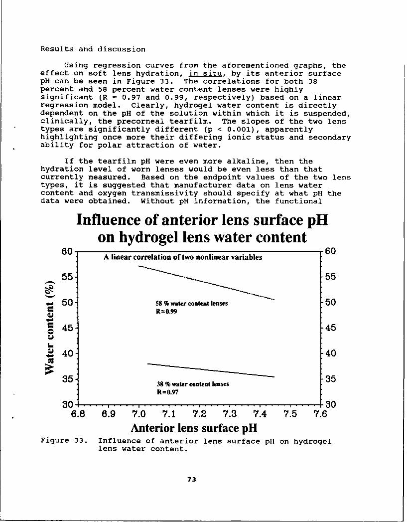
Results and discussion
Using regression curves from the aforementioned graphs, theeffect on soft lens hydration, in situ, by its anterior surfacepH can be seen in Figure 33. The correlations for both 38percent and 58 percent water content lenses were highlysignificant (R = 0.97 and 0.99, respectively) based on a linearregression model. Clearly, hydrogel water content is directlydependent on the pH of the solution within which it is suspended,clinically, the precorneal tearfilm. The slopes of the two lenstypes are significantly different (p < 0.001), apparentlyhighlighting once more their differing ionic status and secondaryability for polar attraction of water.
If the tearfilm pH were even more alkaline, then thehydration level of worn lenses would be even less than thatcurrently measured. Based on the endpoint values of the two lenstypes, it is suggested that manufacturer data on lens watercontent and oxygen transmissivity should specify at what pH thedata were obtained. Without pH information, the functional
Influence of anterior lens surface pHon hydrogel lens water content
60 A linear correlation of two nonlinear variables 60
55 -- S-- 55
* 50 58 % water content lenses 50,= R=0.99
=45 45
S40 40
38% water content lenses
R=0.97
30 1 ...... .. .... .... .... .... ......... .... 306.8 6.9 7.0 7.1 7.2 7.3 7.4 7.5 7.6
Anterior lens surface pHFigure 33. Influence of anterior lens surface pH on hydrogel
lens water content.
73
performance inferences made from such data are meaningless withno useful clinical relevancy.
As an adjunct to the above, if lens manufacturers providedlinear behavior models similar to Figure 33, patient care couldbe individually customized by the clinical measurement ofprecorneal tearfilm pH. Two brands of lenses, identical in allpackaged parameters (power, thickness, base curve, diameter, andwater content) save ionicity, would provide differing physicalfits and oxygen availability on the eye, because of different insitu water contents.
Conclusions
Hydrogel lens water content is directly dependent onprecorneal tearfilm pH. Other perceived contributors to theprocess (tear availability, stability, and osmolarity) likelyhave little clinical contribution. The clinical measurement ofthe precorneal tearfilm, prior to fitting a prospective patient,could assist in the lens selection process, if manufacturersprovided pH-related material behavior charts.
Corneal thickness measurement differences
Introduction
Increases in central corneal thickness (CCT) associated withcontact lens wear have been linked to tissue hypoxia. Cornealswelling has been induced by thick hydrogel lenses, nitrogen-fedgoggles, and metabolic inhibitors. However, the degree andlocation of induced edema varies with the method of stressapplied. The exact mechanism(s) responsible for in vivo softcontact lens-induced corneal swelling have not been fullyelucidated. As a result, clinical modeling of corneal thicknessas a function of contact lens wearing experience has not beenobtained. In an effort to more fully understand the cornealstresses induced by soft contact lens wear, those subjectsfitting with soft lenses only, had their CCT measured by twoseparate methods of pachometry at every examination.
Methods
Two methods of corneal thickness measurement were used:ultrasound pachometry, and optical pachometry. The ultrasoundmethod uses a hand-held sensor with an ultrasound-emitting coneset at a nominal speed of 1630 meters per second. When the endof the cone is placed perpendicularly onto the apex of thecornea, the emitted sound waves are reflected back from surfaceswhere there is a change in refractive index (Dunn, Edmonds, andFry, 1969), e.g. theoretically at the posterior corneal border.
74

The time taken for an emitted wave to traverse the cornea andreflect back to the cone is used to calculate the tissuethickness for the preset ultrasound speed. Variability inultrasound tissue thickness measurement can be induced byinterpatient variation in ultrasound velocity, point of soundreflection, and differences in tissue hydration (Salz et al.,1983; Gordon, Bogges, and Molinari, 1990).
The optical pachometry method was associated with a highmagnification contact specular microscope. The contact specularmicroscope uses an applanation cone to flatten a specific area ofthe central cornea in a process similar to that used inapplanation tonometry. Initially, the optical system is zeroedby focusing the microscope onto the surface of the cone. Once aclear image of the cone's surface is obtained, the cone is movedforward to applanate the central cornea. The optical system thenis focused toward the subject so that a clear image of theendothelium is obtained. Since this type of specular microscopeimages the posterior surface of the endothelium (reference), theoptical distance from cone surface to the endothelium wouldrepresent the corneal thickness. The purpose in using twodifferent measurement techniques was merely to establishintertest reliability and document test cost/performance results.
Results and discussion
Figure 34 is a simple correlative plot of ultrasound CCT asa function of optical CCT. There was a considerable degree ofdata spread with a mild correlation (R=0.56). The two methodswere significantly different by t-test (p<0.05). Mean ultrasoundCCT was 0.542 mm +/- 0.04 SD; mean optical CCT was 0.562 mm +/-0.04 SD. A frequency histogram of CCT, broken down by bothmethod and eye tested (Figure 35) highlights the essentiallynormal distribution of the results and the different centraltendencies of the two pachometry techniques.
Since the two techniques did not correlate well, a furtheranalysis of the data was indicated. A 2-way analysis of variance(ANOVA) of CCT by method and long-term lens wear experiencerevealed some surprising differences (p<0.001; Figure 36).Initial and 24-hour exams obtained essentially like measurements,although the ultrasound technique obtained thicker CCTmeasurements on the initial exam. However, after 1 week in thestudy and longer, the ultrasound CCT measurement wasprogressively and significantly thinner than the opticalmeasurements. A 2-way ANOVA of CCT by method and number of dayscurrent lens wear revealed a very similar relationship, althoughthe maximum mean difference was not as great as for length ofcontact lens wearing experience (p<0.001; Figure 37).
75
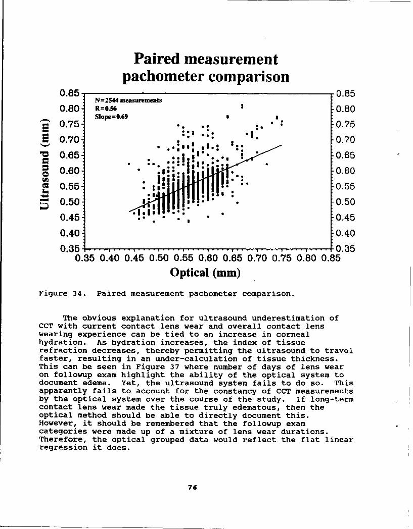
Paired measurementpachometer comparison
0.85 0.85N=2544 measurements
0.80 R=0.56 2 0.80Slope =0.69 • I
• 0.75- 0.75
0.70 :• .:. 0.70
'~0.65 0.65
0.60 ego* 0.60
C 0.55- 0.55
S0.5 0 . 0 .5 00.45 . 0.45
0.40 0.40
0.35 .... .... ................................ 0.350.35 0.40 0.45 0.50 0.55 0.60 0.65 0.70 0.75 0.80 0.85
Optical (mm)
Figure 34. Paired measurement pachometer comparison.
The obvious explanation for ultrasound underestimation ofCCT with current contact lens wear and overall contact lenswearing experience can be tied to an increase in cornealhydration. As hydration increases, the index of tissuerefraction decreases, thereby permitting the ultrasound to travelfaster, resulting in an under-calculation of tissue thickness.This can be seen in Figure 37 where number of days of lens wearon followup exam highlight the ability of the optical system todocument edema. Yet, the ultrasound system fails to do so. Thisapparently fails to account for the constancy of CCT measurementsby the optical system over the course of the study. If long-termcontact lens wear made the tissue truly edematous, then theoptical method should be able to directly document this.However, it should be remembered that the followup examcategories were made up of a mixture of lens wear durations.Therefore, the optical grouped data would reflect the flat linearregression it does.
76

Pachometry frequency histogram200 2002Ultrasound OD
180 Ultrasound OS 180SOptical OD
160 -Optical OS 160
140 140
120 1200
S100 100
"• 80 80
; 60 60
40 40
20 200 .500 0
.450 .500 .550 .600 .650
Corneal thickness groupings (mm)
Figure 35. Pachometry frequency histogram.
Earlier contact lens studies on animal models have shownthat there are a number of corneal changes associated with lenswear. These changes include: epithelial thinning, stromaltissue depletion, and increased fluid accumulation, both intra-and extracellularly. This combination of cellular tissue loss,paired with fluid accumulation within the tissue couldtheoretically result in a net v-'alance in actual CCT. If thisholds true, then followup exam optical measurements of CCT wouldremain constant. In addition, ultrasound measurements wouldunderestimate actual CCT as a result of two contributing factors:loss of cellular tissue and abnormal fluid accumulation. Sincecontact lens wear has been shown to progressively influence thesetwo factors, the progressive CCT thinning by ultrasoundmeasurement documented in this study can thus be accounted for.Conversely, ultrasound CCT measurements consistently overestimatecorneal thickness in the initial and no lens wear categories.This implies that in the relative absence of clinical edema,ultrasound methods slightly overestimate actual CCT.
77
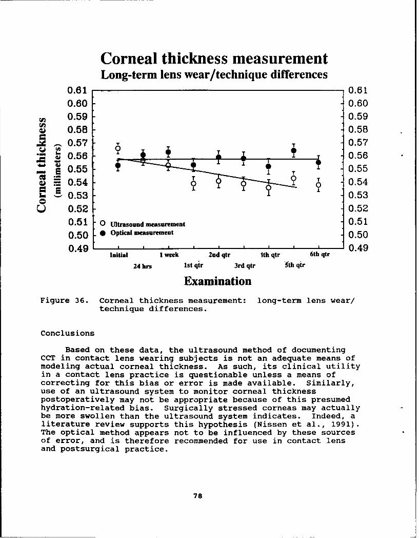
Corneal thickness measurementLong-term lens wear/technique differences
0.61 0.610.60 0.600.59 0.590.58 - 0.58
O.5 T 0.57" " •0.56 - -P•_ • • , 0.56
S0.55 T 0.55
E. 0.54 1 0.54-i 0.53 0.53
S0.52 0.520.51 0 Ultrasound measurement 0.510.50 0 Optical measurement 0.50
0.49 ' a a I - I I I I 0.49Initial I week 2nd qtr ith qtr 6th qtr
24 hrs 1st qtr 3rd qtr 5th qtr
ExaminationFigure 36. Corneal thickness measurement: long-term lens wear/
technique differences.
Conclusions
Based on these data, the ultrasound method of documentingCCT in contact lens wearing subjects is not an adequate means ofmodeling actual corneal thickness. As such, its clinical utilityin a contact lens practice is questionable unless a means ofcorrecting for this bias or error is made available. Similarly,use of an ultrasound system to monitor corneal thicknesspostoperatively may not be appropriate because of this presumedhydration-related bias. Surgically stressed corneas may actuallybe more swollen than the ultrasound system indicates. Indeed, aliterature review supports this hypothesis (Nissen et al., 1991).The optical method appears not to be influenced by these sourcesof error, and is therefore recommended for use in contact lensand postsurgical practice.
78
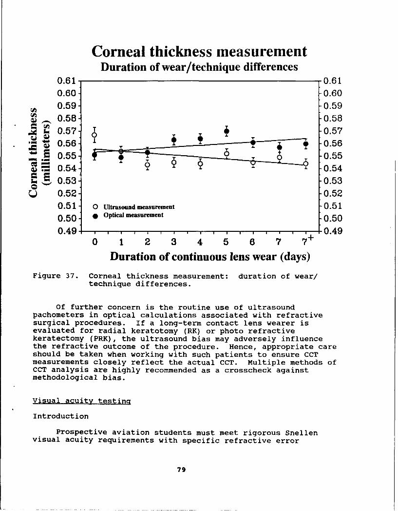
Corneal thickness measurementDuration of wear/technique differences
0.61 0.610.60 0.600.59 0.590.558 0.58
, 0.57'- T 0.570-S0.56 -- 0.56
0.54 9 0.550.54 A. A. -0.54
0.53- -0.53Q 0.52 0.52
0.51 0[ Ultrasound measurement -0.510.50 Optical measurement 0.50
0.49 , , , , , , , , , , , , , , , 0.490 1 2 3 4 5 6 7 7+
Duration of continuous lens wear (days)
Figure 37. Corneal thickness measurement: duration of wear/technique differences.
Of further concern is the routine use of ultrasoundpachometers in optical calculations associated with refractivesurgical procedures. If a long-term contact lens wearer isevaluated for radial keratotomy (RK) or photo refractivekeratectomy (PRK), the ultrasound bias may adversely influencethe refractive outcome of the procedure. Hence, appropriate careshould be taken when working with such patients to ensure CCTmeasurements closely reflect the actual CCT. Multiple methods ofCCT analysis are highly recommended as a crosscheck againstmethodological bias.
Visual acuity testing
Introduction
Prospective aviation students must meet rigorous Snellenvisual acuity requirements with specific refractive error
79
allowances. However, rated aviators have no refractive errorrestrictions as long as they are correctable to a visual acuityof 20/20 or better in each eye. The research protocol had toadhere to the Snellen 20/20 requirement or the volunteer subjectcould not be permitted to wear contact lenses. Beyond the mereadministrative requirement, good vision always has been animportant aviation safety issue. Therefore, safety of flightserved as a strong incentive to monitor visual acuity throughoutthe course of the study.
Over the many years since its inception, Snellen visualacuity has served as an excellent screening tool for appraisingvisual function. It is still the standard for clinical visualassessment. However, a number of investigators have sought todevelop a more sensitive means of assessing visual function(Ginsburg, 1984). Since the Snellen method uses high contrast,black letters against a white background, it is tappingessentially only suprathreshold function. The newer methods seekto monitor visual performance near threshold levels of function.Therefore, two different methods of visual acuity determinationwere used as a check against both gross and subtle changes invisual acuity associated with contact lens wear.
Methods and materials
A standard Snellen acuity projector chart was used to assessvisual acuity at the start and end of each examination. Inaddition, a low contrast (nominally 8 percent) printed acuitychart was used at the start and end of each exam, at the same 20-foot test distance as the Snellen chart, but at a dimmer level ofchart illuminance. Subjects were tested monocularly and asked toread the smallest line of letters they could see clearly andcomfortably. Subjects were given credit for an entire line ifthey could successfully identify at least four out of six letterspresented. On followup exams, entrance acuities were throughcontact lenses and exit acuities through the habitual spectaclecorrection. The reverse crder occurred on initial contact lensfitting exams.
Results and discussion
Statistical data analyses of entrance and exit acuities wereunrevealing. overall visual acuity through contact lenses werenot significantly different than through the habitual spectaclecorrection; this held internally for both Snellen and lowcontrast methods (p=0.67 and 0.52, respectively). These findingsare reassuring that contact lens removal and the cornealexamination procedures used in the protocol did not adverselyaffect the vision of test subjects. No instances, and nocomplaints, of postlens wear "spectacle blur" were recorded.Visual performance assessed by both acuity methods as a function
so
... .... .... ....

of type or brand of contact lens was also not statisticallysignificant (p=0.24). None of these statistical observationswere surprising, since these results represent subjects who weresuccessful at meeting the visual acuity restriction ofmaintaining 20/20 visual acuity.
A surprising finding, however, is the relationship betweenSnellen acuity and low contrast/low illuminance acuity.Frequency histograms of all acuity responses emphasize thedifference between the two tests (Figure 38; p<O.001). A broaderfrequency distribution exists for the low contrast/lowilluminance test, even though under Snellen conditions allsubjects are of 20/20 acuity or better. From the data, it can beseen that a pilot can possess good Snellen acuity, yet exhibitpoor low contrast/low illuminance acuity. Concurrently, it isequally probable that a pilot with good Snellen acuity canexhibit or possess superior low contrast/low illuminance acuity.Snellen acuity fails to differentiate between the two extremes ofthe normal distribution.
Matched acuity responsefrequency distribution
2400 Illmnance: 4236 measurements Illuminance:
"• 2200 106 liu for each acuity test 31 lux 2200
2000 238 subjects Conts 2000
S1800 Test distance: 8 percent 1800S1600 20 feet
S1400 1400
1200 11200
o1000 1000
S800 800
600 600
400 4002001 200
0 0•0W/a n/i mi/S UW a 30/ U 30/40 0/U a/U go/mo
Standard projected Low illumination/low contrastSnellen acuity Bailey-Lovie acuity
Figure 38. Matched acuity response frequency distribution.
82.
Plots of low contrast/low illuminance acuity as a functionof age, for both the spectacle and contact lens wearing con-ditions, demonstrate the absence of age-related, or spectacle/contact influences (Figure 39). Refractive astigmia (correctedwith spectacles; not entirely corrected with spherical softlenses) does not correlate with the low illuminance/low contrastresponses, either. On the other hand, the contact lens-wearingcondition correlates inversely in a marginally significantfashion (mean R = -0.12. Therefore, some of the low illumi-nance/low contrast acuity distribution spread is due to uncor-rected astigmatism in the contact lens condition. However, acorrelation of 0.12 means that less than 2 percent of theobserved effect or spread is due to refractive astigmatism.Correlations by each factor are in Table 2.
Operational studies have suggested that there are superior,average, and poor visual performers with regard to targetdetection, recognition, and identification. However, noscreening tests have been established and standardized that arecapable of consistently differentiating between the groups ofoperational visual performers. Without paired acuity andoperational testing, there is no proof that the extremes of thelow contrast/low illuminance distribution would correlate ormatch operational visual performance. However, the advantage ofbeing able to screen for superior visual performers prior toflight training does merit additional study.
Conclusions
Internal comparisons of visual acuity by either Snellen orlow contrast/low illuminance methods are not influenced by typeof contact lens worn, by the use of spectacles, by age, or byentrance and exit assessment. External comparisons of the twoacuity methods emphasize statistically significant differencesthat have potential for future use in identifying superior visualperformers. If this potential is realized, then standards forvisual assessment in military aviation, and the military ingeneral, will need to be changed.
Case report: endothelial Quttata
Introduction
One particular AH-64 subject, age 27, exhibited on theinitial exam an endothelial appearance indicative of physiologicstress (Figures 40 and 41). Even though the standard slit lampexam was negative, the specular microscope revealed bilateralcentral corneal "guttata," or dark areas scattered within the
82

Low illumination - low contrast acuityas a function of subject age
100- 1009 0. Contact lens-wearing condition N= 1307 90
%90,9
0080 .. 00 80
570 70"••" • ::60 * . ..... oooo oo . 60
•-- e 50 0 *e eoo eoooooooooo ooooe oo 50
WP 40 .. o ..... o40€• 30 °*°°l*ee* ••8 30
= 20 * 20
10 ... 1015 20 25 30 35 40 45 50
Age (years)
Low illumination - low contrast acuityas a function of subject age
100 10090. Spctade.-aeearg condition N 12319090
80 * *80 . 50
= 70 70
• 60 8 :, * 600 50 50
4 4040 * *******.*****.******.** *4
, 30 * 30SC.. 00 0 COOCO0 0 0
S20 20
10l .... I..0............... ....... 015 20 25 30 35 40 45 50
Age (years)
Figure 39. Low illuminance - low contrast acuity as a functionof subject age (contact lens and spectacle-wearingconditions).
83

Table 2.
Low illuminance/low contrast acuityAge vs acuity and correction
OD(contacts) OS(contacts) OD(spectacles) OS(spectacles)
Age 0.02 0.02 -0.04 -0.05 {R value645 662 634 647 (N
0.70 0.64 0.30 0.20 (p value
Cylinder-0.11 -0.13 0.03 0.01 (R value
152 152 152 152 (N0.08 0.06 0.69 0.82 (p value
endothelial mosaic. Guttata have been documented in associationwith a number of cornea-stressing conditions, and their presencesometimes can be contraindicative for contact lens wear.
Figure 40. Endothelial photomicrograph, right eye, initial exam.
$4
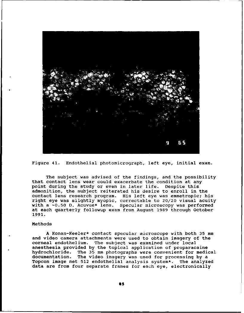
Figure 41. Endothelial photomicrograph, left eye, initial exam.
The subject was advised of the findings, and the possibilitythat contact lens wear could exacerbate the condition at anypoint during the study or even in later life. Despite thisadmonition, the subject reiterated his desire to enroll in thecontact lens research program. His left eye was emmetropic; hisright eye was slightly myopic, correctable to 20/20 visual acuitywith a -0.50 D. Acuvue* lens. Specular microscopy was performedat each quarterly followup exam from August 1989 through October1991.
Methods
A Konan-Keeler* contact specular microscope with both 35 mmand video camera attachments were used to obtain imagery of thecorneal endothelium. The subject was examined under localanesthesia provided by the topical application of proparacainehydrochloride. The 35 mm photographs were convenient for medicaldocumentation. The video imagery was used for processing by aTopcon image net 512 endothelial analysis system*. The analyzeddata are from four separate frames for each eye, electronically
65
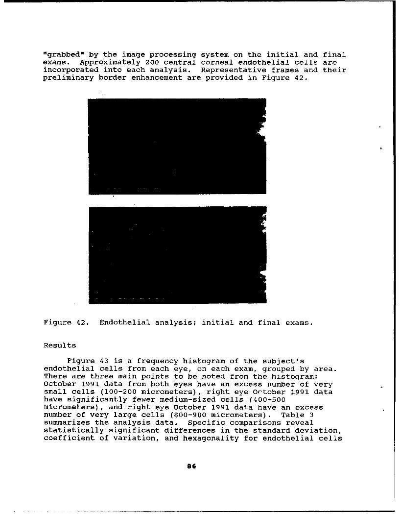
"grabbed" by the image processing system on the initial and finalexams. Approximately 200 central corneal endothelial cells areincorporated into each analysis. Representative frames and theirpreliminary border enhancement are provided in Figure 42.
Figure 42. Endothelial analysis; initial and final exams.
Results
Figure 43 is a frequency histogram of the subject'sendothelial cells from each eye, on each exam, grouped by area.There are three main points to be noted from the histogram:October 1991 data from both eyes have an excess iiumber of verysmall cells (100-200 micrometers), right eye October 1991 datahave significantly fewer medium-sized cells (400-500micrometers), and right eye October 1991 data have an excessnumber of very large cells (800-900 micrometers). Table 3summarizes the analysis data. Specific comparisons revealstatistically significant differences in the standard deviation,coefficient of variation, and hexagonality for endothelial cells
86

Endothelial cell size distribution50-, eM oD, Aug'89; N =204 cells
-CM OS, Aug '89; N = 207 cellsCM1 OD, Oct '91; N=201 cels
S40 1 OS, Oct '91; N=205 cells 40
g30 30
00o• 20 20
a., 10 10
100-200 800-400 500-00 700-4-- 00-00 600-700 80-90
Cell area groupings (square micrometers)
Figure 43. Endothelial cell size distribution.
of the right eye after over 2 years of soft contact lens wearingexperience. For all other factors there were no significantchanges, except for the smaller minimum cell size for both eyeson the final exam. Based on existing literature, all other datafall within the normal, expected ranges.
Table 3.
Endothelial analysis data
August 1989 October 1989OD OS OD OS
Minimum 235.7 247.9 193.2 193.1Maximum 716.6 722.4 864.5 726.5Mean 449.7 453.0 440.2 458.5SD 113.9 117.3 132.6 113.4CV 25.4 25.9 30.1 24.7Density 2223.7 2207.5 2272.0 2181.0Hexagonality 44.8 44.8 42.0 44.0
87
Discussion
With the nonlens-wearing eye serving as this subject'scontrol, these data verify that soft contact lens wear directlyaffects endothelial cell size variability as measured via contactspecular microscopy. Complicating this case is the presence ofcentral corneal guttata, which, in a contact lens wearer,sometimes can be indicative of lens-induced physiological stress.Theoretically, the presence of guttata prior to exposure to thestresses associated with contact lens wear would suggest theexistence of an ongoing degenerative process. This should makethe tissue more susceptible to the physiological load imposed bycontact lens wear. However, data in this case report demonstratethat other indicators of endothelial pathology (mean cell areaand cell density) do not significantly change over time, or as aresult of contact lens wear. Indirectly then, these data suggestan apparently stable abnormality in this subject.
It is conceivable that the presence of dark, nonreflectiveareas of the endothelium on specular microscopy may not be asensitive enough descriptor to differentiate basement membraneabnormalities from extra- and intracellular edema. Therefore,the physical presence of so-called endothelial guttata is notnecessarily indicative of acute physiological stress or disease.These data highlight the need to be able to clinicallydifferentiate between possible subsets of guttata in order toquickly identify those requiring immediate therapeutic attention.
Schirmer tear test
Introduction
Tear production was measured as a part of the overallphysiological assessment of contact lens wear. Since theresearch was segregated organizationally into original protocolsubjects and Desert Shield/Storm subjects, this aspect ofresearch was additionally separated by method of tear productionmeasurement. Original protocol subjects had the Schirmer teartest administered under topical anesthesia, while the DesertShield/Storm subjects had the test done without topicalanesthesia (there was concern about possible topical anestheticinstability in the desert heat).
Tear production measurement has always been difficult toquantify on a consistent basis. Variability can be induced by:inconstant filter paper capillary flow, and pore wettability;distinct individual irritation reflex tearing responses totopical anesthesia and/or from filter paper irritation withoutanesthesia; and varied tear surface tension, viscosity, and
88

evaporation characteristics. As a result, Schirmer tear testinghas been of greater descriptive and relative comparative valuethat as a quantitative standard.
Methods and materials
All participants were fitted with extended wear contactlenses that were used on a flexible wear disposable basis. Dur-ation of extended wear was individually governed by the subjectbased on physical comfort and/or visual acuity. Subjects rangedin age from 19 to 47. Because of combat exclusions, there wereonly two female subjects, so sex-based variation is not presentin these data. A modified Schirmer tear test was performed onboth initial and followup exams using Alcon* brand standardizedsterile strips (35 mm long and 5 mm wide).
Two subject groups were used. One consisted of AH-64"Apache" battalion and special operations aviation regiment air-crew fitted with contact lenses as part of a generalized researchprotocol. This group was administered the modified tear testunder topical anesthesia. The second group consisted of generalaviation aircrew fitted with contact lenses under the aegis ofthe research protocol in response to specific deployment needsrelated to Operation Desert Shield/Storm. These subjects wereadministered the modified tear test without the benefit ofanesthesia because of concerns for solation decompensation attemperatures much greater than 74*F. Since many members of bothgroups saw service in Southwest Asia, subject crossover onfollowup examination from 1 group to the other did occur.However, subject crossover could be seen as a stabilizinginfluence helping to minimize unpaired statistical analysiserror. Therefore, the data set overlaps were not truncated.
The modified Schirmer tear test varied from the acceptedmethod in tear strip placement. The intent of the test was todetermine the total amount of strip wetting possible by a sub-ject. Therefore, in order to maximize tear capture the stripswere placed just temporal to the lower lid punctum; this place-ment helped standardize consistent strip placement by providing aprominent physical reference point that also minimized cornealirritation.
Results
Mean tear strip wetting on all exams are recorded on Table4. The data are broken down by original protocol vs. DesertShield/Storm, and by type of contact lens worn. There werestatistically significant differences noted between both com-parisons. Figures 44 and 45 are percent distribution histogramsof tear strip wetting for both initial and followup exams (Figure44 without anesthetic, and Figure 45 with anesthetic). Test
89

strip wetting data were broken down in]9 equal sized 5-mm groups.The initial, anesthetized gruup consisted of 258 subjects given amean of 6 followup exams. The second, unanesthetized group con-sisted of 344 subjects given a mean of 3 followup exams. Statis-tical compari.on (t-test) revealed no significant difference be-tween initial and followup exams within each separate conditionand wetting grouping. However, the with and without anestheticsample comparison between the condition groups was statisticallysignificant (p<0.01).
Discussion
A recent publication suggested contact lens wearers mayproduce more tears than nonlens wearers (Occhipinti et al.,1988). However, the obvious stability of the contact lensfollowup data compared to the initial prelens-wear data,indicates that contact lens wear does not influence tearproduction over time. This contradicts the observation made byOcchipinti et al. However, there was a distinct difference notedbetween type of contact lenses worn. The mean tear productivity
Table 4.
Schirmer tear test.
Subject Mean wetting(mm) Lens type Mean wetting(mm)group +/- SD +/- SD
Original 58% water 13.28 +/- 1.56anesthetic 13.922 +/- 1.67 38% water 14.44 +/- 1.89protocol RGP 9.51 +/- 1.391(238)
No anesth. 20.852 +/- 1.72 58% water 21.34 +1- 1.98DS/S 38% water 19.45 +/- 2.19protocol RGP none fitted(344)1 p<0.001
2 p<0.0001
90

Schirmer tear test(without anesthetic)
100 100. Initial exams
90 M Followup exams 90
80 80
S70 70
60 60
S50 -50
. 40 40
S30 30
20 20
10 10
0. 00-5 6-10 11-15 1Q-20 21-25 26-30
Test strip wetting (mm)Figure 44. Schirmer tear test (without anesthetic).
Schirmer tear test(with anesthetic)
100 100100 Initial exams
90 - Followup exams 90
880 80
m 70 70*a6 60
PC 50 50
S40 40
30 30
20 20
*10' 10
00-5 6-10 11-15 16-20 21-25 26-30
Test strip wetting (mm)Figure 45. Schirmer tear test (with anesthetic).
91

of RGP lens wea- rs was significantly less than that of the softlens wearers. Rý,P fits were conceptually reserved for subjectswith a spherical equivalent of plano, and for high astigmats. Itis possible there was a bias toward dry eye subjects, as well;otherwise, it must be concluded that RGP lens wear is less tear-stimulating than soft lens wear.
From Table 4, we see mean strip wetting was significantlygreater for the without anesthetic condition or group. This isgraphically illustrated by the low percentage of subjects fallingwithin the 0-5 mm wetting block and the large percentage ofsubjects falling in the 26-30 mm wetting block in the"nonanesthetic condition" creating a skewed orientation acrossthe test strip wetting groupings (Figure 44). On the other hand,subject distribution in the "with anesthetic condition" is moreevenly distributed across the test strip wetting groupings(Figure 45).
These descriptive distinctions highlight the advantage oftopical anesthesia in test administration. Failure to use ananesthetic will bias the data toward higher values as a result ofreflex tear production. This observation is supported by notingthat the percentage exhibiting 0-5 mm wetting "with anesthetic"was over three times greater than "without anesthetic." Thisindicates the relative requirement for topical anesthesia inorder to identify or isolate a truly chronic dry eye. Inconclusion, the Schirmer tear test has clear categorical value incomparing specific groups and conditions.
Operations Desert Shield and Desert Storm
Introduction
In September 1990, the general aviation version of adevelopmental chemical protective mask was identified for earlyfielding in Southwest Asia without an available spectacleoutsert. The Army Surgeon General, in response to an AviationSystems Command (AVSCOM) request, granted a blanket waiver toexisting regulations so that all aircrew deploying to SouthwestAsia could be fitted with contact lenses. Therefore, allametropic aircrew (pilots, crewmembers, maintenance personnel,and medical support personnel) deploying to Southwest Asia wereexamined on a volunteer basis for possible contact lens wearunder the aegis of the ongoing Armywide contact lens researchprotocol. Specific data on aircraft, aircrew job distribution,and age are provided in Figures 46-49.
Eleven Army optometrists and 11 Army ophthalmic techniciansperformed the additional examinations at over a dozen U.S.locations and 3 locations in Europe. Four of the teamspermanently deployed to Saudi Arabia in direct support, and for
92

Primary aircraft distribution140 140
N=344
120 120
S100 100
• 80 80
Iwo 60 60
S40 40
20 20
0 0AH-i AH-4 CH-47 OH-68 OV-1 UH-1 UH-60
Type of aircraft
Figure 46. Primary aircraft distribution.
the duration, of Operation(s) Desert Shield/Storm. The originalArmywide Apache and Special Ops protocol included 238 subjects,while the Desert Storm portion (general aviation) added 344subjects. Approximately 450 of the 582 contact lens-wearingsubjects served in Southwest Asia on Operation(s) DesertShield/Storm.
Methods and materials
Every volunteer subject was given an initial, 24-hour, and1-week examination at the location of deployment mobilization.The basic exam included refraction and visual acuity, slit lampexamination, tear BUT assessment, and a Schirmer tear test(without topical anesthesia). Lens application and removaltraining were provided after accomplishment of a successful fit.Quarterly followup exams were conducted on-location in SaudiArabia. Twelve weeks of materials (48 lenses and 4 boxes of unitdose wetting solution) were issued at the 1-week exam and aftereach quarterly followup exam.
93

Aircrew job distribution200 200N=344180 180
160 160
S140 140S120 120
1•1oo 100
80 80
S60 60
40 40
20 20
0o 0Pilot Crew- Medical Maintenance
member personnel personnel
Figure 47. Aircrew job distribution.
The original, Armywide protocol used a three-tier contactlens fitting system, with the initial lens of choice being amoderate to high water content disposable extended wear softlens. Backup lenses consisted of a low water content standardextended wear soft lens utilized on a disposable basis, and arigid gas permeable (RGP) lens used with a chemical disinfectionsystem. Both types of soft lenses had analogous diameters andbase curves (14.0 mm and 8.8 mm, respectively). The RGP lenseswere not utilized on Operation Desert Storm because of concernsfor possible foreign body intrusion from blowing dust and dirt.Limited RGP use by Desert Storm participants from the originalArmy-wide protocol confirmed this concern.
The soft lens wearers were provided with a sterile,unpreserved, unit-dose wetting solution for use as needed.Because of the problems in achieving adequate hygiene in extremefield conditions, an extended wear regimen was selected overdaily wear. Since contaminated solutions and cases have beenimplicated as a possible source of infection, the elimination ofthe storage, cleaning, and disinfection processes was projectedto balance out the increased risk of infection by extended wear.
94

Aircrew age distribution25 25
N=344
20 20
15 15
10 10
'S 5 5
0 015 20 25 30 35 40 45 50
Figure 48. Aircrew age distribution.
Desert Shield/Storm subjects were advised to follow aconservative 3 day/2 night wearing schedule. The subjects wereinstructed that the night of lens removal was to be passedwithout any new lens wear; worn soft lenses were to be discardedand new lenses applied in the morning.
Results
There were 501 volunteer subjects; 344 subjects weresuccessfully fitted from the two soft contact lens types used inthe original protocol. The 157 unsuccessful attempts fell into 4general groupings (Figure 50). The refractive error distributionvaried from -5.50 D to +2.75 D, peaking at -1.00 D (Figure 51).Subjects ranged in age from 18 to 47. Despite the adverseenvironmental conditions, quarterly followup exam results did notvary in a clinically significant manner from those at the initialfittings (Figures 52-60). However, some statisticallysignificant differences were noted.
95
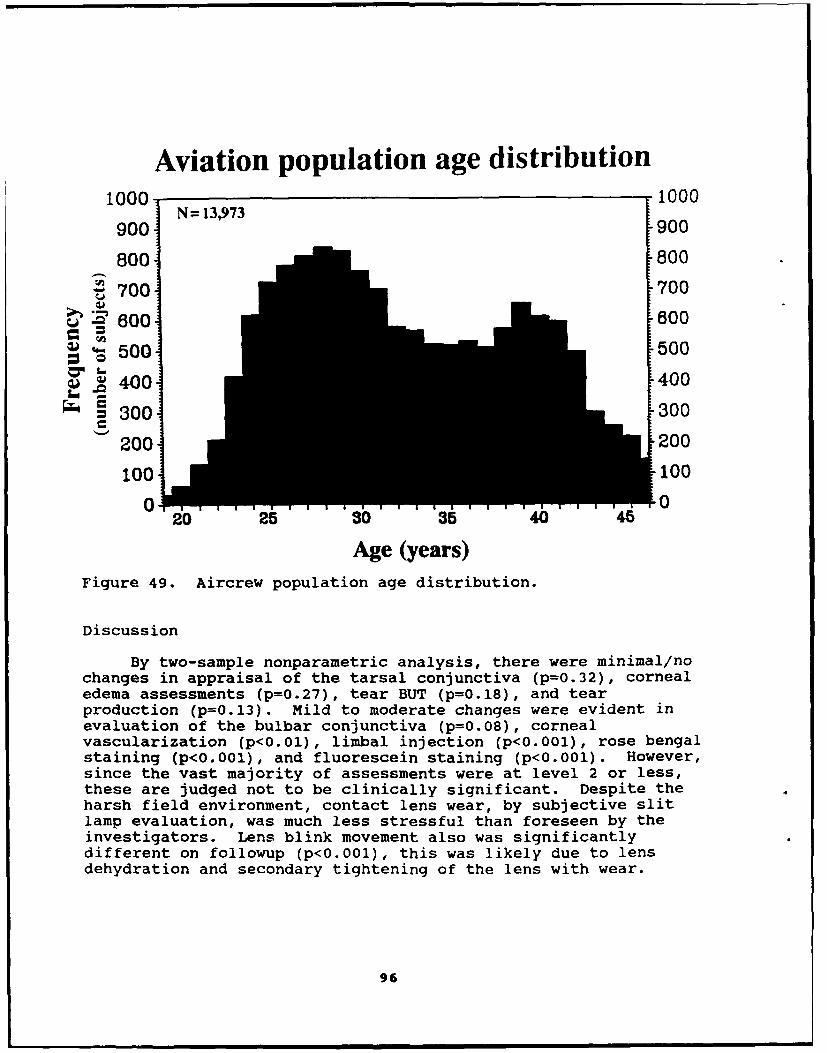
Aviation population age distribution1000 1000
900 N= 13,973 900
800 800
" 700 700
S800 600
S 500 500
S• 400 400
S300 300
2001 200
100 1000 00-2 10 11125 30 35 40 465
Age (years)Figure 49. Aircrew population age distribution.
Discussion
By two-sample nonparametric analysis, there were minimal/nochanges in appraisal of the tarsal conjunctiva (p=0.32), cornealedema assessments (p=0.27), tear BUT (p=0.18), and tearproduction (p=O.13). Mild to moderate changes were evident inevaluation of the bulbar conjunctiva (p=0.08), cornealvascularization (p<0.01), limbal injection (p<0.001), rose bengalstaining (p<0.001), and fluorescein staining (p<0.001). However,since the vast majority of assessments were at level 2 or less,these are judged not to be clinically significant. Despite theharsh field environment, contact lens wear, by subjective slitlamp evaluation, was much less stressful than foreseen by theinvestigators. Lens blink movement also was significantlydifferent on followup (p<0.001), this was likely due to lensdehydration and secondary tightening of the lens with wear.
96

Unsuccessful fit distribution100 100
N= 15790 90
80 80
S70 70
S60 60* __ 50 50
S40 40
= 30 30
20 20
10 10
0 0Medical Physical High Bifocalreasons poor fit astigmia dependent
Reasons for unsuccessful fitFigure 50. Unsuccessful fit distribution.
Individual assessments of contact lens wear by the subjectswas high in garrison, field, and combat conditions, as weresubjective performance assessments. Contact lenses were worn incombat by aircrew members serving on seven types Army aircraft.Combat missions included: attack, troop transport, equipmenttransport, surveillance, intelligence, and medical evacuation.The Apache radar interdiction mission into Iraq on 16 January1991 consisted of several contact lens wearers, including themission commander.
One case of ulcerative keratitis occurred during the courseof the deployment; there were no ulcers documented during thecombat phase of the operation. Three ulcers developed duringpreparations to redeploy back to home bases, or within 2 weeksafter return home. Of these four ulcer patients, one wasinitially enrolled in the original research protocol, and one wasenrolled in the operational protocol. Based on the 344 DesertShield/Storm subjects, plus the roughly i00* original AH-64protocol subjects that served in Southwest Asia, the annualized
97

Refractive error distributionS120 120
. 110 N=688 110
Coll 100 1000 90 90
S80 80S70 70
60 60~50 50
S40 40S30 30
20 20S10 10
0 0-5.00 -4.00 -3.00 -2.00 -1.00 0 +1.00 +2.00
Refractive power (diopters)
Figure 51. Refractive error distribution.
manifest risk for ulcerative keratitis over the 6-monthdeployment, was 4/222 lens wearers/year, or 18/1000/year. Itshould be remembered that three of the four ulcers occurred onredeployment activities; this was perhaps associated with theeuphoria over mission success and a lack of attention to lenshygiene issues. Furthermore, the combined risk of all studysubjects (all original AH-64 protocol subjects plus DesertShield/Storm subjects) from November 1988 through September 1991,equated to 1/112/year, or 8.93/1000/year. This combined riskfalls well within the wide range of risk estimates (2.1/1000/yearto 48/1000/year) proposed for nonaphakic extended soft lens wearin the civilian literature.
Conclusions
Based on the clinical evaluations, subjective feedback, andocular health risk appraisal data, contact lens wear by Armyaircrew is a viable alternative to spectacle wear. However,because of unique difficulties encountered by presbyopes, highastigmats, and those with extreme corneal curvatures (either veryflat or very steep), a sizeable portion of spectacle-wearing
98

Tarsal conjunctival irritation100 100
. -- Initial exams Severity extent90 = Followup exms 0 ........ None -90
I ........ Minimal80........ Mild 80
3 ........ ModerateS70 4. evere 70
"" 60 60
50o 50
S40 40
S30 30
Ow 20- 20
10t 100 0
0 1 2 3 4Grading
Figure 52. Tarsal conjunctival irritation.
aircrew will not be able to successfully wear contact lenses.Consequently, routine contact lens wear represents only a partialsolution to Army aviation's spectacle incompatibility problem.Therefore, developmental hardware alternatives must be includedin future system programming or many Army air crewmembers will beprevented from performing certain flight duties.
Assessment of subjective questionnaire data
Introduction
Verbal feedback from the vast majority of aviators wasexceedingly positive in nature. However, we were concerned thatvolunteers were reluctant to report adverse information on aface-to-face basis, so a questionnaire also was used in anattempt to gain information on the program. Specific interestwas not limited to performance aspects of lens wear, but alsoincluded a rating of the training program and clinicalprocedures.
99
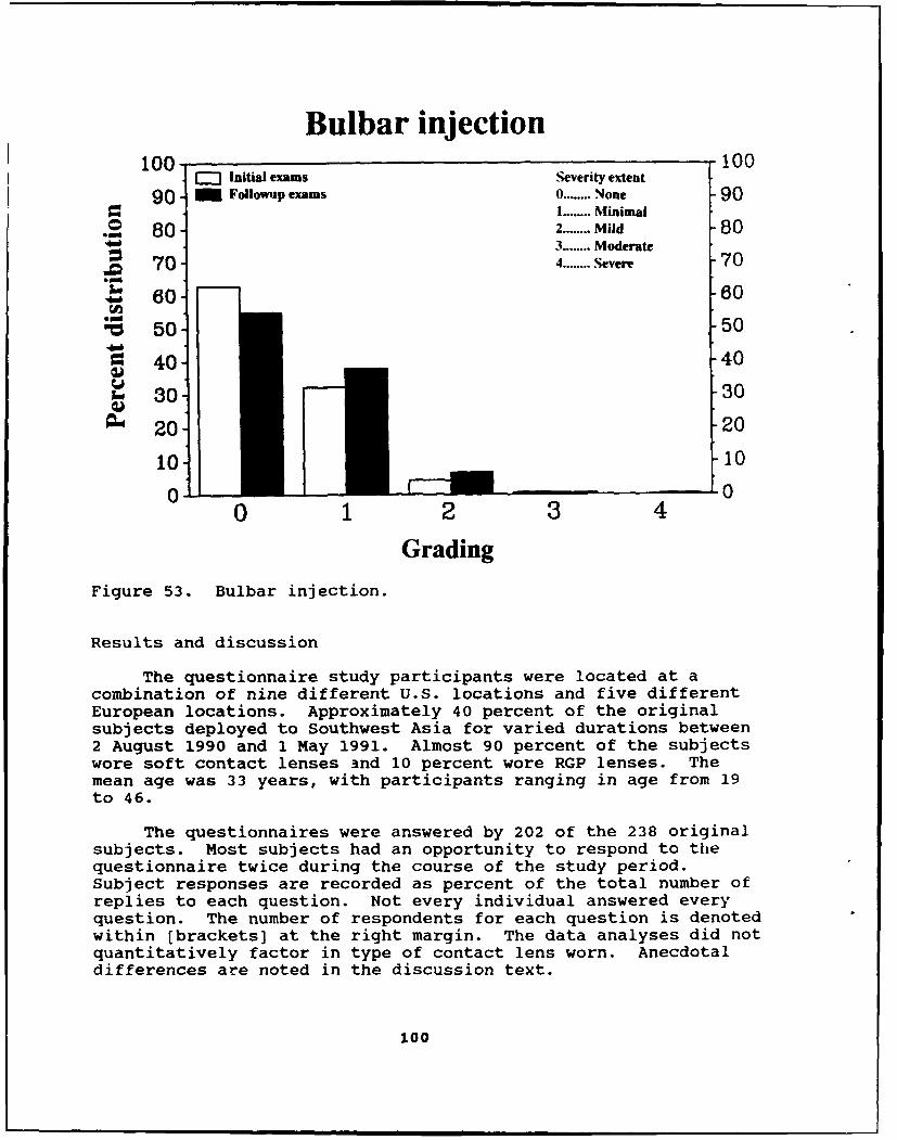
Bulbar injection100 r-" Initial exams Severity extent 10
90 m Followup exams 0 ........ None 90I ........ Minimal
S80 2 ........ Mild 80"A 3 ........ Moderate
70 4 ........ Severe 70
.m 60 60
"PC 50 50
40 40
30 30
S2 0 20
10 10
0 1 2 3 4
Grading
Figure 53. Bulbar injection.
Results and discussion
The questionnaire study participants were located at acombination of nine different U.S. locations and five differentEuropean locations. Approximately 40 percent of the originalsubjects deployed to Southwest Asia for varied durations between2 August 1990 and 1 May 1991. Almost 90 percent of the subjectswore soft contact lenses and 10 percent wore RGP lenses. Themean age was 33 years, with participants ranging in age from 19to 46.
The questionnaires were answered by 202 of the 238 originalsubjects. Most subjects had an opportunity to respond to thequestionnaire twice during the course of the study period.Subject responses are recorded as percent of the total number ofreplies to each question. Not every individual answered everyquestion. The number of respondents for each question is denotedwithin [brackets] at the right margin. The data analyses did notquantitatively factor in type of contact lens worn. Anecdotaldifferences are noted in the discussion text.
100
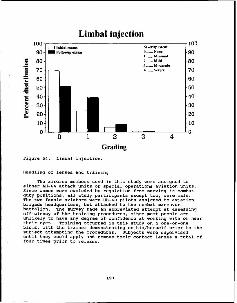
Limbal injection100- 100' Initial exams Seventy extent90 - Followup exams 0 ........ None 90
I ........ MinimalS80 2 ........ Mild 80
.N 3 ........ ModerateS70 4 ........ Severe 70
• 60 60
S50 50
40 40
30 30
W 20 "20
10_ -10
0 1 2 3 4
Grading
Figure 54. Limbal injection.
Handling of lenses and training
The aircrew members used in this study were assigned toeither AH-64 attack units or special operations aviation units.Since women were excluded by regulation from serving in combatduty positions, all study participants except two, were male.The two female aviators were UH-60 pilots assigned to aviationbrigade headquarters, but attached to the combat maneuverbattalion. The survey made an abbreviated attempt at assessingefficiency of the training procedures, since most people areunlikely to have any degree of confidence at working with or neartheir eyes. Training occurred in this study on a one-on-onebasis, with the trainer demonstrating on his/herself prior to thesubject attempting the procedures. Subjects were superviseduntil they could apply and remove their contact lenses a total offour times prior to release.
101

Corneal edema100 =3- Initial exams Severity extent 100
90 - Followup exams 0 ...... None 901 ........ Minimal
80 2 ........ Mild 803-...... Moderate
70 4 ...... Severe 70
16 60 60
, 50 50
40 40
, 30 30
• 20 20
10 100 oil 0
0 1 2 3 4
Grading
Figure 55. Corneal edema.
102
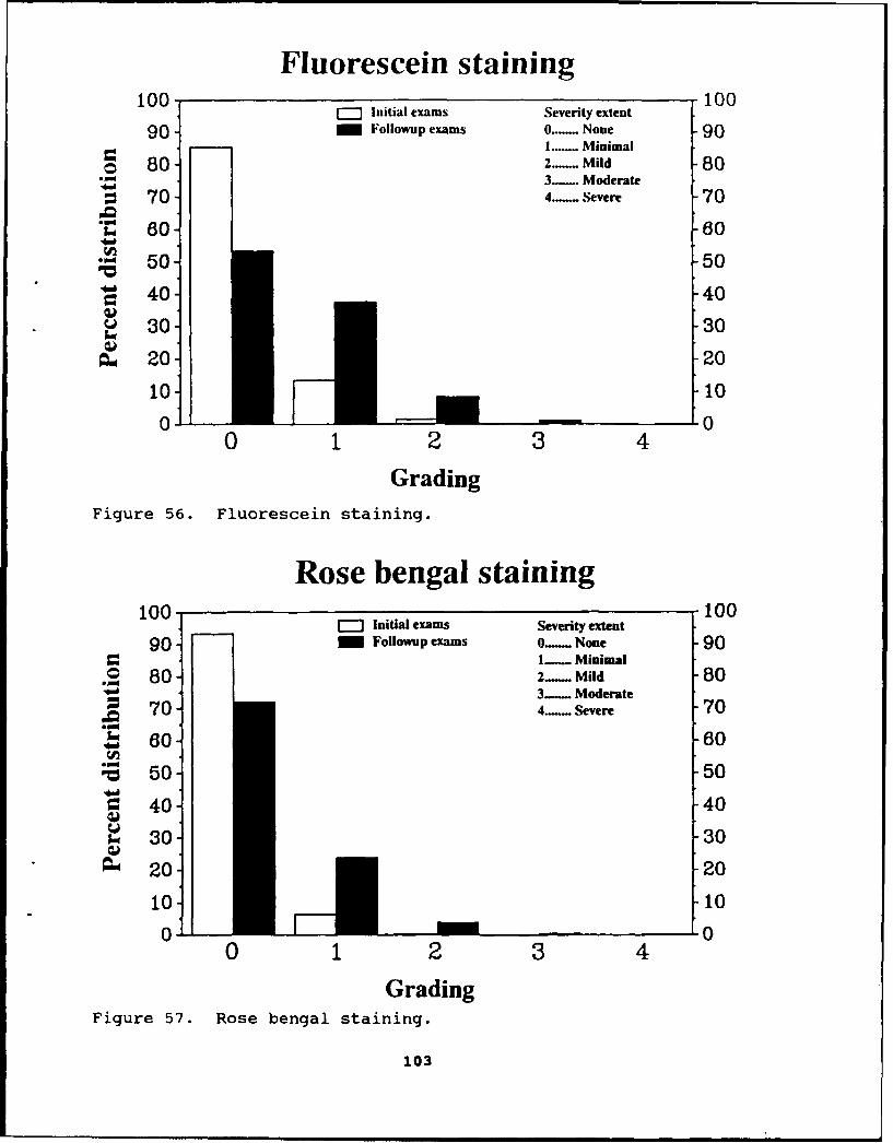
Fluorescein staining100 __1001-" Initial exams Severity extent
90 - Followup exams 0...... None 901 -....... Minimal
o 80 2..... Mild 80"3- Moderate
S70 4..-- Severe 70
• 60 60
"50 50w40"0 40 40
S30 30
S20 20
101 10lO il (- l
0 1 2 3 4Grading
Figure 56. Fluorescein staining.
Rose bengal staining100 r-l Initial exams Severity extent 100
90 - Followup exams O- None 901-. Minimal
* 80 2-. Mild 803-. Moderate
70 4.- Severe 70l, 60 60
S50 50
40o 40
S30 30
- 20 -20
10 _10
0.. 00 1 2 3 4
GradingFigure 57. Rose bengal staining.
103

Tear breakup time(with lenses)
100 100Initial exams
90- Followup exams 90
80 80
70 70
. 60 60
50 50
" 40- 40
S30 30
O, 20 -20
10 l100 •0
0-5 6-10 11-15 16-20 21-25 26-30
Time (seconds)Figure 58. Tear breakup time (with lenses).
Tear breakup time(without lenses)
100 100[-]7 Initial exams
90 / Followup exams 90
o 80 80
, 70 70
W~ 60- 60• -• 50 50
S40 40
S30-3
A 20 20
10 l10
0-5 6-10 11-15 16-20 21-25 26-30
Time (seconds)
Figure 59. Tear breakup time (without lenses).
104
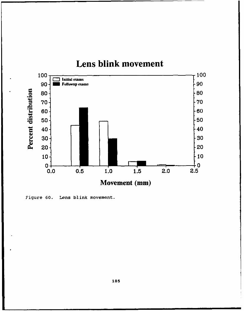
Lens blink movement100 100
100 .Initial exams
90 - Followup exams 90
o 80 80
S70 70
. 60 60
, 50- -50
40- 40
. 30 30
S201 20
10 10
0 t a 00.0 0.5 1.0 1.5 2.0 2.5
Movement (mm)
Figure 60. Lens blink movement.
105

Please rate your experiences in applying your lenses. (359]40% No problems what-so-ever.48% Minimal problems11% Minor problems
1% Moderate problems<1% Severe problems
Please rate your experiences in removing your lenses. [359]74% No problems what-so-ever.22% Minimal problems
3% Minor problems<1% Moderate problems<1% Severe problems
Looking back over the course of your contact lens wearingexperience, please evaluate the training program effectiveness inteaching you the following aspects: [357]
Excellent Good Fair Poor IneffectiveApplication 74% 25% 1% 0% 0%Removal 78% 22% 0% 0% 0%
The above results and their quantitative statisticalcomparison (chi square=47.323; p< 0.0001) suggest that lensremoval is an easier procedure to master than lens application.It should be noted that a high chi square value and a very smallprobability significance level (p) suggest a significantdifference between the two response sets. Although 12 percent ofrespondents felt lens application itself was fairly difficult,only 1 percent felt the shortcoming in lens application was dueto any kind of training deficiency. A similar, but smallerpattern exists for lens removal with 5 percent expressing minorto severe problems, while 0 percent implicated any kind oftraining deficiency causing lens removal difficulties. Indeed,the assessments of application and removal training are notstatistically significant (chi square=2.432; p=0.296). It isimportant to the success of any future clinical program thatthese established levels of training be met or exceeded.
Comfort/vision
In this section we wished to examine the perceived linkbetween contact lens comfort and subjective quality of vision.In civilian practice, if a clinician is not able to effect a
106

comfortable contact lens fit, then the final fitting success iscompromised no matter how clear the optical or visual result.The following questions were used:
In general, how comfortable are your contact lenses? [356]52% Very comfortable40% Comfortable
7% Neither comfortable nor uncomfortable<1% Uncomfortable
1% Very uncomfortable
How do you rate your vision with contact lenses as opposed toyour vision with glasses? (357]
51% Much better with contact lenses21% Slightly better with contact lenses17% No difference between contact lenses and glasses10% Slightly better with glasses
1% Much better with glasses
Responses claiming superior comfort exceeded the superiorquality of vision responses. This qualitative comparison ofvision between glasses and contact lenses is possibly anindicator of the limited contact lens choice available in thestudy. A secondary implication is that a comfortable fit is notenough to satisfy the occupational vision needs of Army aircrew.An increase in available lens types and parameters would likelyincrease the long-term wear success rate.
Environmental conditions
There was some concern that certain weather conditions orproblems with glare sensitivity would interfere with flightoperations. Teather issues were not necessarily a problem forApache pilots because their cockpits are enclosed and haveheating/air conditioning available. However, the specialoperations units fly with their aircraft doors off. Therefore,weather conditions would be potentially debilitating for them.
107
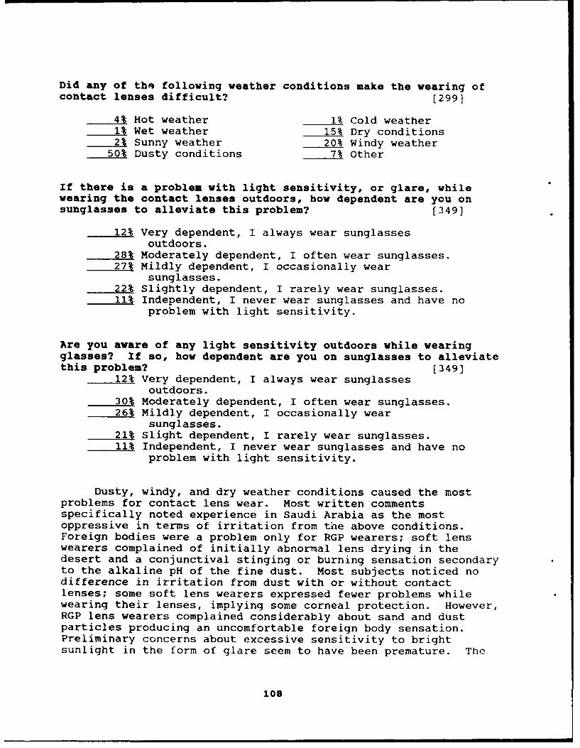
Did any of thq following weather conditions make the wearing ofcontact lenses difficult? [299]
41 Hot weather 1% Cold weather1% Wet weather 15% Dry conditions2% Sunny weather 20% Windy weather
50% Dusty conditions 7% Other
If there is a problem with light sensitivity, or glare, whilewearing the contact lenses outdoors, how dependent are you onsunglasses to alleviate this problem? [349]
12% Very dependent, I always wear sunglassesoutdoors.
28% Moderately dependent, I often wear sunglasses.27% Mildly dependent, I occasionally wear
sunglasses.22% Slightly dependent, I rarely wear sunglasses.11% Independent, I never wear sunglasses and have no
problem with light sensitivity.
Are you aware of any light sensitivity outdoors while wearingglasses? If so, how dependent are you on sunglasses to alleviatethis problem? [349]
12% Very dependent, I always wear sunglassesoutdoors.
30% Moderately dependent, I often wear sunglasses.26% Mildly dependent, I occasionally wear
sunglasses.21% Slight dependent, I rarely wear sunglasses.11% Independent, I never wear sunglasses and have no
problem with light sensitivity.
Dusty, windy, and dry weather conditions caused the mostproblems for contact lens wear. Most written commentsspecifically noted experience in Saudi Arabia as the mostoppressive in terms of irritation from the above conditions.Foreign bodies were a problem only for RGP wearers; soft lenswearers complained of initially abnormal lens drying in thedesert and a conjunctival stinging or burning sensation secondaryto the alkaline pH of the fine dust. Most subjects noticed nodifference in irritation from dust with or without contactlenses; some soft lens wearers expressed fewer problems whilewearing their lenses, implying some corneal protection. However,RGP lens wearers complained considerably about sand and dustparticles producing an uncomfortable foreign body sensation.Preliminary concerns about excessive sensitivity to brightsunlight in the form of glare seem to have been premature. The
108
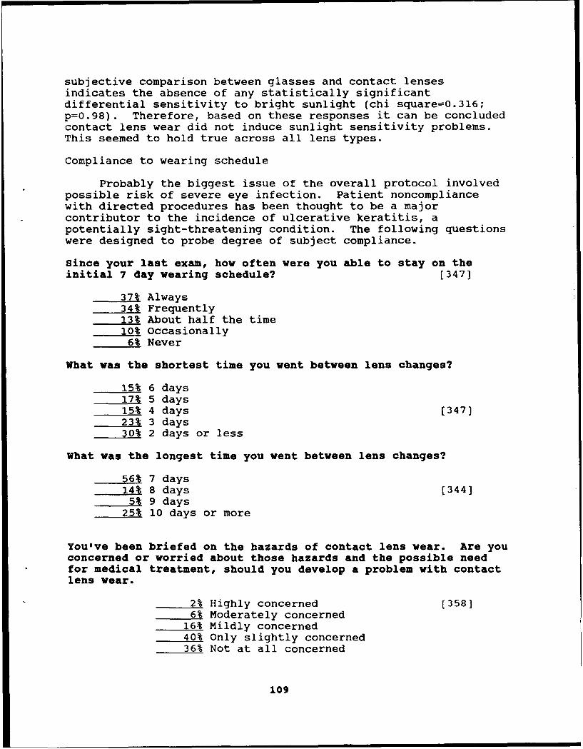
subjective comparison between glasses and contact lensesindicates the absence of any statistically significantdifferential sensitivity to bright sunlight (chi square=0.316;p=0.98). Therefore, based on these responses it can be concludedcontact lens wear did not induce sunlight sensitivity problems.This seemed to hold true across all lens types.
Compliance to wearing schedule
Probably the biggest issue of the overall protocol involvedpossible risk of severe eye infection. Patient noncompliancewith directed procedures has been thought to be a majorcontributor to the incidence of ulcerative keratitis, apotentially sight-threatening condition. The following questionswere designed to probe degree of subject compliance.
Since your last exam, how often were you able to stay on theinitial 7 day wearing schedule? (347]
37% Always34% Frequently13% About half the time10% Occasionally
6% Never
What was the shortest time you went between lens changes?
15% 6 days17% 5 days15% 4 days (347)23% 3 days30% 2 days or less
What was the longest time you went between lens changes?
56% 7 days14% 8 days (344]
5% 9 days25% 10 days or more
You've been briefed on the hazards of contact lens wear. Are youconcerned or worried about those hazards and the possible needfor medical treatment, should you develop a problem with contactlens wear.
2% Highly concerned [358]6% Moderately concerned
16% Mildly concerned40% Only slightly concerned36% Not at all concerned
109

Reported longest and shortest wearing durations wereanalyzed by the Kolmogorov-Smirnov (KS) method of distributionanalysis. A uniform distribution pattern was rejected (p=0.04,indicating the hypothesized distribution is not the correct one)and a normal distribution pattern centered around 7 dayscontinuous wear was accepted (p=0.99). There were both intra-and intersubject variations in typical wearing time duration.While the verbally reported mean wearing time was 4.5 days,approximately 1/4th of the subjects wore their lenses for aslittle as 2 days at one time or another (mostly RGP lenswearers), and the same fraction admit to having worn a pair oflenses 10 days or longer (soft lens wearers, exclusively). Thislatter figure is of some concern. Despite repeated cautions atquarterly exams about the dangers of overwear, a significantnumber of subjects exceeded the recommended limit at some pointin the study. This situation is exacerbated by the fact that 75percent of the subjects really aren't overly concerned about theocular health risks routine extended contact lens wear can pose.This was a statistically significant deviation from the expectednormal distribution around the mildly concerned response (KS;p=0.0016). This public health/preventive medicine issue must bestressed repeatedly in all routine clinical contact lens programsto ensure potentially avoidable infections can be prevented.
General physical lens wear
The general areas of lens comfort and clarity of vision wereprobed in greater detail with an emphasis on comparing flightactivities to garrison activities. The intent of the followingquestions was to see if operational conditions placed any greaterstress visually on the aviator.
Since your last exam, did you experience any of the followingproblems while flying? Check only those that apply. [358]
FrequencyNever Rarely Occasionally Often
Eye irritation 51% 43% 6% 0%Eye pain 65% 35% 0% 0%Blurred vision 52% 47% <1% <1%Dry eye 41% 43% 15% 1%Light sensitive 60% 32% 7% 1%
If any of the above occurred, how bothersome was it?Severity
Minor Moderate SevereEye irritation 91% 9% 0%Eye pain 98% 2% 0%Blurred vision 93% 7% 0%Dry eye 90% 9% 1%Light sensitive 93% 7% 0%
110

Since your last exam, did you experience any of the followingproblems in garrison activities? Check only those that apply.
Frequency [358)Never Rarely Occasionally Often
Eye irritation 57% 34% 9% 0%Eye pain 68% 30% 2% 0%Blurred vision 53% 40% 7% <1%Dry eye 47% 38% 15% <1%Light sensitive 58% 34% 8% <1%
If any of the above occurred, how bothersome was it?Severity
Minor Moderate SevereEye irritation 83% 15% 2%Eye pain 93% 2% 5%Blurred vision 91% 9% 0%Dry eye 92% 8% 0%Light sensitive 91% 9% 0%
Although garrison activities yielded slightly higher ratingsof physical discomfort and/or visual complaints than flightactivities, there was not a statistically significant difference.Severity comparisons of flight vs. garrison activities were alsonot significantly different, reinforcing results of the frequencyquery. Clearly, in-flight activities do not put a greater stresson contact lens wear. A conscious or unconscious bias could haveslanted responses away from flight-related complaints. However,one subject, when briefed on the questionnaire results, commentedthat while flying he is totally unaware of many minor irritationsthat normally get his attention on the ground. Based onsubsequent researcher flight experience, we are inclined to agreewith the latter explanation.
Night vision goggles
Since the special operations units had considerableexperience flying under night vision goggles, we attempted toassess visual performance with glasses vs. contact lenses. Thenumber of respondents to this question was restricted to aircrewexperienced in flight under NVGs.
ill

Please evaluate night vision goggle (NVG) operations whilewearing contact lenses. [121]
78% Much greater readiness and effectiveness withcontact lenses
21% Somewhat greater readiness and effectiveness withcontact lenses
1% No difference between contact lenses and glasses0 Somewhat greater readiness and effectiveness with
glasses0 Much greater readiness and effectiveness with
glasses
Have you noticed any of the following during NVG flight, andunder what visual correction conditions? [126)
With spectacles Observation With contact lensesNo Yes No Yes69% 31% Halos 92% 8%47% 53% Reflections 99% <1%54% 46% Glare 96% 4%54% 46% Decreased field of view 100% 0%
100% 0% Altered color sensitivity 99% <1%
Since 99 percent of the respondents felt greater readinessand effectiveness on NVG operations while wearing contact lenses,it is probable that NVG flight in contact lenses is lessdistracting and thereby less hazardous than in spectacles. Only1 percent felt there was no difference between glasses andcontacts, and no one rated glasses over contacts. Furthermore,when specifically questioned about visual distractors, subjectsmore often noted problems with spectacles. The presence ofhalos, reflections, glare, and decreased field of viewrepresented significant problems for spectacle wear compared tocontact lens wear (chi square=22.72; p=0.00014). Althoughpossible spectacle-incompatibilities within general aviation werenot of immediate interest in this study, this comparisonhighlights the potential benefit of a routine clinical contactlens program for general aviation. Improved NVG flight safetymay be a secondary benefit of contact lens use.
M-43 protective mask
The Apache M-43 protective mask was specifically designedfor compatibility with the HDU portion of the Apache IHADSS.However, fielding of the mask has not been total, and relativelyfew Apache pilots have had much in-flight experience with themask. The following questions were developed for a grossassessment of the mask and the ability to wear contact lensesunder the mask.
112
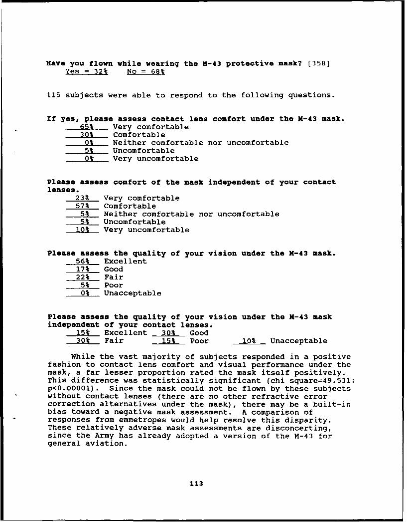
Have you flown while wearing the M-43 protective mask? [358]
Yes = 32% No = 68%
115 subjects were able to respond to the following questions.
If yes, please assess contact lens comfort under the M-43 mask.65% Very comfortable30% Comfortable
0% Neither comfortable nor uncomfortable5% Uncomfortable0% Very uncomfortable
Please assess comfort of the mask independent of your contactlenses.
23% Very comfortable57% Comfortable
5% Neither comfortable nor uncomfortable5% Uncomfortable
10% Very uncomfortable
Please assess the quality of your vision under the X-43 mask.56% Excellent17% Good22% Fair
5% Poor0% Unacceptable
Please assess the quality of your vision under the X-43 maskindependent of your contact lenses.
15% Excellent 30% Good30% Fair 15% Poor 10% Unacceptable
While the vast majority of subjects responded in a positivefashion to contact lens comfort and visual performance under themask, a far lesser proportion rated the mask itself positively.This difference was statistically significant (chi square=49.531;p<0.00001). Since the mask could not be flown by these subjectswithout contact lenses (there are no other refractive errorcorrection alternatives under the mask), there may be a built-inbias toward a negative mask assessment. A comparison ofresponses from emmetropes would help resolve this disparity.These relatively adverse mask assessments are disconcerting,since the Army has already adopted a version of the M-43 forgeneral aviation.
113

Safety of flight
A major investigative concern involved adequacy of flightsafety assessment. While an earlier portion of the questionnairelooked at possible distractions created by contact lens wear,this section sought to examine activity changes induced by lenswear. Baseline activity in the form of control transfer wasinitially documented, then contact lens-induced control transferwas assessed. Subjects were asked to perform a safety of flightassessment based on their experiences.
What was the typical flight duration during the past quarter?2.74 hours (mean)
Approximately how often, during a typical flight prior to yourparticipation in this study, did you have to hand over thecontrols because of an activity not directly related to themission (ie, adjust seat, stretch legs, adjust helmet)?
1.2 (mean) [358]
Approximately how often, during a typical flight during thisstudy, did you have to hand over the controls because of anactivity not directly related to the mission (ie, tend to contactlenses)? 1.1 (mean) [358]
Have you had to use the rewetting drops during flight? [358]Yes = 28% No = 72%
If yes, how often during a typical flight? [100]79% Rarely to never19% 1-2 times per flight
2% 3-5 times per flight0 6-8 times per flight0 >8 times per flight
Have you ever had to hand over the controls in order to tend toyour contact lenses? Yes = 13% No = 87% [359]If so, what activity was required? Add Rewettinc Drops
... and how often did this occur within the course of a typicalflight? 1.1 (mean)
Have you ever had to remove your contact lenses while in flight?Yes = 4% No = 96% [114]
114

How often has this occurred? 1
Please assess the impact these activities had on safety offlight 98% No impact [296]
2% Slight impact<1% Moderate impact
0% _ Severe impact
Have you had to use the rewetting drops during nonflightactivities? [358]
Yes = 91% No = 9%
If yes, how often? [326]37% Rarely to never49% 1-2 times a day13% 3-5 times a day
1% 6-8 times a day0% >8 times a day
Based on the response data, contact lens wear did notinterfere with flight operations and the transfer of the flightcontrols any more than any other "housekeeping activity" (i.e.,adjust seat, stretch legs, adjust helmet). The application ofwetting solution was the primary lens maintenance activityreported, although five subjects reported having to remove a lensin the cockpit at least once during the course of the study.While 28 percent reported using the wetting solution in thecockpit, only 13 percent reported having to transfer the controlsto tend to their lenses. These two responses agree, since it canbe assumed that during a typical flight any one pilot in a two-pilot aircraft would have the controls roughly half the time.The reported use of wetting solution was more closely associatedwith garrison activities. This could be accounted for because ofthe time disparity between a 2.74 hour flight and the balance ofthe duty day. However, bias and/or attention factors could beinfluencing this response. The final safety assessment wasoverwhelmingly positive despite acknowledgment of some requiredcontact lens-related activity in the cockpit.
Final assessments
One last set of questions was used to assess overallapproval of contact lenses and their influence on flying ability,readiness, and combat effectiveness. A final question attemptedto place the subject in a policy-making position concerning theroutine fitting of contact lenses for all aviators in the hopesof minimizing any blatant bias that may exist.
115
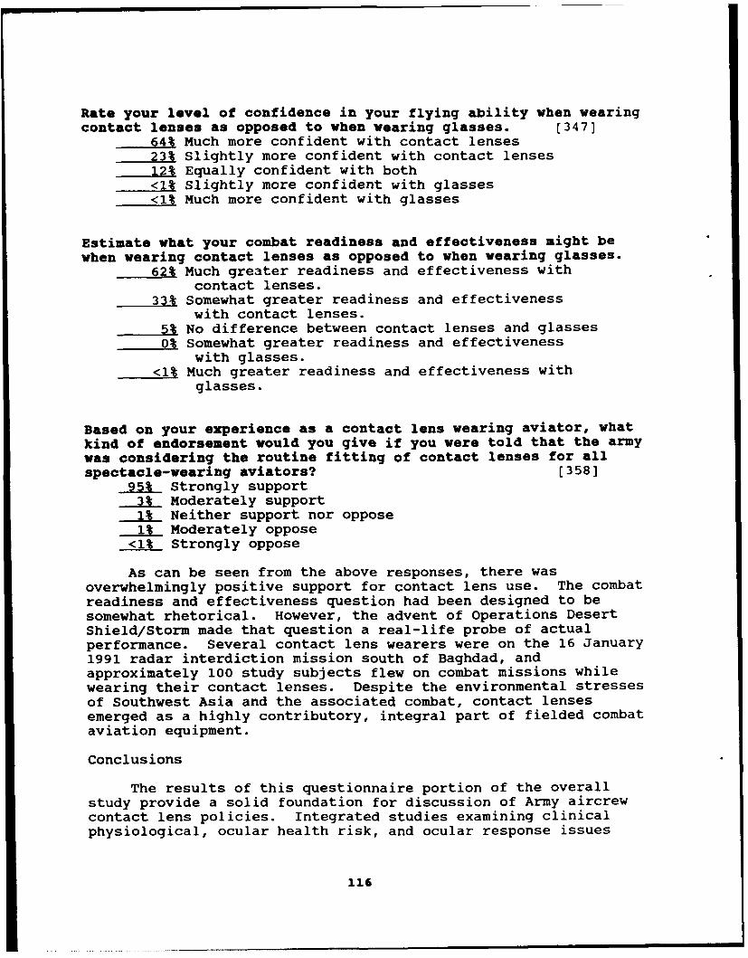
Rate your level of confidence in your flying ability when wearingcontact lenses as opposed to when wearing glasses. [347]
64% Much more confident with contact lenses23% Slightly more confident with contact lenses12% Equally confident with both<1% Slightly more confident with glasses<1% Much more confident with glasses
Estimate what your combat readiness and effectiveness might bewhen wearing contact lenses as opposed to when wearing glasses.
62% Much greater readiness and effectiveness withcontact lenses.
33% Somewhat greater readiness and effectivenesswith contact lenses.
5% No difference between contact lenses and glasses0% Somewhat greater readiness and effectiveness
with glasses.<1% Much greater readiness and effectiveness with
glasses.
Based on your experience as a contact lens wearing aviator, whatkind of endorsement would you give if you were told that the armywas considering the routine fitting of contact lenses for allspectacle-wearing aviators? [358]
95% Strongly support3% Moderately support1% Neither support nor oppose1% Moderately oppose
<1% Strongly oppose
As can be seen from the above responses, there wasoverwhelmingly positive support for contact lens use. The combatreadiness and effectiveness question had been designed to besomewhat rhetorical. However, the advent of Operations DesertShield/Storm made that question a real-life probe of actualperformance. Several contact lens wearers were on the 16 January1991 radar interdiction mission south of Baghdad, andapproximately 100 study subjects flew on combat missions whilewearing their contact lenses. Despite the environmental stressesof Southwest Asia and the associated combat, contact lensesemerged as a highly contributory, integral part of fielded combataviation equipment.
Conclusions
The results of this questionnaire portion of the overallstudy provide a solid foundation for discussion of Army aircrewcontact lens policies. Integrated studies examining clinicalphysiological, ocular health risk, and ocular response issues
116

will merge to form the comprehensive database necessary todelineate a final policy determination on contact lens use byArmy aircrew. Based on the subjective questionnaire responsespresented here, we conclude that contact lens wear is suitablefor routine Army aircrew use and widely accepted by studysubjects as a mission-essential means of overcoming hardware-spectacle compatibility problems.
Overall conclusions
Review of the pertinent issues follows the specific issuesequence established in the report.
a 1. The refractive error distribution in the overall study,compared to the refractive error distribution of all ratedaviators from the AEDR, reveals no significant difference betweenthe two groups (Lattimore and Schrimsher, 1992; in review). Asimilar statistical unity was established in this report betweenAH-64 subjects and Desert Shield/Storm subjects. Therefore,Figure 3 can be used by logistics personnel in the initialstockage of lens powers if a routine contact lens program isinitiated. Lack of fitting success was primarily due toexcessive astigmatism and presbyopia.
2. The subject age distribution matches the AEDR agedistribution. The bimodal characterization suggests periodicfluctuations in the prevalence of spectacle wear among aviatorsfor any 1 year. As a result, prevalence of spectacle wear shouldshow an upswing in the near future, but then fall off in 5 to 7years.
3. Mean corneal limbal vascular development slowly progressesin a linear fashion as a function of amount of time in the study.At the documented rate, exempting individual extremes, it wouldnot represent a significant clinical problem for the wear ofcontact lenses through a normal career.
4. Rose bengal and fluorescein staining suggest the practicallimits of extended soft contact lens wear to be 2 to 5 days,respectively.
5. Tear BUT assessments, using different types of fluorescein,are in fact documenting different aspects of tear film stability.Therefore, one can not substitute for the other. Both testsshould be done on the initial exam, and repeated on followupexams when there are subjective patient complaints expressed.
6. The threat of in-flight sudden incapacitation from contactlenses was shown not to exist. Although lens wearers wereinvolved in mishaps, lenses were judged to have been present, but
117
noncontributory to the mishap. The projected safety of flightissue did not develop.
7. There were minimal numbers of minor contact lens relatedcomplications. This was perhaps minimized by close followup, anddetailed subject instruction on how to best care for their eyesand lenses in order to avoid complications.
8. The ability of a soft lens to drape across the cornea isstrictly a reflection of the lenses base curve and diameter.Lens thickness and water content do not significantly affectfront lens surface curvature or ability to mask astigmatism. Asa result, any routine program should include an array of lensparameters in order to optimize physical lens fit for eachpatient.
9. Corneal aesthesiometry, although a valuable research tool,is not indicated for clinical use because present contact lensmaterials do not significantly affect corneal sensationthresholds.
10. Sterile, peripheral corneal infiltrates developed in sixsubjects. They did not present as acute cases, however. All sixwere detected on followup exam. This highlights the requirementto stress to prospective patients the need for immediateevaluation at the onset of symptoms if a routine program isinstituted. On the other hand, the medical support system shouldhave expert personnel collocated with participating units inorder to ensure ready accessibility. Peripheral, extended wear,soft contact lens-related ulcers are likely a localized responseto endogenous metabolic by-products that are coalesced orconcentrated at the periphery of the lens near the limbus, muchas carbon dioxide bubbles coalesce in the same location upondecreased ambient barometric pressure.
11. Bacterial ulcers represent a true threat to the vision ofcontact lens patients, particularly so in the case of soft,extended wear lenses. While the incidence of ulcerativekeratitis was not outside the ranges predicted by the civilianliterature, from a clinical perspective any ulcer is anunsatisfactory result. In order to minimize this potentiallycatastrophic complication, planned replacement, flexible wearparadigms (with a 3-day/2-night maximum wearing time) should bepursued in garrison. Wearing time during field operations shouldbe as brief as allowed by mission requirements.
12. Translens oxygen flux data indicate a relative loss ofoxygen flow after 5 days of extended wear. Beyond that time lessand less oxygen reaches the cornea. Paradoxically, the low watercontent, nonionic lenses provided better flux after 1 week ofwear than the high water content, ionic lenses. Therefore, a
118

metabolically maximized lens selection for extended wear beyond 3to 5 days continuous wear, if such a wearing time wereoperationally necessary, would be a nonionic, low water contentlens that has an absolutely minimum center thickness.
13. The water component pH of a soft lens takes 2 or more daysto fully stabilize to that of the precorneal tearfilm, and thisprocess does not vary with lens type or packaged pH. It ispossible that the transition period affects lens optics, and thatthe best possible visual performance may not be obtained untilthe lenses have been in place for a couple of days. Packaging ofthe lenses at a pH equivalent to that of human tears wouldcircumvent this process. Any routine lens selection programshould investigate solution pH of the various brands of lenses.
14. Water content measurements indicate a process of soft lenshabituation to the precorneal tearfilm environment. Both typesof soft lenses dehydrated over the first 3 or 4 days of wear; thehigher water content lenses dehydrated to a greater relativedegree on a normalized scale. This process did not vary bymeasurement technique or by environmental condition. Thisdehydration process, as well, might limit optimum visualperformance until stabilization is reached.
15. The linear correlation of water content as a function ofanterior lens surface pH is very high for both lenses. The lensperformance data provided by manufacturers often is based on thepackaged condition, not the in situ condition. This can bemisleading to a busy clinician, and leads to a recommendationthat actual in situ material performance data be made availableto practitioners routinely providing contact lens care.
16. Ultrasound pachometry, at the moment, is not appropriate forcentral corneal thickness assessments when there is a risk ofcorneal edema. Consistently, ultrasound measurementsunderestimated corneal thickness compared to optical thicknessmeasurements on corneas that have recently supported soft contactlenses. As a result, ultrasound pachometry is not recommendedfor contact lens practice.
17. Snellen visual acuity with high contrast letters is not anaccurate measure of visual performance. The use of low contrast,preprinted charts under low illuminance conditions is capable ofsegregating poor visual performers from adept visual performers.Results are not contaminated by astigmatism, or by age in therange from 19 to 46. This technique should be adopted as anadditional test for all soldiers requiring use of advancedvisionic and electro-optical systems.
18. The anatomic term 'endothelial guttata' likely includes avariety of conditions that are expressed by dark areas within the
119

endothelial matrix on specular microscopy. A clinical means ofidentifying each specific condition is therefore required but notcurrently available. In this case report, the subject was stablein the nonlens wearing eye and exhibited endothelialpolymegethism to the same degree as eyes without the presence ofguttata.
19. Contact lens wearers in this study did not produce moretears on followup as compared to their initial exams. However,as a group, RGP wearers produced fewer tears than soft lenswearers. Schirmer tear testing with a topical anesthetic hasgreater clinical usefulness than testing without an anesthetic.
20. Operations Desert Shield and Storm highlighted theoperational usefulness of soft contact lens wear; it alsorevealed foreign body problems associated with RGP lenses.Clinical assessments and subjective evaluations were verypositive in light of expectations associated with the severeenvironmental conditions encountered in Southwest Asia. Contactlens wear by Army aircrew has been combat tested and shown tohave a legitimate role in Army aviation.
21. Subjective questionnaire data simply confirm the aboveconclusions. The lenses were widely accepted by study subjectsas mission-essential equipment.
22. The final recommendation is that a routine soft contact lensprogram be instituted for Army aircrew. Planned replacement(spherical and toric) daily wear paradigms are recommended forgarrison use. Those same brands of lenses worn in garrisonshould be used on a flexible wear, periodic disposable basis inthe field, with a limitation on extended wear of 3 days/2 nights.In some instances, daily disposable wear may be indicated, andshould be accounted for in the budgetary process. Theserecommendations are made in order to minimize corneal stress andsecondary complications. Dedicated optometrists should beauthorized and assigned against Aviation Brigade Tables ofOrganization and Equipment to ensure immediate clinical supportis available in the field environment. Finally, the practicalityof bifocal contact lens use needs to be evaluated.
120

References
Abelson, M.B., Udell, I.J., and Weston, J.H. 1981. Normalhuman tear pH by direct measurement. Archives ofophthalmology. 99:301-304.
Alfonso, E., Mandelbaum, S., Fox, M., and Forster, R.1986. Ulcerative keratitis associated with contact lenswear. American Journal of ophthalmology. 101:429-33.
Allansmith, M.R., Baird, R.S., and Greiner, J.V. 1979.Vernal conjunctivitis and contact lens-associated giantpapillary conjunctivitis compared and contrasted.American journal of ophthalmology. 87:544-55.
Andres, S., Garcia, M.L., Espina, M.M., Valero, J., andValls, 0. 1988. Tear pH, air pollution, and contactlenses. American journal of optometry and physiologyoptics. 65:627-631.
B:chman, W.G. 1988. Extended-wear soft and rigid contactlenses: an operational evaluation among Army aviators.Fort Rucker, AL: USAARL Report No. 88-17.
Bachman, W.G., Leibrecht, B.C., Crosley, J.K., Price, D.R.,Bentley, G., and Leas, P. 1987. An operationalevwluation of extended-wear soft contact lenses in anarmored division. Fort Rucker, AL: USAARL Report No.87-12.
Barr, R.E., and Silver, I.A. 1973. Effects of cornealenvironment on oxygen tension in the anterior chamberof rabbit. Investigative ophthalmology and visualscience. 12:140-4.
Barr, R.E., and Schoessler, J.P. 1980. Corneal endothelialresponse to rigid contact lenses. American journal ofoptometry and physiology optics. 57:267-74.
Benjamin, W.J. 1986. Corneal oxygen, how much is enough?International contact lens clinic. 13:58.
Benjamin, W.J., and Hill, R.M. 1986. Corneal oxygendemand: the closed eye interval. Graefes archivesof ophthalmology. 224:291-4.
Bergmanson, J.P.G. 1989. Histopathological analysis of thecorneal epithelium after contact lens wear. Journal ofthe American optometric association. 32:125-9.
121
Bergmanson, J.P.G., and Chu, L.W.-F. 1982. Contact lensinduced corneal epithelial injury. American journalof optometry and physiology. 59:500-6.
Bergmanson, J.P.G., Ruben, C.M., and Chu, L.W.-F. 1985.Epithelial morphological response to soft hydrogelcontact lenses. British journal of ophthalmology.69:373-9.
Bernstein, I.H., and Brodrick, J. 1981. Contrastsensitivity through spectacles and soft contact lenses.American journal of optometry and physiology optics.58:309-313.
Braithwaite, M. 1983. The use of contact lenses by armyaircrew. Journal of the Royal Army Medical Corps(British). 129:43-5.
Brennan, D.H., and Girvin, J.K. 1985. The flightacceptability of soft contact lenses: an environmentaltrial. Aviation, space, and environmental medicine.56:43-8.
Burns, R.P., and Roberts, H. 1970. Effect of contactlenses on corneal metabolism. In: Symposium on OcularPharmacology and Therapeutics Transaction. New OrleansAcademy Ophthalmology. Mosby, St. Louis. 73-9.
Caldwell, D.R., Kastl, P.R., Dabezies, O.H., Miller, K.R.,and Hawk, T.J. 1982. The effect of long term hard lenswear on the corneal endothelium. Contact andinterocular lens medical journal. 8:87-92.
Carney, L.G., and Hill, R.M. 1976. Human tear pH.Archives of ophthalmology. 94:821-824.
Casebeer, J.C. 1973. Effects of air travel on contact lenswearers. American journal of ophthalmology. 75:165-6.
Chalupa, E., Swarbrick, H.A., Holden, B.A., and Sjostrand,J. 1987. Severe corneal infections associated withcontact lens wear. Ophthalmology. 94:17-22.
Chen, F.S., and Maurice, D.M. 1990. The pH in theprecorneal tearfilm and under a contact lens measuredwith a fluorescent probe. Experimental eye research.50:251-259.
Coles, W.H., and Jaros, P.A. 1984. Dynamics of ocularsurface pH. British journal of ophthalmology.68:549-552.
122
Corboy, J.M., and Tannehill, J.C. 1973. Effects of airtravel on contact lens wearers. American iournal ofophthalmology. 75:166-7.
Crosley, J.K., Braun, E.G., and Bailey, R.W. 1974. Soft(hydrophilic) contact lenses in U.S. Army aviation; aninvestigative study of the B&L "Soflens." Americanjournal of optometry and Physiology optics. 51:470-7.
Crosley, J.K., and Bachman, W.G. 1985. The use of extendedwear soft contact lenses in the aviation environment, astudy-specific protocol. Fort Rucker, AL: USAARL.
Cuklanz, H.D., and Hill, R.M. 1969. Oxygen requirementr, ofcontact lens systems. American Journal of optometry.46:662-5.
Dennis, R. 1988. Wear of soft contact lenses by tacticalaircrew. Considerations in Contact Lens Use UnderAdverse Conditions - a Symposium, 1988. Brooks AirForce Base, TX.
Department of the Army. 1986. preventive medicine.Washington, DC. Department of the Army. AR 40-5.
Department of the Army. 1976. Temporary flyingrestrictions due to exogenoug factors. Washington, DC.Department of the Army. AR 40-8.
Department of the Army. 1986. ophthalmic services.Washington, DC. Department of the Army. AR 40-63.
Department of the Army. 1987. Standards of medicalfitness. Washington, DC. Department of the Army.AR 40-501. Issue Number 1.
Dille, J.R., and Booze, C.F. 1980. The 1976 accidentexperience of civilian pilots with static physicaldefects. Aviation. space, and environmental medicine.51:182-4.
Dille, J.R., and Booze, C.F. 1982. The prevalence ofvisual deficiencies among 1979 general aviation accidentairmen. Aviation. space. and environmental medicine.53:179-82.
Draeger, J. 1981. Should pilots wear contact lenses?Southern iournal of optometry. August:26-7.
123

Dunn, F., Edmonds, P.D., and Fry, W.J. 1969. Absorptionand dispersion of ultrasound in biological media. In:Schwan, H.P., (ed.). Biological engineering.pp 205-332. New York: McGraw-Hill Book Co.
Efron, N., and Brennan, N. 1987. In search of the criticaloxygen requirement of the cornea. Contax. July 1987;5.
Efron, N., and Holden, B.A. 1986. A review of some commoncontact lens complications. Optician. 192:17-33.
Efron, N., Wohl, A., Toma, N.G., Jones, L.W.J., and Lowe, R.1991. Pseudomonas corneal ulcers associated with dailywear of disposable hydrogel contact lenses.International contact lens clinic. 18:46-9.
Eng, W.G., Rasco, J.L., and Marano, J.A. 1978. Lowatmospheric pressure effects of wearing soft contactlenses. Aviation, space, and environmental medicine.49:73-5.
Farris, R.L., Kubota, Z., and Mishima, S. 1971. Epithelialdecompensation with corneal contact lens wear. Archivesof ophthalmology. 85:651.
Fatt, I. 1969. Oxygen tension under a contact lens duringblinking. American journal of optometry. 46:654-9.
Fatt, I., and Hill, R.M. 1970. Oxygen tension under acontact lens during blinking - a comparison of theoryand experimental observation. American journal ofoptometry. 47:50-5.
Fatt, I., and St. Helen, R. 1971. Oxygen tension under anoxygen permeable contact lens. American journal ofoptometry and physiology optics. 48:545-8.
Fatt, I., and Linn, D. 1976. Oxygen tension under soft orhard gas-permeable contact lens in the presence of tearpumping. American journal of optometry and physiologyoptics. 53:104-8.
Fisch, B. 1990. Corneal ulcers and contact lens wear.Spectrum. May:48-57.
Fischer, F.H., and Wiederholt, M. 1982. Human precornealtearfilm pH measured by microelectrodes. Graefesarchives of ophthalmology. 218:168-170.
124

Flynn, W.J., Block, M.G., Provines, W.F., Tredici, T.J., andKullmann, R.D. 1985. Soft contact lens wear during G,acceleration. Brooks Air Force Base, TX: U.S. AirForce School of Aerospace Medicine TR-85-84.
Flynn, W.J., Miller, R.E., Tredici, T.J., Block, M.G.,Kirby, E.E., and Provines, W.F. 1987. Contact lenswear at altitude: subcontact lens bubble formation.Aviation. space, and environmental medicine. 58:1115-8.
Flynn, W.J., Miller, R.E., Tredici, T.J., and Block, M.G.1988. Soft contact lens wear at altitude: effects ofhypoxia. Aviation, space, and environmental medicine.59:44-8.
Forgie, R.E. 1981. Problems arising from the wearing ofhead equipment. In Personal Visual Aides for Aircrew.Lectures Series No. 115. 4.1-4.4. Advisory Group forAerospace Research and Development. Neuilly-sur-Seine,France.
Friend, J. 1983. Corneal metabolism and biochemistry. In:The cornea. Smolin, G., and Thoft, R.A. (eds.).Boston/Toronto: Little, Brown and Company.
Galentine, P.G., Cohen, E.J., Laibson, P.R., Adams, C.P.,Michaud, R., and Arentsen, J.J. 1984. Corneal ulcersassociated with contact lens wear. Archives ofophthalmoloQy. 102:891-4.
Gauvreau, H.K. 1976. Effects of wearing the Bausch andLomb "Soflens" while skydiving. American journal ofoptometry and physiology optics. 53:236-42.
Ginsburg, A.P. 1984. A new contrast sensitivity vision testchart. American journal of optometry & physiologicaloptics. 61:403-407.
Gordon, A., Boggess, E.A., and Molinari, J.F. 1990.Variability of ultrasonic pachometry. Optometry andvisual science. 67:162-7.
Grant, T., Kotow, M., and Holden, B.A. 1987. Hydrogelextended wear: current performance and future options.Contax. May:5.
Grey, C.P. 1986. Changes in contrast sensitivity duringthe first hour of soft lens wear. American journal ofoptometry and Physiology optics. 63:702-707.
125

Halliday, H.E. 1985. A feasibility study of the proposedmethodology in assessing the potential use of extendedwear contact lenses in a combat aviation environment.Small Business Innovation Research Program (SBIRP)Contract No. DAMDI7-85-C-5082. U.S. Department of Defense.
Hapnes, R. 1980. Soft contact lens worn at a simulatedaltitude of 118,000 feet. Archives of Ophthalmology.58:90-5.
Hirst, L.W., Auer, C., Cohn, J., Tseng, S.C., andKhodadoust, A.A. 1984. Specular microscopy of hardcontact lens wearers. Ophthalmology. 91:1105-11.
Holden, B.A., and Mertz, G.W. 1984. Critical oxygen levelsto avoid corneal edema for daily and extended wearcontact lenses. Investigative ophthalmology and visualscience. 25:1161-7.
Holden, B.A., Ross, R., and Jenkins, J. 1987. Hydrogelcontact lenses impede carbon dioxide efflux from thehuman cornea. Current eye research. 6:1283-1290.
Holden, B.A., Sweeney, D.F., and Sanderson, G. 1984. Theminimum precorneal oxygen tension to avoid cornealedema. Investigative ophthalmology and visual science.24:218-22.
Holden, B.A., Sweeney, D.F., and Seeger, R.G. 1986.Epithelial erosions caused by thin high water contentlenses. Clinical and experimental optometry. 69:103-7.
Holden, B.A., Sweeney, D.F., and Vannas, A. 1985. Effectsof long term extended contact lens wear on the humancornea. Investigative ophthalmology and visual science.26:1489-91.
Jagerman, L.S. 1973. Effects of air travel on contact lenswearers. American journal of ophthalmology. 75:533.
Kamiya, C. 1982. A study of corneal endothelial responseto contact lenses. Contact lens and intraocular lensmedical journal. 8:92-5.
Klyce, S.D. 1981. Stromal lactate accumulations canaccount for corneal oedema osmotically followingepithelial hypoxia in the rabbit. Journal ofphysiology. 321:49-64.
126

LaPiana, F.G. 1980. Army ophthalmology in Viet Nam.In: Combat ocular problems, proceedings conference.
-San Francisco: Letterman Army Institute of Research.
Lattimore, M.R. 1988a. The use of disposable extended wearsoft contact lenses in the Fort Rucker AH-64 aviationenvironment: a study-specific protocol. Fort Rucker,AL: U.S. Army Aeromedical Research Laboratory.
Lattimore, M.R. 1988b. Glucose concentration profiles ofnormal and ultraviolet radiation-exposed rabbit corneas.Experimental eye research. 47:699-704.
Lattimore, M.R., and Cornum, R. 1989. The use of extendedwear contact lenses in the AH-64 operationalenvironment: an Armywide study. Protocol. Fort Rucker, AL:U.S. Army Aeromedical Research Laboratory.
Lattimore, M.R. 1990. Contact lens anterior surface pH.International contact lens clinic. 17:228-231.
Lattimore, M.R. 1992. Contact lenses in the U.S. Armyattack helicopter environment: an interim report.Journal of the American optometric association. Inpress.
Lattimore, M.R., and Schrimsher, 1992. Refractive errordistribution and incidence among U.S. Army aviators.In review.
Lattimore, M.R., and Varr, W.F. 1991. Disposable soft lensulcerative keratitis in an Army aviator: a casereport. Aviation. space, and environmental medicine.62:888-9.
Mandell, R.B., and Farrell, R. 1980. Corneal swelling atlow atmospheric oxygen pressures. InvestiQativeophthalmologv and visual science. 19:697-700.
Markovits, A. 1988. Job demands in naval aviation.Considerations in contact lens use under adverseconditions - a symposium. Brooks Air Force Base, TX.
McCarey, B.E., and Wilson, L.A. 1982. pH, osmolarity, andtemperature effects on the water content of hydrogelcontact lenses. Contact intraocular lens medicaljournal. 8:158-167.
127
McNally, J.J., Chalmers, R.L., and Payor, R. 1987. Cornealepithelial disruption with extremely thin hydrogellenses. Clinical and experimental optometry.70:106-11.
Mishima, S., Gasset, A., Klyce, S.D., and Baum, J.L.1966. Determination of tear volume and tear flow.Investigative ophthalmology and visual science.5:264-71.
Molinari, J.F., and Socks, J.F. 1987. A comparativeevaluation between flexible and rigid contact lenswearers in hyperbaric environments. Annual meeting ofthe American Academy of Optometry. December.
Mondino, B.J., Weissman, B.A., Farb, M.D., and Pettit, T.H.1986. Corneal ulcers associated with daily wear andextended wear contact lenses. American journal ofophthalmology. 102:58-65.
Mondino, B.J. 1988. Inflammatory diseases of theperipheral cornea. Ophthalmology. 95:463-72.
Nilsson, K., and Rengstorff, R.F. 1979. Continuous wearingof Duragel contact lenses by Swedish Air Force pilots.American journal of optometry and physiology optics.56:356-8.
Nissen, J., Hjortdal, J.O., Ehlers, N., Frost-Larsen, K.,and Sorensen, T. 1991. A clinical comparison ofoptical and ultrasonic pachometry. Actaophthalmologica. 69:659-63.
Norn, M.S. 1988. Tear fluid pH in normals, contact lenswearers, and pathological cases. Acta ophthalmologica.66:485-489.
Occhipinti, J.R., Mosier, M.A., LaMotte, J., and Monji, G.T.1988. Fluorophotometric measurement of human tearturnover rate. Current eye research. 7:995-1000.
Omerod, L.D., and Smith, R.E. 1986. Contact lens-associated microbial keratitis. Archives ofophthalmology. 104:79-83.
Osborn, G.A., and Zantos, S.G. 1988. Corneal desiccationstaining with high water content lenses. CLAO journal.14:81-5.
128

O'Neal, M.R., and Poise, K.A. 1985. In-vivo assessment ofmechanisms controlling corneal hydration. Investigativeophthalmology and visual science. 26:849-53.
Patel, S., Farrell, J., and Bevan, R. 1989. Relationbetween precorneal tearfilm stability and tearproduction rate in normal eyes. Optometry andvisual science. 66:300-303.
Poggio, E.C., Glynn, R.J., Schein, O.D., Seddon, J.M.,Shannon, M.J., Scardino, V.A, and Kenyon, K.R. 1989.The incidence of ulcerative keratitis among users ofdaily wear and extended-wear soft contact lenses.New England journal of medicine. 321:779-83.
Polishuk, A., and Raz, D. 1975. Soft hydrophilic contactlenses in civil and military aviation. Aviation, space.and environmental medicine. 46:1188-90.
Polse, K.A., and Mandell, R.B. 1970. Critical oxygentension at the corneal surface. Archives ofophthalmology. 84:505-8.
Polse, K.A. 1979. Tear flow under hydrogel contact lenses.Investigative ophthalmology and visual science.18:409-13.
Rao, G.N. 1984. Complications due to daily wear of softcontact lenses. In: Contact lenses, the CLAO guide tobasic science and clinical Practice. 2nd ed. Boston:Little, Brown, and Company. 42.1-42.7.
Rao, G.N., Aquavella, J.V., Goldberg, S.H., and Berk, S.L.1984. Pseudophakic bullous keratopathy.Ophthalmology. 91:1135-40.
Rao, G.N., Shaw, E.L, Arthur, E.J., and Aquavella, J.V.1979. Endothelial cell morphology and cornealdeturgescence. Annals of ophthalmology. 11:885-9.
Refojo, M.F., Koch, J.M., and Leong, F.-L. 1989. Theeffect of lid closure on overnight corneal swelling withtwo types of silicone rubber lenses in rabbit eyes.ARVO Supplement. 30:480.
Rengstorff, R.H. 1965. Contact lenses and basic trainingin the U.S. Army. Military medicine. 130:419-21.
Rengstorff, R.H. 1972. Spectacles and contact lenses: Asurvey of military trainees. Military medicine.137 : 13-4.
129
Riley, M.V. 1969. Glucose and oxygen utilization by therabbit cornea. Experimental eye research. 8:193-6.
Rolando, M., Refojo, M.F., and Kenyon, K.R. 1983.Increased tear evaporation in eyes with kerato-conjunctivitis sicca. Archives of ophthalmology.101:557-558.
Rouwen, A.J.P., and Rosenbrand, R.M. 1986. High watercontent soft lenses used for flexible daily/extendedwear for military personnel. International evecare.2:435-40.
Salz, J..J, Azen, S.P., Bernstein, J., Caroline, P.,Villasenor, R.A., and Schanzlin, D.J. 1983. Evaluationand comparison of sources of variability in themeasurement of corneal thickness with ultrasonic andoptical pachymeters. Ophthalmic surgery. 14:750-4.
Sarver, M.D., Baggett, D.A., Harris, M.G., and Louie, K.1981. Corneal edema with hydrogel lenses and eyeclosure: effect of oxygen transmissibility. Americanjournal of ophthalmology and physiology optics.58:386-92.
Schein, O.D., Glynn, R.J., Poggio, E.C., Seddon, J.M.,Kenyon, K.R., and the microbial keratitis study group.1989. The relative risk of ulcerative keratitis amongusers of daily wear and extended-wear soft contactlenses. New England journal of medicine. 321:773-8.
Schoessler, J.P. 1983. Corneal endothelial polymegethismassociated with extended wear. International contactlens clinic. 10:148-51.
Schoessler, J.P., and Woloschak, M.J. 1981. Cornealendothelium in veteran PMMA contact lens wearers.International contact lens clinic. 8:19-22.
Schoessler, J.P., Barr, J.T., and Freson, D.R. 1984.Corneal endothelial observations of silicone elastomercontact lens wearers. International contact lensclinic. 11:337-40.
Schrimsher, R., and Lattimore, M.R. 1991. Prevalence ofspectacle wear among U.S. Army aviators. Optometry andvisual science. 68: 542-545.
Simon, D.R., and Bradley, M.E. 1980. Adverse effects ofcontact lens wear during decompression. Journal ofAmerican medical association. 244:1213-14.
130
Smelser, G.K., and Chen, O.K. 1955. Physiological changesinduced by contact lenses. Archives of ophthalmology.53:676-9.
Snyder, A. 1982. Corneal thickness and endothelial cellchanges associated with daily and extended contactlens wear. Master's thesis. Columbus, Ohio: Ohio StateUniversity.
Stefansson, E., Wolbarsht, M.L., and Landers, M.B. 1983.The corneal contact lens and aqueous humor hypoxia incats. Investigative ophthalmology and visual science.24:1052-4.
Stefansson, E., Foulkes, G.N., and Hamilton, R.C. 1987.The effect of corneal contact lenses on the oxygentension in the anterior chamber of the rabbit eye.Investigative ophthalmology and visual science.28:1716-9.
Stehr-Green, J.K., Bailey, T.M., Brandt, F.H., Carr, J.H.,Bond, W.W., and Visvesvara, G.S. 1987. Acanthamoebakeratitis in soft contact lens wearers. Journal of theAmerican medical association. 258:57-60.
Stehr-Green, J.K., Bailey, T.M., and Visvesvara, G.S. 1989.The epidemiology of Acanthamoeba keratitis in the UnitedStates. American journal of ophthalmology. 107:331-6.
Stocker, E.G., and Schoessler, J.P. 1985. Cornealendothelial polymegethism induced by PMMA contact lenswear. Investigative ophthalmology and visual science.26:857-63.
Sweeney, D.F. 1985. The clinical significance of cornealendothelial polymegethism. Investigative ophthalmologyand visual science. (Association for research in visionand optometry. Supplement). 26:53.
Sweeney, D.F., and Holden, B.A. 1987. Silicone elastomerlens wear induces less overnight corneal edema thansleep without lens wear. Current eye research.6:1391-4.
Thoft, R.A., and Friend, J. 1971. Corneal epithelialglucose utilization. Archives of ophthalmology.88:58-61.
Thoft, R.A., and Friend, J. 1975. Biochemical aspects ofcontact lens wear. American journal of ophthalmology.80:139-42.
131
Training and Doctrine Command Combined Arms Test Activity(TCATA). 1986. Controlled investigation of contactlenses and operational performances (CICLOPS). FortHood, TX: Test Report FT 484, RCS ATTE-3.
Tredici, T.J., and Flynn, W.J. 1987. The use of contactlenses by USAF aviators. Aviation, space, andenvironmental medicine. 58:438-43.
Van Norren, D. 1984. Contact lenses in the militaryservice. American Journal of optometry andphysiology optics. 61:441-7.
Wagner, L., Polse, K., and Mandell, R. 1980. Tear pumpingand edema with contact lenses. Investigativeophthalmology and visual science. 19:1397-1400.
Weinberg, R.J. 1977. Deep corneal vascularization causedby aphakic soft contact lens wear. American journal ofophthalmology. 83:121-124.
Weissman, B.A., Mondino, B.J., Pettit, T.H., and Hofbauer, J.D.1984. Corneal ulcers associated with extended-wear softcontact lenses. American journal of ophthalmology. 97(4):476-81.
Woo, G.C.S., and Hess, R.F. 1979. Contrast sensitivityfunction and soft contact lenses. Internationalcontact lens clinic. 6:171-176.
Zagrod, M.E., and Connor, C.G. 1988. Contact lens inducedcorneal endothelial polymegethism: functionalsignificance and possible mechanisms. American journalof optometry and physiology optics. 65:233-8.
Zantos, S.G., and Holden, B.A. 1977. Transient endothelialcell changes soon after wearing soft contact lenses.American journal of optometry and physiology optics.54:856-60.
Zantos, S.G. 1984. Corneal infiltrates, debris andmicrocysts. Journal of the American optometricassociation. 55:196-198.
132
Appendix A.
Initial examination
133

Initial contact lens exam
Name Rank
SSN Unit
Date/Time Age
Aircraft: AH-64, UH-60, OH-58.Job title: pilot, crew, FS.
1. Habitual R,:OD
OS
2. Visual acuity: OD(spectacle R,)
OS
3. Low contrast/ ODlow illuminanceacuity: OS(spectacle Rj)
4. Keratometry readings:
OD
OS
Auto-refractor readingsOD
OS
5. Tear osmolarity: OD mOsm.
OS mOsm.
134

6. Slit lamp examination:
Observation Classificationcode
I. Limbal injection
A. Severity
No injection ...................................... 0Minimal (within normal limits) ................... 1Mild ............................................. 2Moderate .......................................... 3Severe ............................................ 4
B. Location
Nasal quadrant only .............................. NTemporal quadrant only ........................... TInferior quadrant only ........................... ISuperior quadrant only ........................... STwo quadrants ..................................... X,XThree quadrants .................................. X,X,XCircumlimbal ...................................... C
II. Bulbar injection
A. Severity
No injection ...................................... 0Minimal .......................................... 1Mild ............................................. 2Moderate .......................................... 3Severe ............................................ 4
B. Location
Superficial vessels (diffuse) .................... ASuperficial vessels (localized) .................. BDeep vessels (diffuse) ........................... CDeep vessels (localized) ......................... DCombined involvement ............................. E
III. Ccrneal edema
A. Severity
No edema .......................................... 0Faint or minimal ................................. 1Mild ............................................. 2
135
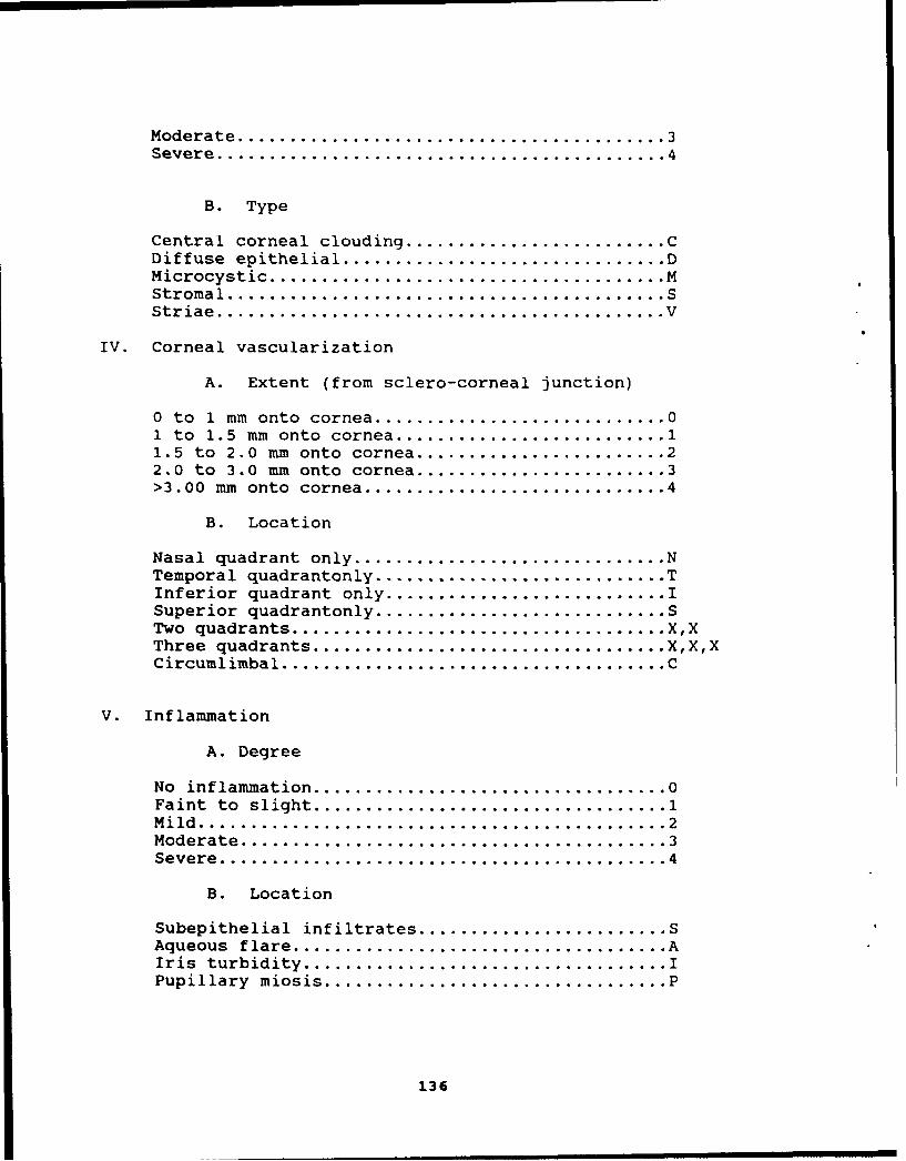
Moderate .......................................... 3Severe ............................................ 4
B. Type
Central corneal clouding ......................... CDiffuse epithelial ............................... DMicrocystic ...................................... MStromal ........................................... SStriae ............................................ V
IV. Corneal vascularization
A. Extent (from sclero-corneal junction)
0 to 1 mm onto cornea ............................ 01 to 1.5 mm onto cornea .......................... 11.5 to 2.0 mm onto cornea ........................ 22.0 to 3.0 mm onto cornea ........................ 3>3.00 mm onto cornea ............................. 4
B. Location
Nasal quadrant only .............................. NTemporal quadrantonly ............................ TInferior quadrant only ........................... ISuperior quadrantonly ............................ STwo quadrants .................................... X,XThree quadrants .................................. X,X,XCircumlimbal ..................................... C
V. Inflammation
A. Degree
No inflammation .................................. 0Faint to slight .................................. 1Mild .............................................. 2Moderate .......................................... 3Severe ............................................ 4
B. Location
Subepithelial infiltrates ........................ SAqueous flare .................................... AIris turbidity ................................... IPupillary miosis ................................. P
136

VI. Tarsal conjunctiva
A. Status
No involvement ................................... 0Faint to slight irritation ....................... 1Mild .............................................. 2Moderate .......................................... 3Severe ............................................ 4
B. Anomaly
Follicles........................................ FPapillae .......................................... PSimple injection ................................. S
VII. Rose bengal staining
A. Severity
No staining ....................................... 0Faint or minimal ................................. 1Mild .............................................. 2Moderate........................................... 3Severe ............................................ 4
B. Type
Bulbar conjunctiva only .......................... BCornea only ....................................... CCombined .......................................... Q
VIII. Fluorescein staining
A. Severity
No staining ....................................... 0Faint or minimal ................................. 1Mild .............................................. 2Moderate .......................................... 3Severe ............................................ 4
B. Type
Abrasion .......................................... AForeign body ..................................... F3:9 ... ........................................... NPunctate ......................................... PUlcer............................................. U
137

7. Tear BUT: OD sec. 8. Corneal OD(without lenses): sensitivity:
Os sec. OS
APPLY TOPICAL ANESTHETIC: Time
9. Schirmer tear OD mm.test (w/topical anesthesia)
OS mm.
10. Corneal OD mm. 11. Corneal OD mm.thickness (ultrasound): thickness (optical):
OS MM. OS mm.
12. Endothelial cell sizeOD
variability:
OS
WAIT 30 MINUTES FROM TIME OF ANESTHETIC INSTILLATION, THEN APPLYCL's
13. pH OD . 14. 02 uptake OD mm/sec.(with lenses) rate
OS_. OS mm/sec.
15. Tear BUT OD sec. 16. Lens blink OD mm.(with lenses): movement:
OS sec. OS mm.
17. Contact lens R,OD
power base curve diameter
manufacturer
OSpower base curve diameter
138

18. Number of lenses provided/used
19. Number of boxes of solutions used
20. Visual acuity: OD(contact lenses)
OS
21. Low contrast/ ODlow illuminanceacuity: OS(contact lenses)
22. Keratometry readings:OD(with contact lenses in place)
OS
Autorefractor: OD(with contact lenses in place)
OS
139
Appendix B.
Followup examination
140
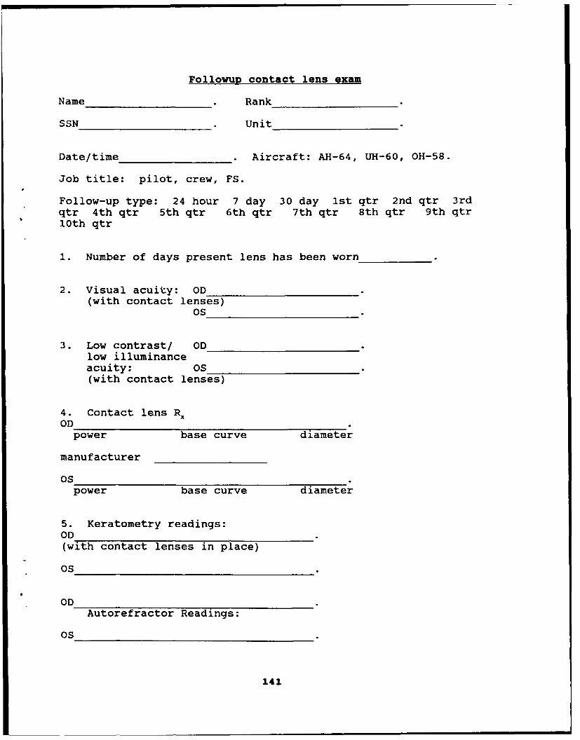
Pollowup contact lens exam
Name Rank
SSN Unit
Date/time Aircraft: AH-64, UH-60, OH-58.
Job title: pilot, crew, FS.
Follow-up type: 24 hour 7 day 30 day 1st qtr 2nd qtr 3rdqtr 4th qtr 5th qtr 6th qtr 7th qtr 8th qtr 9th qtr10th qtr
1. Number of days present lens has been worn
2. Visual acuity: OD(with contact lenses)
OS
3. Low contrast/ ODlow illuminanceacuity: OS(with contact lenses)
4. Contact lens RXOD
power base curve diameter
manufacturer
OSpower base curve diameter
5. Keratometry readings:OD(with contact lenses in place)
OS
ODAutorefractor Readings:
OS
141
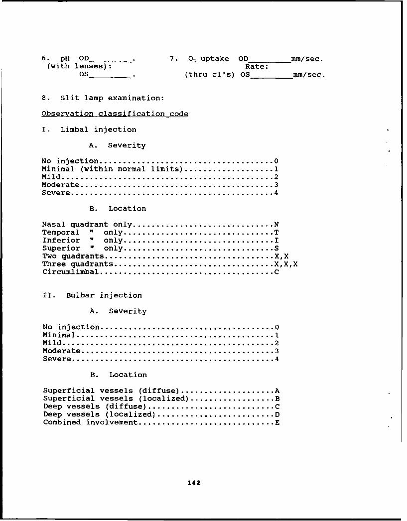
6. pH OD 7. 02 uptake OD mm/sec.(with lenses): Rate:
OS (thru cl's) OS mm/sec.
8. Slit lamp examination:
Observation classification code
I. Limbal injection
A. Severity
No injection ...................................... 0Minimal (within normal limits) ................... 1Mild .............................................. 2Moderate .......................................... 3Severe ............................................ 4
B. Location
Nasal quadrant only .............................. NTemporal " only ................................ TInferior " only ................................ ISuperior " only ................................ STwo quadrants ..................................... X,XThree quadrants .................................. X,X,XCircumlimbal ...................................... C
II. Bulbar injection
A. Severity
No injection ...................................... 0Minimal .......................................... 1Mild .............................................. 2Moderate .......................................... 3Severe ............................................ 4
B. Location
Superficial vessels (diffuse) .................... ASuperficial vessels (localized) .................. BDeep vessels (diffuse) ........................... CDeep vessels (localized) ......................... DCombined involvement ............................. E
142

III. Corneal edema
A. Severity
No edema .......................................... 0Faint or minimal ................................. 1Mild .............................................. 2Moderate .......................................... 3Severe ............................................ 4
B. Type
Central corneal clouding ......................... CDiffuse epithelial ............................... DMicrocystic ...................................... MStromal ........................................... SStriae ............................................ V
IV. Corneal vascularization
A. Extent (from sclero-corneal junction)
0 to 1 mm onto cornea ............................ 01 to 1.5 mm onto cornea .......................... 11.5 to 2.0 mm onto cornea ........................ 22.0 to 3.0 mm onto cornea ........................ 3>3.00 mm onto cornea ............................. 4
B. Location
Nasal quadrant only .............................. NTemporal " only ................................ TInferior " only ................................ ISuperior " only ................................ STwo quadrants .................................... X,XThree quadrants .................................. X,X,XCircumlimbal ...................................... C
V. Inflammation
A. Degree
No inflammation .................................. 0Faint to slight .................................. 1Mild .............................................. 2Moderate......................................... 3Severe............................ .............. 4
143

B. Location
Sub-epithelial infiltrates ....................... SAqueous flare .................................... AIris turbidity .................................... IPupillary miosis ................................. P
VI. Tarsal conjunctiva
A. Status
No involvement .................................... 0Faint to slight irritation ....................... 1Mild .............................................. 2Moderate .......................................... 3Severe ............................................ 4
B. AnomalyFollicles ......................................... F
Papillae .......................................... PSimple injection ................................. S
9. Tear OD mOsm.osmolarity:
OS mOsm.
10. Tear BUT OD sec. 11. Lens blink OD mm.(with lenses): movement:
OS sec. OS mm.
Remove old lenses
VII. Rose bengal staining
A. SeverityNo staining ....................................... 0Faint or minimal .................................. 1Mild .............................................. 2Moderate .......................................... 3Severe ............................................ 4
B. Type
Bulbar conjunctiva only .......................... BCornea only ....................................... CCombined .......................................... Q
144
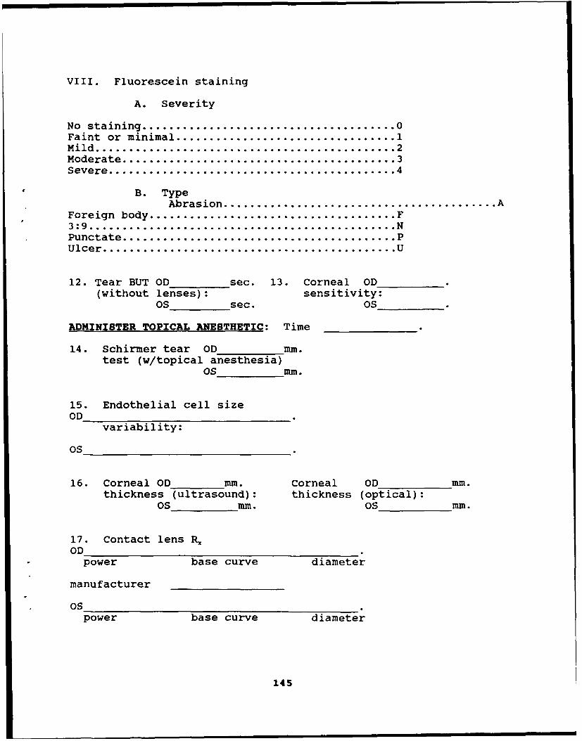
VIII. Fluorescein staining
A. Severity
No staining ....................................... 0Faint or minimal .................................. 1Mild ............................................. 2Moderate .......................................... 3Severe ........................................... 4
B. TypeAbrasion .......................................... A
Foreign body ...................................... F3:9 .............................................. NPunctate .......................................... PUlcer ............................................ U
12. Tear BUT OD sec. 13. Corneal OD(without lenses): sensitivity:
OS sec. OS
ADMINISTER TOPICAL ANESTHETIC: Time
14. Schirmer tear OD mm.test (w/topical anesthesia)
OS mm.
15. Endothelial cell sizeOD
variability:
OS
16. Corneal OD mm. Corneal OD mm.thickness (ultrasound): thickness (optical):
OS mm. OS mm.
17. Contact lens R,OD
power base curve diameter
manufacturer
OSpower base curve diameter
145
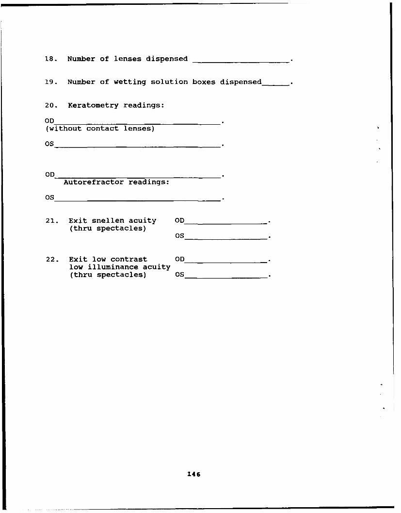
18. Number of lenses dispensed
19. Number of wetting solution boxes dispensed
20. Keratometry readings:
OD(without contact lenses)
OS
ODAutorefractor readings:
OS
21. Exit snellen acuity OD(thru spectacles)
OS
22. Exit low contrast ODlow illuminance acuity(thru spectacles) OS
146

Appendix C.
List of manufacturers
Allergan Inc.2525-T DuPont DriveIrvine, CA 92715
Marco1316 San Marco Blvd.Jacksonville, FL 32247
Johnson & Johnson (Acuvue)501 GeorgeNew Brunswick, NJ 08903
Konan Camera Research Institute, Inc.10-29 MiyanishichoNishinomiyashi Hyogo
Richmond Products (Aesthesiometer)1021 S. Rogers CircleSuite 6Boca Raton, FL 33487-2894
147

Appendix D.
Volunteer agreement affidavit
148

VOLUNTEER AGREEMENT AFFIDAVITFor useat il us forn. w.e ^A 70-25; the atooi4aw v sonyi OTSG
PRIVACY ACT OF 1974
AkMOrftry- t0 USC 3013. 4A USC 3101. and 10 USC 1071-1037
Princitet Purs~oaa: To daamun~en .eiuntar paitmoosaan in mae Cenwc.M lrvesteg~a~tin and Pw.aeacr Ptogram. SSN anid Mafw a.ljdus wow teauma Joe edetlrlmonatf nd Mw-ata puviNs-
AouIuno U"S: The SSN4 and !wna some idi ba uaee for adeindclawn and locauno pvwca.i fnlotrrnaesan darved liarn ma stud3y-11 be "and go oaturwnea the stust. i,,oewriesasion 01 fyte~ca provamgs adl.c"Imt fla clamun: amd lot the nwridawyr
teeoilarq at erwac~ai caretaxts "a tamred lby law. kintreagion may be Jurreer" 10 Fwdaral. Stale and ra~a aaein.*-
014cloaure: The lurrmuw,~t of yce SSN and homes addess a mandatory and necassary to prvead@j idntiltecon and to contact ywit tulAe im4Wnaman~ smaulat uws yme heall mayr be adversay atuleid. Failure to wxavee mae intotmatim maypreaue Vow, vountary pgttcipsataoin uat riveA VTigteog stuay.
PART A(1) - VOLUNTEER AFFIDAVIT
V0duntoWe Sublecm in Approved Oepenmens of UW Army Res~atch StUdbee
V01utitt WOW a11119 tsPrsOAM of AR 40-38 "n AR 70-25 are aurthonize an necs~siaay Med"a came for injuy or disease
*¶,ct 1s To6 Proxmna resAl of Vweu para1roahomn such studaOI.
r.avw*g luff C24uacty to c1ietW arld hawig anaaaned my _ ______bir.tday. do hereby voluwlter/9"v consent as lega
reoesentaaave fo to__ _ _ __ _ _ _ _ __ _ _ _ _ _ &parecmatm "The Use of
-Extended Wear Contact Lenses in the Aviation Environment: An Army Wide Study"
unciefrUOdietri~fon MAJ Morris Lattimore, OD. , Ph.D.
codcoa U.S. Army Aeromedical Research Laboratory
The impticabolst Of my vviwwajy parthopa m~nsir as WOga IinWWflauv duration and p 1,1: q of OW research study MW melthodsand means by wfuch it is 10 be coldiculad: and ame worvermence rld hazads Mu:t may reasonaWl be expectled have been Opiuedto me by
See next page
I have been grme an OPPWfUnat 1ý ask~ Questions oorcwranit Mie KWatvogatasna study. Any such Quesitons were answered to ay fullamo complete sabsiacwrn. Should any funrte questlow rse conem Wlamngy nghtLhe rignts 0f Uev person I represent On study-related injury. I may COnL=
Command Judge Advocate, Telephone (301) 663-2065 or AUTOVON 343-2065
-U.S. Army Medical Research & Development Command, Ft. Detrick, Frederick, MD 21702-5012(laNW.. Wdfeoo WWd PitfaO AWWRO Of #he..ia ekc^u A,". Cada
,..ndrwsunK "at I may at any itme during ame course of this study revoke mny consent and wlehrawehave te person, I mepresewtuw,mtrawfl from We swin~ witrioult turerw Penrlary or 'xs ca benefttM howeaver. Wie person I represent mray be reausted (mlelorayYoeunuwer or requestat: (CseJan volunteerl to unederg cenaa exailnwataio it, in Mre opinion of Ve attending Pflysioaf WC~fexainananons are .~aa for myrthe person I reoresients hearth amd weii-being. My/fte person I represent's refu"sao1 panhoipalewui invoove no Penalty at low o4 beniefits to elivan I ainrine pason I reprsent as Ortemsise enaed.
PART A (2) - ASSENT VOLUNTEER AFFIDAVIT (MINOR CHILD)
_________________________ SSN _ _________having fllf
ccntyto consent and having attained my __________bnrvtoay. do nfleec votuntwe for _________
_______________________________________to Partictoa ta ________n______
ýcVjer L-0 oirec:jon of
concut.azx. at
(Cofltile, on Roveai.,
DA FORM 5303-R, MAY 88 PRVOuS EmorNS APE OBSOLET
149
PART A(2) - ASSENT VOLUNTEER AFFIDAVIT (MINOR CHILD) lCont-4t
The orwianon of my voiu.nuay pabpamn ow nature. dufabOfl 6nd pups 01 twe research sUil e meod "OL mendfan. bV
wl d1h Kt is 0 be wcdaied; am~ 9w rcrewc1s and hazads Vw8 mazy reasorW~a~ be expecteld Kavit been exptaaned to mew by
-MAJ Morris Lattimore, MAJ Rhopda Cornum, or Dr. Kent Jensen
I have been qwan copomat lo ask questuios corcern* 9"s tNea*&WrW mtudy. Any sud¶ 4questuls werei wwlsrd to my kAand axrvte &=d Shot Ad y turUw queswn In~ cormng my n~hlt I may wxnt
Command Judge Advocate, Telephone (301) 663-2065 or AUTOVON 343-2065
at U.S. Army Medical Research & Development Command, Ft. Detrick, Frederick, MD 21702-PIONI. Addr"IL - ft w MGM^O Of PODO 0MU00J 4-4Co 5012
1 underUM4 M~al I may aft "any i dunng ow courm of VU3 study re~vkea my asseni ard mgthdraw from the stucy w~ol hattepenalty ortoss al bartaft txxwkwe. I may be retquesied io undergo CWUAR exarnweauon d. in toe op~o oft ow ananimg physaU1suc axarrftaa am vecemay W my h&aM and weI-biO. Wy rakusl to pancoweaza wwil ove nto penlty or loss dl branas 10wfo I arn o~mwwia I q.,
PART B TO BE COMPLETED BY INVESTIGATOR
P4TRL)CTK)NS FOR ELEAAEN" OF IPFORUED coNSNT: (Provede & dei&Od~ explauuanw mi acc~eo.venawih ApPO~jndj &. AR 40-38 ofAR 70-2n4
SEE ATTACHED INFORMED BRIEFING DOCUMENTS
FITTING OPTOMETRIST: ____________________
I doC] do not C] (check one & indied) consent to the incluSion of this form in my outpatient medicaltreatment record.
SrGNAT~lWE OF VICLUI.0lmDT INTR FLGLGAKA fv~m
PEFSAAN ~ ADOOPESS OF VMA#JTt7ER TYPED NAUE OF WTrN-SS
SX14ATU5E OF WITNESS DATE
REVERSE OF DA FORM 5303-R. MAY 8.8
150

VOLUNTEER INFORMED CONSENT BRIEFING: "The Use of Extended Wear Contact
Lenses in the AH-64 Aviation Environment: An Army-Wide Study"
Principal Investigator: MAJ Morris R. Lattimore, Jr.
INTRODUCTION
Volunteers are being sought for participation in a research studyinvolving the use of extended wear contact lenses. The purpose of the studyis to determine if ametropic AH-64 aircrewmembers can safely and effectivelyutilize contact lenses in lieu of their spectacles during flight operations.Volunteer contributions to this study may well help enhance the Army's combatreadiness, and will provide valuable information concerning the viability ofa possible system-wide endorsement of contact lens wear. Volunteers will beprovided with contact lenses to supplement current spectacles; the study isprojected to end in September, 1991. At that time, a final decision on theuse of contact lenses by certain aviation groups will have to be made.
POTENTIAL BENEFITS TO PARTICIPANTS
The only direct, tangible benefit to study participants is theopportunity to wear extended wear contact lenses on a no-cost trial basis.The decision whether to volunteer or not is strictly an individual one.There will be no adverse consequences if participation is declined, or iflater withdrawal from the study is desired.
QUESTIONNAIRES
Several questionnaires will need to be filled out during this study.These will ask for observations and perceptions of c ntact lens-weareffectiveness; they will also ask for some background information.Additionally, from time to time the unit flight surgeon will need to bebriefed on any observations you may have regarding operational aspects ofcontact lens wear.
151
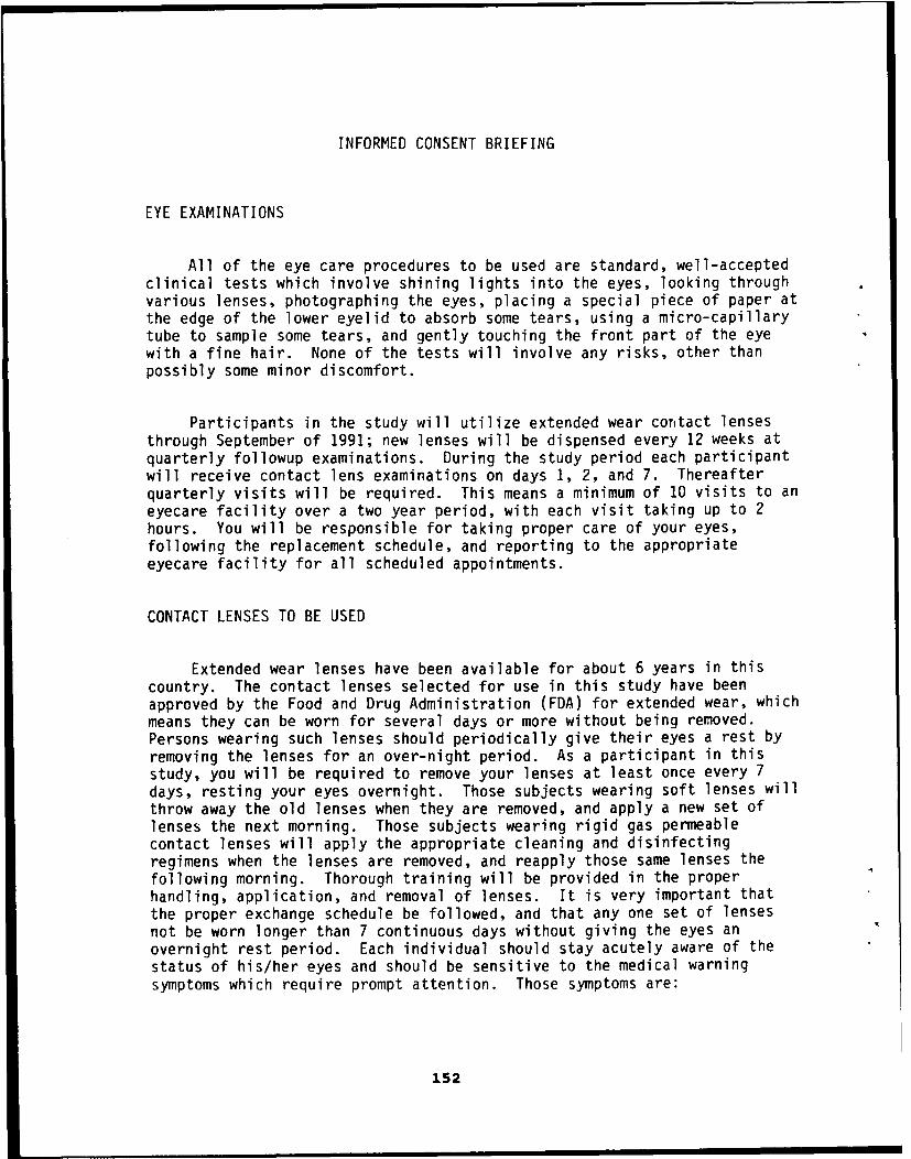
INFORMED CONSENT BRIEFING
EYE EXAMINATIONS
All of the eye care procedures to be used are standard, well-acceptedclinical tests which involve shining lights into the eyes, looking throughvarious lenses, photographing the eyes, placing a special piece of paper atthe edge of the lower eyelid to absorb some tears, using a micro-capillarytube to sample some tears, and gently touching the front part of the eyewith a fine hair. None of the tests will involve any risks, other thanpossibly some minor discomfort.
Participants in the study will utilize extended wear contact lensesthrough September of 1991; new lenses will be dispensed every 12 weeks atquarterly followup examinations. During the study period each participantwill receive contact lens examinations on days 1, 2, and 7. Thereafterquarterly visits will be required. This means a minimum of 10 visits to aneyecare facility over a two year period, with each visit taking up to 2hours. You will be responsible for taking proper care of your eyes,following the replacement schedule, and reporting to the appropriateeyecare facility for all scheduled appointments.
CONTACT LENSES TO BE USED
Extended wear lenses have been available for about 6 years in thiscountry. The contact lenses selected for use in this study have beenapproved by the Food and Drug Administration (FDA) for extended wear, whichmeans they can be worn for several days or more without being removed.Persons wearing such lenses should periodically give their eyes a rest byremoving the lenses for an over-night period. As a participant in thisstudy, you will be required to remove your lenses at least once every 7days, resting your eyes overnight. Those subjects wearing soft lenses willthrow away the old lenses when they are removed, and apply a new set oflenses the next morning. Those subjects wearing rigid gas permeablecontact lenses will apply the appropriate cleaning and disinfectingregimens when the lenses are removed, and reapply those same lenses thefollowing morning. Thorough training will be provided in the properhandling, application, and removal of lenses. It is very important thatthe proper exchange schedule be followed, and that any one set of lensesnot be worn longer than 7 continuous days without giving the eyes anovernight rest period. Each individual should stay acutely aware of thestatus of his/her eyes and should be sensitive to the medical warningsymptoms which require prompt attention. Those symptoms are:
152

INFORMED CONSENT BRIEFING
1. Noticeably blurred vision, other than of short duration.2. Eye irritation which is not eliminated by either comfort drops,
or the application of a new lens.3. Excessive or unusual redness of the eyes.4. Apparent infection of the eyes.5. Eye pain of any duration.6. Excessive tear flow.7. Swelling of eyelids or eye tissues.8. Mucous discharge around the eyes.9. Extreme light sensitivity of recent onset.
RISKS OF WEARING CONTACT LENSES
The only significant source of risk to participants in this studyis the actual wearing of the contact lenses in the performance of militaryand flight duties. The wearing of extended-wear contact lenses has beenassociated with the following effects:
1. Minor, temporary risks that are usually not serious and donot last very long.
a. Mild watering of the eyes.b. Mild sensitivity to light.c. Temporarily blurred vision.d. Slight redness of the eyes.e. Faint sensation of dryness of the eyes.f. Mild feeling of irritation to the eyes.g. Mild eye pain.h. Slight swelling of the cornea or eyelids.
2. Serious and possibly permanent risks.
a. Abnormal growth of blood vessels into the cornea.b. Scarring of the cornea.c. Subtle changes in the cornea which reduce vision.d. Eye infections, possibly leading to surgical
replacement of the cornea, or loss of an eye.e. Decreased corneal capacity to cope with
fluid build-up, which can lead to surgicalreplacement of the cornea, or loss of the eye.
SAFEGUARDS
This project has been approved by the Surgeon General of the U.S.Army. The study has been planned for maximum safety and will be closelymonitored by eyecare professionals. Many procedures have been built in toensure the safety of study participants. These safeguards include:
153

INFORMED CONSENT BRIEFING
1. Thorough eye examinations.2. Contact lenses will not be prescribed if deemed medically
unsuitable.3. Training will be provided in the safe use and care of the
lenses.4. Lenses will be replaced on a periodic basis.5. Contact lens wear will temporarily be suspended if
medically indicated.6. Participation will be discontinued if medically indicated.7. Medical facilities will be briefed on the project in case
of a medical emergency.
Based on preliminary studies, in-flight risks for a standardflight profile are minimal. However, as a safeguard, subjects will beinstructed that they can not fly with another contact lens wearing subject;this will also be documented on the formal waiver, which requires bothcommander and individual signature. Back-up spectacles must be carried atall times in case of lens-wearing difficulties.
HANDLING DATA
Other than the flight waiver, only information arising from seriousmedical incidents is to be placed in the individual's medical record. Upontermination or conclusion of the study an appropriate entry will be made inthe data record. All research data files will be kept in the strictestconfidence in accordance with regulations. Raw data forms and computerfiles will have limited access and will be used for research purposes only.No individual information will be released without expressed writtenconsent.
At the end of the study (30 September 1991), or at withdrawal fromthe study, all contact lens wear by the former subject will be totallydiscontinued, unless otherwise specified by separate documentation.
Research point of contact for this protocol is Major MorrisLattimore, AV 558-6807.
I have received a copy of this volunteer consent package.
SIGNATURE OF VOLUNTEER DATE SIGNED
DISINTERESTED WITNESS DATE SIGNED
154
Appendix E.
Volunteer registry data sheet
155
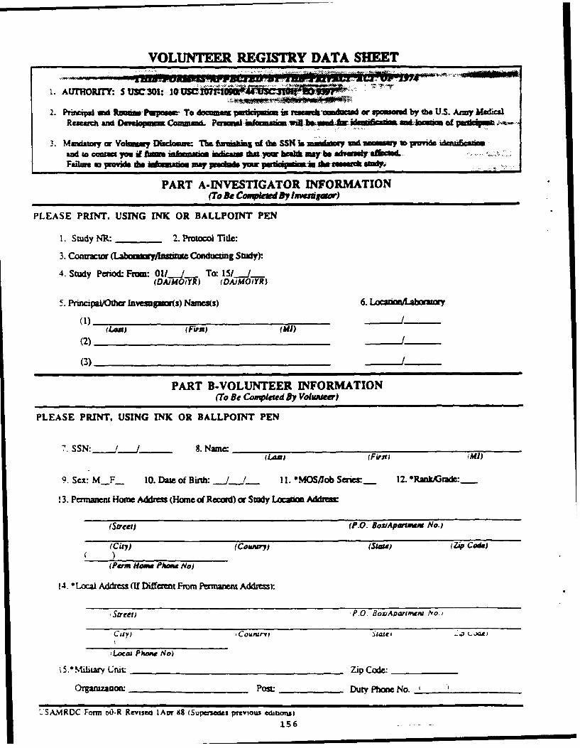
VOLUNTEER REGISTRY DATA SHEET
1. AU=ro ,: 5 USC 301: 10 0
2. Prbi** ida u Rzmtimm toposew To doarn p WI in -conducte&ad or sposoedt by the U.S. A~my MadicalResearh and Dm•.pm 3KCaam P OImmd,.of pwd ...-
3. Mwviawiy or Vabmmmy Dkcaxclmu The farminuhg of do SSN Is muandway xdamom w~ w~avide dulenaamud to contac rm df f0 inwasaim imica *4a yow besa may be advamey aefied.Faflw to provi doiok mmdm may inds ymc 91~ '1mu *sdY
PART A-INVESTIGATOR INFORMATION
(To Be Conwlu By lw.rgla,)
PLEASE PRINT. USING INK OR BALLPOINT PEN
1. Study NR: 2. Protocol Tide:
3. Coauzwto (Labommayjlliusat Conducting Study)Y
4. Study Period- From: 011_J To: 11__..J_(DAJMOIYR) (DAJMOIYR)
5. PrinapauiOh Invempmos) Names(s) 6.1
(Lanj f Firn)p((2)
(3)
PART B-VOLUNTEER INFORMATION(To Be Competed By Voluwnw)
PLEASE PRINT, USING INK OR BALLPOINT PEN
7. SSN:______. _ 8. Name: _ _ _ _ _ _ _ _ _ _ _ _
(LA• (FaraD •Mi)
9. Sex: MF_ 10. Date of Birth: ./__J / 11. "MOS/Job Saries- 12. "Rankrde:
13. Pernunent Home Addres (Home of Reconr) or Study l oc~abam AMdd
(Street) (P.O. BoziAparume No.)
(City) (Cowwry) (Stata (Zip Code)
(Perm Hoom Phone No)
14. "Local Address (If Different From Pemanenm Addres):
,Street) P.O. BoziAvariment jvo,
cuiy) i Counarvp Stafei D.• LJat)
iLocma Phone No)
i5." ýitarv Uruit Zip Code:
Orgamzanon: Post Duty Phone No. _
.'SAMRDC Form o0-R Revisea lAin 88 (Supetiefs pT•vious ecabonsi156 . .
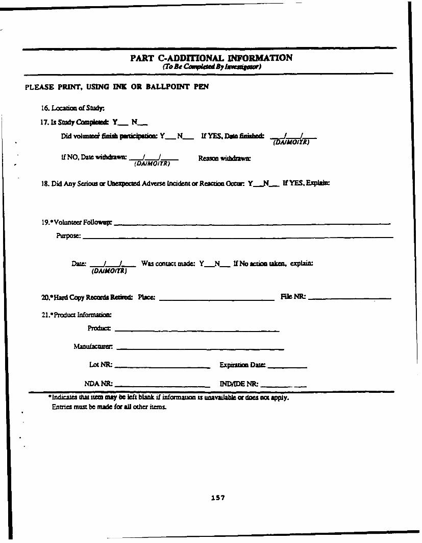
PART C-ADDrrIONAL INFORMATION
(TO Be Cpdiewe BY InmmlSW)
PLEASE PRINT, USING INK OR BALLPOINT PEN
16. 1Location of Study.
17. Is Study Compleled Y. N__
Did volutmeei flinu a juioiuon: r- K If YES. Daft fihisld JJ(DA/MOIYR)
if NO. Date widxidanwu.-J Ream wizhdzw(DAIMOIYR)
18. Did Any Serious or Unexpected, Advezse 1ncident or Reaton (kcw. Y__N IfYES, Expaial
19.*Volunieer FoUowWp
Purpose:
Date. J Was contact made: Y N- If No sew takm~ explain(DAIOMOY)
0.*Hazd Copy Recw&d Rniedift Place: M_________________ e NR: ____________
2l.*Product laformazion
Produc__ ___
mnufEa_ ___ ___ ___
LotNR.__________ Expusnion Dame____
NDA M- __________ ND/DE NIL____
alndzcazfes iha item may be left blank if inorm==uon unavailab or doe =o appy.Entres must be made for ail otwheriems.
15S7

Appendix F.
Medical identification card
In Support of a Research Prolect,
RANK NAME
Has Been Fitted With CONTACT LENSES
In case ot serious injury or disability call
or contact
as soon as possible.
USMVNCC(USAARL i F. 246. L Jan 85
158

Appendix G.
Qualitative data.
159
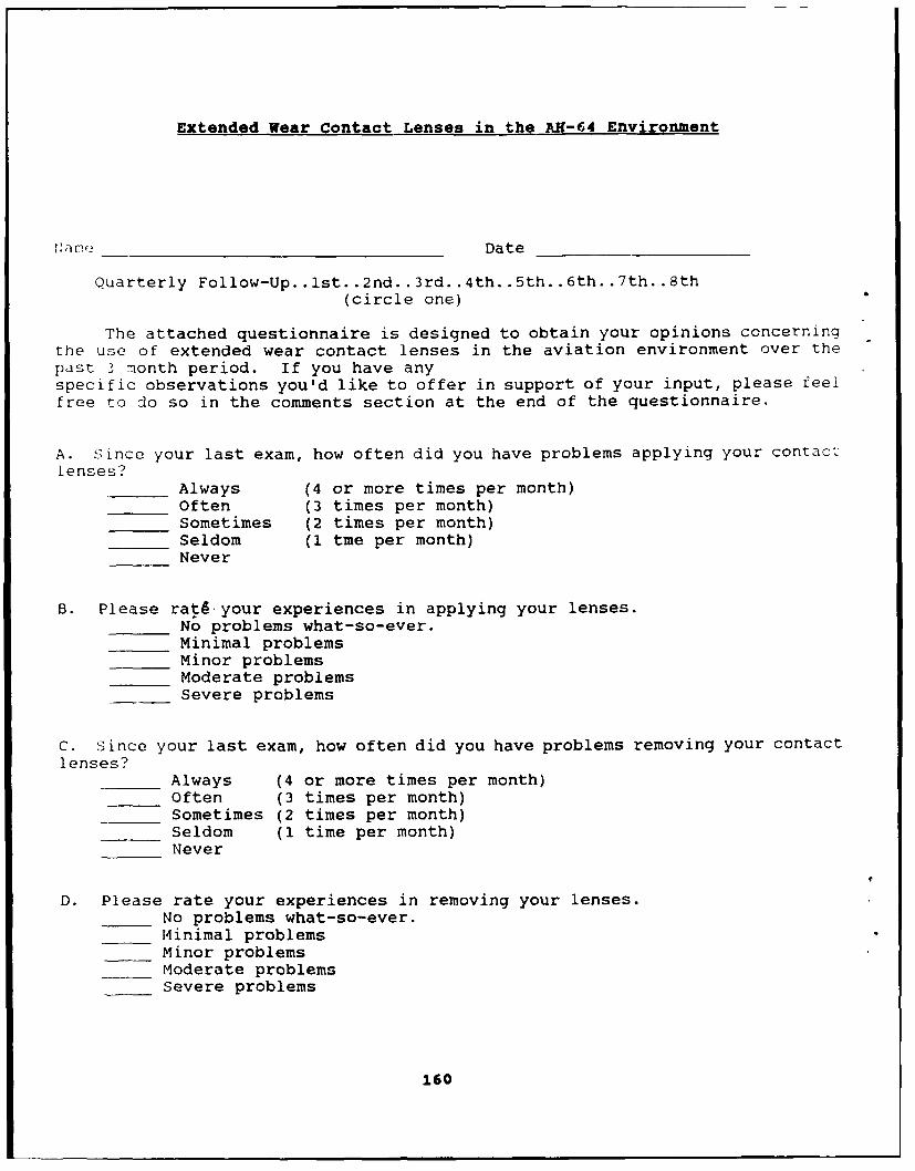
Extended Wear Contact Lenses in the AH-64 Environment
it:a m ( Date
Quarterly Follow-Up..1st..2nd..3rd..4th..5th..6th..7th..8th(circle one)
The attached questionnaire is designed to obtain your opinions concerningthe use of extended wear contact lenses in the aviation environment over thepast 3 month period. If you have anyspecific observations you'd like to offer in support of your input, please feelfree to do so in the comments section at the end of the questionnaire.
A. Since your last exam, how often did you have problems applying your contactlenses?
Always (4 or more times per month)Often (3 times per month)Sometimes (2 times per month)Seldom (1 tme per month)Never
B. Please ratA your experiences in applying your lenses.No problems what-so-ever.Minimal problemsMinor problemsModerate problemsSevere problems
C. Since your last exam, how often did you have problems removing your contactlenses?
Always (4 or more times per month)Often (3 times per month)Sometimes (2 times per month)Seldom (1 time per month)Never
D. Please rate your experiences in removing your lenses.No problems what-so-ever.Minimal problemsMinor problemsModerate problemsSevere problems
160

Extended Wear Contact Lenses in the AH-64 Environment
Looking back over the course of your contact lens wearing experience, pleaIseevaluate the training program effectiveness in teaching you the followingaspects:
Excellent Good Fair Poor Ineffective
Application
R Removal 1
-.ea ringschedule
F. In general, how comfortable are your contact lenses?
Very comfortableComfortableNeither comfortable nor uncomfortableUncomfortableVery uncomfortable
G. How do you rate your vision with contact lenses as opposed to your visionwith glasses.
Much better with contact lensesSlightly better with contact lensesNo difference between contact lenses and glassesSlightly better with glassesMuch better with glasses
H. Rate your level of confidence in your flying ability when wearing contactlenses as opposed to when wearing glasses.
Much more confident with contact lensesSlightly more confident with contact lensesEqually confident with bothSlightly more confident with glassesMuch more confident with glasses
161
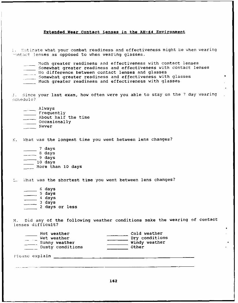
Extended Wear Contact Lenses in the AH-64 Environment
!. !:t Iate what your combat readiness and effectiveness might be when wearing-ntact lenses as opposed to when wearing glasses.
Much greater readiness and effectiveness with contact lensesSomewhat greater readiness and effectiveness with contact lensesNo difference between contact lenses and glassesSomewhat greater readiness and effectiveness with glassesMuch greater readiness and effectiveness with glasses
Since your last exam, how often were you able to stay on the 7 day wearing.,chedule?
AlwaysFrequentlyAbout half the timeOccasionallyNever
K. What was the longest time you went between lens changes?
7 days8. days9 days10 daysMore than 10 days
L. What was the shortest time you went between lens changes?
6 days5 days4 days3 days2 days or less
M. Did any of the following weather conditions make the wearing of contactlenses difficult?
Hot weather Cold weatherWet weather Dry conditionsSunny weather Windy weatherDusty conditions Other
i'lease explain
162

Extended Wear Contact Lenses in the AR-64 Environment
N. Since your last exam, did you experience any of the tollowing problem:bw.hile flying, and if so, how bothersome were they to you? Check only those that.•pply.
Frequency SeverityRarely Occasionally Often Minor Moderate Severe
Eye irritationEye painBlurred visionDry eyeLight sensitivity__
0. Since your last exam, did you experience any of the following problems ingarrison activities, and if so, how bothersome were they to you? Check onlythose that apply.
Frequency SeverityRarely Occasionally Often Minor Moderate Severe
Eye irritationEye painBlurred visionDry eyeLight sensitivity__
P. If there is a problem with light sensitivity, or glare, while wearing thecontact lenses outdoors, how dependent are you on sunglasses to alleviate thisproblem?
Very dependent, I always wear sunglasses outdoors.Moderately dependent, I often wear sunglasses.Mildly dependent, I occasionally wear sunglasses.Slightly dependent, I rarely wear sunglasses.Independent, I never wear sunglasses and have no problem
with light sensitivity.
Q. Are you aware of any light sensitivity outdoors while wearing glasses? Ifso, how dependent are you on sunglasses to alleviate this problem?
Very dependent, I always wear sunglasses outdoors.Moderately dependent, I often wear sunglasses.Mildly dependent, I occasionally wear sunglasses.Slight dependent, I rarely wear sunglasses.Independent, I never wear sunglasses and have no problem
with light sensitivity.
163
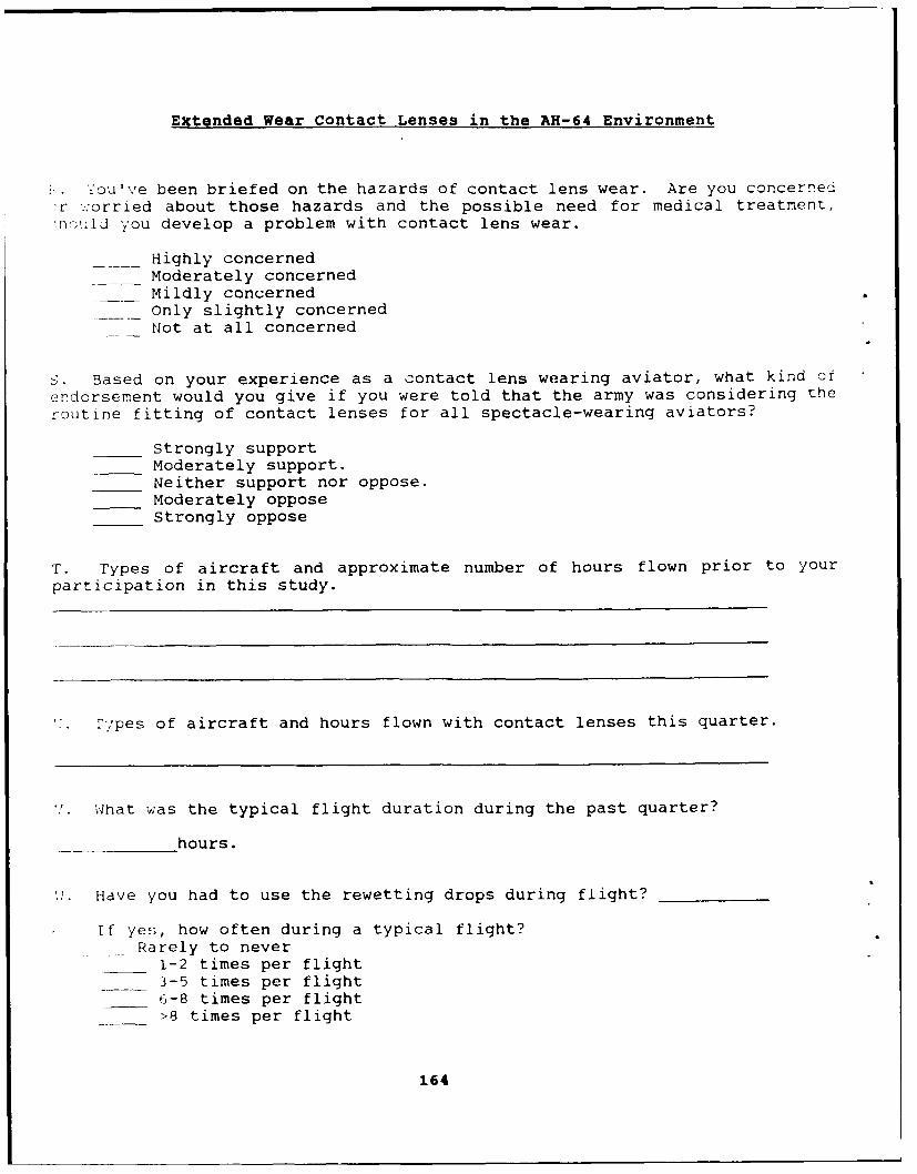
Extended Wear Contact Lenses in the AH-64 Environment
You've been briefed on the hazards of contact lens wear. Are you concerned-r .:orried about those hazards and the possible need for medical treatment,*nmIJd you develop a problem with contact lens wear.
Highly concernedModerately concernedMildly concernedonly slightly concernedNot at all concerned
Based on your experience as a contact lens wearing aviator, what kind ofendorsement would you give if you were told that the army was considering theroutine fitting of contact lenses for all spectacle-wearing aviators?
Strongly supportModerately support.Neither support nor oppose.Moderately opposeStrongly oppose
T. Types of aircraft and approximate number of hours flown prior to yourparticipation in this study.
".Fpes of aircraft and hours flown with contact lenses this quarter.
V. What was the typical flight duration during the past quarter?
hours.
Have you had to use the rewetting drops during flight?
if yes, how often during a typical flight?Rarely to never
1-2 times per flight3-5 times per flight6-8 times per flight>8 times per flight
164
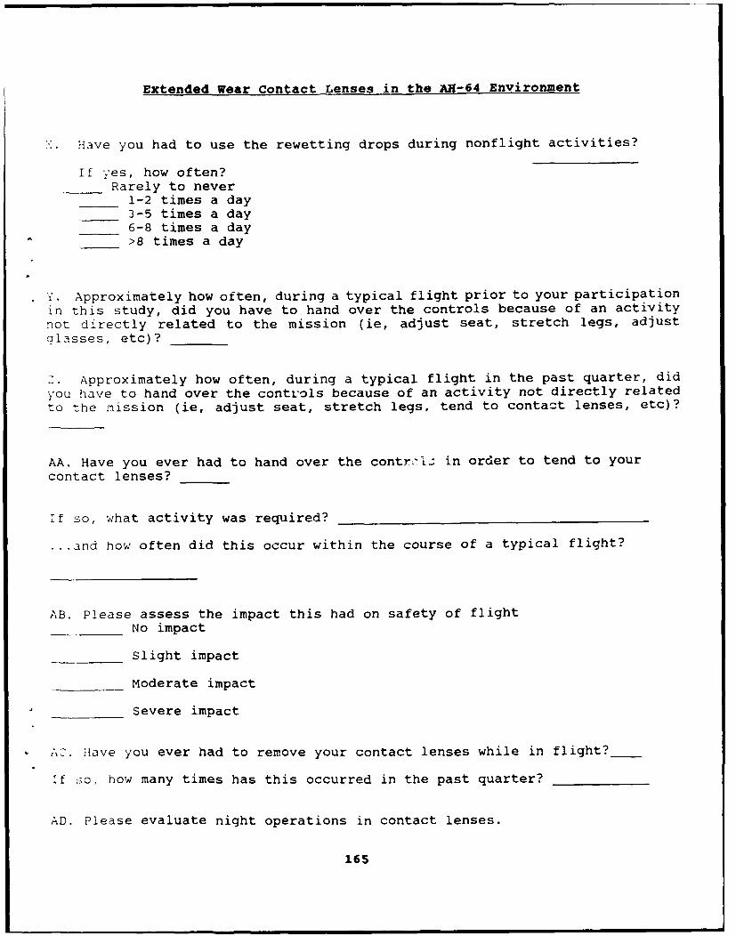
Extended Wear Contact Lenses in the AH-64 Environment
Have you had to use the rewetting drops during nonflight activities?
If yes, how often?Rarely to never
1-2 times a day3-5 times a day6-8 times a day>8 times a day
y. Approximately how often, during a typical flight prior to your participationin this study, did you have to hand over the controls because of an activitynot directly related to the mission (ie, adjust seat, stretch legs, adjustglasses, etc)?
Z. Approximately how often, during a typical flight in the past quarter, didyou have to hand over the controls because of an activity not directly relatedto the mission (ie, adjust seat, stretch legs, tend to contact lenses, etc)?
AA. Have you ever had to hand over the contr.'iz in order to tend to yourcontact lenses?
if so, what activity was required?
... and how often did this occur within the course of a typical flight?
AB. Please assess the impact this had on safety of flight
No impact
Slight impact
Moderate impact
Severe impact
AC. i{ave you ever had to remove your contact lenses while in flight?_
If s0. how many times has this occurred in the past quarter?
AD. Please evaluate night operations in contact lenses.
165
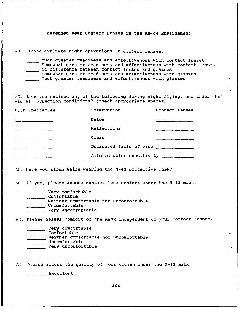
Extended Wear Contact Lenses in the AH-64 Environment
AD. Please evaluate night operations in contact lenses.
Much greater readiness and effectiveness with contact lensesSomewhat greater readiness and effectiveness with contact lensesNo difference between contact lenses and glassesSomewhat greater readiness and effectiveness with glassesMuch greater readiness and effectiveness with glasses
AE. Have you noticed any of the following during night flying, and under what
visual correction conditions? (check appropriate spaces)
With spectacles Observation Contact Lenses
Halos
Reflections
Glare
Decreased field of view
Altered color sensitivity
AF. Have you flown while wearing the M-43 protective mask?
AG. If yes, please assess contact lens comfort under the M-43 mask.
Very comfortableComfortableNeither comfortable nor uncomfortableUncomfortableVery uncomfortable
AH. Please assess comfort of the mask independent of your contact lenses.
Very comfortableComfortableNeither comfortable nor uncomfortableUncomfortableVery uncomfortable
AI. Please assess the quality of your vision under the M-43 mask.
Excellent
166

Extended Wear Contact Lenses in the AH-64 Environment
AI. Please assess the quality of your vision under the M-43 mask.
ExcellentGoodFairPoorUnacceptable
AJ. Please assess the quality of your vision under the M-43 mask independentof your contact lenses.
ExcellentGoodFairPoorUnacceptable
Additional comments
167

Extended Wear Contact Lenses in the AH-64 Environment
V. Looking back over the course of your contact lens wearing experience, please(,valuate the training program effectiveness in teaching you the following.Ispects:
Excellent Good Fair Poor Ineffective
Application
Removal
Wea ringschedule
F. In general, how comfortable are your contact lenses?
Very comfortableComfortableNeither comfortable nor uncomfortableUncomfortableVery uncomfortable
G. How do you rate your vision with contact lenses as opposed to your visionwith glasses.
Much better with contact lensesSlightly better with contact lensesNo difference between contact lenses and glassesSlightly better with glassesMuch better with glasses
H. Rate your level of confidence in your flying ability when wearing contactlenses as opposed to when wearing glasses.
Much more confident with contact lensesSlightly more confident with contact lensesEqually confident with bothslightly more confident with glasses
Much more confident with glasses
168
rT RUCKER 120106
Initial distribution
Commander, U.S. Army Natick Research, U.S. Army Avionics ResearchDevelopment and Evaluation Center and Development Activity
AT[N: STRNC-MIL (Documents ATTN: SAVAA-P-TPLibrarian) Fort Monmouth, NJ 07703-5401Natick, MA 01760-5040
U.S. Army Communications-Electronics* Col. Otto Schramm Filho Command* c/o Brazilian Army Commission ATTN: AMSEL-RD-ESA-D
h Office-CEBW Fort Monmouth, NJ 077034632 Wisconsin Avenue NWWashington, DC 20016 Library
Naval Submarine Medical Research LabCommander/Director Box 900, Naval Sub BaseU.S. Army Combat Surveillance Groton, CT 06349-5900
and Target Acquisition LabATTN: DELCS-D CommanderFort Monmouth, NJ 07703-5304 Man-Machine Integration System
Code 602Commander Naval Air Development Center10th Medical Laboratory Warminster, PA 18974ATTN: AudiologistAPO New York 09180 Commander
Naval Air Development CenterNaval Air Development Center ATTN: Code 602-B (Mr. Brindle)Technical Information Division Warminster, PA 18974Technical Support DetachmentWarminster, PA 18974 Commanding Officer
Armstrong LaboratoryCommanding Officer, Naval Medical Wright-Patterson
Research and Development Command Air Force Base, OH 45433-6573National Naval Medical CenterBethesda, MD 20814-5044 Director
Army Audiology and Speech CenterDeputy Director, Defense Research Walter Reed Army Medical Center
and Engineering Washington, DC 20307-5001ATTN: Military Assistant
for Medical and Life Sciences Commander, U.S. Army InstituteWashington, DC 20301-3080 of Dental Research
ATTN: Jean A. Setterstrom, Ph. D.Commander, U.S. Army Research Walter Reed Army Medical Center
Institute of Environmental Medicine Washington, DC 20307-5300Natick, MA 01760
169

Naval Air Systems Command U.S. Army Materiel SystemsTechnical Air Library 950D Analysis AgencyRoom 278, Jefferson Plaza II ATTN: AMXSY-PA (Reports Processing)Department of the Navy Aberdeen Proving GroundWashington, DC 20361 MD 21005-5071
Director, U.S. Army Human U.S. Army Ordnance CenterEngineering Laboratory and School Library
ATTN: Technical Library Simpson Hall, Building 3071Aberdeen Proving Ground, MD 21005 Aberdeen Proving Ground, MD 21005
Commander, U.S. Army Test U.S. Army Environmentaland Evaluation Command Hygiene Agency
ATTN: AMSTE-AD-H Building E2100Aberdeen Proving Ground, MD 21005 Aberdeen Proving Ground, MD 21010
Director Technical Library Chemical ResearchU.S. Army Ballistic and Development Center
Research Laboratory Aberdeen Proving Ground, MDATTN: DRXBR-OD-ST Tech Reports 21010--5423Aberdeen Proving Ground, MD 21005
CommanderCommander U.S. Army Medical ResearchU.S. Army Medical Research Institute of Infectious Disease
Institute of Chemical Defense SGRD-UIZ-CATTIN: SGRD-UV-AO Fort Detrick, Frederick, MD 21702Aberdeen Proving Ground,MD 21010-5425 Director, Biological
Sciences DivisionCommander, U.S. Army Medical Office of Naval ResearchResearch and Development Command 600 North Quincy SLieetATTN: SGRD-RMS (Ms. Madigan) Arlington, VA 22217Fort Detrick, Frederick, MD 21702-5012
CommanderDirector U.S. Army Materiel CommandWalter Reed Army Institute of Research ATTN: AMCDE-XSWashington, DC 20307-5100 5001 Eisenhower Avenue
Alexandria, VA 22333HQ DA (DASG-PSP-O)5109 Leesburg Pike CommandantFalls Church, VA 22041-3258 U.S. Army Aviation
Logistics School ATTN: ATSQ-IT)NHarry Diamond Laboratories Fort Eustis, VA 23604ATTN: Technical Information Branch2800 Powder Mill RoadAdelphi, MD 20783-1197
170
Veadquarters (ATMD) Commander'U.S. Army Training U.S. Army Aviation Systems Command
and Doctrine Command ATTN: SGRD-UAX-AL (MAJ Gillette)ATTN: ATBO-M 4300 Goodfellow Blvd., Building 105Fort Monroe, VA 23651 St. Louis, MO 63120
Structures Laboratory Library U.S. Army Aviation Systems CommandUSARTL-AVSCOM Library and Information Center Branch
, NASA Langley Research Center ATIN: AMSAV-DILS Mail Stop 266 4300 Goodfellow Boulevard
Hampton, VA 23665 St. Louis, MO 63120
Naval Aerospace Medical Federal Aviation AdministrationInstitute Library Civil Aeromedical Institute
Building 1953, Code 03L Library AAM-400APensacola, FL 32508-5600 P.O. Box 25082
Oklahoma City, OK 73125Command SurgeonHQ USCENTCOM (CCSG) CommanderU.S. Central Command U.S. Army AcademyMacDill Air Force Base FL 33608 of Health Sciences
ATTN: LibraryAir University Library Fort Sam Houston, TX 78234(AUL/LSE)Maxwell Air Fore Base, AL 36112 Commander
U.S. Army Institute of Surgical ResearchU.S. Air Force Institute ATTN: SGRD-USM (Jan Duke)
of Technology (AFIT/LDEE) Fort Sam Houston, TX 78234-6200Building 640, Area BWright-Patterson AAMRL/I IEXAir Force Base, OH 45433 Wright-Patterson
Air Force Base, OH 45433Henry L. TaylorDirector, Institute of Aviation John A. Dellinger,University of Illinois-Willard Airport Southwest Research IpstituteSavoy, IL 61874 P. 0. Box 28510
San Antonio, TX 78284Chief, Nation Guard BureauATTN: NGB-ARS (COL Urbauer) Product ManagerRoom 410, Park Center 4 Aviation Life Support Equipment4501 Ford Avenue A'ITN: AMCPM-ALSEAlexandria, VA 22302-1451 4300 Goodfellow Boulevard
St. Louis, MO 63120-1798
171

Commander Aeromechanics LaboratoryU.S. Army Aviation U.S. Army Research and Technical Labs
Systems Command Ames Research Center, M/S 215-1ATTN: AMSAV-ED Moffett Field, CA 940354300 Goodfellow BoulevardSt. Louis, MO 63120 Sixth U.S. Army
A'ITN: SMACommanding Officer Presidio of San Francisco, CA 94129Naval Biodynamics LaboratoryP.O. Box 24907 CommanderNew Orleans, LA 70189-0407 U.S. Army Aeromedical Center
Fort Rucker, AL 36362Assistant CommandantU.S. Army Field Artillery School U.S. Air Force SchoolATTN: Morris Swott Technical Library of Aerospace MedicineFort Sill, OK 73503-0312 Strughold Aeromedical Library Technical
Reports Section (TSKD)Commander Brooks Air Force Base, TX 78235-5301U.S. Army Health Services CommandATTN: HSOP-SO Dr. Diane DamosFort Sam Houston, TX 78234-6000 Department of Human Factors
ISSM, USCHQ USAF/SGPT Los Angeles, CA 90089-0021Boiling Air Force Base, DC 20332-6188
U.S. Army White SandsU.S. Army Dugway Proving Ground Missile RangeTechnical Library, Building 5330 ATTN: STEWS-IM-STDugway, UT 84022 White Sands Missile Range, NM 88002
U.S. Army Yuma Proving Ground U.S. Army Aviation EngineeringTechnical Library Flight ActivityYuma, AZ 85364 ATITN: SAVTE-M (Tech Lib) Stop 217
Edwards Air Force Base, CA 93523-5000AFFTC Technical Library6510 TW/TSTL Ms. Sandra G. HartEdwards Air Force Base, Ames Research CenterCA 93523-5000 MS 262-3
Moffett Field, CA 94035CommanderCode 3431 Commander, Letterman Army InstituteNaval Weapons Center of ResearchChina Lake, CA 93555 ATTN: Medical Research Library
Presidio of San Francisco, CA 94129
172
"i'ommander Italian Army Liaison OfficeU.S. Army Medical Materiel Building 602
Development Activity Fort Rucker, AL 36362Fort Detrick, Frederick, MD 21702-5009
Directorate of Training DevelopmentCommander Building 502U.S. Army Aviation Center Fort Rucker, AL 36362Directorate of Combat DevelopmentsBuilding 507 ChiefFort Rucker, AL 36362 USAHEL/USAAVNC Field Office
P. 0. Box 716U. S. Army Research Institute Fort Rucker, AL 36362-5349Aviation R&D ActivityATTN: PERI-IR Commander U.S. Army Aviation CenterFort Rucker, AL 36362 and Fort Rucker
ATITN: ATZQ-CGCommander Fort Rucker, AL 36362U.S. Army Safety CenterFort Rucker, AL 36362 Chief
Test & Evaluation Coordinating BoardU.S. Army Aircraft Development Cairns Army Air Field
Test Activity Fort Rucker, AL 36362ATTN: STEBG-MP-PCairns Army Air Field MAJ Terry NewmanFort Rucker, AL 36362 Canadian Army Liaison Office
Building 602Commander U.S. Army Medical Research Fort Rucker, AL 36362
and Development CommandATTN: SGRD-PLC (COL Schnakenberg) German Army Liaison OfficeFort Detrick, Frederick, MD 21702 Building 602
Fort Rucker, AL 36362MAJ John WilsonTRADOC Aviation LO LTC Patrice CottebruneEmbassy of the United States French Army Liaison OfficeAPO New York 09777 USAAVNC (Building 602)
Fort Rucker, AL 36362-5021Netherlands Army Liaison Office
* Building 602 Australian Army Liaison Office* Fort Rucker, AL 36362 Building 602
Fort Rucker, AL 36362
British Army Liaison Office
Building 602 Dr. Garrison RaprnundFort Rucker, AL 36362 6 Burning Tree Court
Bethesda, MD 20817
173
Commandant, Royal Air Force U.S. Army Research and TechnologyInstitute of Aviation Medicine Laboratories (AVSCOM)Farnborough Hampshire GU14 65Z UK Propulsion Laboratory MS 302-2
NASA Lewis Research CenterCommander Cleveland, OH 44135U.S. Army Biomedical Research
and Development Laboratory Dr. Christine SchlichtingATTN: SGRD-UBZ-I Behavioral Sciences DepartmentFort Detrick, Frederick, MD 21702 Box 900, NAVUBASE NLON
Defense Technical Information OCC Groton, CT 06349-5900
Selection Dr. Eugene S. ChanningCameron Station 7985 Schooner CourtAlexandra, VA 22313 Frederick, MD 21701-3273
Commander, U.S. Army Foreign Science LTC Gaylord Lindsey (5)and Technology Center USAMRDC Liaison at Academy
AIFRTA (Davis) of Health Sciences220 7th Street, NE ATTN: HSHA-ZAC-FCharlottesville, VA 22901-5396 Fort Sam Houston, TX 78234
Director, Aviation Medicine ClinicApplied Technology Laboratory TMC #22, SAAFUSARTL-AVSCOM Fort Bragg, NC 28305ATTN: Library, Building 401Fort Eustis, VA 23604 Dr. A. Kornfield, President
Biosearch CompanyU.S. Air Force Armament 3016 Revere Road
Development and Test Center Drexel Hill, PA 29026Eglin Air Force Base, FL 32542
NVEODCommander, U.S. Army Missile AMSEL-RD-ASID
Command (Attn: Trang Bui)Redstone Scientific Information Center Fort Belvior, VA 22060ATI'N: AMSMI-RD-CS-R
/ILL Documents Commander and DirectorRedstone Arsenal, AL 35898 USAE Waterways Experiment Station
ATTN: CEWES-IM-MI-RDr. H. Dix Christensen Alfrieda S. Clark, CD Dept.Bio-Medical Science Building, Room 753 3909 Halls Ferry Road
Post Office Box 26901 Vicksburg, MS 39180-6199Oklahoma City, OK 73190
CA AVNDirector HQ DAACArmy Personnel Research Establishment Middle WallopFarnborough, Hants G014 6TD UK Stockbridge Hants S020 8D7 UK
174

Mr. Richard ThornleyILS Manager, APACHEBox 84Westland Helicopters LimitedYeovil, Somerset BA202YB UK
I
175

SUPPLEMENTARY
INFORMATION
05/24/93 13:42 10205 255 6937 USAARL 2002
Acknowledcments
The authors wish to thank the volunteer subjects and theircommanders for their enthusiastic support of this exhaustiveresearch effort. Sinzere appreciation is also extended to SGTVincent Reynoso who provided technical support and to Ms. CarolynJohnson who provided :Uerical support.
05/24/93 13:42 V205 255 6937 USAARL a 003
This page intentionally left blank.
-- ~m~ ii

05/24/93 13:43 V'205 255 6937 USAARL Z 004
Table of contents
List of figures .............................................. i
List of tables ............................................ viii
List of appendixes ......................................... viii
Introduction ................................................. I
Purpose ................................................... I
Approach ..................................................... 1
Military significance...................... 2
Background .............................................. 2Statement of the specific problem .................. 2Literature review introduction ..................... 3Aviation literature review ........... .............. 4Related ana:omy and physiology ..................... 9
Precorneal teartilm ........................... 9Cornea ........................................ * 0
Introduction to soft contact lenses ............... 12Review of clinical issues .......................... 13
Superficial punctate keratitis ................ 13Neovascularization ............................ 14Giant papillary conjunctivitis ................ .14Superior limbic keratoconjunctivitis .......... 15Bacteriaa infection ........................... 15Acanthamoeba infection ........................ 16
Detailed review of ocular considerations ........... 16Oxyqer ........................................ 16Ocular surface pH issues ...................... !8Epitheiial morphology ......................... 20Cornea.* metabolism ............................ 20Endothelial morphology ........................ 22Endothelial function .......................... 21
Su*xLmary statemer ........................................... 22
Materials and methods ........................................ 23Subjects ....................... ............................. 23Medical personnel and facilities .......... .................. 24
Professiona.. requirements .......................... 24Technical sipport ..................................... 25Facilities ......................................... 25Medical equipment .................................. 25"tens materials ..................................... .. 25
Procedures ....................... ............................ . 26Initial exanination and fitting .................... 26Followup examinations .............................. 26
iii
05/24/93 13:44 e'205 255 6937 USAARL z 005
I
Table of contents (Continued)
Wearinq sch'edule ................................... 27Data collection ..................................... 27Eye-related symptoms and medical emergencies ....... 28Subjective assessment procedures................... 28Lens physical assessment ........................... 28Medical support data ................................ 28Experimental design/data analysis .................. 29Human use justification ............................ 29Regulation compliance .............................. 29Health and safety of volunteers .................... 29
Risks ....... .................................. 29Medical safeguards ............................ 31Medical monitor ................................ 1
Individual privacy and handling of data ............ 31
Results and discussi.n ........................................ 31Introductory description .............................. 31
General technical report ........................... 31Slit 2amp examination.......................... 33Safety concerns ............................... 43Subjec- sentiment ............................. 44Ocular health .................................. 44Synopsis ...................................... 44
Detailed analyses ........................................ 4ECorn)ea. curvature assessment ....................... 45
Intro iction .................................. 4SMethods ........................................ 45Results ....................... ....................... 45Discussion .................................... 46
Corneal aes-hesiometer assessment .................. 47Irtrot&ction .................................. 47Mcthoc ....................................... 48Result!; and discussion ........................ 49Ccncltsion ..................................... 51
Sterile peripheral infiltrates ..................... 52Ba:ckgr-:und ..................................... 52DIscu~s ion .................. ...................... 52
Bacter:al ricers .................................... 54Bazkground ..................................... 54Discussion .................. ...................... 55
Hydrogel translens oxygen flux ..................... 56Ir~trodlction ................................... 56Mct .ods 5........................................57Results and discussion ........................ . 57Ccnclusions .................................... 5
Anterior lens surface pP ........................... 59Introduction ................................... 59Materials and mathods .......................... 60Results ....................................... 61
iv