
The use of cyclosporin in a child with generalized pustular psoriasis
22
Correspondence The vesicular pemphigoid phenotype may be related to antibodies to a 200 kDa antigen in the lower lamina lucida SIR, There are various bullous diseases showing autoantibodies to the basement membrane zone (BMZ). Bullous pemphigoid (BP) sera react with the 230 kDa BP antigen (BP230) and/or the 180 kDa BP antigen (BP180). Epidermolysis bullosa acquisita (EBA) sera react with the 290 kDa EBA antigen (type VII collagen). The sera of cicatricial pemphigoid and linear IgA bullous dermatosis show heterogeneous reactivity. In addition, a 200 kDa antigen located in the deeper portion of the lamina lucida has been identified as a novel antigen in a patient with atypical bullous lesions, 1 and patients with long- standing psoriasis associated with blister formation. 2,3 We present two patients with vesicular pemphigoid-like clinical features, who showed anti-BMZ antibodies against the 200 kDa protein. Patient 1, a 64-year-old Japanese man, developed itchy erythematous skin lesions up to 1 cm in size, associated with crusts and erosions (Fig. 1). Only a few vesicles were seen. An annular arrangement of lesions was occasionally seen. He never developed large tense bullae characteristic of BP. This patient did not respond well to systemic steroids. However, the skin lesions quickly cleared, leaving slight scarring, after administration of dapsone, 100mg daily. The dosage was tapered to 50 mg daily without recurrence. Patient 2, a 64-year-old Japanese man, developed vesicles and erosions up to 0·5cm in size, over the entire body. The lesions on the palms and soles resembled palmoplantar pustulosis. Neither large tense bullae nor an annular arrangement of lesions was seen. This patient responded well to a combination of oral prednisolone, 40 mg daily and dapsone, 75mg daily. This case was reported in a Japanese journal in 1982, without any results of antigen studies. 4 In both cases, histopathological studies showed subepi- dermal bullae with infiltration predominantly of neutrophils. Direct immunofluorescence (IF) showed IgG and C3 deposition at the BMZ. On indirect IF of 1 mol/L NaCl-split skin, IgG in the sera reacted only with the dermal side of the split. On immunoblotting (IB) of normal human epidermal extracts, 5 the sera did not react with either BP230 or BP180. On IB of recombinant protein of the BP180 NC16a domain, which is known to be recognized by almost all BP sera, 6 the sera showed no reactivity. In contrast, on IB of human dermal extracts, 7 the sera reacted exclusively with a 200 kDa protein, while control EBA sera reacted with the 290 kDa EBA antigen. The 200 kDa band showed exactly the same migration on the gel as in previously reported cases. 1–3 The present two patients showed only small skin lesions, corresponding to vesicular pemphigoid, and the skin lesions were responsive to dapsone. Linear IgG deposition along the BMZ was detected bydirect IF. However, in contrast to BP, the sera of these patients reacted with the dermal side of 1 mol/L NaCl-split skin on indirect IF. On IB of various antigen sources including normal epidermal extracts, dermal extracts and recombinant protein of the BP180 NC16a domain, these sera reacted only with a 200 kDa protein present in the dermal extracts, but not with any other known autoantigens. This 200 kDa antigen has been detected in a patient with atypical bullous lesions 1 and in patients with psoriasis. 2,3 This study indicates that a subset of patients with the vesicular pemphigoid phenotype may also react with this antigen, although the nature of this 200 kDa antigen has not been characterized. Department of Dermatology, Y.INOH Kurume University School of Medicine, T.NISHIKAWA* Fukuoka, Japan T.HASHIMOTO *Department of Dermatology, Keio University School of Medicine, Tokyo, Japan References 1 Zillikens D, Kawahara Y, Ishiko A et al. A novel IgG-mediated British Journal of Dermatology 1998; 139: 738–759. 738 q 1998 British Association of Dermatologists Figure 1. Small crusted and erosive lesions are evident on the trunk.
Transcript of The use of cyclosporin in a child with generalized pustular psoriasis

Correspondence
The vesicular pemphigoid phenotype may be related toantibodies to a 200 kDa antigen in the lower lamina lucida
SIR, There are various bullous diseases showing autoantibodiesto the basement membrane zone (BMZ). Bullous pemphigoid(BP) sera react with the 230 kDa BP antigen (BP230) and/orthe 180 kDa BP antigen (BP180). Epidermolysis bullosaacquisita (EBA) sera react with the 290 kDa EBA antigen(type VII collagen). The sera of cicatricial pemphigoid andlinear IgA bullous dermatosis show heterogeneous reactivity.In addition, a 200 kDa antigen located in the deeper portion ofthe lamina lucida has been identified as a novel antigen in apatient with atypical bullous lesions,1 and patients with long-standing psoriasis associated with blister formation.2,3 Wepresent two patients with vesicular pemphigoid-like clinicalfeatures, who showed anti-BMZ antibodies against the200 kDa protein.
Patient 1, a 64-year-old Japanese man, developed itchyerythematous skin lesions up to 1 cm in size, associated withcrusts and erosions (Fig. 1). Only a few vesicles were seen. Anannular arrangement of lesions was occasionally seen. Henever developed large tense bullae characteristic of BP. Thispatient did not respond well to systemic steroids. However, theskin lesions quickly cleared, leaving slight scarring, afteradministration of dapsone, 100 mg daily. The dosage wastapered to 50 mg daily without recurrence.
Patient 2, a 64-year-old Japanese man, developed vesiclesand erosions up to 0·5 cm in size, over the entire body. Thelesions on the palms and soles resembled palmoplantarpustulosis. Neither large tense bullae nor an annulararrangement of lesions was seen. This patient respondedwell to a combination of oral prednisolone, 40 mg daily anddapsone, 75 mg daily. This case was reported in a Japanesejournal in 1982, without any results of antigen studies.4
In both cases, histopathological studies showed subepi-dermal bullae with infiltration predominantly of neutrophils.Direct immunofluorescence (IF) showed IgG and C3 depositionat the BMZ. On indirect IF of 1 mol/L NaCl-split skin, IgG in thesera reacted only with the dermal side of the split. Onimmunoblotting (IB) of normal human epidermal extracts,5
the sera did not react with either BP230 or BP180. On IB ofrecombinant protein of the BP180 NC16a domain, which isknown to be recognized by almost all BP sera,6 the serashowed no reactivity. In contrast, on IB of human dermalextracts,7 the sera reacted exclusively with a 200 kDa protein,while control EBA sera reacted with the 290 kDa EBA antigen.The 200 kDa band showed exactly the same migration on thegel as in previously reported cases.1–3
The present two patients showed only small skin lesions,corresponding to vesicular pemphigoid, and the skin lesionswere responsive to dapsone. Linear IgG deposition along theBMZ was detected by direct IF. However, in contrast to BP, thesera of these patients reacted with the dermal side of 1 mol/LNaCl-split skin on indirect IF. On IB of various antigen sourcesincluding normal epidermal extracts, dermal extracts and
recombinant protein of the BP180 NC16a domain, these serareacted only with a 200 kDa protein present in the dermalextracts, but not with any other known autoantigens. This200 kDa antigen has been detected in a patient with atypicalbullous lesions1 and in patients with psoriasis.2,3 This studyindicates that a subset of patients with the vesicularpemphigoid phenotype may also react with this antigen,although the nature of this 200 kDa antigen has not beencharacterized.
Department of Dermatology, Y.INOH
Kurume University School of Medicine, T.NISHIKAWA*Fukuoka, Japan T.HASHIMOTO
*Department of Dermatology,Keio University School of Medicine,Tokyo, Japan
References
1 Zillikens D, Kawahara Y, Ishiko A et al. A novel IgG-mediated
British Journal of Dermatology 1998; 139: 738–759.
738 q 1998 British Association of Dermatologists
Figure 1. Small crusted and erosive lesions are evident on the trunk.

subepidermal blistering disease with autoantibodies against a200-kD antigen of the basement membrane zone. J Invest Dermatol1996; 106: 465–70.
2 Chen K-R, Shimizu S, Miyakawa S et al. Coexistence of psoriasisand an unusual IgG-mediated subepidermal bullous dermatosis:identification of a novel 200-kDa lower lamina lucida targetantigen. Br J Dermatol 1996; 134: 340–6.
3 Saeki H, Hayashi N, Komine M et al. A case of generalized pustularpsoriasis followed by bullous disease: an atypical case of bullouspemphigoid or a novel bullous disease? Br J Dermatol 1996; 134:152–5.
4 Hashimoto T, Sugiura M, Kurihara S, Nishikawa T. A case ofpolymorphic pemphigoid. Jpn J Clin Dermatol 1982; 1982: 121–3.
5 Sugi T, Hashimoto T, Hibi T, Nishikawa T. Production ofhuman monoclonal anti-basement membrane zone (BMZ)antibodies from a patient with bullous pemphigoid (BP) byEpstein-Barr virus transformation. Analyses of the heterogeneity ofanti-BMZ antibodies in BP sera using them. J Clin Invest 1989; 84:1050–5.
6 Matsumura K, Hashimoto T, Ohata Y, Nishikawa T. Majority ofbullous pemphigoid and herpes gestationis sera react with NC16adomain of the 180 kD bullous pemphigoid antigen. Arch DermatolRes 1996; 288: 507–9.
7 Dmochowski M, Hashimoto T, Bhogal BS et al. Immunoblottingstudies of linear IgA disease. J Dermatol Sci 1993; 6: 194–200.
Inducible nitric oxide synthase expression on a rheumatoidnodule and in rheumatoid papules
SIR, Rheumatoid arthritis (RA) sometimes presents character-istic cutaneous manifestations.1 Rheumatoid nodules are therepresentative skin manifestations occurring in 20–30% ofCaucasian patients with RA.2 Nitric oxide (NO), which hasrecently been implicated as a mediator of immune andinflammatory responses, is suggested to play a part in RA.3,4
An increased production of nitrite in synovial fluids andsera,4,5 and an increased NO synthase (NOS) activity incirculating mononuclear cells6 have been reported in patientswith RA. Local expression of inducible NOS (iNOS) at both themessenger and the protein level is also detected in synovialtissue of RA.7,8 In this study, we have examined theimmunohistochemical expression of iNOS on a rheumatoidnodule and in rheumatoid papules in a patient with RA.
A biopsy was taken from a subcutaneous nodule on theright elbow and from two superficial nodules on the dorsalaspect of the distal interphalangeal joint of the right indexfinger and left middle finger of a 56-year-old woman who hadbeen diagnosed as having RA and sarcoidosis. She had severalsubcutaneous and intradermal nodules on her elbows, fingersand head. The biopsied tissues were cut into two: one half wasfixed in 10% formalin solution, and the other was embedded inOCT compound (Miles Inc., Elkhart, IN, U.S.A.), snap-frozenimmediately in liquid nitrogen, and stored at ¹ 80 8C. Routinehaematoxylin and eosin staining revealed palisading granu-lomas with collagen deposition in their centre and numerousareas of cellular infiltration at the periphery of the lesions.These changes were compatible with rheumatoid nodulesand rheumatoid papules. In addition, central necrosis wasnoted in the rheumatoid nodules. Immunohistochemistry wasperformed using 5-mm thick cryostat sections prepared on
poly L-lysine-coated slides. These were air-dried, treated withmethanol containing 0·3% hydrogen peroxide for 15 min, andthen stained with a standard avidin–biotin–peroxidasetechnique (Nichirei Co., Tokyo, Japan) using human anti-iNOS polyclonal antibody (Affinity BioReagents Inc., CO,U.S.A.) (diluted in phosphate-buffered saline, 1 : 250) for 2 h.CD62 was also used as a marker of macrophages (GenosysBiotechnologies Inc., Cambridge, U.K.). The sections weredeveloped with 3,30-diaminobenzidine solution as chromogen,then counterstained with haematoxylin, dehydrated, clearedand mounted. Negative controls were prepared by omitting theprimary antibody and substituting rabbit IgG.
Results revealed positive staining for iNOS on infiltratingmononuclear cells around the central necrotic area in therheumatoid nodule (Fig. 1a), and in rheumatoid papules(Fig. 1b). Most of the infiltrating mononuclear cells were alsopositive for anti-CD62 antibody (data not shown). Cellsimmunoreactive for iNOS were also detected on endothelialcells of both the rheumatoid nodule and the rheumatoidpapules. However, fibroblasts did not show positive staining.
In this study, iNOS was detected in the rheumatoid noduleand papules in a distribution suggesting that the main sourceof NO in cutaneous lesions is mononuclear cells andendothelial cells. Fibroblasts around the vessels were hardlystained. In the synovial tissue of RA, most cells expressingiNOS were shown by Sakurai et al. to be synovial lining cells,endothelial cells and chondrocytes,7 while another paperreported that iNOS was positively stained on fibroblasts andrarely stained on macrophages.8
Rheumatoid papules occur on the fingers in most cases, andare located on the more superficial dermis compared withrheumatoid nodules. Histologically, both present palisadinggranulomas with collagen degeneration in the centre. NOupregulates matrix metalloproteinase production9 and isimplicated in the interleukin-1b-mediated inhibition ofproteoglycan synthesis.10 We suggest that NO released frominfiltrating mononuclear cells and/or endothelial cells mayplay a part in the regulation of the extracellular matrix,leading to the induction of rheumatoid nodules and rheuma-toid papules. On the other hand, NO may contribute only togranuloma formation. It is not yet known whetheriNOS expression is specific for rheumatoid nodules orpapules, or whether it is expressed in other granulomatoustissues. Further studies are necessary to clarify the mechanismof the formation of rheumatoid nodules and rheumatoidpapules.
Department of Dermatology, T.YAMAMOTO
Tokyo Medical and T.MATSUNAGA
Dental University School of Medicine, H.YOKOZEKI
1-5-45 Yushima, I.KATAYAMA*Bunkyo-ku, K.NISHIOKA
Tokyo 113, Japan
*Department of Dermatology,Nagasaki University School of Medicine,Nagasaki, Japan
CORRESPONDENCE 739
q 1998 British Association of Dermatologists, British Journal of Dermatology, 139, 738–759

References
1 Jorizzo JL, Daniels JC. Dermatologic conditions reported in patientswith rheumatoid arthritis. J Am Acad Dermatol 1983; 8: 439–57.
2 Kaye BR, Kaye RL, Bobrove A. Rheumatoid nodules. Review of anew classification, with a report of four seronegative cases. Am JMed 1990; 76: 279–92.
3 Vladutiu AO. Role of nitric oxide in autoimmunity. Clin ImmunolImmunopathol 1995; 76: 1–11.
4 Farrell AJ, Blake DR, Palmer RMJ, Moncada S. Increasedconcentrations of nitrite in synovial fluid and serum samplessuggest increased nitric oxide synthesis in rheumatic diseases. AnnRheum Dis 1992; 51: 1219–22.
5 Grabowski PS, England A, Dykhuizen R et al. Elevated nitric oxideproduction in rheumatoid arthritis. Arthritis Rheum 1996; 39:642–7.
6 St Clair EW, Wilkinson WE, Lang T et al. Increased expression ofblood mononuclear cell nitric oxide synthase type 2 in rheumatoidarthritis patients. J Exp Med 1996; 184: 1173–8.
7 Sakurai H, Kohsaka H, Liu M-F et al. Nitric oxide production andinducible nitric oxide synthase expression in inflammatoryarthritides. J Clin Invest 1995; 96: 2357–63.
8 McInnes IB, Leung BP, Field M et al. Production of nitric oxide inthe synovial membrane of rheumatoid and osteoarthritis patients.J Exp Med 1996; 184: 1519–24.
9 Murrell GAC, Jang D, Williams RJ. Nitric oxide activatesmetalloprotease enzymes in articular cartilage. Biochem BiophysRes Commun 1995; 206: 15–21.
10 Hauselmann HJ, Oppliger L, Michel BA et al. Nitric oxide andproteoglycan biosynthesis by human articular chondrocytes inarginate culture. FEBS Lett 1994; 352: 361–4.
740 CORRESPONDENCE
q 1998 British Association of Dermatologists, British Journal of Dermatology, 139, 738–759
Figure 1. Immunohistochemical staining ofinducible nitric oxide synthase (iNOS) in arheumatoid nodule and rheumatoidpapules. (a) Mononuclear cells around thecentral necrotic area (N) in the rheumatoidnodule were positively stained for iNOS(original magnification, × 420). (b)Immunoreactive cells were noted onmononuclear cells in rheumatoid papules,but fibroblasts around the vessels did notshow positive staining (originalmagnification × 400).

Palmoplantar keratoderma associated with hypothyroidism
SIR, We report a patient who had hypothyroidism withacquired palmoplantar keratoderma which was reversiblewith thyroid hormone replacement. Analysis of intercellularstratum corneum lipids (SCL) was performed pre- and post-treatment in the light of increased understanding of the partSCL play in disorders of desquamation.
A 67-year-old woman presented with a 6-month history ofworsening dry skin and palmoplantar keratoderma which wasrecalcitrant to topical corticosteroids and keratolytic agents.Her past medical history was remarkable for a thyroid goitre;however, she had discontinued thyroid hormone supplemen-tation for 1 year. Examination revealed symmetrical palmo-plantar keratoderma with fissuring and generalized dry, scalyskin. Keratoderma of the soles was diffuse (Fig. 1a) whereasthe palms demonstrated thenar accentuation. Her facewas pale and puffy with loss of the outer two-thirds ofher eyebrows. The thyroid gland was enlarged, lobulated andnon-tender.
Laboratory examination revealed chronic thyroiditis withthyroid-stimulating hormone (TSH) 177·5 mU/mL (normalrange 0·4–4·0), antithyroid microsomal antibody 1 : 25600
(1 : 400 in 8% of the normal population), and antithyro-globulin antibody 1 : 2560 (1 : 20 in 4% of the normalpopulation). L-thyroxine sodium was started at 25 mg/dayand gradually increased to 100 mg/day with normalization ofthe TSH level. The patient noted considerable improvement ofher keratoderma and xerosis after 1 month and completeresolution after 9 months of thyroid hormone replacement(Fig. 1b). Topical treatments were not prescribed.
Pre- and post-treatment tape strip samples of stratumcorneum from the palm, sole and extremities were analysedfor ceramides, fatty acids, cholesterol and triglycerides bya previously described method of high performance thinlayer chromatography and densitometry; lipid weights werenormalized to the protein content of the samples.1 Nosignificant differences between pre- and post-SCL, whenpalm/sole and extremity were analysed together or separately,were detected (results not shown).
Hypothyroidism is a rare cause of acquired palmoplantarkeratoderma which is reversible with thyroid hormonereplacement, much like the diffuse scalp alopecia associatedwith hypothyroidism.2 In 1952, Shaw et al. first reported anassociation between myxoedema and palmoplantar kerato-derma.3 To the best of our knowledge, five other cases,
CORRESPONDENCE 741
q 1998 British Association of Dermatologists, British Journal of Dermatology, 139, 738–759
Figure 1. The patient’s soles. (a)Pretreatment: diffuse verrucoushyperkeratosis. (b) Post-treatment: resolvedkeratoderma.
Table 1. Keratoderma associated with hypothyroidism
Patient no. Age/sex Keratoderma: palm Keratoderma: sole Response to thyroid hormone Reference
1 61/F diffuse/yellow diffuse/yellow clear (3 months) Shaw et al.3
2 46/M severe hyperkeratosis severe hyperkeratosis improved (10 days) Tan & Sarkany6
3 63/F hyperkeratotic plaques diffuse verrucous hyperkeratosis improved (3 months); Hodak et al.5
clear (1 year)4 67/M hyperkeratotic plaques NR improved (1 month); Good et al.4
clear (3 months)5 59/M hyperkeratotic plaques verrucous hyperkeratosis clear (1 month) Good et al.4
6 67/F hyperkeratotic plaques diffuse verrucous hyperkeratosis improved (1 month); present caseclear (9 months)
NR, not reported.

including the present case, have been reported in the literatureto date (Table 1).4–6 Clinically, these patients are middle-agedto elderly with an equal sex distribution. Distinctive featuresof the keratoderma include the marked severity, sometimesverrucous nature of the hyperkeratosis, and more limitedinvolvement of the palms and diffuse involvement of thesoles. Biopsy, when done, shows hyperkeratosis and acan-thosis. A striking feature in all reported cases of keratodermaassociated with hypothyroidism is the lack of responseto topical corticosteroids and keratolytics and rapid response(10 days to 3 months) to thyroid hormone replacementtherapy.
The cause of palmoplantar keratoderma, or even morecommonly xerosis, secondary to hypothyroidism, remainsunknown. Our lipid analysis was based on recent findings indisorders of desquamation of abnormalities in intercellularSCL7.For example, reduced levels of SCL are seen in winterxerosis while elevated levels of SCL, specifically cholesterolsulphate, are seen in recessive X-linked ichthyosis secondaryto a defective cholesterase sulphatase enzyme. No significantdifferences in SCL were detected pre- and post-treatment in ourpatient despite the dramatic response to therapy, suggestingthat the keratoderma/xerosis associated with hypothyroidismresults from a factor or factors other than lipid content.However, it is impossible to draw conclusions on abnormalitiesin SCL from an investigation of a single patient. Furthermore,our SCL analysis was performed on body sites, the palms andsoles, not previously investigated.
In conclusion, palmoplantar keratoderma associated withhypothyroidism is a condition which is reversible with thyroidhormone replacement. Hypothyroidism should be consideredin the differential diagnosis of new-onset keratoderma.
Department of Dermatology, J.J.MILLER
University of Pennsylvania, D.ROLING
2 Rhoads Pavilion, E.SPIERS
3600 Spruce Street, A.DAVIES*Philadelphia, A.RAWLINGS*PA 19104, U.S.A. J.LEYDEN
*Unilever Research US,Edgewater,NJ, U.S.A.
References
1 Conti A, Rogers J, Verdejo P et al. Seasonal influences on stratumcorneum ceramide 1 fatty acids and the influence of topicalessential fatty acids. Int J Cosmet Sci 1996; 18: 1–12.
2 Freinkel RK, Freinkel N. Hair growth and alopecia in hypo-thyroidism. Arch Dermatol 1972; 106: 349–52.
3 Shaw WM, Mason EH, Kaltz EG. Hypothyroidism, liver damage andvitamin A deficiency as factors in hyperkeratosis. Arch DermatolSyph 1952; 66: 197–203.
4 Good JM, Neill SM, Rowland Payne CME et al. Keratoderma ofmyxoedema. Clin Exp Dermatol 1988; 13: 339–41.
5 Hodak E, David M, Feuerman EJ. Palmplantar keratoderma inassociation with myxedema. Acta Derm Venereol (Stockh) 1986; 66:354–7.
6 Tan OT, Sarkany I. Severe palmar keratoderma in myxoedema. ClinExp Dermatol 1977; 2: 287–8.
7 Rawlings AV, Scott IR, Harding CR et al. Stratum corneummoisturization at the molecular level. Dermatol Found 1994; 28: 1–12.
Atrichia with papular lesions: successful genetic counsellingabout having a child
SIR, Atrichia with papular lesions is a rare inherited skindisorder characterized by congenital atrichia of the wholebody with numerous papular lesions. In most cases, the fetalscalp hair is normal at birth, but this hair is shed within thefirst few months, after which no further growth occurs. Thedisease was first described by Damste and Prakken,1 andreported cases have been siblings,1–3 sporadic cases1,4,5 and acase of father and child.6 These reports strongly indicate thatthe disease is mainly transmitted by autosomal recessiveinheritance. We report a patient with this condition who wasable to have a child after receiving genetic counselling.
A 27-year-old Japanese woman presented for geneticcounselling. Her paternal grandmother and maternalgrandfather were cousins. Her parents and siblings hadnormal hair. She had had normal scalp hair at birth, but thiswas shed at 6 months of age and thereafter no furthergrowth occurred. Examination revealed a complete absenceof scalp hair, eyebrows, eyelashes and axillar and pubic hair(Fig. 1a). Numerous miliary-sized papules covered extensiveareas of the occipital region, nape of the neck, axillae,buttocks and thighs (Fig. 1b). The papules were solid andyellow-white or reddish, but otherwise produced no symp-toms. The patient exhibited no abnormalities of physical ormental development. A skin biopsy specimen from the napeof the neck showed well-developed sebaceous glands andkeratinous cyst formation in the mid-dermis (Fig. 1c).
The patient had married 2 years previously. Because of worrythat she might have a baby with the same skin problem, she hadundergone induced abortion four times, even though she andher partner were eager to have a child. At her request, geneticcounselling was carried out at our clinic for genetic skindiseases.7 The procedure consisted of the following steps:validation of the diagnosis, ascertainment of family historyand mode of inheritance, estimation of heritable risk, explana-tion of the situation to the client, and decision by the client.Reports of sibling cases and parental consanguinity with thispedigree supported an autosomal recessive mode of inheritance.Therefore, the patient was homozygous for the mutant alleles.Her husband was either heterozygous for the mutant andnormal alleles (carrier) or homozygous for the normal alleles. Ifthe husband were homozygous, the baby would be a carrier, butphenotypically normal. If the husband were heterozygous(carrier), the baby would stand a 50% chance of being affected.Therefore, the key point was the incidence of carriers in thegeneral population. In order to estimate the incidence of carrierrisk, the Hardy–Weinberg law was applied.8 Briefly, if theincidence of the normal gene equals p, and the incidence of themutant gene equals q, so that p þ q ¼ 1, then the incidence ofnon-carriers, carriers and affected individuals will be p2, 2pqand q2, respectively. We estimated the incidence of the disease to
742 CORRESPONDENCE
q 1998 British Association of Dermatologists, British Journal of Dermatology, 139, 738–759

be < 1 in 500,000. Therefore, q2 ¼ 1/500,000 and q ¼ 1/707.Then, P ¼ 1¹q ¼ 1 ¹1/707 ¼ 706/707. Thus, the incidence ofcarriers is 2pq ¼ 2 × 706/707 × 1/707 ¼ 0·002824. The risk ofoccurrence in the child is half the incidence of carriers, i.e.0·00141. We explained to the patient that the risk of having anaffected child was approximately 0·001–0·002, although thebaby would be a carrier, and suggested that avoidance ofreproduction was unnecessary. However, the final decision wasleft to the discretion of the client. She accepted our advice andfinally decided to have a baby. As a result, she became pregnantand had an unaffected child.
Our patient was over-anxious that her baby would have thesame skin problem. Genetic counselling specifically refers tothe process of providing an individual and/or family memberswith information about a disease, its genetic risk, surveillance,available therapy and testing. One of the prime requirementsof an effective genetic counselling programme is identificationof those individuals in the population who are at risk of havingan affected child so that they can be offered genetic advice. We
have given genetic counselling to about 500 patients withheritable skin disease over the last 20 years.7 Unlike otherdiseases, the characteristics of genetic counselling for heritableskin diseases must take into account physical appearance,which is very important for clients. Even if physicalappearance is the only problem caused by the disease, theclient may decide to avoid reproduction, even though thedisease is not life-threatening or functionally disabling. In fact,the only problem for our patient was atrichia and papularlesions. Genetic counselling may help to overcome patients’misunderstanding or ignorance about genetic skin diseases.
Department of Dermatology, K.NOMURA*Hirosaki University School of Medicine, I.HASHIMOTO
Hirosaki, Japan*Present address: Department of Dermatology,Aomori Prefectural Central Hospital,Higashi-Tsukurimichi 2-1-1, Aomori 030-0913, Japan
CORRESPONDENCE 743
q 1998 British Association of Dermatologists, British Journal of Dermatology, 139, 738–759
Figure 1. (a) The clinical appearance of the patient. (b) A close view of the numerous papular lesions on her neck. (c) A skin biopsy from the neckshowing keratinous cysts in the dermis (haematoxylin and eosin; original magnification, × 100).

References
1 Damste TJ, Prakken JR. Atrichia with papular lesions; a variant ofcongenital ectodermal dysplasia. Dermatologica 1954; 108: 114–21.
2 Castillo VL, Ruiz-Maldonado R, Carnevale A. Atrichia with papularlesions and mental retardation in two sisters. Int J Dermatol 1974;13: 261–5.
3 Ishii Y, Kusuhara T, Nagata T. Atrichia with papular lesionsassociated with gastrointestinal polyposis. J Dermatol 1979; 6:116–9.
4 Loewenthal LJA, Prakken JR. Atrichia with papular lesions.Dermatologica 1961; 122: 85–9.
5 Misciali C, Tosti A, Fanti PA et al. Atrichia and papular lesions:report of a case. Dermatology 1992; 185: 284–8.
6 Kanzler MH, Rasmussen JE. Atrichia with papular lesions. ArchDermatol 1986; 122: 565–7.
7 Hashimoto I, Katabira Y, Sugawara M et al. Genetic counseling forskin disease. A statistical analysis of 182 cases. Dermatologica 1983;167: 197–203.
8 Young ID. Risk estimation in genetic counseling. In: Principles andPractice of Medical Genetics (Rimon DL, Michael-Connor J, Pyeritz RE,eds), 3rd edn. New York: Churchill Livingstone, 1996; 521–33.
Focal aseptic osteitis underlying neutrophilic dermatosis
SIR, Neutrophilic dermatoses are characterized histologicallyby a sterile infiltration of neutrophils throughout the dermisand/or hypodermis.1 They are often associated with variousdiseases including myeloproliferative disorders, rheumatoidarthritis and inflammatory bowel diseases.1 Systemic mani-festations related to sterile neutrophilic infiltrates of differentorgans, mainly the lung, digestive tract and joints, are well-recognized complications of neutrophilic dermatoses.1,2 Neu-trophilic involvement of the bone is extremely rare1 and onlyfew cases of multifocal sterile osteomyelitis have been reportedin patients with neutrophilic dermatoses.3–6 We report apatient with Crohn’s disease, who developed a focal asepticosteitis directly underlying the site of a profound neutrophilicdermatosis of the lower leg.
A 25-year-old man presented with a 2-week history ofpainful swelling involving the pretibial area of his lower leftleg. He also complained of bloody diarrhoea and a 3-kg weightloss. On admission, the patient was febrile (38·3 8C) andabdominal palpation was sensitive. Examination demon-strated a deep abscess-like lesion, < 2·3 cm in diameter,along the pretibial area of the lower left leg, which wasextremely tender to gentle palpation. Laboratory findings wereas follows: erythrocyte sedimentation rate 100 mm in the firsthour (normal, N: <20), C-reactive protein 235 mg/L (N: <5),haemoglobin 10·3 g/dL (N: 13–15), white cell count17·5 × 109/L (N: 4–10) (70% polymorphonuclear cells), andplatelets 318 × 109/L (N: 150–400). Blood protein immuno-electrophoresis and quantitative immunoglobulins werenormal. Both blood and stool cultures were negative.Autoantibody screening, especially for rheumatoid factor,antinuclear antibodies and cryoglobulin, was negative.Colonoscopy revealed pancolitis, with histologically activegranulomatous colitis characteristic of Crohn’s disease.Gastroscopy and small bowel barium study were normal.
Histological examination of a skin biopsy of the pretibial lesionshowed dense infiltrates of neutrophils throughout both thedermis and hypodermis, associated with dermal endothelialswelling without evidence of vasculitis; there were nogranulomas, which excluded a diagnosis of metastatic Crohn’sdisease. Direct immunofluorescence was negative. Bothroutine and special histological stains and cultures of skinbiopsy specimens for aerobic and anaerobic bacteria, myco-bacteria and fungi were negative.
Bone X-ray showed a focal cortical bone erosion with well-defined margins, involving the site underlying the left pretibiallesion (Fig. 1).99mTc-DPD bone scintigraphy detected anincreased uptake which was localized in the left tibial area,without evidence of multifocal sterile osteomyelitis or anarthritic component. Owing to the risk of transcutaneousinfection, a bone biopsy from the left tibial osteolytic area wasnot performed. The diagnosis of focal aseptic neutrophilicosteitis complicating a profound neutrophilic dermatosis in apatient with Crohn’s disease was made. The patient wastreated with prednisone, 1 mg/kg per day. The cutaneouslesion completely healed within 5 weeks. Cortical bone erosionregressed within 1 month and disappeared after 3 months ofsteroid treatment. After 2·5 years follow-up, the patientremains free of both bone and cutaneous features.
This unusual case is reminiscent of one reported bySamlaska et al.7 who described a patient with cranial osteolysiscomplicating an overlying pyoderma gangrenosum of thefrontoparietal scalp. The clinical features of the abscess-likelesion described herein were quite different from those ofSweet’s syndrome, pyoderma gangrenosum or Sneddon–Wilkinson disease. Such abscess-like lesions have previouslybeen described in patients with Crohn’s disease.8 In ouropinion, they should be included within the spectrum ofneutrophilic dermatoses because of their histological char-acteristics, their concurrence with both fever and bloodneutrophilia, and their association with inflammatorybowel diseases. In the present case, a diagnosis of aseptic
744 CORRESPONDENCE
q 1998 British Association of Dermatologists, British Journal of Dermatology, 139, 738–759
Figure 1. Bone X-ray: focal osteolysis of the left tibia (arrow). Theosteolysis has occurred under the cutaneous lesion at the left pretibialarea.

neutrophilic osteitis could reasonably be made despite theabsence of bone biopsy for several reasons: (i) the deep lesion,with histologically intense neutrophilic infiltration through-out both the dermis and hypodermis, exactly overlay the site ofbone changes; (ii) all routine and special histological stainsand cultures of skin biopsy specimens for any infectiousprocess were negative; and (iii) rapid and complete resolutionof the focal osteolysis was obtained with oral steroids, withoutany antimicrobial therapy. We suggest that aseptic neutro-philic osteitis directly underlying a cutaneous neutrophilicdermatosis lesion should be considered to be part of the bonemanifestations associated with neutrophilic dermatoses.Physicians, particularly dermatologists, should be aware ofthe existence of such aseptic neutrophilic bone changes inpatients with neutrophilic dermatoses.
Acknowledgments
The authors thank Mr Richard Medeiros for his advice inediting the manuscript.
Departement de Medecine Interne, I.MARIE
CHU de Rouen-Boisguillaume, A.BOYER
76031 Rouen Cedex, France F.HERON
*Service de Dermatologie et Groupe de P.JOLY*Recherche en Immunopathologie, H.LEVESQUE
Hopital Charles Nicolle, CHU Rouen, E.THOMINE†76031 Rouen Cedex, France H.COURTOIS
†Laboratoire d’Anatomie et deCytologie Pathologiques,Groupe de Recherche en Immunopathologie,Hopital Charles Nicolle, CHU Rouen,76031 Rouen Cedex, France
References
1 Vignon-Pennamen MD, Wallach D. Neutrophilic disease: a review ofextracutaneous neutrophilic manifestations. Eur J Dermatol 1995;5: 449–55.
2 Gunawardena DA, Gunawardena KA, Ratnayaka RMRS, Van-santhanathaan NS. The clinical spectrum of Sweet’s syndrome(acute febrile neutrophilic dermatosis). A report of 18 cases. Br JDermatol 1975; 92: 363–73.
3 Baron F, Sybert VP, Andrews RG. Cutaneous and extracutaneousneutrophilic infiltrates (Sweet syndrome) in three patients withFanconi anemia. J Pediatr 1989; 115: 726–9.
4 Edwards TC, Stapleton FB, Bond MJ, Barrett FF. Sweet’s syndrome withmultifocal, sterile osteomyelitis. Am J Dis Child 1986; 140: 817–18.
5 Majeed HA, Kalaawi M, Mohanty D Et al. Congenital dyserythro-poietic anemia and chronic recurrent multifocal osteomyelitis inthree related children and the association with Sweet syndrome intwo siblings. J Pediatr 1989; 115: 730–4.
6 Sundaram M, McDonald D, Engel E et al. Chronic recurrentmultifocal osteomyelitis: an evolving clinical and radiologicalspectrum. Skeletal Radiol 1996; 25: 333–6.
7 Samlaska CP, Smith RA, Myers JB et al. Pyoderma gangrenosumand cranial osteolysis: case report and review of the paediatricliterature. Br J Dermatol 1995; 133: 972–7.
8 Andre M, Frances C, Aumaitre O, Piette JC. Abces disseminesaseptiques: association aux dermatoses neutrophiliques et auxmaladies inflammatoires chroniques de l’intestin. Ann DermatolVenereol 1997; 124: 404–5.
Compartmental necrosis: an unusual complication of legulceration
SIR, We report two patients with leg ulceration which wascomplicated by extensive muscle necrosis tracking proximallyalong the leg.
Patient 1 was an 18-year-old white woman who developed ahaemorrhagic necrotic ulcer resembling pyoderma gangreno-sum over the posterolateral aspect of her right lower leg. Thecause of the ulcer remained uncertain. She had been admitted2 weeks previously with an extensive iliofemoral venousthrombosis for which she was anticoagulated. For analgesiashe was given diclofenac 25 mg three times daily for 6 days. Shewas also taking ranitidine 150 mg twice daily. Her analgesiawas subsequently changed to coproxamol. Investigationsrevealed a positive anticardiolipin antibody (40 gplu/mL,normal: 0–10 gplu/mL). The presence of an anticardiolipinantibody would have predisposed her to the venous thrombosis.It may also have been relevant to the ulceration as cutaneousnecrosis resembling pyoderma gangrenosum has been
CORRESPONDENCE 745
q 1998 British Association of Dermatologists, British Journal of Dermatology, 139, 738–759
Figure 1. (a) Patient 1; following debridement. (b) Patient 2: the figureshows an ulcer over the Achilles tendon and a swelling in the rightcalf. (c) Patient 2; following debridement.

described in the lupus anticoagulant syndrome.1 We wereunable to establish whether she did have the lupus anti-coagulant as she was on warfarin by this time. As the clinicalfeatures of the ulcer were suggestive of pyoderma gangrenosumshe was started on prednisolone 40 mg daily. Antibiotictreatment was also commenced with intravenous benzylpenicillin and flucloxacillin for 2 days, followed by oral penicillinV and flucloxacillin.
Unfortunately, there was no clinical improvement. Asurgical opinion was sought and clinical examination atthat time demonstrated that necrotic material could beexpressed from the ulcer by massaging the calf distally. Atsurgery the whole posterolateral compartment was found tobe necrotic. It was laid open, debrided and allowed to heal bygranulation (Fig. 1a). Culture of the necrotic material grewmixed faecal organisms including anaerobes. Her antibiotictreatment was changed to Augmentin.
Patient 2 was an 83-year-old man who was admitted withleucocytoclastic vasculitis on the legs complicated by necroticulceration on the right heel. No underlying cause for thevasculitis was found. His antinuclear factor and rheumatoidfactor were both negative and cryoglobulins were not detected.A swab from the ulcer grew Staphylococcus aureus and he wascommenced on intravenous, then oral flucloxacillin andpenicillin. He was also commenced on diclofenac 50 mgthree times daily for analgesia. His antistreptolysin-O titre wasnormal. Over the following month he developed a swelling inthe right calf proximal to the ulcer (Fig. 1b). The possibility of avenous thrombosis was considered, but excluded by ultra-sound. The swelling persisted over the following 3 weeks andbecame softer. It was noted that gentle compression of the calfswelling resulted in expression of necrotic material from theulcer on the heel. Therefore the calf was explored surgically:the posterior compartment showed extensive necrosis whichwas laid open and debrided (Fig. 1c). Culture of the necroticmaterial grew mixed faecal organisms. He was commenced onoral metronidazole.
Multiple factors probably contributed to our patients’ tissuenecrosis. They both had underlying systemic illnessesassociated with necrosis. In the first patient this was theantiphospholipid syndrome, while the second patient hadvasculitis. It has been proposed that non-steroidal anti-inflammatory drugs (NSAIDs) may play a part in thepathogenesis of necrotizing fasciitis:2 both of our patientshad taken diclofenac prior to the development of burrowingnecrosis in their legs. Therefore, this drug could havecontributed in part to the necrosis. The proposed mechanismswhereby NSAIDs play a part in the development of necrotizingfasciitis include NSAID-induced inhibition of granulocyteadherence, activation and phagocytosis.3 The role of bacterialinfection is very doubtful. Mixed faecal organisms werecultured from the necrotic tissue in both cases, but thesewere more likely to have been opportunistic pathogens ratherthan the primary cause of the necrosis. Haemolytic strepto-cocci and staphylococci are the organisms classically asso-ciated with necrotizing fasciitis4 and these were not identifiedin the debrided tissue.
The compartmentalized necrosis which we have described is arare complication of leg ulceration which is not well recognized.Necrotizing fasciitis has been described as a complication ofvaricose ulceration,5 but we felt that it would be inappropriate toclassify these two cases as necrotizing fasciitis as the skinoverlying the necrotic tracks was unaffected, the necrosis wascompartmentalized and the clinical progression was slow. Wewould recommend that the possibility of tracking necrosisshould be considered in leg ulcers which fail to heal, particularlywhen associated with systemic problems such as vasculitis orthe antiphospholipid syndrome. Patients on NSAIDs may also bemore at risk. A useful clinical sign is massage of the leg aroundthe ulcer to elicit discharge of necrotic material. There was noprospect of our patients’ ulcers healing until the tracks ofnecrotic tissue were opened and debrided. Therefore, promptsurgical treatment is essential.
Warwick Hospital, T.T.YEK
Warwick R.CHARLES-HOLMES
CV34 5BW, U.K. P.ROSE
P.BLACKLAY
References
1 Jean-Jacques G, Jean-Jacques B. Cutaneous manifestations asso-ciated with the presence of lupus anticoagulant. J Am Acad Dermatol1986; 15: 211–19.
2 Brun-Buisson CJL, Saada M, Trunet P et al. Haemolytic strepto-coccal gangrene and non-steroidal anti-inflammatory drugs. BrMed J 1985; 290: 1786.
3 Abramson S, Edelson H, Kaplan H et al. Inhibition of neutrophilactivation by non-steroidal anti-inflammatory drugs. Am J Med1984; 77 (Suppl. 4B): 3–6.
4 Leppard BJ, Seal DV. The value of bacteriology and serology in thediagnosis of necrotising fasciitis. Br J Dermatol 1983; 109: 37–44.
5 Tharakaram S, Keczkes K. Necrotising fasciitis. Int J Dermatol 1988;27: 585–8.
Curly hair following radiotherapy epilation
SIR, We report a 54-year-old woman who, at the age of 50,underwent a modified radical mastectomy of the left breast foran intraductal carcinoma of the breast with stromal invasionand positive left axillary lymph nodes (pT2 pN1). Periopera-tively, systemic treatment with chemotherapeutic agents(namely, cyclophosphamide, fluorouracil, and methotrexate)was started and given for three monthly cycles. Following this,the patient received radiotherapy to the operation site for2 months. After the initiation of chemotherapy, the patientsuffered complete hair loss. Four months after her treatmentwas finished, she noticed regrowth of normal-appearing,straight hair on her scalp.
Four years later, she presented with an isolated tumour inthe left temporal lobe of the brain. The lesion was diagnosed asa metastasis from her breast carcinoma. Surgical excision ofthe metastasis was performed, accompanied by irradiation ofthe whole brain with 3600 cGy, (with 5580 cGy to the lefttemporal region), delivered in 20 fractions, giving fivefractions per week. Radiotherapy resulted in the completeloss of scalp hair.
746 CORRESPONDENCE
q 1998 British Association of Dermatologists, British Journal of Dermatology, 139, 738–759

Approximately 5 months after the irradiation, regrowth ofcurly scalp hair became evident (Fig. 1). The patient deniedtaking any drugs that might have been associated withdevelopment of curly hair, e.g. sodium valproate, azathio-prine, etretinate or isotretinoin.1–4 Light microscopy of thecurly hair showed no abnormality. The woman refused ascalp biopsy.
The cause of the curling hair is unclear. Some authors founda relationship between the cross-sectional shape of the hairshaft and the form of the hair with a rather flattened cross-section in black African subjects and a fairly circular shape inOrientals. Others have demonstrated that the follicle formdetermines the hair form, with a helical curved appearance inthe black African follicle, a type of straight rod in the Orientalfollicle, and a variation between these extremes in Cauca-sians.5 Sometimes a change in hair form from straight to curlyis seen in puberty, which is possibly linked to an increase inpuberal hair follicle length resulting in a bending of thedownward growing follicle.5 Furthermore, curly hair docu-mented in a patient treated with etretinate was thought to bedue to a retinoid-induced atrophic effect on sebaceous glandsadjacent to the outer root sheath of the hair, which caused thehair follicle to bend, probably by contraction of the connective
tissue.5 Interestingly, in transplanted hair, which frequentlybecomes curly, a loss of the normally orderly cuticular scalepattern results in a defective surface, lacking cuticular scalegaps to be filled in by sebum, and this is probably alsoresponsible for the curliness.6
Whereas the effect of irradiation on hair structures has beenknown for many years, including cessation of mitoses, fibrillarappearance of the shaft with reduced diameter, separation ofthe shaft from the basilar portion and shortening of the hairfollicle with replacement by fibrous connective tissue,7 theexact mechanism by which radiotherapy causes curling ofhair is unclear, and further studies are warranted.
Department of Dermatology and M.MOHRENSCHLAGER
Allergy Biederstein, Technical University C.GRAEFE
of Munich, 8-0802 Munich, Germany D.ABECK
J.RING
References
1 Koranda FC, Dehmel EM, Kahn G et al. Cutaneous complications inimmunosuppressed renal homograft recipients. JAMA 1974; 229:419–24.
2 Jeavons PM, Clark JE, Harding GFA. Valproate and curly hair. Lancet1977; 1(8007): 359.
3 Van der Pijl JW, Bouwes Bavinck JN, De Fijter JW. Isotretinoin andazathioprine: a synergy that makes hair curl? Lancet 1996; 348:622–3.
4 Matthews RS. Congenital ichthyosiform erythroderma. Br JDermatol 1984; 111 (Suppl. 26): 75.
5 Lindelof B, Forslind B, Hedblad MA et al. Human hair form.Morphology revealed by light and scanning electron microscopyand computer aided three-dimensional reconstruction. Arch Der-matol 1988; 124: 1359–63.
6 Nelson BR, Griffiths CEM, Stough DB et al. Curly lusterless hair:anatomic surface changes on transplanted hair shafts. J DermatolSurg Oncol 1993; 19: 1129–33.
7 Tessmer CF. Radiation effects in skin. In: Pathology of Irradiation(Berdjis CC, ed.) Baltimore: Williams & Wilkins Company, 1973;146–69.
Persistent impairment of taste resulting from terbinafine
SIR, Terbinafine is an antifungal agent that belongs to theallylamine class. It was introduced in 1991 and has becomewidely used, both topically and systemically. It is effective inthe treatment of common cutaneous dermatophyte infectionsand is generally well tolerated. The commonest side-effects arenausea, abdominal pain and allergic skin reactions. Tastedisturbance is a rare side-effect occurring in 0·6% of patientstaking the oral form of the drug. Previously documented caseshave been transient, with a median time to recovery of42 days.1 We report a case of persistent taste disturbancefollowing oral terbinafine.
A 46-year-old woman presented with a 16-year history ofdystrophic toenails. Before referral, she had been unsuccess-fully treated with topical antifungal preparations and a 6-month course of griseofulvin. She was otherwise well and tookno regular medication. Examination revealed hyperkeratosis
CORRESPONDENCE 747
q 1998 British Association of Dermatologists, British Journal of Dermatology, 139, 738–759
Figure 1. Curly hair in a 54-year-old woman following radiotherapy(dorsal view).

and distal crumbling of both great toenails. Culture of nailclippings confirmed infection with Trichophyton rubrum, andshe commenced treatment with oral terbinafine, 250 mg daily,in March 1995. Six weeks later, she noticed that she wasunable to taste coffee or tea and was requiring more salt in hercooking. This was shortly followed by complete loss of taste.She stopped taking terbinafine immediately. After stoppingterbinafine, she has partial recovery of taste. Unfortunately,recovery was incomplete and for 3 years she continued to havedifficulty tasting sugar and salt in her food. There was nosignificant past medical or psychiatric history and she had hadno symptoms to suggest neurological, gastrointestinal or ear,nose and throat problems. She has a normal varied diet andher body mass index is normal at 20. Routine blood testsincluding a serum zinc level were normal. We found no otherexplanation for her persistent taste disturbance. The patienthas accepted her disability and is keen to continue leading anormal life. She has declined further investigation.
Terbinafine is well absorbed after oral administration with abioavailability of more than 70%. It is a lipophilic drug thatconcentrates in the dermis, epidermis and adipose tissue. It ismetabolized in the liver and subsequently excreted by thekidneys.2 The taste disturbance associated with terbinafine isthought to be caused by receptor dysfunction through theinhibition of cytochrome p450-dependent enzymes, althoughthe exact mechanism is not clear. The ability to taste bitter andsalty food is most commonly impaired as a result of therelatively low number of receptors for these two tastescompared with those for sweet and sour tastes.3 A recentstudy identified low body mass index and advanced age aspredisposing factors for the development of taste disturbancewith terbinafine.4 It has been suggested that a higher tissueconcentration of the drug occurred in slim people, and thereduction in taste buds with increasing age compounded theproblem. The same study identified reduced dietary intake ofprotein and zinc in some patients with taste disturbance. Zincis known to play a part in gustatory and olfactory functions,and zinc salts have been used in the treatment of disorders oftaste and smell. Although our patient does not appear to be inthe high-risk category for taste loss with terbinafine, she hasdeveloped this distressing and persistent side-effect.
This appears to be the first documented case of persistenttaste loss after treatment with terbinafine, and it has resultedin a considerable problem for the affected woman. Wetherefore suggest that patients should be warned about thisuncommon adverse effect. However, terbinafine remains aneffective treatment for the dermatophyte infections, withmany benefits over the older generation of antifungal agents.
Department of Dermatology, J.L.BONG
Monklands Hospital, T.W.LUCKE
Airdrie ML6 0JS, U.K. C.D.EVANS
References
1 O’Sullivan DP, Needham CA, Bangs A et al. Post marketing surveillanceof oral terbinafine in U.K. Br J Clin Pharmacol 1996; 42: 559–65.
2 Abdel-Rahman SM, Nahata MC. Oral terbinafine: a new anti-fungalagent. Ann Pharmacother 1997; 31: 445–56.
3 Henkin RI. Drug induced taste and smell disturbance. Drug Safety1994; 11: 318–77.
4 Stricker BH, Van Riemsdijk MM, Sturkenboom MC et al. Taste lossdue to terbinafine: a case control study of potential risk factor. Br JClin Pharmacol 1996; 42: 313–18.
Hydroxyurea dermopathy with a dermatomyositis-likeeruption and a large leg ulcer
SIR, Hydroxyurea (HU) is an effective treatment for a variety ofmyeloproliferative disorders. It inactivates the enzyme ribo-nucleotide reductase, causing inhibition of cell DNA synthesisand cell death in S phase.1 A distinct cutaneous reaction tolong-term administration of HU has been characterized anddesignated HU dermopathy.2 We report a patient with HUdermopathy who developed a large leg ulcer while on long-term treatment with HU for chronic myelogenous leukaemia.
A 52-year-old Japanese woman was diagnosed as havingchronic myelogenous leukaemia in August 1990. FromDecember 1991, she was treated with subcutaneous inter-feron-alpha and HU (oral, 500 mg to 1·5 g daily), achievinggood clinical response. However, in March 1994, painful
748 CORRESPONDENCE
q 1998 British Association of Dermatologists, British Journal of Dermatology, 139, 738–759
Figure 1. Painful erythema and cutaneous ulcers are evident on thelateral malleolus and heel (a). Erythematous scaly patches are seenover the dorsal aspects of the fingers (b).

erythema and shallow cutaneous ulcers appeared on the rightheel. These eruptions healed under a 3-week admission with bedrest, topical antibiotics, debridement and occlusive dressings.After discharge, new cutaneous ulcers formed, increased innumber, and developed on both lateral malleoli, feet and toes (Fig.1a). These ulcers enlarged and became tender. Interferon-alphawas stopped in April 1995 because of general fatigue. Since then,HU was given alone at a dose of 500 mg to 2·5 g daily by mouthbased on evaluation of laboratory findings. During a 2-monthadmission between May and July 1995, the ulcers were treatedwith bed rest and topical treatments as described above, andgradually healed. The lesions worsened again soon afterdischarge, and became more refractory to topical treatmentsincluding potent steroid ointments. A new ulcer developed on theouter side of the lower left leg; on examination this lesionmeasured 5 × 10 cm. At this time, ichthyosis affecting the trunkand limbs, hyperpigmentation of the face and longitudinalmelanonychia on the fingernails were noted.
Since December 1995, erythematous, scaly patches withtenderness had developed over the dorsa of the hands andfingers, with prominent nail fold telangiectasia resemblingcutaneous manifestations of dermatomyositis (Fig. 1b). Theseeruptions were also refractory and resulted in shallow ulcers.Laboratory tests showed normal serum chemistry. Low titre(1 : 40) antinuclear antibody was found, but no antibodies toextractable nuclear antigens were detected. Cryoglobulins,cryofibrinogens and circulating anticardiolipin antibodies werenot detected. No circulating immune complexes were found.Concentrations of C3, C4 and CH50 were within normal limits.A biopsy specimen from the erythematous lesion on the dorsumof the right hand showed slight atrophy of the epidermis,hyperkeratosis, hypogranulosis, and focal degeneration of thebasal keratinocytes. Hydropic degeneration and lichenoidlymphocytic inflammation at the dermoepidermal junctionwere prominent. Within the dermis, telangiectasia withthickening of the vessel walls and endothelial cell swellingwere observed, but there was no vasculitis.
Based on the clinical and histopathological findings, HUdermopathy was diagnosed. Owing to the refractory natureand development of these cutaneous lesions, we changed theHU regimen to another form of chemotherapy in December1996. After discontinuing HU, the hand eruption improvedwithin 4 weeks, the longitudinal melanonychia diminished4 months later, and the cutaneous ulcers had completelyhealed within 3 months without recurrence.
Cutaneous complications have been described in patientsreceiving long-term maintenance therapy with HU.2–8 Daoud etal.2 described a cutaneous reaction to HU and referred to thislichenoid eruption as HU dermopathy. HU dermopathy developsseveral years after the initiation of HU treatment.3 Acral areasare the most frequently affected. The most common histologicalfindings are interface dermatitis and a focal lichenoid reactionwith epidermal atrophy and endothelial swelling.2
In general, leg ulcers caused by HU are extremely painful,and mostly located over the medial or lateral malleoli. In somecases, the ulcers develop after local trauma.4,5 Although arelation between a dermatomyositis-like eruption and leg
ulcers has not been discussed, in many cases, the differentforms of eruption coexist,2,6–8 and furthermore, both theseeruptions may improve after discontinuation of this drug. Wetherefore consider that these two distinctive clinical variantsare caused by the same mechanism and that they should beincluded in the same category.
Our patient had various cutaneous manifestations such as adermatomyositis-like eruption, pigmentation, melanonychiaand cutaneous ulcers. Initially, the cutaneous ulcers appearedon the right heel, and then developed over both lateralmalleoli, feet and toes; moreover, a lower leg ulcer developedduring the long-term HU therapy and became larger than inpreviously reported cases. These ulcers became more refrac-tory but continued to be shallow. The histopathologicalcharacteristics of HU dermopathy support the shallowness.Based on the anatomical localization of the ulcers, amicrotrauma seems to trigger ulceration. The pathogenesisof the clinical and histological changes remains uncertain.The latency in onset of distinctive skin lesions, their chronicslow progression while the patient is receiving the drug, andsubsequent healing after cessation suggest that the mechan-ism may be chronic cumulative cytological damage.2,5 Finally,the present case shows that HU dermopathy is a sign tochange the HU regimen to another form of chemotherapy.
Department of Dermatology and M.SUEHIRO
*Second Department of Internal Medicine, S.KISHIMOTO
Kyoto Prefectural University of Medicine, T.WAKABAYASHI
465 Kawaramachi-Hirokoji, A.IKEUCHI
Kamigyo-ku, Kyoto 602–0841, H.MIYAKE
Japan H.TAKENAKA
A.OKANO*H.HIRAI*
C.SHIMAZAKI*H.YASUNO
References
1 Yarbro JW. Mechanism of action of hydroxyurea. Semin Oncol 1992;19: 1–10.
2 Daoud MS, Gibson LE, Pittelkow MR. Hydroxyurea dermopathy: aunique lichenoid eruption complicating long-term therapy withhydroxyurea. J Am Acad Dermatol 1997; 36: 178–82.
3 Kennedy BJ, Smith LR, Goltz RW. Skin changes secondary tohydroxyurea therapy. Arch Dermatol 1975; 111: 183–7.
4 Nguyen TV, Margolis DJ. Hydroxyurea and lower leg ulcers. Cutis1993; 52: 217–19.
5 Montefusco E, Alimena G, Gastaldi R et al. Unusual dermatologictoxicity of long-term therapy with hydroxyurea in chronicmyelogenous leukemia. Tumori 1985; 72: 317–21.
6 Papi M, Didona B, Depita O et al. Multiple skin tumors on light-exposed areas during long-term treatment with hydroxyurea. J AmAcad Dermatol 1993; 28: 485–6.
7 Richard M, Truchetet F, Friedel J et al. Skin lesions simulatingchronic dermatomyositis during long-term hydroxyurea therapy. JAm Acad Dermatol 1989; 21: 797–9.
8 Sigal M, Crickx B, Blanchet P et al. Lesions cutanees induites parl’utilisation au long cours de l’hydroxyuree. Ann Dermatol Venereol1984; 111: 895–900.
CORRESPONDENCE 749
q 1998 British Association of Dermatologists, British Journal of Dermatology, 139, 738–759

Acral erythema induced by chemotherapy with cisplatin
SIR, Chemotherapy-induced acral erythema is a cutaneousreaction associated with the use of various systemic chemo-therapeutic agents, usually administered in high doses. Wereport a patient who developed a toxic erythema of the handsduring systemic treatment with cisplatin.
A 50-year-old woman was diagnosed as having cancer ofthe nasopharynx, which was treated with radiotherapy in1994. She developed multiple metastases to the lungs andbones in May 1996. Intravenous treatment with cisplatin wasinitiated and continued for 7 months. Cisplatin was adminis-tered as a continuous infusion at a dose of 20 mg/m2 i.v., ondays 1–5, every 3 weeks. Chemotherapy resulted in remission,and the patient was free of disease when last examined,5 months after cessation of therapy. Three months after thestart of treatment with cisplatin, she developed an acutepainful, symmetrical erythema of the palms, fingers and dorsalaspects of both hands. The eruption was well demarcated andmore pronounced on the dorsal parts of the finger joints.Tenderness and associated oedema caused restriction of thefine movements of the fingers (Fig. 1).
Treatment was started by immersing the hands in a boricacid solution, followed by the application of a preparationcontaining 10% urea, 2% salicylic acid and betamethasonepropionate. The erythema improved greatly with desquamationduring a 2-week period, except for a few patches mainly overthe interphallangeal joints. Every effort to discontinue treat-ment was followed by the reappearance of the exanthem. Theerythema resolved completely after the withdrawal of cisplatin.
Histopathological examination was consistent with a toxicreaction and revealed mild vacuolar degeneration of the basalcell layer, oedema of the papillary dermis as well as a mildperivascular lymphohistiocytic infiltrate. Eccrine sweat glandchanges were not identified. Direct and indirect immunofluor-escence performed with guinea-pig and monkey oesophaguswas negative. When autologous normal human skin was usedas substrate, circulating antibodies against the cytoplasm ofepidermal keratinocytes were detected. These antibodies weredirected against keratins, as found by Western blot analysis ofthe patient’s serum against normal human epidermal anddermal extracts. These results are not specific, becausecirculating antibodies against keratins may be found innumerous conditions, as well as in healthy individuals.
Chemotherapy-induced acral erythema has been describedin patients receiving cytotoxic chemotherapy, under a varietyof names including painful red hands, palmoplantarerythema, palmoplantar erythrodysaesthesia syndrome,hand–foot syndrome and Burgdorf reaction.1 It has beenmainly associated with the use of fluorouracil (FU), doxo-rubicin and cytosine arabinoside,2–4 but also with hydro-xyurea, methotrexate, mercaptopurine, cyclophosphamideand mitotane.5,6 Our patient developed this eruption afterintravenous treatment with cisplatin. The eruption is char-acterized by a painful, well-defined, symmetrical erythema ofthe palms, fingers and soles, often preceded by a prodrome ofdysaesthesia. The exanthem may be associated with oedemaand blister formation and usually resolves with desquamation
in a 1–2-week period. There has been a report of a case in theliterature, in which only the face was affected.7 Our patientdemonstrated lesions on the palms and dorsa of both hands.
The onset of the reaction may occur earlier (from 24 h to 2–3 weeks) with bolus rather than with low-dose continuouschemotherapy (up to 2–10 months). This is in accordance withour case, in which cisplatin was administered as a continuousinfusion, and the lesions developed 3 months after the start oftherapy. The delayed onset, in the fourth cycle of treatment,suggests a cumulative dose mechanism, as appears to be the casewith 5-FU acral erythema. Some authors believe that disconti-nuation of chemotherapy is essential in the resolution of lesions,and thus speculate that this cutaneous response represents a toxicerythema,6 while in other cases the rash has cleared regardless ofthe perseverance with cancer therapy.8 We noticed an improve-ment in the exanthem merely by the application of local agents,without discontinuing cisplatin. However, the erythema resolvedonly after chemotherapy was discontinued.
The underlying pathophysiology is unclear, and thehistopathological features of this condition are consistentwith a toxic reaction, as also found in our case. This reactionhas been postulated to be dose dependent. The greatimprovement of the erythema in our patient, however, andthe absence of any specific immunological abnormalities, mayindicate that acral erythema is not always a dose-dependenttoxic or allergic reaction. The localization and the predilectionof the rash for highly vascular areas suggest that local factorssuch as trauma, temperature, blood flow and a highconcentration of eccrine glands may also play a part.1,3
First Medical Propaedeutic Department, and D.VAKALIS
*Department of Dermatology, D.IOANNIDES*Aristotle University Medical School, E.LAZARIDOU*51 Chalkidikis Street, 546 43, G.MATTHEOU-VAKALI*Thessaloniki, Greece A.TEKNETZIS*
References
1 Baack BR, Burgdorf WHC. Chemotherapy-induced acral erythema.J Am Acad Dermatol 1991; 24: 457–61.
750 CORRESPONDENCE
q 1998 British Association of Dermatologists, British Journal of Dermatology, 139, 738–759
Figure 1. Symmetrical erythema of the dorsal aspects of both hands isseen. It is more pronounced on the dorsal aspects of the finger joints.

2 Lokich JJ, Moore C. Chemotherapy-associated palmar–plantarerythrodysesthesia syndrome. Ann Intern Med 1984; 101: 798–800.
3 Crider MK, Jansen J, Norins AL, McHale MS. Chemotherapy-induced acral erythema in patients receiving bone marrowtransplantation. Arch Dermatol 1986; 122: 1023–7.
4 Levine LE, Medenica MM, Lorinez AL et al Distinctive acralerythema occurring during therapy for severe myelogenousleukemia. Arch Dermatol 1985; 121: 102–4.
5 Silver FS, Espinoza LR, Hartmann RC. Acral erythema andhydroxyurea. Ann Intern Med 1983; 98: 675.
6 Cox GJ, Robertson DB. Toxic erythema of palms and soles associatedwith high-dose mercaptopurine chemotherapy. Arch Dermatol1986; 122: 1413–14.
7 Arias F, Valcayo A, Illarramendi JJ et al. Acral erythema andintrahepatic 5-fluorouracil infusion. J Eur Acad Dermatol Venereol1997; 8: 259–60.
8 Shall L, Lucas GS, Whittaker JA, Holt PJA. Painful red hands: a side-effect of leukaemia therapy. Br J Dermatol 1988; 119: 249–53.
Primary cutaneous large B-cell lymphoma of the legs andmalignant melanoma: coincidence or association?
SIR, Primary cutaneous large B-cell lymphoma (LBCL) of thelegs has been individualized recently as a distinct clinico-pathological cutaneous B-cell lymphoma (CBCL) affectingmainly elderly patients and with an intermediate clinicalbehaviour.1 The occurrence of a non-Hodgkin’s lymphoma(NHL) in a patient followed up for malignant melanoma (MM)is rare. We report here a case of LBCL of the legs associatedwith MM, and we discuss the relationship between NHL andMM.
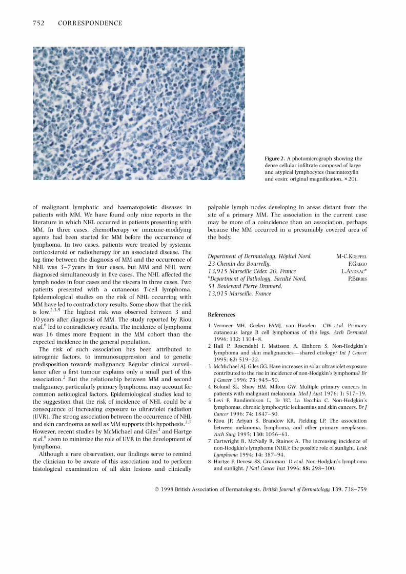
A 68-year-old man was referred in February 1989 for a MMon the back (superficial spreading type, Clark level IV, Breslowthickness 1·24 mm). The treatment consisted of wide excision.Regional lymph nodes were not palpable. A work-up failed tonote any abnormal findings. In April 1990, he developed asubcutaneous mass on the right thigh (Fig. 1). Histologicalexamination showed a very dense, non-epidermotropiccellular infiltrate involving the entire dermis and extendinginto the subcutis. The infiltrate was mainly composed of largeand atypical lymphocytes with round nuclei, immaturechromatin, one or several nucleoli and scanty cytoplasm(Fig. 2). Immunoblasts were present but rare. Immunopheno-typical staining showed that the cells were of the B phenotype,IgM positive and l chain positive. Clinical examination wasotherwise normal, as were routine laboratory examination,serum and urinary protein immunoelectrophoresis. Serologi-cal tests for human immunodeficiency virus type 1 (HIV1),HIV2 and human T-cell leukaemia virus type 1 were negative.The bone marrow was normal, as was computed tomography(CT) scanning of the thorax, abdomen and pelvis.
A diagnosis of primary cutaneous LBCL was made, forwhich the patient was administered six cycles of chemother-apy of mitoxantrone, cyclophosphamide, vincristine andprednisolone. Electron beam therapy (40 Gy) was given tothe resected mass area. The patient remained in completeremission until April 1992, when the appearance of vitiligo ledus to discover an enlarged lymph node in the left axilla.
Histological examination revealed metastatic MM. A repeat CTscan check of the thorax and abdomen was negative.Treatment with interferon a2a (2–6 million units subcuta-neously three times weekly) was given for 6 months from June1992. Between March 1994 and December 1996, the patienthad four relapses of the CBCL affecting the lower limbs only,without any lymphadenopathy or systemic extension. Eachrelapse was treated with different courses of chemotherapy,combined with localized electron beam therapy (cumulativedose, 143 Gy). In December 1997, the patient continued incomplete remission, some 4 months after the last treatment.
The incidences of NHL and MM have increased substantiallyover the last few decades. The occurrence of NHL and MM inthe same patient is still rare. When MM and NHL areassociated, either of them could occur first. There is a strongerassociation between initial NHL and the development ofsubsequent MM than the reverse. The relative risk of MM inpatients with NHL was about twofold2,3 lower than that ofsquamous cell carcinoma (about fivefold).2 MM mostly occursbetween 5 and 10 years after the lymphoma, and the risk doesnot increase with time.
In 1976, Boland et al.4 first reported the increased incidence
CORRESPONDENCE 751
q 1998 British Association of Dermatologists, British Journal of Dermatology, 139, 738–759
Figure 1. The tumour is evident on the right thigh.
of malignant lymphatic and haematopoietic diseases inpatients with MM. We have found only nine reports in theliterature in which NHL occurred in patients presenting withMM. In three cases, chemotherapy or immune-modifyingagents had been started for MM before the occurrence oflymphoma. In two cases, patients were treated by systemiccorticosteroid or radiotherapy for an associated disease. Thelag time between the diagnosis of MM and the occurrence ofNHL was 3–7 years in four cases, but MM and NHL werediagnosed simultaneously in five cases. The NHL affected thelymph nodes in four cases and the viscera in three cases. Twopatients presented with a cutaneous T-cell lymphoma.Epidemiological studies on the risk of NHL occurring withMM have led to contradictory results. Some show that the riskis low.2,3,5 The highest risk was observed between 3 and10 years after diagnosis of MM. The study reported by Riouet al.6 led to contradictory results. The incidence of lymphomawas 16 times more frequent in the MM cohort than theexpected incidence in the general population.
The risk of such association has been attributed toiatrogenic factors, to immunosuppression and to geneticpredisposition towards malignancy. Regular clinical surveil-lance after a first tumour explains only a small part of thisassociation.2 But the relationship between MM and secondmalignancy, particularly primary lymphoma, may account forcommon aetiological factors. Epidemiological studies lead tothe suggestion that the risk of incidence of NHL could be aconsequence of increasing exposure to ultraviolet radiation(UVR). The strong association between the occurrence of NHLand skin carcinoma as well as MM supports this hypothesis.2,7
However, recent studies by McMichael and Giles3 and Hartgeet al.8 seem to minimize the role of UVR in the development oflymphoma.
Although a rare observation, our findings serve to remindthe clinician to be aware of this association and to performhistological examination of all skin lesions and clinically
palpable lymph nodes developing in areas distant from thesite of a primary MM. The association in the current casemay be more of a coincidence than an association, perhapsbecause the MM occurred in a presumably covered area ofthe body.
Department of Dermatology, Hopital Nord, M-C.KOEPPEL
23 Chemin des Bourrelly, F.GREGO
13,915 Marseille Cedex 20, France L.ANDRAC**Department of Pathology, Faculte Nord, P.BERBIS
51 Boulevard Pierre Dramard,13,015 Marseille, France
References
1 Vermeer MH, Geelen FAMJ, van Haselen CW et al. Primarycutaneous large B cell lymphomas of the legs. Arch Dermatol1996; 132: 1304–8.
2 Hall P, Rosendahl I, Mattsson A, Einhorn S. Non-Hodgkin’slymphoma and skin malignancies—shared etiology? Int J Cancer1995; 62: 519–22.
3 McMichael AJ, Giles GG. Have increases in solar ultraviolet exposurecontributed to the rise in incidence of non-Hodgkin’s lymphoma? BrJ Cancer 1996; 73: 945–50.
4 Boland SL, Shaw HM, Milton GW. Multiple primary cancers inpatients with malignant melanoma. Med J Aust 1976; 1: 517–19.
5 Levi F, Randimbison L, Te VC, La Vecchia C. Non-Hodgkin’slymphomas, chronic lymphocytic leukaemias and skin cancers. Br JCancer 1996; 74: 1847–50.
6 Riou JP, Ariyan S, Brandow KR, Fielding LP. The associationbetween melanoma, lymphoma, and other primary neoplasms.Arch Surg 1995; 130: 1056–61.
7 Cartwright R, McNally R, Staines A. The increasing incidence ofnon-Hodgkin’s lymphoma (NHL): the possible role of sunlight. LeukLymphoma 1994; 14: 387–94.
8 Hartge P, Devesa SS, Grauman D et al. Non-Hodgkin’s lymphomaand sunlight. J Natl Cancer Inst 1996; 88: 298–300.
752 CORRESPONDENCE
q 1998 British Association of Dermatologists, British Journal of Dermatology, 139, 738–759
Figure 2. A photomicrograph showing thedense cellular infiltrate composed of largeand atypical lymphocytes (haematoxylinand eosin: original magnification, × 20).

Classical Kaposi’s sarcoma and chronic lymphocyticleukaemia in the same skin biopsy. Report of two cases
SIR, The association of Kaposi’s sarcoma (KS) with leukaemias(acute myeloid leukaemia, hairy-cell leukaemia and chroniclymphocytic leukaemia: CLL) has been reported by numerousauthors; in particular, KS and CLL have been observed quitefrequently in the same patient, but the simultaneous presenceof the two diseases in a lymph node or a skin lesion has seldombeen reported.1–5 We describe two cases in which both KS andCLL were detected in the same cutaneous lesion.
Patient 1 was a 73-year-old Sardinian man admitted inOctober 1995 with generalized KS. Multiple lesions weredistributed over the lower and upper extremities, the trunkand the neck. Lesions slightly elevated above the skin surface,covered by normal skin, were seen over the trunk. He alsopresented bullous lesions on the lower extremities. There wasinvolvement of the mucous membranes of the mouth andconjunctiva. Multiple skin biopsies were taken from differentlesions. A skin biopsy of a typical KS lesion from the right armrevealed not only KS but also a leukaemic infiltrate. In theupper and middle dermis there were spindle cells withinterspersed bundles of collagen, vascular channels lined byendothelial cells, extravasation of erythrocytes and a markedinfiltrate of small monomorphous lymphocytes with incon-spicuous nucleoli and scant cytoplasm. The same histologicalfeatures were seen in the deep dermis in a biopsy of a slightlyelevated lesion covered by normal skin from the back. Finally,a bullous lesion from the lower extremities showed a largebulla in the subepidermic dermis and featured a typical KSlesion. The lesions had first appeared 20 years previously, buthad never been investigated. Examination showed hepato-splenomegaly, and firm multiple lymph nodes were palpablein several areas. The patient’s mother had been affected byKS.
A cervical lymph node biopsy showed diffuse architectural
effacement by a monomorphous infiltrate of small lympho-cytes with round nuclear contours and scant cytoplasmwithout apparent mitotic activity. Immunophenotypic studyrevealed positive staining for CD20, CD45 and CD5. Bonemarrow aspirate smears revealed in the paratrabecularlocation marked hypercellularity with nodular infiltration bysmall lymphocytes, expressing CD20. The lymph node biopsyand bone marrow aspirate smears led to the diagnosis of CLL.
Patient 2 was an 83-year-old Sardinian man, withangiomatous plaques and small isolated nodules on theupper and lower extremities, and first seen in March 1996.His past history revealed CLL of 13 years’ duration but he hadnot undergone immunosuppressive therapy. The skin lesionshad appeared about 6 months before our examination. Severalskin biopsies were performed. These showed a highlyvascularized lesion with a multicentric proliferation ofspindle-shaped cells accompanied by erythrocyte extravasa-tion. Intracytoplasmic hyaline globules were evident in a fewcells. Furthermore, in the same sample there was a diffusemonomorphous infiltrate of small lymphocytes with hyper-chromic nuclei (Fig. 1).
While the association of KS with lymphoproliferativedisorders in the same patient has often been noted, only afew instances of such an association in the same biopsyspecimen have been observed. These include an associationwith Hodgkin’s disease in the same lymph node;6 withmalignant lymphoma and angiofollicular hyperplasia in thesame lymph node;7 and with follicular lymphoma in the samecutaneous lesions.8 KS and CLL not infrequently affect thesame patient, but the observation of the coexistence of bothdiseases in the same skin lesion or in the same lymph node isexceedingly rare: indeed, in the literature there are only fivecases. Of these, Weshler et al.1 and Koren et al.3 described casesin which these two diseases appeared in a single lymph node,but where there was no other manifestation of KS elsewhere inthe patient. According to Weshler et al. the presence of the two
CORRESPONDENCE 753
q 1998 British Association of Dermatologists, British Journal of Dermatology, 139, 738–759
Figure 1. Photomicrograph showing typicalfeatures of Kaposi’s sarcoma with aleukaemic infiltrate (haematoxylin andeosin; original magnification, × 20).

diseases in the same lymph node could have been coincidental,and the authors doubted whether the two diseases couldderive from a common stem cell; indeed, if there had been ahistogenetic connection between the two diseases, they wouldhave expected to find such associations more frequently. Baronet al.5 described a case in which both KS and CLL were detectedin the same cutaneous lesions. To explain this coexistence,they suggested the possibility of local interactions between twoneoplastic populations through the secretion of angiogenicand growth factors and cytokines.
Institutes of Dermatology, F.COTTONI
*Pathological Anatomy and I.M.MASIA
†Haematology and Endocrinology, S.COSSU*University of Sassari, M.A.MONTESU
Viale Mancini 5, 07100 Sassari, S.PARDINI†Italy G.MASSARELLI*
References
1 Weshler Z, Leviatan A, Krasnokuki D, Kopolovitch J. PrimaryKaposi’s sarcoma in lymph nodes concurrent with chroniclymphatic leukemia. Am J Clin Pathol 1979; 71: 234–7.
2 Contu L, Carcassi C, La Nasa G et al. A case of classical Kaposi’ssarcoma in B-cell chronic lymphocytic leukemia (B-CLL). Tumori1986; 72: 365–74.
3 Koren G, Okon E, Zlotnick A. Coexistence of Kaposi’s sarcoma andchronic lymphocytic leukemia in the same lymph node. ActaHaematol (Basel) 1985; 74: 234–5.
4 Sachs W, Gray R. Kaposi’s sarcoma and lymphatic leukemia. ArchDermatol Syphilol 1945; 51: 325–9.
5 Baron JM, Pizarro A, Berrocal A et al. Kaposi’s sarcoma associatedwith chronic B lymphocytic leukemia. Coexistence of both diseasesin skin lesions. Med Clin (Barc) 1991; 96: 700–2.
6 Massarelli G, Tanda F, Denti S. Primary Kaposi’s sarcoma andHodgkin’s disease in the same lymph node. Am J Clin Pathol 1982;78: 107–11.
7 Xerri L, Guigou V, Lepid H et al. Lymphadenopathic tumorexhibiting intermingled features of Kaposi’s sarcoma, malignantlymphoma, and angiofollicular hyperplasia. Arch Pathol Lab Med1991; 115: 1162–6.
8 Robert C, Blanc F, Moulonguet I et al. Lymphome folliculaire etmaladie de Kaposi affectant successivement les memes territoirescutanes. Ann Dermatol Venereol 1990; 117: 949–51.
The use of cyclosporin in a child with generalized pustularpsoriasis
SIR, Generalized pustular psoriasis (GPP) is a serious andresistant form of psoriasis which is rare during the first decadeof life. We describe the use of cyclosporin to treat GPP in a 9-year-old boy.
A boy, born in September 1987, started to suffer fromguttate psoriasis when aged 2 years. There was no familyhistory of psoriasis. When aged 9 years, he developed for thefirst time a superficial pustular eruption on an erythematousbase, which spread all over the body (Fig. 1). Geographiclesions were seen on the tongue. On examination, he was
pyrexial (39 8C) and had erythema and numerous pustulesoccurring in clusters. Vital signs were normal. Routinelaboratory tests were normal. Serum creatinine was73 mmol/L (normal: 50–120). Cultures from the throat andpustules showed no fungal or bacterial growth.
A skin biopsy specimen showed changes compatible withpustular psoriasis, namely acanthosis with elongation of reteridges, parakeratosis and spongiosis with subcorneal andintraepidermal collections of neutrophils. Direct immunofluor-escence was negative. Wet dressings and low-potency topicalcorticosteroids were given initially, but no improvement wasachieved. While the flares of fever and pustules persisted, westarted cyclosporin at 3 mg/kg per day. Control was achievedafter 2 weeks of therapy and the dosage was lowered to 2 mg/kg per day at the second month. Recurrence of some lesionsfollowed the tapering of the dosage to 1 mg/kg per day at theeighth month of therapy. The relapses could not be controlledby topical corticosteroids and the dosage of cyclosporin wasincreased to 3 mg/kg per day with satisfactory control of thedisease. During treatment with cyclosporin, full blood count,
754 CORRESPONDENCE
q 1998 British Association of Dermatologists, British Journal of Dermatology, 139, 738–759
Figure 1. Clinical appearance of the patient prior to cyclosporintherapy.

liver and kidney function and creatinine clearance weremonitored every 2 weeks while he was in hospital and everymonth thereafter. Serum creatinine and creatinine clearanceremained within the normal range. The treatment wasstopped after 11 months. The patient is still free of psoriasisin his second month of follow-up.
Pustular psoriasis may appear as localized pustularpsoriasis, which runs a chronic course, or as a more severecondition, GPP. GPP is particularly rare during childhood.There are fewer than 100 children with GPP described in theliterature. It mainly affects boys. There is a history of precedingguttate psoriasis in 12% of those who present with GPP in thefirst decade, in contrast to the figure of 85% for adults withGPP.1,2 Our patient had had guttate psoriasis since the age of2 years.
Cyclosporin is an immunosuppressive agent that is effec-tively used in the treatment of chronic plaque psoriasis.Experience with localized forms of pustular psoriasis hasinvolved a greater number of patients, in whom cyclosporinwas shown to be effective at a dosage of 2·5–14 mg/kg perday.3,4 Experience gained from transplant patients reveals thatthe immune reactivity of small children seems to be enhancedand to necessitate more intensive immunosuppression.Besides, the pharmacokinetics of cyclosporin, includingintestinal absorption, distribution in body fluids and tissues,metabolism and elimination, is age-dependent. Therefore,children need comparatively higher doses of cyclosporin thanadults to achieve adequate blood levels. A newly introducedoral formulation of cyclosporin (Neoral , Novartis), whichincorporates the drug in a microemulsion preconcentrate, hasshown an improvement in dose linearity and a faster, moreconsistent rate of absorption with less pharmacokineticvariability.5 The superiority of this new formulation may bethe reason for the result achieved at a lower dosage in ourpatient.
Therapeutic agents used in childhood GPP include systemicadrenocorticotrophic hormone, a corticosteroid, methotrex-ate, etretinate or acitretin, and PUVA. Cyclosporin appears tobe an effective alternative treatment.
Department of Dermatology, N.ALLI
Ankara Numune Hospital, E.GUNGOR
06100 Altindag, Ankara, Turkey G.KARAKAYALI
N.LENK
F.ARTUZ
References
1 Khan SA, Peterkin GAG, Mitchell PC. Juvenile generalized pustularpsoriasis: a report of five cases and a review of the literature. ArchDermatol 1972; 105: 67–72.
2 Zelickson BD, Muller SA. Generalized pustular psoriasis inchildhood: report of thirteen cases. J Am Acad Dermatol 1991; 24:186–94.
3 Meinardi MMHM, Westerhof W, Bos JD. Generalized pustularpsoriasis (von Zumbush) responding to cyclosporin A. Br J Dermatol1987; 116: 269–70.
4 Matsumura N, Takematsu H, Saijo S et al. Exanthematic type ofpustular psoriasis consisting of two types of pustular lesion. ActaDerm Venereol (Stockh) 1991; 71: 442–4.
5 Brodehl J. Cyclosporine therapy for transplantation in pediatricpatients. Consensus statements on the optimal use of cyclosporinein pediatric patients. Transplant Proc 1994; 26: 2759–62.
Topical tacrolimus for pyoderma gangrenosum
SIR, Pyoderma gangrenosum is a chronic inflammatory skindisease, characterized clinically by the recurring developmentof destructive and painful skin ulcers. In 50–75% of cases anassociated condition can be identified including inflammatorybowel disease, rheumatoid arthritis, chronic autoimmunehepatitis, non-Hodgkin’s lymphoma, acute and chronicmyeloid leukaemia and carcinoma of the colon, prostate andbreast.
The mainstay of therapy is the systemic administration ofhigh doses of corticosteroids, usually in combination withother immunosuppressive agents such as methotrexate,azathioprine or cyclophosphamide.1 Recently, there havebeen several reports on the efficacy of cyclosporin A in thetreatment of pyoderma gangrenosum.2 However, its use inpyoderma gangrenosum is limited due to systemic side-effectsand the possible deterioration of underlying disease. Topicalforms of cyclosporin have been tried in inflammatorydermatoses, but they do not seem to be effective, presumablydue to insufficient penetration of the skin. Tacrolimus hasalmost the same effects at the cellular level as cyclosporin, is10–100 times more potent and has a lower molecular weight.Recent studies in atopic dermatitis suggest that topicaltacrolimus penetrates human skin and is effective in thecontrol of cutaneous inflammation. Here we report thesuccessful treatment of two patients suffering from pyodermagangrenosum with tacrolimus ointment alone and in combi-nation with systemic cyclosporin.
The first patient, a 48-year-old man, was diagnosed ashaving pyoderma gangrenosum in November 1995 on thebasis of clinical and histopathological findings. Laboratory,sonographic and radiological examinations revealed noevidence for an associated disease. A 10 × 7 cm ulcerativelesion in the left inguinal region was treated with systemicprednisolone (120 mg/day) and low-dose cyclosporin (4 mg/kg per day) and healed within 4 months. One year later, thepatient developed two ulcers (3–5 cm in diameter) afterminimal trauma at the lower left leg. Systemic steroids werestarted and combined with the topical application ofcyclosporin in olive oil.3 This treatment failed to inducesignificant improvement and low-dose oral cyclosporin wassuccessfully re-administered over 3 months. Two monthsafter cessation of therapy, the patient presented with a 2·5 cminflammatory nodule showing the beginnings of ulcerationabove the right clavicula. We decided to start a monotherapyof this early lesion with topical tacrolimus. Tacrolimusointment (0·1%) was prepared according to a recentlypublished formula,4 with 1 mg capsules (Prograf , FujisawaPharmaceutical Company, Osaka, Japan) as the source oftacrolimus. Rapid clearing of the lesion was achieved by
CORRESPONDENCE 755
q 1998 British Association of Dermatologists, British Journal of Dermatology, 139, 738–759

topical application of this ointment twice daily for 3 weeks.During this time, serum levels of tacrolimus, determined 10–12 h after the last application on days 2, 4 and 10, werebelow the therapeutic range (5–15 mg/L). New inguinalulcerations that developed several weeks later respondedfaster to a combination of systemic cyclosporin and topicaltacrolimus than those which had previously been treatedwith systemic cyclosporin alone. The combination therapythus allowed a reduction of the cyclosporin dose andtreatment time.
The second patient, a 27-year-old woman with a 4-year-
history of ulcerative colitis, presented with acute inflamma-tory ulcers of pyoderma gangrenosum at the medial andlateral malleoli of both feet (5–10 cm in diameter). The ulcershad developed from erythematous nodules over the last3 weeks before admission. She was taking 5-aminosalicylicacid (3 g/day) and budesonide (9 mg/day) for the treatment ofulcerative colitis. Endoscopy revealed an active left-sidedcolitis, and laboratory tests showed a marked elevation ofacute phase reactants, a thrombocytosis and leucocytosis.Systemic cyclosporin was started at 5 mg/kg per day. Inaddition, one large ulcer (left medial malleolus; Fig. 1a) wastreated with 0·1% tacrolimus ointment applied once dailyunder occlusion with a hydrocolloid dressing (Comfeel Plus,Coloplast, Hamburg, Germany) for 3 weeks, while the otherlesions were treated with hydrocolloid dressings only. After3 weeks, all lesions were treated with colloid dressings aloneuntil complete remission was achieved. The dose of cyclos-porin was tapered to 2 mg/kg per day over 3 months anddiscontinued thereafter. The additional treatment with tacro-limus ointment clearly accelerated the clinical response of thepyoderma gangrenosum lesions: a marked reduction in thesize of the ulcer ($ 50%) occurred after 5 weeks in the lesionreceiving tacrolimus ointment, but not before 10 weeks in theother lesions. Again, systemic tacrolimus levels remainedbelow 5 mg/L during topical treatment. The ulcers remainedhealed at 5 months (Fig. 1b).
In contrast to steroids and cytostatic agents, cyclosporinand tacrolimus (FK 506), both of which are members of themacrolide family of immunosuppressive antibiotics, have theireffect mainly by an action on T lymphocytes. After complexformation with cytosolic binding proteins, they interfere withthe activation of the transcription factor NF-AT, and inhibitthe transcription of proinflammatory cytokine genes includinginterleukin (IL)-2, IL-4, interferon-g and tumour necrosisfactor-a.5 Systemic therapy with immunosuppressive macro-lides has been shown to be effective in a variety of lymphocyte-driven inflammatory dermatoses including psoriasis, atopicdermatitis and pyoderma gangrenosum. To circumventassociated adverse effects, topical treatment strategies havealso been evaluated. From several investigations it can beconcluded that the percutaneous penetration of cyclosporininto intact skin is too small to deliver effective concentrationswith topical formulations.6 Resorption and topical immuno-suppression have been given as reasons for the application ofcyclosporin preparations on ulcerated lesions of pyodermagangrenosum,3 but the effect on the lymphocytic vasculitis inthe advancing border of the lesion, possibly the main target ofmacrolide therapy, appears questionable. Intralesionalinjection of cyclosporin may be an alternative,7 but is apainful procedure and difficult to perform on larger pyodermagangrenosum lesions. In contrast to cyclosporin, topicalpreparations of tacrolimus have been shown to give effectivecontrol of cutaneous inflammation in animal and humanmodels of contact dermatitis6 and in atopic dermatitis.8 Fromour cases we conclude that topical tacrolimus alone may beeffective in the treatment of early lesions of pyodermagangrenosum and may be beneficial in combination with
756 CORRESPONDENCE
q 1998 British Association of Dermatologists, British Journal of Dermatology, 139, 738–759
Figure 1. Extensive ulceration due to pyoderma gangrenosum in a 27-year-old woman with ulcerative colitis (a). In addition to a 3-monthcourse of systemic cyclosporin, this lesion was treated with tacrolimusointment under occlusion for 3 weeks. The healed appearance at5 months (b).

systemic cyclosporin in the treatment of advanced lesions.Side-effects were not noted, and pain caused by an ulcer inpatient 2 ceased as early as 5 days after occlusive treatmentwith tacrolimus ointment was started. Topical tacrolimus maytherefore offer an additional treatment option in cases wheresystemic immunosuppressive therapy is limited by adverseeffects or associated disorders, and in the control of relapsingpyoderma gangrenosum after cessation of systemic immuno-suppression.
Department of Dermatology, K.REICH
Georg-August-University, Von-Siebold Strasse 3, C.VENTE
37075 Gottingen, Germany C.NEUMANN
References
1 Chow RKP, Ho VC. Treatment of pyoderma gangrenosum. J AmAcad Dermatol 1996; 34: 1047–60.
2 Martis WL, Ellis CN, Griffiths CEM, Lazarus GS. Treatment ofpyoderma gangrenosum with cyclosporin. Arch Dermatol 1992;182: 1060–4.
3 Theissen U, Luger TA, Schwarz T. Erfolgreiche topischeAnwendung von Cyclosporin A bei Pyoderma gangraenosum.Hautarzt 1996; 47: 132–5.
4 Aoyama H, Tabata N, Tanaka M et al. Successful treatment ofresistant facial lesions of atopic dermatitis with 0. 1% FK506ointment (Letter). Br J Dermatol 1995; 133: 494–6.
5 Schreiber SL, Crabtree GR. The mechanism of action of cyclosporinA and FK506. Immunol Today 1992; 13: 136–42.
6 Lauerma AI, Maibach HI. Topical FK506—clinical potential orlaboratory curiosity. Dermatology 1994; 188: 173–6.
7 Mrowietz U, Christophers E. Clearing of pyoderma gangrenosum byintralesional cyclosporin A. Br J Dermatol 1991; 125: 499.
8 Ruzicka T, Bieber T, Schopf E et al. A short-term trial oftacrolimus ointment for atopic dermatitis. European TacrolimusMulticenter Atopic Dermatitis Study Group. N Engl J Med 1997;337: 816–21.
A systematic review of five systemic treatments for severepsoriasis
SIR, We wish to draw readers’ attention to some of theproblems associated with a recent systematic review oftreatments for severe psoriasis by Spuls et al.1 The authorshave clearly undertaken a lot of work to produce this review,and they are to be congratulated for highlighting the shortageand poor quality of clinical trials for commonly usedtreatments for psoriasis. Similar problems have been noticedin other fields of dermatology.2 The paper has also been helpfulin estimating the magnitude of potential benefit of particulartreatments in selected, albeit undefined, groups of patients.
Quantitative pooling of published, randomized, controlledstudies (meta-analysis) is problematic enough in itself,3 but toattempt quantitative pooling of data from non-randomized,uncontrolled studies is very hazardous. It is difficult to makeany meaningful comparisons of the effectiveness of treatmentsreviewed in the article by Spuls et al.1 based on clearance ratesand the proportions of patients with good, moderate and poorresponse, because of the large potential for selection bias inpatients selected for different treatment modalities. We do not
know whether the baseline scores for patients in differenttreatment modalities were different. Such differences can lead toproblems, such as regression to the mean and artificially inflatedtreatment responses in some groups when compared withothers. It is also unclear to us whether a 90% improvement inaverage global score is clinically equivalent to a 90% change inbody surface area involvement without knowing more about theclinical meaning of such changes, the anchors used for thescales and the psychometric properties of those scales. In ouropinion, it is very difficult to make any comparisons between thetreatments highlighted in this systematic review, and we wouldrecommend caution to any person attempting to developguidelines for the treatment of psoriasis based on these results.Occasionally, meta-analysis of observational studies is donewhen no randomized, controlled studies are ethically possible oravailable.4 Where possible, such quantitative summaries shouldbe confined to randomized, controlled clinical trial data. Weagree with the authors that more high-quality, randomized,controlled trials comparing commonly used treatments forplaque psoriasis are now clearly needed.
Queen’s Medical Centre, H.C.WILLIAMS
Department of Dermatology, A.LI WAN PO*Nottingham NG 7 2UH, U.K. D.MURREL†*Department of Pharmaceutical Science, L.NALDI‡University of Nottingham, T.DIEPGEN§Nottingham NG7 2RD, U.K.†Department of Dermatology,The St George Hospital,Kogarah, NSW 2217, Australia‡Clinica Dermatologica,Ospedali Rinniti di Bergamo,24100 Bergamo, Italy§Department of Dermatology,University of Erlangen, 8520 Erlangen, Germany
References
1 Spuls PI, Whitkam L, Bossuyt PMM, Bos JD. A systematic review offive systemic treatments for severe psoriasis. Br J Dermatol 1997;137: 943–9.
2 Bigby M, Stern RS, Bigby JA. An evaluation of method reporting inuse in clinical trials in dermatology. Arch Dermatol 1985; 121:1394–9.
3 Egger M, Davey-Smith G, Phillips AN. Meta-analysis, principles andprocedures. Br Med J 1997; 315: 1533–7.
4 Egger M, Schneider M, Davey-Smith G. Meta-analysis, spuriousprecision? Meta-analysis of observational studies. Br Med J 1998;315: 140–4.
Reply
Sir, We agree with Dr Williams and his colleagues thatquantitative pooling of published randomized controlledstudies is preferred above the pooling of data from randomizedand non-randomized uncontrolled studies together. Greatcaution is required in the interpretation of these findings, aswe have indicated in our paper. With Dr Williams andcolleagues, we firmly believe that opinion-based medicine
CORRESPONDENCE 757
q 1998 British Association of Dermatologists, British Journal of Dermatology, 139, 738–759

should be replaced by evidence-based medicine. We believethat our review is a starting point in the development ofevidence-based practice guidelines for the treatment of severepsoriasis. The systematic review highlights the scarcity of goodquality data on the effectiveness of the available treatmentmodalities in severe psoriasis. Future trials are clearly needed.
Departments of Dermatology and P.I.SPULS
*Biostatistics and Epidemiology, L.WHITKAMP
University of Amsterdam, P.M.M.BOSSUYT
Academical Medical Centre, J.D.BOS
PO Box 22770, 1100 DE Amsterdam,The Netherlands
The pustular eruption of ulcerative colitis
SIR, I was interested to read the report by Sarkany et al.1 of apatient with ulcerative colitis who developed a pustulareruption which they attributed to Sweet’s syndrome. However,I think that their interpretation is incorrect: it certainlystretches the concept of Sweet’s disease.
About 16 years ago, a 35-year-old woman was admittedwith severe ulcerative colitis and a widespread pustulareruption involving the trunk and limbs with scattered bullaecontaining pus (Fig. 1a). She was treated by the admittingphysicians with oral antibiotics but became worse. The puswas sterile and, after a few days, some of the pustules brokedown to form ulcers (Fig. 1b). At this time I was asked to seeher and she clearly had pyoderma gangrenosum with amixture of bullae containing pus and ulcerative lesions typicalof pyoderma gangrenosum. She was treated with highdose oral prednisolone and this led to a prompt healing of alllesions.
I believe that the patient described by Sarkany andcolleagues had bullous pyoderma gangrenosum associatedwith ulcerative colitis.
Department of Dermatology, A.P.WARIN
Royal Devon and Exeter Hospital (Wonford),Exeter EX2 5DW, U.K.
References
1 Sarkany RPE, Burrows NP, Grant JW et al. The pustular eruption ofulcerative colitis: a variant of Sweet’s syndrome? Br J Dermatol1998; 138: 365–6.
Reply
SIR, We are most interested in Dr Warin’s case, in which thepatient’s vesiculopustular eruption was a form of bullouspyoderma gangrenosum. A similar case has been describedpreviously, in which some pustules eventually developed intoulcers of pyoderma gangrenosum.1
However, in contrast to Dr Warin’s case, no ulcers everdeveloped in our patient or in four other colitis patients2–4
reported to have this syndrome of pustular eruption, fever andneutrophilia. In one of these cases,4 there were no ulcers eventhough the eruption was present for several months. Thismakes it more difficult to describe our case as ‘bullouspyoderma gangrenosum’. Whether this entity is described asatypical Sweet’s syndrome, as a variant of pyoderma gang-renosum, or simply as the ‘pustular eruption of ulcerativecolitis’, perhaps we can all agree that it does represent adistinctive neutrophilic dermatosis that affects patients withulcerative colitis.
Departments of Dermatology and R.P.E.SARKANY
*Histopathology, Addenbrooke’s Hospital, N.P.BURROWS
Cambridge CB2 2QQ, U.K. J.W.GRANT*R.J.PYE
P.G.NORRIS
758 CORRESPONDENCE
q 1998 British Association of Dermatologists, British Journal of Dermatology, 139, 738–759
Figure 1. (a) The pustular eruption.(b) Ulcerated lesion typical of pyodermagangrenosum.

References
1 Callen JP, Woo TY. Vesiculopustular eruption in a patient withulcerative colitis. Arch Dermatol 1985; 121: 399–404.
2 Barnes L, Lucky AW, Bucuvalas JC et al. Pustular pyodermagangrenosum associated with ulcerative colitis in childhood. J AmAcad Dermatol 1986; 15: 608–14.
3 O’Loughlin S, Perry HO. A diffuse pustular eruption associated withulcerative colitis. Arch Dermatol 1978; 114: 1061–4.
4 Fenske NA, Gern JE, Pierce D et al. Vesiculopustular eruption ofulcerative colitis. Arch Dermatol 1983; 119: 664–9.
General practitioner excision of malignant melanoma
SIR, In 1991, an audit of malignant melanoma excision(n ¼ 42) was performed in the Mid and North StaffordshireHealth Authorities. The speciality of the medical practitionerwho performed the initial excision of each of the melanomas
was as follows: dermatologist 22 (52%), plastic or generalsurgeon 10 (24%) and general practitioner 10 (24%). Thesefigures are far more worrying than those found in the study ofKorshid et al.1 Further audits should be performed to see if therehas been a reduction in excisions by general practitioners.
Department of Dermatology, J.S.C.ENGLISH
Queen’s Medical Centre,University Hospital,Nottingham NG7 2UH, U.K.
References
1 Khorshid SM, Pinney E, Newton Bishop JA. Melanoma excision bygeneral practitioners in North-East Thames region, England. Br JDermatol 1998; 138: 412–17.
News and Notices
The first World Congress of the International Academy ofCosmetic Dermatology, 28–31 January 1999, St Julian’s,Malta
The inaugural meeting of the International Academy ofCosmetic Dermatology will be held in Malta on 28–31January 1999. It will include sessions on general dermatology,cosmetic dermatology, cosmetic surgery and cosmetic chem-istry. Information is available from Dr L.E.Millikan, IACDSecretary General, Tulane University Medical Center, 1430Tulane Avenue SL73, New Orleans, Louisiana 70112, USA.Fax: +1 504 587 7382; e-mail: [email protected]
8th Congress of the European Academy of Dermatology andVenereology, 29 September–3 October 1999, Amsterdam,The Netherlands
The 8th Congress of the European Academy of Dermatologyand Venereology will be held on 29 September–3 October1999 at the Amsterdam RAI Congress Centre. Furtherinformation can be obtained from Eurocongress Management,J. van Gooyenkade 11, 1075 HPAmsterdam, The Netherlands.Tel.: +31 20 679 3411; fax: +31 20 673 7306, e-mail:[email protected]
Third International Conference on Adjuvant Therapy ofMalignant Melanoma, 19–20 March 1999, London, UK
The Third International Conference on Adjuvant Therapy of
Malignant Melanoma will be held at the Royal College ofPhysicians, London, UK of 19 and 20 March 1999. Furtherdetails can be obtained from Dr Spyros Retsas, Chairman of theConference, at CCI Limited, 2 Palmerston Court, PalmerstonWay, London SW8 4AJ, UK. Tel.: +44 (0)171 720 0800; fax:+44 (0)171 720 7177;e-mail: melanoma@confcomm. demon.co.ukConference website:http://www.confcomm.demon.co.uk/INTRODUCTION.html
Fourth International Symposium on Cosmetic Efficacy,Strategies for the 21st Century, 10–12 May 1999, NewYork, NY, U.S.A.
International experts from industry, academia and regulatoryagencies will meet to address issues related to identifyinginnovative approaches to improving cosmetic efficacy assess-ment. The two-day meeting will include special lectures ontopics such as fragrances, colour of skin and cosmetic anddrug products in the treatment of acne. There will also bepanel discussions and scientific poster sessions. The meetingis co-sponsored by the Departments of Dermatology atthe College of Physicians and Surgeons of Columbia University,Case Western Reserve University and the University ofAachen. For additional information contact: ISCE, 820West Superior Avenue, Suite 340, Cleveland, Ohio 44113-1800, U.S.A. Tel.: þ216/579/9344; fax.: þ216/579/9333;e-mail: [email protected]
Erratum
Varma S, Lanigan SW. Pseudoporphyria caused by nabumetone. Br J Dermatol 1998; 138: 549–50.
The dose of nabumetone should read 1 gtwice daily, not 2 gtwice daily as stated.
CORRESPONDENCE 759
q 1998 British Association of Dermatologists, British Journal of Dermatology, 139, 738–759


![PUSTULAR PSORIASIS RESPONDING TO PROBIOTICS – A NEW … 4/9... · 2012-11-22 · With anecdotal reference [4] of probiotics helping ... PUSTULAR PSORIASIS RESPONDING TO PROBIOTICS](https://static.fdocuments.in/doc/165x107/5e63831b9667476f8503b6bc/pustular-psoriasis-responding-to-probiotics-a-a-new-49-2012-11-22-with.jpg)


![A case of simultaneous acute generalized exanthematous ...AGEP is Generalized Pustular Psoriasis (GPP) [8] “GPP shares many of the clinical and immunological findings and the presentation](https://static.fdocuments.in/doc/165x107/5fddc90e28884f1c641049be/a-case-of-simultaneous-acute-generalized-exanthematous-agep-is-generalized-pustular.jpg)