
The Urinary System Ch 24 Human Anatomy Sonya Schuh-Huerta, Ph.D. C. Babaian.
74
The Urinary System The Urinary System Ch 24 Ch 24 Human Anatomy Sonya Schuh-Huerta, Ph.D. Sonya Schuh-Huerta, Ph.D. C. Babaian
-
Upload
thomasina-harper -
Category
Documents
-
view
223 -
download
0
Transcript of The Urinary System Ch 24 Human Anatomy Sonya Schuh-Huerta, Ph.D. C. Babaian.
The Urinary SystemThe Urinary SystemCh 24Ch 24
Human Anatomy
Sonya Schuh-Huerta, Ph.D.Sonya Schuh-Huerta, Ph.D.
C. Babaian
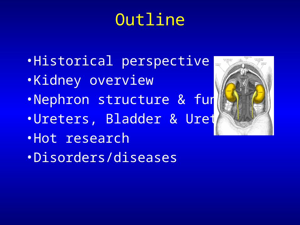
Outline
• Historical perspective
• Kidney overview
• Nephron structure & function
• Ureters, Bladder & Urethra
• Hot research
• Disorders/diseases
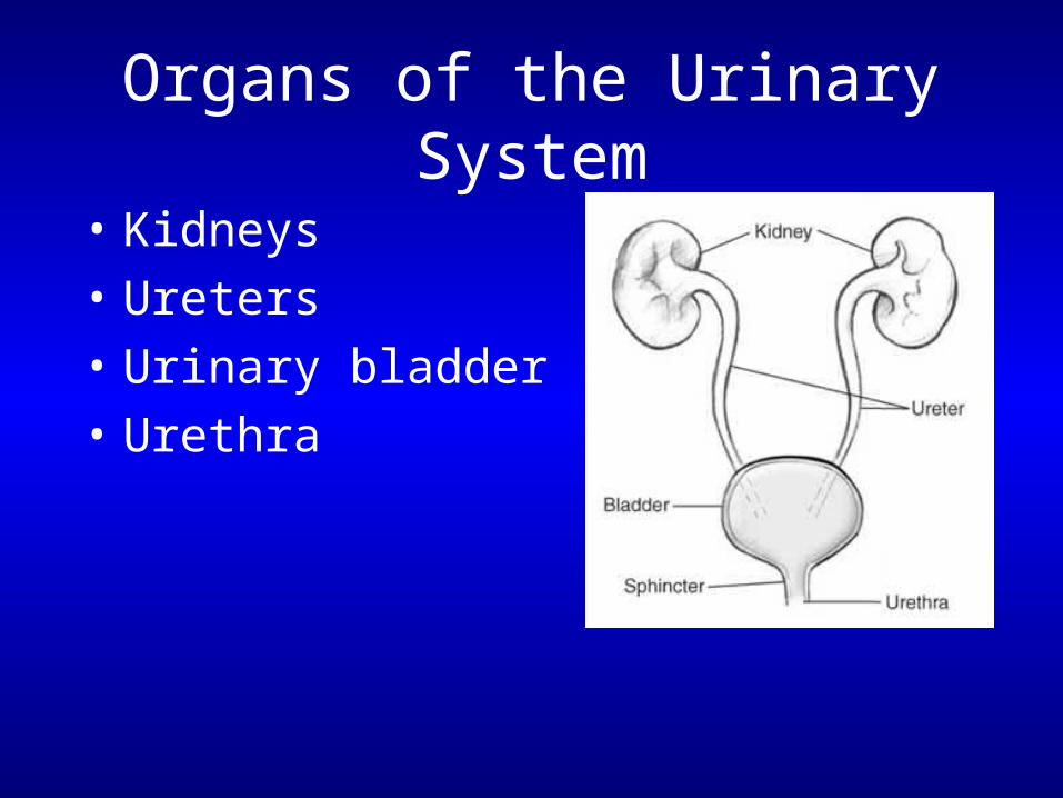
Organs of the Urinary System
• Kidneys
• Ureters
• Urinary bladder
• Urethra
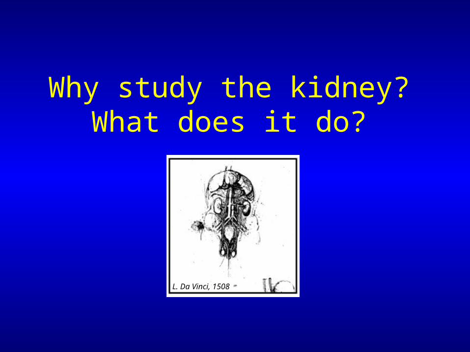
Why study the kidney?What does it do?
L. Da Vinci, 1508
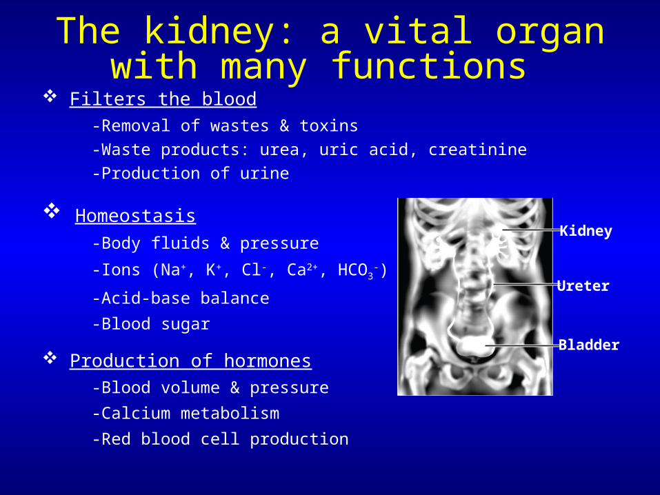
The kidney: a vital organ with many functions
Filters the blood
-Removal of wastes & toxins
-Waste products: urea, uric acid, creatinine
-Production of urine
Homeostasis
-Body fluids & pressure
-Ions (Na+, K+, Cl-, Ca2+, HCO3-)
-Acid-base balance
-Blood sugar
Production of hormones
-Blood volume & pressure
-Calcium metabolism
-Red blood cell production
Kidney
Ureter
Bladder

• Historical interest in resolving urine’s composition & relation to health
• Alchemists of Medieval Europe thought urine contained gold!
• Urea isolated in 1773; 1st organic compound to be artificially synthesized
Urine color wheel
Gerrit Dou, 1617
What can urine tell us?
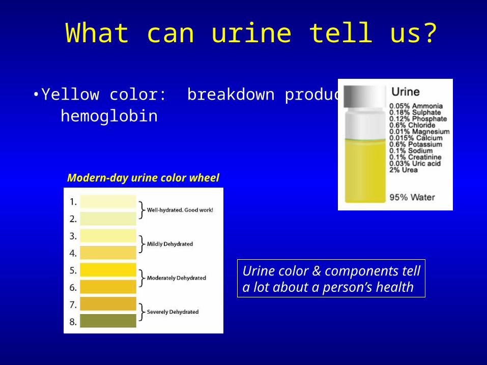
• Yellow color: breakdown products of hemoglobin
Urine color & components tella lot about a person’s health
What can urine tell us?
Modern-day urine color wheel
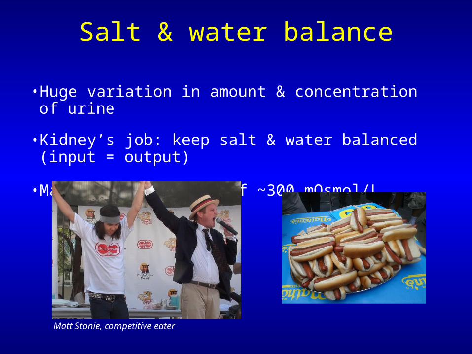
• Huge variation in amount & concentration of urine
• Kidney’s job: keep salt & water balanced (input = output)
• Maintain osmolarity of ~300 mOsmol/L
Salt & water balance
Matt Stonie, competitive eater
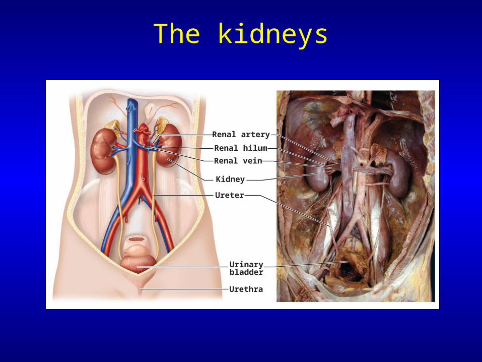
The kidneys
Renal artery
Renal hilum
Renal vein
Kidney
Ureter
Urinarybladder
Urethra
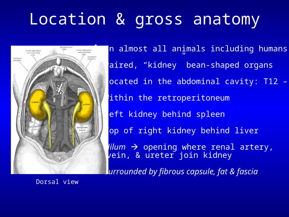
Location & gross anatomy
In almost all animals including humans
Paired, “kidney” bean-shaped organs
Located in the abdominal cavity: T12 – L3
Within the retroperitoneum
Left kidney behind spleen
Top of right kidney behind liver
Hilum opening where renal artery, vein, & ureter join kidney
Surrounded by fibrous capsule, fat & fasciaDorsal view
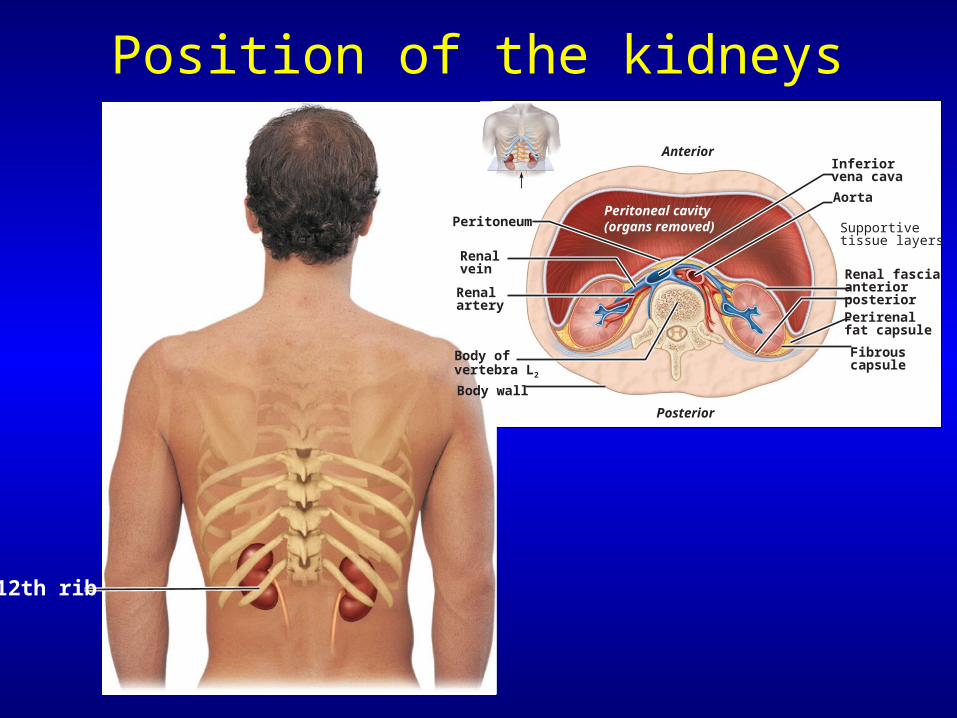
Position of the kidneys
12th rib
Body wall
Perirenalfat capsule
Renalartery
Renalvein
Inferiorvena cava
Aorta
Fibrouscapsule
Renal fasciaanteriorposterior
Supportivetissue layers
Body ofvertebra L2
PeritoneumPeritoneal cavity(organs removed)
Anterior
Posterior
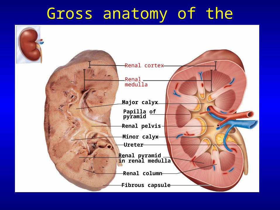
Gross anatomy of the kidney
Renal cortex
Renalmedulla
Major calyx
Papilla ofpyramid
Renal pelvis
Ureter
Minor calyx
Renal column
Renal pyramidin renal medulla
Fibrous capsule
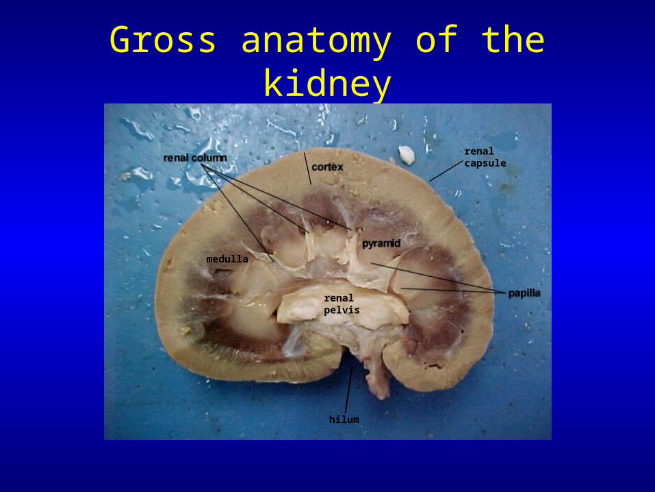
Gross anatomy of the kidney
medulla
hilum
renal capsule
renal pelvis
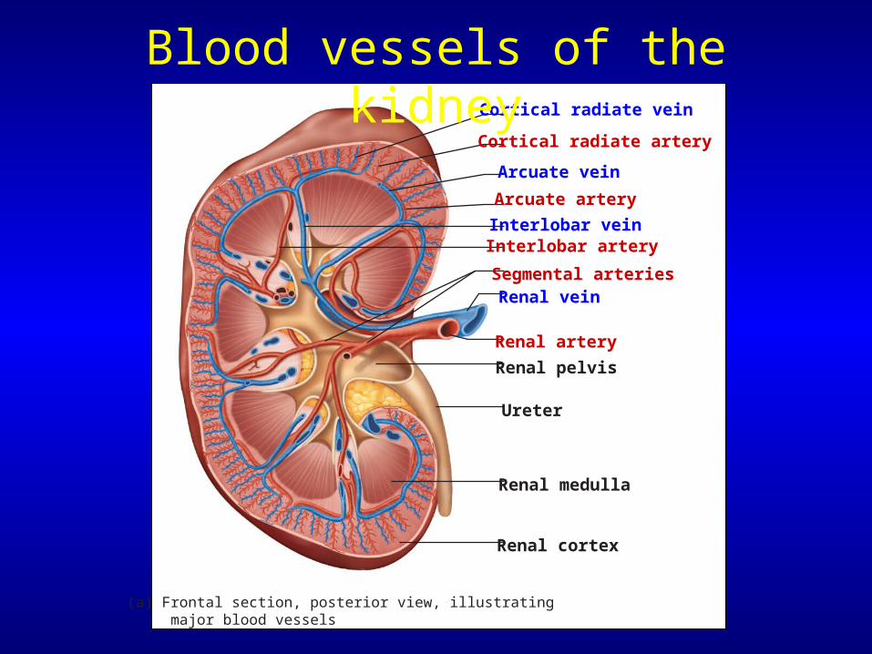
Cortical radiate vein
Cortical radiate artery
Arcuate vein
Arcuate artery
Interlobar veinInterlobar artery
Segmental arteries
Renal artery
Renal vein
Renal pelvis
Ureter
Renal medulla
Renal cortex
(a) Frontal section, posterior view, illustrating major blood vessels
Blood vessels of the kidney
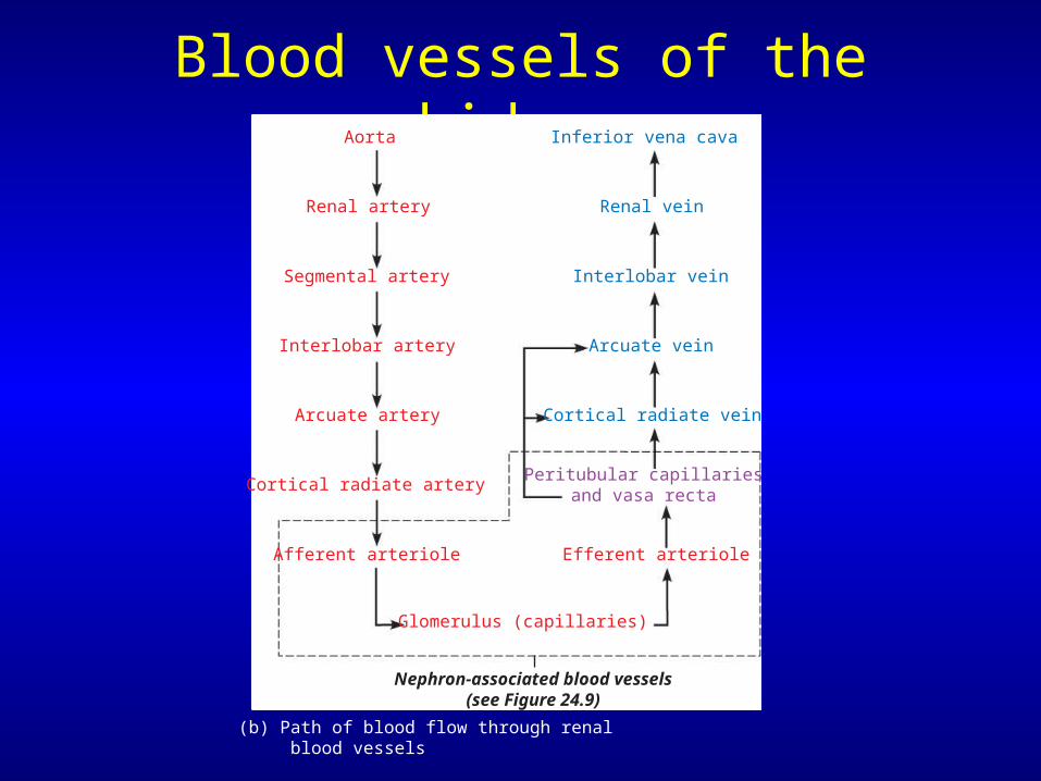
Blood vessels of the kidneyAorta
Renal artery
Segmental artery
Interlobar artery
Arcuate artery
Cortical radiate artery
Afferent arteriole
Glomerulus (capillaries)
Nephron-associated blood vessels(see Figure 24.9)
Inferior vena cava
Renal vein
Interlobar vein
Arcuate vein
Cortical radiate vein
Peritubular capillariesand vasa recta
Efferent arteriole
(b) Path of blood flow through renal blood vessels
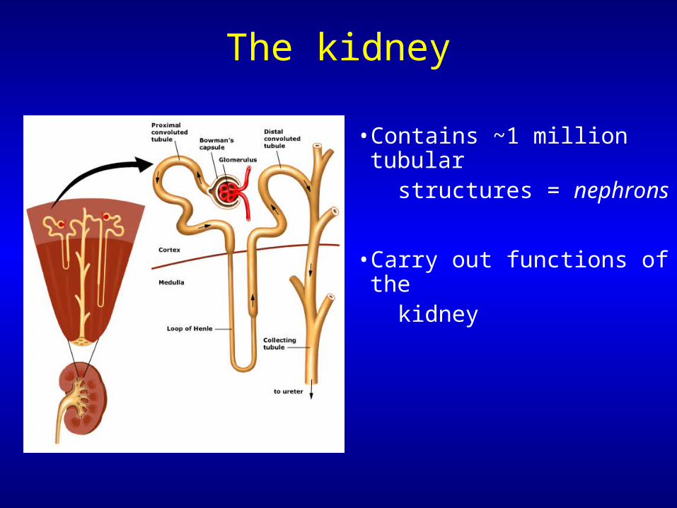
The kidney
• Contains ~1 million tubular structures = nephrons
• Carry out functions of the kidney
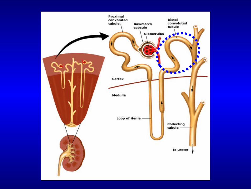
At the microscopic level
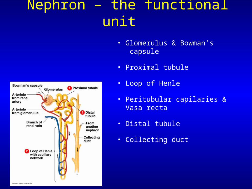
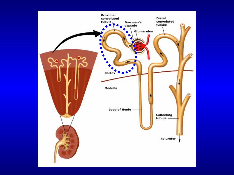
• Glomerulus & Bowman’s capsule
• Proximal tubule
• Loop of Henle
• Peritubular capilaries & Vasa recta
• Distal tubule
• Collecting duct
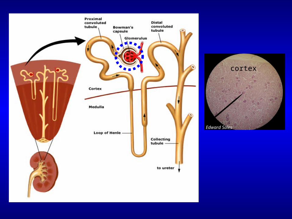
Nephron – the functional unit
Edward Sales
cortex
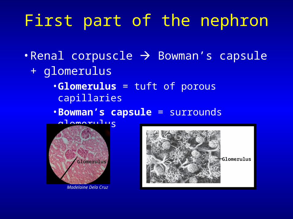
First part of the nephron
• Renal corpuscle Bowman’s capsule + glomerulus
• Glomerulus = tuft of porous capillaries • Bowman’s capsule = surrounds glomerulus
GlomerulusGlomerulus
Madelaine Dela Cruz
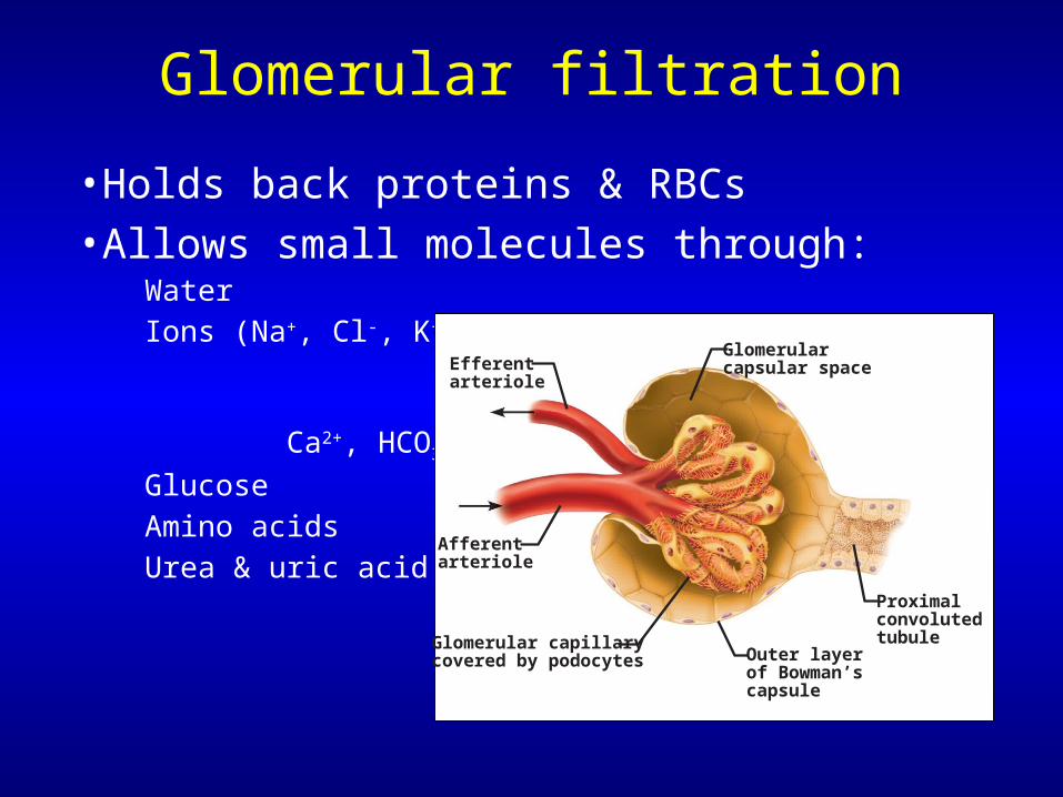
Glomerular filtration
• Holds back proteins & RBCs• Allows small molecules through:
WaterIons (Na+, Cl-, K+,
Ca2+, HCO3-)
GlucoseAmino acidsUrea & uric acid
Glomerular capillarycovered by podocytes
Proximalconvolutedtubule
Outer layerof Bowman’s capsule
Afferentarteriole
Glomerularcapsular spaceEfferent
arteriole

Glomerular filtration
• Glomeruli
– Porous capillaries– Fed & drained by
afferent & efferent arterioles
•Efferent arteriole has smaller diameter
– Filter 1 liter of fluid every 8 minutes!•180 liters of fluid per day•Total blood volume is filtered ~60X/day!
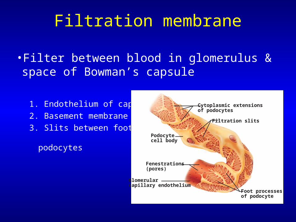
Filtration membrane
• Filter between blood in glomerulus & space of Bowman’s capsule
1. Endothelium of capillary
2. Basement membrane
3. Slits between foot processes of podocytes
Glomerularcapillary endothelium
Fenestrations(pores)
Podocytecell body
Foot processesof podocyte
Filtration slits
Cytoplasmic extensionsof podocytes
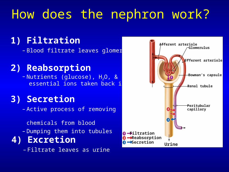
How does the nephron work?
1) Filtration– Blood filtrate leaves glomeruli
2) Reabsorption– Nutrients (glucose), H2O, & essential ions taken back into blood
3) Secretion– Active process of removing
undesirable chemicals from blood
– Dumping them into tubules
4) Excretion– Filtrate leaves as urine
Afferent arterioleGlomerulus
Efferent arteriole
Bowman’s capsule
Renal tubule
Peritubularcapillary
Urine
FiltrationReabsorptionSecretion
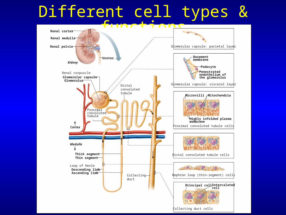
Different cell types & functions
Fenestrated endothelium of the glomerulus
Microvilli
Cortex
Medulla
Podocyte
Basementmembrane
Mitochondria
Highly infolded plasmamembrane
Proximal convolutedtubule
Distalconvolutedtubule
Descending limbLoop of Henle
Ascending limb
Glomerular capsuleRenal corpuscle
Glomerulus
Thick segment
Collectingduct
Intercalatedcell
Principal cell
Thin segment
Proximal convoluted tubule cells
Glomerular capsule: parietal layer
Glomerular capsule: visceral layer
Distal convoluted tubule cells
Nephron loop (thin-segment) cells
Collecting duct cells
Renal cortex
Renal medulla
Renal pelvis
UreterKidney
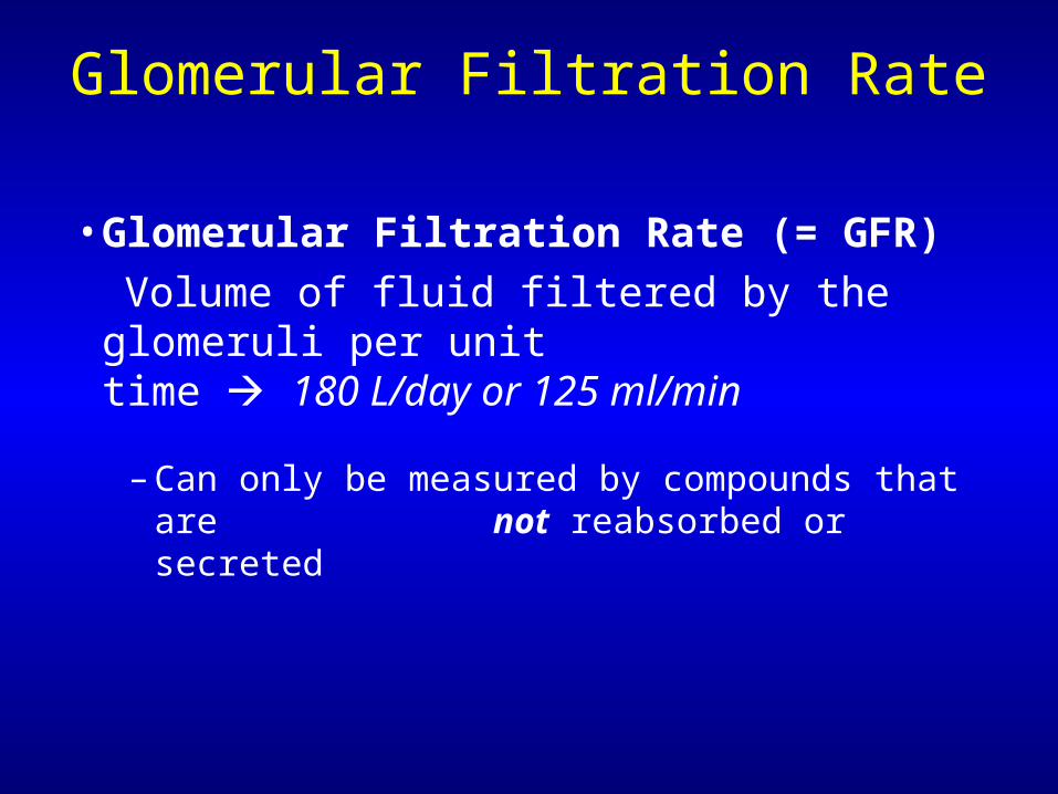
Glomerular Filtration Rate
• Glomerular Filtration Rate (= GFR)
Volume of fluid filtered by the glomeruli per unit time 180 L/day or 125 ml/min
– Can only be measured by compounds that are not reabsorbed or secreted
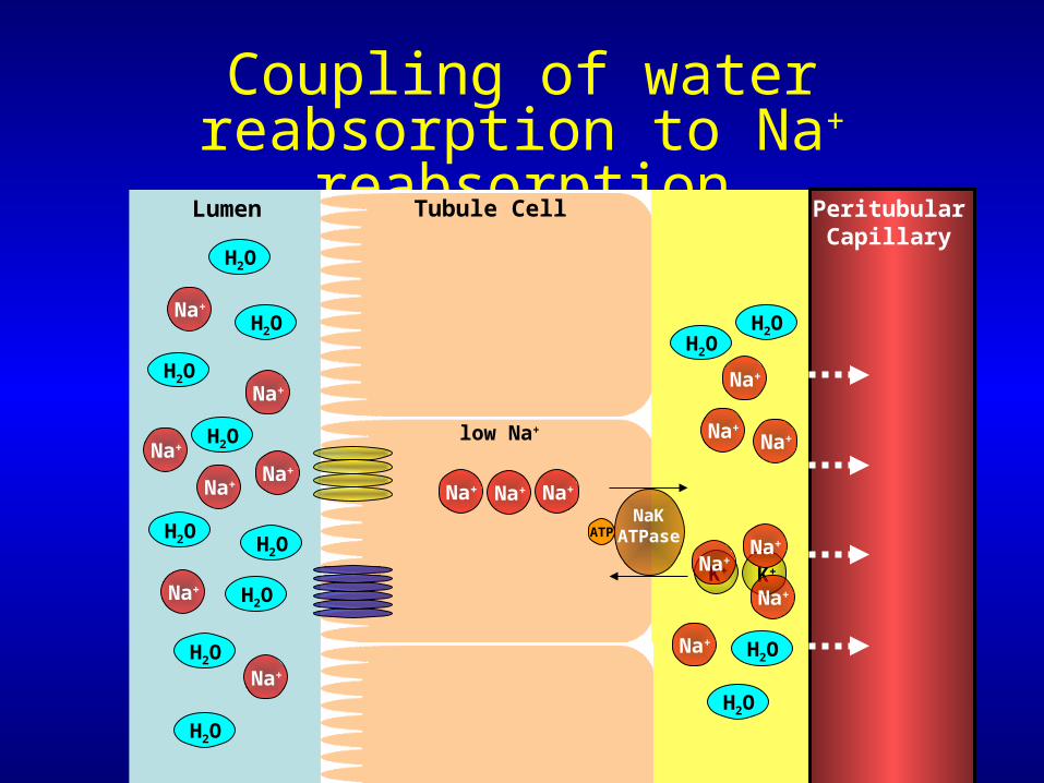
Coupling of water reabsorption to Na+ reabsorption
Tubule Cell PeritubularCapillary
Lumen
H2O
Na+
Na+
Na+
Na+
Na+
Na+ Na+ Na+
K+K+
H2O
H2O
H2O
H2O
H2O
H2O
Na+
H2O
H2O
Na+
ATPNaK
ATPase
low Na+
Na+
Na+Na+
Na+
Na+
Na+
Na+
H2O
H2O
H2OH2O
Proximal convoluted tubule
• Most solutes are reabsorbed here• All glucose is reabsorbed• Drugs/toxins are secreted• Filtrate is isoosmotic
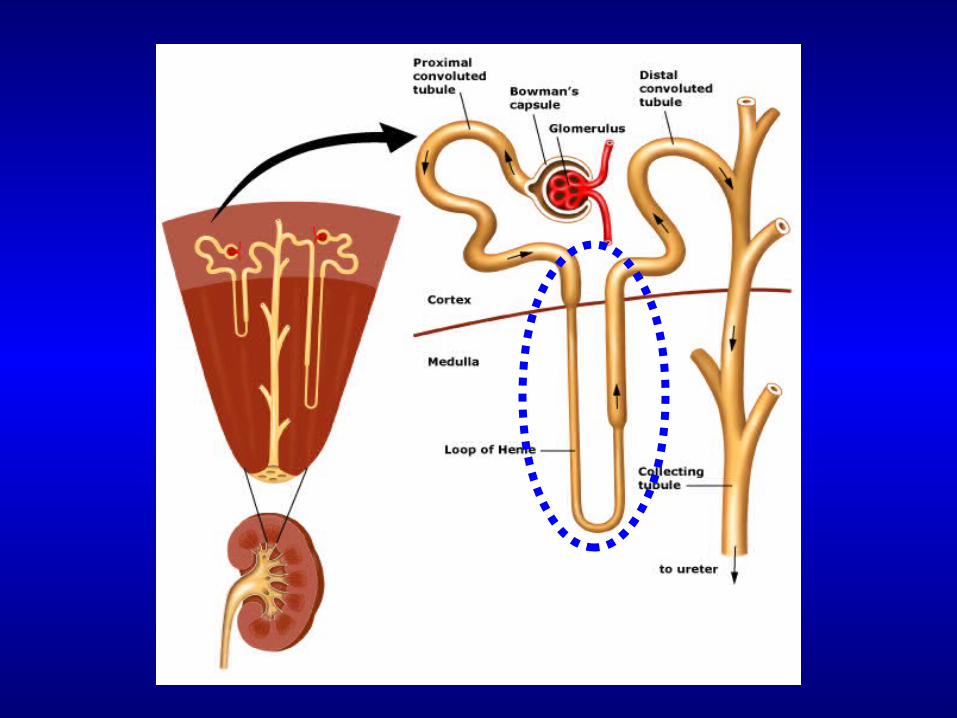
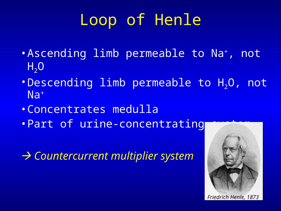
Loop of Henle
• Ascending limb permeable to Na+, not H2O• Descending limb permeable to H2O, not Na+
• Concentrates medulla• Part of urine-concentrating system
Countercurrent multiplier system
Friedrich Henle, 1873
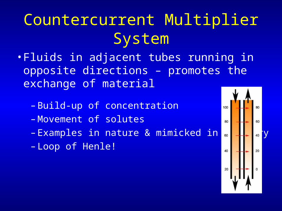
Countercurrent Multiplier System
• Fluids in adjacent tubes running in opposite directions – promotes the exchange of material
– Build-up of concentration– Movement of solutes– Examples in nature & mimicked in industry– Loop of Henle!
Loop of Henle
Corticomedullaryjunction
Afferent arterioleEfferentarteriole
Vasa recta
Peritubularcapillaries
Efferentarteriole
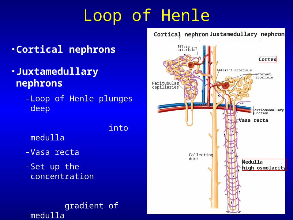
Cortical nephron Juxtamedullary nephron
• Cortical nephrons
• Juxtamedullary nephrons
–Loop of Henle plunges deep into medulla
–Vasa recta
–Set up the concentration gradient of medulla
–Create driving force for maximal H2O reabsorption from collecting ducts
Cortex
Medullahigh osmolarity
Collectingduct
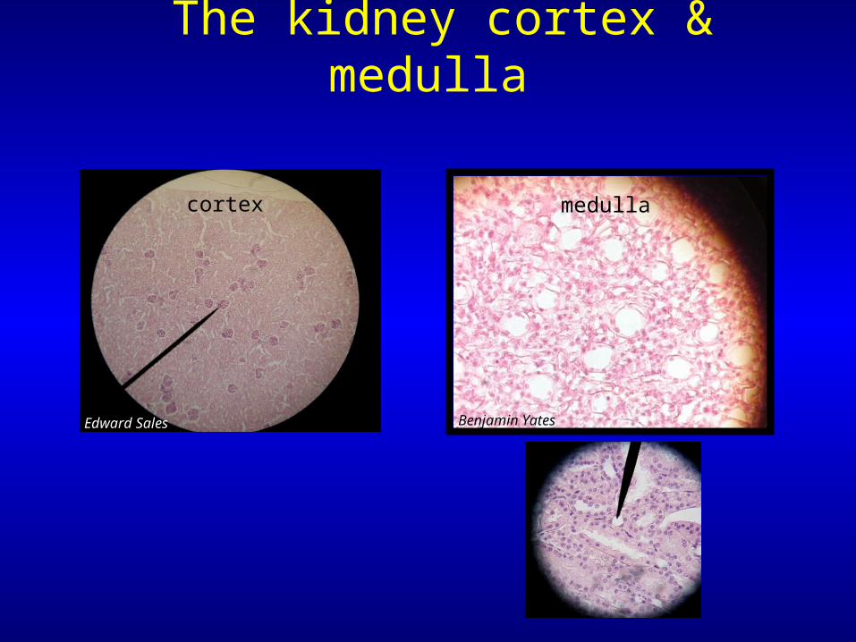
The kidney cortex & medulla
Edward Sales
cortex
Benjamin Yates
medulla
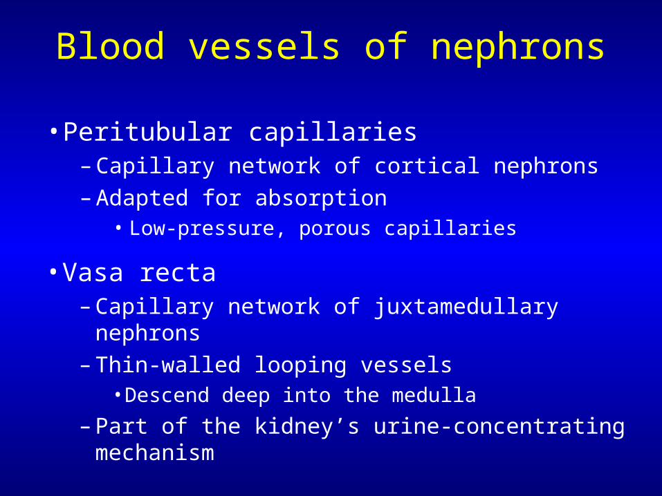
Blood vessels of nephrons
• Peritubular capillaries– Capillary network of cortical nephrons– Adapted for absorption
• Low-pressure, porous capillaries
• Vasa recta– Capillary network of juxtamedullary nephrons– Thin-walled looping vessels
• Descend deep into the medulla
– Part of the kidney’s urine-concentrating mechanism
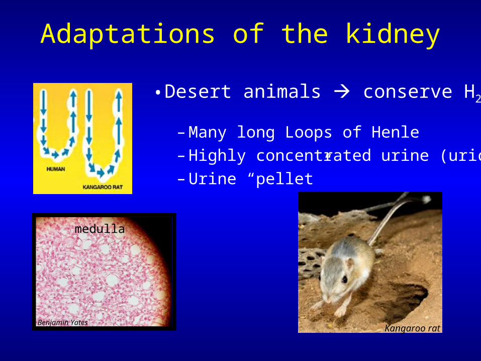
Adaptations of the kidney
• Desert animals conserve H2O
– Many long Loops of Henle– Highly concentrated urine (uric acid)– Urine “pellet”
Kangaroo ratBenjamin Yates
medulla
Distal convoluted tubule
• Permeable to (actively pumps) Na+ out of tubule• Impermeable to H2O• Filtrate hypoosmotic
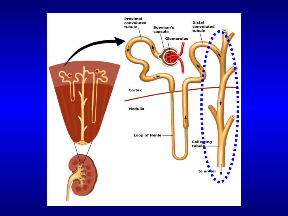
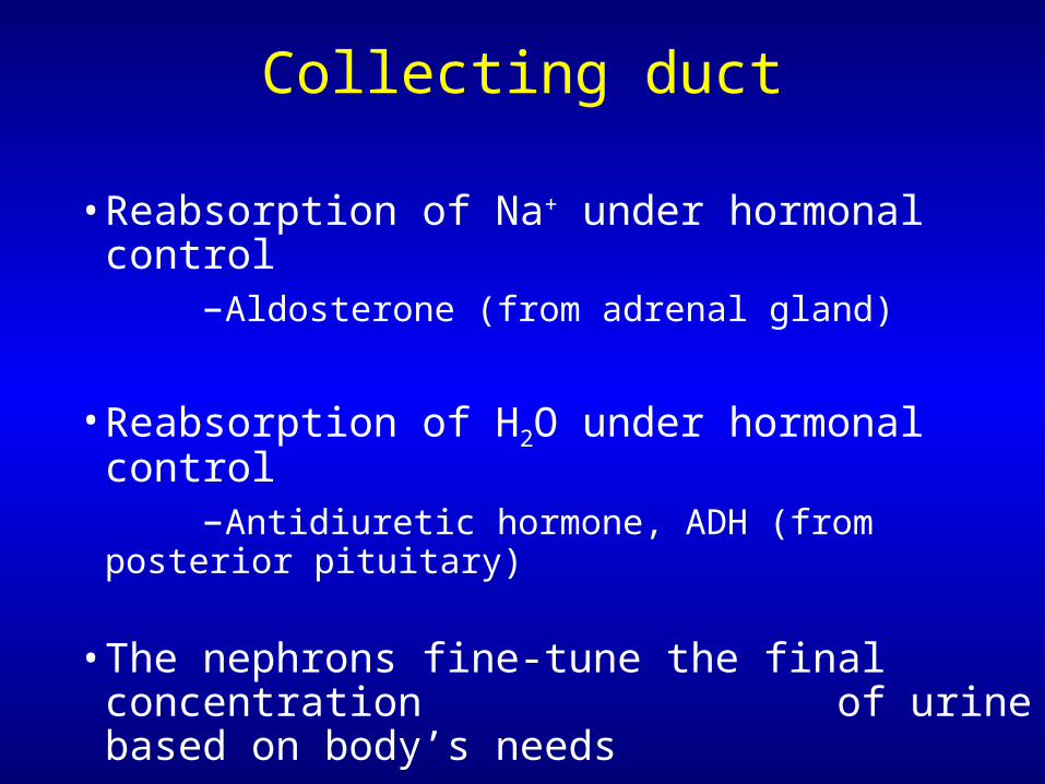
Collecting duct
• Reabsorption of Na+ under hormonal control –Aldosterone (from adrenal gland)
• Reabsorption of H2O under hormonal control –Antidiuretic hormone, ADH (from posterior pituitary)
• The nephrons fine-tune the final concentration of urine based on body’s needs
How does the nephron fine tunethe reabsorption of Na+
and water?
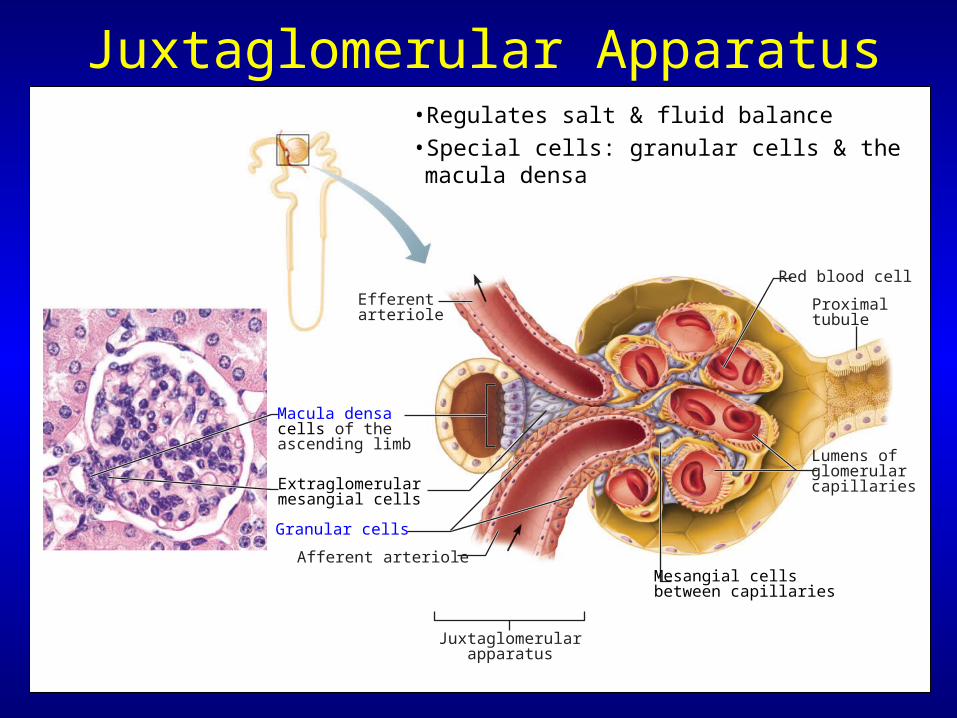
Juxtaglomerular Apparatus
Red blood cell
Proximaltubule
Lumens of glomerularcapillaries
Efferentarteriole
Macula densa cells of the ascending limb
Granular cells
Extraglomerularmesangial cells
Afferent arteriole
Juxtaglomerularapparatus
Mesangial cellsbetween capillaries
• Regulates salt & fluid balance
• Special cells: granular cells & the macula densa
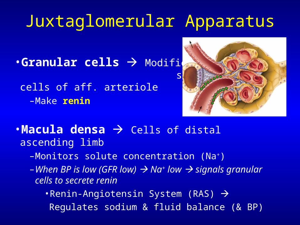
Juxtaglomerular Apparatus
• Macula densa Cells of distal ascending limb–Monitors solute concentration (Na+)
–When BP is low (GFR low) Na+ low signals granular cells to secrete renin
•Renin-Angiotensin System (RAS)
Regulates sodium & fluid balance (& BP)
• Granular cells Modified smooth muscle cells of aff. arteriole
–Make renin
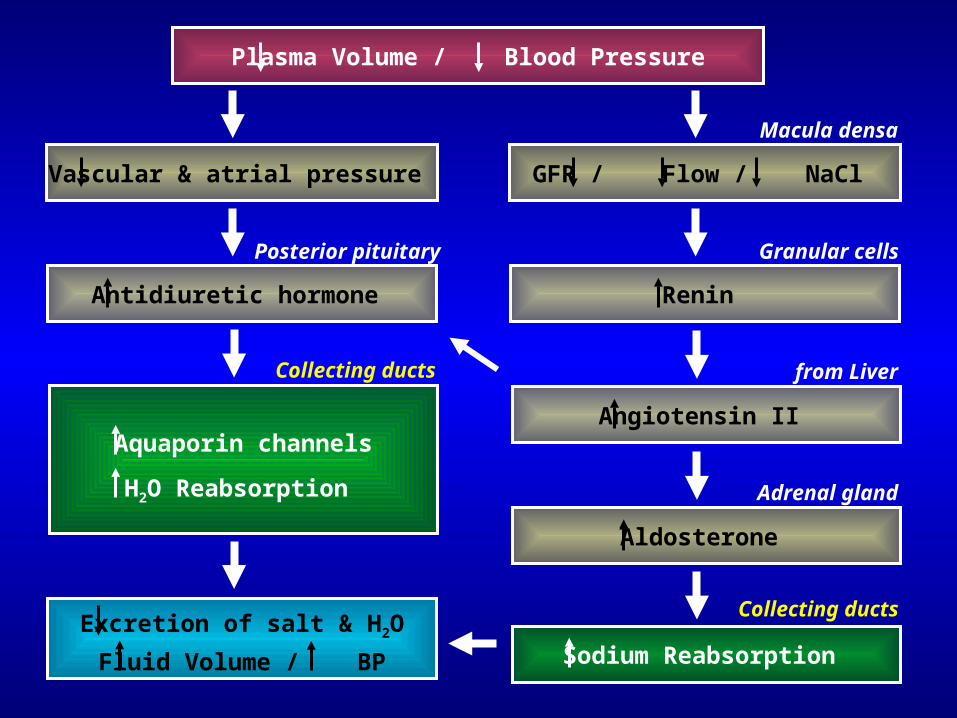
Plasma Volume / Blood Pressure
GFR / Flow / NaCl
Macula densa
Renin
Granular cells
Angiotensin II
from Liver
Aldosterone
Adrenal gland
Sodium Reabsorption
Collecting ducts
Vascular & atrial pressure
Antidiuretic hormone
Posterior pituitary
Aquaporin channels
H2O Reabsorption
Collecting ducts
Excretion of salt & H2O
Fluid Volume / BP
• Many drugs interrupt steps of this system
• Used to treat high BP (hypertension), congenital heart disease, kidney disease, etc. diuretics
• Caffeine & alcohol also diuretics
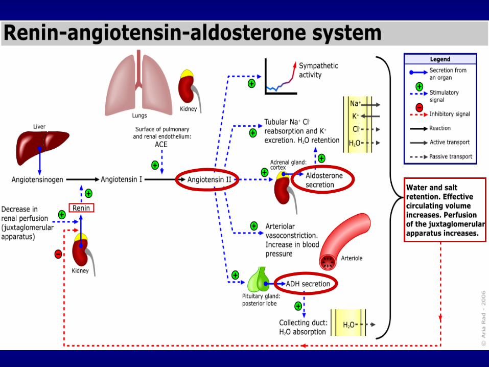
Renin-Angiotensin System

Rest of the urinary system: Ureters
• Carry urine from the kidneys to the urinary bladder
• Oblique entry into bladder prevents backflow of urine
• Histology of ureter– Mucosa transitional epithelium– Muscularis 2 layers
• Inner longitudinal layer• Outer circular layer
– Adventitia typical connective tissue
Transitional Epithelium – Review…
• Description: – Has characteristics of stratified cuboidal &
stratified squamous– Superficial cells dome-shaped when bladder
is relaxed, squamous when full• Function: permits distension of urinary organs
by urine• Location: epithelium of urinary bladder, ureters,
proximal urethra
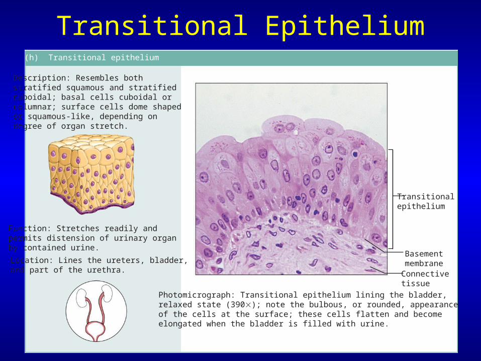
Transitional Epithelium(h) Transitional epithelium
Description: Resembles both stratified squamous and stratified cuboidal; basal cells cuboidal or columnar; surface cells dome shaped or squamous-like, depending ondegree of organ stretch.
Function: Stretches readily and permits distension of urinary organ by contained urine.
Location: Lines the ureters, bladder, and part of the urethra.
Photomicrograph: Transitional epithelium lining the bladder,relaxed state (390); note the bulbous, or rounded, appearanceof the cells at the surface; these cells flatten and becomeelongated when the bladder is filled with urine.
BasementmembraneConnectivetissue
Transitionalepithelium
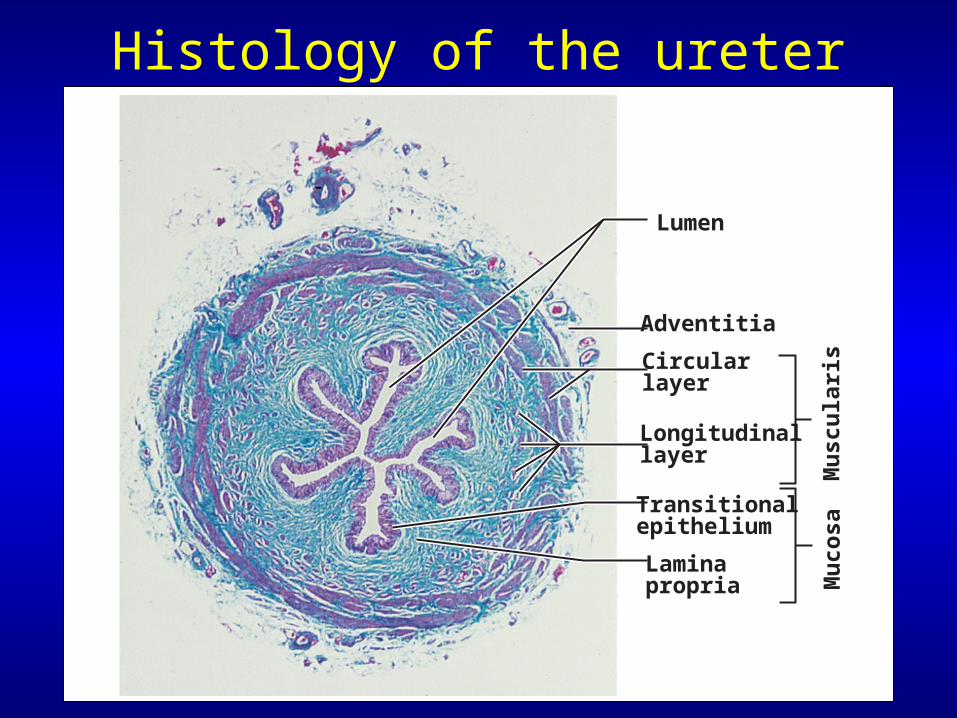
Histology of the ureter
Lumen
Adventitia
Circularlayer
Longitudinallayer
Transitionalepithelium
Laminapropria M
uco
saM
usc
ula
ris
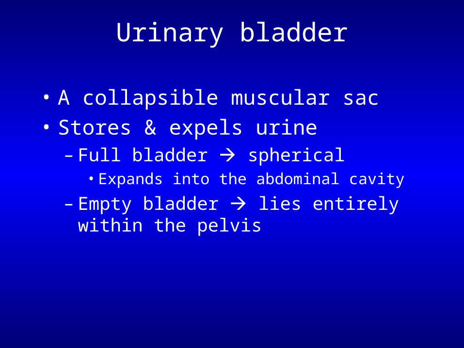
Urinary bladder
• A collapsible muscular sac
• Stores & expels urine– Full bladder spherical
• Expands into the abdominal cavity
– Empty bladder lies entirely within the pelvis
Urinary bladder
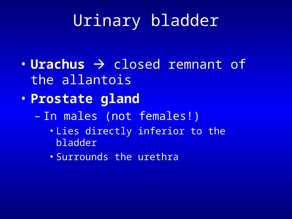
• Urachus closed remnant of the allantois
• Prostate gland– In males (not females!)
• Lies directly inferior to the bladder• Surrounds the urethra
Urinary bladder
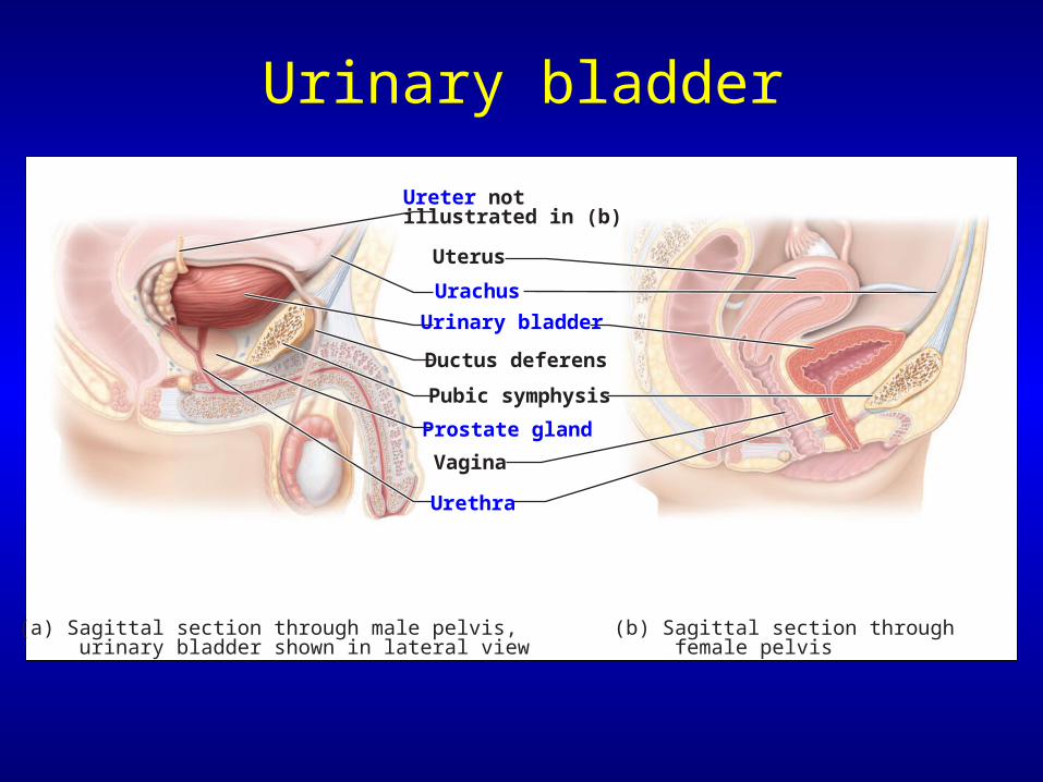
Ureter notillustrated in (b)
Uterus
Urinary bladder
Ductus deferens
Pubic symphysis
Prostate gland
Vagina
Urethra
(a) Sagittal section through male pelvis, urinary bladder shown in lateral view
(b) Sagittal section through female pelvis
Urachus
Urinary bladder
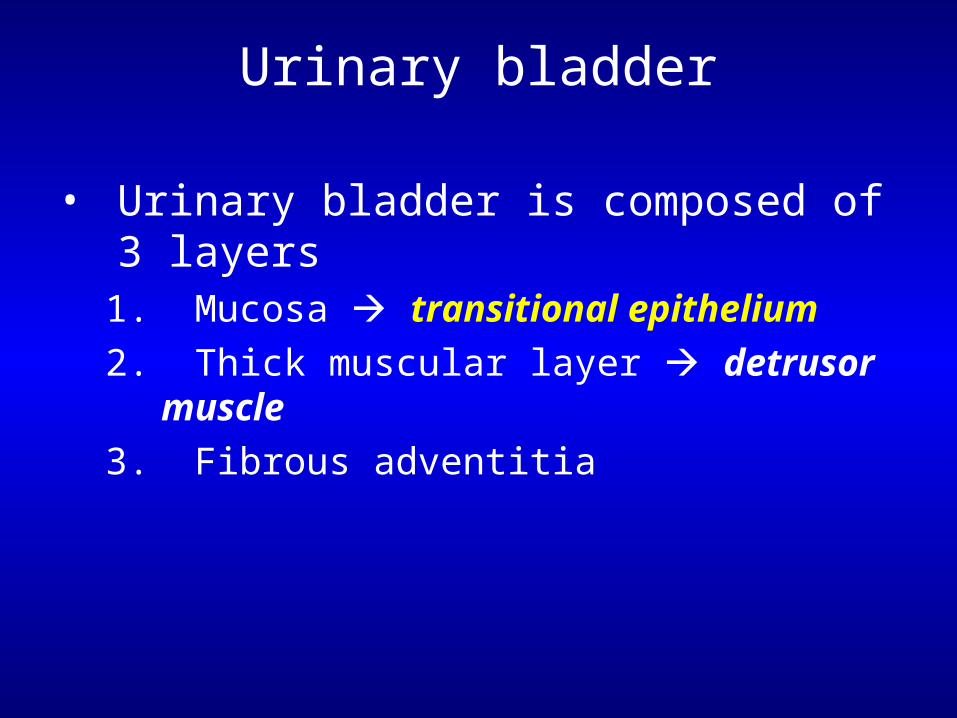
• Urinary bladder is composed of 3 layers1. Mucosa transitional epithelium
2. Thick muscular layer detrusor muscle
3. Fibrous adventitia

Histology of the urinary bladder
Basementmembrane
Laminapropria
Transitionalepithelium
(a) Micrograph of the bladder wall (17X) (b) Epithelium lining the lumen of the bladder (360X)
Lumen of bladder
Adventitia(with fat cells)
Muscularlayer(detrusor)
Transitionalepithelium
Laminapropria
Urethra
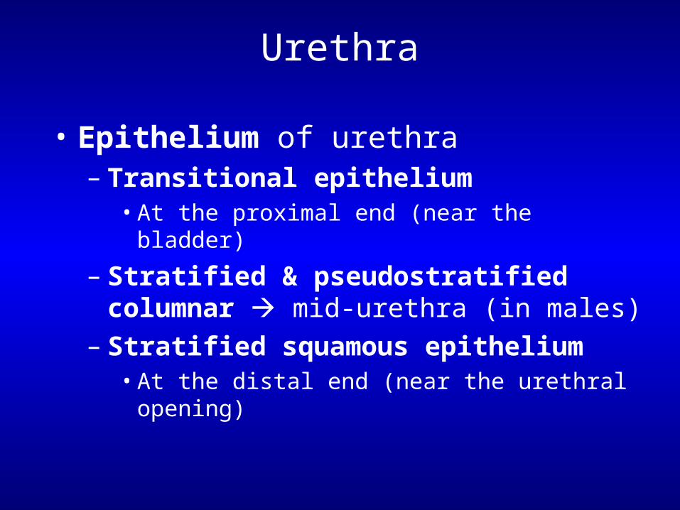
• Epithelium of urethra– Transitional epithelium
• At the proximal end (near the bladder)
– Stratified & pseudostratified columnar mid-urethra (in males)
– Stratified squamous epithelium• At the distal end (near the urethral opening)
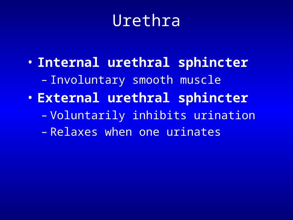
Urethra
• Internal urethral sphincter– Involuntary smooth muscle
• External urethral sphincter– Voluntarily inhibits urination– Relaxes when one urinates
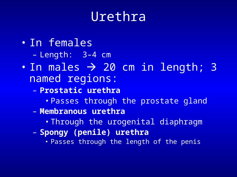
Urethra
• In females– Length: 3–4 cm
• In males 20 cm in length; 3 named regions:– Prostatic urethra
• Passes through the prostate gland– Membranous urethra
• Through the urogenital diaphragm – Spongy (penile) urethra
• Passes through the length of the penis
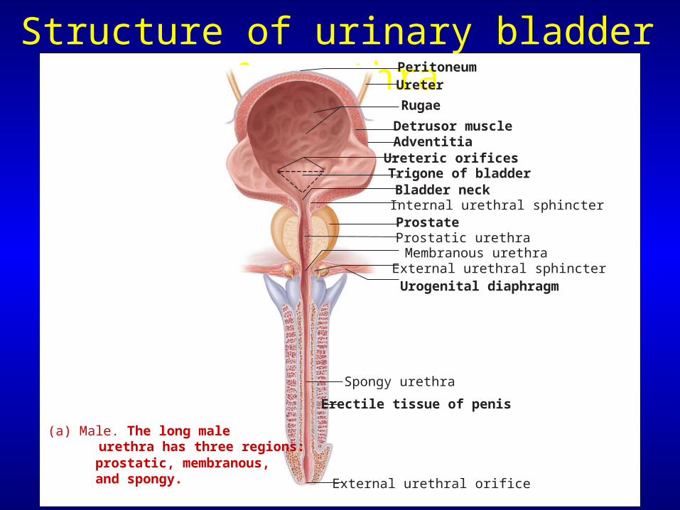
Structure of urinary bladder & urethraUreter
Trigone of bladder
Prostate
Membranous urethraProstatic urethra
Peritoneum
Rugae
Detrusor muscle
Bladder neckInternal urethral sphincter
External urethral sphincterUrogenital diaphragm
Spongy urethra
Erectile tissue of penis
Ureteric orificesAdventitia
(a) Male. The long male urethra has three regions: prostatic, membranous, and spongy. External urethral orifice
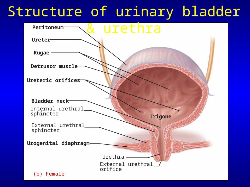
Ureter
Trigone
Peritoneum
Rugae
Detrusor muscle
Bladder neck
Internal urethralsphincter
External urethralsphincter
Urogenital diaphragm
UrethraExternal urethralorifice
Ureteric orifices
(b) Female
Structure of urinary bladder & urethra
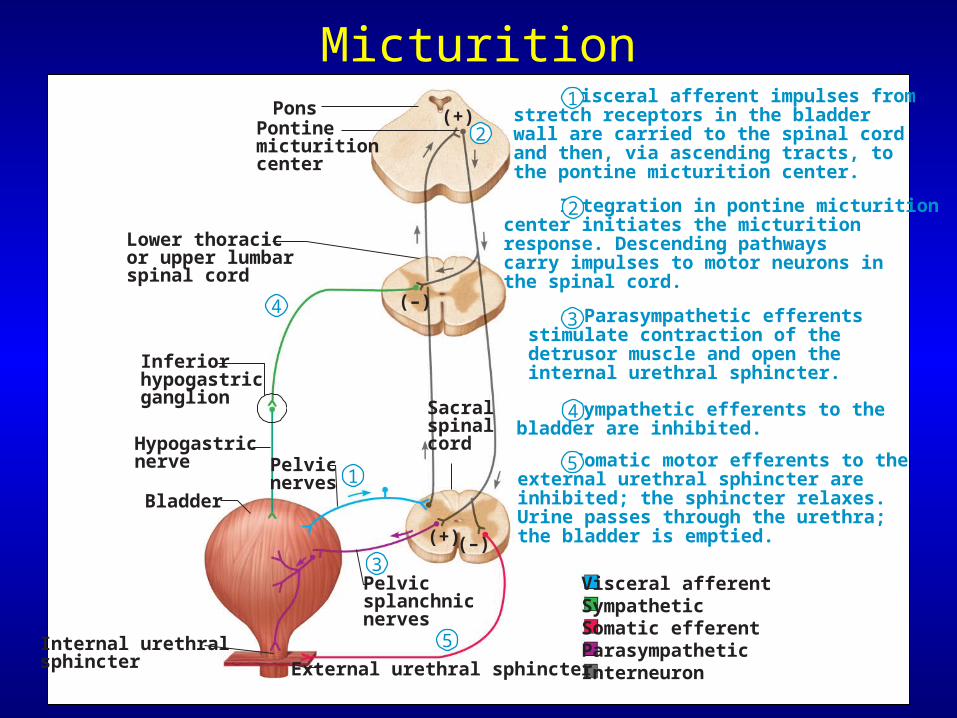
Micturition
Hypogastricnerve
Bladder
Pelvicnerves
Pelvicsplanchnicnerves
PonsPontinemicturitioncenter
Lower thoracicor upper lumbarspinal cord
Sacral spinalcord
Internal urethralsphincter External urethral sphincter
Inferiorhypogastricganglion
Visceral afferent impulses from stretch receptors in the bladder wall are carried to the spinal cord and then, via ascending tracts, to the pontine micturition center.
Integration in pontine micturition center initiates the micturition response. Descending pathways carry impulses to motor neurons in the spinal cord.
Parasympathetic efferents stimulate contraction of the detrusor muscle and open the internal urethral sphincter.
Somatic motor efferents to the external urethral sphincter are inhibited; the sphincter relaxes. Urine passes through the urethra; the bladder is emptied.
Sympathetic efferents to the bladder are inhibited.
(–)
(–)
(+)
(+)
Visceral afferentSympatheticSomatic efferentParasympatheticInterneuron
1
2
3
4
51
2
3
4
5
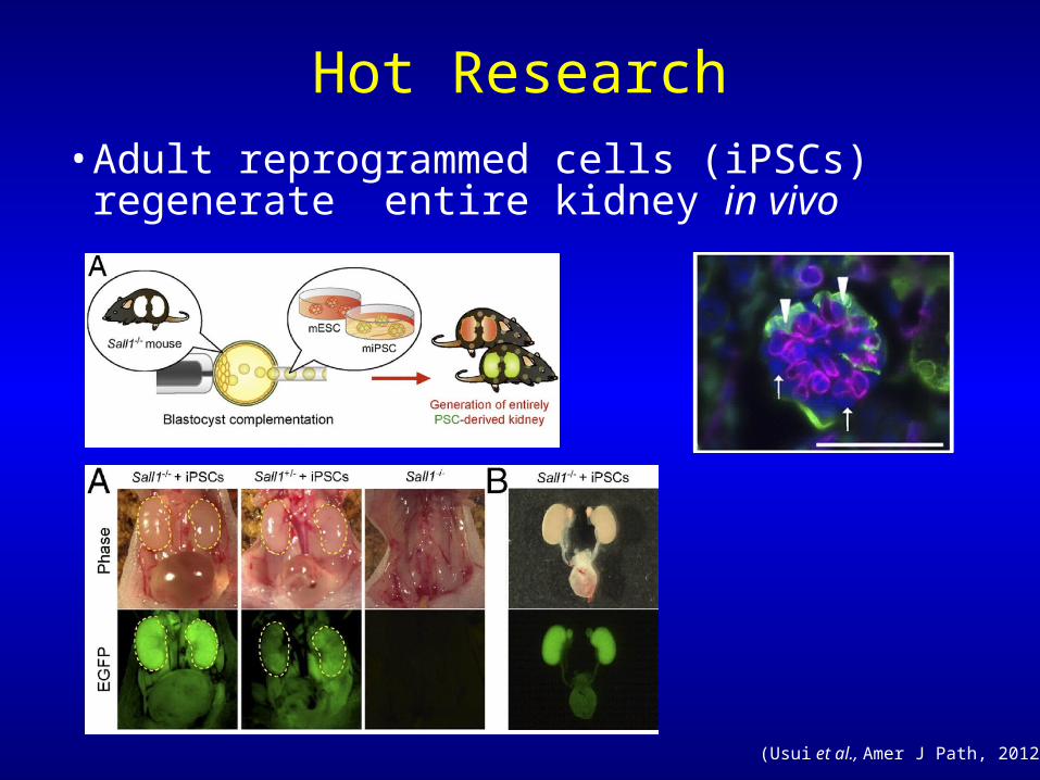
Hot Research• Adult reprogrammed cells (iPSCs) regenerate
entire kidney in vivo
(Usui et al., Amer J Path, 2012)
Disorders affecting the kidneys
• Diabetes mellitus (“flowing through” “sweet”)– Huge sugar load glucose in the urine & causes
osmotic diuresis; can result in low BP, coma & death
• Diabetes insipidus– Lack antidiuretic hormone (ADH)
massive amount of dilute urine & dehydration
• Renal calculi (kidney stones)– Too much salt, Ca2+, uric acid precipitates out of
solution & blocks urine flow; very painful
Disorders affecting the kidneys
• Renal Disease & Failure
–Glomerular dysfunction
–Slowed GFR, massive reabsorption of salt & water, severe hypertension, edema
–Dialysis & Kidney Transplants
Disorders of the urinary system
• Urinary tract infections– More common in females
– Burning sensation during micturition
• Bladder cancer– 3% of cancers more common in men
• Kidney cancer– Arises from epithelial cells of nephron tubules

Disorders of the urinary system
• Congenital defects of the urogenital tract – Hypospadias
–Abnormal placement of urethral orifice (opening); urethra opens anywhere
along the urethral groove on ventral side of penis or scrotum –One of most common birth defects in boys (~1 out of 125 boys) –More severe forms interfere with urination & sexual function! –Most forms are correctable with surgery
Disorders of the urinary system
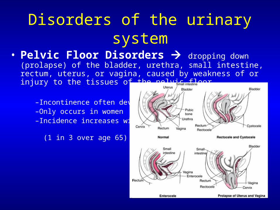
• Pelvic Floor Disorders dropping down (prolapse) of the bladder, urethra, small intestine, rectum, uterus, or vagina, caused by weakness of or injury to the tissues of the pelvic floor
–Incontinence often develops –Only occurs in women –Incidence increases with age
(1 in 3 over age 65)
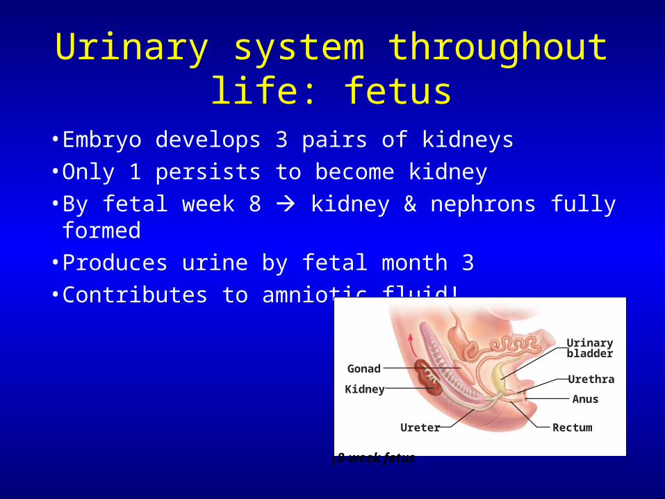
• Embryo develops 3 pairs of kidneys• Only 1 persists to become kidney• By fetal week 8 kidney & nephrons fully formed• Produces urine by fetal month 3• Contributes to amniotic fluid!
Gonad
Kidney
Urinarybladder
Urethra
Anus
Ureter Rectum
8-week fetus
Urinary system throughout life: fetus
• Kidney & bladder function declines with advancing age– Nephrons decrease in size & number– Tubules less efficient at secretion & reabsorption– Filtration declines – Recognition of desire to urinate is delayed– Loss of muscle tone in bladder & sphincters– Incontinence can develop
• And Yes,… exercise can help! Kegels
Urinary system throughout life: adult
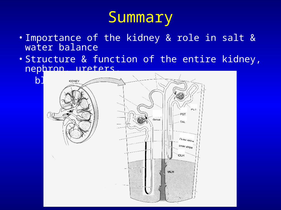
Summary• Importance of the kidney & role in salt & water balance • Structure & function of the entire kidney, nephron, ureters, bladder & urethra
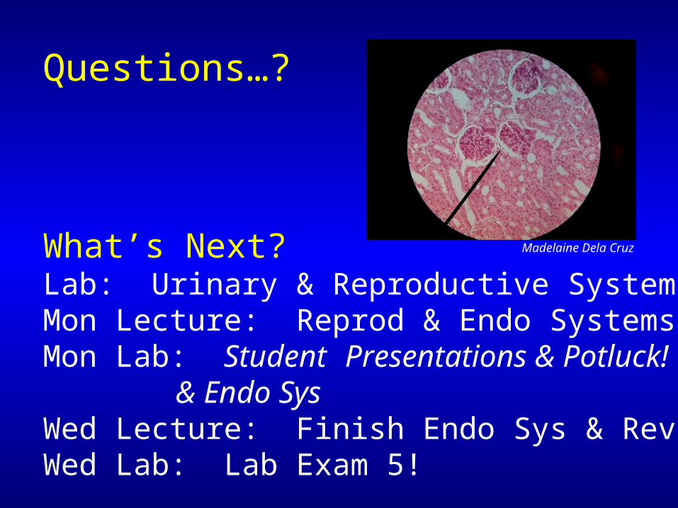
Questions…?
What’s Next?Lab: Urinary & Reproductive SystemsMon Lecture: Reprod & Endo Systems Mon Lab: Student Presentations & Potluck! & Endo SysWed Lecture: Finish Endo Sys & ReviewWed Lab: Lab Exam 5!
Madelaine Dela Cruz
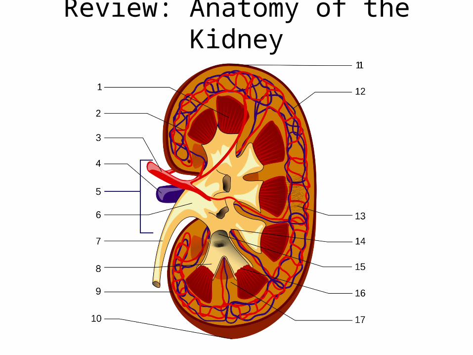
Review: Anatomy of the Kidney
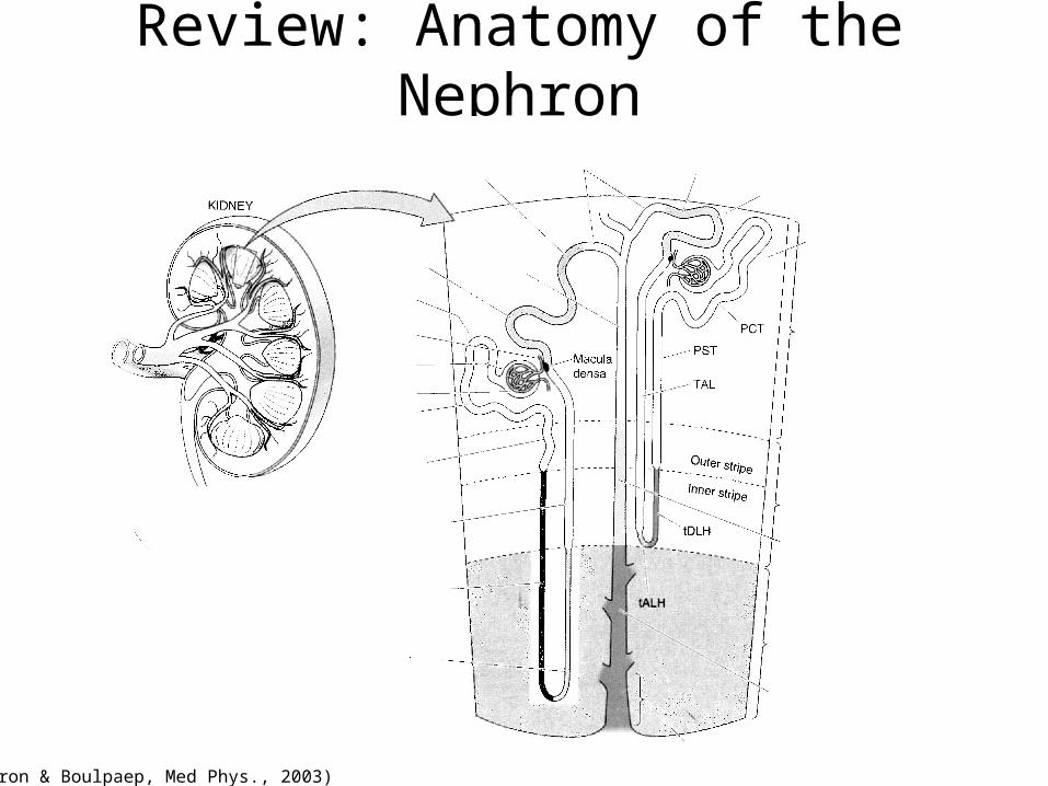
Review: Anatomy of the Nephron
(Boron & Boulpaep, Med Phys., 2003)
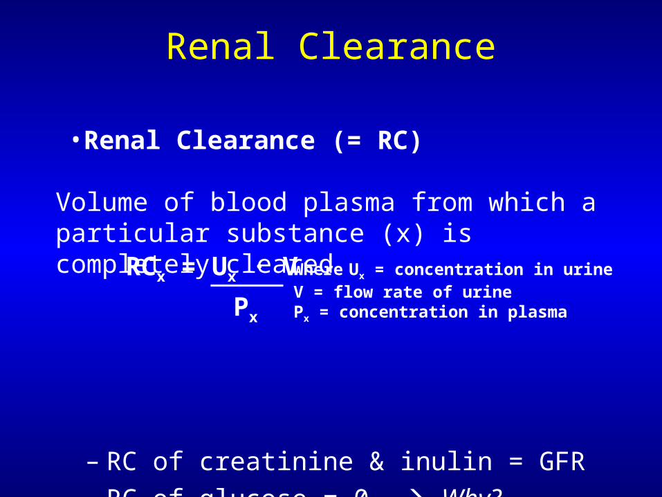
•Renal Clearance (= RC) Volume of blood plasma from which a particular substance (x) is completely cleared
– RC of creatinine & inulin = GFR– RC of glucose = 0 Why?
Renal Clearance
RCx = Ux · V
Px
Where Ux = concentration in urineV = flow rate of urinePx = concentration in plasma