The Thorax. The Thorax – what is in it and what do you need to know? Thoracic wall and diaphragm...
46
The Thorax
-
Upload
alex-dowdle -
Category
Documents
-
view
243 -
download
0
Transcript of The Thorax. The Thorax – what is in it and what do you need to know? Thoracic wall and diaphragm...

The Thorax
The Thorax – what is in it and what do you need to know?
• Thoracic wall and diaphragm• Surface anatomy• Thoracic cavity• Mediastinum• Heart• Lungs and their surrounding layers• Main blood vessels and nerves

Sternum and ribs

Angle of Louis

Surface anatomy chest

Important surface anatomy
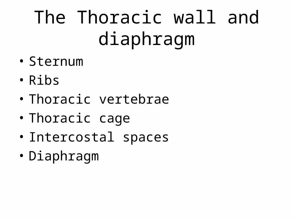
The Thoracic wall and diaphragm
• Sternum• Ribs• Thoracic vertebrae• Thoracic cage• Intercostal spaces• Diaphragm


Chest wall

Diaphragm and chest movements

Thoracic cavity
Heart• Chambers• Valves• Blood supply• Conducting system• PericardiumLungsPleura


Surface anatomy

Heart – structure
• Cardiac muscle – similar to skeletal muscle• Surrounded by pericardial sac – two layers
visceral and parietal• Supported on a fibrous ring (physical support,
supports valves, prevents stretching)• Has its own blood and nerve supply• Myocardium (Muscle), Endocardium (Inner
epithelial ), Epicardium (Outer)

Heart structure
• Pericardium• Outer tougher layer
(fibrous) parietal• Inner (serous) thinner
layer – visceral• Potential space between
is filled with fluid
Outer fibrous
serous
visceral

Supportive skeleton
• Fibrous ring • Supports the bases of the
four main valves

4 main chambers
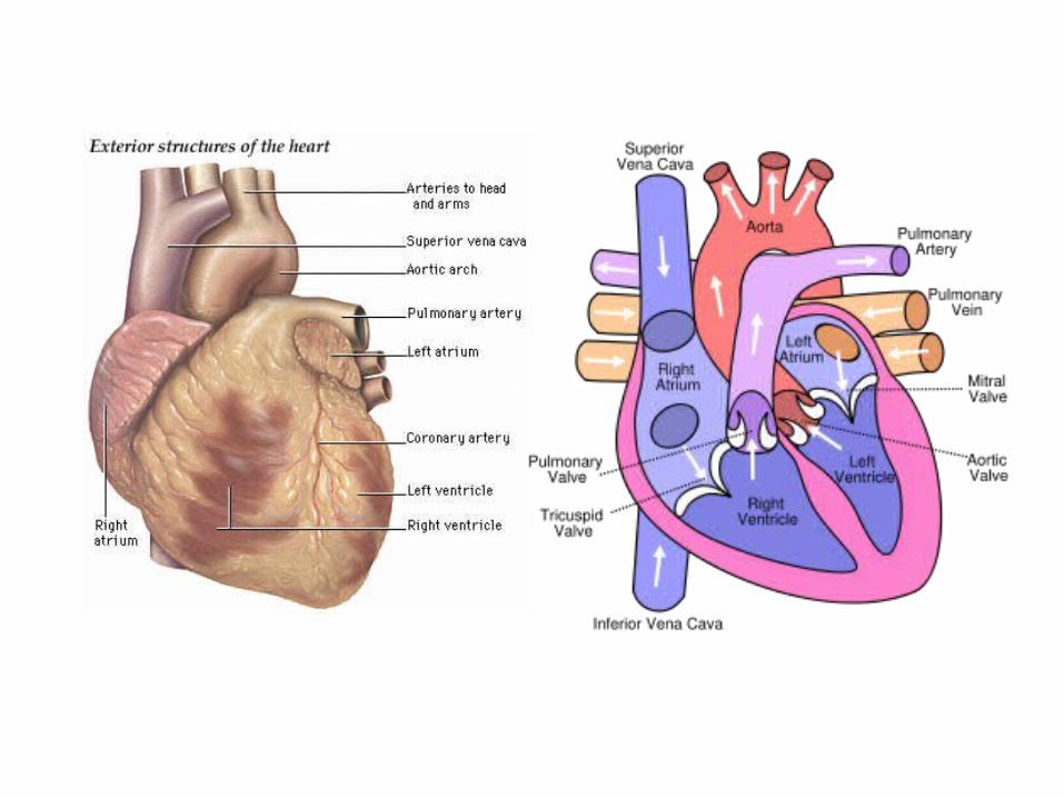
• Right atrium – receives blood back from SVC and IVC
• Right ventricle – thick walled and sends blood to lungs
• Left atrium – thin walled receives blood from lungs
• Left ventricle – very thick and sends blood to body – main muscular pump
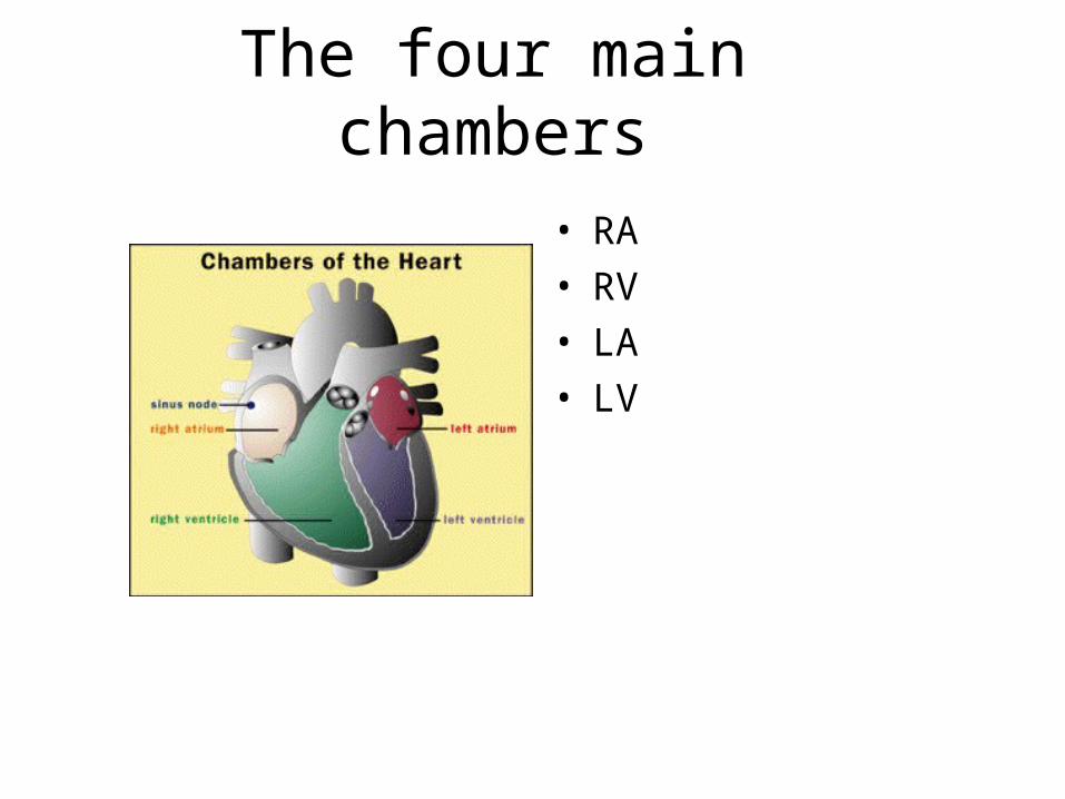
The four main chambers
• RA• RV• LA• LV

4 valves
• Triscuspid – between RA and RV• Pulmonary – between RV and Pulmonary
artery• Mitral – Between LA and LV• Aortic – Between LV and Aortic trunk Valves are made of cusps of fibrous tissue, supported on the fibrous ring
– Mitral and Triscuspid have papillary muscles and chordae which support them from below

The valvesSVC
Pulmonary valve
RA
TV
IVC
LV
Papillary muscle
Mitral valve
Aortic valve
Aorta

The valves
• These close behind blood and when functioning prevent back flow
• They can become narrowed ( stenosis)• Leaking ( Incompetence)• Damaged – by infection or ischaemia to
muscles that support them

Papillary muscle and chordae
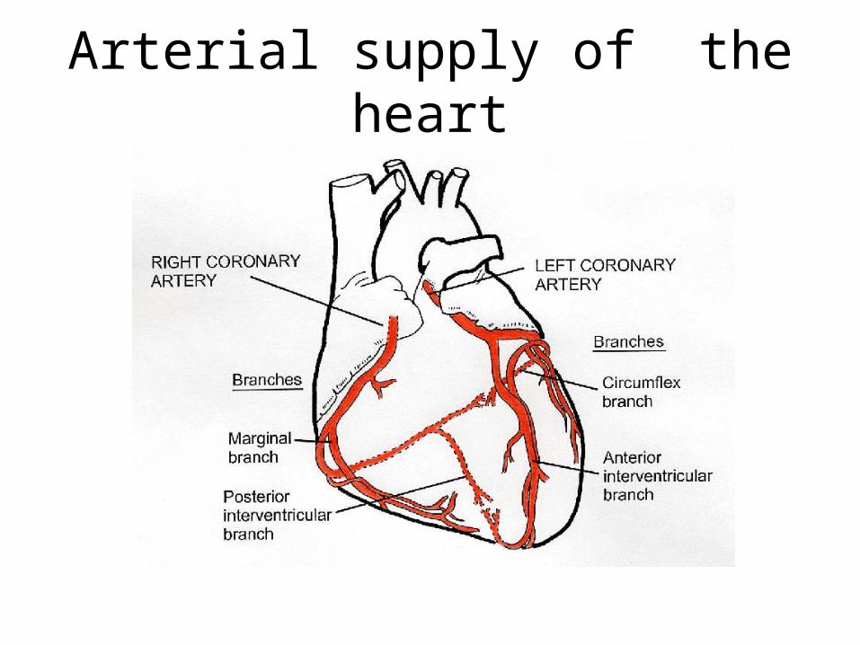
Arterial supply of the heart

Blood supply of the heart
• Right coronary artery – arises anterior aortic sinus (supplies mainly right and inferior)
• Left coronary artery – arises left posterior aortic sinus ( supplies bulk of LV via main branches the circumflex and Left anterior descending – these are the commonest vessels to become occluded)

The electrical conducting system of the heart
• Specialised cardiac cells • Control the sequence of events causing the cardiac cycle• Damage to these can cause heart rhythm abnormalities
Consists• SA node ( starting point)• AV node ( between atrium and ventricles)• AV bundle of HIS• Branching fibres R and L

The conducting system
SA node or pacemaker
AV node
AV bundle
Spreading branches

Nerve supply to the heart
• Sympathetic – Speeds up heart via Adrenergic nerves and adrenaline, nor adrenaline release acting on cardiac adrenoceptors (and can be affected by drugs which act as agonists e.g. Salbutamol, caffeine, or antagonists e.g. B blockers)
• Parasympathetic – Slows the heart down via the Vagus nerve (occurs in a feint)

The aorta and main vessels
• Large vessels• Thick elasticated walls in health• Subject to damage from turbulence, pressure,
chemical damage such as smoking• Atheroma tends to occur mainly at junctions

Normal arterial structure
• Outer connective layer• Inner endothelial layer• Middle layers of fibrous and elasticated tissue

Normal artery and early atheroma
• Normal artery
• Atheroma developing
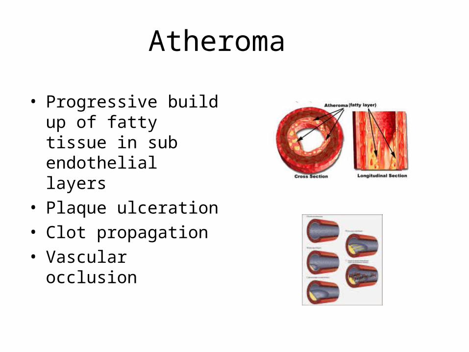
Atheroma
• Progressive build up of fatty tissue in sub endothelial layers
• Plaque ulceration• Clot propagation• Vascular occlusion

What is a heart attack?

What do you need to know about the heartBy next week it will help you if can …
• Describe where the heart is in the chest• Describe its main anatomical features• Describe its blood supply• Describe its nerve supply• Know the main branches of the arterial
system• Understand the main features of the structure
of the blood vessels
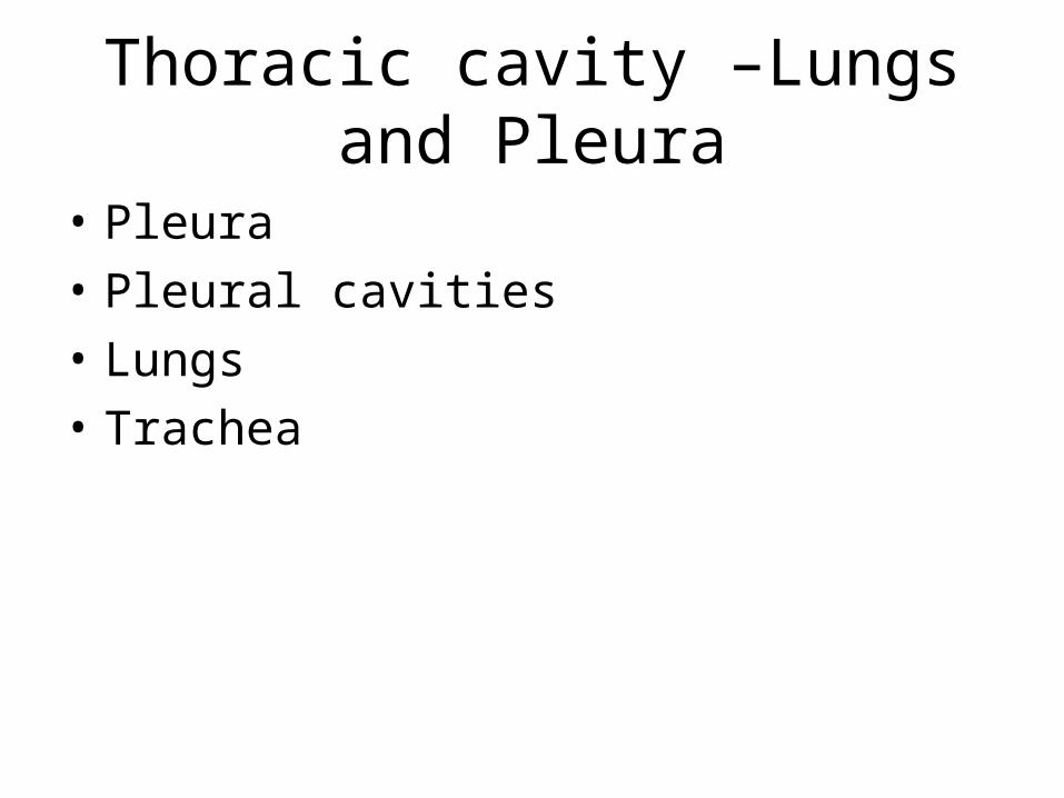
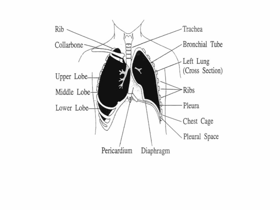
Thoracic cavity –Lungs and Pleura
• Pleura• Pleural cavities• Lungs• Trachea

Anatomy of the chest – using it to understand symptoms and signs
Upper airways• Larynx • TracheaLower airways• Bronchi • Bronchioles • AlveoliThe rest of the chest• Pleural cavity• Pleura• Lung support tissue• Lung vasculature• Heart
Do not forget• Blood itself• The control of breathing

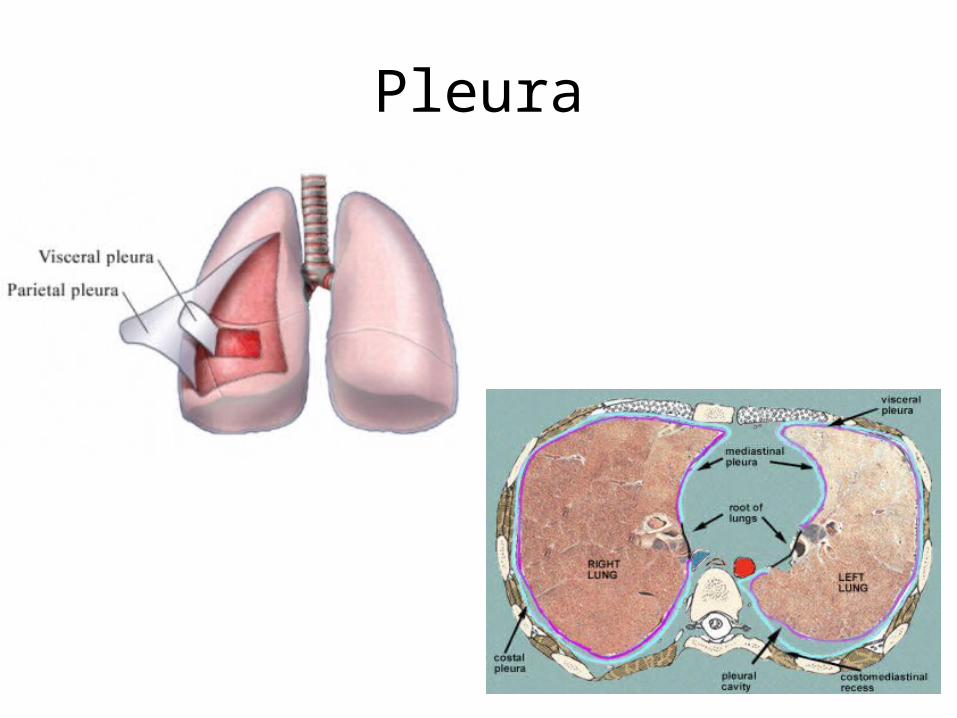
Pleura
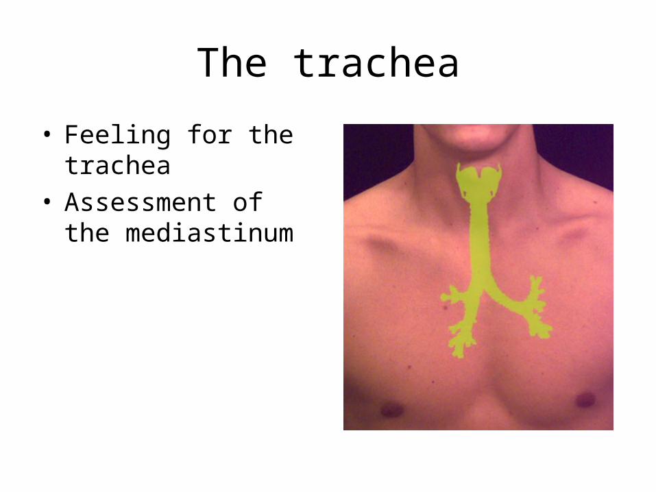
The trachea
• Feeling for the trachea• Assessment of the
mediastinum

Surface anatomy - Which part of the lung is where?

chest x ray

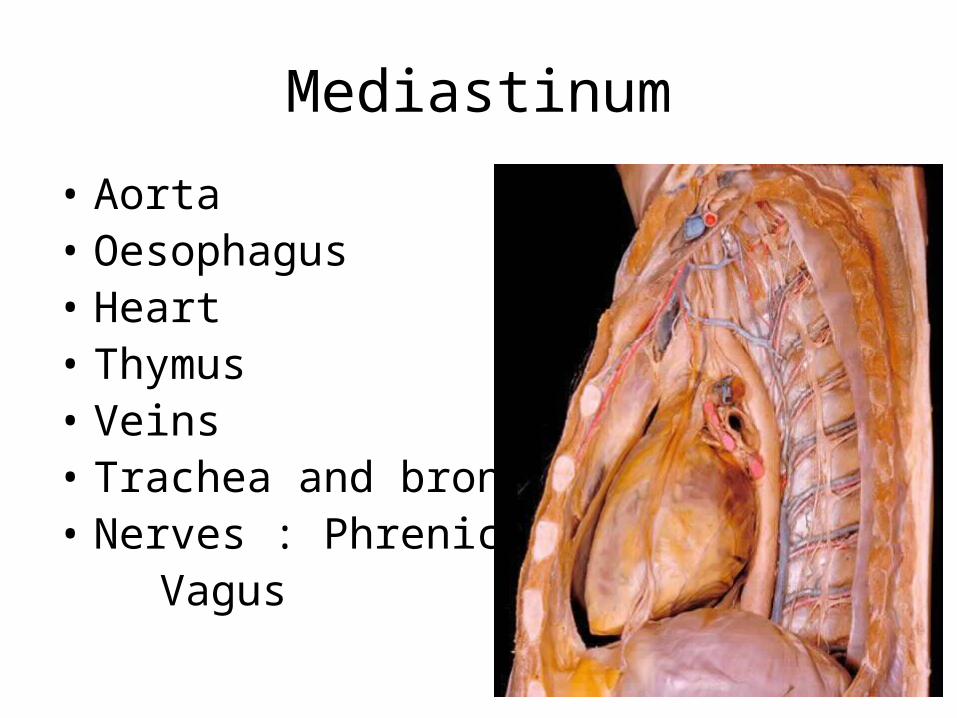
Mediastinum
• Aorta• Oesophagus• Heart• Thymus• Veins• Trachea and bronchi• Nerves : Phrenic and Vagus

Questions and the on line quiz