The tannosome is an organelle forming - Annals of Botany - Oxford
12
The tannosome is an organelle forming condensed tannins in the chlorophyllous organs of Tracheophyta Jean-Marc Brillouet 1 , Charles Romieu 2 , Benoı ˆt Schoefs 3 , Katalin Solymosi 4 , Ve ´ronique Cheynier 1 , He ´le `ne Fulcrand 1 , Jean-Luc Verdeil 2,5 and Genevie `ve Cone ´je ´ro 5,6, * 1 UMR SPO INRA/SupAgro/UM I, 2 UMR AGAP INRA/CIRAD/SupAgro, Montpellier, France, 3 EA2160 MMS, LUNAM, University of Le Mans, France, 4 Department of Plant Anatomy, Eo ¨tvo ¨s University, Budapest, Hungary, 5 Plate-forme d’Histocytologie et d’Imagerie Cellulaire Ve ´ge ´tale (PHIV) and 6 UMR BPMP INRA/CNRS/SupAgro/UM II, Montpellier, France * For correspondence. E-mail [email protected] Received: 24 January 2013 Returned for revision: 8 April 2013 Accepted: 10 June 2013 Published electronically: 11 September 2013 † Background and Aims Condensed tannins (also called proanthocyanidins) are widespread polymers of catechins and are essential for the defence mechanisms of vascular plants (Tracheophyta). A large body of evidence argues for the synthesis of monomeric epicatechin on the cytosolic face of the endoplasmic reticulum and its transport to the vacuole, although the site of its polymerization into tannins remains to be elucidated. The aim of the study was to re-examine the cellular frame of tannin polymerization in various representatives of the Tracheophyta. † Methods Light microscopy epifluorescence, confocal microscopy, transmission electron microscopy (TEM), chemical analysis of tannins following cell fractionation, and immunocytochemistry were used as independent methods on tannin-rich samples from various organs from Cycadophyta, Ginkgophyta, Equisetophyta, Pteridophyta, Coniferophyta and Magnoliophyta. Tissues were fixed in a caffeine–glutaraldehyde mixture and examined by TEM. Other fresh samples were incubated with primary antibodies against proteins from both chloro- plastic envelopes and a thylakoidal chlorophyll-carrying protein; they were also incubated with gelatin–Oregon Green, a fluorescent marker of condensed tannins. Coupled spectral analyses of chlorophyll and tannins were carried out by confocal microscopy on fresh tissues and tannin-rich accretions obtained through cell fractionation; chemical analyses of tannins and chlorophylls were also performed on the accretions. † Key Results and Conclusions The presence of the three different chloroplast membranes inside vacuolar accretions that constitute the typical form of tannin storage in vascular plants was established in fresh tissues as well as in purified organelles, using several independent methods. Tannins are polymerized in a new chloroplast-derived organelle, the tannosome. These are formed by pearling of the thylakoids into 30 nm spheres, which are then encapsulated in a tan- nosome shuttle formed by budding from the chloroplast and bound by a membrane resulting from the fusion of both chloroplast envelopes. The shuttle conveys numerous tannosomes through the cytoplasm towards the vacuole in which it is then incorporated by invagination of the tonoplast. Finally, shuttles bound by a portion of tonoplast ag- gregate into tannin accretions which are stored in the vacuole. Polymerization of tannins occurs inside the tannosome regardless of the compartment being crossed. A complete sequence of events apparently valid in all studied Tracheophyta is described. Key words: Tannosome, organelle, condensed tannins, proanthocyanidins, polymerization, chloroplast, tonoplast, vacuole, Tracheophyta, vascular plants. INTRODUCTION Condensed tannins, also called proanthocyanidins, are present in most vascular plants and are thought to play diverse roles. They provide defence against herbivores and pathogens, and protec- tion against UV radiation. These secondary metabolites are poly- mers of catechins belonging to the vast family of flavonoids. Sucrose gradient sub-cellular fractionation, molecular biology and immunocytochemical approaches have suggested that flavo- noids are synthesized from phenylpropanoids by a multienzy- matic complex loosely bound to the cytosolic face of the endoplasmic reticulum (Wagner and Hrazdina, 1984; Hrazdina et al., 1987; Burbulis and Winkel-Shirley, 1999; Saslowsky and Winkel-Shirley, 2001). It has been hypothesized that tannin inclusions, viewed by transmission electron microscopy (TEM) in cell suspension cultures and calluses from various gymnosperms (Constabel, 1969; Chafe and Durzan, 1973; Parham and Kaustinen, 1977), also originate from the endoplas- mic reticulum. However, since these pioneering reports, no ultra- structural and morphological research has been conducted into the ontogenesis of intracellular tannin-forming elements in ter- restrial plants. In particular, no ultrastructural studies of chloro- plasts, as the source of all plant aromatics through the shikimate pathway (Herrmann, 1995), have been performed, despite the fact that the synthesis of some flavonoids (Saunders and McClure, 1976; Zaprometov and Nikolaeva, 2003) is known to take place in isolated chloroplasts. Few ultrastructural data have been related to the production of phenolic compounds by the chloroplast. Putative phenolics produced by poorly defined organelles vaguely resembling amyloplasts in Eucalyptus ray parenchyma were described by Wardrop and Cronshaw (1962), and chloroplasts in the reproductive organs of Cornus by # The Author 2013. Published by Oxford University Press on behalf of the Annals of Botany Company. All rights reserved. For Permissions, please email: [email protected] Annals of Botany 112: 1003 – 1014, 2013 doi:10.1093/aob/mct168, available online at www.aob.oxfordjournals.org Downloaded from https://academic.oup.com/aob/article-abstract/112/6/1003/2768923 by guest on 22 November 2018
Transcript of The tannosome is an organelle forming - Annals of Botany - Oxford

The tannosome is an organelle forming condensed tannins in thechlorophyllous organs of Tracheophyta
Jean-Marc Brillouet1, Charles Romieu2, Benoıt Schoefs3, Katalin Solymosi4, Veronique Cheynier1,Helene Fulcrand1, Jean-Luc Verdeil2,5 and Genevieve Conejero5,6,*
1UMR SPO INRA/SupAgro/UM I, 2UMR AGAP INRA/CIRAD/SupAgro, Montpellier, France, 3EA2160 MMS, LUNAM, Universityof Le Mans, France, 4Department of Plant Anatomy, Eotvos University, Budapest, Hungary, 5Plate-forme d’Histocytologie et
d’Imagerie Cellulaire Vegetale (PHIV) and 6UMR BPMP INRA/CNRS/SupAgro/UM II, Montpellier, France* For correspondence. E-mail [email protected]
Received: 24 January 2013 Returned for revision: 8 April 2013 Accepted: 10 June 2013 Published electronically: 11 September 2013
† Background and Aims Condensed tannins (also called proanthocyanidins) are widespread polymers of catechinsand are essential for the defence mechanisms of vascular plants (Tracheophyta). A large body of evidence arguesfor the synthesis of monomeric epicatechin on the cytosolic face of the endoplasmic reticulum and its transport tothe vacuole, although the site of its polymerization into tannins remains to be elucidated. The aim of the studywas to re-examine the cellular frame of tannin polymerization in various representatives of the Tracheophyta.† Methods Light microscopy epifluorescence, confocal microscopy, transmission electron microscopy (TEM),chemical analysis of tannins following cell fractionation, and immunocytochemistry were used as independentmethods on tannin-rich samples from various organs from Cycadophyta, Ginkgophyta, Equisetophyta,Pteridophyta, Coniferophyta and Magnoliophyta. Tissues were fixed in a caffeine–glutaraldehyde mixture andexamined by TEM. Other fresh samples were incubated with primary antibodies against proteins from both chloro-plastic envelopes and a thylakoidal chlorophyll-carrying protein; they were also incubated with gelatin–OregonGreen, a fluorescent marker of condensed tannins. Coupled spectral analyses of chlorophyll and tannins werecarried out by confocal microscopy on fresh tissues and tannin-rich accretions obtained through cell fractionation;chemical analyses of tannins and chlorophylls were also performed on the accretions.† Key Results and Conclusions The presence of the three different chloroplast membranes inside vacuolar accretionsthat constitute the typical form of tannin storage in vascular plants was established in fresh tissues as well as in purifiedorganelles, using several independent methods. Tannins are polymerized in a new chloroplast-derived organelle, thetannosome. These are formed by pearling of the thylakoids into 30 nm spheres, which are then encapsulated in a tan-nosome shuttle formed by budding from the chloroplast and bound by a membrane resulting from the fusion of bothchloroplast envelopes. The shuttle conveys numerous tannosomes through the cytoplasm towards the vacuole inwhich it is then incorporated by invagination of the tonoplast. Finally, shuttles bound by a portion of tonoplast ag-gregate into tannin accretions which are stored in the vacuole. Polymerization of tannins occurs inside the tannosomeregardless of the compartment being crossed. A complete sequence of events apparently valid in all studiedTracheophyta is described.
Key words: Tannosome, organelle, condensed tannins, proanthocyanidins, polymerization, chloroplast, tonoplast,vacuole, Tracheophyta, vascular plants.
INTRODUCTION
Condensed tannins, also called proanthocyanidins, are present inmost vascular plants and are thought to play diverse roles. Theyprovide defence against herbivores and pathogens, and protec-tion against UV radiation. These secondary metabolites are poly-mers of catechins belonging to the vast family of flavonoids.Sucrose gradient sub-cellular fractionation, molecular biologyand immunocytochemical approaches have suggested that flavo-noids are synthesized from phenylpropanoids by a multienzy-matic complex loosely bound to the cytosolic face of theendoplasmic reticulum (Wagner and Hrazdina, 1984; Hrazdinaet al., 1987; Burbulis and Winkel-Shirley, 1999; Saslowskyand Winkel-Shirley, 2001). It has been hypothesized thattannin inclusions, viewed by transmission electron microscopy(TEM) in cell suspension cultures and calluses from various
gymnosperms (Constabel, 1969; Chafe and Durzan, 1973;Parham and Kaustinen, 1977), also originate from the endoplas-mic reticulum. However, since these pioneering reports, no ultra-structural and morphological research has been conducted intothe ontogenesis of intracellular tannin-forming elements in ter-restrial plants. In particular, no ultrastructural studies of chloro-plasts, as the source of all plant aromatics through the shikimatepathway (Herrmann, 1995), have been performed, despite thefact that the synthesis of some flavonoids (Saunders andMcClure, 1976; Zaprometov and Nikolaeva, 2003) is known totake place in isolated chloroplasts. Few ultrastructural datahave been related to the production of phenolic compounds bythe chloroplast. Putative phenolics produced by poorly definedorganelles vaguely resembling amyloplasts in Eucalyptus rayparenchyma were described by Wardrop and Cronshaw (1962),and chloroplasts in the reproductive organs of Cornus by
# The Author 2013. Published by Oxford University Press on behalf of the Annals of Botany Company. All rights reserved.
For Permissions, please email: [email protected]
Annals of Botany 112: 1003–1014, 2013
doi:10.1093/aob/mct168, available online at www.aob.oxfordjournals.org
Dow
nloaded from https://academ
ic.oup.com/aob/article-abstract/112/6/1003/2768923 by guest on 22 N
ovember 2018
Juhasz et al. (1969). Compounds thought to be phenolics,because tests for the presence of proteins, lipids and polysacchar-ides were negative, were found within the thylakoid lumen fromlower epidermal cells of Nymphaea indica (van Steveninck andvan Steveninck, 1980), leaves of Haberlea rhodopensis(Georgieva et al., 2010) and in cotyledons, leaves and fruitpeels of a few other species (Keresztes and Sarvari, 2001).However, none of the above-mentioned studies identified theexact nature of the putative phenolics observed in the plastids.More recently, gold immunolabelling showed that threeenzymes of the phenylpropanoid and flavonoid pathways,cinnamate-4-hydroxylase (C4H) (Chen et al., 2006), chalconesynthase (CHS) (Tian et al., 2008) and anthocyanidin reductase(ANR) (Wang et al., 2010), were primarily located in the chloro-plast of developing grape berries.
Based on these data, a detailed ultrastructural and morpho-logical study was carried out to examine the possible involve-ment of chloroplasts in the ontogenesis of tannin-formingelements in vascular plants.
MATERIALS AND METHODS
Plant materials
Leaflets from fern (Dryopteris sp., Pteridophyta), horsetail(Equisetum arvense L., Equisetophyta), Cycas revoluta Thunb.(Cycadophyta), Ginkgo biloba L. (Ginkgophyta) and persim-mon (Dyospiros kaki L., Magnoliophyta, Eudicots), petiolesfrom Chamaerops humilis L. and stalks from Saccharum L. sp.(Magnoliophyta, Monocots), needles from Pinus sylvestrisL. and scales from Cupressus macrocarpa Hartw. ex GeorgeGordon (Coniferophyta), and pistils and fruits from grapevine(Vitis vinifera L., Magnoliophyta, Eudicots) were collected inthe Montpellier City botanical garden.
Light microscopy
Tannin were visualized after fixation, dehydration andembedding of the tissues in resin (Brillouet and Escoute, 2012)by the dimethylaminocinnamaldehyde (DMACA) technique(Treutter, 1989; Cadot et al., 2006).
Fluorescence labelling
Tannin were visualized by dipping sections from grapevinepistils in 0.01 M phosphate-buffered saline (PBS) containingOregon Greenw 488-conjugated gelatin (Invitrogen, USA;10 mg mL21) for 1 h, then washing in PBS (3× 15 min); controlswere obtained by applying gelatin onto tannin-free tissues, i.e.young stem from Hedera helix L. and leaflet from PhyllostachisSiebold & Zucc. sp. (Supplementary Data Fig. S1).
Visualization of chloroplast membrane intrinsic proteins wascarried out as follows: sections were dipped successivelyat 20 8Cin the following media: 4 % paraformaldehyde in 0.01 M PBS for1 h; 0.1 M glycine in PBS for 15 min; PBS (3× 15 min); 5 %bovine serum albumin (BSA) in PBS (blocking buffer, 3 h);anti-Lhcb1 (LHCII type I chlorophyll a/b-binding thylakoidalmembrane protein), anti-TIC40 (translocon complex fromchloroplast inner membrane) or anti-TOC75 (transloconcomplex from chloroplast outer membrane) rabbit antibodies
(Agrisera, Sweden; 10 mg mL21 in blocking buffer, overnightat 4 8C); PBS (3× 15 min); and secondary anti-rabbit AlexaFluorw 633-conjugated IgGs (Invitrogen, USA; 7 mg mL21 inblocking buffer, 1 h). Controls were run (1) with pre-immunerabbit serum instead of primary antibody and (2) with secondaryanti-rabbit IgGs only.
Confocal and epifluorescence microscopy
Confocal imaging was performed with a Zeiss Axiovertmicroscope 200M 510 META fitted with a Plan-Apochromat×63/1.2 W Zeiss objective. Excitations were obtained forgelatin–Oregon Green with an Ar laser at 488 nm (bandpass500–530 nm), for antibodies with an He–Ne laser at 633 nm(band pass .650 nm) and for tannins with a 405 nm bluediode (bandpass 505–550 nm). Images were processed byHuygens (http://www.svi.nl/HuygensSoftware), then Image J(http://rsbweb.nih.gov/ij/) software: stacked images (16 bits)were converted to RGB (red, green, blue) stacks, then, afterthresholding, converted to HSB (hue, saturation, brightness)stacks; then histograms (hue angle) were expressed for eachpixel (as a percentage of total pixels) and fitted with a polyno-mial. For images acquired in lambda scanning mode, the emis-sion spectra were obtained on sample ROIs (regions of interest)by spectral acquisition (lambda stack, excitation at 405 nm).The detection bandwidth was set to collect emissions from 400to 750 nm, using an array of 32 photomultiplier tube (PMT)detectors, each with a 10.7 nm bandwidth. The method of linearunmixing was applied with advanced iterative and one residualchannel. Epifluorescence imaging was performed with an ZeissAxiophot microscope [DAPI (4’,6-diamidino-2-phenylindole)filter, band-path .470 nm).
Transmission electron microscopy
Specimens were dipped in 50 mM sodium cacodylate buffer(pH 7.0) containing 6 % glutaraldehyde (w/v) (Sironval et al.,1968) and 1 % caffeine (w/v) for 6 h, then treated with 1 %osmium tetroxide (w/v) in water for 1 h. After dehydration,they were embedded in Epon EmBed 812. Sections werestained with 0.2 % Oolong tea (Sato et al., 2008). Ultrathin sec-tions (60 nm) were visualized byan H-7100 Hitachi transmissionelectron microscope with 75 kV accelerating voltage.
Purification and characterization of tannin accretions
Leaflets (5 g) from Dyospiros and Ginkgo, and pericarp (5 g)from Vitis, were gently ground between two alternately rotatingdiscs equipped with stainless steel grids in 50 mL of cold potas-sium phosphate buffer (pH 7.2) containing 0.3 M sorbitol, 1 %polyvinylpyrolidonne (mol. wt 40 000 Da), 1 % ascorbic acidand 0.34 % EDTA. After filtration on Miracloth, the slurry wascentrifuged (1000 g, 20 min, 4 8C), and the supernatant was dis-carded; the pellet was resuspended in the same buffer, homoge-nized and centrifuged again. After addition of 15 mL of the samebuffer, the pellet was homogenized with a Potter Elvejhem, thencentrifuged at 100 000 g for 1 h on a cushion (90 mL) of 72 %sucrose (w/v; specific gravity d ¼ 1.268). Chloroplasts werestacked at the interface between buffer and 72 % sucrose, while
Brillouet et al. — Formation of tannins in vascular plants by the tannosome1004
Dow
nloaded from https://academ
ic.oup.com/aob/article-abstract/112/6/1003/2768923 by guest on 22 N
ovember 2018

green accretions sedimented at the tube bottom; the latter wererecovered, frozen in liquid nitrogen then stored at –80 8C.
Condensed tannins were analysed by the phloroglucinol tech-nique according to Michodjehoun-Mestres et al. (2009): briefly,accretions were washed with distilled water with repeated centri-fugation, after which methanol containing 5 % phloroglucinol, 1% ascorbic acid and 0.2 N HCl was added. After heating at 90 8Cfor 6 min, the medium was neutralized with an equal volume of 2% sodium acetate. The phloroglucinol adducts resulting fromthe depolymerization were analysed by HPLC-DAD-MS (high-performance liquid chromatography-diode array detection-massspectrometry). Condensed tannins were also detected qualita-tively in tannin accretions by the DMACA technique (Treutter,1989).
Chlorophylls were spectrophotometrically measured in CHCl3/CH3OH (2:1) extracts at l ¼ 666 nm according to Jodłowska andLatała (2011); results were expressed as chlorophyll a equivalents.
RESULTS
Examination of tannin-containing cells by electron, light,epifluorescence and confocal microscopy, and biochemicalcharacterization of tannin-containing bodies
For this study on the polymerization of tannins in plants, veryyoung chlorophyllous organs (pistils, immature small fruits, leaf-lets and pedicels), in which the rate of formation of tannins ishighest, were selected (Cohen et al., 2012). The study was first con-ducted on V. vinifera, then extended to plants from other divisionsin the Tracheophyta [Cycadophyta, Ginkgophyta, Equisetophyta,Pteridophyta, Coniferophyta, and Magnoliophyta (Eudicots andMonocots)].
Sections from pistils of V. vinifera were examined by TEM, andcircular osmiophilic and finely granular structures (Fig. 1A) wereobserved in the vacuole of tannin-containing cells. After specificstaining of flavan-3-ols with DMACA, sections obtained fromthe same materials revealed in light microscopy numerous blue-green stained spheres inside the vacuole (average diameter0.5 mm) (Fig. 1B). When fresh sections of the same materialswere observed in epifluorescence with a DAPI filter, the vacuoleappeared as containing several spherical accretions (diameter3 mm) intensely fluorescing (blue) and including numerous cor-puscles (diameter 0.5 mm)emitting the red fluorescence ofchloro-phyll (Fig. 1C; see also Fig. 3G, H). These chlorophyllous bodiesmust not be confused with chloroplasts which exhibited far largersizes and an ellipsoidal (non-spherical) morphology (long axis 2–3 mm, short axis 1–2 mm) (Supplementary Data Fig. 2).
Since there was no method for visualizing tannins specifically,as the DMACA technique detects both monomeric flavan-3-olsand condensed tannins, a technique was developed based on theunique property of tannins to form insoluble complexes with pro-teins (Hagerman and Butler, 1981). Gelatin coupled to OregonGreen, afluorophore excitable at 488 nm, was applied to fresh sec-tions of the same materials, and small fluorescing spheres (diam-eter 0.5 mm) aggregated in a roughly circular accretion (diameterapprox. 10 mm) (Fig. 1D–F) were observed in confocal micros-copy; image analysis of the merged processed image (Fig. 1G)revealed a continuum between almost-red and tannin-filledalmost-green spheres (Fig. 1H). Noticeably, pure red or greenspheres were notobserved.The gelatin–OregonGreenfluorescent
probe did not show non-specific adsorption onto other organelles,membranes or cell walls (Supplementary Data Fig. 1).
With the aim of further characterizing the mixed (tannins/chlorophyll) bodies, they were purified from several organs ofdiverse plants through high speed centrifugation on a 72 % (w/v) sucrose cushion (Fig. 2A). Abundant chloroplasts werestacked at the (buffer–sucrose) interface, while most, if not allthe DMACA-reactive material sedimented at the bottom of thetube, i.e. at a specific gravity .1.268. (Fig. 2A). These elementsshowed the same typical pattern as that observed on fresh tissueslides under epifluorescence examination (compare Figs 1C and2A). These heavy elements were subjected to biochemical ana-lyses of condensed tannins and chlorophyll. Compositional ana-lyses confirmed the presence of condensed tannins (Fig. 2B) ofvarious chemical structures according to the division andgenus of origin (Table 1): Ginkgo tannins were almost exclusive-ly made of epigallocatechin while Vitis tannins were mainly con-stituted of epicatechin; Dyospiros tannins were built with amixture of epigallo- and epicatechin, and they were highly gal-loylated, in contrast to Ginkgo tannins which were not. Theirdegrees of polymerization were in the range of 10–30 mono-mers. The starter and extension units were of the same naturein Ginkgo and Dyospiros, in contrast to Vitis where catechinwas the terminal unit. Chlorophylls were also present indiverse relative proportions to tannins.
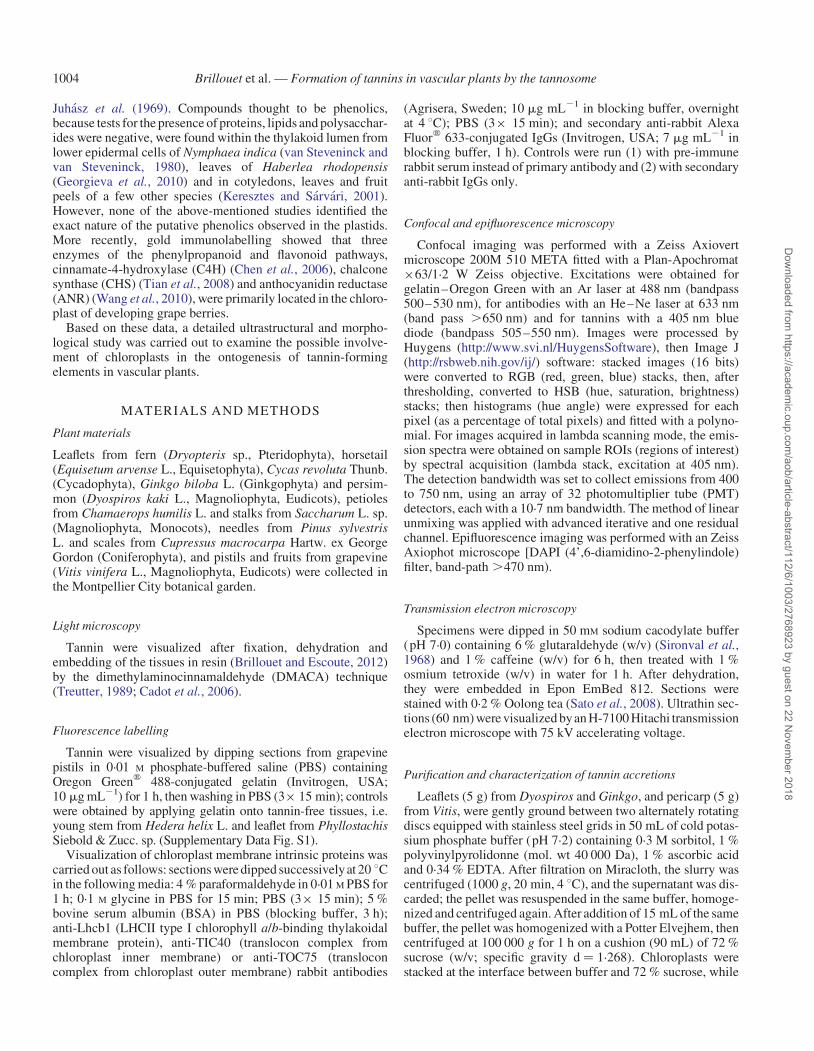
An examination of these purified heavyspecies by spectral ana-lysis revealed particles (diameter 0.5 mm) exhibiting a doubleautofluorescence emission spectrum (Fig. 3A–D): a large emis-sion range with several maxima in the 500–600 nm range,typical of condensed tannins (Fig. 3E, F), as well as chlorophyllautofluorescencewith the characteristic maximum ofphotosystemII (lem 685 nm; Franck et al., 2002). Depending on the plant oforigin (Ginkgo or Vitis) and within the same population of parti-cles, relative proportions of both signals were variable, suggestinga progressive filling of these chlorophyllous bodies with tannins.Coming back to a native tissue (Vitis pericarp), the same observa-tions were carried out on minute vacuolar entities (diameter0.5 mm) (Fig. 3G, H); chloroplasts, as expected, showed thechlorophyll signal only.
Examination of tannin-containing cells by TEM
It should be remembered that, in TEM, all simple phenolics(e.g. quercetin glycosides, chlorogenic acids, and flavan-3-olssuch as epicatechin) are solubilized in water–ethanol mixturesand then dehydrated prior to resin embedding; thus, osmiophilicmaterials observed are the remnants of condensed tannins whichhave not undergone solubilization due to their complexation withcaffeine (Mueller and Greenwood, 1978).
Explants from pistils or very young fruits (diameter 1 mm)were observed by TEM. Functional ellipsoidal chloroplasts(long axis 2–3 mm, short axis 1.5–2 mm) were visible with well-constituted grana thylakoids and the two well-preserved envel-oping membranes (Fig. 4A). Other forms of differentiatingchloroplasts were observed (Fig. 4B): they were characterizedby a swelling with a tendency to circularization [a decrease ofthe (long/short axis) ratio] and by an unstacking of grana.Unstacked thylakoidal lamellae were slightly swollen andincluded osmiophilic material that appeared as black spots anddark grey deposits in these inflated zones. Furthermore, these
Brillouet et al. — Formation of tannins in vascular plants by the tannosome 1005
Dow
nloaded from https://academ
ic.oup.com/aob/article-abstract/112/6/1003/2768923 by guest on 22 N
ovember 2018

lamellae emitted grossly circular structures (diameter 30 nm),including black spots, from their ends into the stroma.Differentiation continued with a massive redistribution of thyla-koidal lamellae (Fig. 4C) which seemed shorter and interlaced;some still contained osmiophilic spots and deposits, whileothers appeared empty. Furthermore, on half of the chloroplastcircumference, both enveloping membranes were no longervisible and were replaced by a diffuse cotton-like mass withinwhich were observed osmiophilic spheres which seemed to beexpulsed from the interior of the chloroplast; these sphericalstructures will hereafter designated as ‘shuttles’. When examin-ing one of these differentiating chloroplasts through a transversesection (Fig. 4B, D), it was observed that unstacking of granawas accompanied by a whirling of these membranes uponthemselves.
Careful examination of the periphery of these chloroplastsshowed budding, where part of the inner plastidial content was
encapsulated in an emerging structure projected into thecytosol (Fig. 4E): this vesicular structure contained portions ofthylakoidal lamellae swollen at regular intervals includingosmiophilic spots attached to the inner face of the membrane,in addition to circular structures (diameter 30 nm). These shut-tles travelled in the cytosol, after having been emitted from theinner plastidial region (Fig. 4F): two possibly coalescing shuttlesmay be seen on this micrograph, with one shuttle showing circu-lar structures (diameter 30 nm) with black spots, whereas theother is filled with similar circular structures themselves filledwith osmiophilic material. Figure 4G shows a well-delineatedfull shuttle within the cytosol.
Careful re-examination of the structures generated by theunstacking of grana thylakoids and incorporated into shuttles(Fig. 4H, I) showed that these lamellae, swollen to a greater orlesser degree, were sectioned at regular intervals into smallspheres (diameter 30 nm) which progressively filled with
2
1
030 60 90 120
Pix
el (
%)
Hue angle (°)
A B C
D
G H
E F
FI G. 1. (A) TEM image of a Vitis fruit cell showing a granular osmiophilic cluster of particles (diameter 0.5 mm) in the vacuole. (B) DMACA staining of vacuolartannins viewed under a light microscope as green-blue particles (diameter as in A). (C) Epifluorescence micrograph (DAPI filter) of chlorophyllous particles (whitearrows) embedded in diffuse blue fluorescence of tannins and aggregated into large pseudo-spherical accretions (diameter range 2–20 mm); note the autofluorescenceof chlorophyll (fuschia red, i.e. red + blue) and tannins (blue). (D–F) Confocal images of clustered particles (diameter range as in A, B) in the vacuole showing (D)chlorophyll autofluorescence, (E) fluorescence of gelatin–Oregon Green bound to tannins and (F) merged images. (G, H) Image analysis of the merged image (F) after
thresholding (G), showing in (H) for each pixel (as percentage/total pixels), its hue angle (8).
Brillouet et al. — Formation of tannins in vascular plants by the tannosome1006
Dow
nloaded from https://academ
ic.oup.com/aob/article-abstract/112/6/1003/2768923 by guest on 22 N
ovember 2018

osmiophilic material. The central osmiophilic mass is attached tothe inner face of the membrane through a small pedicel (Fig. 4I,yellow arrowhead). These structures, henceforth referred to as‘tannosomes’, were also observed entirely filled and with avery regular 30 nm diameter (Fig. 4J).
It was observed that the shuttles ended their journey in thecytosol by penetrating into the vacuole: some contained emptyor filling tannosomes and swollen thylakoids (Fig. 5A, C, D)and others even contained a complete thylakoidal system gener-ating tannosomes (Fig. 5B). It was clearly seen that when ashuttle penetrates into the vacuole, it wraps itself in a portionof tonoplast (Fig. 5B, insert), entrapping a corona of cytosolbetween the tonoplast and the shuttle membrane. This is particu-larly visible in Fig. 5E showing a shuttle having just passed intothe vacuole in which some tannosomes have started accumulat-ing osmiophilic material. Figure 5F–H shows tannosomes inshuttles progressively filling until their inner volume is com-pletely darkened as in Fig. 1A.
Finally, antibodies raised against epitopes from three intrinsicmembrane proteins of the chloroplast, LHCII type I chlorophylla/b-binding (Lhcb1) (Fig. 6A), and translocon complexes fromthe inner and outer envelope membranes, (TIC40, and TOC75,
respectively) were applied on fresh sections of pistil from Vitis(Fig. 6D, G, respectively); autofluorescence of tannins was alsoobserved under confocal microscopy (Fig. 6B, E, H). In allcases, and similarly to what was viewed in Fig. 1D–H, smallspheres (diameter 0.5 mm) were observed. These spheres exhib-ited markings of different intensities against the above-mentionedproteins; the more intense the marking, the lower the amount oftannins present. Image analyses of merged images (Fig. 6C, F, I)revealed a continuum between almost-green and tannin-filledalmost-blue spheres (Fig. 6J, K, L). Noticeably, pure green orblue spheres were not observed. This not only confirms the pres-ence of membranes of thylakoidal origin containing chlorophyllin addition to tannins in shuttles, but it also suggests a fusion ofchloroplast envelope membranes during their ontogenesis bybudding off from the chloroplast. Similar images were obtainedwith purified tannin accretions (data not shown). The entire dataset clearly indicates that the spheres seen in Figs 1B, D–F and 6are tannin shuttles in the vacuole.
The ontogenesis of the tannosome, a chlorophyllous organ-elle, achieving the polymerization of condensed tannins, andthat of the shuttle ensuring their transport into the vacuole, arepresented in Fig. 7.
EGCG(ext)
EGC(ext)
EGCG(ter)
ECG(ter)
ECG(ext)
EC(ext)
C(ext)
Time (min)
Time (min)
Phloroglucinol
Ascorbic acid
0 10 30 50
0 10 30 50
Dyospirosleaflet
Arb
itrar
y un
its (
280
nm)
ch
72 %sucrose
DM
AC
A (
+)
Vitispericarp
EC(ext)
EGC(ext)ECG(ext) ECG(ter)C(ext)A
rbitr
ary
units
(28
0 nm
)
C(ter)
Tanninaccretions
A B
FI G. 2. Purification and characterization of tannin accretions. (A) High speed centrifugation (100 000 g) of a Vitis extract on a 72 % sucrose cushion: chloroplasts (ch)were stacked at the top of the sucrose cushion while chlorophyllous tannin accretions sedimented at the tube bottom. Accretions showed a positive reaction withDMACA and were observed by epifluorescence microscopy (DAPI filter) as those seen in Fig. 1C (bar 2 mm). (B) Typical HPLC chromatograms of phloroglucinol
adducts from condensed tannins present in tannin accretions from Dyospiros leaflet and Vitis pericarp.
Brillouet et al. — Formation of tannins in vascular plants by the tannosome 1007
Dow
nloaded from https://academ
ic.oup.com/aob/article-abstract/112/6/1003/2768923 by guest on 22 N
ovember 2018
Examination of sections from different organs taken fromtannin-producing plants from diverse botanical divisionsshowed, in all cases, the existence of shuttles containing tanno-somes similar to those observed in Vitis (Supplementary DataFig. S3). Moreover, as seen in Fig. 1C, groups of shuttles exhibit-ing the double fluorescence of chlorophyll and condensedtannins similar to those observed in Vitis were seen in all thetested Tracheophytes (Supplementary Data Fig. S4). When ex-haustively examining sections from tannin-free plant materials(e.g. H. helix young stem or Phyllostachis leaflet), shuttleswere never observed whatever the tissue concerned.
DISCUSSION
Light, epifluorescence and spectral confocal microscopy, anddirect chemical analysis, showed that remnants of the chloroplastmembranes, including chlorophylls, are intimately associatedwith tannins, in the large electron-dense accretions representingthe final stage in the accumulation of proanthocyanidin polymerswithin plant vacuoles. Such an association was validated on puri-fied accretions from distant Tracheophytes exhibiting differenttannin structures. Ontogenesis of these accretions was describedin detail by TEM, confirming their plastidial origin.
Ontogenesis of tannin-forming structures in the present model
The first signs of chloroplast differentiation leading ultimatelyto the production of condensed tannins are a moderate inflationof the plastid, and an unstacking and slight swelling of the granathylakoids, similar to those triggered by UV stress in plants (Heet al., 1994; Selga and Selga, 1998; Kostina et al., 2001). Itshould be pointed out that UV-B is a well known upregulator ofphenolic biosynthesis (Jansen et al., 1998; Berli et al., 2011).Simultaneously, or soon afterwards, osmiophilic materialemerges in the thylakoidal lumen; suchdark intralumenal depositswere designated as phenolics without further characterization in avariety of plants and tissues (Juhasz et al., 1969; Chafe andDurzan, 1973; van Steveninck and van Steveninck, 1980;Keresztes and Sarvari, 2001; Georgieva et al., 2010). Anothersymptom is a whirling of thylakoids upon themselves: again,Selga and Selga (1998; Fig. 2A) observed such a phenomenonin the mesophyll of tomato leaf irradiated with excessUV-A.The thylakoids then begin to pearl,generating tannosomes.
These organelles are then encapsulated in shuttles, resultingfrom the budding off from the chloroplast with fusion of bothplastid envelopes. Striking images of plastids expellingmembrane-bound vesicles directly into the vacuole with inwardfolding of the tonoplast were published by Gifford and Stewart
Intensity
Intensity
Intensity
Intensity
4000
3000
2000
2
1
1
3
2
2
3
2
1
2
1
2
1
1000
2 µm 2 µm
0
1·0
0·8
0·6
0·4
0·2
0
450 500 550 600Emission wavelength (nm)
500 600 700Emission wavelength (nm)
1·0
0·8
0·6
0·4
OW
ep
0·2
0
Emission wavelength (nm)500 590 680
650 700
4000
3000
2000
1000
20 µm
ch
0450 500 550 600Emission wavelength (nm)
650 700
2
A B
F H
DC
GE
FI G. 3. Subcellular localization of chlorophyll and tannins in Vitis and Ginkgo. (A, B) Confocal image and spectral analysis of purified Vitis tannin accretions showing(1, 2) the chlorophyll emission spectrum (lem 685 nm) in addition to increasing autofluorescence of tannins (lem 500–650 nm range). (C, D) Confocal image andspectral analysis of purified Ginkgo tannin accretions showing (1, 2) the same phenomenon. (E, F) Confocal image and spectral analysis of grapevine tannins; (1)region of interest. (G, H) Confocal image and spectral analysis of the outer region of a Vitis pistil showing (2) vacuolarclusters of chlorophyllous tinyspheres exhibiting
fluorescence of tannins and (3) photosynthetic chloroplasts. Abbreviations: ch, chloroplast; ow, ovary wall; ep, epidermis.
Brillouet et al. — Formation of tannins in vascular plants by the tannosome1008
Dow
nloaded from https://academ
ic.oup.com/aob/article-abstract/112/6/1003/2768923 by guest on 22 N
ovember 2018

(1968, figs 17–19) on Bryophyllum and Kalanchoe (for compari-son, see Fig. 5B–D). Finally, the vesicle bound by its membraneand a fragment of tonoplast pinched off at its base was completelyreleased as a vacuolar inclusion (see Fig. 5E). Thus, it is temptingto identify their vesicles with shuttles. These authors mentionedthat inclusions could be phenolics on the basis of FeCl3 staining;they also stated that the inclusion could, additionally, containlipids. The presence in the inner volume of shuttles of numeroustannosomes bound by thylakoidal lipoproteic membranes is con-sistent with theirobservations. These vacuolar shuttles which latercoalesce into vacuolar tannin accretions were designated as‘complex bodies called coarcervates or aggregates of catecholmaterials . . . ’ (i.e. ortho-diphenols) bound by ‘a precipitationmembrane’ by Reed and Dufrenoy (1942) in zinc-deficientapricot leaves. Mueller and Beckman (1976) reported in theirwork on phenolic-storing cells in cotton plants that no positiveorigin for the phenolic material could be detected in TEM; none-theless, their fig. 12 shows small vacuoles containing black parti-cles (diameter 20 nm) which could well account for tannosomes,not to be confused with surrounding free ribosomes. Chafe andDurzan (1973; figs7, 8)described the proliferation of30 nmmem-branevesicleswithinasmall tannin-containingvacuole incell sus-pension cultures of white spruce: again, these vesicles were of thesame size as the tannosomes described herein, of too small a diam-eter to be reticulum-derived vesicles. Whatley (1971, plates 2 and4, figs 6 and 2, respectively) reported the occurrence of irregularlyspaced highly osmiophilic deposits along the outer chloroplastmembrane (diameter 0.1 mm) in Equisetum telmateia leaves;they may be compared with osmiophilic shuttles observed in
Fig. 4B, C. Similar deposits were observed in cold-grownBrassica napus (Stefanowska et al., 2002; fig. 2A). More recently,Abrahams et al. (2003; fig. 6D) described osmiophilic globules inthe endothelium of the arabidopsis tds-4-1 mutant which uponmagnification were built of many smaller aggregate vesicles(diameter 15 nm): these vesicles, which were not commented onfurther, may well, according to their aggregation into 0.2–2 mmglobules, account for tannosomes packed into shuttles. Althoughmorphologically similar to autophagosomes (Kilonsky, 2005),lomasomes and plasmalemmasomes (Marchant and Moore,1973), the ontogenesis of the tannosome shuttle clearly differenti-ates it from these sub-compartments.
Finally, chemical analysis unambiguously revealed that densetannin accretions, purified by sucrose density ultracentrifuga-tion, contained both chlorophylls and condensed tannins ofsimilar structure to those present in integral tissues (Souquetet al., 1996). Taken together, these data unambiguously demon-strate a plastidial origin of tannin polymers.
Tannosomes and their tannosome shuttle hosts were observedin chlorophyll-containing parts of several Tracheophyta fromdiverse divisions. Therefore, it can be concluded that the forma-tion of tannins by tannosomes is widely distributed amongTracheophyta.
The accepted vs. new model
The current model for the synthesis of the C6–C3–C6 flavon-oid skeleton is based on the existence of a multienzymaticcomplex loosely bound to the rough endoplasmic reticulum
TABLE 1. Composition of isolated tannin accretions (proanthocyanidins; mmol)*
Dyospiros kaki: Leaflet Ginkgo biloba: Leaflet Vitis vinifera: Pericarp
Extension unitsEpigallocatechin (EGC) 0.665 2.274 0.722Epigallocatechin3-O-gallate (EGCG)
0.125 – –
Catechin (C) 0.024 0.006 0.203Epicatechin (EC) 0.183 0.127 4.874Epicatechin 3-O-gallate(ECG)
0.034 – 0.163
Terminal unitsEpigallocatechin (EGC) – 0.071 –Epigallocatechin3-O-gallate (EGCG)
0.065 – –
Catechin (C) – – 0.148Epicatechin (EC) – 0.006 –Epicatechin 3-O-gallate(ECG)
0.030 – 0.069
Molar ratio toepicatechin
EGC/EGCG/EC/ECG 3.64:1.04:1.00:0.35 EGC/EGCG/EC/ECG 17.92:0.00:1.00:0.00 EGC/EGCG/EC/ECG 0.15:0.00:1.00:0.05
Mole per cent ofgalloylated units†
22.6 0.0 3.8
Mole per cent oftrihydroxylated units‡
75.9 91.6 11.7
Mean degree ofpolymerization§
11.9 32.4 28.4
Total chlorophylls(mg mg21 tannins)
12.7 4.6 3.4
* Total micromoles in the pellet from ultracentrifugation.† (S galloylated units/S all units) × 100.‡ (S units trihydroxylated on the B ring/S all units) × 100.§ (S all units/S terminal units).
Brillouet et al. — Formation of tannins in vascular plants by the tannosome 1009
Dow
nloaded from https://academ
ic.oup.com/aob/article-abstract/112/6/1003/2768923 by guest on 22 N
ovember 2018

(Wagner and Hrazdina, 1984; Hrazdina et al., 1987; Burbulis andWinkel-Shirley, 1999). Therefore, flavonoid monomers wouldbe accumulated in the vacuolar storage pool (Debeaujon et al.,2001) through different possible routes (i.e. tonoplast transportof 3′-O-epicatechin glucoside through MATE1; Zhao andDixon, 2009). However, the mechanism of polymerization intocondensed tannins remains totally unknown (Zhao et al.,2010), while even the nature of precursor(s) remains hypothetic-al (Pang et al., 2013). In any event, direct polymerization in theacid vacuolar sap would lead to a denaturation of vacuolarenzymes and tonoplast transporters by neoformed tannins. Themodel presented in this work describes a highly compartmented
system for the polymerization of tannins: in fact, depending onthe progress of the tannosome journey, the organelle is boundby one (the tannosome stage), two (the shuttle stage) andfinally three (the vacuolar stage) membranes. Condensedtannins are permanently separated from proteins outside differ-entiating thylakoids, therefore preventing denaturation of theseproteins, which would be lethal. The present model thus fitsthe prerequisite of Davies and Schwinn (2006) that ‘Any enzym-atic polymerization process would need to evolve a mechanismfor avoidance of inhibitory–PA interactions’.
The role of chloroplasts in the synthesis of phenolics must notbe overlooked: in fact, through its shikimate pathway (Herrmann,
A B C
D
E
GJ
I
F H
FI G. 4. TEM image of (A) a chloroplast with stacked thylakoids (granum). (B) A differentiating swollen chloroplast with unstacked pearling thylakoids (black-linedblue arrowheads) emitting vesicles (red arrowheads), the tannosomes, at their ends (white line for transverse section). (C) A differentiating chloroplast emitting tanno-some shuttles into the cytoplasm. (D) Transverse section of a differentiating chloroplast showing pearling thylakoids wound on themselves. (E) Budding off from achloroplast of a tannosome shuttle containing pearling thylakoids and tannosomes. (F) Two coalescing tannosome shuttles travelling in the cytoplasm and showingeither emptyor filled tannosomes. (G) A filled tannosome shuttle in the cytoplasm. (H) Magnified view of pearling thylakoids and their derived tannosomes with one orseveral osmiophilic grains bound to the inner side of their membrane. (I) Time sequence of the ontogenesis and filling of the tannosome from the thylakoids in a Vitiscell; note the yellow arrowhead showing attachment of the osmiophilic deposit through a pedicel to the inner face of the membrane. (J) Very regular distribution of fulltannosomes in a shuttle. Abbreviations: cy, cytoplasm; fe, ferritin; m, mitochondrion; mb, outer and inner chloroplast membranes; sh, shuttle; sl, stromal lamella; st,
stroma; t, tonoplast; v, vacuole.
Brillouet et al. — Formation of tannins in vascular plants by the tannosome1010
Dow
nloaded from https://academ
ic.oup.com/aob/article-abstract/112/6/1003/2768923 by guest on 22 N
ovember 2018

1·5
1·0
0·5
0
4
3
2
1
0
3
2
1
0120 180 240
Hue angle (°)
Pix
el (
%)
Pix
el (
%)
Pix
el (
%)
Merge
Merge
MergeTannins
Tannins
Tannins
Anti-TOC75
Anti-TIC40
Anti-Lhcb1 1 mm
1 mm
1 mm
A B C J
KFED
G H I L
FI G. 6. Subcellular localization of LHCII type I chlorophyll a/b-binding protein (lhcb1), and translocon complex proteins from the inner (TIC 40) and outer (TOC 75)chloroplast envelopes, and tannins, in a Vitis cell. Confocal images showing clustered shuttles in the vacuole marked with (C) anti-Lhcb1, (G) anti-TIC40 and (J)anti-TOC75 antibodies and Alexa Fluor 633-conjugated rabbit secondary antibody, and (B, F, J) tannin autofluorescence; (C, F, I) merged images from (A, B), (D,E) and (G, H), respectively; (J–L) image analyses of (C, F, I), respectively, after thresholding, showing for each pixel (as percentage/total pixels), its hue angle (8).
FI G. 5. (A–D) Entrance into the vacuole of shuttles (A) containing tannosomes, (B) containing coiled pearling thylakoids and tannosomes (the shuttle is bound by aninner membrane and a tonoplast portion as its outer membrane – insert, magnification of the two membranes), (C) with well constituted pearling thylakoids and tanno-somes and (D) containing filling tannosomes. (E) A shuttle in the vacuole containing empty and filling tannosomes and bound by its membrane and a tonoplast ring.
(F–H) Progressive filling of tannosomes inside shuttles. Abbreviations: cw, cell wall; cy, cytoplasm; t, tonoplast; v, vacuole.
Brillouet et al. — Formation of tannins in vascular plants by the tannosome 1011
Dow
nloaded from https://academ
ic.oup.com/aob/article-abstract/112/6/1003/2768923 by guest on 22 N
ovember 2018

1995), this organelle is the sole source of aromatic compounds inplant cells. Tian et al. (2008) reported a novel localization ofchalcone synthase in the chloroplast; and, interestingly, this or-ganelle has been repeatedly described as containing flavonoids[e.g catechin (Kefeli and Turetskaia, 1966; Liu et al., 2009)] ithas even been reported as having the capacity to synthesize perse flavonols (Zaprometov and Bukhlaeva, 1970; Saito, 1974;Saunders and McClure, 1976; Zaprometov and Nikolaeva,2003) and catechin (Zaprometov, 1966; Zaprometov andKolonkova, 1967). In the same vein, Chen et al. (2006) andWang et al. (2010), respectively, reported plastidial localizationsof cinnamate-4-hydroxylase (C4H) and anthocyanidin synthase(ANS). All these data converge on the chloroplast possessing themachinery for the synthesis of flavan-3-ols, possible precursorsof condensed tannins.
Whatever the location of the tannosome (stroma, cytosol orvacuole), polymerization starts from the inner face of the
thylakoidal membrane of the tannosome, synthesized materialbeing pushed into the intrathylakoidal lumen until completefilling. This continuum can be viewed as a dynamic phenom-enon: chlorophyllous shuttles progressively fill with tannins.Finally, the possibility exists that synthesis of monomers pro-ceeds in the plastidial stroma or the stromal medium surroundingtannosomes in shuttles, polymerization occurring during activepermeation of these precursors through the thylakoidal mem-brane of the tannosome with final storage in its lumen.
SUPPLEMENTARY DATA
Supplementary data are available online at www.aob.oxford-journals.org and consist of the following. Figure S1: confocalimages of controls of gelatin–Oregon Green on tannin-freeplant materials. Figure S2: chloroplasts from Vitis mesocarpcells viewed by light and epifluorescence microscopy. Figure
IntrathylakoidallumenGrana Stromal
lamellaInflating
thylakoids
Pearlingthylakoid
Tannins
Stroma
Shuttle
Innermembrane
Outermembrane
Stroma
Tannosome
CYTOSOLShuttle
membrane
Tonoplast
Stroma
Tonoplast
VacuoleCytosol
FI G. 7. Schematic of the time course of differentiation of chloroplast with unstacking and inflation of grana thylakoids, their pearling into tannosomes, the encap-sulation of tannosomes into shuttles with progressive filling, their journey through the cytosol and their final storage in the vacuole. Not to scale.
Brillouet et al. — Formation of tannins in vascular plants by the tannosome1012
Dow
nloaded from https://academ
ic.oup.com/aob/article-abstract/112/6/1003/2768923 by guest on 22 N
ovember 2018

S3: various aspects of tannosome shuttles from diverse plantdivisions. Figure S4: epifluorescence micrographs of tanninaccretions from diverse divisions.
ACKNOWLEDGEMENTS
We thank Dr Chantal Cazevieille and Cecile Sanchez [Centre deRessources en Imagerie Cellulaire (CRIC), UniversiteMontpellier I, Montpellier, France] and Elodie Jublanc (INRA,Montpellier, France) for their technical assistance in transmissionelectron and confocal microscopy, respectively. Thanks are alsodue to Marc Lartaud (PHIV, Montpellier, France) for processingconfocal images, Dr Jean-Francois Briat (INRA-BPMP,Montpellier, France) and Professor Francis Marty for criticalreading of the manuscript. Dr Mary Kelly (Faculty of Pharmacy,Montpellier, France) and the referees are also thanked forhelping to make significant improvements to this paper.
LITERATURE CITED
Abrahams S, Lee E, Walker AR, Tanner GJ, Larkin PJ, Ashton AR. 2003.The Arabidopsis TDS4 gene encodes leucoanthocyanidin dioxygenase(LDOX) and is essential for proanthocyanidin synthesis and vacuole devel-opment. The Plant Journal 35: 624–636.
Berli FJ, Fanzone M, Piccoli P, Bottini R. 2011. Solar UV-B and ABA areinvolved in phenol metabolism of Vitis vinifera L. increasing biosynthesisof berry skin polyphenols. Journal of Agricultural and Food Chemistry59: 4874–4884.
Brillouet JM, Escoute J. 2012. A new technique for visualizing proanthocyani-dins by light microscopy. Biotechnic and Histochemistry 87: 195–200.
Burbulis IE, Winkel-Shirley B. 1999. Interactions among enzymes of theArabidopsis flavonoid biosynthetic pathway. Proceedings of the NationalAcademy of Sciences, USA 96: 12929–12934.
Cadot Y, Minana Castello MT, Chevalier M. 2006. Flavan-3-ol compositionalchanges in grape berries (Vitis vinifera L. cv Cabernet Franc) before verai-son, using two complementary analytical approaches, HPLC reversed phaseand histochemistry. Analytica Chimica Acta 563: 65–75.
Chafe SC, Durzan DJ. 1973. Tannin inclusions in cell suspension cultures ofwhite spruce. Planta 113: 251–262.
Chen J-Y, Wen P-F, Kong W-F, Pan O-H, Wan S-B, Huang W-D. 2006.Changes and subcellular localizations of the enzymes involved in phenyl-propanoid metabolism during grape berry development. Journal of PlantPhysiology 163: 115–127.
Cohen SD, Tarara JM, Gambetta GA, Matthews MA, Kennedy JA. 2012.Impact of diurnal temperature variation on grape berry development,proanthocyanidin accumulation, and the expression of flavonoid pathwaygenes. Journal of Experimental Botany 63: 2655–2665.
Constabel F. 1969. Uber die Entwicklung von Gerbstoffzellen in Calluskulturenvon Juniperus communis L. Planta Medica 17: 101–115.
Davies K, Schwinn KE. 2006. Molecular biology and biotechnology of flavo-noids biosynthesis. In: Andersen ØM, Markham KR. eds. Flavonoids –chemistry, biochemistry and applications. Boca Raton, FL: CRC Press,143–218.
Debeaujon I, Peeters AJ, Leon-Kloosterziel KM, Koornneef M. 2001. TheTRANSPARENT TESTA12 gene of Arabidopsis encodes a multidrug sec-ondary transporter-like protein required for flavonoid sequestration invacuoles of the seed coat endothelium. The Plant Cell 13: 853–871.
Franck F, Juneau P, Popovic R. 2002. Resolution of the photosystem I andphotosystem II contributions to chlorophyll fluorescence of intact leavesat room temperature. Biochimica et Biophysica Acta 1556: 239–246.
Georgieva K, Sarvari E, Keresztes A. 2010. Protection of thylakoids againstcombined light and drought by a luminal substance in the resurrectionplant Haberlea rhodopensis. Annals of Botany 105: 117–126.
Gifford EM, Stewart KD. 1968. Inclusions of the proplastids and vacuoles in theshoot apices of Bryophyllum and Kalanchoe. American Journal of Botany55: 206–279.
Hagerman AE, Butler HG. 1981. The specificity of proanthocyanidin–proteininteractions. Journal of Biological Chemistry 256: 4444–4497.
He J, Huang LK, Whitecross MI. 1994. Chloroplast ultrastructure changes inPisum sativum associated with supplementary ultraviolet (UV-B) radiation.Plant, Cell and Environment 17: 771–775.
Herrmann KM. 1995. The shikimate pathway: early steps in the biosynthesis ofaromatic compounds. The Plant Cell 7: 907–919.
Hrazdina G, Zobel AM, Hoch HC. 1987. Biochemical, immunological and im-munocytochemical evidence for the association of chalcone synthase withendoplasmic reticulum membranes. Proceedings of the National Academyof Sciences, USA 84: 8966–8970.
Jansen MAK, Gaba V, Greenberg BM. 1998. Higher plants and UV-B radi-ation: balancing damage, repair and acclimation. Trends in Plant Science3: 131–135.
Jodłowska S, Latała A. 2011. The comparison of spectrophotometric methodand high-performance liquid chromatography in photosynthetic pigmentanalysis. OnLine Journal of Biological Sciences 11: 63–69.
Juhasz GD, Danos B, Rakovan N. 1969. Licht- und elektronenmikroskopischeUntersuchung gerbstoffhaltiger Zellen in den reproduktiven Organen derCornus-arten. Annales Universitatis Scientiarum Budapestinensis deRolando Eotvos Nominatae Sectio Biologica 12: 158–161.
Kefeli VI, Turetskaia R. 1966. Localisation of natural phenolic inhibitors incells of willow leaves. Dokladi Akademii Nauk SSSR 170: 472–475.
Keresztes A, Sarvari E. 2001. Investigations into the ‘inverse contrast’ ofchloroplast thylakoids. Acta Botanica Croatica 60: 253–265.
Kostina E, Wulff A, Julkunen-Tiitto R. 2001. Growth, structure, stomatalresponses and secondary metabolites of birch seedlings (Betula pendula)under elevated UV-B radiation in the field. Trees 15: 483–491.
Liu Y, Gao L, Xia T, Zhao L. 2009. Investigation of the site-specific accumula-tion of catechins in the tea plant [Camellia sinensis (L.) O. Kuntze] viavanillin-HCl staining. Journal of Agricultural and Food Chemistry 57:10371–10376.
Marchant R, Moore RT. 1973. Lomasomes and plasmalemmasomes in fungi.Protoplasma 76: 235–247.
Michodjehoun-Mestres L, Souquet JM, Fulcrand H, Meudec E, Reynes M,Brillouet JM. 2009. Characterisation of highly polymerised prodelphini-dins from skin and flesh of four cashew apple (Anacardium occidentaleL.) genotypes. Food Chemistry 114: 989–995.
Mueller WC, Beckman CH. 1976. Ultrastructure and development of phenolic-storing cells in cotton roots. Canadian Journal of Botany 54: 2074–2082.
Mueller WC, Greenwood AD. 1978. The ultrastructure of phenolic-storing cellsfixed with caffeine. Journal of Experimental Botany 29: 757–764.
Pang Y, Cheng X, Huhman DV, et al. 2013. Medicago glucosyltransferaseUGT72L1: potential roles in proanthocyanidin biosynthesis. Planta 238:139–154.
Parham RA, Kaustinen HM. 1977. On the site of tannin synthesis in plant cells.Botanical Gazette 138: 465–467.
Reed HS, Dufrenoy J. 1942. Catechol aggregates in the vacuoles of cells of zincdeficient plants. American Journal of Botany 29: 544–551.
Saito K. 1974. Possible site of flavonoid synthesis in the photosynthetic appar-atus. Biochemical Journal 144: 431–432.
Saslowsky D, Winkel-Shirley B. 2001. Localization of flavonoid enzymes inArabidopsis roots. The Plant Journal 27: 37–48.
Sato S, Adachi A, Sasaki Y, Ghazizadeh M. 2008. Oolong tea extract as a sub-stitute for uranyl acetate in staining of ultrathin sections. Journal ofMicroscopy 229: 17–20.
Saunders JA, McClure JW. 1976. The occurrence and photoregulation of flavo-noids in barley plastids. Phytochemistry 15: 623–626.
Selga M, Selga T. 1998. Interrelated transformations of chloroplast ultrastructureand morphogenesis of plants caused by UV-A radiation. In: Garab G. ed.Photosynthesis: mechanism and effects. Amsterdam: Kluwer AcademicPublishers, 2413–2416.
Sironval C, Kirchman R, Bronchart R, Michel JM. 1968. Sur le freinage del’accumulation des chlorophylles dans les feuilles primordiales dePhaseolus vulgaris L. var. Commodore a la suite d’une irradiation g; photo-restauration en lumiere continue. Photosynthetica 2: 57–67.
Souquet JM, Cheynier V, Brossaud F, Moutounet M. 1996. Polymericproanthocyanidins from grape skins. Phytochemistry 43: 509–512.
Stefanowska M, Kuras M, Kacperska A. 2002. Low temperature-induced mod-ifications in cell ultrastructure and localization of phenolics in winteroilseedrape (Brassica napus L. var. oleifera L.) leaves. Annals of Botany 90:637–645.
van Steveninck ME, van Steveninck RFM. 1980. Plastids with denselystainingthylakoid contents in Nymphoides indica. II. Characterization of stainablesubstance. Protoplasma 103: 343–360.
Brillouet et al. — Formation of tannins in vascular plants by the tannosome 1013
Dow
nloaded from https://academ
ic.oup.com/aob/article-abstract/112/6/1003/2768923 by guest on 22 N
ovember 2018
Tian L, Wan SB, Pan QH, Zheng YJ, Huang WD. 2008. A novel plastid local-ization of chalcone synthase in developing grape berry. Plant Science 175:431–436.
Treutter D. 1989. Chemical reaction detection of catechins and proanthocyani-dins with 4 dimethylaminocinnamaldehyde. Journal of Chromatography467: 185–193.
Wagner GJ, Hrazdina G. 1984. Endoplasmic reticulum as a site of phenylpro-panoid and flavonoid metabolism in Hippeastrum. Plant Physiology 74:901–906.
Wang H, Wang W, Li H, Zhang P, Zhan J, Huang W. 2010. Gene transcriptaccumulation, tissue and subcellular localization of anthocyanidin synthase(ANS) in developing grape berries. Plant Science 179: 103–113.
Wardrop AB, Cronshaw J. 1962. Formation of phenolic substances in the rayparenchyma of angiosperms. Nature 193: 90–92.
Whatley JM. 1971. The chloroplasts of Equisetum telmateia Erh.: a possible de-velopmental sequence. New Phytologist 70: 1095–1102.
Zaprometov MN. 1966. The rate of catechin formation in young shoots of teaplants. In: Biochemistry and advanced technology of tea production.Moscow: Nauka, 32–36.
ZaprometovMN, BukhlaevaVY. 1970.The productsof photosynthesis in Scotspine (Pinus sylvestris L.). Soviet Plant Physiology 17: 277–279.
Zaprometov MN, Kolonkova SV. 1967. Biosynthesis of phenolic compounds inthe chloroplast of tea plant. Dokladi Akademii Nauk SSSR 176: 470–473.
Zaprometov MN, Nikolaeva TN. 2003. Chloroplasts isolated from kidney beanleaves are capable of phenolic compound biosynthesis. Russian Journal ofPlant Physiology 50: 623–626.
Zhao J, Dixon RA. 2009. MATE transporters facilitate vacuolar uptake of epica-techin 3’-O-glucoside for proanthocyanidin biosynthesis in Medicago trun-catula and Arabidopsis. The Plant Cell 21: 2323–2340.
Zhao J, Pang Y, Dixon RA. 2010. Update on biosynthesis of proanthocyanidins.The mysteries of proanthocyanidin transport and polymerization. PlantPhysiology 153: 437–443.
Brillouet et al. — Formation of tannins in vascular plants by the tannosome1014
Dow
nloaded from https://academ
ic.oup.com/aob/article-abstract/112/6/1003/2768923 by guest on 22 N
ovember 2018