The Synthesis of Size-Adjustable Superparamagnetism Fe3O4 ...
10
NANO EXPRESS Open Access The Synthesis of Size-Adjustable Superparamagnetism Fe 3 O 4 Hollow Microspheres Chao Xu 1,2 , Xiaolong Lu 1,2 and Honglian Dai 1,2* Abstract One hundred fifty to 300-nm-sized monodisperse iron oxide (Fe 3 O 4 ) hollow microspheres were synthesized by the one-pot hydrothermal method. The morphology and crystal structure of the as-prepared hollow microspheres was characterized by scanning electron microscopy, X-ray diffraction, transmission electron microscopy, and high-resolution transmission electron microscopy, while the magnetic property was investigated by vibrating sample magnetometer. We found that the particle size of the hollow microspheres was related to the amount of sodium citrate, polyacrylamide (PAM), and urea. The hollow structure of Fe 3 O 4 microspheres has high magnetization saturation values ranging in 49.10–75.41 emu/g. Keywords: Fe 3 O 4 hollow microspheres, size-adjustable, Hydrothermal method, Superparamagnetism Background Hollow magnetic iron oxide (Fe 3 O 4 ) microspheres have some characteristics, such as crystallinity, uniform sizes, biocompatibility, and surface area, and possess cavity and magnetic responsiveness [1]. These specific character- istics highlight Fe 3 O 4 potential as a nanomaterial. There are many reports of preparing hollow porous magnetic Fe 3 O 4 microspheres [2–10], and one-pot hydrothermal method is one of them [1, 11]. The advantages of the products obtained by this method are uniform sizes, crys- tallinity, and regular shapes [8]. Shanhu et al. [4] reported synthesizing of hollow nanospheres characterized by 290 nm in diameter and saturation magnetization reach- ing 83.0 emu/g. Lu-Ping et al. [12] obtained magnetite hollow spheres with an average diameter of about 310 nm and saturation magnetization 68 emu/g. However, they prepared solely single-size microspheres; there are few reports found about the synthesis of hollow Fe 3 O 4 mi- crospheres with adjustable size. Xuan et al. [13, 14] demonstrated the correlation between the size of the Fe 3 O 4 nanoparticles and their magnetic properties. High saturation magnetization microsphere is sensitive to external magnetic field. Large microsphere cavities are able to hold many of guest molecules, which have potential to be used as drug carriers. Herein, we are reporting a one-pot hydrothermal method to fabricate size-adjustable Fe 3 O 4 hollow microspheres. In this work, a modified hydrothermal method [15] was developed to fabricate Fe 3 O 4 microspheres. FeCl 3 ·6H 2 O was used as the iron source; the size of the microspheres was adjusted by using different amounts of sodium citrate, urea, and polyacrylamide (PAM). All the Fe 3 O 4 micro- sphere products are superparamagnetic and can form self- assembled secondary structure from the primary grains of the size about 18 nm. In addition, we synthesized hollow microspheres with sizes varying from 150 to 300 nm, the values of saturation magnetization were from 49.10 to 75.41 emu/g. The as-prepared Fe 3 O 4 hollow microspheres have good hydrophilic, biocompatible, nontoxicity proper- ties, and strong magnetic responsiveness, which allow them to serve as ideal candidates for practical applications such as magnetic resonance imaging, magnetic separation, and targeted drug delivery [16]. * Correspondence: [email protected] 1 State Key Laboratory of Advanced Technology for Materials Synthesis and Processing, Wuhan University of Technology, Wuhan 430070, People’s Republic of China 2 Biomedical Materials and Engineering Research Center of Hubei Province, Wuhan University of Technology, Wuhan 430070, People’s Republic of China © The Author(s). 2017 Open Access This article is distributed under the terms of the Creative Commons Attribution 4.0 International License (http://creativecommons.org/licenses/by/4.0/), which permits unrestricted use, distribution, and reproduction in any medium, provided you give appropriate credit to the original author(s) and the source, provide a link to the Creative Commons license, and indicate if changes were made. Xu et al. Nanoscale Research Letters (2017) 12:234 DOI 10.1186/s11671-017-1986-z
Transcript of The Synthesis of Size-Adjustable Superparamagnetism Fe3O4 ...
NANO EXPRESS Open Access
The Synthesis of Size-AdjustableSuperparamagnetism Fe3O4 HollowMicrospheresChao Xu1,2, Xiaolong Lu1,2 and Honglian Dai1,2*
Abstract
One hundred fifty to 300-nm-sized monodisperse iron oxide (Fe3O4) hollow microspheres were synthesized by theone-pot hydrothermal method. The morphology and crystal structure of the as-prepared hollow microspheres wascharacterized by scanning electron microscopy, X-ray diffraction, transmission electron microscopy, and high-resolutiontransmission electron microscopy, while the magnetic property was investigated by vibrating sample magnetometer.We found that the particle size of the hollow microspheres was related to the amount of sodium citrate,polyacrylamide (PAM), and urea. The hollow structure of Fe3O4 microspheres has high magnetization saturationvalues ranging in 49.10–75.41 emu/g.
Keywords: Fe3O4 hollow microspheres, size-adjustable, Hydrothermal method, Superparamagnetism
BackgroundHollow magnetic iron oxide (Fe3O4) microspheres havesome characteristics, such as crystallinity, uniform sizes,biocompatibility, and surface area, and possess cavityand magnetic responsiveness [1]. These specific character-istics highlight Fe3O4 potential as a nanomaterial. Thereare many reports of preparing hollow porous magneticFe3O4 microspheres [2–10], and one-pot hydrothermalmethod is one of them [1, 11]. The advantages of theproducts obtained by this method are uniform sizes, crys-tallinity, and regular shapes [8]. Shanhu et al. [4] reportedsynthesizing of hollow nanospheres characterized by290 nm in diameter and saturation magnetization reach-ing 83.0 emu/g. Lu-Ping et al. [12] obtained magnetitehollow spheres with an average diameter of about 310 nmand saturation magnetization 68 emu/g. However, theyprepared solely single-size microspheres; there are fewreports found about the synthesis of hollow Fe3O4 mi-crospheres with adjustable size. Xuan et al. [13, 14]demonstrated the correlation between the size of theFe3O4 nanoparticles and their magnetic properties.
High saturation magnetization microsphere is sensitiveto external magnetic field. Large microsphere cavitiesare able to hold many of guest molecules, which havepotential to be used as drug carriers. Herein, we arereporting a one-pot hydrothermal method to fabricatesize-adjustable Fe3O4 hollow microspheres.In this work, a modified hydrothermal method [15] was
developed to fabricate Fe3O4 microspheres. FeCl3·6H2Owas used as the iron source; the size of the microsphereswas adjusted by using different amounts of sodium citrate,urea, and polyacrylamide (PAM). All the Fe3O4 micro-sphere products are superparamagnetic and can form self-assembled secondary structure from the primary grains ofthe size about 18 nm. In addition, we synthesized hollowmicrospheres with sizes varying from 150 to 300 nm, thevalues of saturation magnetization were from 49.10 to75.41 emu/g. The as-prepared Fe3O4 hollow microsphereshave good hydrophilic, biocompatible, nontoxicity proper-ties, and strong magnetic responsiveness, which allowthem to serve as ideal candidates for practical applicationssuch as magnetic resonance imaging, magnetic separation,and targeted drug delivery [16].
* Correspondence: [email protected] Key Laboratory of Advanced Technology for Materials Synthesis andProcessing, Wuhan University of Technology, Wuhan 430070, People’sRepublic of China2Biomedical Materials and Engineering Research Center of Hubei Province,Wuhan University of Technology, Wuhan 430070, People’s Republic of China
© The Author(s). 2017 Open Access This article is distributed under the terms of the Creative Commons Attribution 4.0International License (http://creativecommons.org/licenses/by/4.0/), which permits unrestricted use, distribution, andreproduction in any medium, provided you give appropriate credit to the original author(s) and the source, provide a link tothe Creative Commons license, and indicate if changes were made.
Xu et al. Nanoscale Research Letters (2017) 12:234 DOI 10.1186/s11671-017-1986-z

MethodsMaterialsIn our study, ferric chloride hexahydrate (FeCl3·6H2O,A.R., Sinopharm Chemical Reagent Co., Ltd) was usedas iron source; urea (A.R., Shanghai chemical reagent co.,Ltd) was used as alkali source. Polyacrylamide (PAM, Mn= 3,000,000) and trisodium sodium citrate were obtainedfrom Sinopharm Chemical Reagent Co., Ltd. All chemicalswere of analytical grade and used without further purifica-tion. The deionized water was prepared by UPT ultrapurewater-polishing system.
Preparation of Fe3O4 Hollow MicrospheresThe superparamagnetism Fe3O4 hollow microsphereswere prepared through a modified hydrothermal reac-tion [15]. Briefly, 2 mmol FeCl3·6H2O, 4 mmol sodiumcitrate, and 6 mmol urea were dissolved in 40 ml deion-ized water. Then, 0.3 g PAM was added under continuousstirring until it was dissolved totally and transferred into aTeflon-lined stainless steel autoclave (80-ml capacity). Theautoclave was heated to 200 °C and maintained for 12 h,then it was cooled to room temperature. The black prod-uct was centrifuged and washed with deionized water andethanol three times and then dried under vacuum over-night for further characterization.
Synthesis of Size-Controllable Fe3O4 Hollow MicrospheresThe above synthetic method can be extended to synthesizedifferent diameter of superparamagnetism Fe3O4 hollowmicrosphere by varying the experiment parameters. Firstly,we changed the sodium citrate amount from 0 to 8 mmolwithout changing other parameters. Following, we in-creased urea amount while other conditions kept the same.Finally, we changed the PAM amount from 0.1 to 0.3 g tosynthesize different sizes of microspheres. A series of
experiments were carried out under different conditions asit has been summarized in Table 1.
Sample CharacterizationThe phase structure of the samples was identified bypowder X-ray diffraction (XRD) on a D8 Advance dif-fractometer using Cu Kα radiation (λ = 1.5418 Å) from10° to 70° at a scanning speed of 4°/min−1. The morph-ology of the samples was observed using field emissionscanning electron microscopy (FESEM, S-4800, HitachiCorp, Japan) and high-resolution transmission electronmicroscopy (HRTEM, JEM-2100F STEM/EDS, JEOL Corp,Japan); all samples were microtomed to ultrathin sectionsfor observation, using a LEICA ULTRACUT UCT. Fourier
Table 1 The detailed experimental conditions for the preparationof superparamagnetic Fe3O4 hollow microspheres
Sample Sodium citrate Urea PAM Time Temperature
1 0 mmol 6 mmol 0.3 g 12 h 200 °C
2 2 mmol 6 mmol 0.3 g 12 h 200 °C
3 3 mmol 6 mmol 0.3 g 12 h 200 °C
4 4 mmol 6 mmol 0.3 g 12 h 200 °C
5 6 mmol 6 mmol 0.3 g 12 h 200 °C
6 8 mmol 6 mmol 0.3 g 12 h 200 °C
7 4 mmol 6 mmol 0.3 g 12 h 200 °C
8 4 mmol 8 mmol 0.3 g 12 h 200 °C
9 4 mmol 10 mmol 0.3 g 12 h 200 °C
10 4 mmol 15 mmol 0.3 g 12 h 200 °C
11 4 mmol 6 mmol 0.1 g 12 h 200 °C
12 4 mmol 6 mmol 0.2 g 12 h 200 °C
13 4 mmol 6 mmol 0.3 g 12 h 200 °C
Other conditions: FeCl3·6H2O 2 mmol, H2O 40 ml
Fig. 1 a XRD pattern of the typical sample. b FT-IR spectrum of the typical sample
Xu et al. Nanoscale Research Letters (2017) 12:234 Page 2 of 10

transform infrared (FT-IR) spectra were recorded on aNicolet6700 (Nicolet, USA) spectrometer. The sampleswere dried and mixed with KBr to be compressed to aplate for measurement. Magnetic investigation wascarried out at 300 K on a JDM-13 vibrating samplemagnetometer.
Results and DiscussionCharacterization of a Typical SampleFigure 1a shows the XRD pattern of sample synthesizedat 200 °C. It was found that the intensities and d valuesof the peaks in the obtained XRD pattern match wellwith the Fe3O4 (JCPDS Card No. 79-0419). In addition,
Fig. 2 a, b SEM image of the typical sample. c TEM image of the typical sample. d SAED pattern from a single sphere
Fig. 3 XRD pattern of different amount of trisodium citrate synthesized sample. a 0 mmol. b 2 mmol. c 3 mmol. d 4 mmol. e 6 mmol. f 8 mmol
Xu et al. Nanoscale Research Letters (2017) 12:234 Page 3 of 10
there are no impurity peaks found. We choose the [311]peak to calculate the average crystallite size of the sam-ple according to the Scherrer formula, the result indi-cates an average crystallite size of 18.07 nm, then thesample went through a vibrating sample magnetometermeasurement and showed superparamagnetic behav-iors; this phenomenon was consistent with the reportof Baoping et al that when the Fe3O4 nanoparticle
diameters are smaller than 30 nm, they would exhibitsuperparamagnetic behaviors [17]. The FT-IR spectros-copy of Fe3O4 microspheres is shown in Fig. 1b. Thepeak at 576 cm-1 was the characterization of the Fe-Ovibrations [18], the absorption peaks at 3430 and1600 cm-1 was ascribed to –OH stretching vibrationand bending vibration [11]. The results showed that thesurface of the microspheres contains some hydrophilic
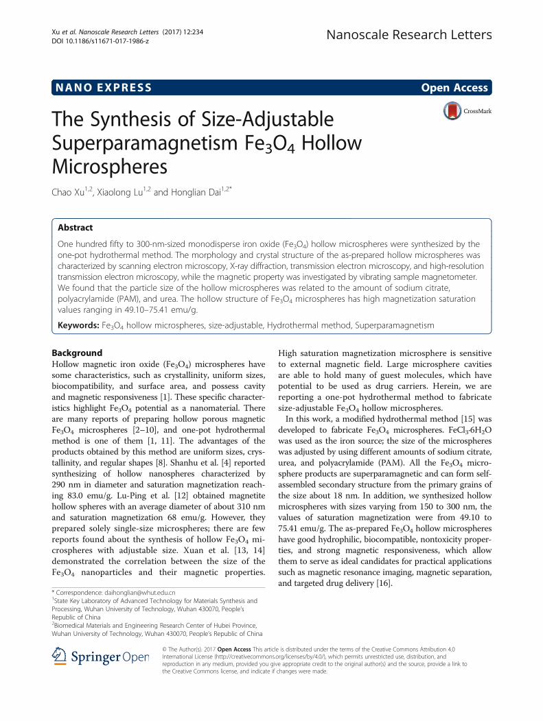
Fig. 4 SEM images of different amount of trisodium citrate synthesized sample. a 3 mmol. b 4 mmol. c 6 mmol. d 8 mmol
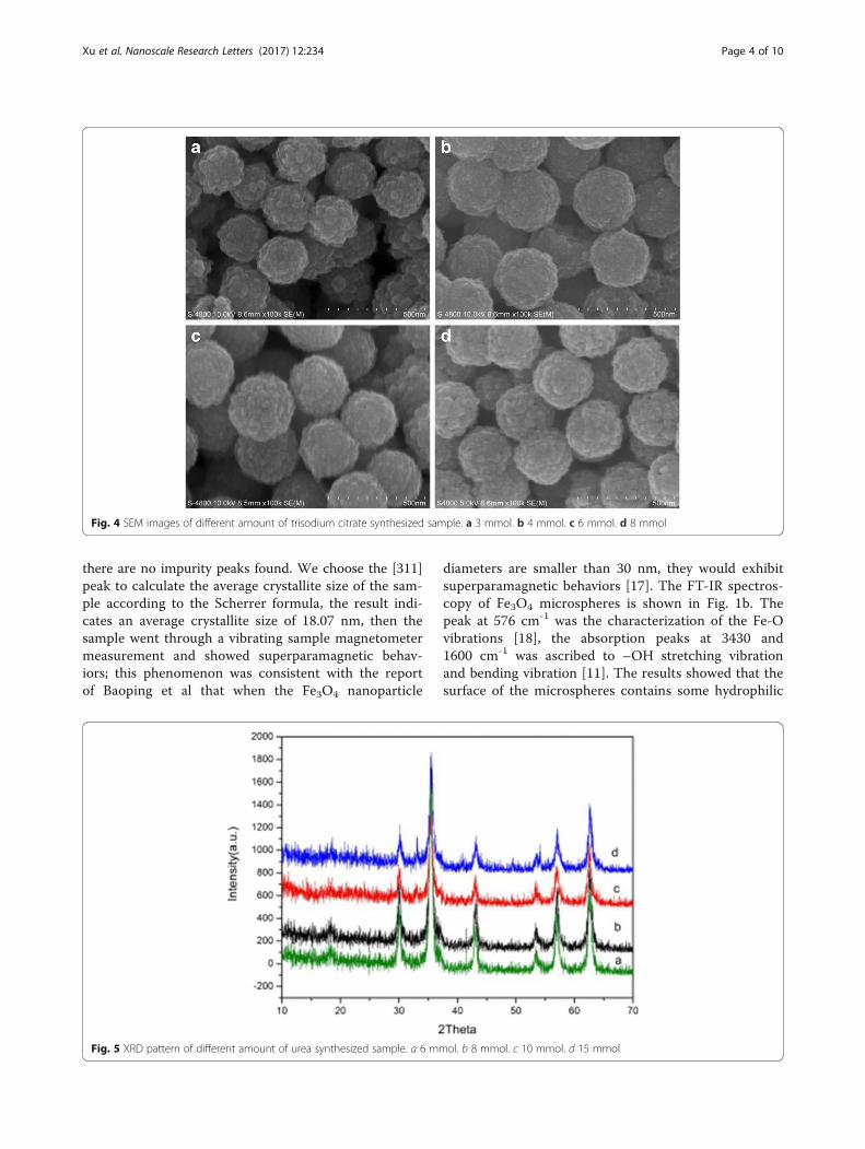
Fig. 5 XRD pattern of different amount of urea synthesized sample. a 6 mmol. b 8 mmol. c 10 mmol. d 15 mmol
Xu et al. Nanoscale Research Letters (2017) 12:234 Page 4 of 10

groups, which may endow microspheres with hydro-philic properties.The morphology of the sample was characterized by
SEM. The SEM images (Fig. 2a) showed that we haveobtained uniform and monodisperse microspheres. Thesamples were cut in ultrathin sections and examined byTEM (Fig. 2b), which showed that the products wereclusters of some small particles with coarse surfaces.
The size of the hollow microspheres was about 300 nmin average. From the single microsphere TEM images(Fig. 2c), it could be found that the spheres had hollowinternal structures. The corresponding SAED patterntaken from an individual microsphere is shown in Fig. 2d.It was found that the sample had polycrystalline struc-tures, which were consistent with the TEM images thatthe microspheres consist of some small particles. From
Fig. 6 SEM images of different amount of urea synthesized sample. a 6 mmol. b 8 mmol. c 10 mmol. d 15 mmol
Fig. 7 XRD pattern of different amount of PAM synthesized sample. a 0.1 g. b 0.2 g. c 0.3 g
Xu et al. Nanoscale Research Letters (2017) 12:234 Page 5 of 10

the inside to the outside, the rings can be indexed to(111), (220), (311), (400), (422), (511), and (440) planesof Fe3O4. All the diffraction rings can be readily indexedto the Fe3O4 phase.
The Effect of Various Factors on Size-Controllable Synthesisof Fe3O4 Hollow MicrospheresThe Effects of the Sodium Citrate on the Size of ProductParticlesThe XRD patterns of the microspheres synthesized withdifferent amounts of sodium citrate are shown in Fig. 3.α-Fe2O3 could be obtained when no sodium citrate isadded (Fig. 3a), and the intensity of α-Fe2O3 graduallydecreased with the increase of sodium citrate amount(Fig. 3b). When the amount of sodium citrate reach upto 3 mmol, the diffraction peaks of α-Fe2O3 disappearedcompletely. Sodium citrate seemed to play a role in theformation of the product. Sodium citrate might act as a
reducing agent under high-temperature conditions [19, 20].Furthermore, sodium citrate could also be used as astabilizer in the system, every sodium citrate molecule con-taining three carboxyl groups, a part of carboxyl groupssubstituted Fe3O4 microspheres surface hydroxyl groups,and formed a monomolecular adsorption layer, which couldreduce the reaction rate and inhibit grain growth [21].From the XRD patterns, it could be found that when
the amount of sodium citrate was 3, 4, 6, and 8 mmol,pure product could be prepared. The influence of thesodium citrate on the morphology of the products wasexamined by SEM. When the sodium citrate amountwas 3 mmol, the diameters of the microspheres wereabout 250 nm (Fig. 4a). At the 4-mmol level, the sizesof the microspheres were 300 nm (Fig. 4b). Further in-creasing the amount up to 6 and 8 mmol, the sizes ofthe microspheres were still 300 nm, and no furthermorphology changes were found (Fig. 4c, d).
Fig. 8 SEM images of different amount of PAM synthesized sample. a, b 0.1 g. c, d 0.2 g e, f 0.3 g
Xu et al. Nanoscale Research Letters (2017) 12:234 Page 6 of 10

The Effects of the Urea on the Size of Product ParticlesThe influence of urea amount on the size of the Fe3O4
hollow microspheres was investigated through samplesas listed in Table 1. The XRD patterns of the micro-spheres synthesized with different amounts of urea areshown in Fig. 5. All peaks of these four samples matchwell with standard Fe3O4 XRD diffraction (JCPDS Card
No. 79-06419). No obvious impurity peaks are found inFig. 5. The sharp peak indicated the high crystallinity ofproducts. It indicated that urea as alkali source in thereaction system did not affect the formation of Fe3O4
crystal grains. In the reaction process, urea was decom-posed to NH3 and provides an alkaline environment forthe solution system [22].
Fig. 9 TEM images of Fe3O4 hollow microspheres at different sizes. a 150 nm. b 200 nm. c 250 nm. d 300 nm
Fig. 10 Schematic illustration of the formation mechanism of Fe3O4 hollow microspheres
Xu et al. Nanoscale Research Letters (2017) 12:234 Page 7 of 10
The morphology and size of the microspheres were ex-amined by SEM. The SEM images showed that when theurea amount was 6 mmol, the diameters of the micro-spheres were about 300 nm (Fig. 6a). When the ureaamount was increased to 8 mmol, the size of the micro-spheres decreased to 250 nm (Fig. 6b). Further increasingthe urea amount to 10 mmol, the size of the microspheresdecreased to 200 nm. When the urea amount reached upto 15 mmol, the size of the microspheres was 150 nm. Itindicated that the amount of urea plays a role in the mat-ter of the size of microspheres. However, with the increaseof urea in the reaction, there were more NH3 and CO2
bubbles that act as a soft template [16], each soft tem-plate might adsorbed less nanoparticles; thus, smallersize of microspheres were obtained after Ostwald ripen-ing process [23, 24].
The Effects of the PAM on the Size of Product ParticlesFigure 7 illustrates the XRD pattern of the samples aslisted in Table 1. All peaks of these samples match wellwith standard Fe3O4 XRD diffraction (JCPDS Card No.79-06419). No obvious impurity peaks were found. Thesharp peak indicated that products had high crystallinity.The SEM images (Fig. 8) showed that when the PAMamount was 0.1 g, the sizes of the microspheres wereabout 200 nm (Fig. 8a, b). Dispersibility and shape of thesamples were not good under such conditions. Increas-ing the amount up to 0.2 g, the dispersibility and shapeof the samples were improved significantly, and the sizesof the microspheres increased to 250 nm (Fig. 8c, d).When the amount of PAM increased up to 0.3 g, thedispersibility and good shape of microspheres are ob-tained and the sizes of the microspheres were about300 nm (Fig. 8e, f ). Peng et al. reported that the polymer
PAM contains a large number of amide ligands, conse-quently stabilizing the primary particles [25]. The polymerPAM might increase the viscosity of the solution, whichmight slow down the movement of nanoparticles, givingmore time to adsorb the primary particles on the surfaceof soft templates and then self-assembled microspheres.With the increase of PAM concentration, nanoparticleshad enough time to self-assemble into larger spheres.
Crystal Structure and Magnetic Property of Fe3O4 hollowMicrospheres with Different SizesWe selected microspheres in the sizes of 150, 200, 250,and 300 nm and cut them in ultrathin sections and ex-amined by TEM (Fig. 9). Fe3O4 microspheres with adiameter of 150 nm (Fig. 9a) were of solid structure.Fe3O4 microspheres with a diameter of about 200 nm(Fig. 9b) interior small nanoparticles were dissolvedgradually. Fe3O4 microspheres with a diameter of 250 nm(Fig. 9c) were characterized as core-shell structure, andshell thickness were about 30 nm. Fe3O4 microsphereswith a diameter of 300 nm (Fig. 9d) interior small nano-particles were dissolved completely, which showed a sig-nificant hollow structure, and the out shell is composed ofprimary nanoparticles. Many cracks on the shell of the mi-crospheres can be clearly observed, indicating the highlyporous structure of the microspheres.Based on the experiment results and discussions above,
we propose that the formation of the hollow spheres is aresult of the dual role of gas bubbles and Ostwald ripeningprocess. The formation mechanism is illustrated in a sche-matic diagram presented in Fig. 10. Urea decomposed intoCO2 and NH3, which acted as soft templates in the reac-tion system (step 1). With the progress of the reaction, theoriginal nanoparticles start to be adsorbed on the surface
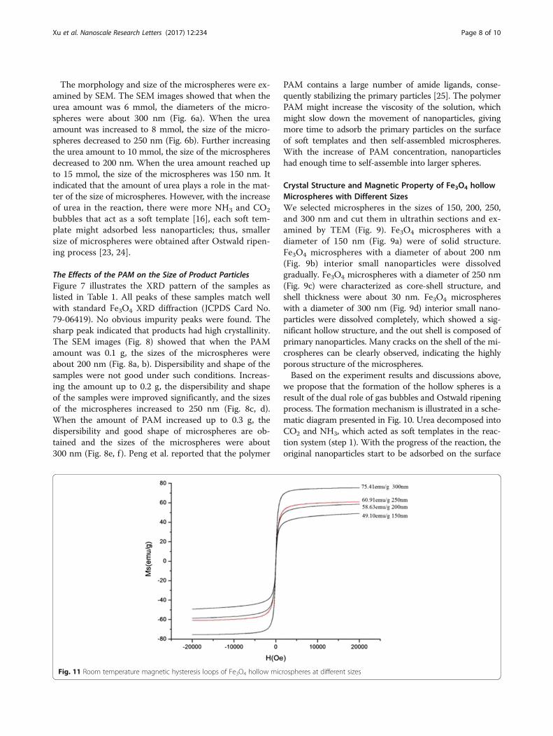
Fig. 11 Room temperature magnetic hysteresis loops of Fe3O4 hollow microspheres at different sizes
Xu et al. Nanoscale Research Letters (2017) 12:234 Page 8 of 10

of the gas bubbles, owing to the high surface energy of thegas bubbles (step 2). Thereafter, the nanoparticles grew onthe surface of the gas bubbles and agglomerated into loosespheres (step 3). Then, gas bubbles easily escaped fromthe loose spheres, which was also leading to form the hol-low cavity (step 4) [26]. Nanocrystals located in the coreregion tend to dissolve owing to the higher surface energythan those nanocrystals on the outer surface, and theinner nanocrystals recrystallization on the outer shell at-tributes to the Ostwald ripening [7, 22]. Once the nano-crystals in the core are dissolved completely, a hollowcavity structure would form.Magnetic characterization of different sizes of micro-
spheres measured at 300 K is shown in Fig. 11. The sat-uration magnetization values were 49.10, 58.63, 60.91,and 75.41 emu/g. The curves showed no remnantmagnetization or coercivity; all microspheres exhibitedsuperparamagnetic behavior at room temperature. Thesaturation magnetization values of prepared Fe3O4 micro-spheres increased gradually when particle size increased,which may be ascribed to the interior hollow cavity struc-ture. The saturation magnetization values varied followingthe changes in sphere size, which allows our Fe3O4 micro-spheres to be controlled easily by an external magneticfield, which is favorable for their applications in thebiomedicine field.
ConclusionsA series of Fe3O4 hollow microspheres with a size of150–300-nm particles were synthesized. The morphologyand structure of the hollow Fe3O4 microspheres werestudied by SEM, TEM, HRTEM, and XRD. We found thatthe size differences of Fe3O4 microspheres were related tothe amounts of sodium citrate, polyacrylamide, and urea.The obtained Fe3O4 microspheres had a hollow struc-ture and exhibited a superparamagnetic behavior withmagnetization saturation values between 49.10 and75.41 emu/g.
AbbreviationsFe3O4: Iron oxide; PAM: Polyacrylamide
AcknowledgementsThe authors thank Bi-Chao Xu of the Core Facility and Technical Support,Wuhan Institute of Virology for her technical support in transmission electronmicroscopy.
FundingThe work was supported by The National Key Research and DevelopmentProgram of China (2016YFC1101605), the Natural Science Foundation ofChina (81190133 and 51172171), the Natural Science Foundation of HubeiProvince (2015CFB551), the Key Technology Research and DevelopmentProgram of Hubei province (2015BAA085), and the Science and TechnologyProject of Wuhan (2015060101010032).
Authors’ ContributionsCX performed the synthesis and characterization of Fe3O4 hollow microspheres,XL participated in the characterization, and HD supervised the conceptual
framework and drafted the manuscript. All authors read and approved the finalmanuscript.
Competing InterestsThe authors declare that they have no competing interests.
Publisher’s NoteSpringer Nature remains neutral with regard to jurisdictional claims inpublished maps and institutional affiliations.
Received: 28 December 2016 Accepted: 8 March 2017
References1. Li D, Qin Q, Duan X, Yang J, Guo W, Zheng W (2013) General one-pot
template-free hydrothermal method to metal oxide hollow spheres andtheir photocatalytic activities and lithium storage properties. ACS ApplMater Interfaces 5(18):9095–9100
2. Jia Y, Yu XY, Luo T, Zhang MY, Liu JH, Huang XJ (2013) Two-step self-assemblyof iron oxide into three-dimensional hollow magnetic porous microspheresand their toxic ion adsorption mechanism. Dalton Trans 42(5):1921–1928
3. Liu M, Wen T, Wu X, Chen C, Hu J, Li J, Wang X (2013) Synthesis of porousFe3O4 hollow microspheres/graphene oxide composite for Cr (vi) removal.Dalton Trans 42(41):14710–14717
4. Liu S, Xing R, Lu F, Rana RK, Zhu J-J (2009) One-pot template-freefabrication of hollow magnetite nanospheres and their application aspotential drug carriers. J Phys Chem C 113(50):21042–21047
5. Ma FX, Hu H, Wu HB, Xu CY, Xu Z, Zhen L, David Lou XW (2015) Formationof uniform Fe3 O4 hollow spheres organized by ultrathin nanosheets andtheir excellent lithium storage properties. Adv Mater 27(27):4097–4101
6. Márquez F, Herrera GM, Campo T, Cotto M, Ducongé J, Sanz JM, Elizalde E,Perales Ó, Morant C (2012) Preparation of hollow magnetite microspheresand their applications as drugs carriers. Nanoscale Res Lett 7(1):1–11
7. Sun Q, Ren Z, Wang R, Chen W, Chen C (2010) Magnetite hollow spheres:solution synthesis, phase formation and magnetic property. J Nanopart Res13(1):213–220
8. Wang X, Huang H, Li G, Liu Y, Huang J, Yang D-P (2014) Hydrothermalsynthesis of 3D hollow porous Fe3O4 microspheres towards catalyticremoval of organic pollutants. Nanoscale Res Lett 9(1):1–5
9. Wang Y, Zhu Q, Tao L (2011) Fabrication and growth mechanism ofhierarchical porous Fe3O4 hollow sub-microspheres and their magneticproperties. CrystEngComm 13(14):4652
10. Zhang J, Yao Y, Huang T, Yu A (2012) Uniform hollow Fe3O4 spheresprepared by template-free solvothermal method as anode material forlithium-ion batteries. Electrochim Acta 78:502–507
11. Ren L, Huang S, Fan W, Liu T (2011) One-step preparation of hierarchicalsuperparamagnetic iron oxide/graphene composites via hydrothermalmethod. Appl Surf Sci 258(3):1132–1138
12. Zhu L-P, Xiao H-M, Zhang W-D, Yang G, Fu S-Y (2008) One-pot template-free synthesis of monodisperse and single-crystal magnetite hollow spheresby a simple solvothermal route. Cryst Growth Des 8(3):957–963
13. Xuan S, Wang F, Wang Y-XJ YJC, Leung KC-F (2010) Facile synthesis of size-controllable monodispersed ferrite nanospheres. J Mater Chem 20(24):5086
14. Xuan S, Wang Y-XJ YJC, Cham-Fai Leung K (2009) Tuning the grain size andparticle size of superparamagnetic Fe3O4 microparticles. Chem Mater21(21):5079–5087
15. Cheng W, Tang K, Qi Y, Sheng J, Liu Z (2010) One-step synthesis ofsuperparamagnetic monodisperse porous Fe3O4 hollow and core-shellspheres. J Mater Chem 20(9):1799
16. Hu P, Yu LJ, Zuo AH, Guo CY, Yuan FL (2009) Fabrication of monodispersemagnetite hollow spheres. J Phys Chem C 113(3):900–906
17. Jia B, Gao L (2008) Morphological transformation of Fe3O4 sphericalaggregates from solid to hollow and their self-assembly under an externalmagnetic field. J Phys Chem C 112(3):666–671
18. Guo S, Li D, Zhang L, Li J, Wang E (2009) Monodisperse mesoporoussuperparamagnetic single-crystal magnetite nanoparticles for drug delivery.Biomaterials 30(10):1881–1889
19. Brown KR, Walter DG, Natan MJ (2000) Seeding of colloidal Au nanoparticlesolutions. 2. Improved control of particle size and shape. Chem Mater12(2):306–313
Xu et al. Nanoscale Research Letters (2017) 12:234 Page 9 of 10

20. Xiong Y, McLellan JM, Yin Y, Xia Y (2007) Synthesis of palladium icosahedrawith twinned structure by blocking oxidative etching with citric acid orcitrate ions. Angew Chem 119(5):804–808
21. Caruso F (2001) Nanoengineering of particle surfaces. Adv Mater 13(1):11–1122. Wang FL, Liu JR, Kong J, Zhang ZJ, Wang XZ, Itoh M, Machida K (2011)
Template free synthesis and electromagnetic wave absorption properties ofmonodispersed hollow magnetite nano-spheres. J Mater Chem 21(12):4314–4320
23. Huo J, Wang L, Irran E, Yu H, Ma L, Gao J, Fan D, Ding W, Amin AM, Tai Y(2012) Synthesis, characterization and magnetic properties of hollowmicrospheres with micro-mesoporous shells assembled from cobalt-basedferrocenyl coordination polymers. J Colloid Interface Sci 367(1):92–100
24. Yuan H, Wang Y, Zhou S-M, Lou S (2011) Fabrication of superparamagneticFe3O4 hollow microspheres with a high saturation magnetization. ChemEng J 175:555–560
25. Peng Y, Xu A-W, Deng B, Antonietti M, Cölfen H (2006) Polymer-controlledcrystallization of zinc oxide hexagonal nanorings and disks. J Phys Chem B110(7):2988–2993
26. Liu X, Li Y, Zhu W, Fu P (2013) Building on size-controllable hollownanospheres with superparamagnetism derived from solid Fe3O4nanospheres: preparation, characterization and application for lipaseimmobilization. CrystEngComm 15(24):4937
Submit your manuscript to a journal and benefi t from:
7 Convenient online submission
7 Rigorous peer review
7 Immediate publication on acceptance
7 Open access: articles freely available online
7 High visibility within the fi eld
7 Retaining the copyright to your article
Submit your next manuscript at 7 springeropen.com
Xu et al. Nanoscale Research Letters (2017) 12:234 Page 10 of 10