THE SUSCEPTIBILITY OF INFANT RHESUS MONKEYS TO
15
THE SUSCEPTIBILITY OF INFANT RHESUS MONKEYS TO POLIOMYELITIS VIRUS ADMINISTERED BY MOUTH A STUDY 0]~ "1~£ DISTRIBUTION OY VIRUS IN --n~ TISSUES OF ORALLY INFECTED ANIMALS* BY DOROTHY M. HORSTMANN, M.D., JOSEPH L. MELNICK, PH.D., ROBERT WARD, M.D., AND M. JOS~ S~ FLEITAS, M.D.~ (From the Section of Preventive Medicine of Yale Uni~rsity School of Medicine, New Haven) (Received for publication, July 16, 1947) The experiments to be described were undertaken with a twofold purpose: (1) to test the susceptibility of infant rhesus monkeys to poliomyelitis virus fed by natural means; and (2), to study the distribution of virus in the tissues of this species when infected by the oral route and to compare the pattern obtained with that reported for cynomolgus monkeys infected orally (1, 2) and for fatal human cases (3-10). Earlier Feeding Experiments. The Susceptibility of Various Monkey Speciea.--The earliest successful attempt to produce infection by the gastrointestinal route is that reported by Leiner and vou Wiesner in 1910 (11). These workers fed large amounts of virus by stomach tube to two monkeys (species not mentioned), one of which de- veloped paralytic poliomyelitis, confirmed by histological sections. They also inocu- lated virus directly into the ileum at laparotomy and produced "characteristic symp- toms" in three of four snimals tested. Between the years 1910-1929, attempts to repeat the work of Lelner and yon Wiesner were largely unsuccessful. Flexner and Lewis (12), Landsteiner, Levaditi, and Pastia (13), Amoss (14), Rhoads (15), Schultz (16), and Thompson (17) all described negative results. Although various monkey species and strains of virus were used in these experi- ments there was no attempt to evaluate such variables until the work of Kling, Levaditi, and their associates L~pine and Homus (18-21). These investigators, between 1929 and 1934, reported a series of experiments testing the susceptibility of both rhesus and cynomolgus monkeys to poliomyelitis virus fed orally, injected by stomach tube, and introduced directly into the intestinal tract at laparotomy. They were able to infect nine of twelve cynomolgus, but failed when rhesus monkeys were used. Bumet and his coworkers confirmed these observations in 1939 (22): eight of fifteen ¢ynomolgus to which virus was administered orally, by pharyngeal swab, or by stomach tube, developed poliomyelitis. (An objection had been raised to the earfier successful experiments of Kllng and his group, on the ground that when infec- tion was produced by the oral route vires may have entered through the olfactory * Aided by a grant from the National Foundation for Infantile Paralysis, Inc. $ Aifredo Hirsch Foundation Fellow of the Academia Nacional de Medlcina, Buenos Aires, Argentina. 3O9
Transcript of THE SUSCEPTIBILITY OF INFANT RHESUS MONKEYS TO
T H E SUSCEPTIBILITY OF INFANT RHESUS MONKEYS TO POLIOMYELITIS VIRUS ADMINISTERED BY MOUTH
A STUDY 0]~ "1~£ DISTRIBUTION OY VIRUS IN --n~ TISSUES OF
ORALLY INFECTED ANIMALS*
BY DOROTHY M. HORSTMANN, M.D., JOSEPH L. MELNICK, PH.D., ROBERT WARD, M.D., AND M. JOS~ S~ FLEITAS, M.D.~
(From the Section of Preventive Medicine of Yale Uni~rsity School of Medicine, New Haven)
(Received for publication, July 16, 1947)
The experiments to be described were undertaken with a twofold purpose: (1) to test the susceptibility of infant rhesus monkeys to poliomyelitis virus f e d by natural means; and (2), to study the distribution of virus in the tissues of this species when infected by the oral route and to compare the pattern obtained with that reported for cynomolgus monkeys infected orally (1, 2) and for fatal human cases (3-10).
Earlier Feeding Experiments. The Susceptibility of Various Monkey Speciea.--The earliest successful attempt to produce infection by the gastrointestinal route is that reported by Leiner and vou Wiesner in 1910 (11). These workers fed large amounts of virus by stomach tube to two monkeys (species not mentioned), one of which de- veloped paralytic poliomyelitis, confirmed by histological sections. They also inocu- lated virus directly into the ileum at laparotomy and produced "characteristic symp- toms" in three of four snimals tested. Between the years 1910-1929, attempts to repeat the work of Lelner and yon Wiesner were largely unsuccessful. Flexner and Lewis (12), Landsteiner, Levaditi, and Pastia (13), Amoss (14), Rhoads (15), Schultz (16), and Thompson (17) all described negative results.
Although various monkey species and strains of virus were used in these experi- ments there was no attempt to evaluate such variables until the work of Kling, Levaditi, and their associates L~pine and Homus (18-21). These investigators, between 1929 and 1934, reported a series of experiments testing the susceptibility of both rhesus and cynomolgus monkeys to poliomyelitis virus fed orally, injected by stomach tube, and introduced directly into the intestinal tract at laparotomy. They were able to infect nine of twelve cynomolgus, but failed when rhesus monkeys were used. Bumet and his coworkers confirmed these observations in 1939 (22): eight of fifteen ¢ynomolgus to which virus was administered orally, by pharyngeal swab, or by stomach tube, developed poliomyelitis. (An objection had been raised to the earfier successful experiments of Kllng and his group, on the ground that when infec- tion was produced by the oral route vires may have entered through the olfactory
* Aided by a grant from the National Foundation for Infantile Paralysis, Inc. $ Aifredo Hirsch Foundation Fellow of the Academia Nacional de Medlcina, Buenos
Aires, Argentina. 3O9
310 ~LIO~JfELI~ VIRUS
pathway. This difficulty was anticipated by Burnet and his colleagues who blocked the nasal pathway in some of their animals by preliminary application of a ZnS04 spray to the nasal mucosa.) Subsecluenfly, cynomolgus monkeys have been infected a number of times by oral administration of virus (1, 23, 24) and other species a few times: M. irus (mordax) 1 in one of eleven trials (23, 26) the green African monkey, Cerco~thecus adhiops s~eus, in one of seventeen (25, 26). Negative results have been obtained with one capuchin (Cebus faIa~ellus) (25), one grivet (Cercop//hecus griseo~iridis), (28), seven vervets, Cerco~ithecus a#ghiops #ygeryJhrus, (27, 28), two M. sinicus (29, 30), and two baboons one of which was of the species Pa#io hacn~ryas (27, 31).
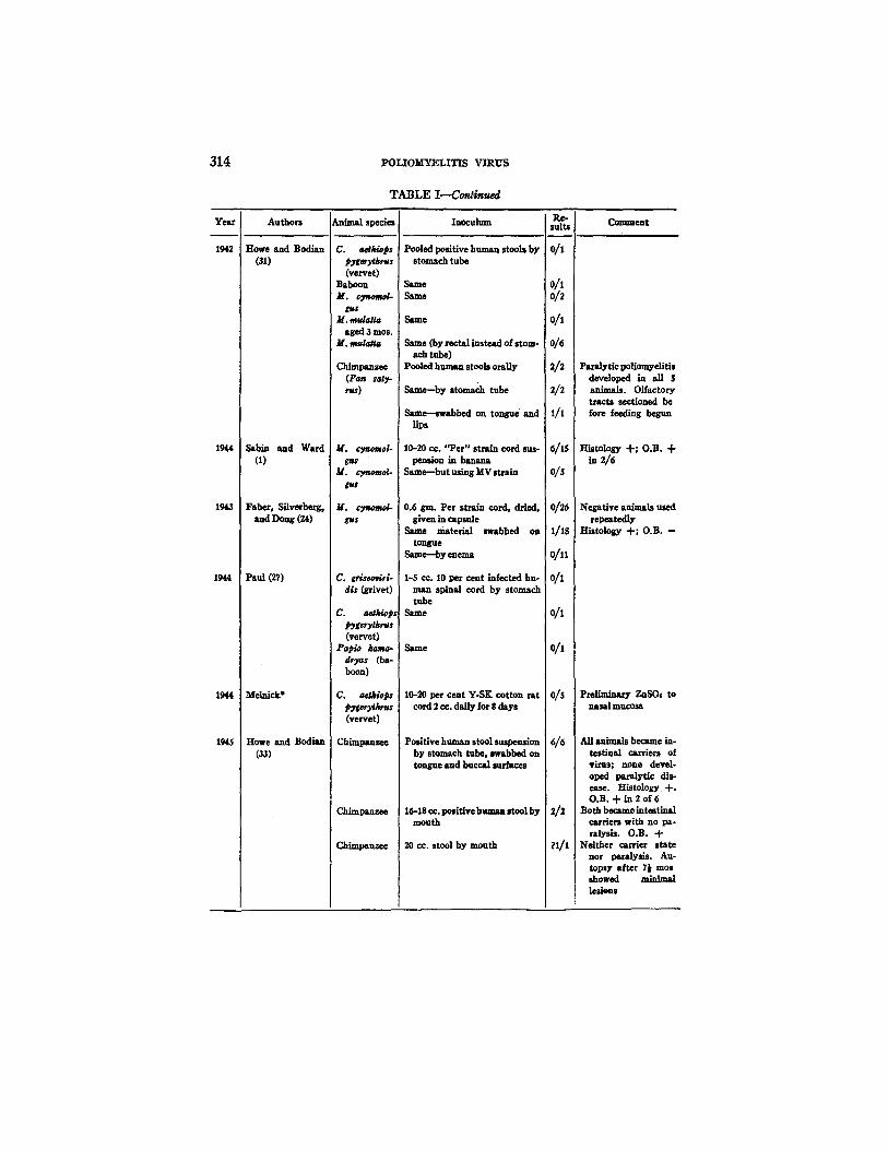
The experimental animal which has proved most susceptible to infection by the gastrointestinal or oral route of virus administration is the chimpanzee. But it was many years before this was appreciated. Landsteiner and Levaditi first attempted such an experiment in 1910 (30) by feeding a chimpanzee 10 cc. of infected monkey cord by stomach tube. The animal remained well and the result was interpreted as negative. However, since it is now known that in chimpanzees infection is often manifested only by a "healthy" carrier state for a period of weeks (33, 34, 35) it is possible that the animal used by Landsteiner and Levaditi did become infected even though it never developed paralysis. Chimpanzees were not used for similar experi- ments until 1942 when Howe and Bodian induced poliomyelitis in five of five animals using infective human stools as the inoculum, which was administered by simple feeding, by stomach tube, and by lingual and buccal swab (31). The olfactory tracts of the animals were sectioned before the experiment was begun in order to block the olfactory pathway. Subsequently, a "healthy" carrier state was produced in each of thirteen chimpanzees tested in Baltimore (33) and in eight chimpanzees in New Haven (34, 35).
The results with chimpanzees are in marked contrast to the experience with rhesus monkeys. In the latter, of the twenty-three reported animals in experiments in which it has been specifically stated that M. mu/a/ta were used for various types of feeding experiments (virus being given by mouth, by stomach tube, and rectal tube) twenty-three negative results have been reported; of these, twenty-two attempts were in young or adult rhesus, while one was in a 3 month old baby rhesus (31). Leiner and von Wiesner (11) reported a single positive, although it is not entirely certain that the species used was M. mu/a~. In recent unpublished experiments from o u r
laboratory, one young rhesus of eight fed the Y-SK strain developed paralytic polio- myelitis (32); previous experience with feeding this strain of virus to young adult rhesus monkeys had been negative (23).
Strain~ of Virus.--The experiments of Sabin (1) indicate the possible r61e of virus strain differences in producing infection by the oral route. Six of fifteen cynomolgus monkeys developed paralyses when fed the recently isolated "Per" strain, while none of five cynoraolgus showed signs of disease when fed the Rockefeller MV strain. Earlier experiments of Clark and his associates (36, 46)and Flexner (5) in which both cynomolgus and rhesus were fed the higMy monkey-adapted MV strain, were
1 T h e r e are at least several branches of the cynomolgu~ family, and the t e r m i n o l o g y is not perfectly clear. Those which have been used include, M. cynomolgus (Java), M. irus (mordax), M. filipenen~ (Philippine Islands), and M. irus ¢a21da (Siam).
D. M. HORSTMANN, 7. L. MELNICK~ R. WARD, AND M. 7. S~ lrLEITAS 311
also negative. Burnet and his group used recently isolated human strains in their successful feeding experiments (22), as did Vignec, Trask, and Paul (23) who used early passages of the Y-SK strain. The successful tests in chimpanzees have been with human stool suspensions (31), fly-contaminated food (34), and the Y-SK strain (35).
Si~ of Pene~aJ, ion.--In attempting to localize the level of the gastrointestinal tract at which the virus penetrates the intact mucous membrane, Faher, Silverberg, and Dong (24) fed virus ("Per" strain) to cynomolgus in capsules, applied it by lingual swab, and injected it by enema. Positive results were obtained only with the lingual swab technique in one of eighteen trials.
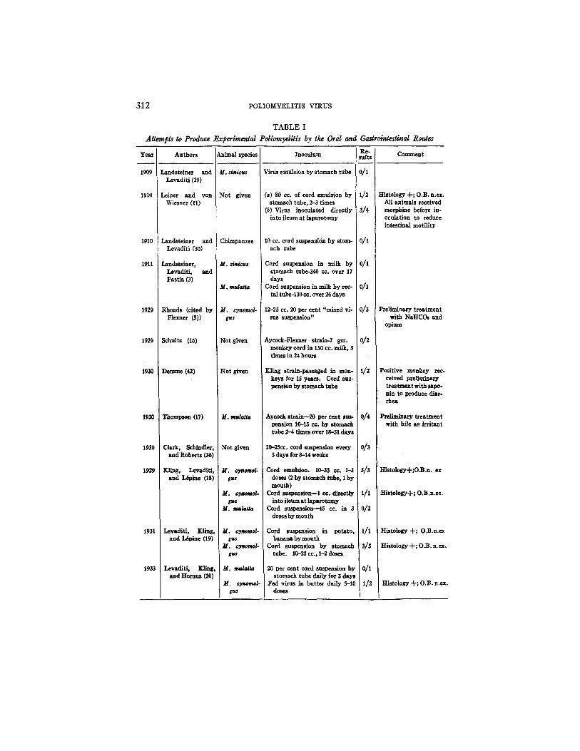
Summary.--Table J summarizes the reported experiments de~ling with the production of poliomyelitis by various oral and gastrointestinal routes. The number of uncontrolled variables in these experiments make it impossible to compare the susceptibility of the different species more directly. Although such a tabulation would make for simplicity and clarity, it would nevertheless be misleading. The table is therefore constructed on chronological lines.
I. The Susceptibility of Infant Rhesus Monkeys to Orally Administered Poliomyelitis Virus
In view of the apparent resistance of young or adult rhesus monkeys to orally administered poliomyelitis virus, the following experiments were planned to test the susceptibility of infant rhesus monkeys to poliomyelitis virus adminis- tered orally. An at tempt was made to reproduce as far as possible a "natural" mode of infection, and simple virus feeding alone was therefore employed. In order to avoid the possibility of infection by the olfactory pathway, the nasal mucosa of each animal was sprayed vigorously with 1 per cent ZnSO4 on two occasions 2 to 5 days apart, before feeding was begun. This procedure has been shown by Schultz and Gebhardt (37) to block the penetration of virus through the nasal mucosa to the olfactory tract.
Materials and M etheds
Monkeys.--Seven infant rhesus monkeys, M. mu/a~ta, ranging in age from 3½ weeks to approTinmtely 4 months and one young adult rhesus were used (see Table II). z Duringthe course of the experiments each animal was observed daily for signs of poliomyelitis, and daily temperature records were kept. Those ~nimals developing fever and paralysis were sacrificed early in the disease in the hope of obtaining a maximum yield of virus from the various tissues. Animals showing no signs of disease were observed for 5 to 6 weeks; they were sacrificed at the end of this period, and sections of their olfactory bulbs, brain stem, and cervical, thoracic, and lumbar cords were made for histological examination.
Virus Slrai~.--A variety of strains were used. They included two rodent-adapted strains (Y-SK and Ph), infective human CN'S and stools, and monkey cord infected with recently isolated human strains. Table II indicates the various strains fed to each animal. Some
i We are indebted to Dr. G. van Wagenen of the Department of Obstetrics and Gynecology, Yale School of Medicine, for most of the infant animals used in these experiments.
312 POLIOMYELITIS VIRUS
Attempts to Produce Ex
Year Authors !Animal species
1909
1910
1910
1911
1929
1929
1930
1930
1930
1929
1931
Landstelner and Levaditi (29)
Leiner and yon Wiesner (11)
Landsteiner and Levaditi (30)
Landstelner, Levaditi, and Pastia (3)
Rhoads (cited by Flexner (5))
schaltz (16)
Demme (42)
Thompson (17)
Clark, Schindler, and Roberts (36)
Kling, Levaditl, and I&pine (18)
Levadlti, gl int , and I~pine (19)
Levaditi, grin f , and HomUs (20)
TABLE I
~erimental Poliomyditis by tke Oral and Gastrointestinal Routes
M. sinicus
Not given
Chimpanzee
M. slni~s
M. mulatM
M. cynoraol- gus
Not given
Not given
M. tuu/atta
Not given
M, cyaomol- gus
M. cynomol- gus
M. mula~ta
M. cyr.omol- gu$
M. cyaomol- gu,~
: M. madc#/a
Inoculum ] sRulte's Comment
Virus emulsion by stomach tube 0/1
(a) 80 co. of cord emulsion by I//2 stomach tube, 2-3 times
(b) Virus inoculated directly i3//4 into ileum at laparotomy
10 cc. cord suspension by store- 0//1 ach tube
Cord suspension in milk by 0/I stomach tube~40 cc. over 17 days
Cord sUspension in milk by rec- 0//I tal tube-130 cc. over 26 days
12-25 cc. 20 per cent "mixed vl- 0//3 rus suspellsion"
Aycock-Flexner strain-7 gin. 0/2 monkey cord in 150 cc. milk, 3 times in 24 hours
Kling strain-passaged in mon- 1/2 keys for 15 years. Cord sus- pension by stomach tube
Aycock strain--20 per cent sus- 0/4 pension 10-15 cc. by stomach tube 2-4 times over 18-51 days
20-25cc. cord suspension every 0/3 5 days for 8-14 weeks
Cord emulsion. 10-35 cc. I-3 3//3 doses (2 by stomach tube, I by mouth)
Cord suspension--1 ec. directly I / I into ileum at lapasotomy
Cord suspension---45 co. in 3 0//2 doses by mouth
Cord suspension in potato, i / l banana by mouth
Cord suspension by stomach 3/5 tube. 10-25 cc., 1-2 doses
20 per cent cord suspension by 0/I stomach tube daily for 3 days
Fed virus in butter daily 5-10 I/2 doses
M. cyr.omo/- gu$
Histology -}-; O,B. n.ex. All animals received morphine before in- oculation to reduce intestinal motility
Preliminary treatment with NaHCO, and
opium
Positive monkey rec- ceived preliminary treatment with $apo- nin to produce diar- rhea
PreH~ary treatment with bile as irritant
Histology-l-;O.B.n. ex
Histology-I-; O.B.n.ex
Histology + ; O.B.n.ex
Histology -[-; O.B.n.ex.
Histology -{'; O.13. n.ex.
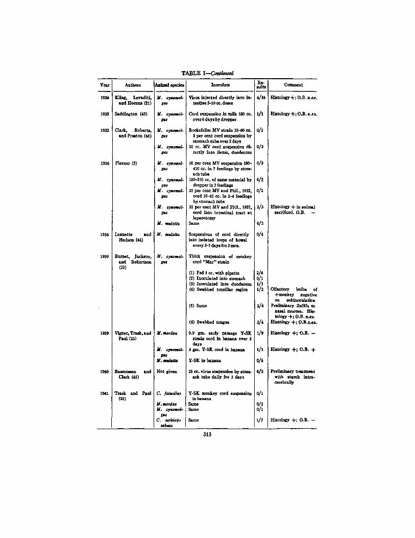
TABLE I--Co~nued
Yes.r
1934
1932
1932
1936
1936
1939
1939
1940
1941
Authors
'glin£, Lovaditi, and Hornet (21)
Saddington (43)
Clark, Roberta, and Preston (46)
Flexner (5)
Lennette and Hudson (44)
Burnet, Jackson, and Rober tson (22)
Vignec, Trask, and Paul (23)
P~e~tuman and Clark (~)
Trask and Paul (25)
Animal species
M. c3~mo/- gu$
M. ~ / - gus
M. c3momol. gu$
M. c3~Aiol- gu~
M. cynomol- gus
gu$ M. cynomol. gus
M. cyaomol-
M. mulatia
M. mulaffa
M. cyewmo~
M. mtorda=
M. c3motw/-
M . ~
Not given
C. .faSuellus
M. tlofda,~ M. cy~mol-
C. aeJkwps saJ~s
Re- Comment Inocuinm sul ts
~Fttus injected directly into in- 4/16 Histology-t-; O.B.n.ex. testine 5-10 co. doses
Cord suspension in milk 180 cc, 1/1 Histology -I-; O.B.n.ex. over 6 days by dropper
i
Rockefeller MV strain 2,5--60 co, 0/2 S per cent cord suspemion by stomach tube over 2 days
10 cc. MV cord s u s l ~ i o u di- 0/3 rectly into ileum, duodenum
10 per cent MV suspsn~on 180- 0/3 410 cc. in 7 feedings by stom- ach tube
180-210 cc. of same material by 0/2 dropper in 7 feedlnp
25 per cent MV and Phil., 1932, 0/2 cord 35--45 cc. in 2--4 feedings by stomach tube
2,5 per cent MV and Phil., 1952, 2/5 Histolosy "1- in animal cord into intestinal tract a t sacrificed. O.B. -- lapsrotomy
Same 0/2
Suspensions of cord directly 0/4 into isolated loops of bowel
every 3-7 days for 5 mos.
Thick suspension of monkey cord "Mar" strain
(1) Fed 1 cc. with pipette 2/6 (2) Inoculated into s tom~h 0/1 (3) Inoculated into duodenum 1/1 (4) Swabbed tonsiilar region 1/2
(S) Same
(6) Swabbed tongue
0.9 gin. early passage Y-SK strain cord in banana over 3 days
3 gin. Y-$K cord in ba~s,m
Y-SK in banana
25 co. virus suspendon by stem- I Itch tube daily for 3 days
Olfactory bulbs ol -I-monkey negative on subinoculation
2/4 pr~limi~sry ZnS04 to nasal mucosa. His- tology -I-; O.B.n.ex.
2/4 Histology -I-; O.B,n.ex,
I /9 Histology -t-; O.B. --
1/1
0/8
0/2
Y-SK monkey cord suspension 0/1 in banana
Same 0/2 Same 0/1
Same 1/7
Histology -I-; O.B. 4-
Pre]imin~ry t rsa~e~t with starch intra- cerebrally
Histology -I-; O.B. --
313
314 I~LIOM'YELITIS VIRUS
T A B L E I - - - C o n ~ m ~
Year
1942
1944
1943
1944
1944
1945
Authors
Howe and Bodian (St)
Stbin and Ward (t)
Faber, Silvecherg, and Doug (24)
Paut (27)
Melnick"
Howe and Bodian (33)
Animal species
C. aethlops pygery~rws (vervet)
BabOOn M. ~ / -
M..mu/4t./a aged 3 mos.
M. nue/a~
Chimpanzee (Pan saO-
M. cynomoI- gu$
M, cynomol-
H. cysomol- ~,ws
C. griseo~ir;- d/s (grlvet)
C. aetklopJ l~g~hrus (vervet)
Papio kar~- dryas (ba- boon)
C. ae~/ops FTgerytkr~ (vervet)
Chimpanzee
Chimpanzee
Cb~panzee
Ioocu]um
Pooled positive hi,mAn stOOls by stomach tube
Same Same
Same
Same (by rectalinstead of stom- ach tube)
Pooled hnm~- stools orally
Same--by stomach tube
Same--swabbed on tongue and llps
10-20 co. "Per" strain cord sus- pension in banana
Same-but using MV strain
0.6 gin. Per strain cord, dried, 8iven in capsule
Same material swabbed on tongue
Same--by enema
1-5 c¢. I0 per cent infected hu- man spinal cord by stomach tube
Same
Same
10-20 per cent Y-SK cotton rat cord 2 cc. daily for 8 days
Positive huma,, stool suspension by stomach tube, swabbed on tongue and buccal surfaces
16-18 co. positive httman stool by mouth
20 cc. stool by mouth
suits
o/1
0/I 0/2
0/t
0/6
2/5
2/2
1/x
6/'15
0/5
0/26
1/18
o/n
o/I
o/1
o/1
o/s
n/t
Comment
Paralytic poHomyeiitis developed in all S animals. Olfactory tracts sectioned be fore feeding begun
Histology + ; O.B. "l- in 2/6
Negative animals used repeatedly
Histology + ; O.B. --
pr~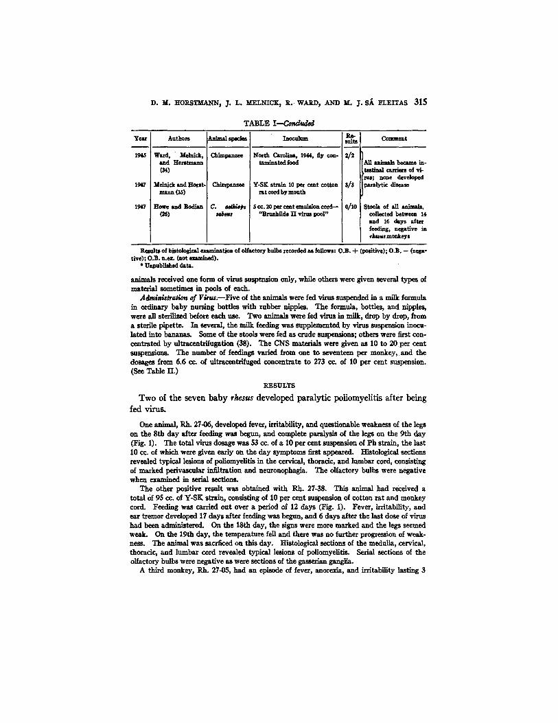
D. M. HORSTMANN, J. L. MELNICK~ R. WARD, AND M. J. S~ ]~LEITAS 315
T ~ L E I - - - C ~ d
Year
1945
1947
1947
Authors
Ward, Melnick, and Horst-mnn
Melnick and Horst- mann OS)
Howe and Bodian (26)
Chb-l~nr~e
ChhnZmlz~
C. ~ p s
~locttlmn
North Carolina, 1944, ~y con- taminated food
Y-SK strain 10 per cent cotton rat cord by mouth
$ cc. 20 per cent emulsion cord-- "Bnmhilde 11 vimJ pool"
suR~ Comment
An an;mAla became in- testinul carriers of vi- rus; none developed
3/3 paralytic disease
o/1o Stooh of an animals, collected between 1,1, and 16 days after feeding, negative in r~us monkeys
Results of h~tological examination of olfactory bulbs recorded as follows: O,B. -I- (positive); O.B. -- (nega- tive); O.B. n,a. (not .~m;.ed).
* Unpublished data.
animals received one form of virus suspension only, whi]e others were given several types of material sometimes in pools of each.
Admi~h~vag~ of Virm.--Five of the ~, ;m, ls were fed virus suspended in a milk formula in ordinary baby nursing bottles with rubber nipples. The formula, bottles, and nipples, were all sterilized before each use. Two An;m~h were fed virus in milk~ drop by drop, from a sterile pipette. In several, the milk feeding was supplemented by virus suspension inocu- lated into bananas. Some of the stools were fed as crude suspensions; others were first con- centrated by ultracentrlfugatlon (38). The CNS materials were given as 10 to 20 per cent suspensions. The number of feedings varied from one to seventeen per monkey, and the dosages from 6.6 cc. of ultracentrifuged concentrate to 273 cc. of 10 per cent suspension. (See Table H.)
RESULTS
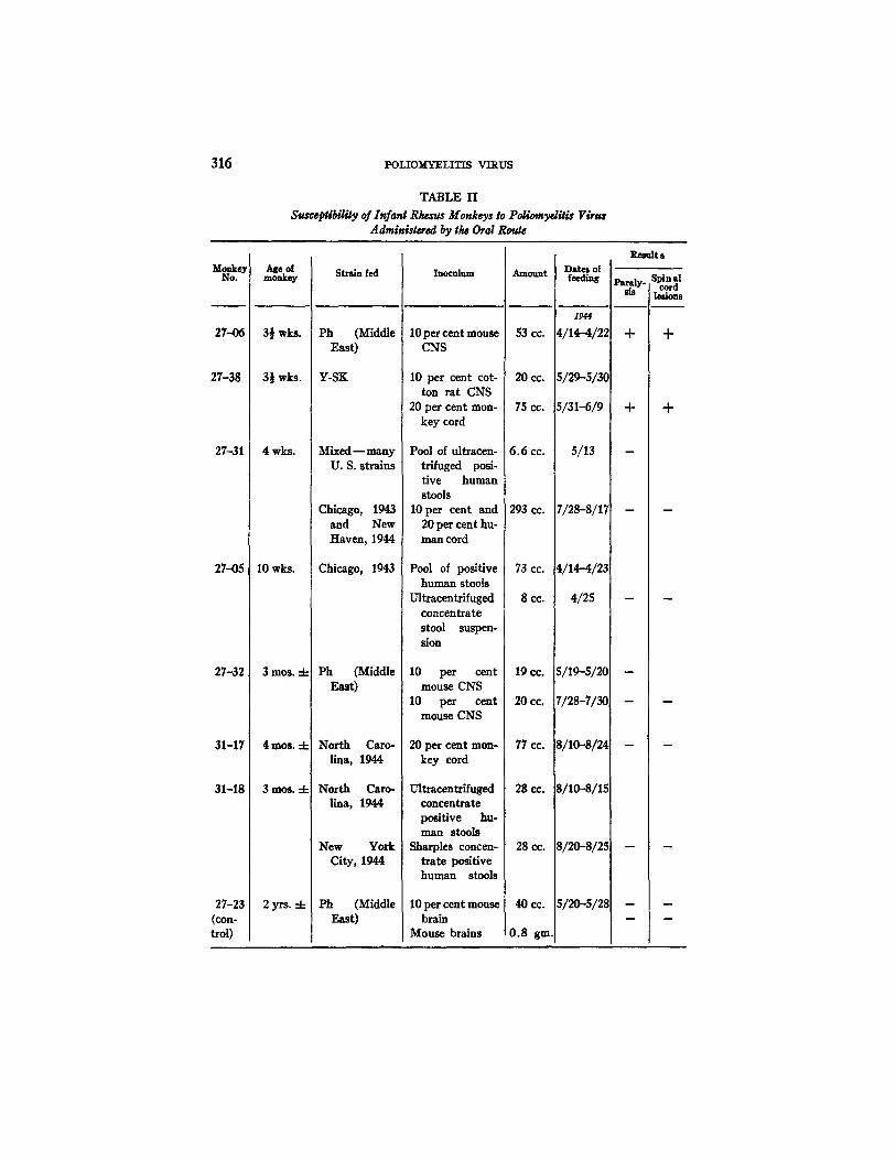
Two of the seven baby rhesus developed paralytic poliomyelitis a/ter being fed virus.
One animal, Rh. 27-06, developed fever, irritability, and questionable weakness of the legs on the 8th day after feeding was begun, and complete paralysis of the legs on the 9th day (Fig. 1). The total virus dosage was 53 cc. of a 10 per cent suspension of Ph strain, the last 10 cc. of which were given early on the day symptoms first appeared. Histological sections revealed typical lesions of poliomyelitis in the cervical, thoracic, and lumbar cord, consisting of marked perivascular infiltration and neuronophagia. The olfactory bulbs were negative when examined in serial sections.
The other positive result was obtained with Rh. 27-38. This animal had received a total of 95 cc. of Y-SK strain, consisting of 10 per cent suspension of cotton rat and monkey cord. Feeding was carded out over a period of 12 days (Fig. 1). Fever, irritability, and ear tremor developed 17 days after feeding was begun, and 6 days after the last dose of virus had been ~dmlnlstered. On the 18th day, the signs were more marked and the legs seemed weak. On the 19th day, the temperature fell and there was no further progression of weak- hess. The animal was sacrficed on this day. Histological sections of the medulla, cervical, thoracic, and lnmbar cord revealed typical lesions of poliomyelitis. Serial sections of the olfactory bulbs were negative as were sections of the gasserian ganglia.
A third monkey, Rh. 27-05, had an episode of fever, anorexia, and irritability lasting 3
316 POLIOMYELITIS VIRUS
TABLE H
Susceptibilily of Infant Rt~'us Monkeys to PoliomydSis Virus Adminlstered by tl~ Oral Route
Monkey No.
27-06
27-38
27-31
2745
27-32
31-17
31-18
27-23 (con- trol)
Age o[ monkey
3½ wks.
3½ wks.
4 wks.
10 wks.
3mos. 4-
4 m o s . -t-
3 m o s . -4-
2yrs. 4-
Strain fed
Ph (Middle East)
Y-SK
Mixed - - many U. S. strains
Chicago, 1943 and New Haven, 1944
Chicago, 1943
Ph (Middle East)
north Caro- lina, 1944
North Caro- lina, 1944
New York City, 1944
Ph (Middle East)
Inoculum
10 per cent mouse CNS
10 per cent cot- ton rat CNS
20 per cent mon- key cord
Pool of ultracen- trlfuged posi- tive human stools
10 per cent and 20 per cent hu- man cord
Pool of positive human stools
Ultracentrifuged concentrate stool suspen- sion
10 per cent mouse CNS
10 per cent mouse CNS
20 per cent mon- key cord
Ultracentrifuged concentrate positive hu- man stools
Sharpies concen- trate positive human stools
10 per cent mouse brain
Mouse brains
Amount
53 cc.
20 cc.
75 cc.
6.6 cc.
293 CC.
73 cc. (
8cc.
19 CO.
20 cc.
77 cc.
2g CO.
28 cc.
40CC. i
0,8 gin.
Dates of feeding
1944
4/1~-4/22
5 ~ 5 ~ C
5~I-6/9
5/13
7/28-8/17
4/14-4/23
4/25
S/19-5/20
7/28-7/3O
S/10-.8/24
8/10-.8/1.5
Sl2O-s/2s
51~-512~
Result s
sis I lesions
+ +
m
m
D. ~ . H O R S T ~ , J. r~. M~LmCK, R. WARD, ~ u . j . S/~ ~Lv.X~AS 317
days which was thought to be compatible with abortive poliomyelitis. This animal had received a total of 73 cc. of crude stool suspension and 8 cc. of ultrscentrifuged concentrate of fecal material. The onset of symptoms was 15 days after virus feeding was begun and 3 days after it had been stopped. Stools collected on the 3rd day of illness and during the subsequent 8 days were pooled, concentrated by ultracentrifugstion, and tested intracere- brally in another rhesus monkey with negative results. Rh. 27-05 was sacrificed 24 days after this mild illness (41 days after the feedings was begun). No lesions were found in three levels of the cord or in the medulla or olfactory bulbs.
The other four animals, as well as the control, remained entirely well during the experi- ment. Two animals (27-31, 27-32) had been given small doses of virus and observed for the usual period for signs of disease. Both remained healthy and after S weeks were fed the same or different strains in large amounts, again with negative results (Table II).
1015 t
103 o
Rh. 27-06
I O 0 ~ . • , • • • * • , * , , • , . , , *
APRIL 14 15 IG 17 18 19 20 21 22 23 24 25 26 ~7 P.8 Z9 3 0 3 /
ul IO&
"~ 104,
(1~ IO3, UJ n
IOZ'
I- lob
IO0 MAY 2s
l l l l l l l l l l l l
- • . . . ° • • • • ' • • • •
30 3 ! J U N i 2 3 4 "5 6 7 8 9 I0
Rh. 27-38
- ~ z:~ :qqn • 4 ' • • • • •
I I 12 13 14 15 16 17
F~G. 1. Course of events in two baby rhesus infected with poliomyelitis virus by the oral route. Arrows indicate times at which virus was fed.
H. Distribution of Poliomyelitis Virus in the Tissues of Two Infant Rhesus Monkeys Infected by the Oral Route
Studies on the localization of poliomyelitis virus in the fatal human disease (3-10) and in cynomolgus monkeys infected by the oral route (1, 2) have shown different distribution patterns. While in neither case does the virus seem to be as strictly neurotropic in its location as was once believed, it is more diffusely distributed throughout the tissues of the cynomolgus monkey than in man. In their series of human autopsies on five bulbar and two spinobulbar types of cases Sabin and Ward (8) found virus primarily in the nervous system, the alimentary tract (including the pharynx), and superficial lymph nodes. The results in less extensive studies of human tissues have been similar (3-7, 9, 10).
318 POLIOIKY]gLITIS VIRUS
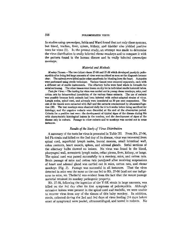
In studies using cynomolgus, Sabin and Ward found that not only these systems, but blood, trachea, liver, spleen, kidney, and bladder also yielded positive tests for virus (1). In the present study, an at tempt was made to determine the virus distribution in orally infected rhesus monkeys and to compare it with the pattern found in the human disease and in orally infected cynomolgus monkeys.
Material and Methods
Mankey T/ssu~.lThe two infant r/wsus 27-06 and 27-38 which developed paralytic polio- myelitis after being fed large amounts of virus were sacrificed as soon as the diagnosis became clear. The animals were killed under ether anesthesia by bleeding from the heart. Autopsies were performed u~ng sterile technique. Various tissues were removed separately, each with a different set of sterile instruments. The olfactory bulbs were fixed whole in formalin for serial sectioning. The other tissues were frozen on dry ice in individual sterile lusteroid tubes.
TesSsfar Virus.--The testing for virus was carried out in young rhesus monkeys, mice, and cotton rats by intracerebral inoculation of the various tissue extracts. The use of rodents was possible because both animals had been infected with rodent-adapted strains of virus. Lymph nodes, spinal cord, and adrenals were inoculated as 20 per cent suspensions. The rest of the tissues were extracted with H20 and the extracts concentrated by ultracentrffuga- tion (38). The test monkeys were observed daily for 4 to 6 weeks before being sacrificed for histology, and the negative rodents were discarded at the end of the observation period. Criteria for a positive test were: the development d typical signs of the disease during life with characteristic histological lesions in the monkey, and the development of signs of the disease only in rodents. Passage to other rodents and to monkeys was carried out in some instances.
Results of the Study of Virus Distribution
A summary of the tests for virus is presented in Table I I I . From Rh. 27-06, fed Ph strain and killed on the 2rid day of its disease, virus was recovered from spinal cord, superficial lymph nodes, buccal mucosa, small intestinal wall, colon contents, heart muscle, spleen, and adrenal glands. Serial sections of the olfactory bulbs showed no lesions. No virus was found in the blood, pharyngeal wall, mesenteric lymph nodes, celiac plexus, liver, kidney, or lungs. The spinal cord was passed successfully to a monkey, mice, and cotton rats. Brain passage of mice and cotton rats paralyzed after receiving suspensions of heart and adrenal gland was carried out in mice, cotton rats, and rhesus monkeys (Fig. 2). Passage was successful in all instances. That the virus detected in mice was the same as the one fed to Rh. 27-06 (and not one indige- nous to mice, viz. Theiler's) was evident from the fact that the mouse passage material retained its monkey pathogenic property.
Rh. 27-38, following the ingestion of the Y-SK strain in large amounts, was killed on the 3rd day after its first symptoms of poliomyelitis. A l t hough extensive lesions were present in the spinal cord and medulla, we were unable to recover virus from any of the tissues of this baby monkey. In addition, stools, collected during the 2nd and 3rd days of virus feeding (14 days before onset of symptoms) were pooled, ultracentrifuged, and tested in rodents. No
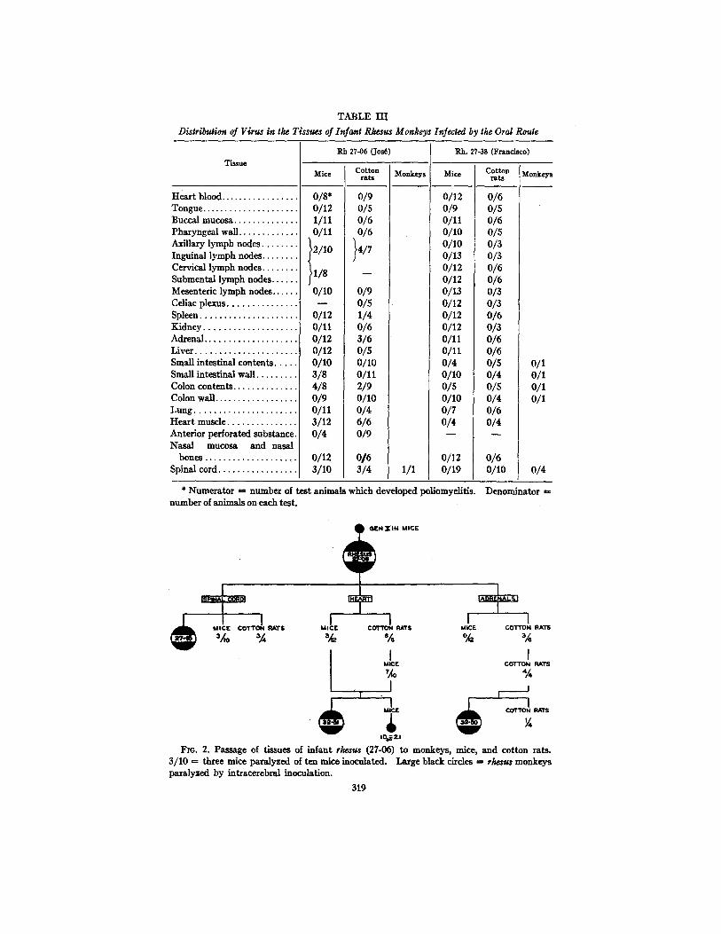
T A B L E I I I
Distribution of Virus in the Tissues of Infant Rhesus Monkeys Infected by the Oral Rouge
Tissue
Hear t blood . . . . . . . . . . . . . . . . . Tongue . . . . . . . . . . . . . . . . . . . . . Buccal mucosa . . . . . . . . . . . . . . Pharyngea l wall . . . . . . . . . . . . . Axillary l ymph nodes . . . . . . . . Inguinal l ymph nodes . . . . . . . . Cervical l ymph nodes . . . . . . . . Submenta l l ymph nodes . . . . . . Mesenteric l ymph nodes . . . . . . Celiac plexus . . . . . . . . . . . . . . Spleen . . . . . . . . . . . . . . . . . . . . Kidney . . . . . . . . . . . . . . . . . . . Adrenal . . . . . . . . . . . . . . . . . . . Liver . . . . . . . . . . . . . . . . . . . . . Small intest inal contents . . . . Small intest inal wall . . . . . . . . Colon contents . . . . . . . . . . . . . Colon wall . . . . . . . . . . . . . . . . . L u n g . . . . . . . . . . . . . . . . . . . . . Hea r t m u s d e . . . . . . . . . . . . . . Anterior perforated subs tance Nasa l mucosa and nasal
bones ...................
Spinal cord ................
/I/8
O/lO
0/12 o/11 0/12 0/12 0/10 3/8 4/8 0/9 0/11 3/12 0/4
0/12 3 /10
Rb. 27-06 (Jos~)
Mice Cotton rats
0/8* 0/12 0/5 1/11 0/6 0/11 0/6
}2/10 }4/7
0/9 o/5 1/4 0/6 3/6 0/5 0/lO o/11 2/9 O/lO 0/4 6/6 0/9
0 / 6 3 /4
Monkeys
1/1
Rh. 27-.38 (Francisco)
Mice
0/9 o/11 O/lO O/lO o/13 o/12 0/12 0/13 0/12 o/12 o/12 o/11 o/11 0/4 O/lO o/s 0/10 0/7 0/4
0/12 0/19
Cotton Monkeys rats
0/6 0/5 0/6 0/5 0/3 0/3 0/6 0/6 0/3 0/3 0/6 0/3 0/6 0/6 o/s o/1 0 / 4 0 /1 o/s o/1 0/4 0/1 0/6 o/4
o/6 0/10 0/4
* Numera to r ,ffi number of tes t animals which developed poliomyelitis. Denomina tor ffi number of animals on each test .
( ) G E N Z l N MIC£
I I I MtC£ COTTON RATS MIC r COTTON RATS MiCE
3/'0 ~A % 'A %
I ' %
I MICIE
4
I COTTON R A l l
I COTTON RATS
"A I
I I
COTTON RATS
¼ to@z,
FIo. 2. Passage of t issues of infant rhesus (27-06) to monkeys , mice, and cot ton rats. 3 /10 = three mice paralyzed of ten mice inoculated. Large black circles ffi rhesus monkeys paralyzed by intracerebral inoculation.
319
320 POLIOMYELITIS VIRUS
virus was recovered. The spinal cord was tested as a 10 per cent suspension in two monkeys with negative results. The cord suspension was then concen- trated by ultracentrifugation and tested in twelve mice, ten cotton rats, and two more monkeys, again with entirely negative results. Ultracentrifuged concentrates of colon contents, colon wall, and small intestinal contents and wall were all negative in monkeys as well as in rodents.
DISCUSSION
It has been demonstrated in these experiments that baby rhesus monkeys can occasionally be infected with poliomyelitis virus by the oral route. In our tests there were two positive results of seven trials. From this small number questions of strain difference~ age of the monkey, and dosage cannot be evalu- ated in the susceptibility of infant rhesus to orally administered virus. But the enormous doses of virus used to transmit the disease and the failure of some animals to become infected on even larger doses, suggest that even the infant rkesus is relatively resistant to virus administered by this peripheral route. Both of the strains which produced infection are rodent-adapted ones related serologicaUy to the Lansing strain (39). A variety of human stool and central nervous system strains either as fresh human material or as early monkey passage CNS failed to produce infection. However, the two positive animals were both 3 to 4 weeks old, whereas three of the four negative ones were 2½ to 4 months old, and only one was 4 weeks of age.
The recovery of virus from the tissues of one baby monkey paralyzed after being fed virus, and failure to recover it from another similarly infected is difficult to understand. Both animals were killed early in the course of disease (48 and 72 hours after onset) and both showed extensive spinal cord lesions histologically. Rh; 27-06, whose tissues were positive for virus, was fed infec- tive material the day symptoms first appeared, 24 hours before being sacrificed. Although it is possible that virus isolated from this an~mnl's buccal mucosa, and perhaps the cervical and submental lymph nodes, may have represented the inoculum of virus which had been ingested 24 hours previously, it seems unlikely that this was the whole explanation for the positive tests for virus isolated from the axillary and inguinal nodes, CNS, or other tissues. The animal whose tissues were negative, had not been fed virus for 6 days before onset of symptoms. The failure to recover virus even from the cord of this second animal, despite the relatively short time between onset and autopsy, and the extensive CNS lesions, is perhaps comparable to the occasional nega- tive tests obtained with human cords of patients dying in the acute stage of the disease.
Although it would be unwise to generalize on the anatomical pattern of dis- tribution of virus from one orally infected rhesus monkey, this pattern appears to be less diffuse than that found by Sabin and Ward (1) for the cynomolgus monkey and closer to that reported for the humah disease (see Table IV).
D. M. HORSTMANN, ~'. L. M.ELNICK, R. WARD, AND M. ~'. SJ~ FLEITAS 321
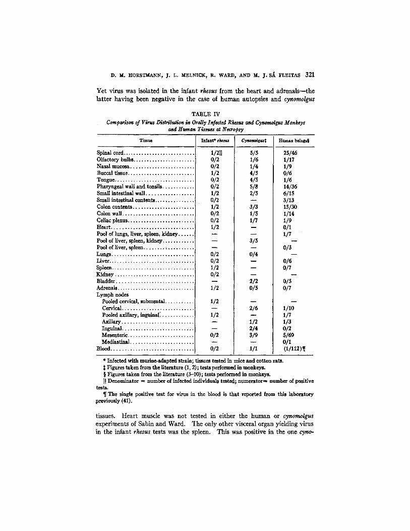
Yet virus was isolated in the infant rhesus from the heart and adrenais--the latter having been negative in the case of human autopsies and cynomolgus
TABLE IV
Comparison of Virus Distribution in Orally Infected Rhesus and Cynomolgus Monkeys and Human Tissuea a~ Necropsy
Tissue Infant* rhesus Cynoraolgus~ Human beings§
Spinal cord. Olfactory bulbs. Nasal mucosa. Ruccal tissue... Tongue ....... Pharyngeal wall and tonsils. Small intestinal wall. Small intestlnM contents . . . . . . . . . . Colon contents Colon wall. Celiac plexus ...... Heart. Pool of lungs, liver, spleen, kidney ...... Pool of liver, spleen, kidney .... Pool of liver, spleen.. Lungs. Liver. Spleen. Kidney ..... Bladder. Adrenals Lymph nodes
Pooled cervical, submental . . . . Cervical. Pooled axiilary, inguinal~ Axillary.. Inguinal. Mesenteric Medlasfinal.
Blood.
1/2[l 0/2 0/2 1/2 0/2 0/2 1/2 0/2 1/2 0/2 0/2 I/2
0/2 0/2 1/2 0/2
1/2
1/2
1/2
o/2
0/2
s/5 1/6 1/4 4/s 4/5 s/8 2/5
8/8 1/5 1/7
3/5
0/4
2/2 o/s
2/6
1/2 2/4 8/9
1/1
25/46 1/17 1/9 0/6 1/6 14/36 6115 3/13 lS/30 1/14 1/9 0/1 1/7
0/3
0/6 O/7
0/5 0/7
1/10 1/7 1/3 0/2 S/59 0/1 (1/112)¶
* Infected with murine-adapted strain; tissues tested in mice and cotton rats. 2~ Figures taken from the literature (I, 2); tests performed in monkeys. § Figures taken from the literature (3-10); tests performed in monkeys. II Denominator = number of infected individuals tested; numerator=~ number of positive
tests. ¶ The single positive test for virus in the blood is that reported from this laboratory
previously (41).
tissues. Heart muscle was not tested in either the human or cy~,wmolgus experiments of Sabin and Ward. The only other visceral organ yielding virus in the infant rhesus tests was the spleen. This was positive in the one cyno-
322 POLIOMYELITIS VIRUS
molgus spleen tested by Sabin and Ward and in a pool of lungs, liver, kidney, and spleen in one of their seven trials with human tissues. The fact that the spleen was positive in the baby rhesus in the absence of detectable virus in the blood suggests that the virus was actually in splenic tissue, and the positive result was not due to contamination by virus in the blood. However, the blood of infected rhesus monkeys (40) and in one instance that of a human being (41) have previously been found to contain virus.
The negative tests with the celiac plexus and the mesenteric lymph nodes are in line with some of the tesults in the human cases and the cynomolgus. The negative results with the pharyngeal wall and other tissues which might have been expected to be positive, may be attributed in part to the incomplete pic- ture which results when only one animal has been studied.
S ~ R Y
Although rhesus monkeys have been generally regarded as refractory to infection with poliomyelitis virus administered by the oral route, two of seven infant rhesus developed paralytic poliomyelitis when fed murine-adapted strains of virus. Preliminary intranasal treatment with zinc sulfate and nega- tive serial sections of the olfactory bulbs of the positive animals ruled out the possibility that infection occurred by way of the olfactory pathway.
Studies on the distribution of virus in the tissues of the infected animals yielded positive results in one animal only. In this instance, virus was widely distributed throughout the body being isolated from spinal cord, buccal mucosa, duodenal wall, colon contents, superficial lymph nodes, spleen, heart, and adrenals.
BIBLIOGRAPHY
1. Sa ,bin, A. B., and Ward, R., cited by Sabin, A. B., J. Mr. Sinai ltosp., 1944, 11 185.
2. Bumet, F. M., Jackson, A. V., and Robertson, E. G., Australian J. Exp. Bid and Med. So., 1940, 18, 361.
3. Landsteiner, K., Levaditi, C., and Pastia, C., Compt. rend. Acad. sc., 1911, 159., 1701. Levaditi, C., Schmutz, E., and Willemin, L., Bull. Acad. re&L, Paris, 1930, series 3, 104, 505.
4. Flexner, S., and Amoss, H. L., J. Exp. Med., 1919, 9.9, 383. 5. Flexner, S., J. Exp. Md. , 1936, 63, 209. 6. Kllug, C., Olin, G., and Gard, S., Compt. rend. Soc. biol., 1938, 129, 451. 7. Kempf, J. E., and Soule, M. H., Proc. So¢. Exp. Biol. and Med., 1940, 43, 476. 8. Sabin, A. B., and Ward, R., J. Exp. Med., 1941, 73, 771. 9. Kessel, J. F., Moore, F. J., Stimpert, F. D., and Fisk, R. T., J. Exp. MaL, 1941
74, 601. 10. Wenner, H. A., and Paul, J. R., Am. J. Meal. S¢., 1947, 9.15, 9. 11. Leiner, C., and yon Wiesner, R., Wien. klin. Woch., 1910, 23, 91. 12. Flexner, S., and Lewis, P. A., J. Exp. Meal., 1910, 19., 227.

D. M. HORSTMANN, J. L. MELNICK, R. WARD, AND M. ~. S~ FLEITAS 323
13. Landsteiner, K., Levaditi, C., and Pastia, M., Ann. Inst. Pasteur, 1911, 25, 833. 14. Amoss, H. L., in Filterable Viruses, (T. M. Rivers, editor) Baltimore, Williams &
Wilkins Co., 1928, 174. 15. Rhoads, C. P., Cited by Flexner (5). 16. Schultz, E. W., Proc. Soc. Exp. Biol. and Meg., 1929, 26, 632. 17. Thompson, R., Y. Exp. Meg., 1930, 51, 777. 18. Kling, C., Levaditi, C., and IApine, P., Bull. Acad. m~d., Paris, 1929, series 3,
102, 158. 19. Levaditi, C., Kling, C., and L6pine, P., Bull. Acad. m~., Paris, 1931, series 3,
105, 190. 20. Levaditi, C., Kling, C., and Hornus, G., Compt. rend. Soc. biol., 1933, 112, 43. 21. Kling, C., Levaditi, C., and Homus, G., Bull. Acad. m~l., Paris, 1934, series 3,
III, 709. 22. Bumet, F. M., Jackson, A. V., and Robertson, E. G., Australian Y. Exp. Biol
and Meg. Se., 1939, I"/, 375. 23. Vignec, A. J., Tmsk, J. D., and Paul, J. R., Proe. Soc. Exp. Biol. and Meg., 1939,
41, 246. 24. Faber, H. K., Silverberg, R. J., and Dong, L., J. Exp. Meg., 1943, 78, 499. 25. Trask, J. D., and Paul, J. R., Y. Exp. Meat., 1941, 73, 453. 26. Howe, H. A., and Bodian, D., Am. Y. Hyg., 1947, 45, 223. 27. Paul, J. R., Fa/e Y. Biol. and Meg., 1944,16, 461. 28. Melniek, J. L., unpublished data. 29. I~ndsteiner, K., and Levaditi, C., Compt. rend. Soe. biol., 1909, 6"/, 787. 30. Landsteiner, K., and Levaditi, C., Ann. Imt. Pasteur, 1910, 24, 833. 31. Howe, H. A., and Bodian, D., Neural Mechanisms in Poliomyelitis, New York,
The Commonwealth Fund, 1942. 32. yon Magnus, H., and Melnick, J. L., unpublished work. 33. Howe, H. A., and Bodian, D., Y. Exp. Meg., 1945, 81, 255. 34. Ward, R., Melnick, J. L., and Horstmann, D.M., Science, 1945, 101, 491. 35. Melnick, J. L., and Horstmann, D. M., a r. Exp. Meg., 1947, 85~ 287. 36. Clark, P. F., Schindler, J., and Roberts, D. J., .I. Bact., 1930, 20, 213 37. Schultz, E. W., and Gebhardt, L. P., Proc. Soe. Exp. Biol. and Meat., 1938, 38, 603. 38. Melnick, J. L., Y. Exp. Meg., 1943, 77, 195. 39. Melnick, J. L., and Ward, R., unpublished data. 40. Melnick, J. L., Proc. Soc. Exp. Biol. and Meg., 1945, fiB, 14. 41. Ward, R., Horstmann, D. M., and Melnick, J. L., Y. Clin. In~., 1946, 25, 284. 42. Demme, H., Deutsch. Z. Nervenheilk., 1930, 11.6, 156. 43. Saddington, R. S., Proc. Soc. Exp. Biol. and Meg., 1932, 9.9,838. 44. Lennette, E. H., and Hudson, H. P., J. Infect. Dis., 1936, 58, 10. 45. Rasmussen, A. F., Jr., and Clark, P. F., Proc. Soc. Exp. Biol. and Meg., 1940,
45, 232. 46. Clark, P. F., Roberts, D. J., and Preston, W. S., Y. Prevent. Meg., 1932, 6, 47.