The Active Management of Impending Cephalopelvic Disproportion ...
Upload
trinhkhanhCategory
view
227download
1
© 2017 Annals of Maxillofacial Surgery | Published by Wolters Kluwer - Medknow112
Case Report ‑ Developmental Deformities
IntRoductIon
The prevalence of Class III malocclusion varies among different ethnic groups and geographic regions with higher frequency in Japanese, Chinese, and Malaysian populations and lower frequency in Indians.[1] In the United States, the prevalence of this type of malocclusion is higher in African-Americans and Hispanics as compared to Caucasians.[2]
Orthodontic camouflage and orthognathic surgery are two modalities to treat skeletal Class III malocclusion in an adult patient. Orthodontic camouflage involves proclination of the maxillary teeth and retraction of the mandibular incisors. This can be done in cases with proclined mandibular incisors. In cases with the retroclined lower anterior teeth, camouflage increases the prominence of chin potentially, adversely affecting esthetics.[3] In such cases, orthognathic surgery involving one or both jaws combined with orthodontic treatment may produce more desirable outcome as compared to orthodontic camouflage. Orthognathic surgery produces a positive impact on the quality of life of patients, improving physical and social aspects. It has been reported to improve emotional aspects in females.[4]
This report focuses on the management of Class III skeletal deformity using a surgical approach. When the option for
surgery has been decided upon, the orthodontist and surgeon have to decide on a pathway forward that will bring about the optimum esthetic, dental, and functional outcome.
SurgeryThe aim of the surgical procedure is to attain the maximum functional and esthetic result while employing the most stable movements for the jaws. These movements normally fall within the envelope of discrepancy as once described by Proffit and Ackermann.[5]
OrthodonticsThe goal of orthodontics is to help the surgeon prepare the patient for a successful surgical outcome. Often, the orthodontist and surgeon need to spend time at the start of treatment deciding on the tooth movements that will allow for the maximum skeletal changes. This happens in three planes of space and decisions have to be made on whether certain movements can be corrected with orthodontic (dental) or surgical (skeletal) interventions.
The Surgical Management of Skeletal Disproportion with Lingual Orthodontics and Three‑dimensional Planning
Patel Krutiben, Chung How Kau, Peter D Waite1, Ahmet Arif Celebi
Departments of Orthodontics and 1Oral and Maxillofacial Surgery, School of Dentistry, University of Alabama at Birmingham, Birmingham, Alabama, USA
This case report describes the successful treatment of a 26-year-old Caucasian male with skeletal and dental Class III malocclusion associated with mild maxillary and mandibular crowding. The patient had anteroposterior and transverse discrepancies with a reverse overjet and bilateral posterior crossbites. The nonextraction treatment plan included aligning and leveling of the teeth in both arches, Le Fort I and bilateral sagittal split osteotomies, and postsurgical correction of the malocclusion. Orthodontic treatment was initiated with custom lingual appliances followed by orthognathic surgery planned with virtual surgical planning. Treatment was concluded with detailed orthodontic finishing, achieving optimum esthetics and function.
Keywords: Class III malocclusion, lingual orthodontics, orthognathic surgery, three-dimensional planning
Access this article online
Quick Response Code:Website: www.amsjournal.com
DOI: 10.4103/ams.ams_55_17
Address for correspondence: Dr. Chung How Kau, Department of Orthodontics, University of Alabama at Birmingham, School
of Dentistry, 1919 7th Avenue South, Birmingham, Alabama 35294, USA. E‑mail: [email protected]
This is an open access article distributed under the terms of the Creative Commons Attribution-NonCommercial-ShareAlike 3.0 License, which allows others to remix, tweak, and build upon the work non-commercially, as long as the author is credited and the new creations are licensed under the identical terms.
For reprints contact: [email protected]
How to cite this article: Patel K, Kau CH, Waite PD, Celebi AA. The surgical management of skeletal disproportion with lingual orthodontics and three‑dimensional planning. Ann Maxillofac Surg 2017;7:112‑6.
Abstract

Krutiben, et al.: 3D surgery with lingual braces
Annals of Maxillofacial Surgery ¦ Volume 7 ¦ Issue 1 ¦ January-June 2017 113
Three‑dimensional planningThe advancements in three-dimensional (3D) planning have allowed the orthodontist and surgeon to be better prepared to give the best outcome for the patient.[6] 3D planning can be done for customized orthodontics, allowing precise and accurate placement of brackets and customization of the dental arch form. The 3D surgical planning is an excellent tool for the orthodontist and surgeon to communicate the surgical movements, plan the final occlusion, and ensure the best outcome for the patient.
This report describes the successful treatment of an adult patient with skeletal Class III malocclusion treated with a complex orthodontic appliance using lingual braces, meticulous 3D surgical planning followed by orthognathic surgery. The final facial esthetics and occlusion were treated to an excellent standard and conclusion.
case RepoRt
Diagnosis and etiologyA 26-year-old male presented to the department of orthodontics with the chief complaint of crowding of his upper teeth. Extraoral examination revealed a bilateral symmetry with increased height of lower third of the face. The patient had slightly concave profile with retruded maxilla and increased nasolabial angle. Intraoral examination revealed bilateral Angle’s Class III molar and canine relationship. He had anterior and posterior crossbites with about 1 mm of reverse overjet and overbite. Maxillary and mandibular arches had mild crowding with mandibular skeletal midline deviated toward the patient’s right [Figure 1].
Lateral cephalometric analysis revealed retruded maxilla in relation to anterior cranial base (SNA, 75.4°) while mandible was in normal sagittal position (SNB, 77.2°). Skeletal Class III relation of maxilla and mandible (ANB, −1.8°, Wits, −4.6 mm) was present with predominance of vertical growth of face (SN-MP, 42.1°). Upper and lower incisors were retruded with respect to anterior cranial base and mandible, respectively (U1‑SN, 94.3°, L1‑MP 78°) [Figure 2 and Table 1]. No abnormalities were detected on panoramic radiograph [Figure 3]. Based on clinical examination and cephalometric analysis, the patient was diagnosed with skeletal Class III malocclusion. The patient was a good candidate for orthodontic treatment combined with orthognathic surgery.
Treatment objectivesThe patient was diagnosed with skeletal and dental Class III malocclusion, mild maxillary and mandibular crowding, anterior and posterior crossbites, and mandibular skeletal asymmetry to the right side. Based on the diagnostic records and consultation with the patient, the following objectives were developed: (1) Aligning and leveling teeth in both arches by arch expansion, (2) correction of the overbite and overjet, (3) correction of the midline, (4) bimaxillary surgery, and (5) postsurgical treatment of malocclusion.
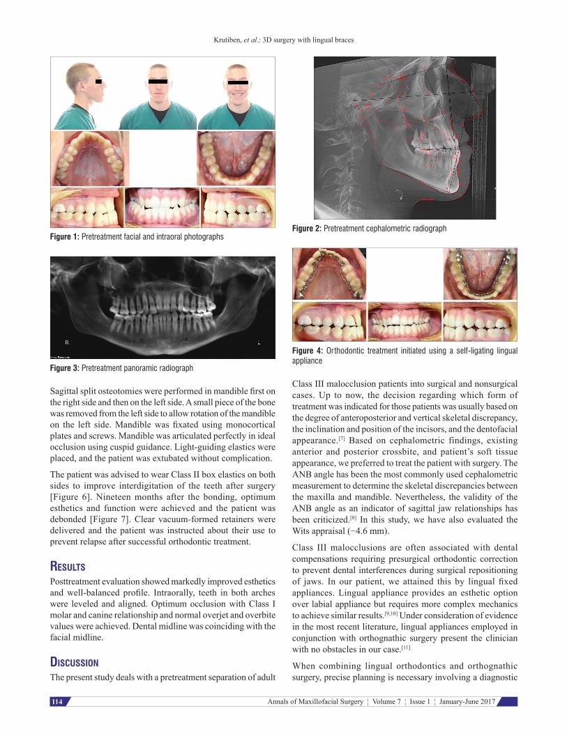
Treatment progressPresurgical orthodontic treatment was initiated using a self-ligating lingual appliance (American Orthodontics, Sheboygan, Wisconsin, USA, 0.018-inch slot) to meet the patient’s esthetic requirements. After a month, the maxillary arch was expanded, and interproximal reduction of the right lateral incisor was performed. An open coil spring was used to create space to derotate the lower right canine [Figure 4]. A power chain was used to close the spaces developed as a result of expansion of the lower arch. Buccal buttons were placed on the lower left canine to derotate the lower right canine [Figure 5]. Seven and half months after initial bonding, presurgical records (upper/lower impressions, cone beam computed tomography [CBCT], and extraoral photographs) were taken and composite buttons were added on all teeth to correct occlusion postsurgically. Virtual surgical planning for Le Fort I and bilateral sagittal split osteotomies (BSSOs) was done using Medical Modeling (VSP, Medical Modeling/3D Systems, Golden, Colorado). This system allows to eliminate requirements of traditional surgery. An interactive web meeting between the surgeon and an engineer was performed to allow for simulation of the procedure, including surgically accurate placement of osteotomies and bony movements. Intermediate and final surgical splints fabricated using Medical Modeling were used to position maxilla and mandible for fixation, respectively. After standard Le Fort I osteotomy with advancement using custom surgical splint maxilla was fixated with 4 mm advancement plate at the left piriform aperture and with 2 mm advancement plate at the right piriform aperture, maxilla was stabilized posteriorly with 1.5 mm plates and screws.
Table 1: Data of the cephalometric analysis
Krutiben, et al.: 3D surgery with lingual braces
Annals of Maxillofacial Surgery ¦ Volume 7 ¦ Issue 1 ¦ January-June 2017114
Figure 1: Pretreatment facial and intraoral photographs
Figure 3: Pretreatment panoramic radiograph
Figure 4: Orthodontic treatment initiated using a self‑ligating lingual appliance
Figure 2: Pretreatment cephalometric radiograph
Sagittal split osteotomies were performed in mandible first on the right side and then on the left side. A small piece of the bone was removed from the left side to allow rotation of the mandible on the left side. Mandible was fixated using monocortical plates and screws. Mandible was articulated perfectly in ideal occlusion using cuspid guidance. Light-guiding elastics were placed, and the patient was extubated without complication.
The patient was advised to wear Class II box elastics on both sides to improve interdigitation of the teeth after surgery [Figure 6]. Nineteen months after the bonding, optimum esthetics and function were achieved and the patient was debonded [Figure 7]. Clear vacuum-formed retainers were delivered and the patient was instructed about their use to prevent relapse after successful orthodontic treatment.
Results
Posttreatment evaluation showed markedly improved esthetics and well‑balanced profile. Intraorally, teeth in both arches were leveled and aligned. Optimum occlusion with Class I molar and canine relationship and normal overjet and overbite values were achieved. Dental midline was coinciding with the facial midline.
dIscussIon
The present study deals with a pretreatment separation of adult
Class III malocclusion patients into surgical and nonsurgical cases. Up to now, the decision regarding which form of treatment was indicated for those patients was usually based on the degree of anteroposterior and vertical skeletal discrepancy, the inclination and position of the incisors, and the dentofacial appearance.[7] Based on cephalometric findings, existing anterior and posterior crossbite, and patient’s soft tissue appearance, we preferred to treat the patient with surgery. The ANB angle has been the most commonly used cephalometric measurement to determine the skeletal discrepancies between the maxilla and mandible. Nevertheless, the validity of the ANB angle as an indicator of sagittal jaw relationships has been criticized.[8] In this study, we have also evaluated the Wits appraisal (−4.6 mm).
Class III malocclusions are often associated with dental compensations requiring presurgical orthodontic correction to prevent dental interferences during surgical repositioning of jaws. In our patient, we attained this by lingual fixed appliances. Lingual appliance provides an esthetic option over labial appliance but requires more complex mechanics to achieve similar results.[9,10] Under consideration of evidence in the most recent literature, lingual appliances employed in conjunction with orthognathic surgery present the clinician with no obstacles in our case.[11]
When combining lingual orthodontics and orthognathic surgery, precise planning is necessary involving a diagnostic

Krutiben, et al.: 3D surgery with lingual braces
Annals of Maxillofacial Surgery ¦ Volume 7 ¦ Issue 1 ¦ January-June 2017 115
setup and profile analysis in close cooperation with the surgeon.[11] Our surgical management was conducted by 3D virtual model surgery (VMO). This recent technique gives the clinician a better and more realistic patent of soft and hard tissue changes after various desired surgical plans compared to manual model surgery.[12] VMO employs the data obtained from CBCT and occlusal anatomy data from study models. It allows accurate surgical planning and movement of osteotomies utilizing surgical splints with eliminating the need for mock surgery on models.
Le Fort I osteotomy with forward repositioning of maxilla and BSSO with mandibular rotation on left side were performed utilizing titanium plates for fixation. Internal
rigid fixation provides better stability in patients with bimaxillary osteotomies as compared to conventional nonrigid fixation methods.[13] During the 1st year after surgery, Class II treatments are more stable as compared to Class III procedures. In 1–5 years after surgery, changes in Class II patients are more frequent than that of Class III patients. Fewer dental changes are noted long term due to dental adaptation following skeletal changes.[14] Combined orthodontic-orthognathic treatment resulted in optimum facial harmony and function while addressing patient’s chief complaint of crowding.
conclusIon
This case report describes combined conventional orthodontic and orthognathic approach used to treat an adult male with dental and skeletal Class III malocclusion. The treatment included aligning and leveling the teeth in both arches, correction of the overbite and overjet, correction of dental midline and posterior crossbite, two-jaw surgery for the correction of the underlying skeletal abnormality, and postsurgical orthodontic treatment to treat the malocclusion. Custom lingual appliances were used to meet the patient’s demand for an esthetic treatment. Virtual surgical modeling provided a detailed planning and precise execution of the surgery. We suggest that multidisciplinary team approach is of utmost importance to achieve the best results while treating patients with skeletal discrepancies.
Declaration of patient consent The authors certify that they have obtained all appropriate patient consent forms. In the form the patient(s) has/have given his/her/their consent for his/her/their images and other clinical information to be reported in the journal. The patients understand that their names and initials will not be published and due efforts will be made to conceal their identity, but anonymity cannot be guaranteed.
Financial support and sponsorshipNil.
Conflicts of interestThere are no conflicts of interest.
RefeRences1. Daniel KH, Yltze PC, Maria FO. Prevalence of angle class III
malocclusion: A systematic review and meta‑analysis. Open J Epidemiol 2012;2:75‑82.
2. Silva RG, Kang DS. Prevalence of malocclusion among Latino adolescents. Am J Orthod Dentofacial Orthop 2001;119:313‑5.
3. Proffit WR, White RP Jr. Combined surgical‑orthodontic treatment: How did it evolve and what are the best practices now? Am J Orthod Dentofacial Orthop 2015;147 5 Suppl:S205‑15.
4. Nicodemo D, Pereira MD, Ferreira LM. Effect of orthognathic surgery for class III correction on quality of life as measured by SF-36. Int J Oral Maxillofac Surg 2008;37:131‑4.
5. Proffit WR, Ackermann JL. A systematic approach to orthodontic diagnosis and treatment planning. In: Graber TM, Swain BF, editors. Current Orthodontic Concepts and Techniques. 3rd ed. Saint Louis: C. V. Mosby; 1985.
6. Van Hemelen G, Van Genechten M, Renier L, Desmedt M, Verbruggen E, Nadjmi N. Three-dimensional virtual planning in orthognathic surgery
Figure 5: Buccal buttons and chains to correct rotation of the lower right canine
Figure 6: Class II box elastics on both sides to correct the malocclusion after surgery
Figure 7: Posttreatment facial and intraoral photographs

Krutiben, et al.: 3D surgery with lingual braces
Annals of Maxillofacial Surgery ¦ Volume 7 ¦ Issue 1 ¦ January-June 2017116
enhances the accuracy of soft tissue prediction. J Craniomaxillofac Surg 2015;43:918‑25.
7. Stellzig-Eisenhauer A, Lux CJ, Schuster G. Treatment decision in adult patients with class III malocclusion: Orthodontic therapy or orthognathic surgery? Am J Orthod Dentofacial Orthop 2002;122:27‑37.
8. Jacobson A. The “Wits” appraisal of jaw disharmony. Am J Orthod 1975;67:125‑38.
9. Creekmore T. Lingual orthodontics – Its renaissance. Am J Orthod Dentofacial Orthop 1989;96:120‑37.
10. Gorman JC, Smith RJ. Comparison of treatment effects with labial and lingual fixed appliances. Am J Orthod Dentofacial Orthop
1991;99:202‑9.11. Pauls HJ. Lingual orthodontics with orthognathic surgery in a severe
class II, division 2 case. J Orofac Orthop 2008;69:135‑45.12. Choi JY, Song KG, Baek SH. Virtual model surgery and wafer fabrication
for orthognathic surgery. Int J Oral Maxillofac Surg 2009;38:1306‑10.13. Law JH, Rotskoff KS, Smith RJ. Stability following combined maxillary
and mandibular osteotomies treated with rigid internal fixation. J Oral Maxillofac Surg 1989;47:128‑36.
14. Proffit WR, Turvey TA, Phillips C. The hierarchy of stability and predictability in orthognathic surgery with rigid fixation: An update and extension. Head Face Med 2007;3:21.