THE SURGICAL IMPORTANCE PERSISTENT LEFT SUPERIOR … · Aleft superior vena cava maybe present...
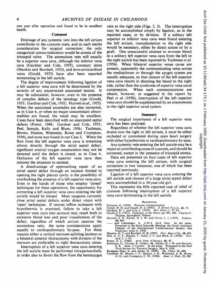
4
THE SURGICAL IMPORTANCE OF A PERSISTENT LEFT SUPERIOR VENA CAVA* BY ELLIOTT S. HURWITTt From the Surgical Division, the Montefiore Hospital, New York City A left superior vena cava may be present simul- taneously with a normal right superior vena cava, or, even more infrequently, as the only superior vena cava. The surgical importance of a persistent left superior vena cava varies depending on its termination into the right or left atrium, as well as the nature and severity of any associated cardio- vascular anomalies. Left Superior Vena Cava Entering Right Atrium. This is not a particularly uncommon anomaly, the vein terminating either directly into the right atrium, or indirectly by way of the coronary sinus. The embryology and frequency of this configuration have been reviewed in a previous communication (Hur- witt, Escher and Citrin, 1955). Since anomalies of the heart and great vessels tend to occur in associa- tion, one may encounter a left superior vena cava draining into the right atrium in the process of other operations, such as in the correction of a patent ductus arteriosus. Under such circumstances the anatomical course of the left superior vena cava is such that it does not interfere with the stated operation, and the vein usually has no surgical importance. With the advent of open heart surgery, however, employing either cardiopulmonary by-pass or hypothermia, a left superior vena cava assumes real surgical significance, regardless of whether it drains into the right or left atrium. Failure to identify and mobilize this vessel, with temporary occlusion for hypothermia and cannulation or occlusion during cardiopulmonary by-pass, may seriously compromise the procedure. The necessity for this manoeuvre has been described in a number of articles, including one from this department (Robinson, Glotzer, Gilbert, Escher and Hurwitt, in press). Left Superior Vena Cava Entering Left Atrium. As a contributing cause of cyanosis, the importance of drainage of any systemic vein into the left auricle has usually been overshadowed by the severity of associated cardiac defects. Subsequent to the summary of the reported cases (Hurwitt et al., 1955), two additional such patients have come to surgery at this hospital. Case Histories Uncorrected Lesions Case 1. A 4 lb. premature infant, one of male twins, was brought to the hospital on January 21, 1957, with complete ectopia cordis (Fig. 1) and severe cyanosis and dyspnoea; the twin was normal. A covering of skin was fashioned over the heart, and the baby died on the- evening of the operation. A left superior vena cava entering the left atrium was only one of the eight major anomalies of the heart and great vessels found at autopsy. A normal right superior vena cava was also present. A motion picture and complete description of this case- have been prepared (Hurwitt and Lebendiger, in press). Case 2. Severe cyanosis and nearly fatal episodes of syncope were the presenting problems in this 18-month- old girl. Investigation in another hospital had estab- lished the presence of both a right and left superior vena cava and a levocardia in the presence of a situs inversus abdominalis. On June 17, 1958, an attempt was made to construct a Blalock shunt between the left subclavian artery and the left pulmonary artery, but irreversible cardiac arrest developed during mobilization of the pulmonary artery. Post-mortem examination disclosed an essentially bilocular heart, with the left superior vena cava draining into the left side of a chamber that was practically a common auricle. Splenic agenesis, described in several such cases, was not present. Corrected Lesions Even when severe associated anomalies of the heart are present, however, interruption of the flow of systemic- venous blood into the left auricle may be beneficial, with or without attempted correction of the other lesions. Diaz and Anido (1949) reported significant improvement * Presented at the Fifth Annual Meeting, the British Association of Paediatric Surgeons, London, July 23, 1958. t Aided by a grant from the New York Heart Association. 1 copyright. on January 21, 2020 by guest. Protected by http://adc.bmj.com/ Arch Dis Child: first published as 10.1136/adc.34.173.1 on 1 February 1959. Downloaded from
Transcript of THE SURGICAL IMPORTANCE PERSISTENT LEFT SUPERIOR … · Aleft superior vena cava maybe present...
THE SURGICAL IMPORTANCE OFA PERSISTENT LEFT SUPERIOR VENA CAVA*
BY
ELLIOTT S. HURWITTtFrom the Surgical Division, the Montefiore Hospital, New York City
A left superior vena cava may be present simul-taneously with a normal right superior vena cava,
or, even more infrequently, as the only superiorvena cava. The surgical importance of a persistentleft superior vena cava varies depending on itstermination into the right or left atrium, as well as
the nature and severity of any associated cardio-vascular anomalies.
Left Superior Vena Cava Entering Right Atrium.This is not a particularly uncommon anomaly, thevein terminating either directly into the right atrium,or indirectly by way of the coronary sinus. Theembryology and frequency of this configuration havebeen reviewed in a previous communication (Hur-witt, Escher and Citrin, 1955). Since anomalies ofthe heart and great vessels tend to occur in associa-tion, one may encounter a left superior vena cava
draining into the right atrium in the process of otheroperations, such as in the correction of a patentductus arteriosus. Under such circumstances theanatomical course of the left superior vena cava issuch that it does not interfere with the statedoperation, and the vein usually has no surgicalimportance.With the advent of open heart surgery, however,
employing either cardiopulmonary by-pass or
hypothermia, a left superior vena cava assumes realsurgical significance, regardless of whether it drainsinto the right or left atrium. Failure to identify andmobilize this vessel, with temporary occlusion forhypothermia and cannulation or occlusion duringcardiopulmonary by-pass, may seriously compromisethe procedure. The necessity for this manoeuvre
has been described in a number of articles, includingone from this department (Robinson, Glotzer,Gilbert, Escher and Hurwitt, in press).
Left Superior Vena Cava Entering Left Atrium.As a contributing cause of cyanosis, the importanceof drainage of any systemic vein into the left auriclehas usually been overshadowed by the severity ofassociated cardiac defects. Subsequent to thesummary of the reported cases (Hurwitt et al., 1955),two additional such patients have come to surgeryat this hospital.
Case HistoriesUncorrected Lesions
Case 1. A 4 lb. premature infant, one of male twins,was brought to the hospital on January 21, 1957, withcomplete ectopia cordis (Fig. 1) and severe cyanosis anddyspnoea; the twin was normal. A covering of skinwas fashioned over the heart, and the baby died on the-evening of the operation. A left superior vena cavaentering the left atrium was only one of the eight majoranomalies of the heart and great vessels found at autopsy.A normal right superior vena cava was also present. Amotion picture and complete description of this case-have been prepared (Hurwitt and Lebendiger, in press).
Case 2. Severe cyanosis and nearly fatal episodes ofsyncope were the presenting problems in this 18-month-old girl. Investigation in another hospital had estab-lished the presence of both a right and left superior venacava and a levocardia in the presence of a situs inversusabdominalis. On June 17, 1958, an attempt was madeto construct a Blalock shunt between the left subclavianartery and the left pulmonary artery, but irreversiblecardiac arrest developed during mobilization of thepulmonary artery. Post-mortem examination disclosedan essentially bilocular heart, with the left superior venacava draining into the left side of a chamber that was
practically a common auricle. Splenic agenesis, describedin several such cases, was not present.
Corrected LesionsEven when severe associated anomalies of the heart
are present, however, interruption of the flow of systemic-venous blood into the left auricle may be beneficial, withor without attempted correction of the other lesions.Diaz and Anido (1949) reported significant improvement
* Presented at the Fifth Annual Meeting, the British Associationof Paediatric Surgeons, London, July 23, 1958.
t Aided by a grant from the New York Heart Association.1
copyright. on January 21, 2020 by guest. P
rotected byhttp://adc.bm
j.com/
Arch D
is Child: first published as 10.1136/adc.34.173.1 on 1 F
ebruary 1959. Dow
nloaded from
ARCHIVES OF DISEASE IN CHILDHOOD
t. A Lt.
R,t Aur. App.
FIG. 1. Case 1. Photograph and diagram of ectopiacordis, with bilateral superior venae cavae.
in a 7-year-old tetralogy case following ligationof a left superior vena cava entering the leftauricle, without any other procedure. Lessen-ing of cyanosis was similarly described in an8-year-old with Eisenmenger's complex, byFeindt and Hauch (1953). Ekstrom (personalcommunication) supplemented constructionof a Blalock shunt in a 12-year-old withtetralogy of Fallot, situs inversus totalis and doublesuperior venae cavae, by ligating the anomalous venacava as it entered the arterial auricle.
Obviously the outlook is much more favourable whenthe left superior vena cava entering the left auricleconstitutes the major cause of cyanosis, or when anyconcomitant intracardiac defects may also be amenableto correction. Examples of each of these situations havenow come to operation at this hospital.
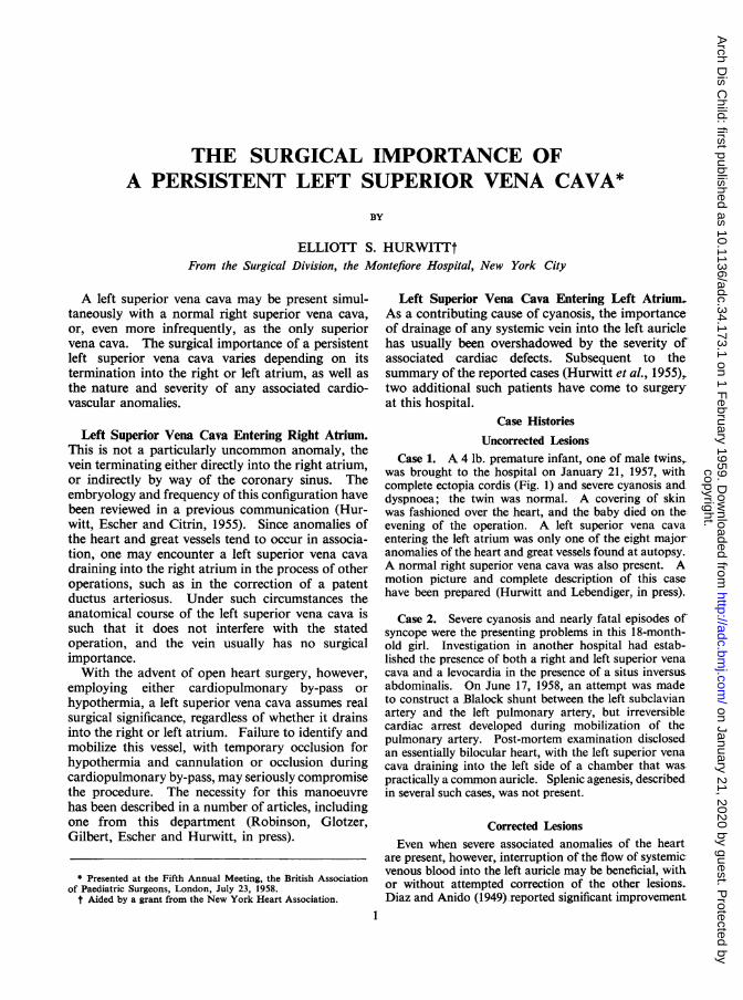
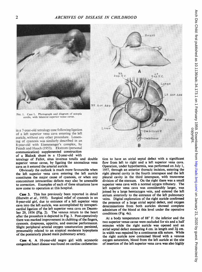
Case 3. This has previously been reported in detail(Hurwitt et al., 1955). Surgical relief of cyanosis in an8-year-old girl, due to entrance of a left superior venacava into the left auricle, was accomplished by intraperi-cardial ligation of the left superior vena cava on Decem-ber 29, 1954 (Fig. 2). The venous return to the heartafter the procedure is depicted in Fig. 3. Post-operativelythere was marked improvement in clubbing of the fingers,cyanosis. dyspnoea, appetite, and exercise performance.Slight peripheral arterial oxygen unsaturation persisted,presumably related to an atypical moderate hypoplasiaof the posteriorly placed main pulmonary artery.
Case 4. A 10-year-old negro girl with acyanoticcongenital heart disease was found on cardiac catheteriza-
L iver
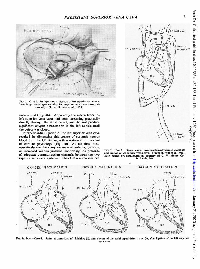
tion to have an atrial septal defect with a significantflow from left to right and a left superior vena cava.Operation, under hypothermia, was performed on July 1,1957, through an anterior thoracic incision, entering theright pleural cavity in the fourth interspace and the leftpleural cavity in the third interspace, with transversedivision of the sternum. On the right there was a smallsuperior vena cava with a normal azygos tributary. Theleft superior vena cava was considerably larger, wasjoined by a large hemiazygos vein, and entered the leftatrium anteriorly to the entrance of the left pulmonaryveins. Digital exploration of the right auricle confirmedthe presence of a large atrial septal defect, and oxygendeterminations from both auricles showed completeadmixture of the blood at this level under the operativeconditions (Fig. 4a).At a body temperature of 880 F. the inferior and the
two superior venae cavae were occluded for six and a halfminutes while the right auricle was opened and anatrial septal defect measuring 4 cm. in length and 21 cm.in width was repaired by a continuous silk suture. Whilethe right auricle now contained blood with a venousoxygen saturation, blood from the left auricle at the siteof insertion of the left superior vena cava was also highly
2
copyright. on January 21, 2020 by guest. P
rotected byhttp://adc.bm
j.com/
Arch D
is Child: first published as 10.1136/adc.34.173.1 on 1 F
ebruary 1959. Dow
nloaded from
PERSISTENT SUPERIOR VENA CAVA
FIG. 2. Case 3. Intrapericardial ligation of left superior vena cava.Note large hemiazygos entering left superior vena cava extraperi-
cardially. (From Hurwitt et al., 1955.)
unsaturated (Fig. 4b). Apparently the return from theleft superior vena cava had been streaming practicallydirectly through the atrial defect, and did not producesignificant oxygen desaturation in the left auricle untilthe defect was closed.
Intrapericardial ligation of the left superior vena cavaresulted in eliminating this source of systemic venousblood from the left atrium, with a restoration to normalof cardiac physiology (Fig. 4c). At no time post-operatively was there any evidence of oedema, cyanosis,or increased venous pressure, confirming the presenceof adequate communicating channels between the twosuperior vena caval systems. The child was re-examined
Rt. SupVC f L oygol
t.iru''AVi,!-Com*. .....i'. ...:!
IlfiacC.
~~~~~~~~~~~~~~~ilac i V.'XFiG. 3. Case 3. Diagrammatic reconstruction of vascular anomaliesand ligation of left superior vena cava. (From Hurwitt el al., 1955.)Both figures are reproduced by courtesy of C. V. Mosby Co.,
St. Louis, Mo.
101.5%
I Lt. SuP.V.C.\.J Y
I
OXYGEN SATURATION
FiG. 4a, b, c.-Case 4. Status at operation: (a), initially; (b), after closure of the atrial septal defect; and (c), after ligation of the left superiorvena cava.
3
V.
OXYGEN SATURATION
1/0# 5 %,
Rtt Sup. V.C.
66LSVj 11 Lt Sup.VC.
OXYGEN SATURATION
81.501,IIII
copyright. on January 21, 2020 by guest. P
rotected byhttp://adc.bm
j.com/
Arch D
is Child: first published as 10.1136/adc.34.173.1 on 1 F
ebruary 1959. Dow
nloaded from
4 ARCHIVES OF DISEASE IN CHILDHOODone year after operation and found to be in excellenthealth.
CommentDrainage of any systemic vein into the left atrium
contributes to the cyanotic state, and as such meritsconsideration for surgical correction; the onlycategorical contra-indication would be atresia of thetricuspid valve. The anomalous vein will usuallybe a superior vena cava, although the inferior venacava (Gardner and Cole, 1955), coronary sinus(Mankin and Burchell, 1953), and levo-atriocardinalveins (Gould, 1953) have also been reportedterminating in the left auricle.The degree of improvement following ligation of
a left superior vena cava will be determined by theseverity of any uncorrected associated lesions. Itmay be substantial, however, even in the presenceof complex defects of the tetralogy type (Gould,1953; Gardner and Cole, 1955; Hurwitt et al., 1955).When the associated anomalies are also corrected,as in Case 4, or when no major concomitant abnor-malities are found, the result may be excellent.Cases have been described with no associated septaldefects (Potter, 1948; Gardner and Cole, 1955;Peel, Semple, Kelly and Blum, 1956; Tuchman,Brown, Huston, Weinstein, Rowe and Crumpton,1956), and none was found in our Case 3. When theflow from the left superior vena cava is streamingalmost directly through the atrial septal defect,significant arterial oxygen unsaturation may not bedetected until the defect is closed, as in Case 4.Occlusion of the left superior vena cava thenrestores the situation to normal.A disadvantage of approaching repair of an
atrial septal defect through an incision limited toopening the right pleural cavity is the possibility ofoverlooking the presence of a left superior vena cava.Even in the hands of those who employ 'closed'techniques for these operations, the opportunity forcorrecting a left superior vena cava entering the leftauricle would be missed. Most surgeons currentlyclose atrial septal defects under direct vision with'open' techniques. If venous inflow occlusion withhypothermia is practised, failure to take a leftsuperior vena cava into account may result both inexcessive blood loss and poor visualization of thedefect, regardless of which atrium receives theanomalous vein; the same considerations applyequally to cardiopulmonary by-pass. For thesereasons either a vertical sternum-splitting incision ora bilateral anterior thoracotomy with division of thesternum are preferable to right thoracotomy alone.
Interruption of a left superior vena cava enteringthe left auricle must be performed intrapericardiallyin order also to divert the flow from the hemiazygos
vein to the right side (Figs. 2, 3). The interruptionmay be accomplished simply by ligation, as in thereported cases, or by division. If a solitary leftsuperior or inferior vena cava were found enteringthe left atrium, transplantation to the right sidewould be necessary, either by direct suture or by agraft. One unsuccessful attempt to re-route bloodin a solitary left superior vena cava from the left tothe right auricle has been reported by Tuchman et al.(1956). When bilateral superior venae cavae arepresent, apparently the communicating veins across.the mediastinum or through the azygos system areusually adequate, so that closure of the left superiorvena cava results in shunting the blood to the rightside, rather than the syndrome of superior vena cavalcompression. When such communications areabsent, however, as suggested in the report byPeel et al. (1956), interruption of the left superiorvena cava should be supplemented by an anastomosis.to the right superior caval system.
SummaryThe surgical importance of a left superior vena
cava has been analysed.Regardless of whether the left superior vena cava
drains into the right or left atrium, it must be eitheroccluded or cannulated during open heart surgerywith either hypothermia or cardiopulmonary by-pass.Any systemic vein entering the left auricle may be a
majoror contributing causeof cyanosis, and should becorrected, except in the presence of tricuspid atresia.Data are presented on four cases of left superior
vena cava entering the left atrium, with surgicalcorrection in two instances, one of which has beenreported previously.
Ligation of a left superior vena cava entering theleft auricle and closure of a large atrial septal defectwere accomplished in a 10-year-old girl.
This represents the fifth reported case of relief ofcyanosis following interruption of a left superiorvena cava terminating in the left auricle.
REFERENCESEkstrom, G. (1954). Personal communication.Feindt, H. R. and Hauch, H. J. (1953). Z. Kreisl.-Forsch., 42, 53.Gardner, D. L. and Cole, L. (1955). Brit. Heart J., 17, 93.Gould, S. E. (1953). Pathology of the Heart, pp.491, 496. Charles C.
Thomas, Springfield, Illinois.Hurwitt, E. S., Escher, D. J. W. and Citrin, L. I. (1955). Surgery,
38, 903.and Lebendiger, A. A.M.A. Arch. Surg. In the press.Presented at the Sixth Scientific Meeting, the North AmericanChapter of the International Cardiovascular Society, SanFrancisco, June 21, 1958.
Mankin, H. T. and Burchell, H. B. (1953). Proc. Mayo Clin., 28, 463.Peel, A. A. F., Semple, T., Kelly, J. C. and Blum, K. (1956). Scot.
med. J., 1, 83.Potter, E. L. (1948). Arch. Path. (Chicago), 46, 87.Robinson, G., Glotzer, P., Gilbert, M., Escher, D. J. W. and Hurwitt,
E. S. Amer. J. Cardiol. In the press. Presented in the NewYork Society for Thoracic Surgery, May 9, 1958.
Rodiguez Diaz, A. and Anido, H. (1949). Dis. Chest, 15, 684.Tuchman, H., Brown, J. F., Huston, J. H., Weinstein, A. B., Rowe,
G. G. and Crumpton, C. W. (1956). Amer. J. Med., 21, 481
copyright. on January 21, 2020 by guest. P
rotected byhttp://adc.bm
j.com/
Arch D
is Child: first published as 10.1136/adc.34.173.1 on 1 F
ebruary 1959. Dow
nloaded from