The Structure Of Normal Skin And The Morphology Of Atopic ... · analyses of mast cells in normal...
8
THE JOURNAL OF INVESTIGATIVE DERMATOLOGY, 67 :305- 312, 1976 Copyright © 1976 by The Williams & Wilkins Co. Vol. 67 , No . 3 Printed in U.S. A. THE STRUCTURE OF NORMAL SKIN AND THE MORPHOLOGY OF ATOPIC ECZEMA MARTIN C. MIHM, JR., M.D., NICHOLAS A. SOTER, M.D., HAROLD F. DVORAK, M.D ., AND K. FRANK AUSTEN, M.D. Departments of Dermatology, Pathology, and Medicine , Harvard Medical School; Dermatology Service and James Homer Wright Laboratories of the Department of Pathology, Massachusetts General Hospital; and Divisions of Dermatology, Departments of Medicine, Robert B. Brigham and Peter Bent Brigham Hospitals, Boston, Massachusetts, U. S. A. The understanding of cutaneous diseases re- quires a thorough knowledge of the anatomy of normal skin. Morphology traditionally has been observed in tissues fixed in different manners, embedded in paraffin, cut 6 to 10 in thickness , and stained with hematoxylin and eosin. Dvorak and co-workers [1] developed a method for the examination of cellular detail in tissues from experimental animals and this technique was ap - plied effectively to human biopsy material [2). Cells such as mast cells and basophils were easily visualized, and the details of cutaneous structures including the vasculature were observed [2,3]. The purpose of this presentation is to provide a brief description of normal cutaneous histology as ob- served in Epon-embedded sections, and to describe the alterations which characterize atopic eczema. MATERIALS AND METHODS Approximately 300 biopsy specimens from the skin of the deltoid regions and the forearms of normal male volunteers, ranging in age from 19 to 34, were utilized for the study of the histology of normal skin. Informed consent was obtained from all volunteers . Biopsy speci- mens were procured with a Keyes 4-mm cutaneous punch, using field anesthesia with 1 or 2% lidocaine without epinephrine . The anesthetic was injected at 4 sites in the periphery of the area to be biopsied. Fixation was carried out for 5 hr at room temperature in a Karnovsky's fixative [4] composed of 2% paraformalde- hyde, 2.5% glutaraldehyde, and 0.025 % CaC1 2 in 0.1 M cacodylate buffer at pH 7.4 [2]. Postfixation in osmium tetroxide was followed by dehydration and embedding in Epon [1]. One-fLm-thick sections, approximately 5 to 8 mm on edge, were stained with Giemsa's reagent. At least t wo observers evaluated all sections at magnifications up to 1000 x . One observer (MM) performed quantitative Reprint requests to: Dr. M. C. Mihm, Jr., Department of Pathology, Massachusetts General Hospital, Fruit Street, Boston, Massachusetts 02114. This study was supported in part by Grants AI 10496, AI 07722, AI 10356, and RR 05486 11 from the National Institutes of Health. Dr. Soter is the recipient of a National Research Service Award (1 F32 AI 05240) and Dr. Dvorak is the recipient of a Research Career Develop- ment Award (1 K04 AI 46352) from the National I nstitutes of Health. Abbreviations: SCV: superficial capillary-venule SVP: superficial venular plexus analyses of mast cells in normal skin of healthy volun- teers as well as in clinically uninvolved and lesional skin of patients with atopic eczema. RESULTS The Histology of Skin in Normal Subjects The normal skin as depicted schematically (Fig. 1) consists of the epidermis, papillary and reticular dermis, and the subcutaneous fat (Fig. 2a). Epidermis. The epidermis is composed of layers [5,6] respectively termed horny, granular, squa- mous, and basal. The most superficial of these is the horny layer or stratum corneum which, on the skin of the forearm, is composed of several lami- nated and loosely attached layers of anucleate keratinized cells. The granular layer is composed of 2 to 3 tightly apposed cell layers that contain minute, deeply blue-staining keratohyaline gran- ules. The squamous layer consists of 5 to 7 layers of polyhedral cells attached to each other by intercel- lular bridges that contain desmosomes [6,7]. These polyhedral cells have prominent nuclei and one or more small nucleoli. The cytoplasm is filled with filamentous-like material that surrounds the nu- cleus and usually runs parallel to the cell margins. The basal cells that lie along the dermoepidermal junction are cuboidal or columnar cells that con- tain blue-grey filamentous-appearing material and are attached to each other and to the overlying squamous cells by intercellular bridges. The basal cells contain aggregates of melanin granules that appear green. They may be irregularly dispersed throughout the cell or form aggregates above the nuclei. These granules may also be present in cells of the squamous layer but in reduced numbers. Scattered among basal cells are melanocytes, the pigment-producing cells, with ample, pale, non- filamentous cytoplasm. They do not connect to adjacent cells with intercellular bridges. These cells possess large oval to round nuclei, a single small nucleolus, and pale, dendritic processes that extend between intercellular spaces of keratino- cytes. Melanocytes exhibit a slightly foamy cyto- plasm that rarely contains melanin granules unless the cell has been activated to produce increased amounts of pigment. Other nonfilamentous cells without intercellular bridges may be observed singly disposed through- 305
Transcript of The Structure Of Normal Skin And The Morphology Of Atopic ... · analyses of mast cells in normal...

THE JOURNAL OF INVESTIGATIVE DERMATOLOGY, 67 :305-312, 1976 Copyright © 1976 by The Williams & Wilkins Co.
Vol. 67 , No . 3 Printed in U.S .A.
THE STRUCTURE OF NORMAL SKIN AND THE MORPHOLOGY OF ATOPIC ECZEMA
MARTIN C. MIHM, JR., M.D., NICHOLAS A. SOTER, M.D., HAROLD F. DVORAK, M.D., AND K. FRANK AUSTEN, M.D.
Departments of Dermatology, Pathology, and Medicine , Harvard Medical School; Dermatology Service and James Homer Wright Laboratories of the Department of Pathology, Massachusetts General Hospital; and Divisions of
Dermatology, Departments of Medicine, Robert B . Brigham and Peter Bent Brigham Hospitals, Boston, Massachusetts, U. S. A.
The understanding of cutaneous diseases requires a thorough knowledge of the anatomy of normal skin. Morphology traditionally has been observed in tissues fixed in different manners, embedded in paraffin, cut 6 to 10 ~m in thickness , and stained with hematoxylin and eosin. Dvorak and co-workers [1] developed a method for the examination of cellular detail in tissues from experimental animals and this technique was ap plied effectively to human biopsy material [2). Cells such as mast cells and basophils were easily visualized, and the details of cutaneous structures including the vasculature were observed [2,3]. The purpose of this presentation is to provide a brief description of normal cutaneous histology as observed in l-~m-thick Epon-embedded sections, and to describe the alterations which characterize atopic eczema.
MATERIALS AND METHODS
Approximately 300 biopsy specimens from the skin of the deltoid regions and the forearms of normal male volunteers, ranging in age from 19 to 34, were utilized for the study of the histology of normal skin. Informed consent was obtained from all volunteers . Biopsy specimens were procured with a Keyes 4-mm cutaneous punch, using field anesthesia with 1 or 2% lidocaine without epinephrine . The anesthetic was injected at 4 sites in the periphery of the area to be biopsied. Fixation was carried out for 5 hr at room temperature in a Karnovsky's fixative [4] composed of 2% paraformaldehyde, 2.5 % glutaraldehyde, and 0.025 % CaC1 2 in 0.1 M cacodylate buffer at pH 7.4 [2]. Postfixation in osmium tetroxide was followed by dehydration and embedding in Epon [1]. One-fLm-thick sections, approximately 5 to 8 mm on edge, were stained with Giemsa's reagent. At least two observers evaluated all sections at magnifications up to 1000 x . One observer (MM) performed quantitative
Reprint requests to: Dr. M. C. Mihm, Jr., Department of Pathology, Massachusetts General Hospital, Fruit Street, Boston, Massachusetts 02114.
This study was supported in part by Grants AI 10496, AI 07722, AI 10356, and RR 05486 11 from the National Institutes of Health. Dr. Soter is the recipient of a National Research Service Award (1 F32 AI 05240) and Dr. Dvorak is the recipient of a Research Career Development Award (1 K04 AI 46352) from the National Institutes of Health.
Abbreviations: SCV: superficial capillary-venule SVP: superficial venular plexus
analyses of mast cells in normal skin of healthy volunteers as well as in clinically uninvolved and lesional skin of patients with atopic eczema.
RESULTS
The Histology of Skin in Normal Subjects
The normal skin as depicted schematically (Fig. 1) consists of the epidermis, papillary and reticular dermis, and the subcutaneous fat (Fig. 2a).
Epidermis. The epidermis is composed of layers [5,6] respectively termed horny, granular, squamous, and basal. The most superficial of these is the horny layer or stratum corneum which , on the skin of the forearm, is composed of several laminated and loosely attached layers of anucleate keratinized cells. The granular layer is composed of 2 to 3 tightly apposed cell layers that contain minute, deeply blue-staining keratohyaline granules. The squamous layer consists of 5 to 7 layers of polyhedral cells attached to each other by intercel-lular bridges that contain desmosomes [6,7]. These polyhedral cells have prominent nuclei and one or more small nucleoli . The cytoplasm is filled with filamentous-like material that surrounds the nucleus and usually runs parallel to the cell margins. The basal cells that lie along the dermoepidermal junction are cuboidal or columnar cells that contain blue-grey filamentous-appearing material and are attached to each other and to the overlying squamous cells by intercellular bridges. The basal cells contain aggregates of melanin granules that appear green. They may be irregularly dispersed throughout the cell or form aggregates above the nuclei. These granules may also be present in cells of the squamous layer but in reduced numbers. Scattered among basal cells are melanocytes, the pigment-producing cells, with ample, pale, nonfilamentous cytoplasm. They do not connect to adjacent cells with intercellular bridges. These cells possess large oval to round nuclei, a single small nucleolus , and pale, dendritic processes that extend between intercellular spaces of keratinocytes. Melanocytes exhibit a slightly foamy cytoplasm that rarely contains melanin granules unless the cell has been activated to produce increased amounts of pigment.
Other nonfilamentous cells without intercellular bridges may be observed singly disposed through-
305

306 MIHM ET AL Vol. 67, No.3
ATOPIC ECZEMA VESICULAR LESION
NORMAL
I I
I~ I
LlCHENIFIED LESION
-il AH
EPIDERMIS
PAPILLARY DERMIS
RETICULAR DERMIS
) _____ )......------)--)-J 2~~~~ SUBCUTIS
FIG. 1. Schematic diagram of the skin of a normal person contrasted with vesicular and lichenified lesions from patients with atopic eczema. ART (artery); SM ART/A (small artery/arteriole) ; SAP (superficial arteriolar plexus); SCV (superficial capillary-venule); SVP1 and SVP2 (components of superficial venular plexus); V/SM VN (venules/small vein); VN (vein); MC (mast cell); L (lymphocyte); MO (monocyte/macrophage): AH (activated histiocyte); VES (vesicle).
out the squamous cell layer. Some of these have been shown to be Langerhans cells which possess a foamy cytoplasm, dendritic processes, and a highly irregular nuclear shape [5].
Subepidermal basement zone. The basal cells rest upon the subepidermal basement zone [5], also called the basement membrane zone, that separates the epidermis from the dermis. Often small, variably sized cellular processes of the basal cells interdigitate with this zone and the papillary dermis. The normal basement membrane zone appears as a thin, variably dense, light pale-green area along the dermoepidermal interface.
Dermis. The papillary dermis (Fig. 2a) lies between the subepidermal basement zone and the reticular dermis. It is composed of collagen fibers that are randomly dispersed but which do not interlace [5,6]. The papillary dermis extends irregularly upward as "pegs" or papillae, which interdigitate with the rete ridges, the irregular columns of epithelium that project downward. These epithelial ridges vary in width and depth in different
areas of the body. Collagen similar to that found in the papillary dermis surrounds appendages and blood vessels.
The reticular dermis is composed of variably sized bundles of collagen fibers that interlace irregularly, especially in the lower one-third of the reticular dermis where many fibers run parallel to the long axis of the skin [5]. Variable numbers of fibroblasts are scattered among these bundles. The collagen fibers of the reticular dermis appear closely apposed in l-,um-thick sections and do not exhibit the artifactual separation characteristic of the paraffin-embedded tissue [5]. Elastic fibers are irregularly distributed throughout the reticular dermis, but are most prevalent in the lower reticular dermis [5]; they stain deep blue and have a wavy, sometimes fragmented appearance [5,8].
Subcutaneous fat. Beneath the dermis and apposing it along an irregular interface is the subcutaneous fat. This is arrayed in lobules delimited by collagenous septa whose fibers arise from the lower reticular dermis.
Sept. 1976 ATOPIC ECZEMA 307
FIG. 2. a: Normal skin with finely fibrillar papillary dermis (PD) and reticular dermis (RD) with its large bundles of collagen. Elastic tissue (ET) stains as dark wavy fibers . Note portion of SVP (Giemsa, x 200) . b. Vesicular lesion demonstrating psoriasiform epidermal hyperplasia, intercellular edema (ED) and compaction of the sev (Giemsa , x 500) .
Microvasculature of the skin. The microvasculature of the human skin [9-13] (Fig. 1) originates in the medium-sized arteries that enter the subcutaneous fat and branch in the lower reticular dermis to form a deep arterial plexus, disposed horizontally to the long axis of the epidermis. From this plexus vertical branches (ART) of diminishing caliber arise and form arcades of small arteries and arterioles (SM ART I A) that extend to the superfi cial reticular dermis where they, in turn , branch into the horizontally oriented, superficial arteriolar plexus (SAP). Smaller afferent vessels arise from this plexus and extend vertically into the dermal papillae where they form hairpin loops which then descend to the lower papillary and upper reticular dermis. The afferent and efferent vessels of this anatomically and functionally distinct system, the superficial capillary venules (SCV) [13], are indistinguishable at 1000x magnification in 1-.um-thick sections taken parallel to the skin surface . The efferent limb of the SCV drains into the horizontally disposed superficial venular plexus (SVP) (Fig. 2a). This plexus is composed of several venular components including interconnecting vertical shunts, which lie above and below the SAP and have been designated SVP l and SVP 2 (Fig. 1) [13] . From the SVP 2 , larger vertically oriented venules and small veins (V ISM VN) drain in a fashion paralleling the arterial system with medium-sized veins (VN) exiting from the lower dermis into the fat. Cutaneous appendages are enveloped by a plexus of venules which correspond to the SVP l and drain into small veins .
Cutaneous venules are surrounded by occasional lymphocytes (L) , some monocyte-macrophages (MO), and a regular complement of mast cells (MC ). The latter cells are irregularly arranged about venules, may appear as polyhedral or rectangular forms or even as elongate fusiform cells, and may manifest dendrite-like cytoplasmic processes extending considerable distances from the cell body. The cytoplasm of mast cells is variably crowded with prominent metachromatically staining (purple) granules that measure approximately 0.5 .um in diameter; the nucleus is oval with distinct chromatin margination and 1 or 2 small nucleoli.
Small lymph capillaries are present at the junction of the p.:lpillary and reticular layers of the dermis. These small vessels are lined by endothelial cells and are surrounded by a rim of elastic fibers. The lymph capillaries drain into variably sized lymphatics that may be observed throughout the dermis and enter in the subcutaneous septa [5 ].
Cutaneous sensory nerves . The sensory nerves are composed of one or more fibers , each of which consists of a strongly osmophilic myelin sheath surrounding a lightly staining central axon . Individual nerve fibers are separated and collectively enveloped by a collagenous perineurium. In the deep dermis large nerve trunks are visible, usually adjacent to blood vessels.
Cutaneous appendages. The hair follicle has been traditionally divided into five portions including the dermal hair papilla, the hair matrix,
308 MIHM ET AL
the hair, and the inner and outer or external root sheath. The external root sheath extends from the epidermis to the hair bulb located variably in the deep dermis or subcutaneous fat [5]. From the skin surface down to the entrance of the sebaceous gland, located in the reticular dermis, the hair follicle undergoes keratinization similar to that of the surface epidermis. The sebaceous glands open into the follicle and have at their perimeter keratinocytes similar to those of the basal layer. In the center of the sebaceous gland lobule are lipid-laden cells whose vacuoles stain pale green.
The eccrine sweat glands begin in a coiled secretory portion in the subcutaneous fat and lower dermis and continue upward as a duct 2 cell layers thick which enters the epidermis at the bottom of a rete ridge. The intraepidermal duct is a specialized structure lined by a layer of luminal cells and a few layers of outer cells that exhibit keratohyaline granules. The lumen is surrounded by a thin cuticle that stains pale blue.
The Histology of Atopic Eczema
Atopic eczema [14-17], an inflammatory skin disorder with an immunologic background [18,19], occurs in patients with a personal and/or family history of atopy consisting of asthma, allergic rhinitis, or urticaria. Some patients have typical skin lesions without an atopic history. The disorder may begin in infancy, childhood, or adulthood. The acute lesions exhibit erythema, edema, and vesiculation that may lead to oozing. The chronic lesions are present as lichenified plaques with prominent skin markings. In adults the lesions are typically localized to the flexural areas, especially the antecubital and popliteal fossae, and may be either acute or chronic. Pruritus is the major symptom.
Biopsy specimens from the antecubital fossae were obtained from 9 patients with atopic eczema whose ages ranged from 23 to 35 years. There were no associated diseases known to affect blood vessels, such as diabetes mellitus or hypertension . Four specimens were obtained from acute vesicular lesions, 5 from lichenified plaques, and 8 from apparently normal skin, 2 to 8 em from the lesional sites. Scoring of l-~m-thick sections was based on a semiquantitative description of variables on a scale of 0 to 4+ (Table). As previously described [3], a score of ± signifies trace pathology, 1+ to 2+ an intermediate or moderate reaction, and 3+ to 4+ a marked change. The number of mast cells was determined quantitatively [2] in each of these specimens and on normal forearm skin from 7 healthy volunteers.
Acute vesicular lesions. Epidermal psoriasiform hyperplasia (::3+) was present in lesional sites of each of the 4 patients with acute lesions; intercellular edema (::2+) with vesiculation was present in 2 patients. An epidermal infiltrate (::1+) consisting predominately of lymphocytes and occasional monocyte/macrophages was regularly observed. Compaction ('" 1 +) of erythrocytes in the
Vol. 67, No.3
SCV was noted without extravasation (Fig 2b). A marked perivenular infiltrate (::2+) and slight intervascular infiltrate (±) about the SVP 1 and SVP 2 was observed in each instance and was comprised of lymphocytes (1+/2+), lymphoblasts (1 + /2 +), and monocyte/macrophages (1 + ) (Fig. 3). Only occasional neutrophils, eosinophils, and basophils were noted; plasma cells were absent. Activated histiocytes (2+) were distributed throughout both the superficial and deep layers of the dermis and often contained melanin. Mast cells were not significantly increased in acute vesicular areas (5.8 ± 0.8/100 ~m) when compared with clinically uninvolved skin (6.8 ± 1.1/100 ~m) or skin from a control population (4.7 ± 0.5/100 ~m). Changes of the SVP (Fig. 3) included endothelial cell hypertrophy, a rare endothelial cell mitosis, and large activated nuclei containing clumped prominent chromatin and prominent nucleoli; there was no evidence of endothelial cell necrosis. Vascular basement membrane alterations included apparent edema, possible reduplication recognized as multilayered concentric lines, and in some instances a homogeneously thickened basement membrane. Pericyte hypertrophy was frequently noted. Arterioles were normal.
Lichenified plaque. Hyperkeratosis (::2+), psoriasiform hyperplasia (~2+), and dyskeratosis ('" 1 +) of the epidermis were noted in the 5 biopsy specimens. In 3 specimens there were varying degrees of intercellular edema (::2+) and slight infiltration by lymphocytes was observed in 4 specimens. Dermal edema was minimal, although compaction of the SVC varied from 0 to 3+/4+ without red blood cell extravasation. A moderate cellular infiltrate (~1 + ) containing predominately monocyte/macrophages as well as lymphocytes was present in both perivenular and intervascular locations (Fig. 4a). The number of mast cells was significantly increased (p < 0.01) in lichenified skin (8.8 ± 0.9/100 ~m) when compared to clinically uninvolved skin (4.3 ± 0.4/100 ~m) or to skin
FIG. 3. Dermis in an acute vesicular lesion with lymphocytes (L) about SVP that exhibits enlarged endothelial cells, slight basement membrane thickening and pericyte hypertrophy. Note mast cell (Me) (Giemsa, x 410).

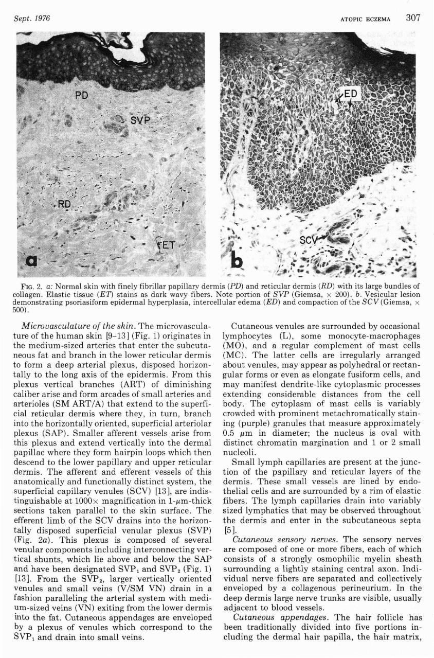
TABLE. Gross appearance and microscopic features of patients with atopic eczema
Vesicul a r areas
Patient JH BT AS PM
Reaction Gross Microscopic
Microscopic Features Epiderm a l chan ges:
H y perkerat os is Dyskeratos is Parakeratos is H y perplas ia
Platelike Psoriasiform
Hair follicle' Cellular infiltra te:
Extent Ly mphocytes Monocytes
Dermal changes : Edema Com p ac t ion Cellular infiltra te :
Extent Perivasc ular Intervasc ular Ly mphocytes Ly mphohl asts Monomacro ph ages Neutrophils Basophils Eos inophils Acti va ted hi st iocy tes Plasma cells Ma~t cellsr!
Fihrin Vessel changes (venous) Nerve
(l L = Les ional skin
L" un
1-1-/2 '1- 0 2 + ±
1 + 1+ ± J::
o 0
o 0 3 - ~ ±
o 3 - ~
2 + ± 1 1-/2 I 1.
2 I- 0
o 0 2 I- /:1 I 1-1-
2+ /:1 1- ± 2 -I- /:1 1- ±
± ±
2 '1- /:1 1- ± 2 + /:3 1- ±
1 + / 2 -I- J::
± 0 o 0 ± ()
2 + 0 o 0 4. 0 8.5 o 0 4 + 2-1-± 0
h U = Clinically unin volved skin
L
1 1- /2 -1 2 1-n -l-
1 1- /'2 1-o ±
o :3 -1- /4 1-
o
2 -II 1- /2 -1-1 I- /'2 -1
o ±
1 -II 1-
± 1 -II -1-
±
±
±
± 2 '1-o 4.7 o
1 1- /2 1-
o
u
o ±
1 1-/2 -1-o o
1 1- /2 : o 1 I
:I.e
±
±
o
± ± o ± ±
± o o o
1 1- /'2 '1 o :3.8 o 1 -1-o
L
1 1-/2 + 2 -1-
~1 + o o
o :3 + o
±
± ()
1 I /2 I
±
1 I 1 i o 1 I I -II I
o o o
1 I /2 I
o 8.2 o 2 1-o
, Involvement with intercellular edema and lymphocyti c infiltra te r! Cells / 100 J..Lm
u L
o 2-1-± 1+
1+ 2 + o 0
:1:: 0
1 + /2 1- 0 o 1 + /'2 1-o 0
o ±
o ±
o 0
o 0 o 1+
2 · ~ ± 2 - ~
o ±
1 '1-/2 I ::Ie 1 1-
± ±
o 0 o 0 o 0 .r. 1 + o () 5 .7 6 .2 o 0 1 -1- 3 I-() 0
u
SO
L
GB
L
o 4 - ~ 4 -1-
3 -1-± 3 - ~
1+ 1 1- /2 -1- 2 + ± o 2 +
o 1+ 1 1-
o 0 o :3 + o
o 2+ o 0
o ± ±
± o
o ±
o 0
o 0 ±
± 0 2 -I- /:1 I-
1 -II I
:I::
1., 1 1-
±
o o o o o 9.0 o 2+ o
2 -1 3 1-
:2 '1- :3 -1-1 -1- ± 1+ 1 + 1 -I- 1 + /2 + :2 -1- :3 -1-o 1+ o 0 o 0 2 -1- :3 ·1-o ±
5.0 8.5 o 0 2 + :3 +
1+/2 ·; - 2 +
Li chenified pl aq ues
.JB
u L u
o 4 + o ± ± 1 + /2 +
1+ 2 + /:3 + 2 -1-o o ±
() 0 o
o 0 o .± 3 + 1 -1-
o () 0
o 0 o o o
o 0 o 0
o 0 o o 0 ±
±
± ± o o o o o o o 5.2 o 2 -1-o
1+ ±
1+ ±
± 0 ± ±
± 0 1+ ±
o 0 o 0 o 0 :3 + 1 + /2 + o 0
10.5 3.0 o 0 4 -1- 1-1-o 0
L
4 + 1 '1-
1-1-o o
o 1-1-o
o o o
o o
BH
u
o
±
o o
o ±
1+
o 1+ o
o o
± ±
o o 0 ± ±
o 0 ± ±
o 0 o 0 o 0 o 0 o 0 9.0 4.7 o 0 4+ 2 + /3 I-3-1- 0
ED
L
4 -i-2 +
1 -i
o ±
o 1 -1-o
1-1-1+ o
± :3 -1- /4 1-
1 -1-
1-1-l 'iL -1 -i-1 -1-o ±
± 3 -1-
.0 11.0 o 4 -1-
2 +
u
o
1-1-o o
o o o
o o o
o o
± ± ± o o ±
o o o o o 4.2 o 4+ o
V1 ~ <";0
N ~
~
~ o "d -(')
t.2l (') N t.2l s:: >
w o ~

310 MIHM ET AL
/
, ,~ '~ -
. I
b
"
/" J
SM ART/A
Vol. 67, No.3
1 / /
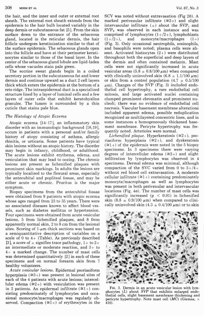
FIG. 4 . a: Dermis in lichenified lesion showing monocyte/macrophage (MO) adjacent to venule with marked basement membrane thickening. Note activated histiocyte (AR), numerous mast cells (MC), and demyelinated cutaneous sensory nerve (N) (Giemsa, x 660). b: Normal cutaneous sensory nerve (N) and arteriole (8M ART/A) are adjacent to affected venule (V) (Giemsa, x 460).
from a control population (4.7 ± 0.5/100 .urn). Numerous activated histiocytes (2+ /3+) were observed. Venular changes (3+/4+) noted about the SVP and deeper venules included endothelial cell hypertrophy with activation of nuclei with prominent nucleoli; variable, often marked degrees of basement membrane thickening; and focal pericyte hypertrophy (Fig. 5). In severe lesions the SCV exhibited similar but less marked changes. Dilated lymphatics were observed in the reticular dermis of 2 specimens. Arterioles appeared unaltered except in biopsy specimens from patient SD in which slight endothelial cell and pericyte hypertrophy were noted.
Cutaneous nerves at all levels of the dermis exhibited alterations including evidence of demyelination and apparent fibrosis (Fig. 4b). Occasionally, small irregular discrete, green-staining vacuolated areas appeared within nerve fibers, and suggested lipid accumulation.
Clinically normal skin. In 7 biopsy specimens, 6 taken 2 em from the lesional site and 1 (BH) 8 em from the lesion, varying but distinct histologic changes were observed. Notable were traces of hyperkeratosis and epidermal hyperplasia, intercellular edema (±/1+), and a slight dermal cellular infiltrate consisting primarily of lymphocytes. All biopsy specimens exhibited significant alterations in most venules varying from 1 + to 4+. These changes included: endothelial cell enlargement with focal luminal obliteration and prominent nuclei with, at times, multiple nucleoli; basement membrane changes varying from slight to marked thickening or multiple laminar reduplication; and prominent pericytes with activated nuclei about almost all affected venules. Dilated lymphatics were observed in two specimens. In 4 specimens variable intercellular edema of the hair follicle (2+/3+) was observed. Sweat glands in all specimens appeared
FIG. 6. Dermis of a lichenified lesion with severely altered venules (V). Note enlarged endothelial cells with prominent nuclei, variable basement membrane thickening, suggestion of reduplication of basement membrane, and pericyte hypertrophy. Infiltrate contains prominent mast cells (MC) and activated histiocytes (AR) (Giemsa, x 410).
unaltered. In 2 specimens fibrosis and focal demyelination of cutaneous sensory nerves were noted.
DISCUSSION
Descriptions [5,17,20,21] of atopic eczema have been based on studies of paraffin-embedded biopsy specimens stained with hematoxylin and eosin or toluidine blue or on ultrastructural studies of infantile eczema [22,23]. Utilization of 1-.um-thick, glutaraldehyde-fixed, Epon-embedded tissue [1,3] stained with Giemsa's reagent avoids the sampling problem inherent in electron microscopy but permits definition of normal skin structure (Fig 2a) and of the inflammatory response [2,3], which is poorly characterized in routinely processed tissue.
In atopic eczema the changes of the epidermis
Sept. 1976
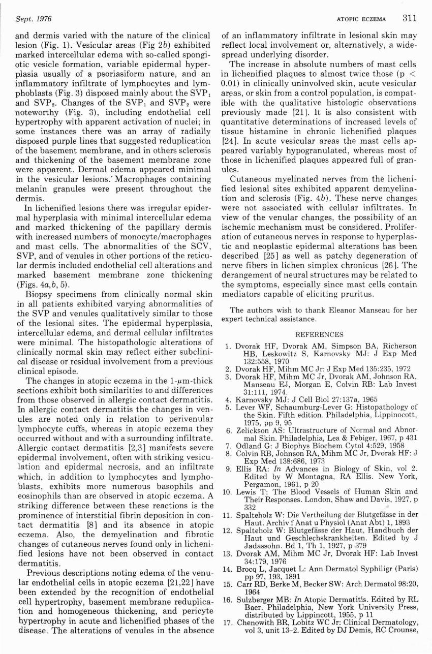
and dermis varied with the nature of the clinical lesion (Fig. 1). Vesicular areas (Fig 2b) exhibited marked intercellular edema with so-called spongiotic vesicle formation, variable epidermal hyperplasia usually of a psoriasiform nature, and an inflammatory infiltrate of lymphocytes and lymphoblasts (Fig. 3) disposed mainly about the SVP 1
and SVP2 • Changes of the SVP 1 and SVP 2 were noteworthy (Fig. 3), including endothelial cell hypertrophy with apparent activation of nuclei; in some instances there was an array of radially disposed purple lines that suggested reduplication of the basement membrane, and in others sclerosis and thickening of the basement membrane zone were apparent. Dermal edema appeared minimal in the vesicular lesions.' Macrophages containing melanin granules were present throughout the dermis.
In lichenified lesions there was irregular epidermal hyperplasia with minimal intercellular edema and marked thickening of the papillary dermis with increased numbers of monocyte/macrophages and mast cells. The abnormalities of the SCV, SVP, and of venules in other portions of the reticular dermis included endothelial cell alterations and marked basement membrane zone thickening (Figs. 4a,b, 5).
Biopsy specimens from clinically normal skin in all patients exhibited varying abnormalities of the SVP and venules qualitatively similar to those of the lesional sites. The epidermal hyperplasia, intercellular edema, and dermal cellular infiltrates were minimal. The histopathologic alterations of clinically normal skin may reflect either subclinical disease or residual involvement from a previous clinical episode.
The changes in atopic eczema in the l-,um-thick sections exhibit both similarities to and differences from those observed in allergic contact dermatitis. In allergic contact dermatitis the changes in venules are noted only in relation to perivenular lymphocyte cuffs, whereas in atopic eczema they occurred without and with a surrounding infiltrate . Allergic contact dermatitis [2,3] manifests severe epidermal involvement, often with striking vesiculation and epidermal necrosis, and an infiltrate which, in addition to lymphocytes and lymphoblasts, exhibits more numerous basophils and eosinophils than are observed in atopic eczema. A striking difference between these reactions is the prominence of interstitial fibrin deposition in contact dermatitis [8] and its absence in atopic eczema. Also, the demyelination and fibrotic changes of cutaneous nerves found only in lichenified lesions have not been observed in contact dermatitis.
Previous descriptions noting edema of the venular endothelial cells in atopic eczema [21,22] have been extended by the recognition of endothelial cell hypertrophy, basement membrane reduplication and homogeneous thickening, and pericyte hypertrophy in acute and lichenified phases of the disease. The alterations of venules in the absence
ATOPIC ECZEMA 311
of an inflammatory infiltrate in lesional skin may reflect local involvement or, alternatively, a widespread underlying disorder.
The increase in absolute numbers of mast cells in lichenified plaques to almost twice those (p < 0.01) in clinically uninvolved skin, acute vesicular ar~as, or skin from a control population, is compatible with the qualitative histologic observations previously made [21]. It is also consistent with quantitative determinations of increased levels of tissue histamine in chronic lichenified plaques [24 J. In acute vesicular areas the mast cells appeared variably hypogranulated, whereas most of those in lichenified plaques appeared full of granules.
Cutaneous myelinated nerves from the lichenified lesional sites exhibited apparent demyelination and sclerosis (Fig. 4b). These nerve changes were not associated with cellular infiltrates. In view of the venular changes , the possibility of an ischemic mechanism must be considered. Proliferation of cutaneous nerves in response to hyperplastic and neoplastic epidermal alterations has been described [25] as well as patchy degeneration of nerve fibers in lichen simplex chronicus [26]. The derangement of neural structures may be related to the symptoms, especially since mast cells contain mediators capable of eliciting pruritus.
The authors wish to thank Eleanor Manseau for her expert technical assistance.
REFERENCES
1. Dvorak HF, Dvorak AM, Simpson BA, Richerson HB, Leskowitz S, Karnovsky MJ: J Exp Med 132:558, 1970
2. Dvorak HF, Mihm MC Jr: J Exp Med 135:235, 1972 3. Dvorak HF, Mihm MC Jr, Dvorak AM , Johnson RA,
Manseau EJ, Morgan E , Colvin RB: Lab Invest 31:111, 1974.
4. Karnovsky MJ: J Cell BioI 27:137a, 1965 5. Lever WF, Schaumburg-Lever G: Histopathology of
the Skin. Fifth edition. Philadelphia , Lippinocott, 1975, pp 9, 95
6. Zelickson AS: Ultrastructure of Normal and Abnormal Skin. Philadelphia, Lea & Febiger, 1967, p 431
7. Odland G: J Biophys Biochem Cytol 4:529 , 1958 8. Colvin RB, Johnson RA, Mihm MC Jr, Dvorak HF: J
Exp Med 138:686, 1973 9. Ellis RA: In Advances in Biology of Skin, vol 2.
Edited by W Montagna, RA Ellis. New York, Pergamon, 1961, p 20
10. Lewis T: The Blood Vessels of Human Skin and Their Responses. London, Shaw and Davis, 1927 , P 332 ~
11. Spalteholz W: Die Vertheilung der Blutgefcisse in der Haut. Archiv f Anat u Physiol (Anat Abt) 1, 1893
12. Spalteholz W: Blutgefasse der Haut, Handbuch der Haut und Geschlechskrankheiten. Edited by J Jadassohn . Bd 1, Th 1, 1927, p 379
13. Dvorak AM, Mihm MC Jr, Dvorak HF: Lab Invest 34: 179, 1976
14. Brocq L, Jacquet L: Ann Dermatol Syphiligr (Paris) pp 97, 193, 1891
15. Carr RD, Berke M , Becker SW: Arch Dermato198:20, 1964
16. Sulzberger MB: In Atopic Dermatitis. Edited by RL Baer. Philadelphia, New York University Press, distributed by Lippincott, 1955, p 11
17. Chenowith BR, Lobitz WC Jr: Clinical Dermatology, vol 3, unit 13-2. Edited by DJ Demis, RC Crounse,

312 MIHM ET AL
RL Dobson, J McGuire. Hagerstown, Md, Harper & Row, 1972, p 1
18. Rostenberg A, Bogdanoff DR: In Immunological Diseases. Edited by M Samter. Boston, Little , Brown, 1965, p 635
19. Champion RH, Parish WE: In Clinical Aspects of Immunology. Third edition. Edited by PGH Gell, RRA Coombs, PJ Lachmann. Oxford, Blackwell, 1975, p 1183
20. Rajka G: Atopic Dermatitis. London, Saunders, 1975 21. Montgomery H: Dermatopathology, vol 1. New York,
Hoeber Medical Division, Harper & Row, 1967 , P 186
22. Prose PH, Sedlis E: J Invest Dermatol 34: 149, 1960 23. Prose PH, Sedlis E, Bigelow M: J Invest Dermatol
45:448, 1965 24. Johnson HH Jr, DeOreo GA, Lascheid WP, Mitchell
F: J Invest Dermatal 34:237, 1960 25. Pawlowski A, Weddell G: Br J Dermatol 78:603, 1966 26. Cowan MA: Arch Dermatol 98:562, 1964
DISCUSSION
Austen: What are your speculations as to the pathogenetic mechanism(s) 'of atopic dermatitis in the light of the natural history of lesions?
Mihm: I have no definite clue as to pathogenesis based on this study. Many of the findings sur.h as the vascular abnormalities and mast cell changes, although they may be secondary to the basic etiologic factors, warrant explanation. The alterations of cutaneous sensory nerves
Vol. 67, No.3
are intriguing, especially in the light of the primary symptom, itching, and they may be related to pathogenesis.
Claman: One of the ·hallmarks of atopic disease in the nose and lungs is an infiltration of eosinophils. Would you discuss why you didn't see many eosinophils in the acute or chronic lesions? Were your patients in fact atopic?
Mihm: We biopsied randomly chosen acute and chronic lesions from patients with atopic eczema. Eosinophils may participate in the development of these lesions, but only serially performed biopsies of lesions from their onset to their fully developed stage will resolve this question.
Lazarus: Please define endothelial cell activation. Mihm: Endothelial cell activation involves an increase
in endothelial cell mass and enlargement of nuclei with slightly irregular chromatin distribution about the nuclear membrane. We have observed these changes in several inflammatory disorders, including allergic contact dermatitis, reactions to intradermally injected protein antigens, and cutaneous necrotizing angiitis .
Provost: A great deal of emphasis has been placed on the possible role of elevated serum IgE levels in the pathogenesis of atopic dermatitis. It should be noted that as many as 20% of atopic dermatitis patients have normal or low serum IgE levels. Recently Johansson has presented evidence that atopic dermatitis patients without physical or historical evidence of asthma or allergic rhinitis have low or normal serum IgE levels. They further stated that serum IgE levels correlated with the presence of asthma and allergic rhinitis.