The strategies of Staphylococcus aureus to develop a ... · Osteomyelitis is the most frequent...
55
The strategies of Staphylococcus aureus to develop a chronic infection Habilitation Submitted to the Medical Faculty of Friedrich-Schiller University Jena From Dr. rer.nat. Lorena Tuchscherr From Buenos aires Vorgelegt am: 13.09.2016
Transcript of The strategies of Staphylococcus aureus to develop a ... · Osteomyelitis is the most frequent...
The strategies of Staphylococcus aureus to
develop a chronic infection
Habilitation
Submitted to the Medical Faculty of
Friedrich-Schiller University Jena
From
Dr. rer.nat. Lorena Tuchscherr
From Buenos aires
Vorgelegt am: 13.09.2016

Gutachter
Internal:
1- .Prof. Dr. med. Bettina Löffler, Institut für Medizinische Mikrobiologie, UKJ,
Jena.
2- Prof. Dr. med. Michael Bauer, Klinik für Anästhesiologie und Intensivmedizin,
UKJ, Jena.
External:
1- Prof. Dr. Andreas Peschel, Institut für Mikrobiologie und Infektionsmedizin,
Univ. Tübingen, Tübingen.
Teaching qualification obtained on 6th July, 2017.

Ehrenwörtliche Erklärung
Ich erkläre hiermit, daß mir die aktuelle Habilitationsordnung der Friedrich-Schiller-Universität
Jena bekannt ist.
Ferner erkläre ich, daß ich die vorliegende Arbeit ohne unzulässige Hilfe Dritter und ohne
Benutzung anderer als der angegebenen Hilfsmittel angefertigt habe. Die aus anderen
Quellen direkt oder indirekt übernommenen Daten und Konzepte sind unter Angabe der
Quelle gekennzeichnet.
Bei der Auswahl und Auswertung folgenden Materials haben mir die nachstehend
aufgeführten Personen in der jeweils beschriebenen Weise entgeltlich/unentgeltlich geholfen:
1. Prof. Dr. Löffler , Prof. Dr Peters und Prof. Dr Sordelli.
2..MTAs und students from Münster: B. Schuhen, K. Broschwig, M. Brück, Marie
Hachmeister and Jennifer Geraci.
3. MTAs from Argentina: S. Soldavini and L. Medina
4. Colleges: S. Niemann, V. Hoerr, C. Kreis
Weitere Personen waren an der inhaltlich-materiellen Erstellung der Arbeit nicht beteiligt.
Insbesondere habe ich hierfür nicht die entgeltliche Hilfe von Vermittlungs- bzw.
Beratungsdiensten in Anspruch genommen. Niemand hat von mir unmittelbar oder mittelbar
geldwerte Leistungen für Arbeiten erhalten, die im Zusammenhang mit dem Inhalt der
vorgelegten Arbeit stehen.
Die Arbeit wurde bisher weder im In- noch Ausland in gleicher oder ähnlicher Form einer
anderen Prüfungsbehörde vorgelegt.
Ich versichere, daß ich nach bestem Wissen die reine Wahrheit gesagt und nichts
verschwiegen habe.
Jena, den 23. June 2016
Dr. Lorena Tuchscherr de Hauschopp

1
Contents
Contents 1
List of publications 2
1. Introduction 4
1.1 S. aureus and Persistence 4
1.2. Objectives of this work 6
2. Results and discussions: Description of main results and papers 7
2.1 Acute phase of infection and host response 7
2.2 In vivo models to study staphylococcal infection 9
2.3 Development and dynamic of small colony variants (SCVs) 12
2.4 Host controlled by S. aureus during persistence: mechanisms of immune escape 22
2.5 Loss of capsule during persistence 24
2.6 Approaches for vaccine development and treatment to eliminate S. aureus 27
3. Conclusions and overview 32
4. List of abbreviations 35
5. References 36
6. Ehrenwörtliche Erklärung 42
7. Curriculum Vitae 43
8. Complete list of publications 46
9. Oral presentations 49
10. Acknowledgements 50
11. Copies of original publications used for this Habilitation 51

2
List of publications
The present Habilitation is based on the following original publications. The publications
where I am corresponding author are indicated (*).
a. Tuchscherr L, Kreis CA, Hoerr V, Flint L, Hachmeister M, Geraci J, Bremer-
Streck S, Kiehntopf M, Medina E, Kribus M, Raschke M, Pletz M, Peters G,
Löffler B. Staphylococcus aureus develops increased resistance to antibiotics
by forming dynamic small colony variants during chronic osteomyelitis. J.
Antimicrob Chemother. 2016 Feb;71(2):438-48. (*)
b. Tuchscherr L, Löffler B. Staphylococcus aureus dynamically adapts global
regulators and virulence factor expression in the course from acute to chronic
infection. Curr Genet. 2015 Jun 30.
c. Tuchscherr L, Bischoff M, Lattar SM, Noto Llana M, Pförtner H, Niemann S,
Geraci J, Van de Vyver H, Fraunholz MJ, Cheung AL, Herrmann M, Völker U,
Sordelli DO, Peters G, Löffler B. Sigma Factor SigB Is Crucial to Mediate
Staphylococcus aureus Adaptation during Chronic Infections. PLoS Pathog.
2015 Apr 29;11(4).
d. Kalinka J, Hachmeister M, Geraci J, Sordelli D, Hansen U, Niemann S,
Oetermann S, Peters G, Löffler B, Tuchscherr L. Staphylococcus aureus
isolates from chronic osteomyelitis are characterized by high host cell invasion
and intracellular adaptation, but still induce inflammation. Int J Med Microbiol.
2014 Nov;304(8):1038-49.
e. Horst SA, Hoerr V, Beineke A, Kreis C, Tuchscherr L, Kalinka J, Lehne S,
Schleicher I, Köhler G, Fuchs T, Raschke MJ, Rohde M, Peters G, Faber C,
Löffler B, Medina E. A novel mouse model of Staphylococcus aureus chronic
osteomyelitis that closely mimics the human infection: an integrated view of
disease pathogenesis. Am J Pathol. 2012 Oct;181(4):1206-14.
f. Tuchscherr L, Medina E, Hussain M, Völker W, Heitmann V, Niemann S,
Holzinger D, Roth J, Proctor RA, Becker K, Peters G, Löffler B.
Staphylococcus aureus phenotype switching: an effective bacterial strategy to
escape host immune response and establish a chronic infection. EMBO Mol
Med. 2011 Mar;3(3):129-41.

3
g. Tuchscherr L, Löffler B, Buzzola FR, Sordelli DO. Staphylococcus aureus
adaptation to the host and persistence: role of loss of capsular polysaccharide
expression. Future Microbiol. 2010 Dec;5(12):1823-32.
h. Tuchscherr L, Heitmann V, Hussain M, Viemann D, Roth J, von Eiff C, Peters
G, Becker K, Löffler B. Staphylococcus aureus small-colony variants are
adapted phenotypes for intracellular persistence. J Infect Dis. 2010 Oct
1;202(7):1031-40.
i. Grundmeier M, Tuchscherr L, Brück M, Viemann D, Roth J, Willscher E,
Becker K, Peters G, Löffler B. Staphylococcal strains vary greatly in their
ability to induce an inflammatory response in endothelial cells. J Infect Dis.
2010 Mar 15;201(6):871-80.
j. Tuchscherr LP, Buzzola FR, Alvarez LP, Lee JC, Sordelli DO. Antibodies to
capsular polysaccharide and clumping factor A prevent mastitis and the
emergence of unencapsulated and small-colony variants of Staphylococcus
aureus in mice.Infect Immun. 2008 Dec;76(12):5738-44.

4
1- Introduction
1.1- S. aureus and Persistence
Staphylococcus aureus is a gram-positive coccal bacterium that is a member of the
Firmicutes. S. aureus is a facultative pathogenic bacterium that can colonize the epithelial
surfaces of humans and domestic animals and can also cause different types of severe
tissue infections. Approximately 20% of individuals are persistently nasally colonized with S.
aureus, and 30% of individuals are intermittently colonized [1, 2]. However, other sites can
be colonized, including the axillae, groin, and gastrointestinal tract [3]. Colonization provides
a reservoir from which bacteria can cause different types of infection [4]. In a study of
bacteraemia, blood isolates were identical to nasal insolates in 82% of patients, suggesting
that S. aureus can occasionally breach epithelial barriers and enter the bloodstream [5].
S. aureus is the main pathogen of osteomyelitis worldwide. Osteomyelitis is a bone infection
that can be associated with high levels of inflammation and bone tissue destruction. The
infection can sometimes develop into a chronic course and may become extremely difficult to
treat with antimicrobials [6].
Osteomyelitis can be categorized into three groups, according to the route used by the
infecting bacterium to gain access to the bone [7]. First, in haematogenous osteomyelitis,
staphylococci access the bone tissue via the bloodstream. This form of osteomyelitis affects
mainly prepubertal children. Second, osteomyelitis develops by way of a contiguous spread
from a local infection after trauma, bone surgery, or joint replacement. Finally, osteomyelitis
can be peripheral to vascular insufficiency. This form of osteomyelitis occurs mostly in
diabetic patients and usually originates from an infected foot ulcer that spreads to the bone.
All its forms can present in the acute or chronic phase and in virtually any bone.
Osteomyelitis is the most frequent cause of non-traumatic limb amputation, as antimicrobial
compounds often fail to clear the infection [6-9].
Despite the availability of effective antimicrobial agents to treat staphylococcal infection, it
continues to be a major cause of morbidity and mortality worldwide [10]. Epidemiological
studies due to the spread of successful clones continue to be reported from virtually every
geographic region [11].
S. aureus is one of the most important pathogens in the healthcare and community setting.
The emergence of antimicrobial-resistant strains, especially those that are resistant to
methicillin, has been a constant feature. Methicillin-resistant Staphylococcus aureus (MRSA)
is a major public health concern and is responsible for both hospital- and community-
associated infections worldwide. It is estimated that MRSA infections within the health care

5
setting alone affected more than 150,000 patients annually in the European Union, with an
additional cost of 380 million Euros [12]. In addition, MRSA has developed as a colonizer and
pathogen of domestic animals and is linked with livestock-associated infections [13].
In addition to resistance development, many mechanisms appear to contribute to the
success of S. aureus as a pathogen. Those mechanisms include commensal colonization,
long-term survival on environmental surfaces and diverse mechanisms of immune host
evasion, and the armamentarium of virulence determinants, often with redundant functions,
are among the most important [14-17]. To establish an infection, S. aureus bears a multitude
of virulence factors, including adhesive surface proteins (adhesins) and toxic compounds that
act in concert, destroy host tissue, and resist the host defence system [10]. Yet, the extent of
endowment with virulence factors can vary widely between clinical isolates. Furthermore, the
expression of almost all virulence factors is controlled by a complex S. aureus regulatory
network that allows for rapid adaptation; for example, the quorum-sensing system accessory
gene regulator (Agr) enhances the expression of toxins and other secreted cytotoxic factors,
and the alternative sigma factor B SigB (σB) modulates stress responses [18, 19]. During
bacteraemia or sepsis, the bacteria need to survive within the bloodstream and to defend
against immune cells. Notably, secreted pore-forming toxins (e.g., -hemolysin/-toxin)
cause inflammation and contribute to sepsis development [20]. To settle an infection in host
tissue, e.g., bone tissue, bacteria first need to adhere to host structures, such as the
extracellular matrix or host cells. For this, S. aureus expresses various surface proteins with
adhesive functions, such as fibronectin-binding proteins (FnBPs), clumping factors (Clfs),
collagen-binding protein (Cna), bone sialoprotein-binding protein (Bbp), the anchorless
protein extracellular adhesion protein (Eap), and the extracellular matrix binding protein
(Emp) [21]. Up until now, a clear association between a single adhesin and the development
of osteomyelitis could not be demonstrated [6]. After initial settling of the infection, the
bacteria must adapt to the host tissue for persistence and to escape from the host immune
system. For this, S. aureus can invade different types of host cells, including osteoblasts [22].
In the past decades, S. aureus was increasingly recognized as a facultative intracellular
pathogen. S. aureus has the ability to invade cells and survive intracellularly for various
periods. Many studies were conducted to investigate whether intracellular S. aureus
contributes to persistent and therapy-refractory infections. S. aureus can be internalized and
survive in endothelial cells, epithelial cells, fibroblasts, osteoblasts and keratinocytes (as non-
professional phagocytic cells). Recently reports even document bacterial survival within
professional phagocytes, such as human monocyte-derived macrophages, and some studies
have examined S. aureus survival in neutrophils [17, 23, 24].

6
The persistence strategy of S. aureus may be the reason for infections that take chronic
courses, which can be extremely difficult to eradicate by antibiotic treatment, even though the
strains are susceptible to antibiotics in vitro. Currently, the mechanism for persistence in the
presence of host defences and antibiotic therapy is not completely understood. The methods
of persistence are not clear and include different mechanisms. The persister cells can be
formed as dormant cells, e.g., non-dividing cells, such as small colony variants (SCVs) [25].
The SCVs are macroscopically slowly growing, without pigmentation, deficient at producing
exotoxins and frequently auxotrophic for menadione, hemine and thymidine [26]. For many
years, the appearance of SCVs has been associated with chronic infections [26, 27].
Osteomyelitis is one type of infection that frequently develops to chronicity [28]. The diverse
strategies of persistent S. aureus include crosstalk between global regulators (agr, sigB and
sarA) and rapid feedback according to the environmental stimulus [29, 30]. This work
provides a summary of the complex steps of staphylococcal infection, from acute infection to
the development of chronicity. The main objective is to understand the biology of persister
staphylococcal cells and offer insights into their role in infection development.
1.2- Objectives of this work:
I investigate the passage of S. aureus from an acute and septic infection to a chronic and
persisting infection. Different tools were used like fluorescent image, real time PCR, cell
culture and animal models. According our results, I focused my research on:
1- To analyse the bacterial regulatory mechanisms and changes in virulence factor
expression which are required to establish a chronic infection..
2- To study the S. aureus mechanism of persistence in professional vs. non-professional
phagocytes.
3- To characterized the formation of staphylococcal small colony variants during the
course of infection.
4- To find possible therapeutic targets to avoid the persistence of S. aureus.

7
2- Results and discussion
2.1- Acute phase of infection and host response
Grundmeier M, Tuchscherr L, Brück M, Viemann D, Roth J, Willscher E, Becker K,
Peters G, Löffler B. Staphylococcal strains vary greatly in their ability to induce an
inflammatory response in endothelial cells. J Infect Dis. 2010 Mar 15;201(6):871-80.
The first step in establishing an infection is bacterial adherence, followed by the invasion of
host cells. Many authors have demonstrated that most clinical staphylococcal isolates exhibit
a strong invasive phenotype [31]. Epithelial or endothelial cells are the first barrier for S.
aureus to start the infection. These cells express molecules that attract, bind and activate
cells from the immune system to defend against the bacteria. To find out how S. aureus can
provoke epithelial/endothelial inflammatory reactions, we used microarray, real-time PCR
and ELISA analysis. We selected 3 invasive strains (Cowan I, 6850 and ST239) with highly
invasive phenotypes for our study. Bacterial internalization was quantified using a flow
cytometry invasion assay and revealed equally efficient uptake of the different strains by
endothelial cells. These results were confirmed by electron microscopy (Fig. 1). The bacteria
were mainly located within phagosomes, and major parts of the endothelial cells were
infected.
Fig. 1: Electron micrographs of endothelial cells infected with Staphylococcus aureus. Confluent human umbilical vein endothelial cells (HUVEC cells) were infected with live S. aureus Cowan I, 6850, or ST239 and incubated for 4 h. Cells were then fixed and processed for electron microscopy. All bacterial strains tested were internalized by endothelial cells, and located mainly within phagosomes.
In staphylococci, the production of virulence factors is mainly controlled by the accessory
gene regulator (agr) system, a quorum-sensing mechanism that controls gene expression

8
according to the bacterial cell density [32]. To quantify the expression of important virulence
factors, including adhesins and secreted toxins, reverse-transcription PCR (RT-PCR, Fig. 2)
was performed. All the strains expressed adhesins (fibronectin and protein A), but the Cowan
I strain failed to express important virulence factors related to the agr system, such as α-toxin
(hla). In contrast, strains 6850 and ST239 strongly expressed the regulator agr and related
virulence factors (Fig. 2A and B).
Fig. 2: Differential expression of genes by Staphylococcus aureus strains Cowan I, 6850, and ST239. A: Results of real-time PCR from S. aureus Cowan I, 6850, and ST239. B: S. aureus Cowan I, 6850, and ST239 were grown for 2, 4, 6, or 8 h, as indicated, and bacterial RNA was extracted and agr expression was analysed by real-time PCR.
To determine whether the invasiveness is directly associated with inflammation, we analysed
the endothelial gene expression in response to different S. aureus strains using microarray
analysis. Gene expression analysis revealed that most of the genes were highly upregulated
following stimulation with strains 6850 and ST239 compared with uninfected cells, whereas
no genes were highly upregulated in response to S. aureus Cowan I. Both strains 6850 and
ST239 induced the upregulation of genes that are involved in the endothelial immune
response, including cytokines, chemokines and adhesion proteins. However, strain Cowan I
failed to highly upregulate any genes that could play a role in the antimicrobial response.
These results were confirmed by real-time PCR and ELISA [33].
To investigate the clinical relevance of our findings, we analysed 12 clinical isolates for their
agr expression and divided them into low-agr-expressing strains and high-agr-expressing
strains. As expected, the high-agr-expressing strains exhibited high hla, haemolytic activity,
and proteases and induced strongly enhanced chemokine expression. In contrast, the low-
agr-expressing strains caused no or only weak chemokine expression as well as virulence
factors, similar to Cowan I.

9
In summary, we found that S. aureus strains could cause different types of infections.
Isolates that express a multitude of virulence factors induce an extensive immune response
and affect many cellular functions. A high level of inflammation might help the host eliminate
S. aureus but could also produce tissue damage, which favours bacterial spread to deep
tissue structures. Strains defective in the agr system invade host cells but do not cause a
huge inflammatory reaction, which might represent an alternative strategy of S. aureus to
evade the host immune system and cause chronic and recurrent infections (Fig. 3).
Fig. 3: Summary of events after S. aureus host cell invasion. In general, after host cell invasion different courses of infection are possible depending on the bacterial strain and its expression of virulence factors by the agr system. If the ingested staphylococci express a multitude of virulence factors, especially a-toxin, then the bacteria induce proinflammatory and cytotoxic effects within the host cells, which often results in the death of the host cell. If the staphylococci downregulate their virulence factor expression, for example due to a defect in the agr system, then they can persist within the host cells without causing damage; this cell looks morphologically intact, although it contains living bacteria. In this intracellular location the bacteria are likely well protected against the host immune system and against antimicrobial treatments additionally this might be a reservoir for chronic infections.
2.2- In vivo models to study staphylococcal infections
Horst SA, Hoerr V, Beineke A, Kreis C, Tuchscherr L, Kalinka J, Lehne S, Schleicher
I, Köhler G, Fuchs T, Raschke MJ, Rohde M, Peters G, Faber C, Löffler B, Medina E.
A novel mouse model of Staphylococcus aureus chronic osteomyelitis that closely
mimics the human infection: an integrated view of disease pathogenesis. Am J
Pathol. 2012 Oct;181(4):1206-14.
To study the intrinsic steps of S. aureus infections, it is necessary to have a model that
resembles human pathogenesis and tools to follow S. aureus during infection. For this, we
established a haematogenous osteomyelitis model in mice to analyse the infection process

10
from the acute to the chronic stage. The animal studies provide experimental proof for the
molecular basis of pathogenesis and the role of the immune system. Additionally, we created
a new tool to label bacteria for detection by magnetic resonance imaging (MRI).
The pathogenesis of osteomyelitis remains poorly understood, partly for lack of experimental
models that closely mimic human disease. Although diverse animal models of osteomyelitis
have been established, they are often lacking the full spectrum of the human disease,
including both the acute and chronic phases. To overcome this limitation, we created a novel
murine model of osteomyelitis in which bone tissue was infected via the bloodstream. The
murine model recapitulates important aspects of acute and chronic stages of osteomyelitis in
humans, as demonstrated by histopathology, MRI, X-ray imaging and electron microscopic
examination (Fig. 4). Histological analysis of infected bones revealed a massive infiltration of
inflammatory cells during the acute phase of the infection. As in the human disease, bone
resorption started to become apparent when the infection entered the chronic phase. We
could also detect the formation of bone sequestra (necrotic bone) in this murine model (Fig.
5).

11
Fig. 4: Sequential MRI and X-ray imaging showing the progression of osteomyelitis in the tibiae of S. aureus-infected mice during acute and chronic phases of infection. A: MRI scans of the right (R) and left (L) tibiae of an uninfected (control) and a S. aureus-infected mouse at progressive times after bacterial inoculation. B: Three-dimensional reconstruction of the tibiae and inflammatory lesions after segmentation of MRI. Inflammatory lesions are coloured orange and pink (right) or red and brown (left) according to the inflammatory depth in the bones. The noninflamed area (the area of the bone without signs of inflammation) of the right leg is shown in magenta and the noninflamed area of the left leg is shown in green. C: Sequential radiography of a left tibia, showing the progression of bone deformation over time. D and E: Whole-body X-ray radiography on day 3 (D) and day 63 (E) of infection.

12
Fig. 5: Histopathological evaluation of mouse and human osteomyelitis during acute and chronic disease stages (H&E stain). In the acute osteomyelitis, massive infiltration of immune cells was detected, whereas in the chronic stage of infection, the resorption lacunae produced by the activity of individual osteoclasts is shown. This pattern is similar in mice and patients.
To summarize these concepts, we have described a novel murine model of S. aureus that
mimics the natural route of infection in haematogenous osteomyelitis. This model can
facilitate the identification of bacterial factors involved in bone tropism and provides an
important platform for microbiological and immunological studies [34].
2.3- Development and dynamic of small colony variants (SCVs)
Tuchscherr L, Medina E, Hussain M, Völker W, Heitmann V, Niemann S, Holzinger
D, Roth J, Proctor RA, Becker K, Peters G, Löffler B. Staphylococcus aureus
phenotype switching: an effective bacterial strategy to escape host immune response
and establish a chronic infection. EMBO Mol Med. 2011 Mar;3(3):129-41.
Löffler B, Tuchscherr L, Niemann S, Peters G. Staphylococcus aureus persistence in
non-professional phagocytes. Int J Med Microbiol. 2014 Mar;304(2):170-6. doi:
10.1016/j.ijmm.2013.11.011. Epub 2013 Dec 1. Review.
Tuchscherr L, Bischoff M, Lattar SM, Noto Llana M, Pförtner H, Niemann S, Geraci
J, Van de Vyver H, Fraunholz MJ, Cheung AL, Herrmann M, Völker U, Sordelli DO,
Peters G, Löffler B. Sigma Factor SigB Is Crucial to Mediate Staphylococcus aureus
Adaptation during Chronic Infections. PLoS Pathog. 2015 Apr 29;11(4):e1004870

13
Tuchscherr L, Löffler B. Staphylococcus aureus dynamically adapts global regulators
and virulence factor expression in the course from acute to chronic infection. Curr
Genet. 2015 Jun 30.
Although S. aureus is primarily considered an extracellular pathogen, recent evidence
suggests that this bacterium can invade a variety of nonprofessional phagocytic cells. The
intracellularity of S. aureus has been implied as immune-evasive strategy, thereby escaping
detection by professional phagocytes. During its intracellular life, S. aureus can change the
phenotype to small colony variants (SCVs) [26, 35-37]. Subpopulations of SCVs have been
found in a wide variety of bacterial species [26, 38, 39], but they have been most extensively
studied in S. aureus. In general, SCVs form small colonies on agar plates (approximately 10
times smaller than the parent strain) due to their slow growth rate and reduced metabolism,
which also explains their decreased susceptibility to a variety of antibiotics [40, 41]. They
express a changed pattern of virulence factors, including the reduced expression of
exotoxins, such as α‐haemolysin (α‐toxin), and an increased expression of adhesins, such as
the fibronectin‐binding proteins (FnBPs) [42]. Furthermore, SCVs are frequently auxotrophic
for menadione and haemin, compounds involved in the biosynthesis of electron transport
chain elements, or thymidine [26, 42, 43].
SCVs recovered from clinical specimens are often not stable and can rapidly revert to their
wild‐type phenotype when subcultivated [44]. For this reason, most knowledge and
laboratory work on SCVs has been obtained with stable site‐directed mutants with mutations
in the electron transport system that mimic the SCV phenotype, e.g., hemB and menD
mutants [45, 46]. Consequently, data on the development and dynamics of SCVs are largely
missing. Further reported mechanisms leading to the formation of SCVs (in vitro and in vivo)
include prolonged exposure to subinhibitory concentrations of antibiotics [45, 47, 48] or to
exoproducts from other bacteria, e.g., Pseudomonas aeruginosa [49]. Moreover, there is
growing evidence that the formation of SCVs could also be due to regulatory mechanisms,
involving global regulators (e.g., sigB, sarA and agr), Clp ATPases [48, 50, 51] or non‐
protein‐coding RNAs as regulatory molecules [52].
There are many open questions regarding the signals and factors that induce the formation
of S. aureus SCVs. However, the central question that needs to be addressed first is whether
the development of SCVs is only a rare, marginal or laboratory phenomenon (possibly due to
gene mutations) or whether the formation of SCVs is an integral part of the normal bacterial
life cycle that is required for adaptation and persistence. In the latter case, a dynamic and
reversible formation of SCVs has to be assumed. The phenomenon of rapid phenotype

14
switching of genetically identical cells (bet‐hedging strategy) has been described for a variety
of other microorganisms [53-55]. Particularly, in a stressful and fluctuating environment,
stochastic differentiation into distinct phenotypes can provide a strong advantage that
promotes bacterial persistence [25, 56, 57].
To analyse the mechanisms of SCV formation, several in vitro cell cultures and in vivo
murine models of S. aureus long‐term infections were used [58, 59]. The recovered bacteria
were analysed for their virulence potential and their dynamic capacity to revert to the wild‐
type phenotype.
First, we infected A549 cells (cell line ATCC CCL-185 and human lung adenocarcinoma) with
S. aureus strain 6850 and the nasal isolate 628. After 4 weeks, we could recover from the
infected host cells viable colonies of both strains with a high phenotypic diversity. The
proportion of SCVs recovered increased up to 90% with increased time of intracellular
persistence. We performed the same experiment using primary cells (HUVEC and
osteoblasts) and obtained the same result after 1 week (Fig. 6A, B).

15
Fig. 6: Staphylococci survive within cultured host cells for 28 days, change phenotypes, and virulence factor expression. A: Epithelial cells (A549) were infected with different staphylococcal strains (6850 or 628) and analysed for 28 days. The number of viable intracellular persisting bacteria was determined weekly by lysing host cells, plating the lysates on agar plates and counting the colonies the following day (n = 3, ±SEM). Electron micrographs of infected cells were performed directly after infection and 28 days post-infection showing morphological intact staphylococci within epithelial cells. B: Percentage of small and very small (SCV) phenotypes (<5 and <10-fold smaller than those of the wild-type phenotype, respectively) recovered over the 28 days (n = 3, between 30 and 300 colonies examined in each sample, ±SEM). Photographs of recovered colonies were performed directly after and 28 days post-infection showing small and SCV colonies. C: Changes in bacterial gene expression (strain 6850) of fibronectin binding protein A (fnbA), α-haemolysin (hla) and agr during the course of infection were determined by real-time PCR (n = 5, ±SD). D: Changes in host cell response measured by the expression of CCL5, CXCL11 and ICAM-1 over time following infection with strain 6850 (n = 6). *p ≤ 0.05, **p ≤ 0.01 and ***p ≤ 0.001 in comparison with values from uninfected cells.

16
We next determined the expression of virulence factors during phenotypic switching and the
innate immune response. Interestingly, we found high expression of FnBPA (fnbA) and
downregulation of hla (α-toxin) and the global accessory gene regulator agr, which controls
the expression of many secreted virulence factors. In parallel, the host response, measured
by the acute inflammatory cytokine levels (CCR5/RANTES or CXCL11/I-ITAC) or ICAM-
1/CD54 levels, was attenuated (Fig. 6C, D). The results clearly showed diminished hla and
agr levels over time, suggesting reduced virulence. Consequently, persisting bacteria appear
to be as silent as possible to avoid provoking the host immune system. In contrast, fnbA
levels increased during long-term persistence, indicating that the bacteria become highly
adhesive and are rapidly taken up by new host cells once they are released. Similar results
were obtained with primary cells (HUVECs and osteoblasts). Furthermore, we analysed this
phenomenon in vivo in tissue samples from osteomyelitis mice (see section “model to study
persistence”) and in human tissue derived from patients with chronic S. aureus infection. In
both cases, we observed an increased rate of SCVs during the process of infection (30
days). Additionally, the temporal RNA expression profiles obtained were similar to the
alterations obtained in cell culture models. Taken together, the bacteria show common
adaptation mechanisms, including the formation of SCVs and downregulation or upregulation
of toxins. Next, we analysed the dynamics of this switching process. For this, we analysed
SCVs obtained from cell culture, animal models and patient samples before and after
subcultivating steps. After 24 h, all SCVs recovered the original wild-type phenotypes,
including the cytotoxic activity (related with increased hla and agr expression), whereas they
showed decreased invasiveness (related with decreased fnb expression) (Fig. 7).
Taken together, these results demonstrate that S. aureus dynamically alters its agr and
virulence factor expression to bypass innate immune defences and establishes conditions
favouring prolonged intracellular persistence. Our research explains that the phenotypic
switching of S. aureus is indeed an essential feature of the staphylococcal life cycle, and the
formation of SCVs in the intracellular milieu produces a chronic infection [58]

17
Fig. 7: S. aureus obtained from clinical tissues revealed changes in phenotype switching. SCV phenotypes recovered from in vitro (cell culture) chronic infection and in vivo (animals) chronic infection models and from an chronic osteomyelitis patient (A26026V) were subcultivated in BHI at 37°C with shaking. Every hour samples were plated on agar plates to determine the percentage of small and SCV phenotypes.
Due to the rapid switching process between wild-type and SCV strains, regulatory
mechanisms that control the expression of almost all virulence factors could be involved in
the phenotype switching mechanism [29, 30] . The main global S. aureus regulators include
the accessory gene regulator (agr), the staphylococcal accessory regulator A (sarA), and the
alternative sigma B factor (sigB) (Fig. 8). To test the function of the global regulatory systems
in the course from acute to chronic infection, we created several knockout strains with
mutations in global regulators. We generated single, double and triple mutants for the
defined factors [29, 30, 59].

18
Fig. 8: General description of global regulators in Staphylococcus aureus. The main global regulators of S. aureus are agr (accessory gene regulator), sarA (staphylococcal accessory regulator A) and sigB (alternative sigma factor B). The interaction between all global regulators determines the expression of virulence factors and adhesins during the course of infection.
All the strains were characterized by LC-MS/MS to provide an overview of the levels of
virulence factor expression in each strain. The data show that the sarA mutant and, even
more, the double- and the triple-mutants released a drastically reduced number of virulence
factors in comparison with the WT strain. As secreted virulence factors are particularly
directed against professional phagocytes, we tested the effect of bacterial supernatants on
neutrophils (PMNs) isolated from humans and mice (Fig. 9). These results suggest that the
agr and sarA systems are required to mount an aggressive and cytotoxic phenotype during
acute infection, while sigB appears to be less important for virulence.
Fig. 9: The combined action of agr and sarA is required for inflammation and cytotoxicity in the acute stage of infection. Cytotoxicity experiments and analysis of host inflammatory responses were performed in polymorphonuclear cells (PMNs) using wild-type strain LS1 and derivate mutants. A and B: PMNs were freshly isolated form human blood (A) and bone marrow of Balb/C mice (B) and 1×10⁶/0.5 ml cells were incubated with 5% v/v of bacterial supernatants for 1 h. Cells were then washed, stained with annexin V and propidium iodide and cell death was measured by flow cytometry.
To analyse the function and interplay of global regulatory systems in the course from acute to
chronic infection, we infected osteoblast and endothelial cell cultures with wild-type and the
knockout strains with mutation in global regulators and analysed their ability to persist
intracellularly for 9 days. In general, the number of intracellular bacteria was decreased
during the whole infection course (Fig. 10A), but considerable differences between the
strains appeared after 9 days (Fig. 10B). The agr/sarA and sigB/sarA double mutants as well
as the triple mutant were able to persist within the intracellular location at significantly higher

19
numbers (up to 100-fold) than the corresponding wild-type strain. In contrast, the sigB
mutants were completely cleared from the host cells within 7 – 9 days, but this effect could
be fully reversed by the complementation of sigB (Fig. 10A)
Interestingly, in the present study, we found that all sigB mutants completely failed to develop
SCV phenotypes after 7 days of intracellular persistence (Fig. 10C). By analysing the
recovered colonies from the sigB mutants, we observed much less phenotypic diversity than
in the wild-type and other mutants, as the plates revealed only uniformly large, white
colonies. These effects could be reversed by complementation of the sigB mutations with an
intact sigB operon, thus proving a clear and specific connection between the bacterial ability
to form dynamic SCVs and the SigB system. These results demonstrate the relationship
between the dynamic formation of SCVs and crosstalk of global regulators. Yet, SigB
promotes bacterial persistence and is highly associated with the bacterial ability to form
SCVs and the adaptation process.
Taken together, our results show that the agr system, which upregulates the expression of
toxins and secreted virulence factors, such as alpha-hemolysin (Hla), is required in the acute
phase of the infection to defend the bacteria against invading immune cells, whereas agr
needs to be silenced during chronic infection to allow bacterial persistence. Downregulation
of agr and related toxins enables the bacteria to silently persist within the intracellular
location without provoking the host immune response and without killing the host cells. In
contrast, the expression of SigB plays a crucial role during the persistence and dynamic
formation of SCVs.

20
Fig. 10: Intracellular persistence of LS1 and mutants after infection of osteoblasts. A: Cultured osteoblasts were infected with S. aureus strain LS1 (WT), mutants of LS1 or complemented mutants and infected cells were analysed for up to 9 days. The number of viable intracellular persisting bacteria was determined every 2 days by lysing host cells, plating the lysates on blood agar plates and counting the colonies on the following day. B: The results after 9 days are shown separately. The results shown are from osteoblast infection experiments, however, similar results were obtained after infection of endothelial cells. C: The percentage of small and very small (SCV) phenotypes (<5 and <10-fold smaller than those of the wild-type phenotypes, respectively) recovered were determined after 7-9

21
days p.i. The values of all experiments represent the mean ± SD of at least three independent experiments performed in duplicates. * P≤0.05 ANOVA test comparing the effects induced by the wild-type strains and the corresponding mutants. D: Photographs of recovered colonies, 7 days post infection of endothelial cells with strains LS1, LS1∆sigB or LS1∆sigB compl.
Summarizing our results (Fig. 11), we developed a schema for chronic infection courses. We
found that S. aureus can persist within host cells at low numbers for several weeks.
Intracellularly, the bacteria change their phenotype and their expression of virulence factors
to avoid activation of the host immune system. Within their host cells, the bacteria are most
likely well-protected against the host immune system and against most antimicrobial
treatments. However, these phenotypes are very dynamic; they can rapidly regain full
virulence when they leave the intracellular environment and can efficiently cause a new
episode of infection. One of the main factors involved in this phenomenon is sigB, which is
involved in the formation of SCVs. This dynamic switching between aggressive and hiding
phenotypes could be an explanation for chronic infections, which are mostly resistant against
antimicrobial treatments [29, 30, 59].
Fig. 11: Summary of chronic staphylococcal infections. S. aureus can persist within host cells at low numbers for several weeks. Intracellular bacteria change their phenotype and their

22
expression of virulence factors to avoid activation of the host immune system. Within host cells the bacteria are likely protected against the host immune system and against most antimicrobial treatments. However, these phenotypes are very dynamic, they can rapidly regain full virulence when they leave the intracellular environment and can efficiently cause a new episode of an infection. This dynamic switching between aggressive and obscure phenotypes could be an explanation for chronic infections, which are largely resistant to antimicrobial treatments.
2.4- Host controled by S. aureus during persistence: mechanisms of immune escape
Tuchscherr L, Heitmann V, Hussain M, Viemann D, Roth J, von Eiff C, Peters G,
Becker K, Löffler B. Staphylococcus aureus small-colony variants are adapted
phenotypes for intracellular persistence. J Infect Dis. 2010 Oct 1;202(7):1031-40. doi:
10.1086/656047.
Kalinka J, Hachmeister M, Geraci J, Sordelli D, Hansen U, Niemann S, Oetermann S,
Peters G, Löffler B, Tuchscherr L. Staphylococcus aureus isolates from chronic
osteomyelitis are characterized by high host cell invasion and intracellular adaptation,
but still induce inflammation. Int J Med Microbiol. 2014 Nov;304(8):1038-49.
During the course of infection, the host and pathogen dynamically interact with each other.
On one hand, the host has a high capacity to kill the bacteria, but on the other hand, S.
aureus can be hidden intracellularly, and the host apparently fails to notice the presence of
the microorganisms. The acute phase is largely determined by the expression of multiple
bacterial factors (e.g., surface bound adhesins and exotoxins) that enable S. aureus to
adhere to host structures, destroy tissue, and invade various types of host cells [29, 33].
Cellular invasion can be followed by intracellular bacterial persistence. However, the host
organism has a sophisticated defence system against bacterial infections. The immune
response involves multiple cytokines that activate professional cells to very rapidly eliminate
all microorganisms [33]. Conversely, S. aureus can successfully evade the host response.
As we described before, one of the mechanisms that S. aureus uses to avoid elimination is
phenotype switching to SCVs. To determine how the host reacts to staphylococcal
persistence, we analysed the host response after bacterial infection. For our study,
endothelial cells were infected with highly virulent wild-type strains (6850), SCV-phenotype
strains (IIb13 and JB1) and the complemented mutant (IIb13 with restored hemB, KM4). The
host response was analysed by microarray and real-time PCR [60].

23
All the strains revealed high cell invasiveness. Infection of endothelial cells with wild-type
phenotypes or SCVs caused considerable differences in gene expression. Most genes of
chemokines and innate immune factors were upregulated after infection with wild-type strains
compared to uninfected control cells. In contrast, SCV-infected endothelial cells showed
reduced gene expression. Thus, infection with a complemented SCV strain (KM4) resulted in
a similar pattern of chemokine expression as wild-type strains. The results were confirmed by
real-time PCR and ELISA (Fig. 12). Moreover, the inflammatory response was analysed for 4
days. The wild-type and complemented mutant showed high chemokine expression during
the first two days. However, 3 days after infection, chemokine release decreased to the level
of uninfected control cells. As expected, the SCV mutant IIb13 did not cause a considerable
increase in chemokine expression at any time point measured [60].
Fig. 11: Confirmation of endothelial gene expression by real-time PCR. RNA was extracted from endothelial cells infected with different Staphylococcus aureus strains (6850, IIB13, KM4 and JB1) for microarray analysis, and results were confirmed by real-time PCR. Real-time PCR of 4 genes (CCL5, CXCL10, CXCL11 and ICAM-1) was performed. Results demonstrate the relative increase in gene expression, compared to the unstimulated cells.

24
Data shown are the mean ± standard error of 3 independent experiments performed in
duplicate. Results are compared with uninfected control cells. **P ⩽ .01.
Our results demonstrate that when located intracellularly, SCVs largely avoid activation of
the host innate defence system and that they do not kill the host cells during persistence.
This finding can be explained by the downregulation of important virulence factors in SCVs,
e.g., -toxin and proteases, which contribute to inflammation and tissue destruction [29, 33].
Another important aspect of host-pathogen co-evolution is to investigate whether
staphylococcal strains have special pre-established characteristics according to the type of
infection [61]. To determine whether staphylococcal virulence factors are associated with
osteomyelitis and contribute to a chronic course of infection, different clinical isolates were
analysed. The comparison between 10 isolates from acute osteomyelitis, 10 isolates from
chronic osteomyelitis, 10 isolates from sepsis and 10 isolates from nasal colonization
demonstrated that the chronic osteomyelitis isolates showed high host cell invasion rates,
low cytotoxicity and the ability to persist and adapt within osteoblasts. Furthermore, isolates
from both acute and chronic osteomyelitis strongly produced biofilm and induced high levels
of host cell inflammation, which may explain the tissue destruction and bone deformation
observed as typical complications of long-lasting bone infections [61]. In this way, we can
assume that some strains are adapted to defined host tissue, and these characteristics are
already fixed in the genome.
Taken together, we can conclude that S. aureus evades the host response by switching
phenotypes to form SCVs, which are more highly invasive and prevent the host immune
response. Nevertheless, some virulence factors are fixed in the genome depending on the
type of infection [60, 61].
2.5- Loss of capsule: One mechanism of persistence
Tuchscherr L, Löffler B, Buzzola FR, Sordelli DO. Staphylococcus aureus adaptation
to the host and persistence: role of loss of capsular polysaccharide expression.
Future Microbial. 2010 Dec;5(12):1823-32. doi: 10.2217/fmb.10.147.
S. aureus is a versatile microorganism that can use different mechanisms to survive and
evade the host response. During long-term persistence, we can distinguish different steps,
including the dynamic adaptation to the tissue (the formation of SCVs, as described before)

25
and, later on, the final adaptation where the bacteria loses genes that are not necessary in its
present environment. An example of this is the loss of the capsule, which is associated with
chronic infection. The high plasticity of the S. aureus genome makes this genus highly
adaptive to environmental changes, which leads to significant phenotypic diversification of S.
aureus clinical isolates [62]. Capsules increase the bacterial virulence by contributing to
phagocytosis resistance [63]. Staphylococcal capsule production was first described in 1931
by Gilbert. Because capsule detection methods were crude (India ink negative staining,
colony morphology on agar plates and in serum-soft agar, and lack of cell-associated
clumping factor), only a few strains of S. aureus were recognized as capsule-positive [64].
These highly encapsulated strains (typified by strains M and Smith diffuse) produced mucoid
colonies and showed strong resistance to phagocytosis and virulence in a mouse model. In
1982, Karakawa and Vann proposed a new capsular polysaccharide-typing test using
adsorbed rabbit antiserum [65]. By this new method, most of the staphylococcal isolates
were found to be encapsulated, and eight capsular serotypes were described. The strongly
encapsulated strains M and Smith diffuse were named serotypes 1 and 2, respectively.
These serotypes are rarely found among clinical isolates. The other serotypes produce non-
mucoid colonies on solid medium, and some authors have defined them as
microencapsulated to distinguish them from the atypical mucoid strains [62, 66].
Eleven capsular serotypes have been described, but most clinical isolates of S. aureus
(isolated from humans or animals) belong to capsular types (CP) 5 or 8. The prevalence of
CP5 or CP8 is dependent on the geographical area [66, 67].
Strains that do not react with antibodies to serotypes 1, 2, 5 or 8 are referred to as non-
typeable (NT) [62, 68]. In this context, a prevalent S. aureus clone in bovines of Argentina
with subclinical mastitis exhibited a deletion of almost the entire cap cluster and was named
NT. The deletion was associated with the presence of an insertion element, IScap [68]. In
those strains, 63 bp of the 3´ end of the capP gene remained in place, confirming that the
cap gene cluster that was initially present in the genome was deleted. It is important to note
that subclinical mastitis is a chronic condition involving long-term survival of the bacteria in
the infected udder [68, 69].
Not all clinically relevant S. aureus isolates from humans produce CP5 or CP8 [12,21].
Furthermore, loss of CP5(8) expression has been associated with the persistence of S.
aureus in the infected host. This hypothesis has been supported by different experimental
and clinical studies. Studies in a mouse model of mastitis have shown that an isogenic
mutant lacking CP expression persisted in higher numbers and for a longer time in the
mammary glands than their capsulated counterparts [70]. Moreover, clinical studies testing

26
staphylococcal osteomyelitis strains from different patients showed a higher proportion of NT
S. aureus in patients with chronic osteomyelitis [71].
To enter the intracellular environment, bacteria have evolved different strategies, such as the
expression of adhesins and downregulation of different regulators and virulence factors,
including CP5(8) microcapsules. Previous studies have demonstrated that the expression of
CP5(8) interfered with bacterial adhesion by masking adhesins [72]. Furthermore, agr
inactivation can be advantageous to S. aureus for intracellular survival. Indeed, an increase
in the capacity for cell invasion occurs after the inhibition of the S. aureus agr system,
wherein the expression of cell wall proteins is elevated [73]. Subsequently, due to their slow
growth rate, SCV populations are not able to reach quorum sensing conditions to activate the
agr system. Additionally, the agr system was inactive in SCVs isolated from the lungs of
cystic fibrosis patients [26, 74].
Taken together, the coordinated expression of S. aureus virulence factors appears to be
critical for the development of an infection. S. aureus can change its lifestyle between
“adherent” and “aggressive” in response to bacterial density sensed by the agr quorum-
sensing system [18]. Then, S. aureus can access the intracellular milieu by exposing high
levels of adhesins because of the loss of the capsule. Inside the cells, the microevolution
continues through the formation of SCVs. The microevolution from the “aggressive” to the
“adherent” phenotype is explained in Fig. 12. Antibodies to CP5(8) opsonize encapsulated S.
aureus (“aggressive phenotype’”) and favour its subsequent removal by professional
phagocytes. At the expected rate for a point mutation, stable mutants that do not express
CP5(8) (non-capsulated) emerge and are selected (“adhesive phenotype”). These mutants
can expose their adhesins to gain access to the intracellular milieu. In this new environment,
the intracellular stress conditions select the formation of SCVs [72].

27
Fig. 12: Microevolution of S. aureus from the “aggressive” to the “adherent” phenotype in vivo. Antibodies to CP5(8) opsonize capsulated S. aureus (‘aggressive phenotype’) and lead to its subsequent removal by professional phagocytes. At the expected rate for a point mutation, stable mutants that do not express CP5(8) (nontypeable [NT] variants) emerge and are selected (‘adhesive phenotype’). If enough time elapses, total selection of a NT, stable S. aureus occurs in the chronically infected host. These NT staphylococci are more efficiently internalized. If loss of CP5(8) occurs due to a mutation in a regulatory system, concomitant
loss of other factors such as ‑hemolysin also occurs, and such S. aureus variants are better
adapted to the intracellular environment. NT variants would precede the emergence of non-capsulated SCVs (from left to right, upper portion of the figure).
2.6- Approaches for vaccine development and treatment to eliminate S. aureus
Tuchscherr LP, Buzzola FR, Alvarez LP, Lee JC, Sordelli DO. Antibodies to capsular
polysaccharide and clumping factor A prevent mastitis and the emergence of
unencapsulated and small-colony variants of Staphylococcus aureus in mice.Infect
Immun. 2008 Dec;76(12):5738-44.
L. Tuchscherr, C.A. Kreis, V. Hoerr , L. Flint, M. Hachmeister, J. Geraci, S. Bremer
Streck, M. Kiehntopf, E. Medina, M. Kribus, M. Raschke, M. Pletz, G. Peters, B.
Löffler. Staphylococcus aureus develops increased resistance against antibiotics by
forming dynamic small colony variants during chronic osteomyelitis. J Antimicrob
Chemother. 2016 Feb;71(2):438-48.
Chronic staphylococcal infections are very difficult to eradicate. The reason for this problem
is the highly sophisticated adaptation mechanisms of S. aureus to the intracellular

28
environment and the ability of this bacteria to manipulate innate and adaptive immune
responses. Additionally, S. aureus is endowed with a multitude of virulence factors. Up until
now, many therapeutic strategies have failed because they were designed to focus on one
virulence factor as a possible target to design a vaccination or treatment. To prevent
staphylococcal infections and avoid the development of chronic infections, a better
understanding of staphylococcal infection strategies is needed. A successful therapy should
take different aspects in consideration: 1- the phase of infection (acute/chronic), 2- the
localization of S. aureus (intracellular/extracellular), 3- the metabolic stage of the bacteria
(active/dormant) and 4- the possible protection provided by biofilm (biofilm). According to the
steps of staphylococcus infection, different approaches were studied (Fig. 13).
Fig. 13: Summary of different approaches investigated to eradicate S. aureus. During the course of infection, there are many stages to interrupt the development of chronic infection. The first step is colonization which can be avoided through pre-treatment with attenuated mutants. The second stage is internalization of S. aureus by host cells. Upon internalization of S. aureus the use of active intracellular antimicrobials such as rifampicin can clear the bacteria. The last stage is a phenotype switch from the aggressive wild type to a non-aggressive phenotype such as SCVs. The use of antibodies against factors (capsule and clfA) essential for staphylococcal cells may prevent this phenotype switching. A new generation of antibiotics may be required to eliminate the non-metabolic and persistent phenotype like SCVs.
The first approach was directly designed to prevent the passage of S. aureus from acute to
chronic infection. We found that during the chronic phase, capsule expression is
downregulated and adhesins are exposed [72]. One of the most important adhesins is
clumping factor (clfs), a cell wall-anchored S. aureus surface protein. Using our mastitis
murine model [70], we tested antibodies against capsular polysaccharide and clumping

29
factor. Clinical trials that targeted the capsule or clumping factor A (ClfA) failed to protect the
recipients against staphylococcal infections [75-77]. We passively immunized lactating mice
with rabbit antibodies to S. aureus capsular polysaccharide (CP) serotype 5 (CP5) or CP8 or
with monoclonal antibodies to ClfA. Mice immunized with antibodies to CP5 or CP8 or with
ClfA had significantly reduced tissue bacterial burdens 4 days after intramammary challenge
with encapsulated S. aureus strains. After several passages in mice passively immunized
with CP-specific antiserum, increasing numbers of stable uncapsulated variants of S. aureus
were cultured from the infected mammary glands. Furthermore, small colony variants (SCVs)
were recovered from the infected mammary glands after several passages in mice passively
immunized with CP-specific antiserum. A combination of antibodies was effective at
eliminating the S. aureus from the mammary glands (Fig. 14A). More importantly, passive
immunization with antibodies to both CP and ClfA fully inhibited the emergence of
uncapsulated "escape mutants" and significantly reduced the appearance of SCVs (Fig.
14B). To conclude, a vaccine formulation comprising CP conjugates plus a surface-
associated protein, adhesin, might be more effective than either antigen alone for the
prevention of chronic S. aureus infection [69].
Fig. 14: A: Passive immunization with antibodies to CP5 and ClfA reduced the intra-mammary bacterial load 96 h after intra-mammary challenge. B: SCVs recovered from the mammary glands of passively immunized mice during the enrichment experiment.
The second approach was to study the effect of antimicrobial treatment against S. aureus in
chronic infection courses. As S. aureus can invade host cells and adapts to the intracellular
environment by forming SCVs, it is likely that they become more tolerant to antibiotics due to

30
their reduced metabolism. To analyse the efficacy of antibiotics in the acute and chronic
stages of bone infections, we performed our established long-term cell culture model in
osteoblasts and our murine haematogenous osteomyelitis model. Antibiotics that were tested
include β-lactams, fluoroquinolones, vancomycin, linezolid, daptomycin, fosfomycin,
gentamicin, rifampicin and clindamycin. Cell culture infection experiments revealed that all
tested antibiotics reduced the bacterial numbers within infected osteoblasts when treatment
was started immediately. In contrast, we found that some antimicrobial compounds (-
lactams, daptomycin, fosfomycin and clindamycin) lost activity against chronically infecting
bacteria when treatment was started only 7 days post infection (Fig. 15). -lactams,
daptomycin and fosfomycin are cell-wall-active antibiotics. These compounds are highly
effective bactericidal antibiotics against fast-growing bacteria, but they apparently rapidly
lose activity when the bacteria slow down their growth rate, which could limit their efficacy
during chronic infections and against SCVs (phenotypic antibiotic resistance/tolerance).
Additionally, clindamycin, which inhibits protein biosynthesis, lost activity against persisting
bacteria, which can be explained by a reduced metabolism in persisting bacteria and the
bacteriostatic mode of action. In contrast, other protein biosynthesis inhibitors (gentamicin
and rifampicin) were still active against persisting bacteria; this might be due to their
bactericidal effect, which is apparently still effective on SCVs. The bactericidal and/or
bacteriolytic effects of moxifloxacin and vancomycin also resulted in high activity against
persisting bacteria at the serum concentrations tested.

31
Fig. 15: Intracellular activity of antibiotics against S. aureus persisting in osteoblasts. Osteoblasts were infected with S. aureus 6850 followed by treatment with antibiotics for 48 h directly after infection or at 7 days post-infection. The number of surviving bacteria were determined by plating and are shown in relation to control (untreated, 100%; 4.36 × 10⁸ ± 8 × 10⁶ cfu/mL) cells. Statistical analysis was performed by ANOVA comparing bacterial numbers in untreated control cells with those in treated cells at the two timepoints (n ≥ 3; ±SD). *P ≤ 0.05.
In our study, we further tested treatment efficacy in our haematogenous osteomyelitis model,
which develops to chronicity and closely mimics human infection. The subcutaneous
application of defined antibiotics (rifampicin, gentamicin, cefuroxime) for 5 days resulted in
serum levels that were similar to the concentrations reached in humans. To evaluate the
success of treatment, we determined the bacterial load in the bones and used MRI to
quantify the areas of inflammation and bone deformation. In the acute phase of infection
(treatment 5 days post infection.), we detected a significant reduction in bacteria after
treatment with rifampicin, whereas during chronic infection (treatment after 6 weeks post
infection), none of the tested antibiotics reduced the numbers of the infecting bacteria (Fig.
16). Furthermore, we observed that low concentrations of gentamicin, moxifloxacin and
clindamycin enhanced the formation/ selection of SCVs, which could promote chronic
infection.

32
Fig. 16: Treatment of a haematogenous osteomyelitis model in mice with rifampicin, gentamicin and cefuroxime. A: Bacterial loads within the tibiae were analysed after treatment in the acute and chronic stages of infection by plating host tissue and counting the number of recovered colonies. B: The percentage of SCVs among the recovered colonies was evaluated for each antibiotic in the acute and chronic stages of infection.
Taken together, we found that the low metabolism of SCVs formed during chronic infection
impede the action of many commonly used antibiotics.
3- Conclusions and overview
In our work, we investigated the biology of staphylococcal infections, especially the
intracellular persistence mechanisms. Using different animal and cellular models, we
identified essential factors that are required for successful acute infection and strategies that
the microorganism uses to establish a chronic infection and escape from the host response.
As model systems, we established long-term cell culture systems using different types of
host cells and a chronic osteomyelitis model in mice that closely mimics the human disease.

33
During the acute phase, S. aureus uses an arsenal of virulence factors to invade and destroy
host cells. We found that many strains induce a huge inflammatory response, especially
enhancing the expression of genes involved in innate immunity. Furthermore, we analysed
the crosstalk of different global bacterial regulators during acute and chronic infections that
mediate the expression of almost all virulence factors. According to our data, all the agr-
and/or sarA-mediated factors are required for an acute infection. Agr/sarA-defective strains
are not able to fight against the immune host response, and they are rapidly cleared from the
host.
An acute infection can develop into a chronic or recurrent infection that becomes extremely
difficult to treat. The transition between acute infection and a successful chronic/persistent
infection involves many bacterial survival strategies. We found that the development of small
colony variants (SCVs) is an essential part of intracellular long-term persistence. We
described that SCVs largely avoid activation of the host response, which enables them to
survive intracellularly for long periods. Most importantly, the development of persistent SCV
phenotypes is a highly dynamic process, as SCVs can rapidly revert back to their fully
virulent wild-type phenotypes and start a new course of infection. This bacterial phenotype
switching is an integral part of the infection process that enables the bacteria to hide inside
host cells. This event is largely mediated by the crosstalk of global regulators. By generating
and testing mutants for defined S. aureus regulatory factors, we could demonstrate that the
stress factor SigB is indispensable for long-term bacterial persistence and the development
of SCVs.
In addition to dynamic persistence mechanisms that are largely mediated by regulatory
mechanisms, we also studied changes in the staphylococcal genome that are related to long-
term persistence. The flexibility of the staphylococcal genome makes this genus highly
adaptive to environmental changes, which leads to significant phenotypic diversification of
staphylococcal clinical isolates. Analysis of clinical isolates from patients with chronic
infection showed an increased rate of a non-capsulated phenotype. The loss of the capsule
allows the exposure of adhesins necessary to invade tissues.
Consequently, characteristic features for persisting bacteria are the formation of dynamic
SCV-phenotypes, which is dependent on the stress factor SigB, the downregulation of
secreted virulence factors (e.g., toxins) and the loss of the capsule. Using these
mechanisms, the bacteria enter a stage of dormancy that enables them to escape from the
immune response and establish a chronic infection. This bacterial population is difficult to
eradicate with conventional antimicrobial treatments. Our antibiotic treatment experiments in
chronic infection models revealed that many commonly used antibiotics lost activity against
chronically infecting bacteria. Additionally, we described one strategy for a possible

34
staphylococcal vaccine. We found that the combination of antibodies against the capsule and
clumping factor is an effective treatment to eliminate S. aureus and to prevent the
appearance of SCVs.
In summary, our research explains S. aureus mechanisms that enable the shift from an acute
and highly inflammatory infection to a persistent state that is extremely difficult to treat. Many
factors are involved in this process, such as global regulators, changes in virulence factor
expression, and loss of the capsule. A deeper understanding of these processes provides
possible candidate molecules to design better therapeutic approaches.

35
4- List of Abbreviations:
S. aureus Staphylococcus aureus
Agr accessory gene regulator
sarA staphylococcal accessory regulator
sigB alternative sigma factor B
hla alpha hemolysin, α-toxin, α-hemolysin
fnbA/B fibronectin binding protein A/B
emp extracellular matrix protein
eap extracellular adherence protein
clfA/B clumping factor A/B
CP(5)8 capsule protein type 5 and 8
NT non-typeable (strains that do not react with
antibodies to capsule types 1, 2, 5 or 8)
LC-MS/MS liquid chromatography–mass spectrometry
WT wild-type
MRI magnetic resonance imaging
MRSA methicillin-resistant Staphylococcus aureus
MSSA methicillin-sensitive Staphylococcus aureus
Cna collagen-binding protein
Bbp bone sialoprotein-binding protein
SCVs small colony variants
ELISA Enzyme Linked Immunosorbent Assay
PCR polymerase chain reaction
RT-PCR Reverse-transcription PCR
H&E staining Hematoxylin and eosin staining
hemB hemin auxotroph strain
menD Menadione auxotroph strain
Thy or TD SCV Thymidine auxotroph strain
Clp molecular chaperone
RANTES/CCL-5 chemotactic for T cells, eosinophils, and
basophils,
CXCL-11/I-TAC Interferon-inducible T-cell alpha
chemoattractant
ICAM/CD54 cell surface glycoprotein
PMN granulocyte polymorphonuclear neutrophil

36
5- References
1. Wertheim, H.F., et al., The role of nasal carriage in Staphylococcus aureus infections. Lancet Infect Dis, 2005. 5(12): p. 751-62.
2. Kluytmans, J.A. and H.F. Wertheim, Nasal carriage of Staphylococcus aureus and prevention of nosocomial infections. Infection, 2005. 33(1): p. 3-8.
3. Lee, C.S., et al., Screening for methicillin-resistant Staphylococcus aureus colonization using sponges. Infect Control Hosp Epidemiol, 2015. 36(1): p. 28-33.
4. Gordon, R.J. and F.D. Lowy, Pathogenesis of methicillin-resistant Staphylococcus aureus infection. Clin Infect Dis, 2008. 46 Suppl 5: p. S350-9.
5. von Eiff, C., et al., Nasal carriage as a source of Staphylococcus aureus bacteremia. Study Group. N Engl J Med, 2001. 344(1): p. 11-6.
6. Wright, J.A. and S.P. Nair, Interaction of staphylococci with bone. Int J Med Microbiol, 2010. 300(2-3): p. 193-204.
7. Lew, D.P. and F.A. Waldvogel, Osteomyelitis. Lancet, 2004. 364(9431): p. 369-79.
8. Bhavan, K.P., et al., The epidemiology of hematogenous vertebral osteomyelitis: a cohort study in a tertiary care hospital. BMC Infect Dis, 2010. 10: p. 158.
9. Fritz, J.M. and J.R. McDonald, Osteomyelitis: approach to diagnosis and treatment. Phys Sportsmed, 2008. 36(1): p. nihpa116823.
10. Lowy, F.D., Staphylococcus aureus infections. N Engl J Med, 1998. 339(8): p. 520-32.
11. DeLeo, F.R. and H.F. Chambers, Reemergence of antibiotic-resistant Staphylococcus aureus in the genomics era. J Clin Invest, 2009. 119(9): p. 2464-74.
12. Kock, R., et al., Methicillin-resistant Staphylococcus aureus (MRSA): burden of disease and control challenges in Europe. Euro Surveill, 2010. 15(41): p. 19688.
13. Verkade, E. and J. Kluytmans, Livestock-associated Staphylococcus aureus CC398: animal reservoirs and human infections. Infect Genet Evol, 2014. 21: p. 523-30.
14. Fluit, A.C., et al., Epidemiology and susceptibility of 3,051 Staphylococcus aureus isolates from 25 university hospitals participating in the European SENTRY study. J Clin Microbiol, 2001. 39(10): p. 3727-32.
15. Foster, T.J., Immune evasion by staphylococci. Nat Rev Microbiol, 2005. 3(12): p. 948-58.
16. Otto, M., Basis of virulence in community-associated methicillin-resistant Staphylococcus aureus. Annu Rev Microbiol, 2010. 64: p. 143-62.

37
17. Garzoni, C. and W.L. Kelley, Staphylococcus aureus: new evidence for intracellular persistence. Trends Microbiol, 2009. 17(2): p. 59-65.
18. Novick, R.P. and E. Geisinger, Quorum sensing in staphylococci. Annu Rev Genet, 2008. 42: p. 541-64.
19. Bischoff, M., et al., Microarray-based analysis of the Staphylococcus aureus sigmaB regulon. J Bacteriol, 2004. 186(13): p. 4085-99.
20. Sonnen, A.F. and P. Henneke, Role of pore-forming toxins in neonatal sepsis. Clin Dev Immunol, 2013. 2013: p. 608456.
21. Foster, T.J., et al., Adhesion, invasion and evasion: the many functions of the surface proteins of Staphylococcus aureus. Nat Rev Microbiol, 2014. 12(1): p. 49-62.
22. Ellington, J.K., et al., Mechanisms of Staphylococcus aureus invasion of cultured osteoblasts. Microb Pathog, 1999. 26(6): p. 317-23.
23. Kubica, M., et al., A potential new pathway for Staphylococcus aureus dissemination: the silent survival of S. aureus phagocytosed by human monocyte-derived macrophages. PLoS One, 2008. 3(1): p. e1409.
24. Lehar, S.M., et al., Novel antibody-antibiotic conjugate eliminates intracellular S. aureus. Nature, 2015. 527(7578): p. 323-8.
25. Lewis, K., Persister cells, dormancy and infectious disease. Nat Rev Microbiol, 2007. 5(1): p. 48-56.
26. Proctor, R.A., et al., Small colony variants: a pathogenic form of bacteria that facilitates persistent and recurrent infections. Nat Rev Microbiol, 2006. 4(4): p. 295-305.
27. von Eiff, C., G. Peters, and K. Becker, The small colony variant (SCV) concept -- the role of staphylococcal SCVs in persistent infections. Injury, 2006. 37 Suppl 2: p. S26-33.
28. Uckay, I., et al., Chronic osteomyelitis. Curr Infect Dis Rep, 2012. 14(5): p. 566-75.
29. Tuchscherr, L. and B. Loffler, Staphylococcus aureus dynamically adapts global regulators and virulence factor expression in the course from acute to chronic infection. Curr Genet, 2015.
30. Tuchscherr, L., et al., Sigma Factor SigB Is Crucial to Mediate Staphylococcus aureus Adaptation during Chronic Infections. PLoS Pathog, 2015. 11(4): p. e1004870.
31. Peacock, S.J., et al., Virulent combinations of adhesin and toxin genes in natural populations of Staphylococcus aureus. Infect Immun, 2002. 70(9): p. 4987-96.

38
32. Novick, R.P., et al., Synthesis of staphylococcal virulence factors is controlled by a regulatory RNA molecule. EMBO J, 1993. 12(10): p. 3967-75.
33. Grundmeier, M., et al., Staphylococcal strains vary greatly in their ability to induce an inflammatory response in endothelial cells. J Infect Dis, 2010. 201(6): p. 871-80.
34. Horst, S.A., et al., A novel mouse model of Staphylococcus aureus chronic osteomyelitis that closely mimics the human infection: an integrated view of disease pathogenesis. Am J Pathol, 2012. 181(4): p. 1206-14.
35. Kahl, B., et al., Persistent infection with small colony variant strains of Staphylococcus aureus in patients with cystic fibrosis. J Infect Dis, 1998. 177(4): p. 1023-9.
36. Proctor, R.A., et al., Persistent and relapsing infections associated with small-colony variants of Staphylococcus aureus. Clin Infect Dis, 1995. 20(1): p. 95-102.
37. Sendi, P., et al., Staphylococcus aureus small colony variants in prosthetic joint infection. Clin Infect Dis, 2006. 43(8): p. 961-7.
38. Haussler, S., et al., Small-colony variants of Pseudomonas aeruginosa in cystic fibrosis. Clin Infect Dis, 1999. 29(3): p. 621-5.
39. Wellinghausen, N., et al., Characterization of clinical Enterococcus faecalis small-colony variants. J Clin Microbiol, 2009. 47(9): p. 2802-11.
40. Chuard, C., et al., Decreased susceptibility to antibiotic killing of a stable small colony variant of Staphylococcus aureus in fluid phase and on fibronectin-coated surfaces. J Antimicrob Chemother, 1997. 39(5): p. 603-8.
41. von Eiff, C., Staphylococcus aureus small colony variants: a challenge to microbiologists and clinicians. Int J Antimicrob Agents, 2008. 31(6): p. 507-10.
42. Vaudaux, P., et al., Increased expression of clumping factor and fibronectin-binding proteins by hemB mutants of Staphylococcus aureus expressing small colony variant phenotypes. Infect Immun, 2002. 70(10): p. 5428-37.
43. Sendi, P. and R.A. Proctor, Staphylococcus aureus as an intracellular pathogen: the role of small colony variants. Trends Microbiol, 2009. 17(2): p. 54-8.
44. Becker, K., et al., Fourier-transform infrared spectroscopic analysis is a powerful tool for studying the dynamic changes in Staphylococcus aureus small-colony variants. J Clin Microbiol, 2006. 44(9): p. 3274-8.
45. von Eiff, C., et al., A site-directed Staphylococcus aureus hemB mutant is a small-colony variant which persists intracellularly. J Bacteriol, 1997. 179(15): p. 4706-12.
46. von Eiff, C., et al., Phenotype microarray profiling of Staphylococcus aureus menD and hemB mutants with the small-colony-variant phenotype. J Bacteriol, 2006. 188(2): p. 687-93.

39
47. Massey, R.C., A. Buckling, and S.J. Peacock, Phenotypic switching of antibiotic resistance circumvents permanent costs in Staphylococcus aureus. Curr Biol, 2001. 11(22): p. 1810-4.
48. Mitchell, G., et al., A role for sigma factor B in the emergence of Staphylococcus aureus small-colony variants and elevated biofilm production resulting from an exposure to aminoglycosides. Microb Pathog, 2010. 48(1): p. 18-27.
49. Mitchell, G., et al., Staphylococcus aureus sigma B-dependent emergence of small-colony variants and biofilm production following exposure to Pseudomonas aeruginosa 4-hydroxy-2-heptylquinoline-N-oxide. BMC Microbiol, 2010. 10: p. 33.
50. Kahl, B.C., et al., Thymidine-dependent Staphylococcus aureus small-colony variants are associated with extensive alterations in regulator and virulence gene expression profiles. Infect Immun, 2005. 73(7): p. 4119-26.
51. Mitchell, G., et al., Staphylococcus aureus SigB activity promotes a strong fibronectin-bacterium interaction which may sustain host tissue colonization by small-colony variants isolated from cystic fibrosis patients. Mol Microbiol, 2008. 70(6): p. 1540-55.
52. Abu-Qatouseh, L.F., et al., Identification of differentially expressed small non-protein-coding RNAs in Staphylococcus aureus displaying both the normal and the small-colony variant phenotype. J Mol Med (Berl), 2010. 88(6): p. 565-75.
53. Beaumont, H.J., et al., Experimental evolution of bet hedging. Nature, 2009. 462(7269): p. 90-3.
54. Epstein, S.S., Microbial awakenings. Nature, 2009. 457(7233): p. 1083.
55. Finkel, S.E. and R. Kolter, Evolution of microbial diversity during prolonged starvation. Proc Natl Acad Sci U S A, 1999. 96(7): p. 4023-7.
56. Balaban, N.Q., et al., Bacterial persistence as a phenotypic switch. Science, 2004. 305(5690): p. 1622-5.
57. Rotem, E., et al., Regulation of phenotypic variability by a threshold-based mechanism underlies bacterial persistence. Proc Natl Acad Sci U S A, 2010. 107(28): p. 12541-6.
58. Tuchscherr, L., et al., Staphylococcus aureus phenotype switching: an effective bacterial strategy to escape host immune response and establish a chronic infection. EMBO Mol Med, 2011. 3(3): p. 129-41.
59. Loffler, B., et al., Staphylococcus aureus persistence in non-professional phagocytes. Int J Med Microbiol, 2014. 304(2): p. 170-6.
60. Tuchscherr, L., et al., Staphylococcus aureus small-colony variants are adapted phenotypes for intracellular persistence. J Infect Dis, 2010. 202(7): p. 1031-40.

40
61. Kalinka, J., et al., Staphylococcus aureus isolates from chronic osteomyelitis are characterized by high host cell invasion and intracellular adaptation, but still induce inflammation. Int J Med Microbiol, 2014. 304(8): p. 1038-49.
62. Cocchiaro, J.L., et al., Molecular characterization of the capsule locus from non-typeable Staphylococcus aureus. Mol Microbiol, 2006. 59(3): p. 948-60.
63. Nanra, J.S., et al., Capsular polysaccharides are an important immune evasion mechanism for Staphylococcus aureus. Hum Vaccin Immunother, 2013. 9(3): p. 480-7.
64. Gilbert, I., Dissociation in an Encapsulated Staphylococcus. J Bacteriol, 1931. 21(3): p. 157-60.
65. Arbeit, R.D., et al., Predominance of two newly described capsular polysaccharide types among clinical isolates of Staphylococcus aureus. Diagn Microbiol Infect Dis, 1984. 2(2): p. 85-91.
66. O'Riordan, K. and J.C. Lee, Staphylococcus aureus capsular polysaccharides. Clin Microbiol Rev, 2004. 17(1): p. 218-34.
67. Sordelli, D.O., et al., Capsule expression by bovine isolates of Staphylococcus aureus from Argentina: genetic and epidemiologic analyses. J Clin Microbiol, 2000. 38(2): p. 846-50.
68. Tuchscherr, L.P., et al., Characterization of a new variant of IS257 that has displaced the capsule genes within bovine isolates of Staphylococcus aureus. Infect Immun, 2007. 75(11): p. 5483-8.
69. Tuchscherr, L.P., et al., Antibodies to capsular polysaccharide and clumping factor A prevent mastitis and the emergence of unencapsulated and small-colony variants of Staphylococcus aureus in mice. Infect Immun, 2008. 76(12): p. 5738-44.
70. Tuchscherr, L.P., et al., Capsule-negative Staphylococcus aureus induces chronic experimental mastitis in mice. Infect Immun, 2005. 73(12): p. 7932-7.
71. Lattar, S.M., et al., Capsule expression and genotypic differences among Staphylococcus aureus isolates from patients with chronic or acute osteomyelitis. Infect Immun, 2009. 77(5): p. 1968-75.
72. Tuchscherr, L., et al., Staphylococcus aureus adaptation to the host and persistence: role of loss of capsular polysaccharide expression. Future Microbiol, 2010. 5(12): p. 1823-32.
73. Buzzola, F.R., et al., Differential abilities of capsulated and noncapsulated Staphylococcus aureus isolates from diverse agr groups to invade mammary epithelial cells. Infect Immun, 2007. 75(2): p. 886-91.
74. Goerke, C. and C. Wolz, Adaptation of Staphylococcus aureus to the cystic fibrosis lung. Int J Med Microbiol, 2010. 300(8): p. 520-5.

41
75. Shinefield, H., et al., Use of a Staphylococcus aureus conjugate vaccine in patients receiving hemodialysis. N Engl J Med, 2002. 346(7): p. 491-6.
76. Josefsson, E., et al., Fibrinogen binding sites P336 and Y338 of clumping factor A are crucial for Staphylococcus aureus virulence. PLoS One, 2008. 3(5): p. e2206.
77. Josefsson, E., et al., Protection against experimental Staphylococcus aureus arthritis by vaccination with clumping factor A, a novel virulence determinant. J Infect Dis, 2001. 184(12): p. 1572-80.

42
6- Ehrenwörtliche Erklärung
Ich erkläre hiermit, daß mir die aktuelle Habilitationsordnung der Friedrich-Schiller-Universität Jena bekannt ist.
Ferner erkläre ich, daß ich die vorliegende Arbeit ohne unzulässige Hilfe Dritter und ohne Benutzung anderer als der angegebenen Hilfsmittel angefertigt habe. Die aus anderen Quellen direkt oder indirekt übernommenen Daten und Konzepte sind unter Angabe der Quelle gekennzeichnet.
Bei der Auswahl und Auswertung folgenden Materials haben mir die nachstehend aufgeführten Personen in der jeweils beschriebenen Weise entgeltlich/unentgeltlich geholfen:
1. Prof. Dr. Löffler , Prof. Dr Peters und Prof. Dr Sordelli.
2..MTAs und students from Münster: B. Schuhen, K. Broschwig, M. Brück, Marie Hachmeister and Jennifer Geraci.
3. MTAs from Argentina: S. Soldavini and L. Medina
4. Colleges: S. Niemann, V. Hoerr, C. Kreis
Weitere Personen waren an der inhaltlich-materiellen Erstellung der Arbeit nicht beteiligt. Insbesondere habe ich hierfür nicht die entgeltliche Hilfe von Vermittlungs- bzw. Beratungsdiensten in Anspruch genommen. Niemand hat von mir unmittelbar oder mittelbar geldwerte Leistungen für Arbeiten erhalten, die im Zusammenhang mit dem Inhalt der vorgelegten Arbeit stehen.
Die Arbeit wurde bisher weder im In- noch Ausland in gleicher oder ähnlicher Form einer anderen Prüfungsbehörde vorgelegt.
Ich versichere, daß ich nach bestem Wissen die reine Wahrheit gesagt und nichts verschwiegen habe.
Jena, den 23. June 2016
Lorena Tuchscherr

43
7- Curriculum Vitae
PERSONAL DATA
Name: Lorena P. N. Tuchscherr de Hauschopp (born: Tuchscherr)
Date of Birth: June 02, 1976.
Place of Birth: Buenos Aires City, Argentina.
Marital status: Married
Children: 2
Citizenship: Argentine.
Country of residence: Germany
Business address: Institute of Medical Microbiology
Erlanger Allee 101
(07747) Jena
Germany
Phone: +49 36419-393628
EDUCATION
MSc, Biology, 2002
Buenos Aires University, School of Sciences. Buenos Aires, Argentina.
PhD, Biology, 2007
Buenos Aires University, School of Sciences. Buenos Aires, Argentina.
Thesis: Loss of capsule expression of Staphylococcus aureus in
chronic infection: causative factors and pathological significance of this
finding. Dr. Daniel O. Sordelli, Advisor.
Post doc, Biology, 2007 to 2014. University of Münster, Germany
LANGUAGES
Spanish (native language).
English
German
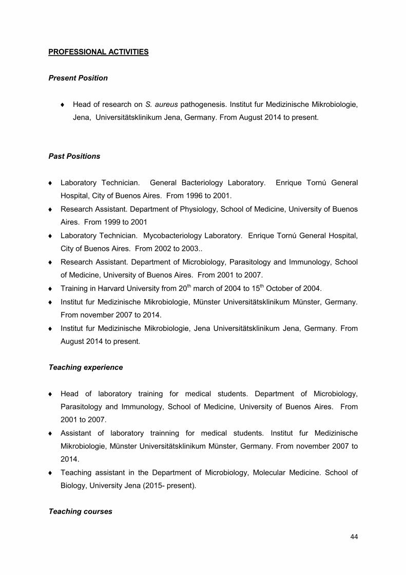
44
PROFESSIONAL ACTIVITIES
Present Position
Head of research on S. aureus pathogenesis. Institut fur Medizinische Mikrobiologie,
Jena, Universitätsklinikum Jena, Germany. From August 2014 to present.
Past Positions
Laboratory Technician. General Bacteriology Laboratory. Enrique Tornú General
Hospital, City of Buenos Aires. From 1996 to 2001.
Research Assistant. Department of Physiology, School of Medicine, University of Buenos
Aires. From 1999 to 2001
Laboratory Technician. Mycobacteriology Laboratory. Enrique Tornú General Hospital,
City of Buenos Aires. From 2002 to 2003..
Research Assistant. Department of Microbiology, Parasitology and Immunology, School
of Medicine, University of Buenos Aires. From 2001 to 2007.
Training in Harvard University from 20th march of 2004 to 15th October of 2004.
Institut fur Medizinische Mikrobiologie, Münster Universitätsklinikum Münster, Germany.
From november 2007 to 2014.
Institut fur Medizinische Mikrobiologie, Jena Universitätsklinikum Jena, Germany. From
August 2014 to present.
Teaching experience
Head of laboratory training for medical students. Department of Microbiology,
Parasitology and Immunology, School of Medicine, University of Buenos Aires. From
2001 to 2007.
Assistant of laboratory trainning for medical students. Institut fur Medizinische
Mikrobiologie, Münster Universitätsklinikum Münster, Germany. From november 2007 to
2014.
Teaching assistant in the Department of Microbiology, Molecular Medicine. School of
Biology, University Jena (2015- present).
Teaching courses

45
26-06-2015 Präsentieren mit Prezi-nicht ohne eine Metapher, Friedrich-Schiller-Universität
Jena
03-07-2015 Teaching in English, Friedrich-Schiller-Universität Jena
22-07-2015 Lehrqualifikation Basic, Friedrich-Schiller-Universität Jena
Experience in research training and role as thesis advisor assistance
2010-2013 Julia Kalinka, phD: “Host-pathogen interactions in S. aureus osteomyelitis”
2011-2015 Vijaya Mysore, phD: “Staphylococcus aureus SCVs use intracellular
persistence in human macrophages as a strategy evade the innate immune
response”
2012-2013 Christine Muth, Bachelor : “Competition between wt and different mutant
on general regulators from S. aureus (sigB, agr, SarA)”
2012-2013 Elvira Vögel, Master : “Untersuchung zum Einfluss der Stringenten
Antwort auf die intrazelluläre Persistenz von Staphylococcus aureus“
2012 Dr. Caroline Kreis, medical research training: “Staphylococcus aureus develops
increased resistance to antibiotics by forming dynamic small colony variants during
chronic osteomyelitis”
2012 Lisa Flint, phD: “SCV-Induktion durch die Gabe verschiedener Antibiotika als
Ursache für die Entwicklung chronischer und Therapie-refraktärer S.aureus-
Infektionen”
2012 Sandra Diekmann, Bachelor: “Influence of tryptophan on SCV formation in S.
aureus”
2013-2014 Anika Blümke, Bachelor : “Phäno- und genotypische
Adaptionsmechanismen in Staphylococcus aureus Osteomyelitis”
2012-2014 Hannah F. Preugschas, Bachelor : “Labeling S. aureus with iron oxide
nano particles with rhodamine”
2013 Marie Hachmeister, Master: “Studien zu Infektionsstrategien von
Staphylococcus aureus in einem chronischen hämatogenen Osteomyelitismodell in
der Maus“
2013-2016 Jennifer Geraci, pHD: “ The role of adhesins and biofilm formation in
chronic Staphylococcus aureus infections”
2015 Rebekka Salzmann, Bachelor: “Test of new potential antimicrobial drugs
against S.aureus: BM-38”
2015-2018 Anke Siegmund, pHD: “Detecting and deciphering Staphylococcus aureus
strategies that cause chronic and persistent infections” (Transregio)
2015-2018 Christine Pöllath, pHD: “Staphylococcus aureus pathogenesis: from

46
sepsis to hematogenous chronic bone infections” (Staphbone, CSCC)
2015-2016 Christian Fritzsch, Master: “Persistence of Staphylococcus aureus within
osteocytes and osteoblasts”
2016-2019 Vandana Pradeep, pHD: “Metabolic cross talk between host and S.
aureus during chronic osteomyelitis” (ILRS, microbial communications)
2016-2019 Marina García-Moreno, pHD: “Staphylococcus aureus pathogenesis: The
role of different host cell types during the passage from sepsis to chronic
osteomyelitis” (CSCC)
FELLOWSHIPS ,AWARDS and Grants
2000-2001 University of Buenos aires Research Fellowship
2001-2002 Carrillo-Oñativia Research Fellowship, Health Department, Argentina
2003-2006 University of Buenos Aires Doctoral Fellowship
2004 International Union for Microbiological Societies. To visit Dr. jean C. Lee's
laboratory, The Chaning Laboratory, Brigham and Women's Hospital
Harvard University
2007-2009 CONICET Internal Post-doctoral Fellowship
2007-2010 2007-2010 Skin staph Post doc position
2010-2012 2010-2012 Transregio/IZKF
2012-2024 SFB
2012 IMF
2016-2019 ILRS, Microbial Communications, HKI
2016-2019 CSCC, Center of sepsis, Jena
Jena, 22nd July , 2016

47
8- Complete list of publications:
The following list of publications is ordered chronologically. The corresponding author is
indicated (*). The impact factor (IF) is from the year of publication. In all the publications from
2016 where the IF is still not available, the IF from 2015 was inidcated. The weighted impact
factor (gIF) was calculated taking into account the impact factor of journal in the year of
publication of the work and the average impact factor for present year (2016 or2015).
Information on the average impact factor was found on the website of the isi web of science.
1: Goldmann O, Tuchscherr L, Rohde M, Medina E. α-Hemolysin enhances Staphylococcus
aureus internalization and survival within mast cells by modulating the expression of β1
integrin. Cell Microbiol. 2016 Jun;18(6):807-19. (IF2014 4.915; gIF2014 1.50)
2: Tuchscherr L, Kreis CA, Hoerr V, Flint L, Hachmeister M, Geraci J, Bremer-Streck S,
Kiehntopf M, Medina E, Kribus M, Raschke M, Pletz M, Peters G, Löffler B. Staphylococcus
aureus develops increased resistance to antibiotics by forming dynamic small colony
variants during chronic osteomyelitis. J Antimicrob Chemother. 2016 Feb;71(2):438-48.
(IF2014 5.313; gIF2014 2.06)
3: Tuchscherr L, Löffler B. Staphylococcus aureus dynamically adapts global regulators and
virulence factor expression in the course from acute to chronic infection. Curr Genet. 2016
Feb;62(1):15-7. (IF2014 6,723; gIF2014 3.04)
4: Grüner M, Tuchscherr L, Löffler B, Gonnissen D, Riehemann K, Staniford MC, Kynast U,
Strassert CA. Selective Inactivation of Resistant Gram-Positive Pathogens with a Light-
Driven Hybrid Nanomaterial. ACS Appl Mater Interfaces. 2015 Sep 23;7(37):20965-71. (IF2014
2.682; gIF2014 1.09)
5: Tuchscherr L, Bischoff M, Lattar SM, Noto Llana M, Pförtner H, Niemann S, Geraci J,
Van de Vyver H, Fraunholz MJ, Cheung AL, Herrmann M, Völker U, Sordelli DO, Peters G,
Löffler B. Sigma Factor SigB Is Crucial to Mediate Staphylococcus aureus Adaptation during
Chronic Infections. PLoS Pathog. 2015 Apr 29;11(4):e1004870. (IF2014 7.562; gIF2014 3.01)
6: Klinger-Strobel M, Lautenschläger C, Fischer D, Mainz JG, Bruns T, Tuchscherr L, Pletz
MW, Makarewicz O. Aspects of pulmonary drug delivery strategies for infections in cystic
fibrosis--where do we stand? Expert Opin Drug Deliv. 2015 Aug;12(8):1351-74. (IF2014 4.84;
gIF2014 2.05)

48
7: Ring J, Hoerr V, Tuchscherr L, Kuhlmann MT, Löffler B, Faber C. MRI visualization of
Staphyloccocus aureus-induced infective endocarditis in mice. PLoS One. 2014 Sep
17;9(9):e107179. (IF2014 3.234; gIF2014 4.41)
8: Kalinka J, Hachmeister M, Geraci J, Sordelli D, Hansen U, Niemann S, Oetermann S,
Peters G, Löffler B, Tuchscherr L. Staphylococcus aureus isolates from chronic
osteomyelitis are characterized by high host cell invasion and intracellular adaptation, but still
induce inflammation. Int J Med Microbiol. 2014 Nov;304(8):1038-49. (IF2014 3.614; gIF2014
1.44)
9: Löffler B, Tuchscherr L, Niemann S, Peters G. Staphylococcus aureus persistence in
non-professional phagocytes. Int J Med Microbiol. 2014 Mar;304(2):170-6. (IF2014 3.614;
gIF2014 1.44)
10: Kneidl J, Mysore V, Geraci J, Tuchscherr L, Löffler B, Holzinger D, Roth J, Barczyk-
Kahlert K. Soluble CD163 masks fibronectin-binding protein A-mediated inflammatory
activation of Staphylococcus aureus infected monocytes. Cell Microbiol. 2014 Mar;16(3):364-
77. (IF2014 4.915; gIF2014 1.50)
11: Hoerr V, Tuchscherr L, Hüve J, Nippe N, Loser K, Glyvuk N, Tsytsyura Y, Holtkamp M,
Sunderkötter C, Karst U, Klingauf J, Peters G, Löffler B, Faber C. Bacteria tracking by in vivo
magnetic resonance imaging. BMC Biol. 2013 May 28;11:63. (IF2013 7.431; gIF2013 5.19)
12: Horst SA, Hoerr V, Beineke A, Kreis C, Tuchscherr L, Kalinka J, Lehne S, Schleicher I,
Köhler G, Fuchs T, Raschke MJ, Rohde M, Peters G, Faber C, Löffler B, Medina E. A novel
mouse model of Staphylococcus aureus chronic osteomyelitis that closely mimics the human
infection: an integrated view of disease pathogenesis. Am J Pathol. 2012 Oct;181(4):1206-
14. (IF2012 4.410; gIF2012 2.263)
13: Niemann S, Ehrhardt C, Medina E, Warnking K, Tuchscherr L, Heitmann V, Ludwig S,
Peters G, Löffler B. Combined action of influenza virus and Staphylococcus aureus panton-
valentine leukocidin provokes severe lung epithelium damage. J Infect Dis. 2012 Oct
1;206(7):1138-48. (IF201 5.848; gIF2012 2.02)
14: Lattar SM, Tuchscherr LP, Centrón D, Becker K, Predari SC, Buzzola FR, Robinson DA,
Sordelli DO. Molecular fingerprinting of Staphylococcus aureus isolated from patients with
osteomyelitis in Argentina and clonal distribution of the cap5(8) genes and of other selected
virulence genes. Eur J Clin Microbiol Infect Dis. 2012 Oct;31(10):2559-66. (IF2012 3.024;
gIF2012 1.26)

49
15: Barbagelata MS, Alvarez L, Gordiola M, Tuchscherr L, von Eiff C, Becker K, Sordelli D,
Buzzola F. Auxotrophic mutant of Staphylococcus aureus interferes with nasal colonization
by the wild type. Microbes Infect. 2011 Nov;13(12-13):1081-90. (IF2011 3.101; gIF2011 1.04)
16: Tuchscherr L, Medina E, Hussain M, Völker W, Heitmann V, Niemann S, Holzinger D,
Roth J, Proctor RA, Becker K, Peters G, Löffler B. Staphylococcus aureus phenotype
switching: an effective bacterial strategy to escape host immune response and establish a
chronic infection. EMBO Mol Med. 2011 Mar;3(3):129-41. (IF2011 10.333; gIF2011 4.64)
17: Tuchscherr L, Löffler B, Buzzola FR, Sordelli DO. Staphylococcus aureus adaptation to
the host and persistence: role of loss of capsular polysaccharide expression. Future
Microbiol. 2010 Dec;5(12):1823-32. (IF2010 2.755; gIF2010 1.16)
18: Tuchscherr L, Heitmann V, Hussain M, Viemann D, Roth J, von Eiff C, Peters G, Becker
K, Löffler B. Staphylococcus aureus small-colony variants are adapted phenotypes for
intracellular persistence. J Infect Dis. 2010 Oct 1;202(7):1031-40. (IF2010 5.961; gIF2010 2.09)
19: Grundmeier M, Tuchscherr L, Brück M, Viemann D, Roth J, Willscher E, Becker K,
Peters G, Löffler B. Staphylococcal strains vary greatly in their ability to induce an
inflammatory response in endothelial cells. J Infect Dis. 2010 Mar 15;201(6):871-80. (IF2010
5.961; gIF2010 2.09)
20: Pardo L, Machado V, Mollerach M, Mota MI, Tuchscherr LP, Gadea P, Gardella N,
Sordelli DO, Vola M, Schelotto F, Varela G. Characteristics of Community-Associated
Methicillin-Resistant Staphylococcus aureus (CA-MRSA) Strains Isolated from Skin and Soft-
Tissue Infections in Uruguay. Int J Microbiol. 2009;2009:472126. (IF2009 N/A; gIF2009 N/A)
21: Lattar SM, Tuchscherr LP, Caccuri RL, Centrón D, Becker K, Alonso CA, Barberis C,
Miranda G, Buzzola FR, von Eiff C, Sordelli DO. Capsule expression and genotypic
differences among Staphylococcus aureus isolates from patients with chronic or acute
osteomyelitis. Infect Immun. 2009 May;77(5):1968-75. (IF2009 4.205; gIF2009 1.53)
22: Tuchscherr LP, Buzzola FR, Alvarez LP, Lee JC, Sordelli DO. Antibodies to capsular
polysaccharide and clumping factor A prevent mastitis and the emergence of
unencapsulated and small-colony variants of Staphylococcus aureus in mice. Infect Immun.
2008 Dec;76(12):5738-44. (IF2008 3.451; gIF2008 1.24)
50
23: Tuchscherr LP, Gomez MI, Buzzola FR, Calvinho LF, Lee JC, Sordelli DO.
Characterization of a new variant of IS257 that has displaced the capsule genes within
bovine isolates of Staphylococcus aureus. Infect Immun. 2007 Nov;75(11):5483-8. (IF2007
3.484; gIF2007 1.34)
24: Buzzola FR, Alvarez LP, Tuchscherr LP, Barbagelata MS, Lattar SM, Calvinho L,
Sordelli DO. Differential abilities of capsulated and noncapsulated Staphylococcus aureus
isolates from diverse agr groups to invade mammary epithelial cells. Infect Immun. 2007
Feb;75(2):886-91. (IF2007 3.484; gIF2007 1.34)
25: Tuchscherr LP, Buzzola FR, Alvarez LP, Caccuri RL, Lee JC, Sordelli DO. Capsule-
negative Staphylococcus aureus induces chronic experimental mastitis in mice. Infect
Immun. 2005 Dec;73(12):7932-7. (IF2005 3.225; gIF2005 1.40)
26: Villar HE, Tuchscherr L, Arena M, Longo L, Laurino G, Hoffman M. [Inhibitory and
bactericidal activity and selection capacity of mutants resistant and tolerant to vancomycin in
Staphylococcus aureus strains]. Enferm Infecc Microbiol Clin. 1999 Nov;17(9):454-7. (IF1999
N/A; gIF1999 N/A)
9- Oral presentations:
“Staphylococcus aureus long-term persistence and the relation with chronic infections” 3rd.
Immunology meeting, Münster, 2012.
“How can we study staphylococcal infection?” IZKF Münster, Annual Meeting Gut
Havichhorst, Münster , 2012
51
10- Acknowledgements
I would like to thank Prof. B. Löffler and Prof. D. Sordelli, my research supervisors, for their
patient guidance, valuable support, enthusiastic encouragement and useful critiques of this
research work.
I would also like to thank Professor Peters for giving me the opportunity to start my research
in Germany. My grateful thanks are also extended to Mrs. S. Soldavini, Mrs. L. Medina, Mrs
B. Schuhen, Mrs. M. Brück, Mrs. K. Broschwig and Mrs. C. Berling for their technical
excellent assistance. I also would like to thank all people who helped me to correct and
handle this work: Mrs. M. Meyer and Mrs. S. Schramm.
I also would like to thank all the students and colleges who collaborated during the process
of the experiments and shared with me all the moments: Dr. C. Cerquetti, Dr. M.
Giacomodonato, Dr. S. Sarnacki, Dr. A. Noto Llana, Dr. S. Ramírez, Dr. M. Gómez, Dr. S.
Lattar, Dr. F. Buzzola, Dr. MS. Barbagelata, Mrs. J. Geraci, Mrs. M. Hachmeister, Mrs. H.
Van de Vyver Dr S. Niemann, Mrs. A. Siegmund and Dr V. Hoerr. My special thanks are
extended to the staff of the Institute of medical microbiology from Buenos Aires, Münster and
Jena.
Finally, I wish to thank specially my mother, Luis, Gabriel, Matias, my grandmother, Deby
and Roger, Mathilde, Margarete, Uschi, Walter, Christian, Monique, Michael, Julia, Ruth,
Bernd, Maren, Niklas, Wiebke, Henning and friends for their support and encouragement
throughout my study. I also would like to thank my husband Heinz and my children, Paula
and Tobias, for their patient and supporting me during this journey.

52
11- Copies of original publications used for this work
The publications are ordered chronologically from the first to the last one. The impact factors
are described on the section 8.