The sRNA RyhB Regulates the Synthesis of the Escherichia ...
14
The sRNA RyhB Regulates the Synthesis of the Escherichia coli Methionine Sulfoxide Reductase MsrB but Not MsrA Julia Bos 1¤a , Yohann Duverger 1 , Benoıˆt Thouvenot 2¤b , Claude Chiaruttini 3 , Christiane Branlant 2 , Mathias Springer 3 , Bruno Charpentier 2 , Fre ´ de ´ ric Barras 1 * 1 Laboratoire de Chimie Bacte ´rienne, Institut de Microbiologie de la Me ´diterrane ´e, Centre National de la Recherche Scientifique-Aix Marseille Universite ´, Unite ´ Mixte de Recherche, Marseille, France, 2 Centre National de la Recherche Scientifique-Universite ´ de Lorraine, Unite ´ Mixte de Recherche, Biopo ˆ le de l’Universite ´ de Lorraine, Campus Biologie Sante ´, Vandœuvre-le ` s-Nancy, France, 3 Unite ´ Propre de Recherche du Centre National de la Recherche Scientifique, Universite ´ Denis Diderot, Paris VII, Institut de Biologie Physico-chimique, Paris, France Abstract Controlling iron homeostasis is crucial for all aerobically grown living cells that are exposed to oxidative damage by reactive oxygen species (ROS), as free iron increases the production of ROS. Methionine sulfoxide reductases (Msr) are key enzymes in repairing ROS-mediated damage to proteins, as they reduce oxidized methionine (MetSO) residues to methionine. E. coli synthesizes two Msr, A and B, which exhibit substrate diastereospecificity. The bacterial iron-responsive small RNA (sRNA) RyhB controls iron metabolism by modulating intracellular iron usage. We show in this paper that RyhB is a direct regulator of the msrB gene that encodes the MsrB enzyme. RyhB down-regulates msrB transcripts along with Hfq and RNaseE proteins since mutations in the ryhB, fur, hfq, or RNaseE-encoded genes resulted in iron-insensitive expression of msrB. Our results show that RyhB binds to two sequences within the short 59UTR of msrB mRNA as identified by reverse transcriptase and RNase and lead (II) protection assays. Toeprinting analysis shows that RyhB pairing to msrB mRNA prevents efficient ribosome binding and thereby inhibits translation initiation. In vivo site directed-mutagenesis experiments in the msrB 59UTR region indicate that both RyhB-pairing sites are required to decrease msrB expression. Thus, this study suggests a novel mechanism of translational regulation where a same sRNA can basepair to two different locations within the same mRNA species. In contrast, expression of msrA is not influenced by changes in iron levels. Citation: Bos J, Duverger Y, Thouvenot B, Chiaruttini C, Branlant C, et al. (2013) The sRNA RyhB Regulates the Synthesis of the Escherichia coli Methionine Sulfoxide Reductase MsrB but Not MsrA. PLoS ONE 8(5): e63647. doi:10.1371/journal.pone.0063647 Editor: Eric Cascales, CNRS UPR9043, France Received November 12, 2012; Accepted April 4, 2013; Published May 9, 2013 Copyright: ß 2013 Bos et al. This is an open-access article distributed under the terms of the Creative Commons Attribution License, which permits unrestricted use, distribution, and reproduction in any medium, provided the original author and source are credited. Funding: This work was made possible by grants from the Centre National de la Recherche Scientifique, the French National Research Agency and Aix-Marseille University. The funders had no role in study design, data collection and analysis, decision to publish, or preparation of the manuscript." Competing Interests: The authors have declared that no competing interests exist. * E-mail: [email protected] ¤a Current address: Department of Molecular Biology, Princeton University, Princeton, New Jersey, United States of America ¤b Current address: Laboratoire MTM/Genclis, Vandœuvre-le `s Nancy, France Introduction Reactive oxygen species (ROS) can damage most macromole- cules, proteins, nucleic acids and lipids [1]. Within proteins, sulfur- containing amino acids, cysteine and methionine (Met) exhibit high sensitivity [2]. In particular, methionine oxidation yields methionine sulfoxide (Met-SO) and eventually methionine sulfone. Like any other oxidative modification, methionine oxidation can have deleterious consequences as it can be accompanied by protein carbonylation, protein aggregation and/or degradation [3,4,5]. However, the oxidation of Met to Met-SO can be reversed by the action of methionine sulfoxide reductases (Msr) [6,7,8,9,10]. Such an ability to reverse methionine oxidation has led credence to the idea that the surface-exposed Met residues act as scavengers for ROS and that reduction by Msr enables proteins to recover activity [11]. Msr proteins are highly conserved among most living organisms [12,13]. There are basically two types, referred to as MsrA and B, which act on two different diastereoisomers, Met-S-SO and Met- R-SO, respectively [8,10,14]. Lack of functional MsrA and/or B have been reported to cause abnormal cellular function in a wide range of organisms from bacteria to humans, including yeast, mice, flies and plants [7,15,16,17,18,19,20,21]. Likewise neuro- logical disorders, shortened life span, and various oxidative stress- related defects have been reported in eukaryotes having reduced Msr activity. As a general trend, bacteria lacking Msr exhibit hypersensitivity to ROS and those that are pathogens have a reduced ability to infect their host [22,23,24,25,26,27,28,29]. Small non-coding RNAs are believed to play a major role in genome expression both in prokaryotes and eukaryotes. The E. coli iron-responsive sRNA RyhB that controls iron metabolism is one of the best studied. Early microarray approaches suggested that RyhB controls directly the expression of about 50 genes in response to iron limitation [30]. RyhB expression is itself negatively regulated by the iron-sensing Ferric uptake regulator (Fur) [31]. Hence, when iron becomes limiting, Fur repression is alleviated, RyhB synthesis is induced and expression of its targets inhibited. Genes targeted by RyhB encode iron-storage and iron- PLOS ONE | www.plosone.org 1 May 2013 | Volume 8 | Issue 5 | e63647
Transcript of The sRNA RyhB Regulates the Synthesis of the Escherichia ...

The sRNA RyhB Regulates the Synthesis of theEscherichia coli Methionine Sulfoxide Reductase MsrBbut Not MsrAJulia Bos1¤a, Yohann Duverger1, Benoıt Thouvenot2¤b, Claude Chiaruttini3, Christiane Branlant2,
Mathias Springer3, Bruno Charpentier2, Frederic Barras1*
1 Laboratoire de Chimie Bacterienne, Institut de Microbiologie de la Mediterranee, Centre National de la Recherche Scientifique-Aix Marseille Universite, Unite Mixte de
Recherche, Marseille, France, 2 Centre National de la Recherche Scientifique-Universite de Lorraine, Unite Mixte de Recherche, Biopole de l’Universite de Lorraine, Campus
Biologie Sante, Vandœuvre-les-Nancy, France, 3 Unite Propre de Recherche du Centre National de la Recherche Scientifique, Universite Denis Diderot, Paris VII, Institut de
Biologie Physico-chimique, Paris, France
Abstract
Controlling iron homeostasis is crucial for all aerobically grown living cells that are exposed to oxidative damage by reactiveoxygen species (ROS), as free iron increases the production of ROS. Methionine sulfoxide reductases (Msr) are key enzymesin repairing ROS-mediated damage to proteins, as they reduce oxidized methionine (MetSO) residues to methionine. E. colisynthesizes two Msr, A and B, which exhibit substrate diastereospecificity. The bacterial iron-responsive small RNA (sRNA)RyhB controls iron metabolism by modulating intracellular iron usage. We show in this paper that RyhB is a direct regulatorof the msrB gene that encodes the MsrB enzyme. RyhB down-regulates msrB transcripts along with Hfq and RNaseE proteinssince mutations in the ryhB, fur, hfq, or RNaseE-encoded genes resulted in iron-insensitive expression of msrB. Our resultsshow that RyhB binds to two sequences within the short 59UTR of msrB mRNA as identified by reverse transcriptase andRNase and lead (II) protection assays. Toeprinting analysis shows that RyhB pairing to msrB mRNA prevents efficientribosome binding and thereby inhibits translation initiation. In vivo site directed-mutagenesis experiments in the msrB59UTR region indicate that both RyhB-pairing sites are required to decrease msrB expression. Thus, this study suggests anovel mechanism of translational regulation where a same sRNA can basepair to two different locations within the samemRNA species. In contrast, expression of msrA is not influenced by changes in iron levels.
Citation: Bos J, Duverger Y, Thouvenot B, Chiaruttini C, Branlant C, et al. (2013) The sRNA RyhB Regulates the Synthesis of the Escherichia coli MethionineSulfoxide Reductase MsrB but Not MsrA. PLoS ONE 8(5): e63647. doi:10.1371/journal.pone.0063647
Editor: Eric Cascales, CNRS UPR9043, France
Received November 12, 2012; Accepted April 4, 2013; Published May 9, 2013
Copyright: � 2013 Bos et al. This is an open-access article distributed under the terms of the Creative Commons Attribution License, which permits unrestricteduse, distribution, and reproduction in any medium, provided the original author and source are credited.
Funding: This work was made possible by grants from the Centre National de la Recherche Scientifique, the French National Research Agency and Aix-MarseilleUniversity. The funders had no role in study design, data collection and analysis, decision to publish, or preparation of the manuscript."
Competing Interests: The authors have declared that no competing interests exist.
* E-mail: [email protected]
¤a Current address: Department of Molecular Biology, Princeton University, Princeton, New Jersey, United States of America¤b Current address: Laboratoire MTM/Genclis, Vandœuvre-les Nancy, France
Introduction
Reactive oxygen species (ROS) can damage most macromole-
cules, proteins, nucleic acids and lipids [1]. Within proteins, sulfur-
containing amino acids, cysteine and methionine (Met) exhibit
high sensitivity [2]. In particular, methionine oxidation yields
methionine sulfoxide (Met-SO) and eventually methionine sulfone.
Like any other oxidative modification, methionine oxidation can
have deleterious consequences as it can be accompanied by
protein carbonylation, protein aggregation and/or degradation
[3,4,5]. However, the oxidation of Met to Met-SO can be reversed
by the action of methionine sulfoxide reductases (Msr) [6,7,8,9,10].
Such an ability to reverse methionine oxidation has led credence
to the idea that the surface-exposed Met residues act as scavengers
for ROS and that reduction by Msr enables proteins to recover
activity [11].
Msr proteins are highly conserved among most living organisms
[12,13]. There are basically two types, referred to as MsrA and B,
which act on two different diastereoisomers, Met-S-SO and Met-
R-SO, respectively [8,10,14]. Lack of functional MsrA and/or B
have been reported to cause abnormal cellular function in a wide
range of organisms from bacteria to humans, including yeast,
mice, flies and plants [7,15,16,17,18,19,20,21]. Likewise neuro-
logical disorders, shortened life span, and various oxidative stress-
related defects have been reported in eukaryotes having reduced
Msr activity. As a general trend, bacteria lacking Msr exhibit
hypersensitivity to ROS and those that are pathogens have a
reduced ability to infect their host [22,23,24,25,26,27,28,29].
Small non-coding RNAs are believed to play a major role in
genome expression both in prokaryotes and eukaryotes. The E. coli
iron-responsive sRNA RyhB that controls iron metabolism is one
of the best studied. Early microarray approaches suggested that
RyhB controls directly the expression of about 50 genes in
response to iron limitation [30]. RyhB expression is itself
negatively regulated by the iron-sensing Ferric uptake regulator
(Fur) [31]. Hence, when iron becomes limiting, Fur repression is
alleviated, RyhB synthesis is induced and expression of its targets
inhibited. Genes targeted by RyhB encode iron-storage and iron-
PLOS ONE | www.plosone.org 1 May 2013 | Volume 8 | Issue 5 | e63647

using proteins and it was proposed that RyhB enables iron sparing
for central metabolism and for essential iron-binding proteins
when iron concentration becomes scarce [30,31,32]. Therefore,
synthesis of a non-essential-iron-using protein would be decreased
and the cell would rather employ an iron-non-using functional
homolog. An illustration of this model is provided by the SodA
and SodB superoxide dismutases. The synthesis of the iron
superoxide dismutase SodB is decreased by RyhB under iron
limitation, whereas the synthesis of SodA, a Mn-superoxide
dismutase is not [30,33]. These iron-responsive regulators, RyhB
and Fur also play a crucial role to reduce the response to oxidative
stress, as free iron is susceptible to react with ROS such as
superoxide anions and to increase ROS production via the Fenton
and Haber Weiss reactions [34]. RyhB regulates gene expression
by basepairing within or near the translation initiation region of
mRNAs. Two mechanisms of action have been proposed for RyhB
to explain its role as a gene expression regulator. In a first
mechanism, the pairing of RyhB to its target mRNA interferes
either positively or negatively with the 30S ribosome subunit
binding and thereby translation [31,35,36]. Alternatively, the
pairing of the sRNA RyhB to the target mRNA can initiate
mRNA degradation even in the absence of translation on the
mRNA target [37].
Early microarray analysis suggested that RyhB levels influence
the expression of msrB but not that of msrA [30]. In addition, MsrB
has been biochemically characterized as an iron binding protein
[38]. Therefore we wished to test the functional and direct
inactivation of the E. coli msrB mRNA by RyhB in vitro and in vivo,
and whether the cell switches to msrA under iron limitation. We
provide evidence that RyhB directly pairs with the 59UTR region
of msrB at two distinct sites and does repress msrB expression by
competing with the ribosome. Secondly, we showed that msrA
expression is not sensitive to changes in iron availability. Thus, in
addition to identifying a new target for the sRNA RyhB and
pointing out a novel mechanism of translational regulation, this
study indicates that in E. coli MsrB is dispensable when iron supply
is low but MsrA is retained to repair even limited methionine
oxidation.
Materials and Methods
Media and growth conditionsDerivatives of E. coli MG1655 strain were used in all
experiments and were cultivated under aerobic conditions at
37uC in Luria Bertani (LB) medium unless stated otherwise.
Plasmids were maintained with ampicillin used at a final
concentration of 100 mg/ml.
Plasmid constructionTranscriptional and/or translational fusions of msrB and msrA
genes to lacZ reporter gene were constructed by inserting msrB
fragment (from 2336 to +360 relative to msrB start codon) or msrA
fragment (from 2416 to +132 relative to msrA start codon) into
plasmids pRS415 (transcriptional fusion) and pRS414 (transla-
tional fusion)[39]. For information, pRS415 contains the lacZ
ORF including the lacZ translation initiation region (TIR) whereas
pRS414 contains the lacZ ORF without the lacZ TIR (‘lacZ). msrB
and msrA genes were amplified from MG1655 chromosomal DNA
with the oligonucleotides B01E/B05B and A01E/A04B (see
Table 1) respectively. The resulting products were digested with
EcoRI and BamHI and ligated into EcoRI/BamHI-digested
pRS415 and pRS414 plasmids, to generate the transcriptional
and translational fusions to lacZ gene. The msrB’-lacZ, msrB’-‘lacZ
and msrA’-‘lacZ constructs were further engineered onto lRS45
phage and integrated into the attachment site (att site) of
IBPC5321 strain as previously described [39], generating strains
JB43 (transcriptional msrB’-lacZ), JB44 (translational msrB’-‘lacZ)
and JB56 (translational msrA’-‘lacZ). Stable lysogens were screened
for single insertion of recombinant by PCR [40] and sequenced
using LacZinv primer (see Table 1). We next introduced the
following Dfur ::kan and DryhB ::cat deletions by P1 transduction in
JB44 generating strains JB46 and JB50 respectively and in JB56 to
generate YD100.
The construct pUC-MsrB was generated by PCR using
chromosomal DNA from MG1655 wild type strain as a template
and primers B01E and B05N (see Table 1). The resulting product
was digested wih EcoRI and NdeI and cloned into the correspond-
ing sites of pUC18. The construct was sequenced using the primer
pUCVERI and transformed into MG1655 wild type strain. The
construct pUC-MsrB-HA has been generated using the same
protocol to that of pUC-MsrB construct. The HA tag was added to
the 3’ end of msrB using the reverse oligonucleotide BHA (see
Table 1). IPTG (0.5 mM) was added to the culture to induce the
synthesis of MsrB and MsrB-HA. Strains used in this study are
listed in Table 2.
Site-directed mutagenesisSite-directed mutagenesis was carried out by PCR using pUC-
MsrB or pUC-MsrB-HA as DNA template. Mutations mut1,
mut2a, and mut2b are introduced into the 5’UTR region of msrB
with the use of primers Bm1for-Bm1rev, Bm2afor-Bm2arev,
Bm2bfor-Bm2brev respectively, containing the suitable nucleotide
mismatches (see Table 1). The amplified plasmid products were
then digested with DpnI restriction enzyme for 2 hours at 37uC to
select for mutation-containing synthesized DNA and 1 ml aliquot
was transformed into DH5 alpha competent cells. Transformation
reactions were plated on LB containing ampicillin. Five colonies
were selected and the positive clones were screened by digestion
and sequenced. Plasmids carrying wild type or mutant alleles were
introduced into DmsrB strain (SMG505). Transformants were used
for Northern blot and Western blot experiments.
Western blottingBacteria cells carrying pUC-MsrB-HA, pUC-MsrBmut2a-HA
and pUC-MsrBmut2b-HA were cultivated in LB medium to
exponential phase. Iron chelator (2–29 dipyridyl) was added for
30 min and 50 ml sample aliquots were collected. French Press
lysates were spun down for 30 min at 13500 rpm. Supernatants
were collected and equal amounts of cellular extracts were
western-blotted. To detect MsrB-HA protein, the membrane was
probed with a mouse anti-HA primary antibody, and with a goat
anti mouse IgG alexa fluor 680 as secondary antibody and
scanned by using an Odyssey IR imaging scanner. Band intensity
was analyzed by using Image J software.
DNA probe labelingRadiolabeled probes used in the Northern blots were generated
from PCR products. msrB, RyhB and 5S PCR products were
generated using the pair of oligonucleotides B1–B4, REco-Rbam
and 5S1-5S2 respectively (see Table 1). After gel extraction and
purification, the PCR products were then used as a template for
[a-32P]dCTP-labeling reaction (Ready-to-go DNA labeling kit,
Amersham). Unincorporated nucleotides were removed by using a
G-50 resin minicolumn. For primer extension assays and toeprint
assays, the 59 end of oligonucleotides 437 and 1451 respectively,
was labeled with [c-32P]ATP (50 mCi) using T4 polynucleotide
kinase (10 U).
Methionine Oxidation and Iron Homeostasis
PLOS ONE | www.plosone.org 2 May 2013 | Volume 8 | Issue 5 | e63647

RNA isolation and Northern blot analysisTotal RNA was isolated from cells at the indicated times using
the SV total RNA isolation system kit as described by the
manufacturer (Promega). For Northern blot analysis, total RNA
(10 mg) was separated on a 1.2% TBE-agarose gel and transferred
to a Hybond N+ nylon membrane. Membranes were UV-
crosslinked and hybridized for 4 hours at 58uC with the suitable
radiolabeled probe. Membranes were washed three times in
0.05%SDS/2X SSC buffer and then twice in 0.1%SDS/0.1X
SSC buffer before being scanned by using a phophorimager
(Molecular Dynamics). Band intensity was analyzed by using
Image J software.
Primer extension assaysHybridization reaction between of 59-end labeled oligonucleo-
tide 437 (4 ng) and purified msrB RNA transcript (100 ng) was
performed in a final volume of 2.5 ml, in the 1X binding buffer
(Tris HCl 10 mM, pH8, MgCl2 10 mM, NaCl 50 mM, KCl
50 mM) for 10 min at 65uC. RyhB sRNA (5 mg) was added to the
reaction mix (7.5 ml) for 10 min at 37uC. The reverse transcription
reaction was performed for 30 min at 42uC by adding 5 ml of the
AMV RT enzyme and dNTPs to the reaction mix. The resulting
cDNA reaction mix was loaded onto a 6% acrylamide/
bisacrylamide denaturing gel along with the products of
dideoxy-sequencing reactions obtained with the same labeled
primer. Signal intensities were evaluated using a phosphorimager
(Molecular Dynamics).
Toeprinting assaysToeprint experiments were carried out using purified 30S
ribosomal subunits (isolated from D10 E. coli strain as described in
[41]), tRNAfMet and AMV reverse transcriptase to detect the
formation of ternary initiation complexes on the long msrB
transcript (450 nt). In a typical experiment, the msrB RNA
transcript (50 fmol) was mixed to a fourfold molar excess of 59-
labeled 437 oligonucleotide primer (complementary to positions
+60 to +73 of msrB). Toeprint reactions were performed as
described in [42]. The toeprint band intensity was analyzed by
using Image J software and normalized to that of full-length
transcript. Data are represented as means 6S.E. from two
independent experiments.
Half-life determinationOvernight cultures were subcultured in LB medium (200 ml) at
37uC to an O.D.600 of 0.3. Each culture was then divided in 2 and
one culture was treated with 2–29 dipyridyl (250 mM) for 15 min at
37uC. Samples of 5 ml started to be collected at different times (0,
2, 4, 6, 8, 10, 12, 15 and 30 min) right after the addition of
Rifampicin (0.3 mg/ml) and were immediately frozen in liquid
nitrogen. Total RNA was extracted from each sample and 10 mg
of total RNA were subjected to a Northern blot analysis using the
specific msrB and 5S probes. Signal intensities were evaluated using
a phosphorimager (Fuji FLA 5100) and quantitated by using
Image J software. msrB signals were normalized to that of 5S RNA.
Table 1. List of oligonucleotides used in this study.
Name Sequence
LacZinv ATCGGTGCGGGCCTCTTC
BO1E CGCGAATTCCCACCAGCTATTTGTTAGTG
BO5B CGCGGATCCGGGTTAACACAATAACGTTCGCCCGTTGG
BO5N GGGCATATGTCAACCGTTGATTTCTTCGCCGTT
AO1E CGCGAATTCCCTTTCAGACCTTCGCGGATG
AO4B CGCGGATCCGTGCCGATCAGGCCTACGCAG
5S1 GCGGTCTGATAAAACAGAATTTGCC
5S2 GCCTGGCAGTTCCCTACTCTCGCAT
BT7 TAATACGACTCACTATAGGGCACCAGCTATTTGTTAGTGA
B1 CCTTCGGCAGAAGAACTG
B4 GCCCGTTGGCTGCGGCCCGTCGGGGAAGACATG
RT7 TAATACGACTCACTATAGGGGCGATCAGGAAGACCCTCGC
Reco GCGGAATTCCGCGATCAGGAAGACC
Rbam GCGGGATCCAAAAAGCCAGCACCCGGC
PUCVERI GGGCATTAGGCACCCCAGGCTTT
BM1for GAATCAGTCACGTAATACAAGCACGATTAAAGTGAGATG
BM1rev CATCTCACTTTAATCGTGCTTGTATTACGTGACTGATTC
BM2afor ACGATTAAAGTGAGATGTGACTCAATGGCTAATAAACCTTCGG
BM2arev CCGAAGGTTTATTAGCCATTGAGTCACATCTCACTTTAATCGT
BM2bfor ACGATTAAAGTGAGATGTGAGATAATGGCTAATAAACCTTCGG
BM2brev CCGAAGGTTTATTAGCCATTATCTCACATCTCACTTTAATCGT
437 GATTCTGCGTCACGCGTGACGC
1429 GGCGGTTCTGTCCCATGATTCTGCGTCACGCGTGACGC
3261 GACTCACTATAGGGGTCACGTAATGTGAGCACGATTAAAG
BHA GACTCTCACGTGCATATGTCATTAAGCGTAGTCTGGGACGTCGTATGGGTAACCGTTGATTTCTTCGCC
doi:10.1371/journal.pone.0063647.t001
Methionine Oxidation and Iron Homeostasis
PLOS ONE | www.plosone.org 3 May 2013 | Volume 8 | Issue 5 | e63647
Data are represented as means 6S.E. from three independent
experiments.
Beta-galactosidase assaysCultures were grown to exponential phase (O.D.600<0.4) in LB
medium supplemented or not with 250 mM 2,29 dipyridyl iron
chelator. The b-Galactosidase assays were carried out as described
(Miller, 1972). The results are reported in Table 3.
In vitro transcription of msrB and RyhBIn vitro transcription of the msrB mRNA and the RyhB sRNA
was performed using 200 units of T7 polymerase in T7
transcription buffer (Ambion), the pUC-RyhB and pUC-MsrB
vectors (0.5 mg) as a DNA template respectively. After 2 h of
incubation at 37uC, the mixture was treated with RNase-free
DNase I for 30 min at 37uC. RNA transcripts were extracted by
phenol/chloroform followed by ethanol-precipitation and were
resuspended in RNase-free water. Concentrations of msrB and
RyhB transcripts were determined by using a WPA BIOWAVE II
spectrophotometer. Dephosphorylated RNA transcripts were 59
end-labeled with [c-32P]ATP using T4 polynucleotide kinase for
30 min at 37uC. Radiolabeled RNAs were purified onto a
denaturing (8 M urea) polyacrylamide gel and eluted overnight
in elution buffer (10 mM Tris-HCl pH 7.5, 300 mM NaCl, 1 mM
EDTA, 1% SDS) followed by ethanol precipitation.
RNases and Lead (II) footprinting32P-59 end-labelled msrB RNA (, 50 fmol) was incubated in
buffer D [150 mM KCl, 1.5 mM MgCl2, 0.2 mM EDTA and
20 mM HEPES (pH 7.9)]. For ribonuclease cleavage, reactions
were initiated by the addition of 1025 U/ml of RNase V1 or
561022 U/ml of RNase T2, and incubated for 5 min at room
temperature. The enzymatic reactions were stopped by extraction
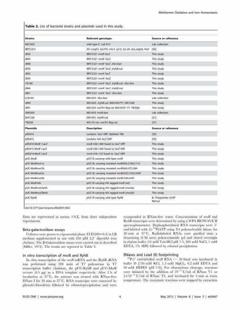
Table 2. List of bacterial strains and plasmids used in this study.
Strains Relevant genotype Source or reference
MG1655 wild type E. coli K12 Lab collection
IBPC5321 thi-1,argE3, lacX74, mlt-li, xyl-5, tsx-29, rpsL,argG6, his4 [56]
JB43 IBPC5321 msrB’-lacZ This study
JB44 IBPC5321 msrB’-‘lacZ This study
JB46 IBPC5321 msrB’-‘lacZ Dfur::kan This study
JB50 IBPC5321 msrB’-‘lacZ DryhB::cat This study
JB55 IBPC5321 msrA’-lacZ This study
JB56 IBPC5321 msrA’-‘lacZ This study
YD100 IBPC5321 msrA’-‘lacZ DryhB::cat Dfur::kan This study
JB64 IBPC5321 msrA’-‘lacZ DryhB::cat This study
JB61 IBPC5321 msrA’-‘lacZ Dfur::kan This study
LCB195 MG1655 Dfur::kan Lab collection
JB99 MG1655 DryhB::cat (MG1655*P1 EM1238) This study
JB91 MG1655 rne701-flag-cat (MG1655* P1 TM528) This study
SMG505 MG1655 msrB::kan Lab collection
EM1238 EM1055 DryhB::cat [31]
TM528 W3110 mlc rne701-flag-cat [57]
Plasmids Description Source or reference
pRS414 contains ‘lacZ ORF (deleted TIR) [39]
pRS415 contains full lacZ ORF [39]
pRS414-MsrB’-‘LacZ msrB-336/+360 fused to lacZ ORF This study
pRS415-MsrB’-LacZ msrB-336/+360 fused to lacZ ORF This study
pRS414-MsrA’-LacZ msrA-416+132 fused to ‘lacZ ORF This study
pUC-MsrB pUC18 carrying wild type msrB This study
pUC-MsrBmut1a pUC18, carrying mutated msrBA9G,G10A,C11U This study
pUC-MsrBmut1b pUC18, carrying mutated msrBG6A,U7C,G8A This study
pUC-MsrBmut2a pUC18, carrying mutated msrBG32C,C33U,A34C This study
pUC-MsrBmut2b pUC18, carrying mutated msrBC33A,A34T This study
pUC-MsrB-HA pUC18 carrying HA tagged-msrB (wt) This study
pUC-MsrBmut2aHA pUC18 carrying HA tagged-msrB (mut2a) This study
pUC-MsrBmut2bHA pUC18 carrying HA tagged-msrB (mut2b) This study
pUC-RyhB pUC19 carrying wild type RyhB B. Charpentier (UHPNancy)
doi:10.1371/journal.pone.0063647.t002
Methionine Oxidation and Iron Homeostasis
PLOS ONE | www.plosone.org 4 May 2013 | Volume 8 | Issue 5 | e63647

with phenol/chloroform and precipitation by ethanol in presence
of sodium acetate (0.3 M) and 2 mg tRNAs.
For lead (II) footprinting, the reactions were initiated by the
addition of 12 mM Pb(II) acetate. After 5 min incubation at room
temperature, reactions were stopped by adding EDTA (0.05 M)
before ethanol precipitation followed by extraction with phenol/
chloroform and a second precipitation by ethanol in the presence
of sodium acetate (0.3 M) and 2 mg tRNAs. As sequence markers,
RNA alkaline hydrolysis ladders (cleavage after each nucleotide)
were generated by incubating in 1 M sodium carbonate at pH9 for
3 min at 96uC. RNase T1 ladders (cleavage after each guanosine)
were generated by incubating the RNA in 1 M sodium hydroxide
citrate for 5 min at 65uC, 2 mg tRNAs then by adding 1U RNase
T1 for 10 min at 65uC. For both enzymatic and chemical probing
reactions, the treated RNA samples were then ethanol-precipitated
in the presence of 0.3 M sodium acetate and washed with 80%
ethanol. RNA pellets were directly resuspended in RNA loading
dye [95% (v/v) formamide, 20 mM EDTA, 0.05% (w/v) each
bromophenol blue and xylene cyanol]. The cleavage products
were separated onto a 10% polyacrylamide 8 M urea-containing
gel and visualized by using a Phosphorimager.
Results
Expression of the msrB gene is repressed under ironstarvation
Transcriptional and translational fusions between the msrB gene
and the lacZ reporter gene were constructed and inserted at the att
site in the E. coli chromosome. The engineered strains namely
JB43 and JB44, respectively, were grown in the presence or
absence of 2,29dipyridyl (2,29dip), an iron chelator, and ß-
galactosidase activity assayed (Table 3). Expression of the
translational fusion was repressed by more than three fold in the
presence of 2,29dip whereas expression of the transcriptional
fusion remained nearly the same in both conditions. To assess
whether the decrease in expression of the translational reporter
observed with the 2,29dip was due to a decrease in both msrB
mRNA and protein levels, we measured the amount of msrB
mRNA and MsrB protein within the cell by Northern and
Western blot analyses, respectively. Levels of both msrB mRNA
and MsrB protein showed a 5-fold decrease under iron limitation
(Figure 1A–B). The sRNA RyhB is known to specifically down-
regulate the expression of numerous genes under iron starvation
[30,31]. Introducing a ryhB mutation in JB44 (JB50) abolished
2,29dip repression, yielding an up constitutive and iron-indepen-
dent phenotype (Table 3). Expression of RyhB is under Fur
repression. Introducing a fur mutation in JB44 (JB46) abolished
2,29dip repression, yielding a down constitutive iron-independent
phenotype (Table 3). Taken together, these results show the
expression of the msrB gene to be regulated by the Fur/RyhB
cascade such that msrB expression decreases under iron starvation.
Expression of the sRNA RyhB reduces the msrB transcriptlevels
The sRNA RyhB promotes the degradation of mRNAs
encoding Fe-using proteins and the synthesis of RyhB is induced
under iron starvation [31,33]. Therefore, we analysed the levels of
both RyhB and msrB transcripts in cells that were submitted to
sudden iron starvation and subsequent iron replenishment
(Figure 1C, 1D). Briefly, cells were grown to mid-exponential
phase, exposed to 2,29dip for 15 minutes and subsequently
exposed to excess iron (100 mM FeSO4). We then carried out
Northern blots with probes specific to msrB and RyhB transcripts.
Results showed that within 5 minutes after 2,29dip exposure, msrB
transcript levels were greatly decreased (Figure 1D lane 3), while
RyhB transcript levels immediately increased (Figure 1D lanes 3–
4). When cells were supplemented with excess iron, msrB transcript
levels reached a maximum within 5 minutes while RyhB transcript
decreased to undetectable levels within 15 minutes (Figure 1D,
lanes 6–7). These observations establish a clear correlation
between the presence of RyhB and the absence of msrB transcripts,
in full agreement with the hypothesis that RyhB induces msrB
transcript degradation.
In order to quantify this putative destabilizing effect, we
determined the half-life of msrB mRNA in the presence and in
the absence of RyhB. To measure the half-life, 2,29dip was added
to the culture for 15 minutes, followed by the addition of
rifampicin to stop transcription. Total RNA was extracted at
different time points and the mRNAs were hybridized with probes
specific either to msrB or to 5S RNA as an internal control
(Figure 2). In the wild type strain, the half-life of the msrB mRNA
was 4.7 times shorter in the presence of 2,29dip. In contrast, in the
ryhB mutant, the half-life of the msrB transcript was the same
regardless of the addition of 2,29dip. Moreover, in a fur mutant
Table 3. Expression of msrA-lacZ and msrB-lacZ fusions in wild type, ryhB deletion or fur deletion strains grown to exponentialphase (O.D.600<0.4) in LB rich medium either in the presence or in the absence of iron chelator (2,29dip, 250 mM).
Type of fusion Strains LB LB +2,29dip Fold expression ±2,29dip
Transcriptional msrB’-lacZ (JB43) 1655635 1502630 0.9x
Translational msrB’-‘lacZ (JB44) 680620 19068 0.3x
Translational msrB’-‘lacZ ryhB ::cat (JB50) 688616 70268 1.0x
Translational msrB’-‘lacZ fur ::kan (JB46) 18166 17269 1.0x
Transcriptional msrA’-lacZ (JB55) 105632 148635 1.4x
Translational msrA’-‘lacZ (JB56) 58614 7964 1.4x
Translational msrA’-‘lacZ ryhB ::cat (JB64) 5366 93613 1.7x
Translational msrA’-‘lacZ fur ::kan (JB61) 5365 60610 1.1x
Translational msrA’-‘lacZ ryhB ::cat fur ::kan(YD100)
3465 47614 1.4x
The level of b-galactosidase expressed from the fusions is given in Miller units. Each value is the average of four independent experiments with three measurementseach. Fold expression in iron starvation is indicated. Standard error is indicated.doi:10.1371/journal.pone.0063647.t003
Methionine Oxidation and Iron Homeostasis
PLOS ONE | www.plosone.org 5 May 2013 | Volume 8 | Issue 5 | e63647

Figure 1. RyhB-dependent down-regulation of msrB mRNAs and proteins. (A) Cultures containing a wild type strain of E. coli were grown toan O.D.600 value of 0.5 then 250 mM 2,29dip was added. After 30 min of incubation with 2,29dip, total RNA and proteins were extracted in parallel,giving total RNA used for the Northern blots (top panel) and soluble protein fraction used for Western blot (bottom panel). The same membrane wasprobed successively for msrB mRNA, RyhB, and 23S RNA (loading control) (top panel). MsrB proteins were probed with anti-HA antibodies (bottompanel). The radioactive probes used are described in Materials and Methods, and Table 1. (B). Quantification of msrB mRNA, RyhB sRNA and MsrBprotein levels (arbitrary units) from experiment described in (A). Band intensity was normalized to that of an internal control (23S for both msrB andRyhB RNA bands; a non-specific protein recognized by anti-HA antibodies for MsrB-HA protein band). (C). Overview of the experiment described in D.Total RNA was extracted at the indicated times (min). (D). Wild type E. coli cells were grown in LB (lanes 1,2,5), LB + 2,29dip (250 mM) (lanes 3,4). Iron(100 mM) was added after 15 min of growth in LB (lane 8) and after 5 or 15 min of pre-incubation with 2,29dip (lanes 6–7). Samples were removed atindicated time points, and total RNA was extracted as described in Materials and Methods. Strain SMG505 (DmsrB) was used as a control (lane 1). Fordetermination of RyhB and msrB RNA amounts, 10 mg of total RNA samples were loaded onto a denaturating agarose gel. After migration, a Northernblot hybridization was performed with a specific oligoprobe for RyhB and msrB respectively. Quantification of msrB and RyhB transcript levels(arbitrary units) are shown below Northern blots panels.doi:10.1371/journal.pone.0063647.g001
Methionine Oxidation and Iron Homeostasis
PLOS ONE | www.plosone.org 6 May 2013 | Volume 8 | Issue 5 | e63647

that synthesizes RyhB at a high-level, the half-life of the msrB
transcript was the same whether 2,29dip was added or not and was
as short as in 2,29dip-exposed wild type cells.
Altogether, these observations fit with the model that iron
depletion induces RyhB synthesis, which in turn favours the
degradation of the msrB mRNA.
The RNA degradosome and the RNA chaperone Hfq arerequired for the RyhB-induced msrB mRNA degradation
Previous characterizations of the mode of action of RyhB
showed that RNase E and the RNA degradosome are required in
the RyhB-mediated degradation of target mRNAs [33]. Therefore
we tested if this also explains the RyhB-mediated decrease in the
levels of the msrB mRNA. In the rne701 mutant, a strain lacking a
functional RNA degradosome, the half-life of the msrB transcript
was increased 1.5-fold and 8-fold, as compared with the wild type
strain, in the presence and in the absence of 2,29dip respectively
(Figure 2). This indicates that the RNA degradosome is involved in
the msrB transcript destabilization observed under iron limiting
conditions.
The RNA chaperone Hfq is essential for RyhB-mediated
regulation of its target genes. Therefore we tested whether Hfq was
involved in msrB regulation. In an hfq mutant, the half-life of the
msrB transcript was only slightly changed by the presence of
2,29dip and was increased 4.6-fold in the presence of 2,29dip as
compared with the wild type strain.
Altogether these observations validate that the RyhB/Hfq/
Rnase E complex is involved in the msrB mRNA decay.
Identification of two RyhB/msrB pairing sites in the 59
UTR of the msrB mRNAIn order to determine whether the RyhB control of msrB
expression was due to a direct interaction between the two RNA
species, the 59 untranslated region (59 UTR) of msrB mRNA was
searched for sequences complementary to sequences in RyhB. Our
results revealed two such potential interaction sites (Figure 3A).
The first site, which we will refer to as Site I, is located between
G1, the first nucleotide to be transcribed, and A12 (Figure 3A).
This 12 nucleotide long sequence exhibits 12 pairings with
sequence within RyhB stem-loop 2 with a total free energy of
225.4 kcal/mol. The second site, which we will refer to as Site II,
is located between A26 and G38 and hence overlaps the Shine-
Dalgarno sequence. This 13 nucleotide long sequence exhibits 12
matches with a region located within RyhB loop 2 (Figure 3A).
Pairing at Site II between RyhB and msrB mRNA is predicted to
be less stable than the pairing at Site I with a free energy of
211.7 kcal/mol. Thus, this sequence analysis raised the possibility
that msrB mRNA possesses two RyhB binding sites. It is
remarkable that the two sites contain duplicates of a 9-nucleotide
sequence (AUGUGAGCA) and therefore predicted to pair with
the same RyhB region, i.e. a sequence of the central stem-loop of
RyhB.
Experimental identification of two RyhB binding sites inthe 59 UTR of msrB mRNA
The validity of the predictions reported above was subsequently
tested by in vitro approaches. First, we asked whether RyhB binding
on msrB mRNA would block reverse transcriptase (RT) elongation
in a primer extension assay (PEA). A shorter version of msrB
mRNA (124 nt; nt +1 to +124) including the whole 59 UTR region
(35 nucleotides) was transcribed in vitro and used as a template for
PEA in the presence and in the absence of RyhB (Figure 3B). In
the presence of RyhB, two primer extension products were
detected that were mapped within Site I (C11) and Site II (C33,
A34)(Fig. 3B; lane 2). As a control, we performed a PEA on the
msrB mRNA alone and observed no specific primer extension
product along the msrB1–124 RNA transcript (Figure 3B; lane 1).
Second, we compared patterns of partial digestion of the 59
UTR of the msrB transcript with RNAses in the absence and in the
presence of RyhB (Figure 4). When the msrB mRNA was digested
by RNase T2 (a single strand-specific endoribonuclease with
preference for adenosine residues) in the presence of RyhB,
cleavages appeared at the 39 end of Site I (bonds between residues
at positions +12 to +14) and Site II (bonds between residues at
positions +40 to +42) while some others were strongly reinforced
downstream of the Site II (bonds between residues at positions +51
to +67) (Figure 4, lane 3). Conversely, the presence of RyhB
afforded protection against RNase T2 cleavages within the binding
Sites I and II (bonds between residues at positions +7 and +8, and
at positions +34 and +35, Figure 4; lane 4). When RNase V1
(specific for double stranded regions and stacked nucleotides) was
Figure 2. Effects of ryhB, fur, rne701 and hfq mutations on msrB transcript stability. msrB transcript stability in wild type (A), ryhB mutant (B),fur mutant (C), hfq mutant (D) and rne701 mutant (E) strains grown at 37uC to an O.D.600 of 0.4, was assayed by Northern blot analysis. After 10 min ofincubation with 2,29dip, rifampicin was added. Samples were removed at the indicated time points after rifampicin addition and total RNA wasextracted as described in Materials and Methods. For determination of msrB mRNA amount, 10 mg of total RNA samples were loaded onto adenaturating agarose gel. After migration, a Northern blot hybridization was performed with a specific oligoprobe for msrB and 5S as an internalcontrol. Band intensity of msrB transcript was normalized to that of 5S RNA. Half-life (seconds) of msrB transcript and the ratio of msrB mRNA half-life62,29dip are indicated for the different strains. Standard errors (SE) are shown.doi:10.1371/journal.pone.0063647.g002
Methionine Oxidation and Iron Homeostasis
PLOS ONE | www.plosone.org 7 May 2013 | Volume 8 | Issue 5 | e63647

used in the presence of RyhB, significant changes in the RNase
V1-digestion pattern were observed with new cleavage products in
the sequence of the predicted binding Site I (bonds between
positions +8 to +11) and Site II (bonds between positions +31/+32,
+33/+34 and +35 to +37) (Figure 4; lane 6). Additional cleavages
were also observed within the coding region of msrB but no
potential RyhB binding sites was detected by sequence analysis
and no prematured RT extension products were observed (data
not shown). Last, digestion of the 59UTR of msrB with lead (II)
acetate (specific for single stranded regions) in the presence of
RyhB (Figure 4, lane 8) showed that the two regions that refer to
Sites I and II were significantly protected from lead (II) digestion
(bonds between positions +5 to +11 and +26 to +38, Figure 4B).
Thus, taken together, PEA, RNase/lead (II) probing analyses
fully establish that RyhB pairing occurs at two sites, referred to
Sites I and II within the short 59 UTR of the msrB mRNA.
Mutagenesis analysis of the RyhB/msrB interaction invitro
In order to validate the binding of RyhB at Sites I and II in vitro,
nucleotide changes were made in the sequences of msrB Sites I and
II. The consequences on RyhB binding were analysed by PEA.
First, mutations (G32C, C33U, A34C) were introduced into the
Site II, yielding an msrB allele referred to as mut2a (Figure 3A).
Results showed only one primer extension product, located within
the Site I (Figure 5A; lane 4). Thus, modifications introduced into
the Site II prevented RyhB to bind at this site. Second, mutations
(G6A, U7C, G8A) were introduced into the Site I, yielding the
mut1 allele (Figure 3A). Results showed only one primer extension
product, located within Site II (Figure 5B; lane 3). Altogether these
mutational analyses further support the notion of an interaction
between RyhB and msrB at both Sites I and II in vitro.
Figure 3. RyhB binds msrB mRNA at two sites. Panel (A) shows the predicted interactions between RyhB and the msrB sense strand referred asSite I and Site II (in yellow). The predicted ribosome-binding site for msrB is underlined. The start codon for msrB is shown underlined and in italics.Mutations in Site I and Site II are shown in red and blue respectively. (B) cDNA extension experiments with wild type msrB1–124 as a template for thereverse transcriptase. Lane 1: extension with no other component added; lane 2: extension with RyhB alone. C, U, A, and G are sequencing lanesobtained using the same radiolabeled primer as in the reverse extension analysis. Reverse transcriptase stops are indicated at positions +11, +33 and+34. Nucleotides involved in Sites I and II are indicated as thin vertical lines. The transcription start of msrB is referred to as the position + 1. Thenumbers on the left indicate sequence positions with respect to the transcription start.doi:10.1371/journal.pone.0063647.g003
Methionine Oxidation and Iron Homeostasis
PLOS ONE | www.plosone.org 8 May 2013 | Volume 8 | Issue 5 | e63647

Figure 4. Changes in msrB RNA accessibility to enzymatic and chemical probes upon RyhB binding. (A) RNases and lead (II) footprinting:59 end-labelled msrB1–124 transcript was subjected to partial digestion with RNase T2 (lanes 3–4), RNase V1 (lanes 5–6) or lead (II) (lanes 7–8) in thepresence (+) or in the absence (-) of RyhB sRNA. Lanes 1 and 2 are control lanes of RT extension on msrB alone (lane 1) or on msrB with RyhB (lane 2).The resulting fragments were then analyzed onto a denaturing sequencing gel. The numbers indicate sequence positions with respect to thetranscription start site. Lanes OH2 and T1 correspond, respectively, to an alkaline hydrolysis ladder, and an RNase T1 digestion ladder obtained indenaturing conditions. The position of G residues that resulted from RNase T1 hydrolysis is given. Circles, arrowheads, and rectangles indicate,respectively, phosphodiester bonds cleavages by RNase T2, RNase V1, and lead (II). Products resulting from a strong (red) or a weak (orange)
Methionine Oxidation and Iron Homeostasis
PLOS ONE | www.plosone.org 9 May 2013 | Volume 8 | Issue 5 | e63647

RyhB binding on the msrB mRNA blocks ribosomebinding
RyhB is known to interfere with translation initiation. There-
fore, we asked whether RyhB binding on msrB mRNA would block
ribosome binding in vitro. For this purpose, toeprint assays were
run. A long version of msrB mRNA (450nt; nt +1 to nt +449)
including the whole 59 UTR region was transcribed in vitro and
used as a template for toeprint assays (Figure 6). When the 30S
ribosomal subunit was added, its binding to the wild type msrB
mRNA blocked RT elongation, resulting in a shortened cDNA
called ‘toeprint’ at position +50 to +52 (Figure 6A–B; lane 3).
However, in the presence of RyhB, the intensity of the toeprint
band was 1666 times decreased (Figure 6A–B; compare lane 3
with lane 4). This result shows that the interaction of RyhB with
msrB mRNA interferes with the binding of the 30S subunit.
We then carried out a toeprint assay with the msrB mut1 mRNA
as a template. No specific primer extension product was observed
on the msrB mut1 mRNA alone (Figure 6A–B; lane 5). In the
presence of the 30S ribosomal subunit, a toeprint was detected
(Figure 6A–B; lane 7). When RyhB was added, a specific primer
extension product was observed at Site II but not at Site I as
predicted (Figure 6A–B; lane 6). When both RyhB and the 30S
subunit were added, the intensity of the toeprint band was
4.2560.6 times reduced (Figure 6A–B; compare lane 7 and lane
8). This observation indicates that the interaction of RyhB with
Site II efficiently interferes with the 30S binding. However since
the interaction of RyhB with Site II does not fully abolish the
toeprinting signal it suggests that the pairing of RyhB at Site I may
contribute to the inhibitory effect of RyhB on the ribosome
binding.
We then tested the effect of mutations in msrB RyhB-binding site
II. As the mut2a mutation used (G32C, C33U, A34C) alters the
Shine-Dalgarno sequence, we expected it to reduce the binding
affinity of the 30S subunit. A Western-blot analysis of the resulting
mutant was carried out and indeed shown to have a very low level
of MsrB production (Figure S1). Therefore a 10-fold higher
concentration of 30S subunit was used. However, even with such
an increased concentration, we failed to observe any detectable
toeprint (data not shown). Consequently, we decided to construct a
new variant, referred to as mut2b. For this, nucleotides C33 and
A34 were substituted for A and U, respectively (Figure 3A). A
Western-blot analysis of the resulting mutant showed that the
MsrB protein was produced at a similar level as the wild type,
showing that the mutation had not altered translation efficiency
(Figure S1). A primer extension analysis of mut2b revealed Site I
but not Site II as expected (Figure 5A). Last, we performed a
toeprint assay on the msrB mut2b mRNA alone and observed no
specific primer extension product (Figure 6A–B; lane 9). When
RyhB was added, a primer extension product was observed at Site
I but no longer at Site II as expected from the mutations
introduced (Figure 6A–B; lane 10). Adding the 30S ribosomal
subunit significantly increased the toeprint band (Figure 6A–B;
enhancement of the cleavages in presence of RyhB are indicated. Reduced levels of cleavages in presence of RyhB are indicated by dark green(strong) and light green (weak) symbols. RyhB-binding sites (Sites I and II) are shown as thin vertical lines. (B). Summary of the RNases/lead (II)footprints of msrB1–124 mRNA in the presence of RyhB based on the results obtained in (A). The translation start codon of msrB is shown in bold andthe Shine Dalgarno sequence is underlined. RyhB Stem Loop 2 (SL2) pairing at Site I and Site II is shown. The same rules as in panel A are utilized forrepresentation of changes in phosphodiester bonds cleavages in presence of RyhB.doi:10.1371/journal.pone.0063647.g004
Figure 5. Mutagenesis analysis of the RyhB/msrB interaction in vitro. (A–B) Autoradiograms of primer extension analysis of msrB mRNA areshown (for details, see Materials and Methods, and Results). (A) with wild type (lanes 1–2), mut2a (lanes 3,4) and mut2b (lanes 5,6) msrB1–124
transcripts as a template for the reverse transcriptase. Lanes 1,3 and 5: extension with no other component added; lanes 2,4 and 6: extension withRyhB. C, U, A, and G are sequencing lanes obtained using the same radiolabeled primer as in the reverse extension analysis. (B) cDNA extensionexperiments with wild type (lanes 1–2) and mut1 (lane 3) msrB1–124 transcripts as templates for the reverse transcriptase. Lane 1: extension with noother component added; lanes 2–3: extension with RyhB. For (A) and (B), reverse transcriptase stops are indicated at positions +11, +33 and +34. Thinvertical lines indicate nucleotides involved in Site I and Site II. The transcription start of msrB is referred to as the position + 1. The numbers to the leftindicate sequence positions with respect to the transcription start site.doi:10.1371/journal.pone.0063647.g005
Methionine Oxidation and Iron Homeostasis
PLOS ONE | www.plosone.org 10 May 2013 | Volume 8 | Issue 5 | e63647
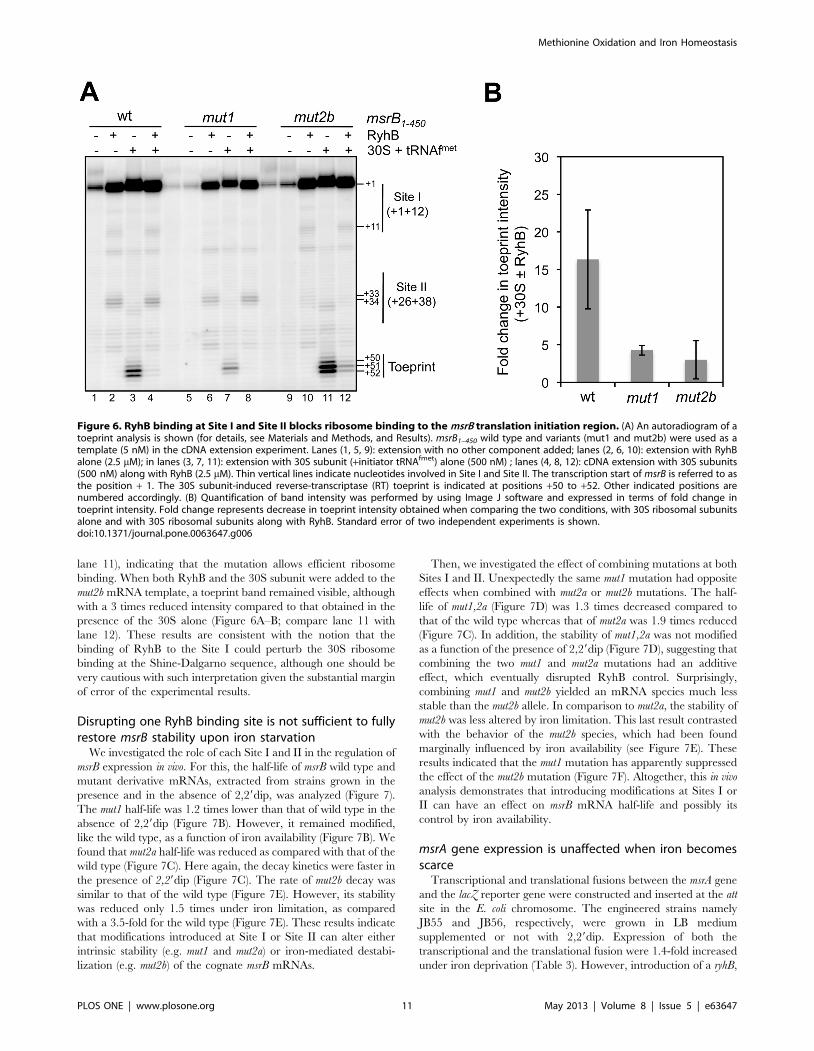
lane 11), indicating that the mutation allows efficient ribosome
binding. When both RyhB and the 30S subunit were added to the
mut2b mRNA template, a toeprint band remained visible, although
with a 3 times reduced intensity compared to that obtained in the
presence of the 30S alone (Figure 6A–B; compare lane 11 with
lane 12). These results are consistent with the notion that the
binding of RyhB to the Site I could perturb the 30S ribosome
binding at the Shine-Dalgarno sequence, although one should be
very cautious with such interpretation given the substantial margin
of error of the experimental results.
Disrupting one RyhB binding site is not sufficient to fullyrestore msrB stability upon iron starvation
We investigated the role of each Site I and II in the regulation of
msrB expression in vivo. For this, the half-life of msrB wild type and
mutant derivative mRNAs, extracted from strains grown in the
presence and in the absence of 2,29dip, was analyzed (Figure 7).
The mut1 half-life was 1.2 times lower than that of wild type in the
absence of 2,29dip (Figure 7B). However, it remained modified,
like the wild type, as a function of iron availability (Figure 7B). We
found that mut2a half-life was reduced as compared with that of the
wild type (Figure 7C). Here again, the decay kinetics were faster in
the presence of 2,29dip (Figure 7C). The rate of mut2b decay was
similar to that of the wild type (Figure 7E). However, its stability
was reduced only 1.5 times under iron limitation, as compared
with a 3.5-fold for the wild type (Figure 7E). These results indicate
that modifications introduced at Site I or Site II can alter either
intrinsic stability (e.g. mut1 and mut2a) or iron-mediated destabi-
lization (e.g. mut2b) of the cognate msrB mRNAs.
Then, we investigated the effect of combining mutations at both
Sites I and II. Unexpectedly the same mut1 mutation had opposite
effects when combined with mut2a or mut2b mutations. The half-
life of mut1,2a (Figure 7D) was 1.3 times decreased compared to
that of the wild type whereas that of mut2a was 1.9 times reduced
(Figure 7C). In addition, the stability of mut1,2a was not modified
as a function of the presence of 2,29dip (Figure 7D), suggesting that
combining the two mut1 and mut2a mutations had an additive
effect, which eventually disrupted RyhB control. Surprisingly,
combining mut1 and mut2b yielded an mRNA species much less
stable than the mut2b allele. In comparison to mut2a, the stability of
mut2b was less altered by iron limitation. This last result contrasted
with the behavior of the mut2b species, which had been found
marginally influenced by iron availability (see Figure 7E). These
results indicated that the mut1 mutation has apparently suppressed
the effect of the mut2b mutation (Figure 7F). Altogether, this in vivo
analysis demonstrates that introducing modifications at Sites I or
II can have an effect on msrB mRNA half-life and possibly its
control by iron availability.
msrA gene expression is unaffected when iron becomesscarce
Transcriptional and translational fusions between the msrA gene
and the lacZ reporter gene were constructed and inserted at the att
site in the E. coli chromosome. The engineered strains namely
JB55 and JB56, respectively, were grown in LB medium
supplemented or not with 2,29dip. Expression of both the
transcriptional and the translational fusion were 1.4-fold increased
under iron deprivation (Table 3). However, introduction of a ryhB,
Figure 6. RyhB binding at Site I and Site II blocks ribosome binding to the msrB translation initiation region. (A) An autoradiogram of atoeprint analysis is shown (for details, see Materials and Methods, and Results). msrB1–450 wild type and variants (mut1 and mut2b) were used as atemplate (5 nM) in the cDNA extension experiment. Lanes (1, 5, 9): extension with no other component added; lanes (2, 6, 10): extension with RyhBalone (2.5 mM); in lanes (3, 7, 11): extension with 30S subunit (+initiator tRNAfmet) alone (500 nM) ; lanes (4, 8, 12): cDNA extension with 30S subunits(500 nM) along with RyhB (2.5 mM). Thin vertical lines indicate nucleotides involved in Site I and Site II. The transcription start of msrB is referred to asthe position + 1. The 30S subunit-induced reverse-transcriptase (RT) toeprint is indicated at positions +50 to +52. Other indicated positions arenumbered accordingly. (B) Quantification of band intensity was performed by using Image J software and expressed in terms of fold change intoeprint intensity. Fold change represents decrease in toeprint intensity obtained when comparing the two conditions, with 30S ribosomal subunitsalone and with 30S ribosomal subunits along with RyhB. Standard error of two independent experiments is shown.doi:10.1371/journal.pone.0063647.g006
Methionine Oxidation and Iron Homeostasis
PLOS ONE | www.plosone.org 11 May 2013 | Volume 8 | Issue 5 | e63647

a fur or a ryhB fur mutations did not change significantly msrA’-‘lacZ
expression levels (Table 3). These results suggested that unlike
msrB, msrA gene expression might be induced by iron availability
but not by a Fur or RyhB dependent mecanism.
Discussion
This study demonstrates that the synthesis of the methionine
sulfoxide reductase MsrB is repressed under iron limitation. We
show this negative regulation to result from the action of the sRNA
RyhB. The involvement of additional trans-acting factors, such as
the transcriptional iron sensing repressor Fur, the RNA chaperone
Hfq and the degradosome-belonging RNase E was also established
[43,44,45,46]. The msrB gene is therefore a new member of the
RyhB regulon, as initially suggested by transcriptomic analysis of
the Masse group [30]. The RyhB-mediated down regulation of
msrB is likely to be related to the fact that MsrB is an iron-binding
enzyme [38].
The mechanism allowing RyhB to control expression of the
msrB gene appears to be original and complex. Indeed, the msrB
gene was found to contain two binding sites for RyhB. The
upstream site is located at the very 59 end of the msrB mRNA while
the second site is located 30 nucleotides downstream and overlaps
the Shine-Dalgarno sequence. Previous studies on RyhB-mediated
regulation of the sodB, sdhD or iscS genes showed that the RyhB-
binding site overlaps the Shine-Dalgarno sequence, thereby
inhibiting translation initiation and provoking mRNA instability
[31,35]. The region of RyhB (loop II) that is used to target msrB is
the same as that pairing with the 59 UTR regions of sodB or sdhD.
Use of molecular techniques allowed us to demonstrate RyhB
binding at both sites in vitro. In Salmonella, the RybB sRNA has
multiple binding sites within the ompCD mRNA but RybB binds
only one site targeting either ompC or ompD mRNA as the pairing
sites are mutually exclusive [47]. Also, a recent study has reported
that SgrS sRNA can have multiple binding sites within a
polycistronic mRNAs [48]. However, the binding sites of SgrS
are present within portions of the mRNA that are encoded by
different genes of the operon. Hence, all of these cases differ from
that described here where the same sRNA can bind to two
different locations within the same mRNA species. Curiously, the
two sites show the same 9 nucleotide sequence. Evidently, this
raises the question of whether two RyhB molecules bind
simultaneously to msrB mRNA or only one at a time at one of
the two sites. Further experiments aimed at establishing the
stoichiometry msrB/RyhB will have to be carried out to get insight
into this question.
Toeprint analysis revealed that the pairing of RyhB with the
msrB mRNA at Site II prevents the binding of the 30S ribosomal
subunit while it remains unclear if pairing at Site I bears any effect
on the binding of the ribosome. The way Site II-bound RyhB
interferes with the ribosome binding is likely to be due to steric
hindrance as Site II overlaps the Shine-Dalgarno sequence. Such a
direct competition between RyhB and the ribosome was demon-
strated for the expression of sodB, sdh and iscS genes. In contrast,
the way Site I-bound RyhB may interfere with ribosome binding,
if it does, is uncertain. In Salmonella typhimurium, the regulatory
sRNA GcvB represses translation by binding to target RNA gltI
and argT at upstream sites, outside the RBS [49]. In Salmonella,
RybB targets sites on omp mRNAs within the coding region [50] or
upstream of the region covered by the ribosome [51]. One
possibility is that it might directly interfere with the binding of the
ribosome. Indeed the ribosome was proposed to cover a region
extending from – 35 nt upstream the start codon to + 19 nt
downstream [52]. An alternative role of the pairing of RyhB at
Site I might be that it stimulates the degradation of msrB mRNA
by RNase E and the degradosome as it has recently been reported
for sodB mRNA by the Masse group [37]. In-depth study of the
decay of mut1 mRNA species is required to assess the possibility of
such a mechanism.
Analysis of msrB alleles that contained modified Site I and/or
Site II highlighted the complexity of the mechanism underlying
RyhB regulation, yet provided us with important information. In
particular, we were able to isolate mutations within the regulatory
Figure 7. Effect of mutations in RyhB-binding Sites I and II on msrB mRNA stability. Northern blot analysis of wild type msrB (A), msrB mut1(B), msrB mut2a (C), msrB mut1,2a (D), msrB mut2b (E), and msrB mut1,2b (F). Strains were grown at 37uC to an O.D.600 of 0.4. After 10 min ofincubation with 2,29dip, rifampicin was added. Samples were removed at the times indicated after rifampicin addition and total RNA was extracted asdescribed in Materials and Methods. Half-life of msrB mRNA was calculated with or without iron chelator. For determination of msrB mRNA amount,10 mg of total RNA samples was loaded on a denaturating 1.2% agarose gel. After migration, a Northern blot hybridization was performed with aspecific oligoprobe for msrB and with 5S as a loading control. Half-life (seconds) of msrB mRNA (wild type and mutants) and the ratio of msrB mRNAhalf-life 62,29dip, are indicated. Band intensity of msrB transcript was normalized to that of 5S RNA.doi:10.1371/journal.pone.0063647.g007
Methionine Oxidation and Iron Homeostasis
PLOS ONE | www.plosone.org 12 May 2013 | Volume 8 | Issue 5 | e63647

sequence of msrB transcript that uncoupled msrB degradation and
iron bioavailability. However, these mutants exhibited additional
unexpected features. One case is the ‘‘double’’ mut1,2a allele,
which contains a mutation at each Site I and II. This allele
exhibited a constitutive-like behaviour as the half-life of the
cognate mRNA, was not altered in bacteria that are grown under
iron limitation. This departs from the behaviours of both ‘‘single’’
mut1 and mut2a alleles, which remain regulated by iron and do not
show any increased stability. Thus, the additive effect of the two
mutations could signify the involvement of the two sites in RyhB-
mediated regulation. However, mut1,2a allele is very poorly
recognized by the 30S ribosomal subunit and, accordingly, it is
very poorly translated. Hence, it is impossible to decipher whether
the loss of the iron regulation is due to the inability of RyhB to
recognize this species and to interfere with ribosome binding or if
reduced ribosome binding efficiency prevents any associated
regulatory mechanism to take place. Moreover, one would have
expected the half-life of this mutated version to be drastically
shortened, as it is no longer protected by ribosomes. Could the two
mutations yield a highly structured mRNA, unable to interact with
any of the trans-acting factors, including the ribosome, RyhB, the
degradosome or a subset of them? A thorough RNase structural
probing is needed prior to further interpret the behaviour of this
species. Another puzzling case is the mut2b allele. In this case, the
sole modification of the Site II was sufficient to alter iron
regulation. In vitro analysis of the mut2b mRNA species revealed
that RyhB no longer interacts with Site II but interacts with Site I.
The iron regulation was altered but not fully abolished and this
was consistent with the notion that the RyhB/Site I interaction
can mediate iron regulation. However, a simple interpretation of
this allele was prevented by the observation that the mut2b
mutation effect could be moderately suppressed by the mut1
mutation. Indeed, the mut1,2b allele produces an mRNA species
which remains under the control of iron bioavailibility with a 2-
fold decrease in its stability upon 2,29 dip treatment. Clearly more
in-depth analysis of the cognate structure is required before we
make any firm conclusion on how they affect the msrB/RyhB
interaction, and the regulation of the msrB expression.
Methionine sulfoxide reductases MsrA and MsrB catalyse the
reduction of S- and R-diastereoisomeric forms of methionine
sulfoxide. Previous studies established that the combined action of
both MsrA and MsrB is required for full repair of oxidized
polypeptides [8,54]. From this biochemical point of view, one
would expect that the two enzymes will be synthesized in the same
conditions. This is likely to be the case in Bacillus subtilis wherein
the two cognate structural genes belong to the same operon [55].
This is also the case of Neisseria wherein both MsrA and MsrB are
actually fused into a single polypeptide [27]. Conversely, in E. coli,
the two genes are physically separated, raising the question of their
co-regulation. Further, we found that MsrB synthesis is decreased
under iron limitation conditions whereas that of MsrA appears
essentially not affected by this limitation. One might speculate that
under iron limitation, damage by iron-mediated Fenton reaction is
reduced and methionine oxidation is infrequent. This might
explain why E. coli reduces MsrB synthesis while saving iron for
essential processes and relies only on MsrA to rescue those few
oxidized polypeptides.
Supporting Information
Figure S1 Western blot analysis of MsrB protein levelsfrom cells expressing wild type msrB and mutant msrB.
Wild type msrB (A) and mutant msrB (mut2a (B) and mut2b (C)).
Strains were grown at 37uC to an O.D.600 of 0.4. After 60 min of
incubation with 2,2’dip, samples were removed and proteins were
extracted as described in Materials and Methods. MsrB proteins
were probed using anti-HA antibodies.
(TIF)
Acknowledgments
The authors are grateful to members of Frederic Barras, Christiane
Branlant and Mathias Springer laboratories for fruitful discussions and
expert technical assistance.
Author Contributions
Conceived and designed the experiments: JB YD BT CC CB MS BC FB.
Performed the experiments: JB YD BT CC BC. Analyzed the data: JB YD
BT CC CB MS BC FB. Wrote the paper: JB YD CC MS BC FB.
References
1. Finkel T, Holbrook NJ (2000) Oxidants, oxidative stress and the biology of
ageing. Nature 408: 239–247.
2. Brosnan JT, Brosnan ME (2006) The sulfur-containing amino acids: an
overview. J Nutr 136: 1636S–1640S.
3. Maisonneuve E, Ducret A, Khoueiry P, Lignon S, Longhi S, et al. (2009) Rules
governing selective protein carbonylation. PLoS One 4: e7269.
4. Maisonneuve E, Ezraty B, Dukan S (2008) Protein aggregates: an aging factor
involved in cell death. J Bacteriol 190: 6070–6075.
5. Moskovitz J, Oien DB (2010) Protein carbonyl and the methionine sulfoxide
reductase system. Antioxid Redox Signal 12: 405–415.
6. Boschi-Muller S, Olry A, Antoine M, Branlant G (2005) The enzymology and
biochemistry of methionine sulfoxide reductases. Biochim Biophys Acta 1703:231–238.
7. Ezraty B, Aussel L, Barras F (2005) Methionine sulfoxide reductases in
prokaryotes. Biochim Biophys Acta 1703: 221–229.
8. Grimaud R, Ezraty B, Mitchell JK, Lafitte D, Briand C, et al. (2001) Repair of
oxidized proteins. Identification of a new methionine sulfoxide reductase. J BiolChem 276: 48915–48920.
9. Sharov VS, Ferrington DA, Squier TC, Schoneich C (1999) Diastereoselectivereduction of protein-bound methionine sulfoxide by methionine sulfoxide
reductase. FEBS Lett 455: 247–250.
10. Sharov VS, Schoneich C (2000) Diastereoselective protein methionine oxidation
by reactive oxygen species and diastereoselective repair by methionine sulfoxidereductase. Free Radic Biol Med 29: 986–994.
11. Luo S, Levine RL (2009) Methionine in proteins defends against oxidative stress.FASEB J 23: 464–472.
12. Koc A, Gladyshev VN (2007) Methionine sulfoxide reduction and the agingprocess. Ann N Y Acad Sci 1100: 383–386.
13. Zhang XH, Weissbach H (2008) Origin and evolution of the protein-repairing
enzymes methionine sulphoxide reductases. Biol Rev Camb Philos Soc 83: 249–
257.
14. Boschi-Muller S, Gand A, Branlant G (2008) The methionine sulfoxide
reductases: Catalysis and substrate specificities. Arch Biochem Biophys 474:
266–273.
15. Hansel A, Heinemann SH, Hoshi T (2005) Heterogeneity and function of
mammalian MSRs: enzymes for repair, protection and regulation. Biochim
Biophys Acta 1703: 239–247.
16. Hansel A, Jung S, Hoshi T, Heinemann SH (2003) A second human methionine
sulfoxide reductase (hMSRB2) reducing methionine-R-sulfoxide displays a tissue
expression pattern distinct from hMSRB1. Redox Rep 8: 384–388.
17. Moskovitz J (2005) Roles of methionine suldfoxide reductases in antioxidant
defense, protein regulation and survival. Curr Pharm Des 11: 1451–1457.
18. Ruan H, Tang XD, Chen ML, Joiner ML, Sun G, et al. (2002) High-quality life
extension by the enzyme peptide methionine sulfoxide reductase. Proc Natl
Acad Sci U S A 99: 2748–2753.
19. Moskovitz J, Berlett BS, Poston JM, Stadtman ER (1997) The yeast peptide-
methionine sulfoxide reductase functions as an antioxidant in vivo. Proc Natl
Acad Sci U S A 94: 9585–9589.
20. Moskovitz J, Bar-Noy S, Williams WM, Requena J, Berlett BS, et al. (2001)
Methionine sulfoxide reductase (MsrA) is a regulator of antioxidant defense and
lifespan in mammals. Proc Natl Acad Sci U S A 98: 12920–12925.
21. Rouhier N, Vieira Dos Santos C, Tarrago L, Rey P (2006) Plant methionine
sulfoxide reductase A and B multigenic families. Photosynth Res 89: 247–262.
22. Ezraty B, Bos J, Barras F, Aussel L (2005) Methionine sulfoxide reduction and
assimilation in Escherichia coli: new role for the biotin sulfoxide reductase BisC.
J Bacteriol 187: 231–237.
Methionine Oxidation and Iron Homeostasis
PLOS ONE | www.plosone.org 13 May 2013 | Volume 8 | Issue 5 | e63647

23. Douglas T, Daniel DS, Parida BK, Jagannath C, Dhandayuthapani S (2004)
Methionine sulfoxide reductase A (MsrA) deficiency affects the survival ofMycobacterium smegmatis within macrophages. J Bacteriol 186: 3590–3598.
24. Singh VK, Moskovitz J (2003) Multiple methionine sulfoxide reductase genes in
Staphylococcus aureus: expression of activity and roles in tolerance of oxidativestress. Microbiology 149: 2739–2747.
25. Hassouni ME, Chambost JP, Expert D, Van Gijsegem F, Barras F (1999) Theminimal gene set member msrA, encoding peptide methionine sulfoxide
reductase, is a virulence determinant of the plant pathogen Erwinia
chrysanthemi. Proc Natl Acad Sci U S A 96: 887–892.26. Denkel LA, Horst SA, Rouf SF, Kitowski V, Bohm OM, et al. (2011)
Methionine sulfoxide reductases are essential for virulence of Salmonellatyphimurium. PLoS One 6: e26974.
27. Skaar EP, Tobiason DM, Quick J, Judd RC, Weissbach H, et al. (2002) Theouter membrane localization of the Neisseria gonorrhoeae MsrA/B is involved
in survival against reactive oxygen species. Proc Natl Acad Sci U S A 99: 10108–
10113.28. Dhandayuthapani S, Blaylock MW, Bebear CM, Rasmussen WG, Baseman JB
(2001) Peptide methionine sulfoxide reductase (MsrA) is a virulence determinantin Mycoplasma genitalium. J Bacteriol 183: 5645–5650.
29. Wizemann TM, Moskovitz J, Pearce BJ, Cundell D, Arvidson CG, et al. (1996)
Peptide methionine sulfoxide reductase contributes to the maintenance ofadhesins in three major pathogens. Proc Natl Acad Sci U S A 93: 7985–7990.
30. Masse E, Vanderpool CK, Gottesman S (2005) Effect of RyhB small RNA onglobal iron use in Escherichia coli. J Bacteriol 187: 6962–6971.
31. Masse E, Gottesman S (2002) A small RNA regulates the expression of genesinvolved in iron metabolism in Escherichia coli. Proc Natl Acad Sci U S A 99:
4620–4625.
32. Masse E, Salvail H, Desnoyers G, Arguin M (2007) Small RNAs controlling ironmetabolism. Curr Opin Microbiol 10: 140–145.
33. Masse E, Escorcia FE, Gottesman S (2003) Coupled degradation of a smallregulatory RNA and its mRNA targets in Escherichia coli. Genes Dev 17: 2374–
2383.
34. Cornelis P, Wei Q, Andrews SC, Vinckx T (2011) Iron homeostasis andmanagement of oxidative stress response in bacteria. Metallomics 3: 540–549.
35. Desnoyers G, Morissette A, Prevost K, Masse E (2009) Small RNA-induceddifferential degradation of the polycistronic mRNA iscRSUA. EMBO J 28:
1551–1561.36. Prevost K, Salvail H, Desnoyers G, Jacques JF, Phaneuf E, et al. (2007) The
small RNA RyhB activates the translation of shiA mRNA encoding a permease
of shikimate, a compound involved in siderophore synthesis. Mol Microbiol 64:1260–1273.
37. Prevost K, Desnoyers G, Jacques JF, Lavoie F, Masse E (2011) Small RNA-induced mRNA degradation achieved through both translation block and
activated cleavage. Genes Dev 25: 385–396.
38. Olry A, Boschi-Muller S, Yu H, Burnel D, Branlant G (2005) Insights into therole of the metal binding site in methionine-R-sulfoxide reductases B. Protein Sci
14: 2828–2837.39. Simons RW, Houman F, Kleckner N (1987) Improved single and multicopy lac-
based cloning vectors for protein and operon fusions. Gene 53: 85–96.40. Powell BS, Rivas MP, Court DL, Nakamura Y, Turnbough CL Jr (1994) Rapid
confirmation of single copy lambda prophage integration by PCR. Nucleic Acids
Res 22: 5765–5766.
41. Makhno VI, Peshin NN, Semenkov Iu P, Kirillov SV (1988) [A modified
method of isolation of "tight" 70S ribosomes from Escherichia coli highly active
at different stages of the elongation cycle]. Mol Biol (Mosk) 22: 670–679.
42. Haentjens-Sitri J, Allemand F, Springer M, Chiaruttini C (2008) A competition
mechanism regulates the translation of the Escherichia coli operon encoding
ribosomal proteins L35 and L20. J Mol Biol 375: 612–625.
43. Afonyushkin T, Vecerek B, Moll I, Blasi U, Kaberdin VR (2005) Both RNase E
and RNase III control the stability of sodB mRNA upon translational inhibition
by the small regulatory RNA RyhB. Nucleic Acids Res 33: 1678–1689.
44. Morita T, Maki K, Aiba H (2005) RNase E-based ribonucleoprotein complexes:
mechanical basis of mRNA destabilization mediated by bacterial noncoding
RNAs. Genes Dev 19: 2176–2186.
45. Geissmann TA, Touati D (2004) Hfq, a new chaperoning role: binding to
messenger RNA determines access for small RNA regulator. EMBO J 23: 396–
405.
46. Moll I, Afonyushkin T, Vytvytska O, Kaberdin VR, Blasi U (2003) Coincident
Hfq binding and RNase E cleavage sites on mRNA and small regulatory RNAs.
RNA 9: 1308–1314.
47. Balbontin R, Fiorini F, Figueroa-Bossi N, Casadesus J, Bossi L (2010)
Recognition of heptameric seed sequence underlies multi-target regulation by
RybB small RNA in Salmonella enterica. Mol Microbiol 78: 380–394.
48. Rice JB, Balasubramanian D, Vanderpool CK (2012) Small RNA binding-site
multiplicity involved in translational regulation of a polycistronic mRNA. Proc
Natl Acad Sci U S A 109: E2691–2698.
49. Sharma CM, Darfeuille F, Plantinga TH, Vogel J (2007) A small RNA regulates
multiple ABC transporter mRNAs by targeting C/A-rich elements inside and
upstream of ribosome-binding sites. Genes Dev 21: 2804–2817.
50. Bouvier M, Sharma CM, Mika F, Nierhaus KH, Vogel J (2008) Small RNA
binding to 59 mRNA coding region inhibits translational initiation. Mol Cell 32:
827–837.
51. Papenfort K, Bouvier M, Mika F, Sharma CM, Vogel J (2010) Evidence for an
autonomous 59 target recognition domain in an Hfq-associated small RNA. Proc
Natl Acad Sci U S A 107: 20435–20440.
52. Huttenhofer A, Noller HF (1994) Footprinting mRNA-ribosome complexes with
chemical probes. EMBO J 13: 3892–3901.
53. Zuker M (2003) Mfold web server for nucleic acid folding and hybridization
prediction. Nucleic Acids Res 31: 3406–3415.
54. Ezraty B, Grimaud R, El Hassouni M, Moinier D, Barras F (2004) Methionine
sulfoxide reductases protect Ffh from oxidative damages in Escherichia coli.
EMBO J 23: 1868–1877.
55. You C, Sekowska A, Francetic O, Martin-Verstraete I, Wang Y, et al. (2008)
Spx mediates oxidative stress regulation of the methionine sulfoxide reductases
operon in Bacillus subtilis. BMC Microbiol 8: 128.
56. Plumbridge J, Soll D (1989) Characterization of cis-acting mutations which
increase expression of a glnS-lacZ fusion in Escherichia coli. Mol Gen Genet
216: 113–119.
57. Morita T, Kawamoto H, Mizota T, Inada T, Aiba H (2004) Enolase in the RNA
degradosome plays a crucial role in the rapid decay of glucose transporter
mRNA in the response to phosphosugar stress in Escherichia coli. Mol Microbiol
54: 1063–1075.
Methionine Oxidation and Iron Homeostasis
PLOS ONE | www.plosone.org 14 May 2013 | Volume 8 | Issue 5 | e63647