The soft-tissue attachment scars in Late Jurassic ... · Cretaceous ammonites, Triassic ceratites...
20
Acta Palaeontol. Pol. 60 (4): 981–1000, 2015 http://dx.doi.org/10.4202/app.00041.2013 The soft-tissue attachment scars in Late Jurassic ammonites from Central Russia ALEKSANDR A. MIRONENKO Mironenko, A.A. 2015. The soft-tissue attachment scars in Late Jurassic ammonites from Central Russia. Acta Palaeonto- logica Polonica 60 (4): 981–1000. Soft-tissue attachment scars of two genera and four species of Late Jurassic craspeditid ammonites from the Russian Platform are described. A previously suggested relationship between lateral attachment scars and ammonoid hyponome is confirmed, however, a new interpretation is proposed for dorsal attachment scars: they could have been areas not only for attachment of the dorsal (nuchal) retractors, but also of the cephalic retractors. The new type of the soft-tissue attachment—anterior lateral sinuses, located between the lateral attachment scars and the aperture of the ammonite body chamber is described. Enclosed elliptical or subtriangular areas in apertural parts of the anterior lateral sinuses were found for the first time. Their presence and location suggest that this structure could have been used for attaching the funnel-locking apparatus, similar to those of coleoids. A transformation of shape and position of lateral attachment scars through the evolution of the Late Jurassic craspeditid lineage starting from platycones (Kachpurites fulgens) to keeled oxycones (Garniericeras catenulatum) is recognized. Key words: Ammonoidea, Craspeditidae, Kachpurites, Garniericeras, attachment scars, paleobiology, Jurassic, Russia. Aleksandr A. Mironenko [[email protected]], Kirovogradskaya st., 28-1-101, 117519 Moscow, Russia. Received 9 November 2013, accepted 24 May 2014, available online 16 June 2014. Copyright © 2015 A.A. Mironenko. This is an open-access article distributed under the terms of the Creative Commons Attribution License (for details please see http://creativecommons.org/licenses/by/4.0/), which permits unrestricted use, distribution, and reproduction in any medium, provided the original author and source are credited. Introduction The earliest information about the mantle attachment scars of the ammonoids appeared in the late 19th century: Waagen (1870) and Trautschold (1871) described large paired lateral lobes, similar to retractor attachment scars of Recent Nautilus. The first comprehensive study of ammonoid soft-tissue at- tachment scars was performed by Crick (1898). He described paired dorsal attachment scars in two Paleozoic and eleven Mesozoic ammonoid genera, including both heteromorphs and normally coiled ammonoids. In addition, Crick showed some pictures of ammonoids with visible paired lateral and unpaired ventral muscle scars. Unpaired ventral muscle scars were described 60 years later (Jones 1961). Jordan (1968) reexamined the material described by Crick (1898) and newly described attachment scars in 22 Mesozoic gen- era in detail. In addition to the previously described paired dorsal, unpaired ventral and paired lateral attachment mus- cle scars, Jordan (1968) discovered the lateral sinus lines (“Einbuchtung”). These sinus lines with an adoral opening are present on each side of the body chamber. Subsequent authors reported an unpaired dorsal scar in the internal (dorsal) sutural lobe in several ammonoids (Bandel 1982; Weitschat 1986; Sarikadze et al. 1990; Weitschat and Bandel 1991; Tanabe et al. 1998; Richter 2002; Richter and Fischer 2002; Klug et al. 2007). Doguzhaeva and Mikhailova (1991) and Landman and Waage (1993) respectively described large paired dorsal attachment scars and paired dorsal and unpaired ventral muscle scars in some Cretaceous hetero- morph ammonoids. Doguzhaeva and Kabanov (1988) as well as Doguzhaeva and Mutvei (1991, 1993a) first described paired lateral attachment muscle scars in a large number of ammonoid shells of three Mesozoic genera: Aconeceras and Deshayesites (Early Cretaceous) and Quenstedtoceras (Middle Jurassic). Doguzhaeva and Mutvei (1996) also sum- marized the data on previously described attachment scars in ammonoid shells. Sharikadze et al. (1990) documented lat- eral sinuses in several ammonoid genera. Dagys and Keupp (1998) suggested that “Einbuchtung” (sensu Jordan 1968) are the same structures as large lateral muscle scars which were described by Doguzhaeva and Mutvei (1991). Subsequently Landman et al. (1999) described previously unknown impressions of the attachment of the soft body to the shell: transverse lines, longitudinal bands and mid-ventral bands in the body chambers of the Late Cretaceous pachy- discid ammonoids. They also observed sinus-like structures in the anterior portion of the body chamber of Aconeceras (Landman et al. 1999: fig. 23B). Dagys and Keupp (1998) and Keupp (2000) found paired or single ventral grooves on
Transcript of The soft-tissue attachment scars in Late Jurassic ... · Cretaceous ammonites, Triassic ceratites...
Acta Palaeontol. Pol. 60 (4): 981–1000, 2015 http://dx.doi.org/10.4202/app.00041.2013
The soft-tissue attachment scars in Late Jurassic ammonites from Central RussiaALEKSANDR A. MIRONENKO
Mironenko, A.A. 2015. The soft-tissue attachment scars in Late Jurassic ammonites from Central Russia. Acta Palaeonto-logica Polonica 60 (4): 981–1000.
Soft-tissue attachment scars of two genera and four species of Late Jurassic craspeditid ammonites from the Russian Platform are described. A previously suggested relationship between lateral attachment scars and ammonoid hyponome is confirmed, however, a new interpretation is proposed for dorsal attachment scars: they could have been areas not only for attachment of the dorsal (nuchal) retractors, but also of the cephalic retractors. The new type of the soft-tissue attachment—anterior lateral sinuses, located between the lateral attachment scars and the aperture of the ammonite body chamber is described. Enclosed elliptical or subtriangular areas in apertural parts of the anterior lateral sinuses were found for the first time. Their presence and location suggest that this structure could have been used for attaching the funnel-locking apparatus, similar to those of coleoids. A transformation of shape and position of lateral attachment scars through the evolution of the Late Jurassic craspeditid lineage starting from platycones (Kachpurites fulgens) to keeled oxycones (Garniericeras catenulatum) is recognized.
Key words: Ammonoidea, Craspeditidae, Kachpurites, Garniericeras, attachment scars, paleobiology, Jurassic, Russia.
Aleksandr A. Mironenko [[email protected]], Kirovogradskaya st., 28-1-101, 117519 Moscow, Russia.
Received 9 November 2013, accepted 24 May 2014, available online 16 June 2014.
Copyright © 2015 A.A. Mironenko. This is an open-access article distributed under the terms of the Creative Commons Attribution License (for details please see http://creativecommons.org/licenses/by/4.0/), which permits unrestricted use, distribution, and reproduction in any medium, provided the original author and source are credited.
IntroductionThe earliest information about the mantle attachment scars of the ammonoids appeared in the late 19th century: Waagen (1870) and Trautschold (1871) described large paired lateral lobes, similar to retractor attachment scars of Recent Nautilus. The first comprehensive study of ammonoid soft-tissue at-tachment scars was performed by Crick (1898). He described paired dorsal attachment scars in two Paleozoic and eleven Mesozoic ammonoid genera, including both heteromorphs and normally coiled ammonoids. In addition, Crick showed some pictures of ammonoids with visible paired lateral and unpaired ventral muscle scars. Unpaired ventral muscle scars were described 60 years later (Jones 1961). Jordan (1968) reexamined the material described by Crick (1898) and newly described attachment scars in 22 Mesozoic gen-era in detail. In addition to the previously described paired dorsal, unpaired ventral and paired lateral attachment mus-cle scars, Jordan (1968) discovered the lateral sinus lines (“Einbuchtung”). These sinus lines with an adoral opening are present on each side of the body chamber. Subsequent authors reported an unpaired dorsal scar in the internal (dorsal) sutural lobe in several ammonoids (Bandel 1982; Weitschat 1986; Sarikadze et al. 1990; Weitschat and Bandel
1991; Tanabe et al. 1998; Richter 2002; Richter and Fischer 2002; Klug et al. 2007). Doguzhaeva and Mikhailova (1991) and Landman and Waage (1993) respectively described large paired dorsal attachment scars and paired dorsal and unpaired ventral muscle scars in some Cretaceous hetero-morph ammonoids. Doguzhaeva and Kabanov (1988) as well as Doguzhaeva and Mutvei (1991, 1993a) first described paired lateral attachment muscle scars in a large number of ammonoid shells of three Mesozoic genera: Aconeceras and Deshayesites (Early Cretaceous) and Quenstedtoceras (Middle Jurassic). Doguzhaeva and Mutvei (1996) also sum-marized the data on previously described attachment scars in ammonoid shells. Sharikadze et al. (1990) documented lat-eral sinuses in several ammonoid genera. Dagys and Keupp (1998) suggested that “Einbuchtung” (sensu Jordan 1968) are the same structures as large lateral muscle scars which were described by Doguzhaeva and Mutvei (1991).
Subsequently Landman et al. (1999) described previously unknown impressions of the attachment of the soft body to the shell: transverse lines, longitudinal bands and mid-ventral bands in the body chambers of the Late Cretaceous pachy-discid ammonoids. They also observed sinus-like structures in the anterior portion of the body chamber of Aconeceras (Landman et al. 1999: fig. 23B). Dagys and Keupp (1998) and Keupp (2000) found paired or single ventral grooves on
982 ACTA PALAEONTOLOGICA POLONICA 60 (4), 2015
the ventral side of internal moulds of Triassic and Jurassic ammonoids, suggesting that these structures may be related to muscle attachment. Meanwhile, Kennedy et al. (2002) restud-ied and described a previously unknown muscle attachment and mantle-related features in the Cretaceous heteromorph Baculites. They uncovered linear iridescent lines and bands that were interpreted as analogous to the transverse lines of Landman et al. (1999). Richter and Fischer (2002) synthe-sized muscle scars on pyritic internal moulds of Jurassic and Cretaceous ammonites, Triassic ceratites and Paleozoic gonia-tites, with extensive reviewing already published descriptions. They demonstrated that the large paired lateral muscle scars are present in many ammonoids belonging to different orders.
Despite the fact that the ammonoid muscle attachment system has been studied in several publications, there are still some blank spots in this field. The functions of the lateral attachment scars remain controversial and some researchers have questioned the relationship of the lateral lobes with muscle attachment (see Jacobs and Landman 1993) or their prevalence among ammonites (Mutvei and Dunca 2007). The functions of the lateral sinuses also remained disput-able (see Doguzhaeva and Mutvei 1996). The relationship between lateral attachment scars and lateral sinuses was un-clear—usually they were found at different ammonoid taxa.
Because of exceptionally good preservation of the in-ner shell layers of craspeditid ammonites in Central Russia, soft-tissue attachment structures were found in anterior parts of their body chambers. These structures shed light on the nature of the lateral sinuses and their relationship with the lat-eral attachment scars. The presence of both large lateral and small dorsal attachment scars on studied ammonites made it possible to compare their structure and make assumptions about their functions. A comparison of the lateral attachment scars in the shells of two ammonite genera from one evo-lutionary lineage (Kachpurites and Garniericeras, ancestor and descendant respectively) shows how the shape and po-sition of the scars changed during the transition of the shell shape (from platyconic to oxyconic).
Institutional abbreviations.—MSU, Collection Number 113, Moscow State University Museum, Russia.
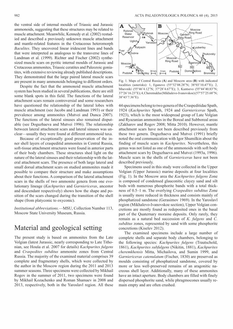
Material and geological settingThe present study is based on ammonites from the Late Volgian (latest Jurassic, nearly corresponding to Late Titho-nian, see Houša et al. 2007 for details) Kachpurites fulgens and Craspedites subditus ammonite zones from Central Russia. The majority of the examined material comprises 39 complete and fragmentary shells, which were collected by the author in the Moscow region during the 2011 and 2013 summer seasons. Three specimens were collected by Mikhail Rogov in the summer of 2011, two specimens were found by Mikhail Kozachenko and Roman Shamaev in 2008 and 2013, respectively, both in the Yaroslavl region. All these
44 specimens belong to two genera of the Craspeditidae Spath, 1924 (Kachpurites Spath, 1924 and Garniericeras Spath, 1923), which is the most widespread group of Late Volgian and Ryazanian ammonites in the Boreal and Subboreal areas (Zakharov and Rogov 2008; Mitta 2010). However, mantle attachment scars have not been described previously from these two genera. Doguzhaeva and Mutvei (1991) briefly noted the oral communication with Igor Shumilkin about the finding of muscle scars in Kachpurites. Nevertheless, this genus was not listed as one of the ammonoids with soft body attachment scars by Doguzhaeva and Mutvei (1993a, 1996). Muscle scars in the shells of Garniericeras have not been described previously.
Specimens used in this study were collected in the Upper Volgian (Upper Jurassic) marine deposits at four localities (Fig. 1). In the Moscow area the Kachpurites fulgens Zone is composed of condensed glauconitic clayey sand and silt beds with numerous phosphorite bands with a total thick-ness of 0.5–1 m. The overlying Craspedites subditus Zone is usually more reduced in thickness and consists mainly of phosphatized sandstone (Gerasimov 1969). In the Yaroslavl region (Mikhalevo-Ivanovskoe section), Upper Volgian con-cretions are mostly found as redeposited ones in the basal part of the Quaternary moraine deposits. Only rarely, they remain as a natural bed succession of K. fulgens and C. subditus zones, represented by sand with some phosphorite concretions (Kiselev 2012).
The examined specimens include a large number of complete shells and separate body chambers, belonging to the following species: Kachpurites fulgens (Trautschold, 1861), Kachpurites subfulgens (Nikitin, 1881), Kachpurites cheremkhensis Mitta, Michailova, and Sumin 1999, and Garniericeras catenulatum (Fischer, 1830) are preserved as moulds consisting of phosphatized sandstone, covered by more or less well-preserved remains of an aragonitic na-creous shell layer. Additionally, many of these ammonites have an intact aperture. Body chambers are filled with finely dispersed phosphorite sand, while phragmocones usually re-main empty and are often crushed.
Fig. 1. Maps of Central Russia (A) and Moscow area (B) with indicated localities (asterisks): 1, Eganovo (55°32’08.28”N; 38°03’10.47”E); 2, Mnevniki (55°46’4.12”N; 37°28’4.67”E); 3, Kuntsevo (55°44’40.83”N; 37°26’16.23”E); 4, Cheremukha (Mikhalevo-Ivanovskoe) (57°57’25.68”N; 38°43’7.36”E).
MIRONENKO—ATTACHMENT SCARS OF JURASSIC AMMONITES 983
Kachpurites first appeared at the end of the Epivirgatites nikitini Chron of the Middle Volgian (Rogov and Zakharov 2009) and flourished in the beginning of the Late Volgian (Kachpurites fulgens Chron). Garniericeras derived from Kachpurites at the beginning of the Craspedites subditus Chron (Sasonova and Sasonov 1983; Kiselev 2012). Kachpurites shells used in this study all come from the K. fulgens Zone and Garniericeras ones from the lower part of the Craspedites subditus Zone. These ammonites are characterized mainly by small to moderate shell size with adult diameters between 3.5 and 9 cm. Kachpurites fulgens is one of the older Late Volgian species of this genus. Kachpurites cheremkhensis and Kachpurites subfulgens are considered to be descendants of K. fulgens. Shells of K. fulgens and K. cheremkhensis have platyconic shells, while shells of K. subfulgens are character-ized by a more involute discoconic shape. K. subfulgens differs from its ancestors in having higher and narrower whorls and a narrower umbilicus. Garniericeras catenulatum, which likely derived from the latest K. subfulgens, has an involute oxyconic keeled shell with shorter body chamber.
To observe the markings on the shells, all specimens were viewed at various angles under incident illumination. Some specimens were examined with an optical microscope.
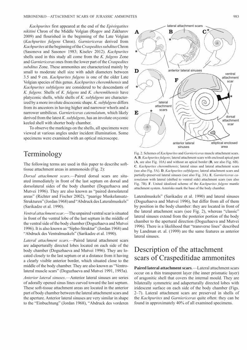
TerminologyThe following terms are used in this paper to describe soft -tissue attachment areas in ammonoids (Fig. 2):Dorsal attachment scars.—Paired dorsal scars are situ-ated immediately in front of the last septum on dorsal and dorsolateral sides of the body chamber (Doguzhaeva and Mutvei 1996). They are also known as “paired dorsolateral areas” (Richter and Fischer 2002), “paarige Muskelansatz-Strukturen” (Jordan 1968) and “Abdruck des Lateralmuskels” (Sarikadze et al. 1990).Ventral attachment scar.—The unpaired ventral scar is situated in front of the ventral lobe of the last septum in the middle of the ventral side of the body chamber (Doguzhaeva and Mutvei 1996). It is also known as “Sipho-Struktur” (Jordan 1968) and “Abdruck des Ventralmuskels” (Sarikadze et al. 1990).Lateral attachment scars.—Paired lateral attachment scars are adaperturally directed lobes located on each side of the body chamber (Doguzhaeva and Mutvei 1996). They are lo-cated closely to the last septum or at a distance from it having a clearly visible anterior border, which situated close to the middle of the body chamber. They are also known as “Ventro-lateral muscle scars” (Doguzhaeva and Mutvei 1991, 1993a).Anterior lateral sinuses.—Anterior lateral sinuses are series of adorally opened sinus lines curved toward the last septum. These soft-tissue attachment areas are located in the anterior part of body chamber between the lateral attachment scars and the aperture. Anterior lateral sinuses are very similar in shape to the “Einbuchtung” (Jordan 1968), “Abdruck des vor deren
Lateralmuskels” (Sarikadze et al. 1990) and lateral sinuses (Doguzhaeva and Mutvei 1996), but differ from all of them by position in the body chamber: they are located in front of the lateral attachment scars (see Fig. 2), whereas “classic” lateral sinuses extend from the posterior portion of the body chamber to the apertural direction (Doguzhaeva and Mutvei 1996). There is a likelihood that “transverse lines” described by Landman et. al. (1999) are the same features as anterior lateral sinuses.
Description of the attachment scars of Craspeditidae ammonitesPaired lateral attachment scars.—Lateral attachment scars occur on a thin transparent layer (the inner prismatic layer) of aragonitic shell that covers the internal mould. They are bilaterally symmetric and adaperturally directed lobes with iridescent surface on each side of the body chamber (Figs. 2–7). Lateral attachment scars are preserved in shells of the Kachpurites and Garniericeras quite often: they can be found in approximately 40% of all examined specimens.
lateral attachment scars
anterior lateral sinuses
A B C
D
E
F
lateralattachment
scars
ventralattachment
scar
dorsalattachment
scar
anterior lateralsinuses
elliptical enclosedarea
Fig. 2. Schemes of Kachpurites and Garniericeras muscle attachment scars. A, B. Kachpurites fulgens; lateral attachment scars with enclosed apical part (A, see also Fig. 10A) and without an apical border (B, see also Fig. 6B). C. Kachpurites cheremkhensis; lateral sinus and lateral attachment scars (see also Fig. 5A). D. Kachpurites subfulgens; lateral attachment scars and partially-preserved lateral sinuses (see also Fig. 3A). E. Garniericeras ca-tenulatum with lateral (shifted to ventral side) attachment scars (see also Fig. 7B). F. United idealized scheme of the Kachpurites fulgens mantle attachment system. Asterisks mark the base of the body chamber.
984 ACTA PALAEONTOLOGICA POLONICA 60 (4), 2015
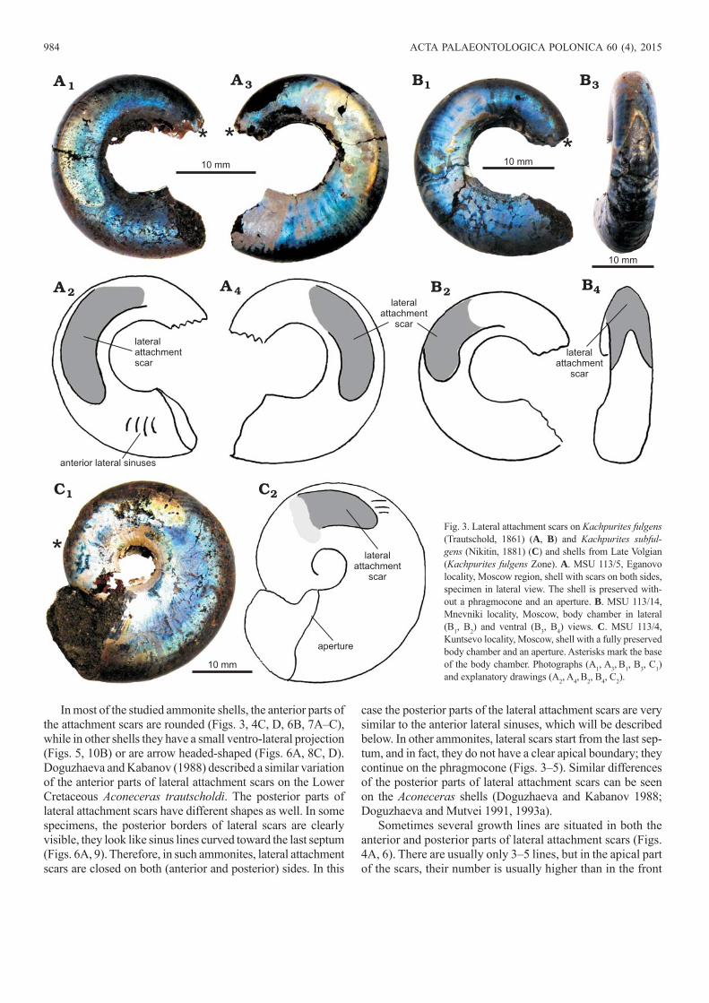
In most of the studied ammonite shells, the anterior parts of the attachment scars are rounded (Figs. 3, 4C, D, 6B, 7A–C), while in other shells they have a small ventro-lateral projection (Figs. 5, 10B) or are arrow headed-shaped (Figs. 6A, 8C, D). Doguzhaeva and Kabanov (1988) described a similar variation of the anterior parts of lateral attachment scars on the Lower Cretaceous Aconeceras trautscholdi. The posterior parts of lateral attachment scars have different shapes as well. In some specimens, the posterior borders of lateral scars are clearly visible, they look like sinus lines curved toward the last septum (Figs. 6A, 9). Therefore, in such ammonites, lateral attachment scars are closed on both (anterior and posterior) sides. In this
case the posterior parts of the lateral attachment scars are very similar to the anterior lateral sinuses, which will be described below. In other ammonites, lateral scars start from the last sep-tum, and in fact, they do not have a clear apical boundary; they continue on the phragmocone (Figs. 3–5). Similar differences of the posterior parts of lateral attachment scars can be seen on the Aconeceras shells (Doguzhaeva and Kabanov 1988; Doguzhaeva and Mutvei 1991, 1993a).
Sometimes several growth lines are situated in both the anterior and posterior parts of lateral attachment scars (Figs. 4A, 6). There are usually only 3–5 lines, but in the apical part of the scars, their number is usually higher than in the front
2A
3A
4A
A 1
2B
3B
4B
B1
10 mm 10 mm
10 mm
lateralattachmentscar
anterior lateral sinuses
lateralattachment
scar
lateralattachment
scar
2CC1
10 mm
aperture
lateralattachment
scar
Fig. 3. Lateral attachment scars on Kachpurites ful gens (Trautschold, 1861) (A, B) and Kachpurites subful-gens (Nikitin, 1881) (C) and shells from Late Volgian (Kachpurites fulgens Zone). A. MSU 113/5, Eganovo locality, Moscow region, shell with scars on both sides, specimen in lateral view. The shell is preserved with-out a phragmocone and an aperture. B. MSU 113/14, Mnevniki locality, Moscow, body chamber in lateral (B1, B2) and ventral (B3, B4) views. C. MSU 113/4, Kuntsevo locality, Moscow, shell with a fully preserved body chamber and an aperture. Asterisks mark the base of the body chamber. Photographs (A1, A3, B1, B3, C1) and explanatory drawings (A2, A4, B2, B4, C2).
MIRONENKO—ATTACHMENT SCARS OF JURASSIC AMMONITES 985
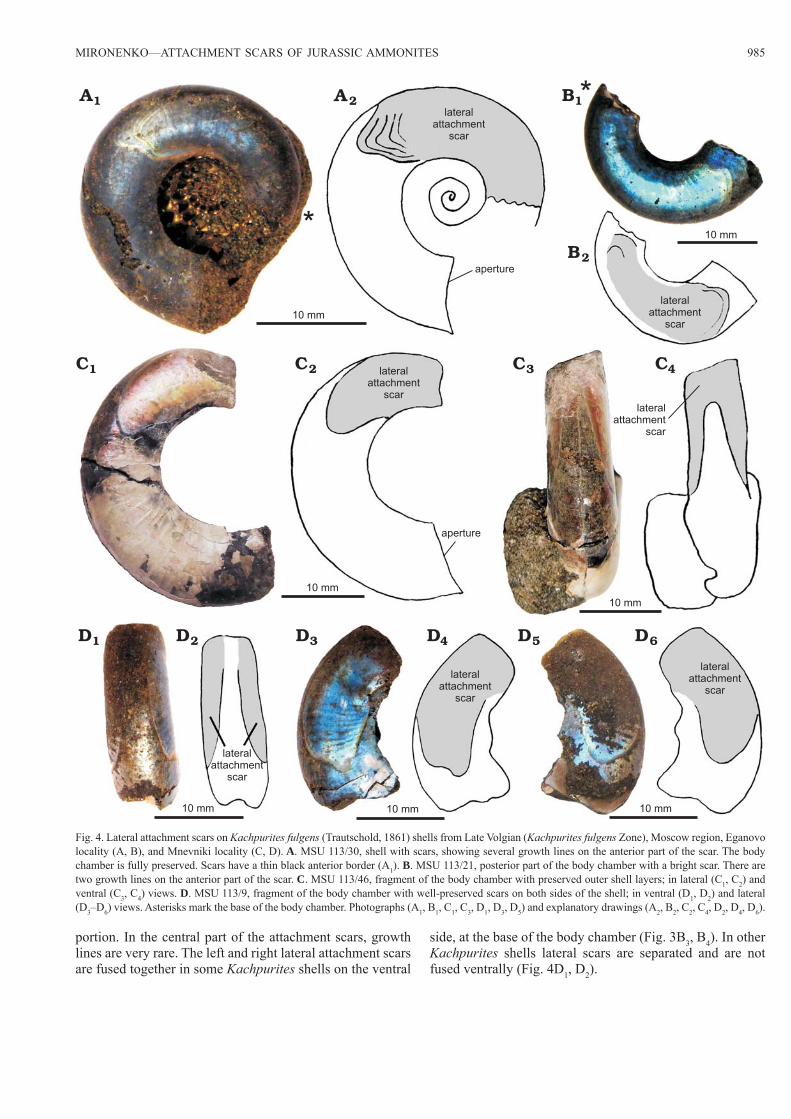
portion. In the central part of the attachment scars, growth lines are very rare. The left and right lateral attachment scars are fused together in some Kachpurites shells on the ventral
side, at the base of the body chamber (Fig. 3B3, B4). In other Kachpurites shells lateral scars are separated and are not fused ventrally (Fig. 4D1, D2).
Fig. 4. Lateral attachment scars on Kachpurites fulgens (Trautschold, 1861) shells from Late Volgian (Kachpurites fulgens Zone), Moscow region, Eganovo locality (A, B), and Mnevniki locality (C, D). A. MSU 113/30, shell with scars, showing several growth lines on the anterior part of the scar. The body chamber is fully preserved. Scars have a thin black anterior border (A1). B. MSU 113/21, posterior part of the body chamber with a bright scar. There are two growth lines on the anterior part of the scar. C. MSU 113/46, fragment of the body chamber with preserved outer shell layers; in lateral (C1, C2) and ventral (C3, C4) views. D. MSU 113/9, fragment of the body chamber with well-preserved scars on both sides of the shell; in ventral (D1, D2) and lateral (D3–D6) views. Asterisks mark the base of the body chamber. Photographs (A1, B1, C1, C3, D1, D3, D5) and explanatory drawings (A2, B2, C2, C4, D2, D4, D6).
aperture
lateralattachment
scar
10 mm
2AA1
2B
B1
2C 3C 4CC1
10 mm
10 mm
10 mm
10 mm
lateralattachment
scar
lateralattachment
scar
lateralattachment
scar
aperture
lateralattachment
scar
lateralattachment
scar
lateralattachment
scar
10 mm 10 mm
2D 3D 4D 5DD1 6D
986 ACTA PALAEONTOLOGICA POLONICA 60 (4), 2015
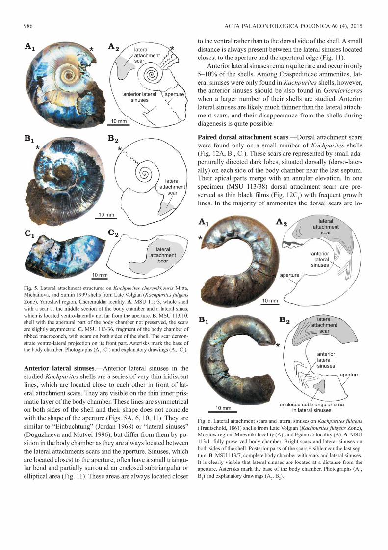
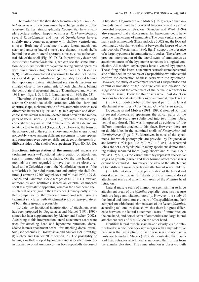
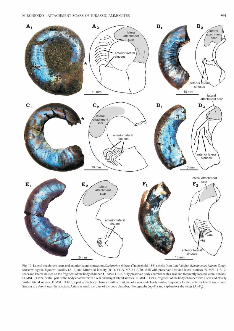
Anterior lateral sinuses.—Anterior lateral sinuses in the studied Kachpurites shells are a series of very thin iridiscent lines, which are located close to each other in front of lat-eral attachment scars. They are visible on the thin inner pris-matic layer of the body chamber. These lines are symmetrical on both sides of the shell and their shape does not coincide with the shape of the aperture (Figs. 5A, 6, 10, 11). They are similar to “Einbuchtung” (Jordan 1968) or “lateral sinuses” (Doguzhaeva and Mutvei 1996), but differ from them by po-sition in the body chamber as they are always located between the lateral attachments scars and the aperture. Sinuses, which are located closest to the aperture, often have a small triangu-lar bend and partially surround an enclosed subtriangular or elliptical area (Fig. 11). These areas are always located closer
to the ventral rather than to the dorsal side of the shell. A small distance is always present between the lateral sinuses located closest to the aperture and the apertural edge (Fig. 11).
Anterior lateral sinuses remain quite rare and occur in only 5–10% of the shells. Among Craspeditidae ammonites, lat-eral sinuses were only found in Kachpurites shells, however, the anterior sinuses should be also found in Garniericeras when a larger number of their shells are studied. Anterior lateral sinuses are likely much thinner than the lateral attach-ment scars, and their disappearance from the shells during diagenesis is quite possible.
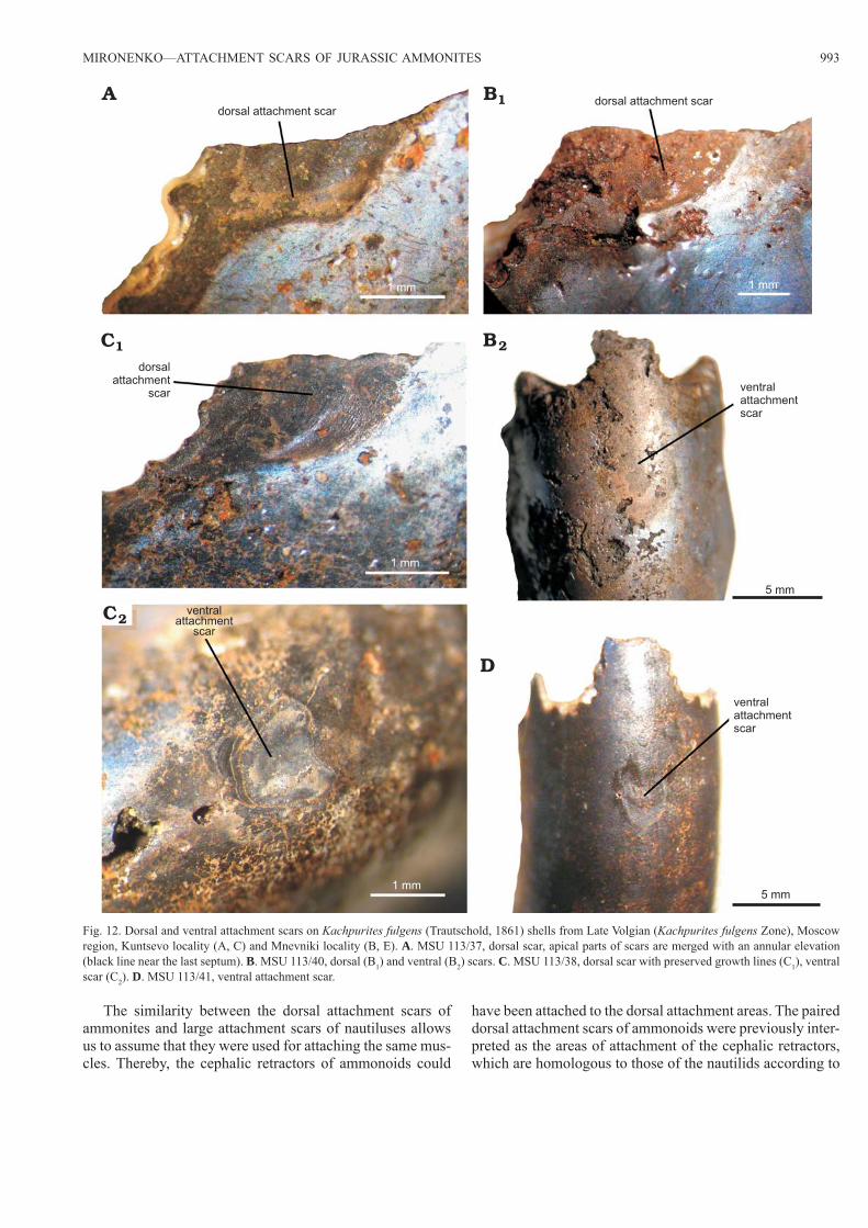
Paired dorsal attachment scars.—Dorsal attachment scars were found only on a small number of Kachpurites shells (Fig. 12A, B1, C1). These scars are represented by small ada-perturally directed dark lobes, situated dorsally (dorso-later-ally) on each side of the body chamber near the last septum. Their apical parts merge with an annular elevation. In one specimen (MSU 113/38) dorsal attachment scars are pre-served as thin black films (Fig. 12C1) with frequent growth lines. In the majority of ammonites the dorsal scars are lo-
Fig. 5. Lateral attachment structures on Kachpurites cheremkhensis Mitta, Michailova, and Sumin 1999 shells from Late Volgian (Kachpurites fulgens Zone), Yaroslavl region, Cheremukha locality. A. MSU 113/3, whole shell with a scar at the middle section of the body chamber and a lateral sinus, which is located ventro-laterally not far from the aperture. B. MSU 113/10, shell with the apertural part of the body chamber not preserved, the scars are slightly asymmetric. C. MSU 113/36, fragment of the body chamber of ribbed macroconch, with scars on both sides of the shell. The scar demon-strate ventro-lateral projection on its front part. Asterisks mark the base of the body chamber. Photographs (A1–C1) and explanatory drawings (A2–C2).
aperture
lateralattachment
scar
10 mm
2AA1
2BB1
2CC1
10 mm
lateralattachmentscar
lateralattachment
scar
10 mm
anterior lateralsinuses
Fig. 6. Lateral attachment scars and lateral sinuses on Kachpurites fulgens (Trautschold, 1861) shells from Late Volgian (Kachpurites fulgens Zone), Moscow region, Mnevniki locality (A), and Eganovo locality (B). A. MSU 113/1, fully preserved body chamber. Bright scars and lateral sinuses on both sides of the shell. Posterior parts of the scars visible near the last sep-tum. B. MSU 113/7, complete body chamber with scars and lateral sinuses. It is clearly visible that lateral sinuses are located at a distance from the aperture. Asterisks mark the base of the body chamber. Photographs (A1, B1) and explanatory drawings (A2, B2).
aperture
lateralattachment
scar
10 mm
2AA1
2BB1
10 mm
lateralattachment
scar
anteriorlateralsinuses
aperture
enclosed subtriangular areain lateral sinuses
anteriorlateral
sinuses
MIRONENKO—ATTACHMENT SCARS OF JURASSIC AMMONITES 987
cated on the dorsal and dorsolateral sides, leaning over the umbilical seam (Jordan 1968: pl. 2: 2, 3; Doguzhaeva and Mutvei 1996: fig. 3; Landman et al. 1999: fig. 21), however, in studied Kachpurites shells the dorsal side is not available for inspection since it is covered with phragmocone remnants, which are difficult to remove. The rarity of findings of dorsal scars in Craspeditidae can be explained by the small size of these scars in relatively small Kachpurites and Garniericeras shells and by the shell thinness in the area of their location.
Unpaired ventral attachment scars.—Ventral attachment scars were also found on a small number of Kachpurites specimens (Fig. 12B2, C2, D). They are marked by small round dark areas in the middle of the ventral side in front of the last septum, situated at a short (several millimeters on studied shells) distance from the last septum, but their apical parts always merge with the annular elevation.
DiscussionShell shape and the position of the attachment scars in the family Craspeditidae.—Several examples of lateral attach-ment scars in ammonoids belonging to different superfami-
lies have been well studied (Doguzhaeva and Kabanov 1988; Doguzhaeva and Mutvei 1991, 1993a; Richter 2002; Klug et al. 2007). However, the detailed preservation of the shells of the Late Volgian craspeditids allows us to compare the shape and position of these scars in four species of a single evolu-tionary line of ammonoids.
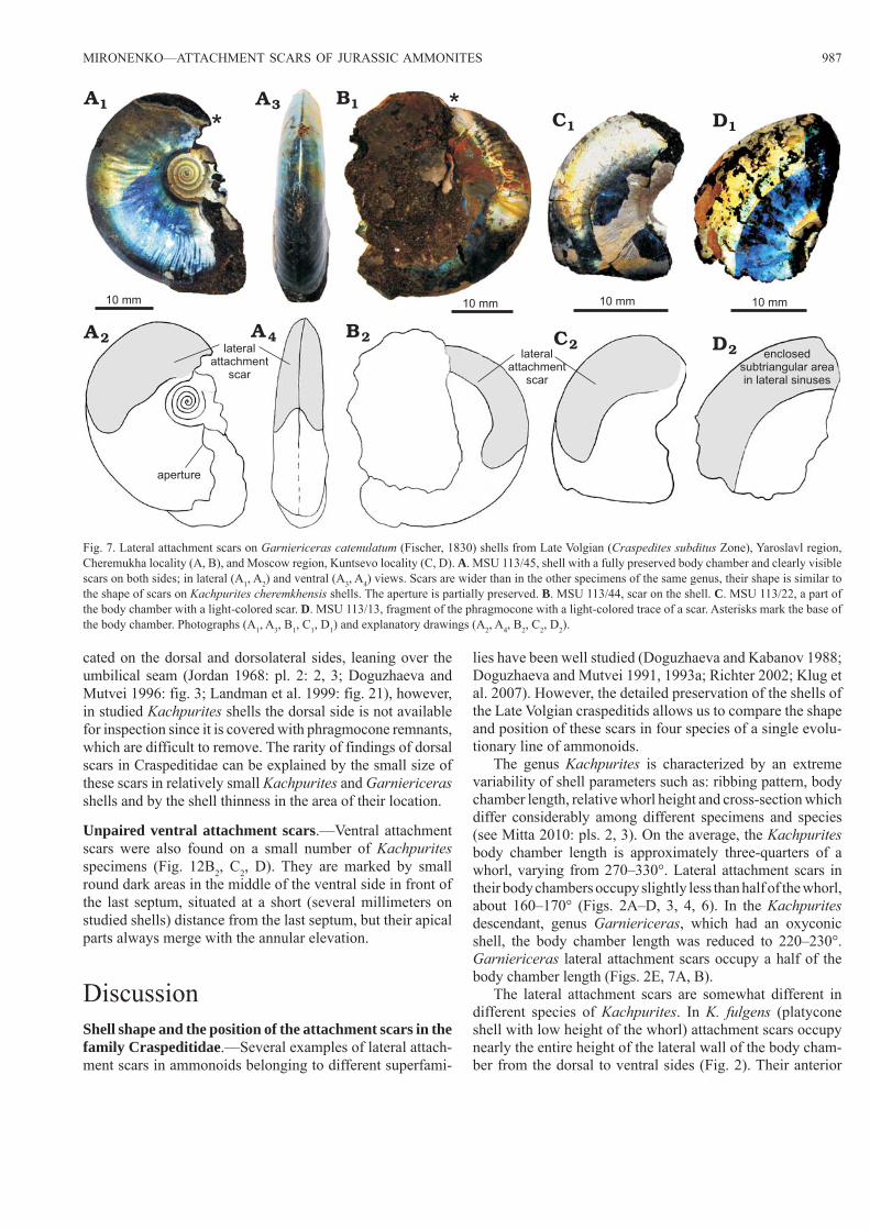
The genus Kachpurites is characterized by an extreme variability of shell parameters such as: ribbing pattern, body chamber length, relative whorl height and cross -section which differ considerably among different specimens and species (see Mitta 2010: pls. 2, 3). On the average, the Kachpurites body chamber length is approximately three-quarters of a whorl, varying from 270–330°. Lateral attachment scars in their body chambers occupy slightly less than half of the whorl, about 160–170° (Figs. 2A–D, 3, 4, 6). In the Kachpurites descendant, genus Garniericeras, which had an oxyconic shell, the body chamber length was reduced to 220–230°. Garniericeras lateral attachment scars occupy a half of the body chamber length (Figs. 2E, 7A, B).
The lateral attachment scars are somewhat different in different species of Kachpurites. In K. fulgens (platycone shell with low height of the whorl) attachment scars occupy nearly the entire height of the lateral wall of the body cham-ber from the dorsal to ventral sides (Fig. 2). Their anterior
Fig. 7. Lateral attachment scars on Garniericeras catenulatum (Fischer, 1830) shells from Late Volgian (Craspedites subditus Zone), Yaroslavl region, Cheremukha locality (A, B), and Moscow region, Kuntsevo locality (C, D). A. MSU 113/45, shell with a fully preserved body chamber and clearly visible scars on both sides; in lateral (A1, A2) and ventral (A3, A4) views. Scars are wider than in the other specimens of the same genus, their shape is similar to the shape of scars on Kachpurites cheremkhensis shells. The aperture is partially preserved. B. MSU 113/44, scar on the shell. C. MSU 113/22, a part of the body chamber with a light-colored scar. D. MSU 113/13, fragment of the phragmocone with a light-colored trace of a scar. Asterisks mark the base of the body chamber. Photographs (A1, A3, B1, C1, D1) and explanatory drawings (A2, A4, B2, C2, D2).
A1
2A
10 mm 10 mm
B1
2B
3A
4Alateral
attachmentscar
aperture
10 mm10 mm
C1
2Clateral
attachmentscar
enclosedsubtriangular areain lateral sinuses
D1
2D
988 ACTA PALAEONTOLOGICA POLONICA 60 (4), 2015
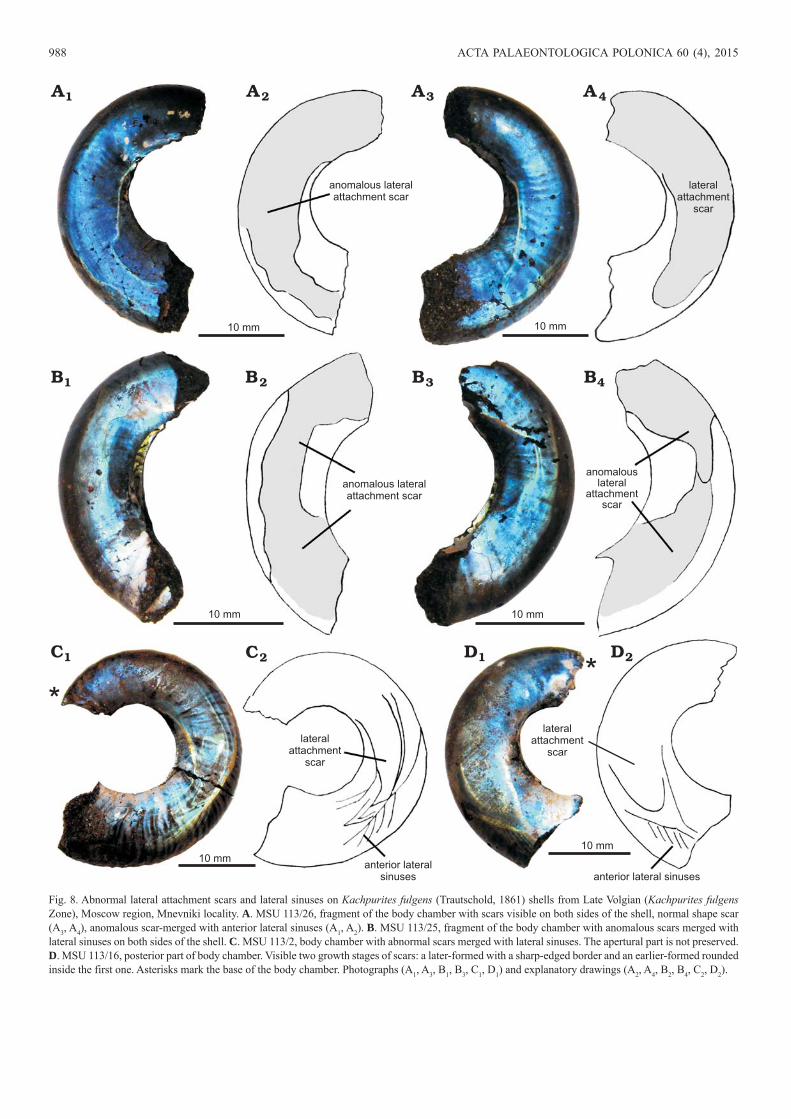
Fig. 8. Abnormal lateral attachment scars and lateral sinuses on Kachpurites fulgens (Trautschold, 1861) shells from Late Volgian (Kachpurites fulgens Zone), Moscow region, Mnevniki locality. A. MSU 113/26, fragment of the body chamber with scars visible on both sides of the shell, normal shape scar (A3, A4), anomalous scar-merged with anterior lateral sinuses (A1, A2). B. MSU 113/25, fragment of the body chamber with anomalous scars merged with lateral sinuses on both sides of the shell. C. MSU 113/2, body chamber with abnormal scars merged with lateral sinuses. The apertural part is not preserved. D. MSU 113/16, posterior part of body chamber. Visible two growth stages of scars: a later-formed with a sharp-edged border and an earlier-formed rounded inside the first one. Asterisks mark the base of the body chamber. Photographs (A1, A3, B1, B3, C1, D1) and explanatory drawings (A2, A4, B2, B4, C2, D2).
lateralattachment
scar
lateralattachment
scar
10 mm
2AA1
B1
2CC1
10 mm
lateralattachment
scar
lateralattachment
scar
10 mm
*
anterior lateralsinuses
10 mm
10 mm
10 mm
anterior lateral sinuses
3A 4A
2DD1
2B 3B 4B
anomalous lateralattachment scar
anomalous lateralattachment scar
anomalouslateral
attachmentscar
anomalouslateral
attachmentscar
MIRONENKO—ATTACHMENT SCARS OF JURASSIC AMMONITES 989
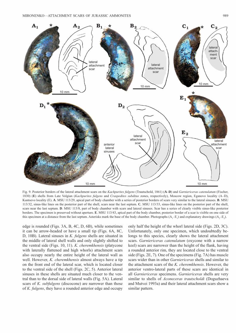
edge is rounded (Figs. 3A, B, 4C, D, 6B), while sometimes it can be arrow-headed or have a small tip (Figs. 6A, 8C, D, 10B). Lateral sinuses in K. fulgens shells are situated in the middle of lateral shell walls and only slightly shifted to the ventral side (Figs. 10, 11). K. cheremkhensis (platycone with laterally flattened and high whorls) attachment scars also occupy nearly the entire height of the lateral wall as well. However, K. cheremkhensis almost always have a tip on the front end of the lateral scar, which is located closer to the ventral side of the shell (Figs. 2C, 5). Anterior lateral sinuses in these shells are situated much closer to the ven-tral than to the dorsal side of lateral walls (Fig. 5A). Lateral scars of K. subfulgens (discocone) are narrower than those of K. fulgens, they have a rounded anterior edge and occupy
only half the height of the whorl lateral side (Figs. 2D, 3C). Unfortunately, only one specimen, which undoubtedly be-longs to this species, clearly shows the lateral attachment scars. Garniericeras catenulatum (oxycone with a narrow keel) scars are narrower than the height of the flank, having a rounded anterior rim, they are located close to the ventral side (Figs. 2E, 7). One of the specimens (Fig. 7A) has muscle scars wider than in other Garniericeras shells and similar to the attachment scars of the K. cheremkhensis. However, the anterior ventro-lateral parts of these scars are identical in all Garniericeras specimens. Garniericeras shells are very similar to shells of Aconeceras trautscholdi (Doguzhaeva and Mutvei 1993a) and their lateral attachment scars show a similar pattern.
Fig. 9. Posterior borders of the lateral attachment scars on the Kachpurites fulgens (Trautschold, 1861) (A–D) and Garniericeras catenulatum (Fischer, 1830) (E) shells from Late Volgian (Kachpurites fulgens and Craspedites subditus zones, respectively), Moscow region, Eganovo locality (A–D), Kuntsevo locality (E). A. MSU 113/29, apical part of body chamber with a series of posterior borders of scars very similar to the lateral sinuses. B. MSU 113/32, sinus-like lines on the posterior part of the shell, scars near the last septum. C. MSU 113/33, sinus-like lines on the posterior part of the shell, scars near the last septum. D. MSU 113/8, part of body chamber with scars and lateral sinuses. Scar has a series of clearly visible sinus-like posterior borders. The specimen is preserved without aperture. E. MSU 113/43, apical part of the body chamber, posterior border of a scar is visible on one side of this specimen at a distance from the last septum. Asterisks mark the base of the body chamber. Photographs (A1–E1) and explanatory drawings (A2–E2).
lateralattachmentscar
10 mm
2AA1 B1
2DD1
10 mm
lateralattachment
scar
lateralattachment
scar
10 mm
10 mm
10 mm
2CC12B
anteriorlateral
sinuses
lateralattach-mentscar
lateralattachment
scar
2E
E1
990 ACTA PALAEONTOLOGICA POLONICA 60 (4), 2015
The evolution of the shell shape from the early Kachpurites to Garniericeras is accompanied by a change in shape of the aperture. Earliest stratigraphically Kachpurites have a sim-ple aperture without lappets or sinuses. K. cheremkhensis, several K. subfulgens, and most of Garniericeras have a slightly more complex aperture with shallow ventrolateral sinuses. Both lateral attachment areas: lateral attachment scars and anterior lateral sinuses, are situated in such shells behind these ventrolateral apertural sinuses, close to the ven-tral side of the shell (Fig. 2C, D, E). In previously described Aconeceras trautscholdi shells, we can see the same situa-tion. Aconeceras shells are oxyconic having curved apertures with two sinuses (Doguzhaeva and Mutvei 1991: text-figs. 8, 9), shallow dorsolateral (presumably located behind the eyes) and deeper ventrolateral (presumably located behind the hyponome). Lateral attachment scars in Aconeceras are situated close to the ventral side of body chambers, behind the ventrolateral apertural sinuses (Doguzhaeva and Mutvei 1991: text-figs. 1, 3, 4, 8, 9; Landman et al. 1999: fig. 23).
Therefore, the position of the lateral attachment muscle scars in Craspeditidae shells correlated with shell form and aperture shape, a characteristic of this ammonite species (see difference between Fig. 2B and D). In platyconic and disco-conic shells lateral scars are located most often on the middle parts of lateral sides (Fig. 2A–C, F), whereas in keeled oxy-conic shells they are shifted to the ventro-lateral side and are located close to the keel (Figs. 2D, 7). However, the form of the anterior part of the scar is a more unique characteristic and noticeably varies among different specimens in one species and sometimes even between different stages of the growth or different sides of the shell of one specimen (Figs. 4D, 8A, D).
Functional interpretation of the ammonoid muscle at-tachment scars.—Functional interpretation of attachment scars in ammonoids is speculative. On the one hand, am-monoids are now regarded to have been more closely re-lated to the Coleoidea than to the Nautiloidea because of the similarities in the radular structure and embryonic shell fea-tures (Lehmann 1976; Doguzhaeva and Mutvei 1992, 1993b; Jacobs and Landman 1993; Kröger et al. 2011). However, ammonoids and nautiloids shared an external chambered shell as a hydrostatic apparatus, whereas the chambered shell is internal or vestigial in the Coleoidea. Consequently, a fur-ther comparison of the observed ammonoid soft tissue at-tachment structures with attachment scars of representatives of both these groups is plausible.
To date, the functional interpretation of attachment scars has been proposed by Doguzhaeva and Mutvei (1991, 1996) somewhat later supplemented by Richter and Fischer (2002). According to this interpretation lateral attachment scars were used for attaching head and hyponome retractors, dorsal (dorso -lateral) attachment scars—for attaching dorsal retrac-tors (see schemes in Doguzhaeva and Mutvei 1991: text-fig. 8; Richter and Fischer 2002: text-fig. 5). The possibility of having a well-developed hyponome (and associated muscles) in normally-coiled ammonoids has been repeatedly discussed
in literature. Doguzhaeva and Mutvei (1991) argued that am-monoids could have had powerful hyponome and a pair of powerful hyponome retractors. Saunders and Ward (1994) also suggested that a strong muscular hyponome could have been the main engine of ammonites. The deep ventral sinus of many early ammonoids (Korn and Klug 2002) and the forward pointing sub-circular ventral sinus between the lappets of some microconchs (Westermann 1990: fig. 2) support the presence of a large hyponome in ammonite soft bodies. Therefore, the previous interpretation of the lateral scars of ammonoids as attachment areas of the hyponome retractors is a logical con-clusion. All modern cephalopods have a ventral hyponome. The shifting of the lateral attachment scars closer to the ventral side of the shell in the course of Craspeditidae evolution could confirm the connection of these scars with the hyponome. However, the study of attachment scars of Craspeditidae and careful examination of the literature calls into question the suggestion about the attachment of the cephalic retractors to the lateral scars. Below are three facts which cast doubt on previous functional interpretations of ammonoid muscle scars:
(i) Lack of double lobes on the apical part of the lateral attachment scars in Kachpurites and Garniericeras shells.
Doguzhaeva and Mutvei (1991, 1993a) pointed out that in several Aconeceras specimens the apical parts of the lateral muscle scars are subdivided into two minor lobes, ventral and dorsal. This was interpreted as evidence of two different muscles attached to this place. However, there are no double lobes in the examined shells of Kachpurites and Garniericeras (Figs. 2–7). Moreover, in most of the speci-mens, for which photographs are provided by Doguzhaeva and Mutvei (1991: pls. 2: 2, 3; 3: 2; 7: 1–3; 8: 1, 3), separated lobes are not clearly visible. In many specimens demonstrat-ing visibly separated lobes (Doguzhaeva and Mutvei 1991: pls. 4: 1, 2; 6: 1, 2) the variant that they represent successive stages of growth (earlier and later formed attachment scars) cannot be excluded. This makes the idea of the attachment of two different muscles to lateral attachment scars unlikely.
(ii) Different structure and preservation of the lateral and dorsal attachment scars. Similarity of the ammonoid dorsal attachment scars and attachment areas of the Nautilus head retractors.
Lateral muscle scars of ammonites seem similar to large attachment areas of the Nautilus cephalic retractors because both are large and situated laterally. However, the study of the dorsal and lateral muscle scars of Craspeditidae and their comparison with the attachment scars of the Recent Nautilus, according to literature data, shows that there is a great differ-ence between the lateral attachment scars of ammonites on the one hand, and dorsal scars of ammonites and large lateral attachment areas of Nautilus on the other hand.
Nautilida lateral muscle scars have a clearly visible ante-rior border, while their backside merges with a myoadhesive band near the last septum. In fact, these scars do not have a posterior boundary. Mutvei (1957) demonstrated that nauti-loid head retractor attachment scars derive their origin from the annular elevation. The same situation is observed with
MIRONENKO—ATTACHMENT SCARS OF JURASSIC AMMONITES 991
Fig. 10. Lateral attachment scars and anterior lateral sinuses on Kachpurites fulgens (Trautschold, 1861) shells from Late Volgian (Kachpurites fulgens Zone), Moscow region, Eganovo locality (A, E) and Mnevniki locality (B–D, F). A. MSU 113/20, shell with preserved scar and lateral sinuses. B. MSU 113/12, scars and lateral sinuses on the fragment of the body chamber. C. MSU 113/6, fully preserved body chamber with a scar and frequently located lateral sinuses. D. MSU 113/39, central part of the body chamber with a scar and bright lateral sinuses. E. MSU 113/47, fragment of the body chamber with a scar and clearly visible lateral sinuses. F. MSU 113/15, a part of the body chamber with a front end of a scar and clearly visible frequently located anterior lateral sinus lines. Sinuses are absent near the aperture. Asterisks mark the base of the body chamber. Photographs (A1–F1) and explanatory drawings (A2–F2).
lateralattachment
scar
10 mm
2AA1 B1
2CC1
10 mm
lateralattachment
scar
lateralattachment scar
10 mm 10 mm
10 mm
2DD1
2B
anterior lateralsinuses
lateralattachment
scar
lateral attachmentscar
2EE1
anterior lateralsinuses
anterior lateralsinuses
anterior lateralsinuses
anterior lateralsinuses
anterior lateralsinuses
2FF1
10 mm
lateralattachment
scar
992 ACTA PALAEONTOLOGICA POLONICA 60 (4), 2015
small dorsal Kachpurites attachment scars: located near the last septum and posteriorly merged with the annular elevation (Fig. 10A, B1, C1). The same location of these scars has been depicted in literature (Jordan 1968: pl. 1: 2, 4). Furthermore, Nautilida lateral muscle scars have clear and frequent growth lines (Chirat 1997: pl. 2: 1; Klug and Lehmkuhl 2004: fig. 3A). Ammonoid paired dorsal attachment scars have a sim-ilar structure: growth lines are clearly visible in Jordan’s illustrations (Jordan 1968: pls. 1: 4; 2: 1, 5) and on the one of Kachpurites specimens (Fig. 10C1).
Lateral attachment scars of ammonoids were often de-picted as part of the annular elevation (Doguzhaeva and Mutvei 1996: fig. 2A, B) as well as Nautilus lateral attach-ment scars. However, lateral attachment scars of Kachpurites and Garniericeras can be located at a distance from the last septum and from the annular elevation, and they can have not only an anterior, but also a posterior boundary (Figs. 6A, 9). Their posterior portion is not merged with the annular eleva-tion, these scars are not surrounded by the myoadhesive band (in contrast to the large attachment areas of the Nautilus: see Klug and Lehmkuhl 2004: figs. 3, 4). A similar structure and position of the posterior part of the lateral scars has been observed in Aconeceras shell (Landman et al. 1999: fig. 23). In central part of ammonoid lateral attachment scars, as a
general rule, no growth lines are visible. Even if growth lines occur in lateral attachment scars (Figs. 4A, 6), they are sig-nificantly widely spaced and not similar to frequent growth lines of dorsal scars (Fig. 12C1).
Lateral and dorsal attachment scars differ not only in the position and structure of scars, but also in the type of their preservation (Dagys and Keupp 1998). On the pyrite internal moulds of ammonoid body chambers and phragmocones, dorsal and ventral attachment scars are represented by black fine-grained pyrite (Zimmermann 1985; Dagys and Keupp 1998; Richter 2002; Richter and Fischer 2002; Paul 2011). However, lateral attachment scars usually are represented by bright yellow pyrite, sometimes bordered by a thin black line (Dagys and Keupp 1998; Keupp 2000; Richter 2002; Richter and Fischer 2002). If the internal mould of the ammonite body chamber is composed of sandstone, phosphorite, sider-ite, etc., lateral attachment structures are preserved as a gen-eral rule if the nacreous layer of the shell is present (but see alternative variants in Richter and Fischer 2002; Klug et al. 2007). On the other hand, the dorsal and ventral attachment scars are often preserved in the mould devoid of the nacreous layer (Sarikadze et. al. 1990; Klug et al. 2007). Difference in types of preservation may indicate a different initial structure of these attachment scars and their different origin.
Fig. 11. Apertural parts of the anterior lateral sinuses of Kachpurites fulgens (Trautschold, 1861) from Late Volgian (Kachpurites fulgens Zone), Moscow region, Eganovo locality (B) and Mnevniki locality (A, C–F). A. MSU 113/24, lateral sinus line in the apertural part of the body chamber. B. MSU 113/7 (see also Fig. 6B), apertural part of the body chamber with lateral sinuses. C. MSU 113/12 (see also Fig. 10B), lateral sinuses in the apertural part of the body chamber. D. MSU 113/23, apertural part of the body chamber with lateral sinuses. E. MSU 113/27, apertural part of the body chamber with a very bright lateral sinus. F. MSU 113/6 (see also Fig. 10C), apertural part of the body chamber. Apertural edge is not preserved. Photographs (A1–F1) and explanatory drawings (A2–F2).
5 mm
2AA1 B1 2CC1
5 mm
5 mm
2DD1
2Banteriorlateral
sinuses
2EE1
subtriangular areaformed fromsinus lines
2FF1
5 mm anterior lateralsinuses
anteriorlateral
sinuses
subtriangulararea formed
from sinus lines
elliptical areaformed fromsinus lines
aperture aperture
aperture aperture
elliptical areaformed fromsinus lines
5 mm
aperture
5 mm
MIRONENKO—ATTACHMENT SCARS OF JURASSIC AMMONITES 993
The similarity between the dorsal attachment scars of ammonites and large attachment scars of nautiluses allows us to assume that they were used for attaching the same mus-cles. Thereby, the cephalic retractors of ammonoids could
have been attached to the dorsal attachment areas. The paired dorsal attachment scars of ammonoids were previously inter-preted as the areas of attachment of the cephalic retractors, which are homologous to those of the nautilids according to
Fig. 12. Dorsal and ventral attachment scars on Kachpurites fulgens (Trautschold, 1861) shells from Late Volgian (Kachpurites fulgens Zone), Moscow region, Kuntsevo locality (A, C) and Mnevniki locality (B, E). A. MSU 113/37, dorsal scar, apical parts of scars are merged with an annular elevation (black line near the last septum). B. MSU 113/40, dorsal (B1) and ventral (B2) scars. C. MSU 113/38, dorsal scar with preserved growth lines (C1), ventral scar (C2). D. MSU 113/41, ventral attachment scar.
1 mm
A B1
C1
1 mm
1 mm5 mm
5 mm
D
2B
1 mm
dorsal attachment scardorsal attachment scar
ventralattachmentscar
dorsalattachment
scar
ventralattachmentscar
ventralattachment
scar2C
994 ACTA PALAEONTOLOGICA POLONICA 60 (4), 2015
various authors (Crick 1898; Jordan 1968; Mutvei 1964). The small dorsal attachment scars are present not only in am-monoid shells, but also in the shells of Bactritida (ammonoid ancestors) and Orthocerida (ancestors of Bactritida) (Mutvei 2002; Kröger et al. 2005; Klug et al. 2008). Latest evidence on the possible origin of the Nautilida from early orthocerids (Kröger et. al. 2011) confirms the possibility of the existence of homologous muscles in ammonoids and nautilids.
(iii) Lack of the lateral attachment scars in the shells of the heteromorph ammonites with hook-like body chambers.
Among a plurality of heteromorphic ammonites, sev-eral taxa (including family Ancyloceratidae) have a shell with a hook-like adult body chamber. Muscle scars in Ancyloceratidae body chambers were first described by Crick (1898: pls. 17, 14, 17) and studied in detail by Doguzhaeva and Michailova (1991). Ancyloceratids have very large paired dorsal attachment scars, larger than in normally coiled ammonoids. At the same time, lateral attachment scars have not been found in their hook-like body chambers, even on well-preserved specimens (Doguzhaeva and Michailova 1991). Very likely muscles, which were attached to the lat-eral scars in normally-coiled ammonoids, were reduced in these heteromorphs.
Heteromorph ammonites with ancyloconic shells have been interpreted by paleontologists as very sluggish swim-mers, mainly pelagic vertical migrants (Westermann and Tsujita 1999), very likely incapable of actively swimming with hyponome propulsion (Doguzhaeva and Michailova 1991).
If we accept the hypothesis of the attachment of both the head and hyponome retractors to the lateral attachment areas, it stands to reason that both these muscles would have been reduced in Ancyloceratidae. Only the dorsal and ven-tral muscles remain. Here it is necessary to clarify what the dorsal muscles were. Richter and Fischer (2002) suggested that dorsal attachment scars can be interpreted as attach-ment sites of a second pair of cephalic retractor muscles. Recent Nautilus has only one pair of cephalic retractors (Mutvei 1957, Bizikov 2008). However, Bizikov (2002) de-scribed nuchal retractors in Nautilus soft bodies—a pair of powerful muscles that originate from the nuchal valves of the collar folds and extend backward to the dorsal wall of the body chamber. Coleoids, for example squids, have two pairs of cephalic retractors, lateral and medial components of the head retractors (Bizikov 2008). Bizikov (2008) also suggested that in Coleoidea soft bodies, lateral segments of the head retractors are homologous to Nautilus cephalic retractors and medial segments apparently are homologous to Nautilus nuchal retractors. Although the question of trans-formation of nuchal retractors, which initially were con-nected with the collar into second pair of cephalic retractors in Coleoidea has not been studied in detail, it is very likely that nuchal retractors could have become medial compo-nents of the head retractors only after the coleoid’s shell became internal and nuchal cartilage appeared. Since am-monites had external shells and they were able to withdraw their soft body very deep into the body chamber (Kröger
2002), nuchal retractors in their soft body likely were im-portant to them: these muscles could have been used for the retraction of the dorsal part of the collar. It can be assumed that dorsal retractors of ammonoids and nuchal retractors of Nautilus are the same muscles and they were connected with the collar, not with the head. But if these arguments are correct, the simultaneous reduction of the head and funnel retractors in the Ancyloceratidae seems unlikely. In the ab-sence of the head retractors these ammonites could not have drawn the head and arms into the shell. Numerous spikes on their shells show that they were forced to defend themselves against predators, so the ability to retract the soft body into the body chamber was important to them.
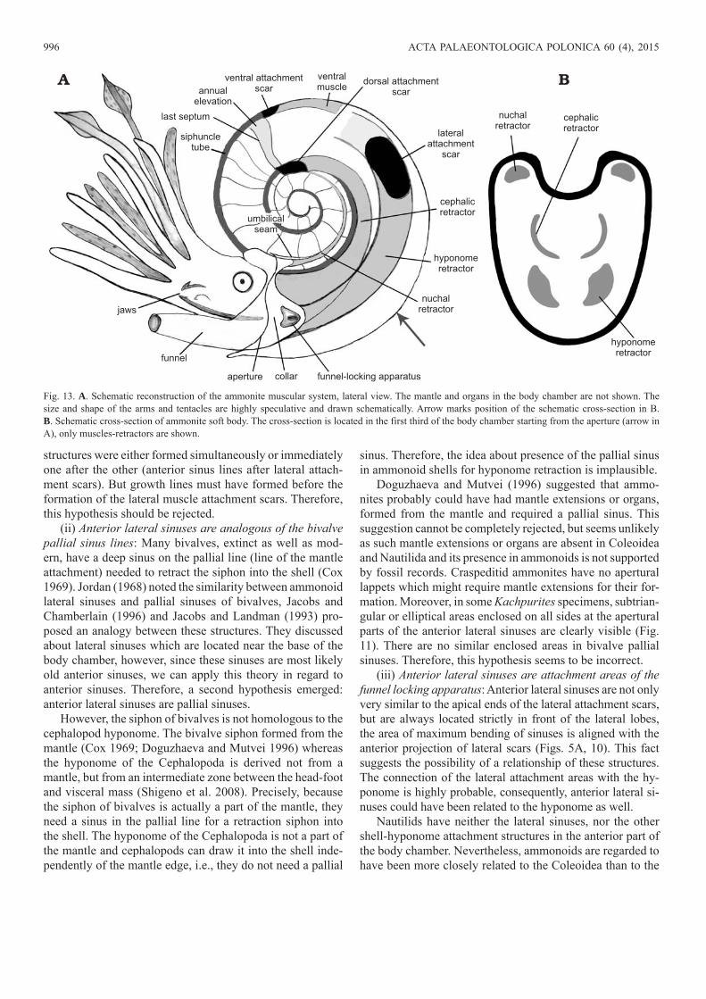
The assumption of the attachment to the lateral attach-ment scars only the hyponome retractors seems more likely. This explains the absence of double lobes in the anterior parts of the Kachpurites and Garniericeras lateral scars, and a reduction of these scars in hook-like ancyloceratid body chambers. Ancyloceratids are not considered by research-ers as active swimmers (Doguzhaeva and Michailova 1991; Westermann and Tsujita 1999) it is very likely that their hyponome and lateral (hyponomic) attachment areas were reduced, but their cephalic and nuchal retractors were saved to retract soft body into the shell.
Cephalic retractors of the ammonoids could have been attached to the dorsal (dorso-lateral) attachment scars along with nuchal (dorsal) retractors. Recent Nautilus has nuchal retractors attached along with cephalic retractors (between cephalic retractors on the dorsal side) and attachment areas of these two types of muscles are fused together (Bizikov 2002, 2008). Very likely, ammonoids had a similar location of these muscles: cephalic retractors were attached to the lateral parts of the paired dorsal attachment scars and nuchal (dorsal) retractors—to the dorsal parts of these scars. Ammonoids could have inherited this organization of muscle attachment from the Nautiloidea, probably from common ancestors with Nautilus (provided that Ammonoidea and Nautilida originated from one evolutionary branch of Nautiloidea see Kröger et. al. 2011). An indirect confirmation of such a de-sign of muscle attachment in ammonoids is a three-lobed an-nular elevation of Bactrites (Devonobactrites) sp. (Kröger et al. 2005: figs. 3, 4). This three-lobed dorsal attachment area is a part of the annular elevation exactly as in Nautilus at-tachment scars and ammonoid paired dorsal scars (but not the ammonoid paired lateral scars). Probably, the central (dorsal) lobe in Devonobactrites was an attachment area of the nuchal retractors, whereas marginal (dorso-lateral) lobes were used for attachment cephalic retractors.
The lateral ammonoid attachment scars (attachment areas of the hyponomic retractors) could have arisen later than the dorsal scars (since Nautilida do not have this type of scars), in Ammonoidea or in their common ancestors—Bactritida (in this case Ammonoidea and Coleoidea could have inher-ited large hyponome retractors from bactrites). However, the question of the evolution of the attachment scars is beyond the scope of this article.
MIRONENKO—ATTACHMENT SCARS OF JURASSIC AMMONITES 995
Relationship between the lateral attachment scars and anterior lateral sinuses.—Study of the lateral attachment scars of Kachpurites and Garniericeras shows that the apical parts of these scars have a different shape. In some specimens muscle scars are visible immediately from the last septum, and in fact they do not have an apical boundary (Figs. 3, 4, 6B). In other specimens lateral attachment scars are located at a distance from the last septum and have a posterior bound-ary (Figs. 6A, 9). This border consists of a series of adorally opened sinus lines curved toward the last septum. The lines are very similar to the anterior lateral sinuses located in the apertural parts of the body chambers (Fig. 10). Similar si-nus-like posterior borders of lateral attachment scars can be seen in previous publications (Doguzhaeva and Mutvei 1991: pls. 2: 2–4; 5: 1; 6: 1, 2; Landman et al. 1999: fig. 23; Keupp 2000: 105; Richter and Fischer 2002: text-fig. 3B, pl. 1B, E, G). If the body chambers are not fully preserved, these poste-rior borders of the attachment scars look like adorally-opened sinus lines, located near the last septum (Crick 1898: pl. 20: 5, 6, 8; Jordan 1968: pl. 10: 1, 3–7). These structures were described by Jordan (1968) as “Einbuchtung”. As usual, the lateral attachment scars and lateral sinuses (“Einbuchtung”) are interpreted as different muscle attachment structures (Doguzhaeva and Mutvei 1996). However, Dagys and Keupp (1998) pointed out that Jordan’s “Einbuchtung” are the same structures as large ventrolateral muscle scars which are de-scribed by Doguzhaeva and Mutvei (1991). The study of the soft-tissue attachment areas of Craspeditidae ammonites confirms this conclusion. The shape of the posterior bor-ders of the Kachpurites and Garniericeras lateral attachment scars (Figs. 6A, 9) is very similar to “Einbuchtung“ or lateral sinuses (Doguzhaeva and Mutvei 1996). Jordan believed that these structures were open toward the aperture (Jordan 1968: pls. 23–26), however, on the one of his photos the anterior border of this structure can be seen (Jordan 1968: pl. 10: 7) therefore it is a typical lateral attachment scar.
Similar lateral sinuses are absent in apical parts of the Nautilida lateral attachment scars (Chirat 1997: pls. 1, 2), in Orthocerida and Bactritida dorsal attachment areas (Kröger et al. 2005: fig. 4) and in ammonoid paired dorsal scars (Fig. 12A, B1, C1). This is one more confirmation of differences between these structures on the one hand and ammonoid lateral attachment scars on the other hand.
In Kachpurites body chambers the series of lateral sinus lines (called “anterior lateral sinuses”) are observed between lateral attachment scars and the aperture (Figs. 5A, 6, 9D, 10). They are similar to “Einbuchtung”, lateral sinuses sensu Doguzhaeva and Mutvei (1996) and posterior borders of the lateral scars by their shape, but differ from them by position on the body chamber: they are always located between the anterior borders of lateral attachments scars and the aperture. In the case of incomplete preservation of the body chamber sometimes it is difficult to determine what kind of sinus lines are visible on the specimen—posterior borders of the lateral attachment scars or parts of the anterior lateral sinuses (for example Sarikadze et al. 1990: abb. 1). Very likely, the simi-
larity between the shape of anterior lateral sinuses and sinus lines in the posterior parts of the lateral attachment scars is not accidental. A sinus-like shape of the posterior borders of the attachment scars could be explained through their forma-tion using the same part of the mantle, but another explana-tion seems more likely: these posterior borders of attachment scars most probably are anterior lateral sinuses, which had been formed during the previous stage of shell growth. It can be assumed that, at each stage of shell growth, old anterior sinuses became an apical part of the new lateral attachment scars, after which the ammonite formed new scars and new anterior sinuses in the new section of the shell.
Functional interpretation of the anterior lateral sinuses.—Interpretation of the functions of the anterior lateral sinuses is complex and speculative, since the modern nautilids do not possess such structures and Coleoidea have no outer body chamber, on which sinus lines could be seen.
Three hypotheses have been put forward as for the func-tion of the anterior lateral sinuses:
(i) Anterior lateral sinuses are growth lines on the inner layer of the body chamber. This hypothesis emerged during examination of the inner surface of Nautilus body chambers. In Recent and fossil Nautilida, there are series growth lines on the inner layer of the body chamber (Klug and Lehmkuhl 2004: fig. 4C). The shape of these lines is similar to the shape of the aperture edge, but not identical to it. The shape of anterior sinuses significantly differs from the shape of the apertural edge, more than the growth lines shape differ, however their frequency and location in the front of the body chamber at a small distance from the aperture resemble nau-tiloid growth lines.
The growth lines are formed by a mantle edge above the nacreous layer slightly behind the apertural margin (Klug and Lehmkuhl 2004). During further growth of shell, muscle attachment scars covered old growth lines. Respectively, if we study the internal mould of the body chamber with the inner layer and muscle scars, these lines are located above the muscle scars. However, in studied Kachpurites shells there are no anterior lateral sinus lines above the attachment scars (seen from the outside through a transparent shell wall). Conversely, we can see lateral attachment scar lobes cover-ing anterior lateral sinuses from our point of view (Figs. 6B, 9D, 10B), or even sinus lines which appear to be growing away from the scar rims (Figs. 6A, 8C, 10D).
A few K. fulgens specimens show an anomalous con-figuration of lateral attachments scars and anterior lateral sinuses: in these specimens anterior parts of the lateral mus-cle scars gradually merged with the lateral sinuses located in front of it without any clear boundaries (Fig. 8A1, A2, B). Such a phenomenon could not have taken place if the anterior lateral sinuses have been formed prior to the formation of the lateral lobes. The cause of this anomaly is still unclear how-ever, such a situation could have arisen during simultaneous formation of lateral attachment scars and lateral sinuses in the apertural part of the shell. It seems very likely that both
996 ACTA PALAEONTOLOGICA POLONICA 60 (4), 2015
structures were either formed simultaneously or immediately one after the other (anterior sinus lines after lateral attach-ment scars). But growth lines must have formed before the formation of the lateral muscle attachment scars. Therefore, this hypothesis should be rejected.
(ii) Anterior lateral sinuses are analogous of the bivalve pallial sinus lines: Many bivalves, extinct as well as mod-ern, have a deep sinus on the pallial line (line of the mantle attachment) needed to retract the siphon into the shell (Cox 1969). Jordan (1968) noted the similarity between ammonoid lateral sinuses and pallial sinuses of bivalves, Jacobs and Chamberlain (1996) and Jacobs and Landman (1993) pro-posed an analogy between these structures. They discussed about lateral sinuses which are located near the base of the body chamber, however, since these sinuses are most likely old anterior sinuses, we can apply this theory in regard to anterior sinuses. Therefore, a second hypothesis emerged: anterior lateral sinuses are pallial sinuses.
However, the siphon of bivalves is not homologous to the cephalopod hyponome. The bivalve siphon formed from the mantle (Cox 1969; Doguzhaeva and Mutvei 1996) whereas the hyponome of the Cephalopoda is derived not from a mantle, but from an intermediate zone between the head-foot and visceral mass (Shigeno et al. 2008). Precisely, because the siphon of bivalves is actually a part of the mantle, they need a sinus in the pallial line for a retraction siphon into the shell. The hyponome of the Cephalopoda is not a part of the mantle and cephalopods can draw it into the shell inde-pendently of the mantle edge, i.e., they do not need a pallial
sinus. Therefore, the idea about presence of the pallial sinus in ammonoid shells for hyponome retraction is implausible.
Doguzhaeva and Mutvei (1996) suggested that ammo-nites probably could have had mantle extensions or organs, formed from the mantle and required a pallial sinus. This suggestion cannot be completely rejected, but seems unlikely as such mantle extensions or organs are absent in Coleoidea and Nautilida and its presence in ammonoids is not supported by fossil records. Craspeditid ammonites have no apertural lappets which might require mantle extensions for their for-mation. Moreover, in some Kachpurites specimens, subtrian-gular or elliptical areas enclosed on all sides at the apertural parts of the anterior lateral sinuses are clearly visible (Fig. 11). There are no similar enclosed areas in bivalve pallial sinuses. Therefore, this hypothesis seems to be incorrect.
(iii) Anterior lateral sinuses are attachment areas of the funnel locking apparatus: Anterior lateral sinuses are not only very similar to the apical ends of the lateral attachment scars, but are always located strictly in front of the lateral lobes, the area of maximum bending of sinuses is aligned with the anterior projection of lateral scars (Figs. 5A, 10). This fact suggests the possibility of a relationship of these structures. The connection of the lateral attachment areas with the hy-ponome is highly probable, consequently, anterior lateral si-nuses could have been related to the hyponome as well.
Nautilids have neither the lateral sinuses, nor the other shell-hyponome attachment structures in the anterior part of the body chamber. Nevertheless, ammonoids are regarded to have been more closely related to the Coleoidea than to the
Fig. 13. A. Schematic reconstruction of the ammonite muscular system, lateral view. The mantle and organs in the body chamber are not shown. The size and shape of the arms and tentacles are highly speculative and drawn schematically. Arrow marks position of the schematic cross-section in B. B. Schematic cross-section of ammonite soft body. The cross-section is located in the first third of the body chamber starting from the aperture (arrow in A), only muscles-retractors are shown.
MIRONENKO—ATTACHMENT SCARS OF JURASSIC AMMONITES 997
Nautiloidea (Lehmann 1976; Jacobs and Landman 1993). Coleoidea have a powerful hyponome and large hyponome retractors, which, as suggested above, could have also been present in ammonoids. Modern Coleoidea have a structure used for connecting the hyponome with the mantle: it is a funnel-locking apparatus (Bizikov 2008). Therefore, a third hypothesis emerged: anterior lateral sinuses are attachment areas of the funnel-locking apparatus.
The funnel-locking apparatus (or “mantle-funnel lock-ing apparatus”) in Coleoidea consists of two pairs of carti-lage-like components, with one pair on each side of the body, at the base of the hyponome (Bizikov 2008). In each pair, one of the components is located on the base of the funnel with another component on the mantle (see Bizikov 2008: fig. 145B in general, figs. 27C, D, 59A, 65A, 70A, 133A for different species). Since ammonoids had an external shell, it can be assumed that in their body, the mantle component of the funnel-locking apparatus could have had an area of support or attachment on the inner surface of the wall of the body chamber.
In different modern coleoids the shape and location of the funnel-locking apparatus is somewhat different, but it is always located in one line or slightly below the place where the hyponome and hyponomic retractors are fused (Bizikov 2008: figs. 23, 53C, 64, 77, 86, 100, 110, 126, 132). This co-incides with the location of the maximum curvature of the an-terior lateral sinus-aligned with the anterior portion of lateral attachment scars. Moreover, in Coleoidea the funnel-locking
apparatus is located laterally (Bizikov 2008: fig. 53C), or much more often, ventro-laterally at a small distance from the mantle edge (see Bizikov 2008: figs. 23, 64, 77, 86, 100, 110, 126, 132). Similarly, the small subtriangular or elliptical enclosed areas in Kachpurites are located ventro-laterally at a small distance from the aperture at the apertural parts of the anterior lateral sinuses (Fig. 11). The presence of these areas and their ventro-lateral location supports the idea of the relationship of the anterior lateral sinuses and funnel-locking apparatus. This hypothesis seems to be best supported by existing data, it can be assumed that anterior lateral sinuses (more precisely enclosed elliptical areas in their apertural parts) were places for attachment mantle components of the funnel-locking apparatus.
However, it seems to be uncertain why ammonoids would need the funnel-locking apparatus. Jacobs and Landman (1993) suggested that the mantle of ammonoids could have been similar in structure to the mantle of coleoids. In this case, the ammonoid funnel-locking apparatus could have been completely homologous to that of the modern Coleoidea. However, the presence of the coleoid-like contractive man-tle inside the rigid ammonoid body chamber seems doubt-ful to several researchers (e.g., Saunders and Ward 1994). Saunders and Ward (1994) proposed another hypothesis, they suggested that the main engine of ammonites was not a mantle having the ability to contract, but a highly developed large muscular hyponome. In this case, thrust was created not by the retraction of the head, like in Recent Nautilus and
A B
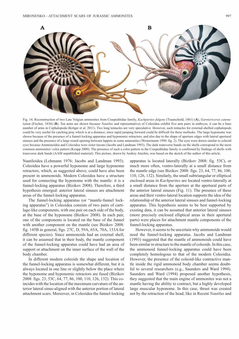
Fig. 14. Reconstruction of two Late Volgian ammonites from Craspeditidae family, Kachpurites fulgens (Trautschold, 1861) (A), Garniericeras catenu-latum (Fischer, 1830) (B). Ten arms are shown because Nautilus and representatives of Coleoidea exhibit five arm pairs in embryos, it can be a base number of arms in Cephalopoda (Kröger et al. 2011). Two long tentacles are very speculative. However, such tentacles for external-shelled cephalopods could be very useful for catching pray which is at a distance; since rapid jumping forward could be difficult for these mollusks. The large hyponome was shown because of the presence of a funnel-locking apparatus and hyponomic retractors, and also due to the shape of aperture edges with lateral apertural sinuses and the presence of a large round opening between lappets in some ammonites (Westermann 1990: fig. 2). The eyes were drawn similar to coleoid eyes because Ammonoidea and Coleoidea were sister taxons (Jacobs and Landman 1993). The dark transverse bands on the shells correspond to the most common ammonites’ color pattern (Keupp 2000). The presence of such a color pattern in the Craspeditidae family is confirmed by findings of shells with transverse dark bands (AAM unpublished material). This picture, drawn by Andrey Atuchin, was based on the sketch of the author of this article.
998 ACTA PALAEONTOLOGICA POLONICA 60 (4), 2015
not due to the compression of the whole muscular mantle, like in Coleoidea, but only due to the compression of a large and highly muscular funnel. If this hypothesis is correct, the hyponome would have functioned like an external hydrojet engine connected to the body and the shell of the ammonite. In this case, the hyponome must have been attached to the body chamber walls for the transfer of momentum to the shell. Without this attachment, the hyponome with connected soft tissues would have only pressed into the body chamber during water ejection and the momentum would have been wasted. An analogy can be drawn to a rocket engine which must be rigidly fixed in the missile airframe, because the rocket and the engine must move as a single unit. It seems very likely that the subtriangular or elliptical areas in lateral sinuses had been used for attachment of the hyponome to the body chamber walls. This provided the rigid fixation of the base of the hyponome, needed for the transfer of momentum from the muscular hyponome to the shell. This attachment would not have been permanent, ammonoids must have been able to fasten and unfasten a hyponome onto the shell walls, enabling them to withdraw it into the body chamber in case of emergency. Therefore, this attachment area worked similarly to the funnel-locking apparatus of the Coleoidea. Possibly it was a basal version of a funnel-locking apparatus. Presuming the presence of the funnel-locking apparatus in both the Ammonoidea and the Coleoidea would imply the presence of this structure in the Bactritida, the last common ancestral clade of these two groups. Speculatively, this appa-ratus initially appeared for transferring the momentum from the hyponome to the shell and later, coleoids began using it for their mantle-contraction swimming. Until the soft tissues of the ammonoid are found, this assumption remains specu-lative. However, a “funnel-based” swimming mechanism as proposed by Saunders and Ward (1994) seems to be possible and this mechanism would have needed some connection between hyponome and body chamber walls.
The prevalence of lateral attachment structures among ammonoids.—Saunders and Ward (1994) suggested that the large lateral attachment scars were found in only a few genera of ammonoids with a short body chamber (brevodomic shells). Their premise was that lateral scars were unique given their rarity among the Ammonoidea. Mutvei and Dunca (2007) have also expressed the idea that except in a few taxa the lateral muscle attachments are not recognized in ammonoids. However, Richter and Fischer (2002) showed the presence of lateral attachment scars not only in the Jurassic and Cretaceous Ammonitida, but also in the Triassic Ceratitida and Devonian Goniatitida. Among these ammonites were mesodomic and longidomic shells (Cheiloceras spp., Tornoceras spp.). From the Russian Platform, large paired lateral attachment scars have been described in ammonoids belonging to four super-families: Deshayesites deshayesi of the Deshayesitoidea (Doguzhaeva and Kabanov 1988), Aconeceras (Sinzowia) trauts choldi of the Haploceratoidea (Doguzhaeva and Mutvei 1991, 1993a, 1996; Landman et al. 1999), Quenstedtoceras
sp. of the Stephanoceratoidea (Doguzhaeva and Mutvei 1991), Kachpurites spp. and Garniericeras catenulatum of the Perisphinctoidea (this article). Most of the shells of these species are brevidomic, however, Kachpurites spp. has a me-sodomic shell (body chamber near 270–330°). It is difficult to imagine that the lateral attachment scars could have appeared independently in four different superfamilies of Ammonitida as well as in Ceratitida and Goniatitida. Apparently, a lateral attachment scars were a basic structural part of ammonoid anatomy. It can be expected that the large paired lateral at-tachment scars will be found in longidomic ammonites if these shells are sufficiently well preserved, especially with inner nacreous layers.
ConclusionsThe paleobiological interpretation of the attachment scars and anterior sinuses, allowed to propose the following new hypothetical reconstruction of the soft tissue attachment sys-tem in ammonite shells (Fig. 13). I propose that the soft tissues were attached to the shell in the following way:• Paired dorsal attachment scars were the attachment sites
of both nuchal and cephalic retractors, exactly the same as in Nautilida. Nuchal (dorsal) retractors could have been attached to the dorsal parts of these areas, while cephalic retractors—to the lateral parts.
• Paired lateral attachment scars were areas for attachment of large hyponome retractors. The shifting of the lateral attachment areas closer to the ventral side of the shell in the course of Craspeditidae evolution could confirm the connection of these scars with the hyponome.
• Anterior lateral sinuses (more precisely their apertural parts with enclosed elliptical areas) are the loci where the mantle component of the funnel-locking apparatus was at-tached. It is likely that the functions of ammonoid funnel -locking apparatus were similar, but probably not identical to functions of this apparatus in Coleoidea (it can be called “proto-funnel-locking apparatus” in this case).
The function of the muscle that was attached to the small unpaired ventral attachment scar remains controversial (Doguzhaeva and Mutvei 1996; Dagys and Keupp 1998). Despite the good preservation of ventral scars on Kachpurites shells, the study of these areas did not contribute anything new to our understanding of their functions.
“Einbuchtung” or lateral sinuses located in the posterior part of body chamber may not have been actually separate attachment areas but only posterior borders of the paired lateral attachment scars.
Based on the presence of attachment areas of the funnel retractors in Coleoidea and Ammonoidea and the possible presence in both groups of a funnel-locking apparatus, it can be assumed that both inherited these anatomic structures from the Bactritida, the last common ancestral clade of these two groups.
MIRONENKO—ATTACHMENT SCARS OF JURASSIC AMMONITES 999
AcknowledgementsI am grateful to have the initial version of my manuscript reviewed by Michail Rogov (Geological Institute of the Russian Academy of Sciences [GIN], Moscow, Russia). The initial draft was greatly im-proved by his comments. I am also grateful to him for the donation of several specimens from the Yaroslavl region for this article. Many thanks to Vyacheslav Bizikov (Russian Federal Research Institute of Fisheries and Oceanography [VNIRO], Moscow, Russia) for some precious ideas and advice on the anatomy of modern Coleoidea and the structure of their muscular system. I also want to thank Roman Shamaev (Rybinsk, Russia) for the donation of specimens for this study. I would like to thank Vitaly Kutin and Alexander Lebedev (both Moscow, Russia) for their help in collecting ammonites during our team excavation. Very accurate and beautiful reconstructions of am-monites based on my sketch were drawn by paleoillustrator Andrey Atuchin (Kemerovo, Russia). Special thanks to Steve Ford (Vancouver, Canada) who went over and edited the manuscript with me and im-proved its English. The author is very grateful to Larisa Doguzhaeva (Swedish Museum of Natural History, Stockholm, Sweden), Christian Klug (Paläontologisches Institut und Museum, Universität Zürich, Switzerland), Neil Landman (American Museum of Natural History, New York, USA) and two anonymous reviewers for the important and helpful comments which greatly improved this article.
ReferencesBandel, K. 1982. Morphologie und Bildung der frühontogenetischen
Gehäuse bei conchiferen Mollusken. Facies 7: 1–198.Bizikov, V.A. 2002. Reanalysis of functional design of Nautilus locomotory
and respiratory systems. American Malacological Bulletin 17: 17–30.Bizikov, V.A. 2008. Evolution of the Shell in Cephalopoda. 444 pp. VNIRO
Publishing, Moscow.Chirat, R. 1997. Les Nautilida post-triasiques. Paléobiologie, paléoéco-
logie, paléobiogéographie et systématique à la lumière des données récemment acquises sur Nautilus. 322 pp. Unpublished Ph.D. Thesis, Université de Caen, Caen.
Cox, L.R. 1969 General features of Bivalvia. In: R.C. Moore (ed.), Treatise on Invertebrate Paleontology, Part N, Mollusca, 6 (1): Bivalvia, 2–129. Geological Society of America and University of Kansas Press, Lawrence, Kansas.
Crick, G.C. 1898. On the muscular attachment of the animal to its shell in some Cephalopoda (Ammonoidea). Transactions of the Linnean Society of London. 2nd Series: Zoology 7: 71–113.
Dagys, A.S. and Keupp, H. 1998. Internal ventral keels in Triassic ceratid ammonoids: description and functional interpretation as muscle scars. Zeitschrift der Deutschen Geologischen Gesellschaft 149: 81–89.
Doguzhaeva, L.A. and Kabanov, G.K. 1988. Muscle scars in ammonoids [in Russian]. Doklady Akademii Nauk SSSR 301: 210–212.
Doguzhaeva, L.A. and Michailova I.A. 1991. New data on the muscular system of heteromorphic ammonoids [in Russian]. Doklady Akademii Nauk SSSR 318: 981–985.
Doguzhaeva, L.A. and Mutvei H. 1991. Organization of the soft body in Aconeceras (Ammonitina), interpreted on the basis of shell morphol-ogy and muscle scars. Palaeontographica A 218: 17–33.
Doguzhaeva, L.A. and Mutvei, H. 1992. Radula of Early Cretaceous ammo-nite Aconeceras (Mollusca: Cephalopoda). Palaeontographica A 223: 167–177.
Doguzhaeva, L.A. and Mutvei, H. 1993a. Shell ultrastructure, muscle scars, and buccal apparatus in ammonoids. Geobios, Memoire Special 26: 111–119.
Doguzhaeva, L.A. and Mutvei, H. 1993b. Structural features of Cretaceous ammonoids indicative of semi- internal or internal shells. In: M.R. House
(ed.), The Ammonoidea: Environment, Ecology, and Evolutionary Change. Systematics Association Special Volume 47: 99–114.
Doguzhaeva, L.A. and Mutvei, H. 1996. Attachment of the body to the shell in ammonoids. In: N.H. Landman, K. Tanabe, and R.A. Davis (eds.), Ammonoid Paleobiology, 43–63. Plenum Press, New York.
Gerasimov, P.A. 1969. Verhnij pod’ârus voljskogo ârusa centralnoj časti Russkoj Platformy. 144 pp. Nauka, Moskva.
Houša, V., Pruner, P., Zakharov, V.A., Kostak, M., Chadima, M., Rogov, M.A., Šlechta, S. Mazuch, M. 2007. Boreal-Tethyan correlation of the Jurassic–Cretaceous boundary interval by magneto- and biostratigra-phy. Stratigraphy and Geological Correlation 15: 297–309.
Jacobs, D.K. and Chamberlain, J.A. 1996. Buoyancy and hydrodynamics in ammonoids. In: N. Landman, K. Tanabe, and R.A. Davis (eds.), Am mo-noid Paleobiology. Topics in Geobiology 13: 169–223.
Jacobs, D.K. and Landman, N.H. 1993. Nautilus—a poor model for the function and behavior of ammonoids? Lethaia 26: 101–111.
Jones, D.L. 1961. Muscle attachment impressions in a Cretaceous ammo-nite. Journal of Paleontology 35: 502–504.
Jordan, R. 1968. Zur Anatomie mesozoischer Ammoniten nach den Struk tur-elementen der Gehäuse-Innenwand. Beihefte zum Geologischen Jahr-buch 77: 1–64.
Kennedy, W.J., Cobban, W.A., and Klinger, H.C. 2002. Muscle attach-ment and mantle-related features in Upper Cretaceous Baculites from the United States Western Interior. Abhandlungen der Geologischen Bundesantstalt Wien 57: 89–112.
Keupp, H. 2000. Ammoniten. Paläobiologische Erfolgsspiralen. 165 pp. Thorbecke Species, Stuttgart.
Kiselev, D.N. 2012. Ob’ekti geologičeskogo naslediâ Âroslavskoj oblasti: stratigrafiâ, paleontologiâ i paleogeografiâ. 304 pp. Ûstitsinform, Moskva.
Klug, C. and Lehmkuhl, A. 2004. Soft-tissue attachment structures and taphonomy of the Middle Triassic nautiloid Germanonautilus. Acta Palaeontologica Polonica 49: 243–258.
Klug, C., Meyer, E.P., Richter, U., and Korn, D. 2008. Soft-tissue imprints in fossil and Recent cephalopod septa and septum formation. Lethaia 41: 477–492.
Klug, C., Montenari, M., Schulz, H., and Urlichs, M. 2007. Soft-tissue attach-ment of Middle Triassic Ceratitida from Germany. In: R.H. Landman, R.A. Davis, and R.H. Mapes (eds.), Sixth International Symposium Cephalopods Present and Past. New Insights and Fresh Perspectives, 205–220. Springer, New York.
Korn, D. and Klug, C. 2002. Ammoneae Devonicae. In: W. Riegraf (ed.), Fossilium catalogus I: Animalia, 138. 375 pp. Backhuys, Leiden.
Kröger, B. 2002. On the ability of withdrawing of some Jurassic ammonoids. In: Summesberger, H., Histon, K., and Daurer, A. (eds.), Cephalopods—Present and Past. Abhandlungen der Geologischen Bundesanstalt Wien 57: 199–204.
Kröger, B., Klug, C., and Mapes, R. 2005. Soft-tissue attachments in ortho cerid and bactritid cephalopods from the Early and Middle Devonian of Germany and Morocco. Acta Palaeontologica Polonica 50: 329–342.
Kröger, B., Vinther, J., and Fuchs, D. 2011. Cephalopod origin and evolu-tion: a congruent picture emerging from fossils, development and mole-cules. BioEssays 33: 602–613.
Landman, N.H. and Waage, K.M. 1993. Scaphitid ammonites of the Upper Cretaceous (Maastrichtian) Fox Hills Formation in South Dakota and Wyoming. Bulletin of the American Museum of Natural History 215: 1–257.
Landman, N.H., Lane, J., Cobban, W.A., Jorgensen, S.D., Kennedy, W.J., and Larson, N.L. 1999. Impressions of the attachment to the soft body to the shell in late Cretaceous pachydiscid ammonites from the Western Interior of the United States. American Museum Novitates 3273: 1–31.
Lehmann, U. 1976. Ammoniten: ihr Leben und ihre Umwelt. 171 pp. Enke, Stuttgart.
Mitta, V.V. 2010. Late Volgian Kachpurites Spath (Craspeditidae, Am-mo no idea) of the Russian Platform. Paleontological Journal 44: 622–631.
1000 ACTA PALAEONTOLOGICA POLONICA 60 (4), 2015
Mutvei, H. 1957. On the relations of the principal muscles to the shell in Nautilus and some fossil nautiloids. Arkiv för Mineralogi och Geologi 10: 219–253.
Mutvei, H. 1964. Remarks on the anatomy of Recent and fossil Cephalo-poda. Stockholm Contributions in Geology 11: 79–102.
Mutvei, H. 2002. Nautiloid systematics based on siphuncular structure and position of muscle scars. Abhandlungen der Geologischen Bundesanstalt 57: 379–392.
Mutvei, H. and Dunca, E. 2007. Connecting ring ultrastructure in the Jurassic ammonoid Quenstedtoceras with discussion on mode of life of ammonoids. In: R.H. Landman, R.A. Davis, and R.H. Mapes (eds.), Sixth International Symposium Cephalopods Present and Past. New Insights and Fresh Perspectives, 239–256. Springer, Dordrecht.
Paul, C.R.C. 2011. Dark bands on pyritic internal moulds of the Early Jurassic ammonites Oxynoticeras and Cheltonia from Gloucestershire, England: interpretation and significance to ammonite growth analysis. Palaeontology 54: 1213–1221.
Richter, U. 2002. Gewebeansatz-Strukturen auf Steinkernen von Ammono-ideen. Geologische Beiträge Hannover 4: 1–113.
Richter, U. and Fischer, R. 2002. Soft tissue attachment structures on pyri-tized internal moulds of ammonoids. Abhandlungen der geologischen Bundesanstalt 57: 139–149.
Rogov, M. and Zakharov, V. 2009. Ammonite- and bivalve-based biostrati-graphy and Panboreal correlation of the Volgian Stage. Science in China Series D, Earth Sciences 52: 1890–1909.
Sarikadze, M.Z., Lominadze T.A., and Kvantaliani, I.V. 1990. Systematische Bedeutung von Muskelabdriicken spatjurassischer Ammonoideae. Zeit-schrift für Geologische Wissenschaften Berlin 18: 1031–1039.
Sasonova, I.G. and Sasonov, N.T. 1983. The Berriasian of the European Realm. Zitteliana 10: 439–446.
Saunders, W.B. and Ward, P.D. 1994. Nautilus is not a model for the func-tion and behavior of ammonoids. Lethaia 27: 47–48.
Shigeno, S., Sasaki, T., Moritaki, T., Kasugai, T., Vecchione, M., and Agata, K. 2008. Evolution of the cephalopod head complex by assembly of multiple molluscan body parts: evidence from Nautilus embryonic development. Journal of Morphology 269: 1–17.
Tanabe, K., Landman, N.H., and Mapes, R.H. 1998. Muscle attachment scars in a Carboniferous goniatite. Paleontological Research 2: 130–136.
Trautschold, H. 1871. Ueber die Erhaltungszustande russischer ammo-niten. Bulletin de la Société Impériale des Naturalistes de Moscou 43 (3–4): 301–306.
Waagen, W. 1870. Ueber die Ansatzstelle der Hafmuskeln beim Nautilus und den Ammoniten. Paleontographica 17: 185–210.
Weitschat, W. 1986. Phosphatisierte Ammonoideen aus der Mittleren Trias von Central-Spitzbergen, Mitteilungen Geologisch-Paläontologisches Institut der Universität Hamburg 61: 249–279.
Weitschat, W. and Bandel, K. 1991. Organic components in phragmocones of Boreal Triassic ammonoids: Implications for ammonoid biology. Palä ontologische Zeitschrift 65: 269–303.
Westermann, G.E.G. 1990. New developments in ecology of Jurassic–Creta-ceous ammonites. In: G. Pallini, F. Cecca, S. Cresta, and M. Santantonio (eds.), Atti II Convegno Internazionale. Fossili. Evoluzione. Ambiente. Pergola 1987, 459–478. Technostampa, Ostra Vetere.
Westermann, G.E.G. and Tsujita, C.J. 1999. Life habits of ammonoids. In: E. Savazzi (ed.), Functional Morphology of the Invertebrate Skeleton, 299–325. John Wiley & Sons, New York.
Zakharov, V.A. and Rogov, M.A. 2008. The Upper Volgian substage in Northeast Siberia (Nordvik Peninsula) and its panboreal correlation based on ammonites. Stratigraphy and Geological Correlation 16 (4): 423–436.
Zimmermann, A. 1985. Strukturelemente der Gehäusewand bei Ammoniten aus dem oberen Pliensbachium von Empelde bei Hannover. Arbeitskreis Paläontologie, Hannover 13 (1/2): 69–71.