The Skeletal System - Yolakhsbiology.yolasite.com/resources/skeletal System Notes.pdf · •Parts...
42
The Skeletal System
Transcript of The Skeletal System - Yolakhsbiology.yolasite.com/resources/skeletal System Notes.pdf · •Parts...

The Skeletal System
The Skeletal System
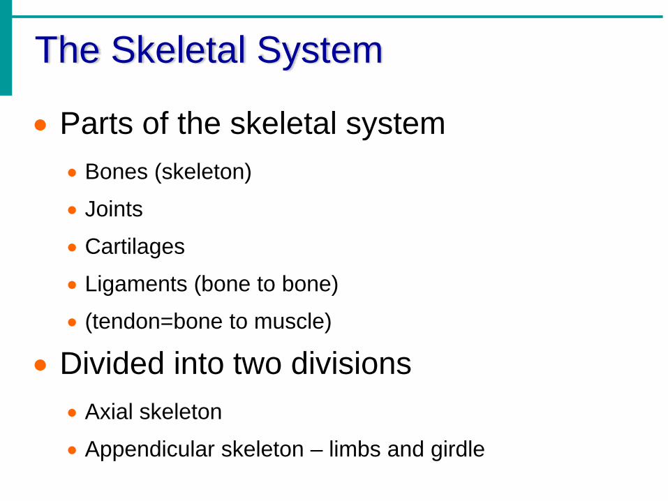
• Parts of the skeletal system• Bones (skeleton)
• Joints
• Cartilages
• Ligaments (bone to bone)
• (tendon=bone to muscle)
• Divided into two divisions• Axial skeleton
• Appendicular skeleton – limbs and girdle


Functions of Bones
• Support of the body
• Protection of soft organs
• Movement due to attached skeletal muscles
• Storage of minerals and fats
• Blood cell formation

Bones of the Human Body
• The skeleton has 206 bones
• Two basic types of bone tissue•Compact bone• Homogeneous
•Spongy bone• Small needle-like
pieces of bone
• Many open spacesFigure 5.2b
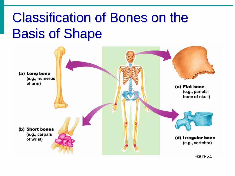
Classification of Bones on the Basis of Shape
Figure 5.1
Classification of Bones
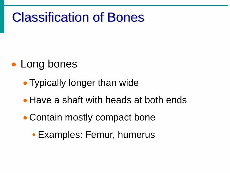
• Long bones
•Typically longer than wide
•Have a shaft with heads at both ends
•Contain mostly compact bone
• Examples: Femur, humerus
Classification of Bones
• Short bones
•Generally cube-shape
•Contain mostly spongy bone
•Examples: Carpals, tarsals

Classification of Bones
• Flat bones
•Thin and flattened
•Usually curved
•Thin layers of compact bone around a layer of spongy bone
•Examples: Skull, ribs, sternum

Classification of Bones
• Irregular bones
• Irregular shape
•Do not fit into other bone classification categories
•Example: Vertebrae and hip

Classification of Bones on the Basis of Shape
Figure 5.1

Gross Anatomy of a Long Bone
• Diaphysis•Shaft
•Composed of compact bone
• Epiphysis•Ends of the bone
•Composed mostly of spongy bone
Figure 5.2a
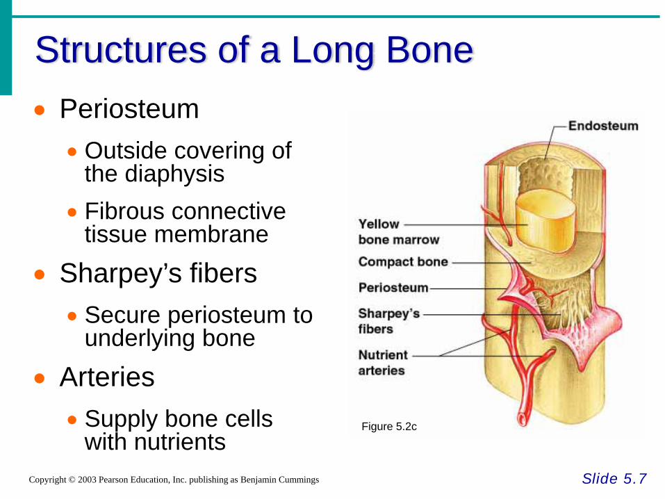
Structures of a Long Bone
Slide 5.7Copyright © 2003 Pearson Education, Inc. publishing as Benjamin Cummings
• Periosteum• Outside covering of
the diaphysis• Fibrous connective
tissue membrane
• Sharpey’s fibers• Secure periosteum to
underlying bone
• Arteries• Supply bone cells
with nutrientsFigure 5.2c

Structures of a Long Bone
• Articular cartilage
•Covers the external surface of the epiphyses
•Made of hyaline cartilage
•Decreases friction at joint surfaces Figure 5.2a

Structures of a Long Bone
• Medullary cavity
•Cavity of the shaft
•Contains yellow marrow (mostly fat) in adults
•Contains red marrow (for blood cell formation) in infants Figure 5.2a

Bone Growth
• Bones are remodeled and lengthened until growth stops
•Bones change shape somewhat
•Bones grow in width

Long Bone Formation and Growth
Figure 5.4a
Types of Bone Cells• Osteocytes
• Mature bone cells
• Osteoblasts• Bone-forming cells
• Osteoclasts• Bone-destroying cells• Break down bone matrix for remodeling and
release of calcium
• Bone remodeling is a process by both osteoblasts and osteoclasts
Bone Fractures
• A break in a bone
• Types of bone fractures•Closed (simple) fracture – break that does not
penetrate the skin
•Open (compound) fracture – broken bone penetrates through the skin
• Bone fractures are treated by reduction and immobilization•Realignment of the bone

Common Types of Fractures
Slide 5.17Copyright © 2003 Pearson Education, Inc. publishing as Benjamin Cummings
Table 5.2
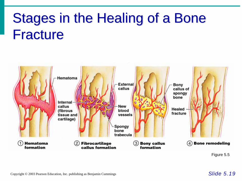
Stages in the Healing of a Bone Fracture
Slide 5.19Copyright © 2003 Pearson Education, Inc. publishing as Benjamin Cummings
Figure 5.5

The Skull
Slide 5 21a
Copyright © 2003 Pearson Education, Inc. publishing as Benjamin Cummings
• Two sets of bones
•Cranium
•Facial bones
• Bones are joined by sutures
• Only the mandible is attached by a freely movable joint

The Skull
Slide 5 21b
Copyright © 2003 Pearson Education, Inc. publishing as Benjamin Cummings
Figure 5.7
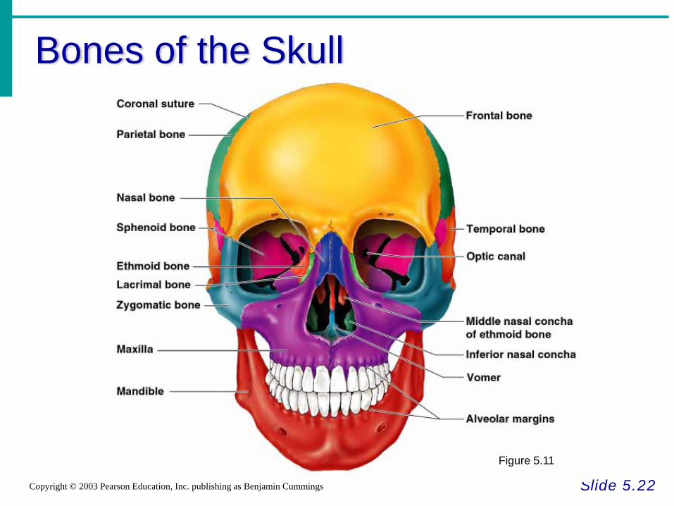
Bones of the Skull
Slide 5.22Copyright © 2003 Pearson Education, Inc. publishing as Benjamin Cummings
Figure 5.11

Human Skull, Superior View
Slide 5.23Copyright © 2003 Pearson Education, Inc. publishing as Benjamin Cummings
Figure 5.8

Human Skull, Inferior View
Slide 5.24Copyright © 2003 Pearson Education, Inc. publishing as Benjamin Cummings
Figure 5.9
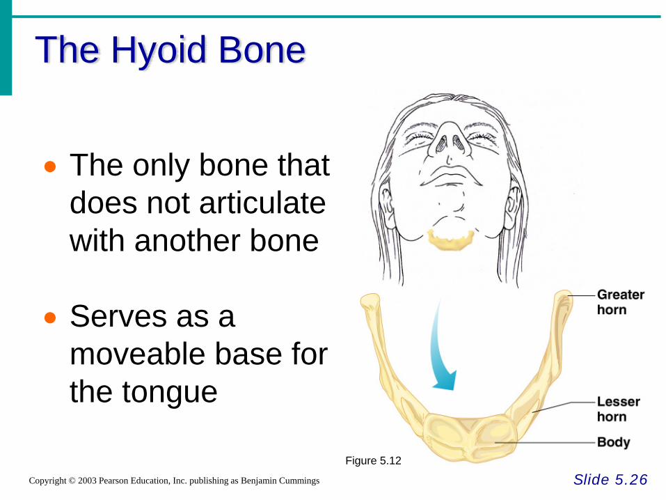
The Hyoid Bone
Slide 5.26Copyright © 2003 Pearson Education, Inc. publishing as Benjamin Cummings
• The only bone that does not articulate with another bone
• Serves as a moveable base for the tongue
Figure 5.12
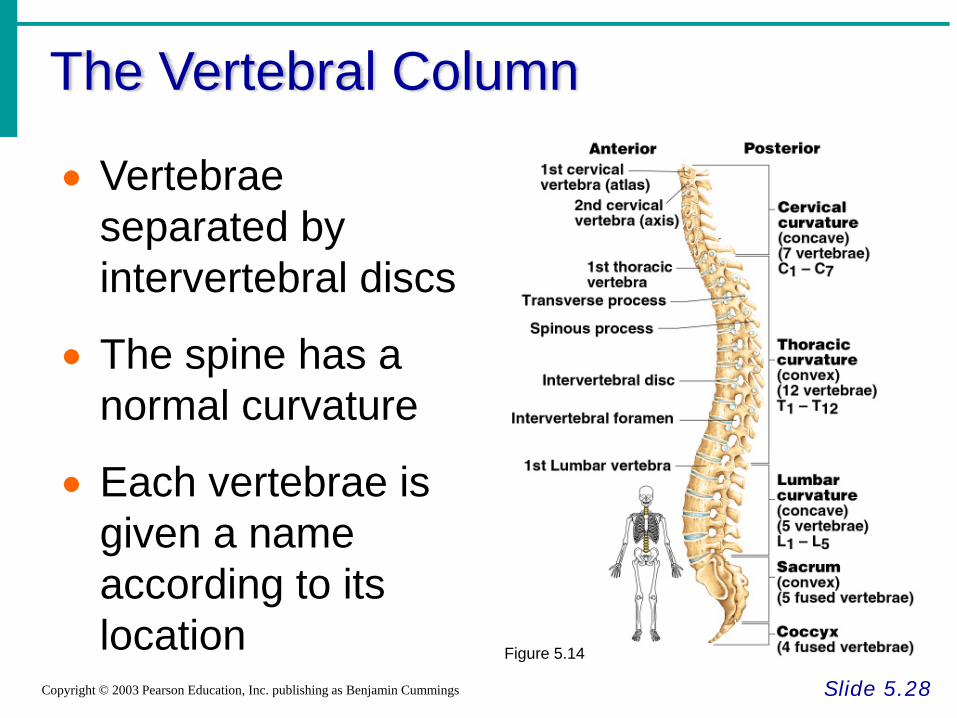
The Vertebral Column
Slide 5.28Copyright © 2003 Pearson Education, Inc. publishing as Benjamin Cummings
• Vertebrae separated by intervertebral discs
• The spine has a normal curvature
• Each vertebrae is given a name according to its location Figure 5.14

Structure of a Typical Vertebrae
Slide 5.29Copyright © 2003 Pearson Education, Inc. publishing as Benjamin Cummings
Figure 5.16
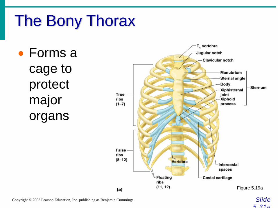
The Bony Thorax
Slide 5 31a
Copyright © 2003 Pearson Education, Inc. publishing as Benjamin Cummings
• Forms a cage to protect major organs
Figure 5.19a
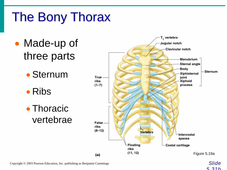
The Bony Thorax
Slide 5 31b
Copyright © 2003 Pearson Education, Inc. publishing as Benjamin Cummings
• Made-up of three parts
•Sternum
•Ribs
•Thoracic vertebrae
Figure 5.19a

Joints
Slide 5.43Copyright © 2003 Pearson Education, Inc. publishing as Benjamin Cummings
• Articulations of bones
• Functions of joints
•Hold bones together
•Allow for mobility
• Ways joints are classified
•Functionally
•Structurally

Functional Classification of Joints
Slide 5.44Copyright © 2003 Pearson Education, Inc. publishing as Benjamin Cummings
• Synarthroses – immovable joints
• Amphiarthroses – slightly moveable joints
• Diarthroses – freely moveable joints
Structural Classification of Joints
Slide 5.45Copyright © 2003 Pearson Education, Inc. publishing as Benjamin Cummings
• Fibrous joints
•Generally immovable
• Cartilaginous joints
• Immovable or slightly moveable
• Synovial joints
•Freely moveable
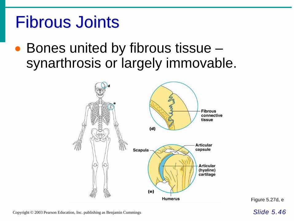
Fibrous Joints
Slide 5.46Copyright © 2003 Pearson Education, Inc. publishing as Benjamin Cummings
• Bones united by fibrous tissue –synarthrosis or largely immovable.
Figure 5.27d, e
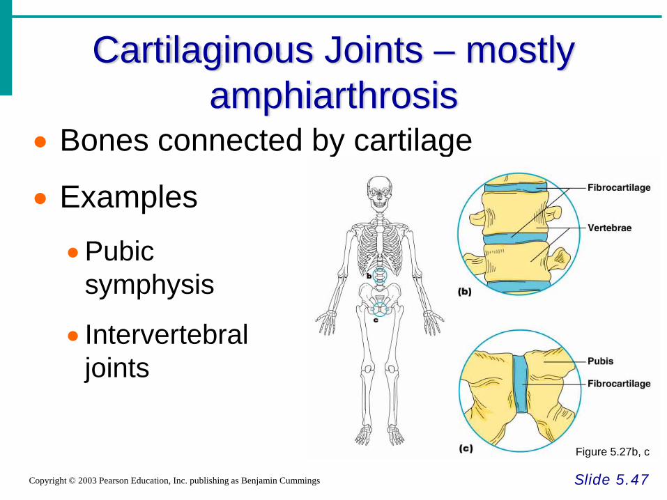
Cartilaginous Joints – mostly amphiarthrosis
Slide 5.47Copyright © 2003 Pearson Education, Inc. publishing as Benjamin Cummings
• Bones connected by cartilage
• Examples
•Pubic symphysis
• Intervertebral joints
Figure 5.27b, c
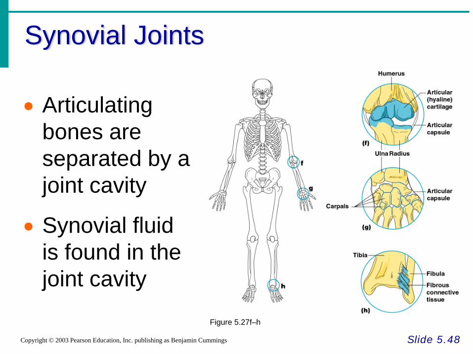
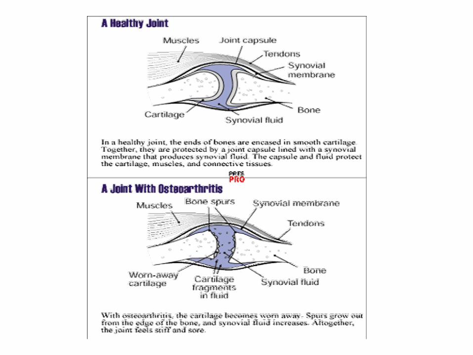
Synovial Joints
Slide 5.48Copyright © 2003 Pearson Education, Inc. publishing as Benjamin Cummings
• Articulating bones are separated by a joint cavity
• Synovial fluid is found in the joint cavity
Figure 5.27f–h
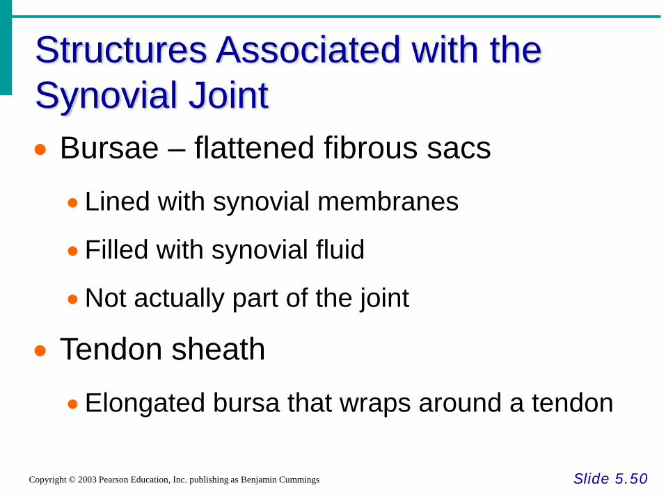
Structures Associated with the Synovial Joint
Slide 5.50Copyright © 2003 Pearson Education, Inc. publishing as Benjamin Cummings
• Bursae – flattened fibrous sacs• Lined with synovial membranes
•Filled with synovial fluid
•Not actually part of the joint
• Tendon sheath•Elongated bursa that wraps around a tendon
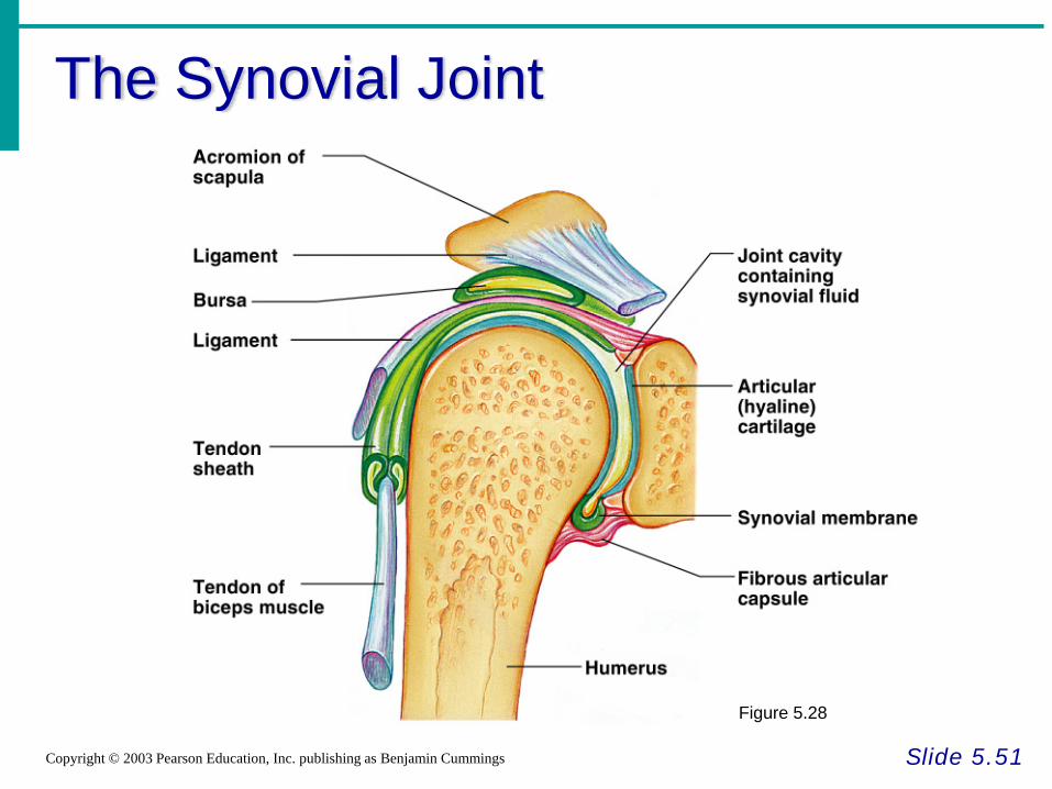
The Synovial Joint
Slide 5.51Copyright © 2003 Pearson Education, Inc. publishing as Benjamin Cummings
Figure 5.28
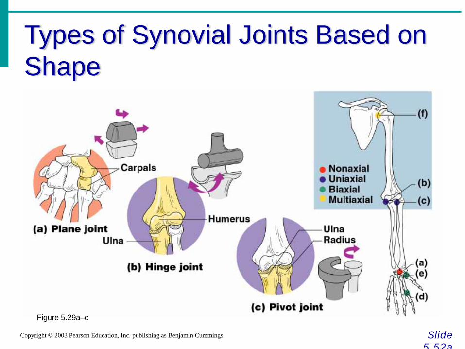
Types of Synovial Joints Based on Shape
Slide 5 52a
Copyright © 2003 Pearson Education, Inc. publishing as Benjamin Cummings
Figure 5.29a–c

Types of Synovial Joints Based on Shape
Slide 5 52b
Copyright © 2003 Pearson Education, Inc. publishing as Benjamin Cummings
Figure 5.29d–f