The Skeletal System - Newcastle ISD · plate (AKA: epiphyseal plate) ... Types of synovial joints...
30
Chapter 5
Transcript of The Skeletal System - Newcastle ISD · plate (AKA: epiphyseal plate) ... Types of synovial joints...
Chapter 5
Structure
Protection
Red blood cell formation
Movement
Storage: fat in yellow marrow, calcium
(hard matrix), phosphorus
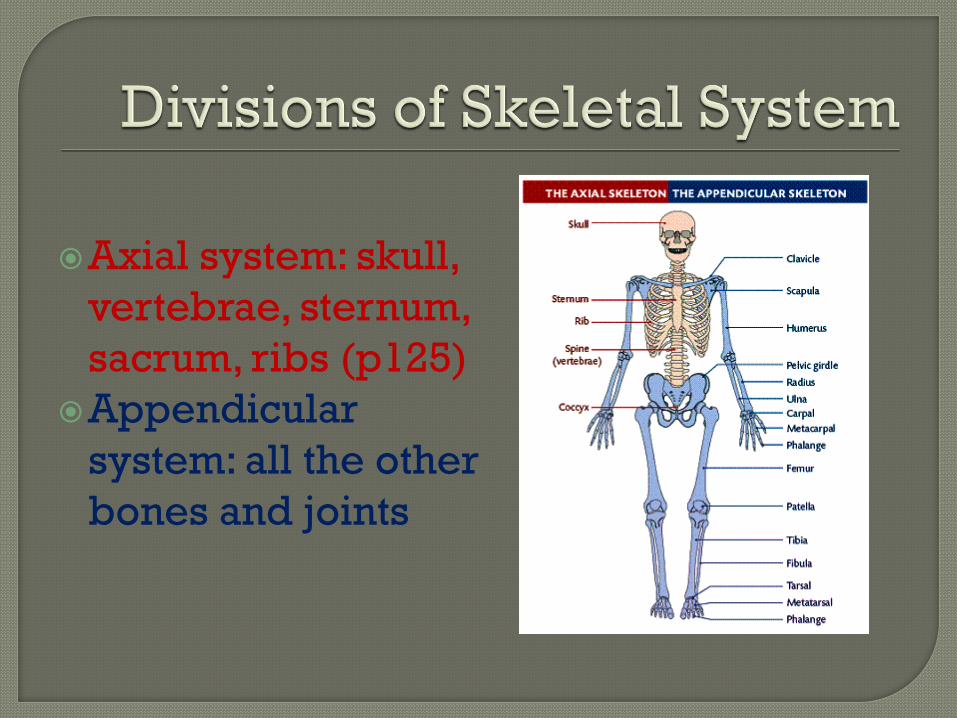
Axial system: skull,
vertebrae, sternum,
sacrum, ribs (p125)
Appendicular
system: all the other
bones and joints
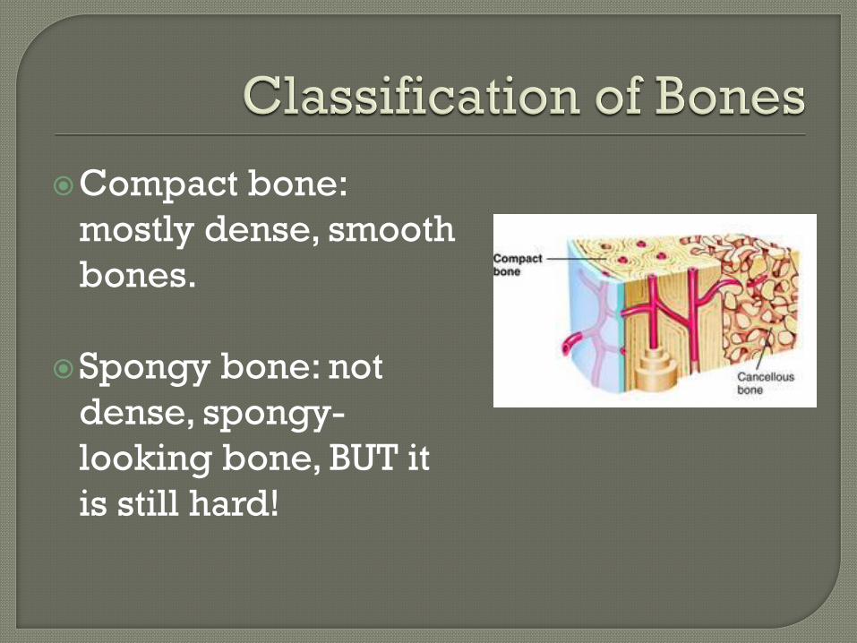
Compact bone:
mostly dense, smooth
bones.
Spongy bone: not
dense, spongy-
looking bone, BUT it
is still hard!
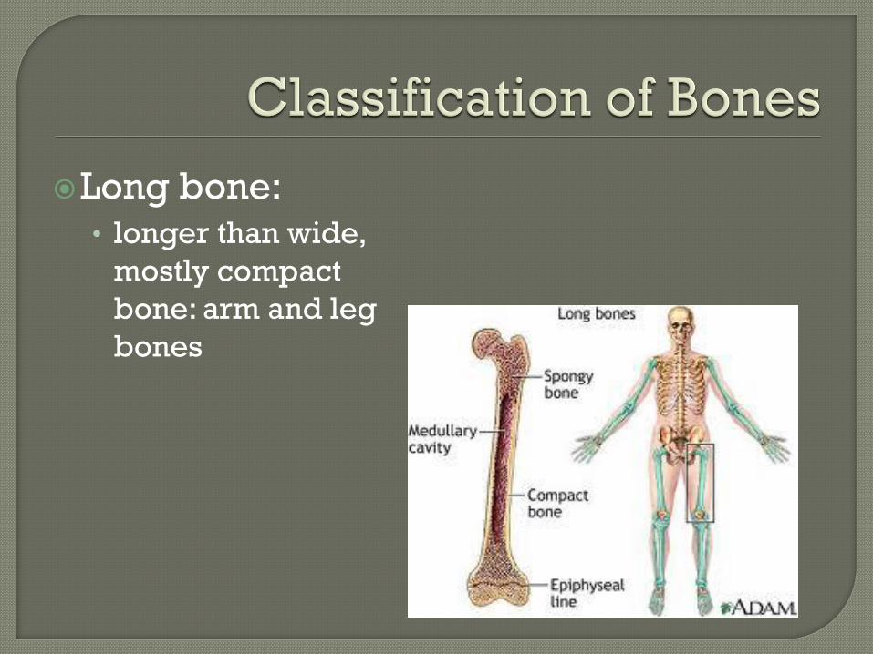
Long bone: • longer than wide,
mostly compact
bone: arm and leg
bones
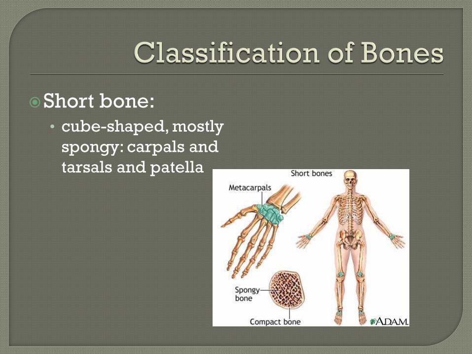
Short bone: • cube-shaped, mostly
spongy: carpals and
tarsals and patella
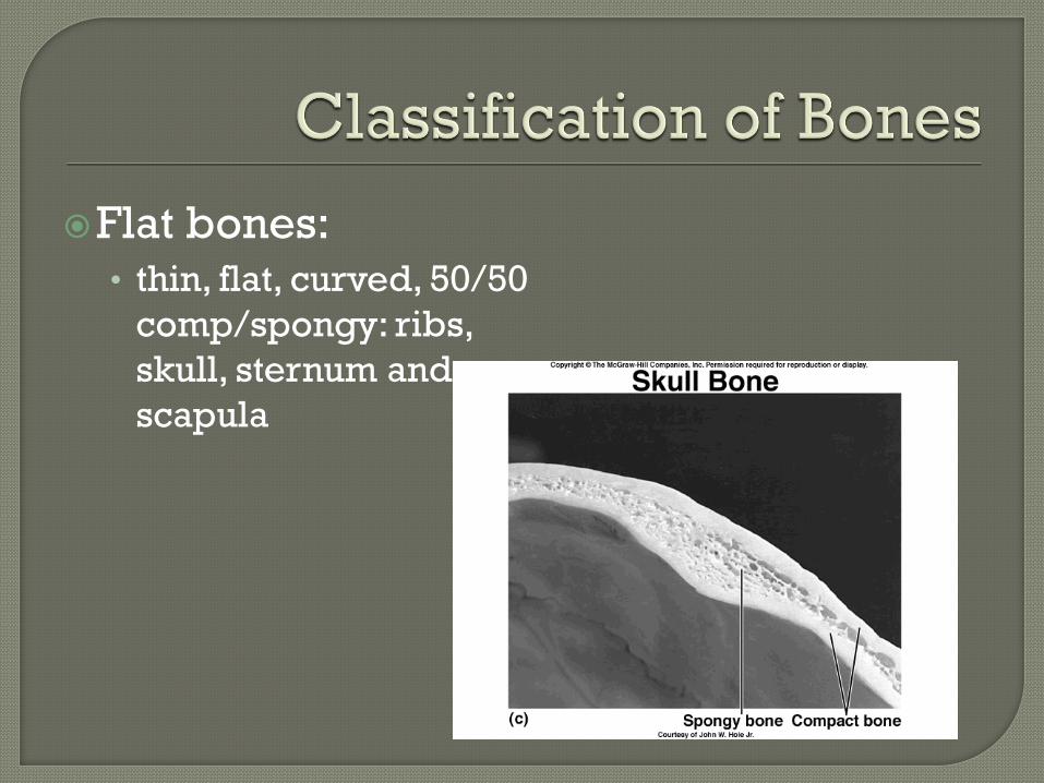
Flat bones: • thin, flat, curved, 50/50
comp/spongy: ribs,
skull, sternum and
scapula
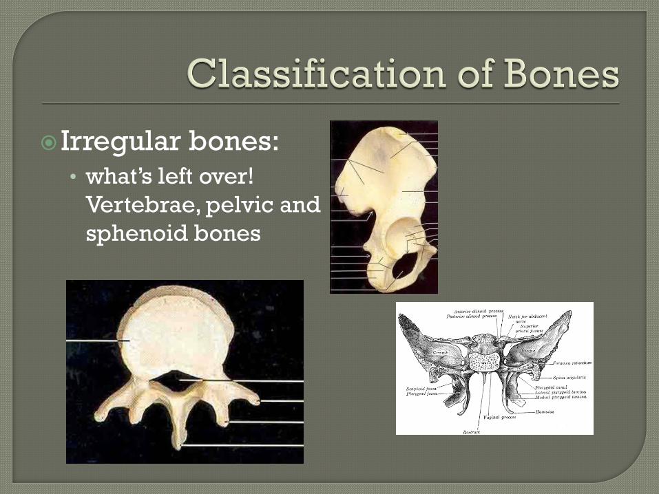
Irregular bones: • what’s left over!
Vertebrae, pelvic and
sphenoid bones
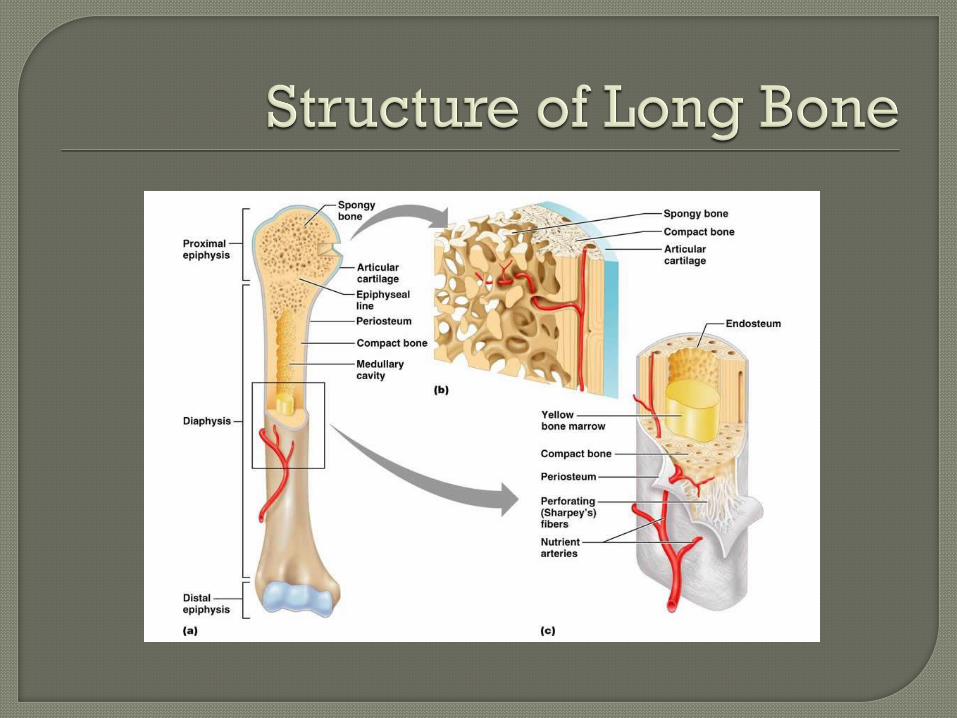
P. 118 Diaphysis: shaft of long bone Epiphysis: ends of diaphysis
(proximal/distal) Articular cartilage: hyaline cartilage lining
the joints of the epiphysis. Epiphyseal line: what’s left of the growth
plate (AKA: epiphyseal plate) Periosteum: bone “skin”, lines the outside Endosteum: lines the medullary cavity
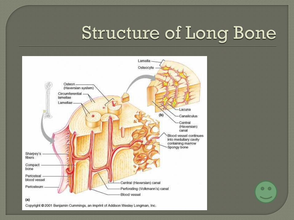
Small to Big
Osteocyte: (“osteo”- bone, “cyte”- cell)
mature bone cell.
Lacunae: hollow area in hard bone matrix
that houses the osteocyte
Lamellae: rings of lacunae
Osteon (Haversian System): system of
lamellae that make up the majority of
compact bone
Blood Canals
Vertical canals = Haversian canal
Horizontal canals = Volkmann’s canal
Capillary canals (surround osteocytes) =
canaliculi
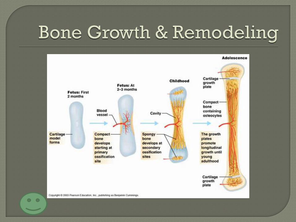
Ossification: bone formation (during fetal
development)
*Remember, fetal skeletons start as
cartilage.
1. hyaline cartilage is covered with bone
formed from cells called osteoblasts.
2. the center of the hyaline cartilage is
“eaten” away forming the medullary
cavity
Length: epiphyseal plate & articular
cartilage grow new cartilage away from
the medullary cavity and new bone
towards the cavity via osteoblasts.
Width: osteoblasts in periosteum add
new bone and osteoclasts (bone eating
cells) in endosteum digest cells near the
marrow
Control: Growth pre-puberty is controlled by
growth and sex hormones. Bone constantly changes via osteoblasts
and osteoclasts • Calcium levels in blood (blood steals from bones
if it has to!)
• Pull of gravity on bones
• Pull of muscles on bones (remember Walle?)
• Stress areas or breaks
Types of fractures (p. 123)
1. simple fracture: the bone does not
protrude from the skin
2. compound fracture: the bone does
protrude from the skin
Treatments:
Closed reduction: put back together
without surgery.
Open reduction: put back together via
surgery, pins, wires, screws, bolts, ect…
The major events of bone repair:
1. hematoma: blood-filled swelling
2. fibrocartilage callus: massive repair
tissue site (cartilage, bone, collagen)
3. bony callus: osteoblasts and
osteoclasts migrate here and replace
fibrocartilage.
4. bony callus remodeling
Articulations: points of connections
between bones (no joint for the hyoid
bone in neck)
Classifications: 1. by function (amount of
motion) & 2. by structure
Functional classes: • Synarthosis: immovable
• Amphiarthrosis: slightly movable
• Diarthrosis: movable
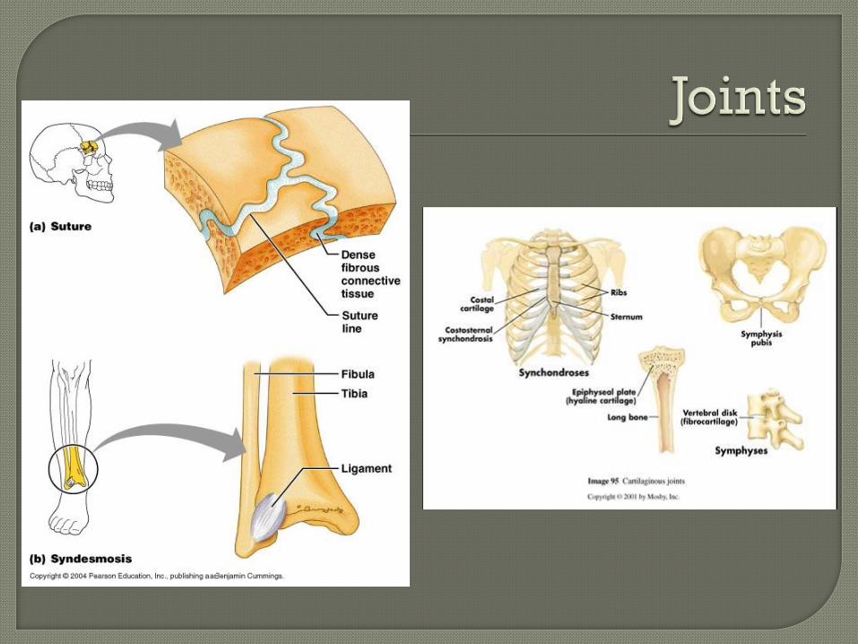
Structural classes (page 148) 1. Fibrous joints: mainly immovable
(synarthrosis) joints connected tightly by fibrous tissue • Examples: sutures of the skull & syndesmoses of the
distal fibula and tibia
2. Cartilaginous joints: mainly semi-movable (amphiarthrosis) joints connected by cartilaginous tissue. • Examples: pubic symphysis, intervertebral joints,
epiphyseal plate & rib/sternum joints
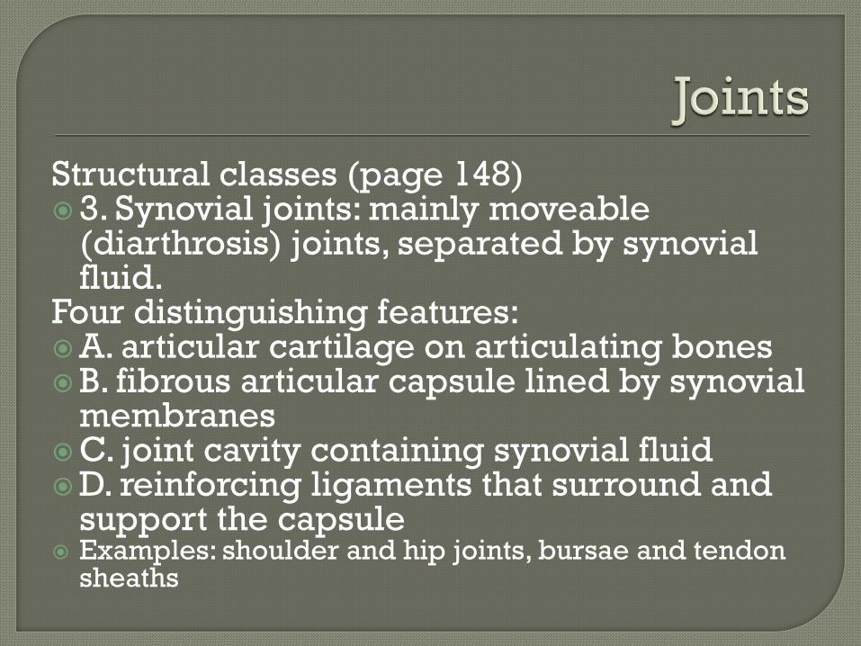
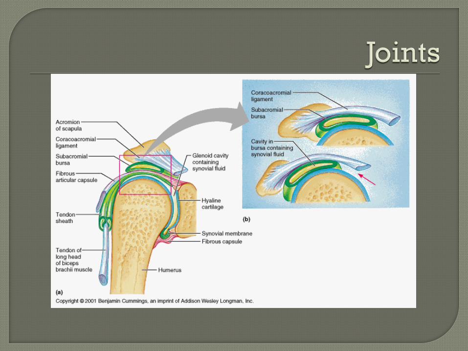
Structural classes (page 148) 3. Synovial joints: mainly moveable
(diarthrosis) joints, separated by synovial fluid.
Four distinguishing features: A. articular cartilage on articulating bones B. fibrous articular capsule lined by synovial
membranes C. joint cavity containing synovial fluid D. reinforcing ligaments that surround and
support the capsule Examples: shoulder and hip joints, bursae and tendon
sheaths
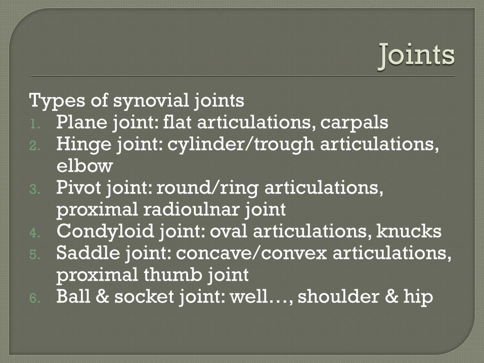
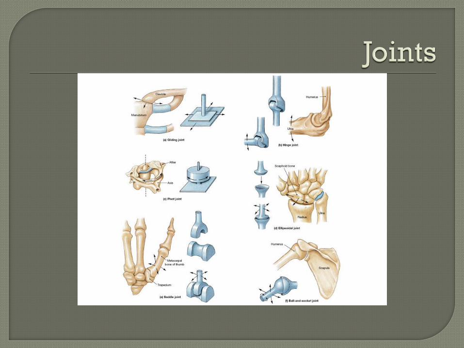
Types of synovial joints 1. Plane joint: flat articulations, carpals 2. Hinge joint: cylinder/trough articulations,
elbow 3. Pivot joint: round/ring articulations,
proximal radioulnar joint 4. Condyloid joint: oval articulations, knucks 5. Saddle joint: concave/convex articulations,
proximal thumb joint 6. Ball & socket joint: well…, shoulder & hip
Inflammatory Disorders of Joints 1. Bursitis, sprains 2. Acute arthritis: usually a bacterial infection
like lyme disease 3. Chronic arthritis:
a. Osteoarthritis: most common form especially in elderly; caused from years of wear and tear
b. Rheumatoid arthritis: autoimmune disease, white blood cells produce a joint eating tissue that ossifies and fuses diarthotic joints
c. Gouty arthritis: accumulation of spiky uric acid crystals in joints (often the large toe)