Saprotrophic proteomes of biotypes of the witches’ broom ...
1
SILVA FENNICASilva Fennica vol. 49 no. 4 article id 1320
Category: research note
www.silvafennica.fiISSN-L 0037-5330 | ISSN 2242-4075 (Online)
The Finnish Society of Forest Science Natural Resources Institute Finland
Abbot O. Oghenekaro 1, Geoffrey Daniel 2 and Fred O. Asiegbu 1
The saprotrophic wood-degrading abilities of Rigidoporus microporus
Oghenekaro A.O., Daniel G., Asiegbu F.O. (2015). The saprotrophic wood-degrading abilities of Rigidoporus microporus. Silva Fennica vol. 49 no. 4 article id 1320. 10 p.
Highlights• Rigidoporus microporus isolates displayed varying saprotrophic capabilities on wood blocks
of Rubber tree (Hevea brasiliensis).• Percentage mass loss of (Hevea brasiliensis) wood blocks caused by the pathogenic Rigi-
doporus microporuswassignificantlyhigherthanthatobservedwiththeendophyticisolate.• The endophytic isolate has very poor saprotrophic ability on Hevea brasiliensis wood blocks.
AbstractSaprotrophic wood-decaying abilities of Rigidoporus microporus (Polyporales, Basidiomycota) syn. Rigidoporus lignosus and the structural alterations induced in wood blocks of Hevea bra-siliensis Muell. Arg were studied. Mass loss of wood blocks was analyzed after 3 and 6 months respectively and the patterns of decay by pathogenic and endophytic isolates of this fungus were investigated using light microscopy. Effects of temperature on growth of the isolates on malt extract agar were also investigated. The R. microporus isolated from a non-H. brasiliensis host caused the highest percentage mass loss (27.2% after 6 months), followed by isolates ED310 (21.1%) and M13 (15.7%), both collected from diseased H. brasiliensis plantations. The isolate initiallyidentifiedasanendophyteshowedverylowsaprotrophicwooddecaycapability(4.3%after 6 months). The optimal temperature for growth of the isolates was 30 °C; except for the endophytic isolate which showed highest growth at 25 °C. Wood samples degraded by the R. microporus isolates showed simultaneous attack of wood cell walls, typical of white rot fungi. Results of the study indicate variability in the wood degrading abilities of the isolates and the potentialdifferencesintheirphysiologyarediscussed.Ourfindingsfurthersupporttheneedfora taxonomical revision of the Rigidoporus genus.
Keywords simultaneousdecay;wooddegradation;whiterot;delignificationAddresses 1 Department of Forest Sciences, University of Helsinki, P.O. Box 27, FI-00014 Uni-versity of Helsinki, Finland; 2 Department of Forest Products/Wood Science, Swedish University of Agricultural Sciences, P.O. Box 7008, SE-75007 Uppsala, SwedenE-mail [email protected] 16 February Revised 18 August 2015 Accepted 19 August 2015Available at http://dx.doi.org/10.14214/sf.1320

2
Silva Fennica vol. 49 no. 4 article id 1320 · Oghenekaro et al. · The saprotrophic wood-degrading abilities of…
1 Introduction
Rigidoporus microporus (Polyporales, Basidiomycota) syn. Rigidoporus lignosus is the most eco-nomically important pathogen of tropical rubber tree (Hevea brasiliensis Muell. Arg) plantations (Liyanage 1997) where it causes white rot disease (WRD). White rot disease was shown as a major problem for 43% of farmers in a smallholdings survey in Malaysia (Sail and Ahmad 2009). In Nigeria, WRD is responsible for 96 % of incidences of root diseases and results in killing of up to fivetreesperhectareperyearinplantations(Omorusi,2012).ThetaxonomyoftheR. microporus complex is problematic. The source of the type specimen of R. microporus (Sw.) Overeem 1788 was from West Indies, but the source of the type specimen of the synonym R. lignosus (Klotzsch) Imazeki is not given in the literature and probably does not exist anymore (Ryvarden, 1976). In an earlier multigene phylogenetic study, we were able to separate R. microporus isolates causing WRD in Africa and Asia into two different groups (Oghenekaro et al. 2014). Non-pathogenic isolatesfromPeruandaherbariumspecimenofasamplefromCubaidentifiedasR. microporus were also separated into different groups (Oghenekaro et al. 2014). White rot disease attack of rubber wood plantations is well known throughout Southeast Asia, central, east and West Africa.
White rot fungi are known as active wood degraders secreting a wide range of hydrolytic and oxidative enzymes involved in plant polymer biomineralization (Daniel 2014). Cellulose degrada-tion is carried out by a range of cellobiohydrolases and endoglucanases (Hori et al. 2013). Lytic polysaccharide monooxygenases are also implicated in cellulose breakdown (Bey et al. 2013). White rot fungi degrade lignin using a plethora of ligninolytic peroxidases and laccases (Daniel 2014). Ligninolytic peroxidases include lignin, manganese and versatile peroxidases that may be produced in different amounts according to the white rot species involved (Floudas et al. 2012; Fernandez-Fueyo et al. 2012). Laccases may also be produced in high amounts by white rot basidi-omycetes and are implicated in various biological processes in addition to lignin biodegradation in both bacteria and fungi (Furukawa et al. 2014). A typical example is Pycnoporus cinnabarius (Jacq.) Fr. which has been shown to secrete large amounts of laccase into culture media (Levas-seur et al. 2014).
Apart from being a serious pathogen, R. microporus is a typical white rot basidiomycete with saprotrophic abilities degrading the major components of wood including lignin. Epidemio-logical studies of H. brasiliensis natural forest and plantations showed high fungal density in soil (Nandris et al. 1988) suggesting a capacity for high biodegradative ability of plant residues by R. microporus. Majority of previous studies on R. microporus has been directed at population biology and molecular phylogeny (Kaewchai et al. 2010; Oghenekaro et al. 2014), pathogenicity (Farid et al. 2009; Kaewchai et al. 2009; Madushani et al. 2013), host-parasite interactions (Nicole et al, 1985; Nicole et al. 1986a; 1986b), peroxidases (Geiger et al. 1989) and laccases, with the latter beingisolated,purifiedandstudiedindetail(Nicoleetal.1992;Bonomoetal.1998;2001;Cam-bria et al, 2000; 2011; 2012). In vitro studies have focused on biological control of the pathogen (Kaewchai and Soytong, 2010; Ogbebor et al. 2015).
There are presently no studies on the effects of temperature on in vitro growth of isolates of the fungus causing WRD and almost nothing is known about the role of the fungus when the tree is eventually killed. The present study primarily focused on assessment of saprotrophic wood decay ability of a tropical rubber tree pathogen (Rigidoporus microporus) compared to isolates described as endophytes or saprotrophs on host and non-host trees. To achieve this, we conducted initial studies to assess wood decay capacity of a sub-set of representative R. microporus isolates includingnon-pathogenicSouthAmericanisolatepreviouslyidentifiedasanendophyte(Martinet al. 2015).

3
Silva Fennica vol. 49 no. 4 article id 1320 · Oghenekaro et al. · The saprotrophic wood-degrading abilities of…
2 Materials and methods
2.1 Fungal isolates
The Rigidoporus microporus isolates used were: ED310 (Nigeria), M13 (Malaysia), MUCL45064 (Cuba) and MS564b (Peru). The isolates, ED310 and M13 were isolated from diseased H. bra-siliensisplantations.MUCL45064was isolatedfromanun-identifiedangiospermin2003andidentifiedasR. microporus according to records from BCC/MUCL [Belgian Co-ordinated col-lections of Micro-organisms (http://bccm.belspo.be/about-us/bccm-mucl)]. MS564b was isolated from sapwood of H. brasiliensisfromthewildandidentifiedasR. microporus (Martin et al. 2015). Isolates ED310, M13 and MS564b are deposited in culture collections of Forest Pathology Group at Department of Forest Sciences, University of Helsinki. The isolates used in this study were derivedfromisolatesthatwereidentifiedinourearlierworkusingmultigenephylogenyandfoundto belong to 4 different clades. The isolates were previously grouped together as R. microporus. The African and Asian isolates were more closely related and were isolated from WRD diseased H. brasiliensis trees (Oghenekaro et al. 2014). The fungi were routinely maintained on 2% w/v malt extract agar plates (MEA).
2.2 Wood samples and decay tests
Wood decay studies were carried out using wood blocks obtained from Rubber tree (H. brasiliensis). Wood blocks were generated from the tree clone NIG 801 (Umar et al. 2010) of H. brasiliensis at the Rubber Research Institute of Nigeria. Three wood blocks measuring 3 x 1 x 0.5 cm were dried at 65 °C to constant mass. Three wood blocks per treatment were then weighed and placed in 100 mlErlenmeyerflaskscontainingvermiculite(fractionsize–1mm)andnutrientsolution(gl–1: NH4NO3–0.6,K2HPO4–0.4,KH2PO4–0.5,MgSO4.7H2O–0.4andglucose–1.0)intheratio1:6 (1 g vermiculite to 6 ml nutrient solution). Flasks containing wood blocks were stoppered with cotton wool and aluminum foil and autoclaved for 20 mins. Three agar square plugs (5 mm2) from actively growing isolates of R. microporuswereinoculatedintoeachflaskwithwoodblocks.Sterileagarsquareplugsof2%MEAinoculatedintoseparateflaskscontainingwoodblocksservedascontrols.Therewerethreereplications.Inoculatedflaskswereplacedinanimprovisedincubationchamber overlaid with wet paper towels to provide humidity and prevent water loss. The chamber was covered with a lid and incubated at 25 °C.Humidityinthechamberwasmaintainedat60–80%by wetting the paper towels periodically at intervals of 2 weeks. Two time points were chosen, with sets of blocks harvested after three months and six months. At the end of each incubation period,thewoodblockswereremovedfromtheflaskandadheringmyceliascrapedoffusingascalpel. Wood blocks were then dried in an oven at 105 °C for 24 hrs. Percentage dry matter loss with respect to the original dry mass of the wood blocks was then calculated.
2.3 Growth rate and temperature studies
Growth rates of the isolates at different temperature (15, 20, 25, and 30 °C) were studied in vitro using MEA. Five mm2 agar plug of an actively growing culture was placed on the center of 9 cm Petri dish containing MEA. Three replicates were prepared for each isolate and temperature. Measurement of the diameter of colony growth was done daily for each replicate. Mean values were calculated.
4
Silva Fennica vol. 49 no. 4 article id 1320 · Oghenekaro et al. · The saprotrophic wood-degrading abilities of…
2.4 Light microscopy observations of wood decay
To observe patterns of fungal colonization and decay by the R. microporus isolates, sections were cut from the wood blocks using a razor blade and examined using light microscopy. Transverse (T.S), radial and tangential longitudinal (L.S) sections were stained with either 1% w/v Safranin O or 1% w/v aniline blue (to stain and visualize hyphae) respectively. Sections were mounted in fresh glycerol on glass slides and observed with a Leica DMLB light microscope with images recorded using a Leica DC 300 camera.
2.5 Statistical analysis
The Levene test was used for testing homogeneity of variances (p > 0.05) and normality of the error variances.Normalityofvariancesanduniformityofresidualswasconfirmedbytheskewnessandkurtosis values for the data distribution. To better satisfy conditions for normality and uniformity of variance, logarithmic values of the data distribution was used for the skewness and kurtosis test. Percentage mass loss was compared separately at 3 and 6 months, using one-way analyses ofvariancetodeterminesignificanceat5%level(p≤0.05).Interactionoftemperatureandisolateonhyphalengthonmediawastestedusingtwo-wayanalysisofvarianceat5%level(p≤0.05).Differences between the means (within groups) of the percentage mass losses caused by the differ-entfungalisolatesweretestedusingTukeyHSD(HonestlySignificantDifference)test.Statisticalanalysis was performed with SPSS 21.0 Statistical package (IBM Corporation, New York, U.S.A).
3 Results
3.1 Mass loss
The R. microporus isolates studied showed large variations in their saprotrophic wood decay ability on rubber wood blocks. Wood blocks were densely colonized with characteristic mycelial strands of R. microporus by three of the isolates while isolate MS564b showed poor and faint coloniza-tion. Isolate MUCL45064 showed the greatest capacity for saprotrophic attack degrading 19.3% and 27% of the wood after 3 and 6 months respectively. This was followed by ED310 (13.7% and 21.2%) and M13 (11.3% and 15.7%) isolates (Fig. 1). The isolate MS564b showed only weak attackand4.3%masslossafter6months.AccordingtoTukey’sHSDtest,therewasnosignificantdifferencebetweenisolatesED310andM13after3months.(p>0.05).Therewasnosignificantdifference in percentage mass loss between the non-pathogenic endophytic isolate (MS564b) and the control both at 3 and 6 months (p > 0.05) (Fig. 1).
3.2 Effects of temperature on growth rate
On MEA, isolates MUCL45064 and MS564b grew at all the temperatures tested while ED310 and M13 did not grow at 15 °C after 6 days (Fig. 2). Highest hyphae growth length after 6 days (78.3 mm) was observed for M13 at 30 °C(Fig.2).AtwowayANOVAatthe5%level(p≤0.05)to test interaction of temperature and isolate on hyphae length on media showed that there was sig-nificanteffect(F=103.26,p=0.001).Consequently,wedidnotconsidertheeffectsoftemperatureand isolate type individually. However, there was generally an increase in growth as temperature increased except with the endophytic isolate, MS564b. For the endophytic isolate, mean growth at 25 °C (52.1 mm) was higher than at 30 °C (41 mm) after 6 days (Fig 2).

5
Silva Fennica vol. 49 no. 4 article id 1320 · Oghenekaro et al. · The saprotrophic wood-degrading abilities of…
Fig. 1. Mass loss of Hevea brasiliensis wood blocks after 3 and 6 months caused by isolates of Rigidoporus micropo-rus. Values with different letters differed significantly within groups according to Tukey HSD test (P < 0.05). Error bars indicate standard errors of the mean.
Fig. 2. Growth of Rigidoporus microporus isolates at different temperatures on malt extract agar after 6 days. Two way ANOVA shows interactive effect of isolates and temperature is significant (P < 0.05) and only this interaction was significant. The F and P values for the time x isolate combinations are indicated. Error bars indicate standard deviations of the mean.
6
Silva Fennica vol. 49 no. 4 article id 1320 · Oghenekaro et al. · The saprotrophic wood-degrading abilities of…
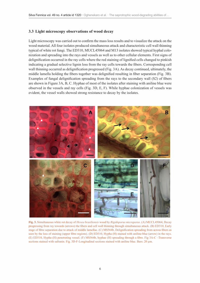
3.3 Light microscopy observations of wood decay
Lightmicroscopywascarriedouttoconfirmthemasslossresultsandtovisualizetheattackonthewood material. All four isolates produced simultaneous attack and characteristic cell wall thinning typical of white rot fungi. The ED310, MUCL45064 and M13 isolates showed typical hyphal colo-nization and spreading into the rays and vessels as well as to other cellular elements. First signs of delignificationoccurredintheraycellswheretheredstainingoflignifiedcellschangedtopinkishindicatingagradualselectiveligninlossfromtheraycellstowardsthefibers.Correspondingcellwallthinningoccurredasdelignificationprogressed(Fig.3A).Asdecaycontinued,ultimately,themiddlelamellaholdingthefiberstogetherwasdelignifiedresultinginfiberseparation(Fig.3B).Examplesoffungaldelignificationspreadingfromtheraystothesecondarywall(S2)offibersare shown in Figure 3A, B, C. Hyphae of most of the isolates after staining with aniline blue were observed in the vessels and ray cells (Fig. 3D, E, F). While hyphae colonization of vessels was evident, the vessel walls showed strong resistance to decay by the isolates.
Fig. 3. Simultaneous white rot decay of Hevea brasiliensis wood by Rigidoporus microporus. (A) MUCL45064, Decay progressingfromraytowards(arrows)thefibersandcellwallthinningthroughsimultaneousattack.(B)ED310,Earlystageoffibreseparationduetoattackofmiddlelamellae.(C)MS564b,Delignificationspreadingfromacrossfibersasseenbythelossofstaining(upperfibreregions).(D)ED310,Hypha(H)stainedwithaniline-blue(arrow)intherays.(E)ED310,Hypha(H)penetratingvessel.(F)MS564b,hyphae(H)spreadingthroughafibre.Fig3A-C–Transversesections stained with safranin. Fig. 3D-F-Longitudinal sections stained with aniline blue. Bars: 20 µm.

7
Silva Fennica vol. 49 no. 4 article id 1320 · Oghenekaro et al. · The saprotrophic wood-degrading abilities of…
4 Discussion
In this study, the results indicated that the R. microporus isolates previously isolated from trees displayed varying abilities to cause mass loss of rubber wood suggesting differences in their sap-rotrophic abilities. Interestingly, the values for percentage mass loss recorded were comparable to other studies using strains of the sapstain fungus Lasiodiploidia theobromae (Pat.) Griffon & Maubl. cultivated on H. brasiliensis and inoculated under similar conditions (Encinas and Daniel 1996; 1997). The isolate MUCL45064 collected from a non-H. brasiliensis host showed higher wood decay ability than the isolates which are proven pathogens on rubber trees. This isolate also had the highest growth on MEA at 25 °C, the same temperature used for the wood decay experiment. Itisdifficulttospeculateonwhetherthehighersaprotrophicabilityofthisisolatealsosuggestsa potential to be pathogenic on H. brasiliensis plantations especially if it undergoes a host-shift. Possibility for a host-shift of R. microporus from other trees to H. brasiliensis or vice-versa has been proven through molecular studies (Oghenekaro et al. 2014). The fungus has recently been reportedforthefirsttimeaspathogeniconArtocarpus nobilis Thw. (Moraceae family) in Sri Lanka (Madushani et al. 2013). An interesting feature concerned the endophytic isolate which showed very poor saprotrophic ability; perhaps this is associated with its behavior as a sapwood endophyte of H. brasiliensis in the natural forest (Martin et al. 2015).
Of the four isolates studied, the MUCL45064, ED310 and M13 isolates showed characteristic mycelial strand development in culture. R. microporus produces rhizomorphs in the soil and also mycelial strands under culture conditions (Richard and Button 1996). The absence of mycelial strands in the non-pathogenic isolate further points to an endophytic life-style. This isolate was previously isolated from the sapwood of healthy H. brasilieneis trees showing no symptoms of WRD (Martin et al. 2015). The effect of different temperature on the growth rates of the fungi also varied among the isolates. The varying saprotrophic abilities of the different isolates suggests that the isolates col-lectively considered as R. microporus (Mycobank, GenBank) and used for this study are most likely different species as suggested by Oghenekaro et al. (2014) through molecular phylogenetic studies. This supports the consensus that the R. microporus/lignosus species complex should be revised.
Light microscopy observations of H. brasiliensisdegradedwoodconfirmedtypicalwhiterot decay. The differential pink and red Safranin staining of cell walls showed evidence of del-ignificationasreportedinpreviousstudies(SrebotnikandMessner1994;SchwarzeandEngels1998).Theisolatescolonizedthewoodblocksthroughtherays,vesselsandthenfibers.Directhyphal penetration through wood cell walls was observed for all isolates except for the endophytic isolate whose hyphae only colonized and spread in the rays. There was widespread distribution of hyphaethroughoutvessels,fibresandtracheidsinthewoodblocksespeciallywithisolateED310(Schwarz et al., 2004; Schwarz 2007). Simultaneous cell wall thinning and preferential degrada-tion of wood cell walls was observed for the MUCL45064, ED310 and M13 isolates. Degrada-tion of cell wall materials particularly around the middle lamella regions was also evident. The saprotrophic decay patterns observed are similar to those reported for other basidiomycete white rot fungi like Heterobasidion annosum (Daniel et al. 1998); Lenzites stereoides (Fr.) Ryvarden and Ganoderma lucidum (Curtis) P. Karst (Nagadesi et al. 2013); Phanerochaete chrysosporium Burdsall (Koyani and Rajput 2014); Schizophyllum commune Fries and Flavodonflavus (Klotzsch) Ryvarden (Padhiar and Albert 2012).
The results of the present study reveal differences in the wood decay capability of host and non-host pathogenic and endophytic isolates as well as in their physiology. The study further sheds light on the potential contribution of the isolates to carbon cycling through their saprotrophic decay activities on dead trees already killed by this fungus. This imperatively should have important implications on the management of trees in rubber plantations infected by the pathogens.

8
Silva Fennica vol. 49 no. 4 article id 1320 · Oghenekaro et al. · The saprotrophic wood-degrading abilities of…
Acknowledgements
We thank the Delta State Government of Nigeria for a postgraduate scholarship to A.O.O. We are also grateful to Academy of Finland for project funding.
References
Bey M., Zhou S., Poidevin L., Henrissat B., Coutinho PM., Berrina J., Sigoillot J. (2013). Cello-Oligosaccharide oxidation reveals differences between two lytic polysaccharide monooxy-genases (family GH61) from Podospora anserina. Applied and Environmental Microbiology 79:488–496.http://dx.doi.org/10.1128/AEM.02942-12.
Bonomo R.P., Boudet A.M., Cozzolino R., Rizzarelli E., Santoro A.M., Sterjiades R., Zappalà R. (1998). A comparative study of two isoforms of laccase secreted by the ‘white- rot’ fungus Rigidoporus lignosus,exhibitingsignificantstructuralandfunctionaldifferences.JournalofInorganicBiochemistry71(3–4):205–211.http://dx.doi.org/10.1016/S0162-0134(98)10057-0.
Bonomo R.P., Cennamo G., Purrello R., Santoro A.M., Zappalà R. (2001). Comparison of three fungal laccases from Rigidoporus lignosus and Pleurotus ostreatus: correlation between conformationchangesandcatalyticactivity.JournalofInorganicBiochemistry83(1):67–75.http://dx.doi.org/10.1016/S0162-0134(00)00130-6.
CambriaM.,CambriaA.,RagusaS.,RizzarelliE.(2000).Production,purificationandpropertiesof an extracellular laccase from Rigidoporus lignosus.ProteinExpressionandPurification18(2):141–147.http://dx.doi.org/10.1006/prep.1999.1126.
Cambria M.T., Ragusa S., Calabrese V., Cambria A. (2011). Enhanced laccase production in white-rot fungus Rigidoporus lignosus by the addition of selected phenolic and aromatic compounds. AppliedBiochemistryandBiotechnology163(3):415–422.http://dx.doi.org/10.1007/s12010-010-9049-2.
Cambria M.T., Gullotto D., Garavaglia S., Cambria A. (2012). In silico study of structural deter-minants modulating the redox potential of Rigidoporus lignosus and other fungal laccases. JournalofBiomolecularStructureandDynamics30(1):89–101.http://dx.doi.org/10.1080/07391102.2012.674275.
Daniel G. (2014). Fungal and bacterial biodegradation: white rots, brown rots, soft rots, and bac-teria. In: Schultz T. et al. (eds.). Deterioration and protection of sustainable biomaterials. ACS SymposiumSeries,AmericanChemicalSociety,Washington,DC.p.23–54.http://dx.doi.org/10.1021/bk-2014-1158.ch002.
Daniel G., Asiegbu F., Johansson M. (1998). The saprotrophic wood-degrading abilities of Het-erobasidium annosum intersterility groups P and S. Mycological Research 102(8):991–997.http://dx.doi.org/10.1017/S0953756297005935.
Encinas O., Daniel G. (1996). Decay capacity of different strains of the blue stain fungus Lasi-odiplodia theobromae on various wood species. Material Und Organismen 30(4):239–258.
Encinas O., Daniel G. (1997). Degradation of the gelatinous layer in aspen and rubberwood by the blue stain fungus Lasiodiplodia theobromae. International Association of Wood Anatomists Journal 18(2):107–115.http://dx.doi.org/10.1163/22941932-90001471.
Farid A.M., Lee S.S., Maziah Z., Patahayah M. (2009). Pathogenicity of Rigidoporus microporus and Phellinus noxius against four major plantation tree species in peninsular Malaysia. Journal ofTropicalForestScience21:289–298.
Fernandez-Fueyo E., Ruiz-Duenas F.J., Ferreira P., Floudas D., Hibbett D.S et al. (2012). Com-parative genomics of Ceriporiopsis subvermispora and Phanerochaete chrysosporium provide
9
Silva Fennica vol. 49 no. 4 article id 1320 · Oghenekaro et al. · The saprotrophic wood-degrading abilities of…
insight into selective ligninolysis. Proceedings of the National Academy of Sciences of the UnitedStatesofAmerica109:5458–5463.http://dx.doi.org/10.1073/pnas.1119912109.
Floudas D., Binder M., Riley R., Barry K., Blanchette R.A. et al. (2012). The Paleozoic origin of enzymatic lignin decomposition reconstructed from 31 fungal genomes. Science 336: 1715–1719.http://dx.doi.org/10.1126/science.1221748.
Furukawa T., Bello F.O., Horsfall L. (2014). Microbial enzyme systems for lignin degradation and their transcriptional regulation. Frontiers in Biology 9(6):448–471.http://dx.doi.org/10.1007/s11515-014-1336-9.
Geiger J.P., Rio B., Nicole M., Nandris D. (1989). Peroxidase production in tissues of the rubber tree following infection by root rot fungi. Physiological and Molecular Plant Pathology 34: 241–256.http://dx.doi.org/10.1016/0885-5765(89)90047-7.
Hori C., Gaskell J., Igarashi K., Samejima M., Hibbett D et al. (2013). Genome wide analysis of polysaccharide degrading enzymes in eleven white and brown rot polyporales provides insight intomechanismsofwooddecay.Mycologia105:1412–1427.http://dx.doi.org/10.3852/13-072.
Kaewchai S., Soytong K. (2010). Application of biofungicides against Rigidoporus microporus causingwhiterootdiseaseofrubbertrees.JournalofAgriculturalTechnology6:349–363.
Kaewchai S., Wang H.K., Lin F., Hyde K.D., Soytong K. (2009). Genetic variation among isolates of Rigidoporus microporus causing white root disease of rubber trees in southern Thailand revealed by ISSR markers and pathogenicity. African Journal of Microbiology Research 3: 641–648.
Kaewchai S., Lin F., Wang H.K., Soytong K. (2010). Characterization of Rigidoporus microporus isolated from rubber trees based on morphology and ITS sequencing. Journal of Agricultural Technology6:289–298.
Koyani R.D., Rajput K.S. (2014). Light microscopic analysis of Tectona grandis L.f. wood inocu-lated with Irpex lacteus and Phanerochaete chrysosporium. European Journal of Wood and Wood Products 72(2):157–164.http://dx.doi.org/10.1007/s00107-013-0763-7.
Levasseur A., Lomascolo A., Chabrol O., Ruiz-Dueñas F.J., Boukhris-Uzan E et al. (2014). The genome of the white-rot fungus Pycnoporus cinnabarinus: a basidiomycete model with a versatile arsenal for lignocellulosic biomass breakdown. BMC Genomics 15(1): 486. http://dx.doi.org/10.1186/1471-2164-15-486.
Liyanage A.S. (1997). Rubber. In: Hillocks R.J., Waller J.M. (eds.). Soilborne diseases of tropical crops.CABInternational,Wallingford,UK.p.331–347.
Madushani H.K. I., Fernando T.H. P.S., Wijesundara R.L. C., Siriwardane D. (2013). First report of white root disease of Artocarpus nobilis in Sri lanka caused by Rigidoporus microporus. JournaloftheNationalScienceFoundationofSriLanka42(2):197–198.
Martin R., Gazis R., Skaltsas D., Chaverri P., Hibbett D.S (2015). Unexpected diversity of basidiomycetous endophytes in sapwood and leaves of Hevea. Mycologia. http://dx.doi.org/10.3852/14-206.
NagadesiP.K.,AryaA.,AlbertS.(2013).Delignificationpatternofwooddecaybywhiterotfungiin teak (Tectona grandis L. f.). Journal of the Indian Academy of Wood Science 10(1):1–8.http://dx.doi.org/10.1007/s13196-013-0085-8.
Nandris D., Nicole M., Geiger J.P. (1988). Root rot diseases of rubber tree. I. Severity, dynamics andcharacterizationofepidemics.CanadianJournalofForestResearch18:1248–1254.http://dx.doi.org/10.1139/x88-192.
Nicole M., Geiger J.P., Nandris D. (1985). Defense reactions of Hevea brasiliensis to root rot diseases. European Journal of Forest pathology 15: 320–322. http://dx.doi.org/10.1111/j.1439-0329.1985.tb01106.x.
Nicole M., Geiger J.P., Nandris D. (1986a). Root rot diseases of Hevea brasiliensis. II.

10
Silva Fennica vol. 49 no. 4 article id 1320 · Oghenekaro et al. · The saprotrophic wood-degrading abilities of…
Some host reactions. European Journal of Forest pathology 16: 37–55. http://dx.doi.org/10.1111/j.1439-0329.1986.tb01050.x.
Nicole M., Geiger J.P., Nandris D. (1986b). Penetration and degradation of suberized cells of Hevea brasiliensis infected with root rot fungi. Physiological and Molecular Plant Pathology 28: 181–185. http://dx.doi.org/10.1016/S0048-4059(86)80062-5.
Nicole M., Chamberland H., Geiger J.P., Lecours N., Valero J., Rio B., Ouellette G.B. (1992). Immunocytochemical localization of laccase L1 in wood decayed by Rigidoporus lignosus. Applied and Environmental Microbiology 58(5): 1727–1739.
Ogbebor N.O., Adekunle A.T., Eghafona O.N., Ogboghodo A.I. (2015). Biological control of Rigidoporus lignosus in Hevea brasiliensis in Nigeria. Fungal Biology 119: 1–6. http://dx.doi.org/10.1016/j.funbio.2014.10.002.
Oghenekaro A.O., Miettinen O., Omorusi V.I., Evueh G.A., Farid M.A., Gazis R., Asiegbu F.O. (2014). Molecular phylogeny of Rigidoporus microporus isolates associated with white rot disease of rubber trees (Hevea brasiliensis). Fungal Biology 118: 495–506. http://dx.doi.org/10.1016/j.funbio.2014.04.001.
Omorusi V.I. (2012). Effects of white root rot disease on Hevea brasiliensis (Muell. Arg.) - chal-lenges and control approach. In: Dhal N.K., Sahu S.C. (eds.). Plant Science. http://www.intechopen.com/books/plant-science/effects-of-white-root-rot-disease-on-Hevea-brasiliensis-muell-arg-challenges-and-control-approach.
Padhiar A., Albert S. (2012). Anatomical studies on decaying wood of Mangifera indica by two white rot fungi Schizophyllum commune and Flavadon flavus. Journal of the Indian Academy of Wood Science 9(2): 143–153. http://dx.doi.org/10.1007/s13196-012-0079-y.
Richard T., Botton B. (1996). Growth and mycelial strand production of Rigidoporus lignosus with various nitrogen and carbon sources. Mycopathologia 134: 83–89. http://dx.doi.org/10.1007/BF00436869.
Ryvarden L. (1976). Type studies in the Polyporaceae 4. Species described by J.F Klotzsch. Mem-oirs of the New York Botanical Garden 28: 199–207.
Sail R.M., Ahmad M. (2009). Enhancing socio-economy of rubber smallholders through effective transfer of technology. National Rubber Economic Conference 2009 (NREC), Nikko Hotel, Kuala Lumpur, 23–24 June, 2009. p. 134–142.
Schwarze F.W.M.R., Engels J. (1998). Cavity formation and the exposure of peculiar structures in the secondary wall (S2) of tracheids and fibres by wood degrading basidiomycetes. Holz-forschung 52: 117–123. http://dx.doi.org/10.1515/hfsg.1998.52.2.117.
Schwarz F.W.R.M., Engels J., Mattheck C. (2004). Fungal strategies of wood decay in trees. Springer, Heidelberg. 185 p.
Schwarze F.W. M.R. (2007). Wood decay under the microscope. Fungal Biology Reviews 21(4): 133–170. http://dx.doi.org/10.1016/j.fbr.2007.09.001.
Srebotnik E., Messner K. (1994). A simple method that uses differential staining and light micros-copy to assess the selectivity of wood delignification by white rot fungi. Applied and Envi-ronmental Microbiology 60: 1383–1386.
Umar H.Y., Esekhade T.U., Idoko S.O., Ugwa I.K.(2010). Production analysis of budded rubber stumps in Rubber Research Institute of Nigeria (RRIN). Journal of Agricultural Science 1: 109–113.
Total of 42 references