The safety of beta-blocker use in chronic obstructive pulmonary disease patients with respiratory...
9
ORIGINAL RESEARCH ARTICLE Open Access The safety of beta-blocker use in chronic obstructive pulmonary disease patients with respiratory failure in the intensive care unit Feyza Kargin 1 , Huriye Berk Takir 1 , Cuneyt Salturk 1 , Nezihe Ciftaslan Goksenoglu 1 , Can Yucel Karabay 2 , Ozlem Yazicioglu Mocin 1 , Nalan Adiguzel 1 , Gokay Gungor 1 , Merih Kalamanoglu Balci 1 , Murat Yalcinsoy 1 , Ramazan Kargin 3 and Zuhal Karakurt 1* Abstract Background: The safety of beta-blockers as a heart rate-limiting drug (HRLD) in patients with acute respiratory failure (ARF) due to chronic obstructive lung disease (COPD) has not been properly assessed in the intensive care unit (ICU) setting. This study aims to compare the use of beta-blocker drugs relative to non-beta-blocker ones in COPD patients with ARF due to heart rate-limiting with respect to length of ICU stay and mortality. Methods: We performed a retrospective (January 2011-December 2012) case-control study in a level III ICU in a teaching hospital. It was carried out in a closed ICU by the same intensivists. All COPD patients with ARF who were treated with beta-blockers (case group) and non-beta-blocker HRLDs (control group) were included. Their demographics, reason for HRLD, cause of ARF, comorbidities, ICU data including acute physiology and chronic health evaluation (APACHE II) score, type of ventilation, heart rate, and lengths of ICU and hospital stays were collected. The mortality rates in the ICU, the hospital, and over 30 days were also recorded. Results: We enrolled 188 patients (46 female, n = 74 and n = 114 for the case and control groups, respectively). Reasons for HRLD (case and control group, respectively) were atrial fibrillation (AF, 23% and 50%), and supraventricular tachycardia (SVT, 41.9% and 54.4%). Patients’ characteristics, APACHE II score, heart rate, duration and type of ventilation, and median length of ICU-hospital stay were similar between the groups. The mortality outcomes in the ICU, hospital, and 30 days after discharge in the case and control groups were 17.6% versus 15.8% (p > 0.75); 18.9% versus 19.3% (p > 0.95) and 20% versus 11% (p > 0.47), respectively. Conclusions: Our results suggest that beta-blocker use for heart rate control in COPD patients with ARF is associated with similar ICU stay length and mortality compared with COPD patients treated with other HRLDs. Keywords: Acute respiratory failure, Arrhythmia, Beta-blockers, COPD, Intensive care unit Background The majority of patients with chronic obstructive pul- monary disease (COPD) have chronic heart failure (CHF) or coronary artery disease (CAD) [1]. The risk of cardiac arrhythmia is increased during acute exacer- bations of COPD [1]. Atrial fibrillation (AF) is fre- quently observed in elderly COPD patients [2], and cardiac arrhythmias are a significant cause of mortality in these patients [3]. COPD patients with CAD generally have elevated heart rates, and beta-adrenergic receptor an- tagonists (beta-blockers) are known to improve the sur- vival of patients with CHF or CAD [4,5]. Previous studies have shown that patients with CAD and coexisting COPD generally failed to receive optimal therapy or appropriate drug dosages for HR reduction. In a recent study, 54% of patients suffered from heart failure in a population with CAD and COPD, and only 52.8% of these patients were receiving beta-blocker therapy. In addition, in the majority of these patients the daily dosages of beta-blockers were * Correspondence: [email protected] 1 Respiratory and Intensive Care Unit, Sureyyapaşa Chest Diseases and Thoracic Surgery Training and Research Hospital, Soyak Yenişehir Manolya Evleri, 34770 Umraniye, Istanbul, Turkey Full list of author information is available at the end of the article © 2014 Kargin et al.; licensee BioMed Central Ltd. This is an Open Access article distributed under the terms of the Creative Commons Attribution License (http://creativecommons.org/licenses/by/2.0), which permits unrestricted use, distribution, and reproduction in any medium, provided the original work is properly cited. The Creative Commons Public Domain Dedication waiver (http://creativecommons.org/publicdomain/zero/1.0/) applies to the data made available in this article, unless otherwise stated. Kargin et al. Multidisciplinary Respiratory Medicine 2014, 9:8 http://www.mrmjournal.com/content/9/1/8
Transcript of The safety of beta-blocker use in chronic obstructive pulmonary disease patients with respiratory...

ORIGINAL RESEARCH ARTICLE Open Access
The safety of beta-blocker use in chronicobstructive pulmonary disease patients withrespiratory failure in the intensive care unitFeyza Kargin1, Huriye Berk Takir1, Cuneyt Salturk1, Nezihe Ciftaslan Goksenoglu1, Can Yucel Karabay2,Ozlem Yazicioglu Mocin1, Nalan Adiguzel1, Gokay Gungor1, Merih Kalamanoglu Balci1, Murat Yalcinsoy1,Ramazan Kargin3 and Zuhal Karakurt1*
Abstract
Background: The safety of beta-blockers as a heart rate-limiting drug (HRLD) in patients with acute respiratoryfailure (ARF) due to chronic obstructive lung disease (COPD) has not been properly assessed in the intensive careunit (ICU) setting. This study aims to compare the use of beta-blocker drugs relative to non-beta-blocker ones inCOPD patients with ARF due to heart rate-limiting with respect to length of ICU stay and mortality.
Methods: We performed a retrospective (January 2011-December 2012) case-control study in a level III ICU in ateaching hospital. It was carried out in a closed ICU by the same intensivists. All COPD patients with ARF who weretreated with beta-blockers (case group) and non-beta-blocker HRLDs (control group) were included. Their demographics,reason for HRLD, cause of ARF, comorbidities, ICU data including acute physiology and chronic health evaluation (APACHEII) score, type of ventilation, heart rate, and lengths of ICU and hospital stays were collected. The mortality rates in the ICU,the hospital, and over 30 days were also recorded.
Results: We enrolled 188 patients (46 female, n = 74 and n = 114 for the case and control groups, respectively). Reasonsfor HRLD (case and control group, respectively) were atrial fibrillation (AF, 23% and 50%), and supraventricular tachycardia(SVT, 41.9% and 54.4%). Patients’ characteristics, APACHE II score, heart rate, duration and type of ventilation, and medianlength of ICU-hospital stay were similar between the groups. The mortality outcomes in the ICU, hospital, and 30 daysafter discharge in the case and control groups were 17.6% versus 15.8% (p > 0.75); 18.9% versus 19.3% (p > 0.95) and 20%versus 11% (p > 0.47), respectively.
Conclusions: Our results suggest that beta-blocker use for heart rate control in COPD patients with ARF is associated withsimilar ICU stay length and mortality compared with COPD patients treated with other HRLDs.
Keywords: Acute respiratory failure, Arrhythmia, Beta-blockers, COPD, Intensive care unit
BackgroundThe majority of patients with chronic obstructive pul-monary disease (COPD) have chronic heart failure(CHF) or coronary artery disease (CAD) [1]. The riskof cardiac arrhythmia is increased during acute exacer-bations of COPD [1]. Atrial fibrillation (AF) is fre-quently observed in elderly COPD patients [2], and
cardiac arrhythmias are a significant cause of mortality inthese patients [3]. COPD patients with CAD generallyhave elevated heart rates, and beta-adrenergic receptor an-tagonists (beta-blockers) are known to improve the sur-vival of patients with CHF or CAD [4,5]. Previous studieshave shown that patients with CAD and coexisting COPDgenerally failed to receive optimal therapy or appropriatedrug dosages for HR reduction. In a recent study, 54% ofpatients suffered from heart failure in a population withCAD and COPD, and only 52.8% of these patients werereceiving beta-blocker therapy. In addition, in the majorityof these patients the daily dosages of beta-blockers were
* Correspondence: [email protected] and Intensive Care Unit, Sureyyapaşa Chest Diseases andThoracic Surgery Training and Research Hospital, Soyak Yenişehir ManolyaEvleri, 34770 Umraniye, Istanbul, TurkeyFull list of author information is available at the end of the article
© 2014 Kargin et al.; licensee BioMed Central Ltd. This is an Open Access article distributed under the terms of the CreativeCommons Attribution License (http://creativecommons.org/licenses/by/2.0), which permits unrestricted use, distribution, andreproduction in any medium, provided the original work is properly cited. The Creative Commons Public Domain Dedicationwaiver (http://creativecommons.org/publicdomain/zero/1.0/) applies to the data made available in this article, unless otherwisestated.
Kargin et al. Multidisciplinary Respiratory Medicine 2014, 9:8http://www.mrmjournal.com/content/9/1/8

very low, which could be explained by the physicians’ hesi-tation to prescribe them due to possible adverse pulmonaryeffects [6]. The current medical treatment approach forCOPD exacerbation is the administration of short-actingbronchodilators (ß2-agonists), with or without anticholiner-gic compounds and corticosteroids [7]. COPD patients witharrhythmias require complex treatment strategies. Studiescomparing the side effects of beta-blockers have yieldedmixed results in patients with arrhythmias and myocardialinfarction (MI) [8-11].There are limited data about patients with acute respira-
tory failure (ARF) in the intensive care unit (ICU) thatdemonstrated the safety of beta-blockers in COPD pa-tients with arrhythmias. In this study, we compared theoutcomes of COPD patients with ARF who received beta-blockers versus other medications for heart rate control.We hypothesized that the use of beta-blockers as a heartrate-limiting drug (HRLD) in COPD patients with ARFwould achieve similar outcomes with respect to mortalityand length of ICU stay as other non-beta-blocker drugs.
MethodsWe performed a retrospective case–control study in theICU of a teaching hospital for chest diseases betweenJanuary 2011 and December 2012. The 22-bed ICU wasclassified as level III and was operated as a closed unit byeight pulmonologist-intensivist specialists 24 hours/dayand 7 days/week. The majority of ARF patients admittedto the ICU had COPD. This study was approved by thelocal ethical committee of the government teaching hos-pital (Sureyyapasa Chest Diseases and Thoracic SurgeryTeaching Hospital-Istanbul-Turkey).
PatientsAll consecutive patients with previously diagnosed COPDwho were admitted to the ICU due to ARF during thestudy period were evaluated. COPD diagnoses were estab-lished by a physician based on airflow obstruction on spir-ometry, defined as a forced expiratory volume in 1 second(FEV1) and forced vital capacity ratio of 70% or less. Wesearched the electronic database system and InternationalClassification of Disease (ICD)-10 coding system and re-corded “J 44” as COPD. Patients were enrolled in thestudy if they had been treated with HRLDs at any timeduring their ICU stay. Patients with asthma or previoususe with diltiazem were excluded from the study, aswere those who had been in the ICU for less than 24hours. The patients were divided into two groups ac-cording to their HRLD treatment: beta-blocker (casegroup) and non-beta-blocker (control group). TheHRLDs administered in the case group included meto-prolol, bisoprolol, and carvedilol, and those given to the
control group were diltiazem, digoxin, amiodarone, orany combination of these.
DataThe presence of comorbidities, including diabetes melli-tus (E 10–11), ischemic heart disease (I 25), and atrialfibrillation (I 48), was determined by the ICD coding inour teaching hospital database. Previous HRLD use wasalso recorded at the time of ICU admission. Any previ-ous hospital admissions were recorded as emergency ser-vice, hospital ward, and other ICU. Body mass index (BMI),reason for HRLD use, causes of ARF, acute physiologicaland chronic health evaluation (APACHE II score) [12], ap-plication and duration of noninvasive (NIV) or invasivemechanical ventilation (IMV), heart rate, C-reactive protein(CRP) level at ICU admission and the peak value duringICU stay, biochemistry values, complete blood counts, spu-tum/tracheal aspirate/bronchial lavage cultures, lengths ofICU and hospital stays, and mortality in the ICU and hos-pital were recorded from the patients’ files. Mortality within30 days of hospital discharge was recorded from the onlinedeceased declaration system to assess short-term mortality.
DefinitionsARFPatients were divided into three groups according to theirarterial blood gas (ABG) results on admission to the ICU.“Hypoxic ARF” was defined if partial arterial oxygenpressure in inspired fractionated oxygen (PaO2/FiO2) was< 300 and partial arterial carbon dioxide pressure (PaCO2)was < 45 mmHg. “Hypercapnic and hypoxemic ARF” wasdefined as PaCO2 > 45 mmHg and PaO2/FiO2 < 300, and“hypercapnicARF” was PaCO2 > 45 mmHg and PaO2/FiO2 > 300 [13,14].
SepsisThe presence of sepsis was defined as the presence of in-fection together with systemic inflammatory response syn-drome [15,16]. Patients who were unresponsive to fluidresuscitation and required vasopressor agents were de-fined as being in septic shock [16].
TreatmentHRLDs used in the ICU were recorded in six major indica-tion subgroups: 1) AF [17], 2) supraventricular tachycardia(SVT) [18], 3) ventricular tachyarrhythmia (VT), 4) con-gestive heart failure CHF [19], 5) hypertension (HT) [20],and 6) suspicion of MI. These drugs were administered or-ally or via intravenous (IV) injection or infusion. The drugsand doses for CHF were as follows: metoprolol (12.5/25 mgonce daily [o.d.], starting dose targeting 200 mg o.d.) withbisoprolol (1.25 mg o.d., starting dose targeting 10 mg o.d),and carvedilol (3.125 mg, twice daily, starting dose targeting25-50 mg), or digoxin (0.25 mg o.d.) [19]. The drug doses
Kargin et al. Multidisciplinary Respiratory Medicine 2014, 9:8 Page 2 of 9http://www.mrmjournal.com/content/9/1/8

for AF treatment were metoprolol (2.5-5 mg IV bolus over2 minutes, maximum 3 doses, oral maintenance dose 100-200 mg, o.d.), bisoprolol (2.5-10 mg, o.d.), carvedilol (3.125-25 mg, twice daily), diltiazem (60 mg twice daily to 360 mgo.d.), digoxin (0.5-1 mg IV, then 0.125-0.5 mg o.d.), or ami-odarone (5 mg/kg in 1 hour, 50 mg/hour maintenance; orin oral form, 100-200 mg once daily) [17]. More than oneHRLDs (except beta-blockers) were defined as diltiazemplus amiodarone and/or plus digoxin. While using thesedrugs, the target heart rate was 80-100/minute [17]. NoHRLDs were administered if patients had septic shock. Thechoice of HRLDs was totally dependent on intensivist staffchoice unless a known contraindication was present (e.g.,diltiazem was not used in patients with lower ejectionfractions).
Treatment of COPD exacerbationA short acting ß2 agonist (salbutamol, 100 mcg perpuff ) and ipratropium bromide (100 mcg/20 mcg perpuff ) were given every 2-4 hours (4 to 10 puffs) via ametered dose inhaler chamber (Aerovent, Altech®, AlteraFirm, Izmir-Turkey) when the patients were under NIV orIMV. A nebular form of salbutamol (2.5 mg/2.5 mL pernebule) was given every 15 minutes to 4 hours, or ipratro-pium bromide/salbutamol (0.5 mg/3.01 mg/2.5 mL pernebule) was given every 2 to 4 hours for patients breathingwithout mechanical ventilation support. Long-acting ß2-agonists were not used in COPD patients with ARF in theICU [7,21]. The long-acting ß2-agonists, such as salme-terol and formeterol, were not used during the ICU stay. Asystemic corticosteroid, prednisolone (30–40 mg per day),was also administered [22]. Theophylline was not used ineligible tachycardic COPD patients. Antibiotics were ad-ministered according to recommended guidelines [22].
Mechanical ventilationNIV was applied initially in the respiratory ward by aspecialized staff (nurse and specialist or resident) if pa-tients were hemodynamically stable, cooperative, hadno organ failure except respiratory failure, their ABGanalysis revealed pH 7.28-7.34, PaCO2 = 45-90 mmHg,PaO2/FiO2 > 200, and the Glasgow coma scale was > 13due to hypercapnic confusion. ICU demand for patientsunder NIV treatment in the ward was determined basedon cognitive function deterioration, lack of cooperation,agitation, hemodynamically unstable conditions (e.g.,hypotension, HT, tachycardia, arrhythmia) that neededclose monitoring, and deterioration of ABG values (in-creased PaCO2, decreased PaO2, and decreased pH level).NIV was provided in pressure assist-control mode withICU mechanical ventilators via a double-tube circuit witha full-face mask [13]. Invasive mechanical ventilation wasapplied in the presence of absolute or relative contraindi-cations for NIV and in NIV failure [13]. Contraindications
of NIV were defined as: 1) absolute, respiratory arrest andunable to fit mask, and 2) relative, medically unstable(hypotensive shock, uncontrolled cardiac ischemia orarrhythmia, upper gastrointestinal bleeding), agitation,uncooperativeness, inability to protect airway, impairedswallowing, excessive secretions not managed by secretionclearance techniques, multiple (two or more) organ fail-ure, and recent upper airway or upper gastrointestinal sur-gery [13,14]. The definition of NIV failure in hypercapnicpatients was no pH improvement, no change or a rise inbreathing frequency after 1–2 hours, and lack of cooper-ation. For hypoxic COPD patients, failure was consideredas no, or a minimal, rise in PaO2/FiO2 after 1-2 hours(< 200) [13]. The Richmond agitation sedation scale(RASS) was used for infusion and assessment of the dailyneed for sedation [23]. When patients met the previouslydescribed criteria for weaning, they were extubated after30 minutes of a successful T-piece trial [24]. After extuba-tion, NIV was applied in cases of moderate respiratorydistress, if there were no contraindications [24].
OutcomesThe primary outcome was mortality (including hospitaland 30 days after hospital discharge), and the secondaryoutcomes were NIV and IMV durations and the lengthof ICU stay.
Statistical analysisA descriptive analysis was used for patient demographicsand ICU data. Case and control groups were comparedwith Mann Whitney U tests for non-parametric continu-ous variables or Student’s t-tests for parametric continu-ous variables. The chi-square test was employed fordichotomous variables. Median and quartiles 1 and 3 wereused to describe non-parametric continuous variables, andmean ± standard deviation (SD) was used for parametriccontinuous variables. Count and percentage were usedwhen applicable. For predicting the mortality rates of ICU,hospital, and 30 days after hospital discharge, logistic re-gression analysis was applied using a model that includedHRLD type, APACHE II score on admission to the ICU,age, reasons for ICU admission, NIV and IMV implications,presence of septic shock, CRP level, and previous HRLDuse. P < 0.05 was accepted as statistically significant.
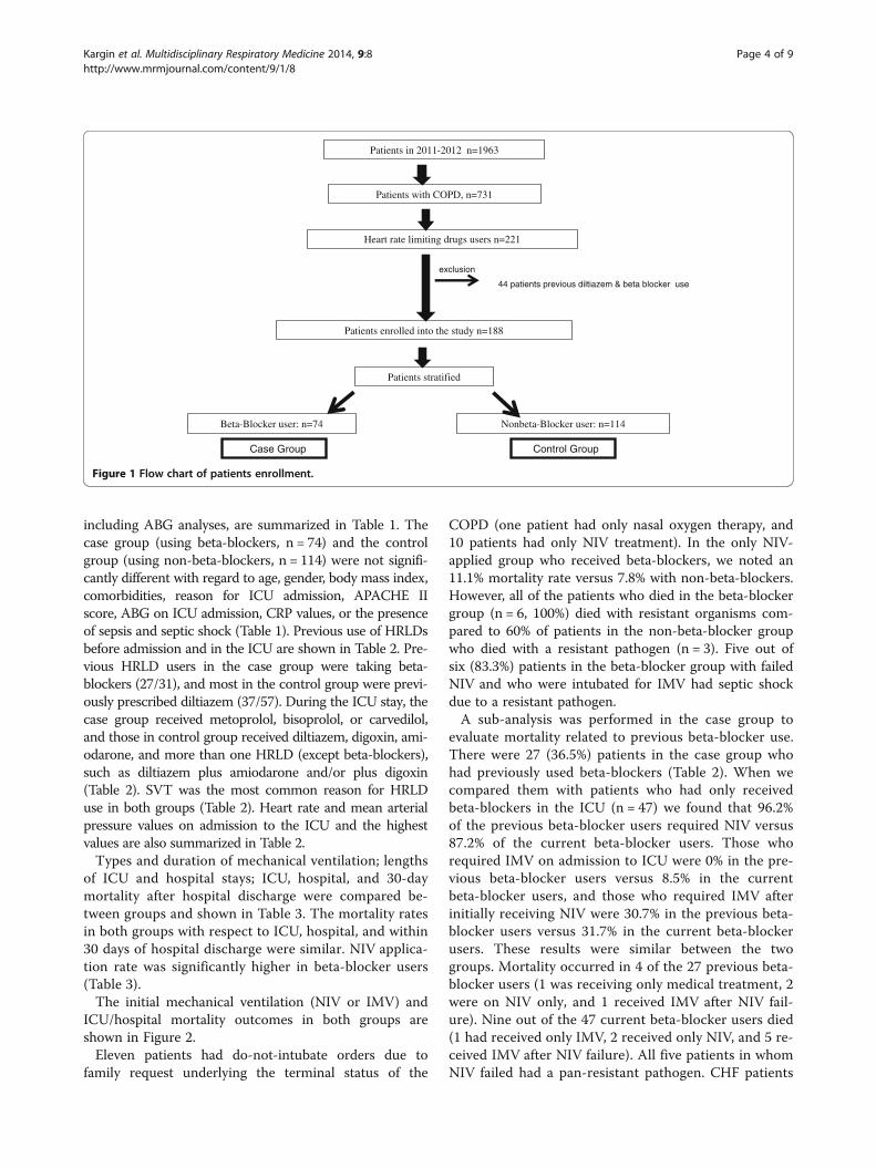
ResultsDuring the study period, a total of 1,964 patients were ad-mitted to the ICU, and 221 patients were using HRLDs.After excluding 44 patients due to simultaneous beta-blocker and diltiazem use, 188 patients were included inthe study. The patient enrollment flow chart is shown inFigure 1.Patient group demographics, comorbidities, reason for
ICU admission, microbiology results, and ICU data,
Kargin et al. Multidisciplinary Respiratory Medicine 2014, 9:8 Page 3 of 9http://www.mrmjournal.com/content/9/1/8
including ABG analyses, are summarized in Table 1. Thecase group (using beta-blockers, n = 74) and the controlgroup (using non-beta-blockers, n = 114) were not signifi-cantly different with regard to age, gender, body mass index,comorbidities, reason for ICU admission, APACHE IIscore, ABG on ICU admission, CRP values, or the presenceof sepsis and septic shock (Table 1). Previous use of HRLDsbefore admission and in the ICU are shown in Table 2. Pre-vious HRLD users in the case group were taking beta-blockers (27/31), and most in the control group were previ-ously prescribed diltiazem (37/57). During the ICU stay, thecase group received metoprolol, bisoprolol, or carvedilol,and those in control group received diltiazem, digoxin, ami-odarone, and more than one HRLD (except beta-blockers),such as diltiazem plus amiodarone and/or plus digoxin(Table 2). SVT was the most common reason for HRLDuse in both groups (Table 2). Heart rate and mean arterialpressure values on admission to the ICU and the highestvalues are also summarized in Table 2.Types and duration of mechanical ventilation; lengths
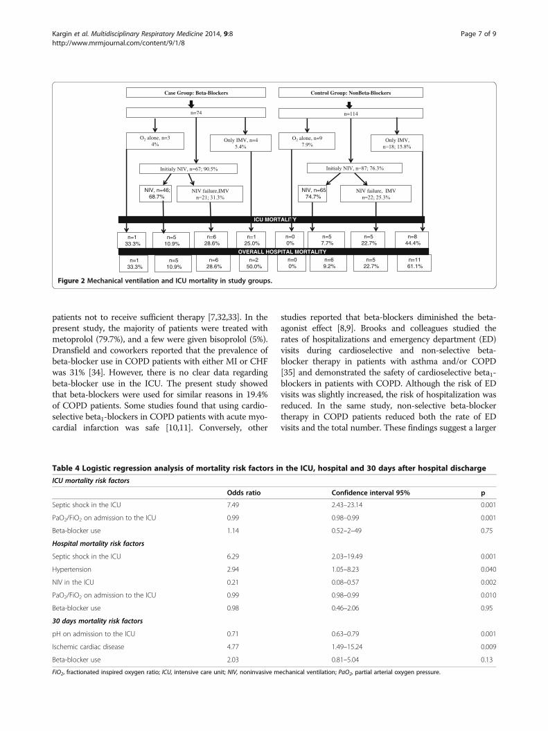
of ICU and hospital stays; ICU, hospital, and 30-daymortality after hospital discharge were compared be-tween groups and shown in Table 3. The mortality ratesin both groups with respect to ICU, hospital, and within30 days of hospital discharge were similar. NIV applica-tion rate was significantly higher in beta-blocker users(Table 3).The initial mechanical ventilation (NIV or IMV) and
ICU/hospital mortality outcomes in both groups areshown in Figure 2.Eleven patients had do-not-intubate orders due to
family request underlying the terminal status of the
COPD (one patient had only nasal oxygen therapy, and10 patients had only NIV treatment). In the only NIV-applied group who received beta-blockers, we noted an11.1% mortality rate versus 7.8% with non-beta-blockers.However, all of the patients who died in the beta-blockergroup (n = 6, 100%) died with resistant organisms com-pared to 60% of patients in the non-beta-blocker groupwho died with a resistant pathogen (n = 3). Five out ofsix (83.3%) patients in the beta-blocker group with failedNIV and who were intubated for IMV had septic shockdue to a resistant pathogen.A sub-analysis was performed in the case group to
evaluate mortality related to previous beta-blocker use.There were 27 (36.5%) patients in the case group whohad previously used beta-blockers (Table 2). When wecompared them with patients who had only receivedbeta-blockers in the ICU (n = 47) we found that 96.2%of the previous beta-blocker users required NIV versus87.2% of the current beta-blocker users. Those whorequired IMV on admission to ICU were 0% in the pre-vious beta-blocker users versus 8.5% in the currentbeta-blocker users, and those who required IMV afterinitially receiving NIV were 30.7% in the previous beta-blocker users versus 31.7% in the current beta-blockerusers. These results were similar between the twogroups. Mortality occurred in 4 of the 27 previous beta-blocker users (1 was receiving only medical treatment, 2were on NIV only, and 1 received IMV after NIV fail-ure). Nine out of the 47 current beta-blocker users died(1 had received only IMV, 2 received only NIV, and 5 re-ceived IMV after NIV failure). All five patients in whomNIV failed had a pan-resistant pathogen. CHF patients
Patients in 2011-2012 n=1963
Patients with COPD, n=731
Heart rate limiting drugs users n=221
Beta-Blocker user: n=74 Nonbeta-Blocker user: n=114
Patients enrolled into the study n=188
Patients stratified
Case Group Control Group
44 patients previous diltiazem & beta blocker use
exclusion
Figure 1 Flow chart of patients enrollment.
Kargin et al. Multidisciplinary Respiratory Medicine 2014, 9:8 Page 4 of 9http://www.mrmjournal.com/content/9/1/8

on beta-blockers before ICU admission (n = 13) or dur-ing the ICU stay (n = 11) had similar mortality rates(14.8% versus 19.1%, p = 0.64).During the study period, 363/1,963 patients died (19.5%)
in the ICU, and the mortality rate among the 731 patientswith COPD who were admitted to the ICU was 13.8%.Among 188 COPD patients using HRLDs, the overall mor-tality rate was 16.5% (31/188).
For predicting ICU, hospital, and 30-day mortality ratesafter hospital discharge, we included APACHE II score onadmission, age, reasons for ICU admission, NIV and IMVimplications, presence of septic shock, CRP level, and pre-vious HRLD use in the binary logistic regression model.The significant predictors and the effects of beta-blockeruse are summarized in Table 4. Beta blocker use was notfound to have a significant effect on mortality rate in the
Table 1 Patient characteristics for groups (case: beta-blockers, control: non-beta-blockers)
Case, n = 74 Control, n = 114 p value
Age, years, median (quartile 1–3) 71 (63–77) 71 (64–75) 0.82
Female/male 18/56 28/86 0.97
BMI, kg/m2, median (quartile 1–3) 23 (22–27) 24 (21–28) 0.82
Comorbidities, n (%) 69 (93.2) 100 (87.7) 0.22
AF, n (%) 56 (75.7) 80 (70.2) 0.41
Diabetes mellitus, n (%) 20 (27.0) 29 (25.4) 0.81
CAD, n (%) 12 (16.2) 14 (12.3) 0.45
Hypertension, n(%) 42(56.8) 73 (64) 0.32
Pre ICU location, n (%)
Emergency service 40 (54.1) 55 (48.2) 0.69
Hospital ward 31 (41.9) 55 (48.2)
Another ICU 3 (4.1) 4 (3.5)
Reason for ICU admission
COPD exacerbation 64 (86.5) 101 (88.6) 0.35
Pneumonia 8 (10.8) 6 (5.3)
Hemodynamic monitoring 0 (0) 3 (2.6)
Postoperative respiratory failure 0 (0) 1 (0.9)
Home ventilator evaluation 2 (2.7) 0 (0)
Sepsis, n (%) 50 (67.6) 76 (66.7) 0.90
Septic shock, n (%) 12 (16.2) 13 (11.4) 0.34
ICU admission APACHE-II value 19 (17–23) 20 (16–24) 0.88
ICU admission CRP mg/L 39 (14–91) 41 (14–125) 0.33
Peak of CRP mg/L median 64 (23–130) 110 (30–178) 0.08
Microbiologic culture, n (%) 51 (68.9) 67 (58.8) 0.16
Positive culture n (%) 18 (35.3) 27 (23.7) 0.58
Resistant pathogen, rate 14/18 23/27 0.43
Arterial blood gases analysis on admission ICU
pH, median (quartile 1–3) 7.28 (7.25-7.37) 7.32 (7.25-7.40) 0.08
PaCO2, mmHg, median (quartile 1–3) 72 (54–86) 73 (59–84) 0.78
PaO2/FiO2, median (quartile 1–3) 160 (130–211) 168 (116–230) 0.65
Type of acute respiratory failure, n(%)
PaO2/FiO2 <300 15 (20.3) 19 (16.7) 0.50
PaO2/FiO2 < 300 and PaCO2 > 45 mmHg 46 (62.2) 79 (69.3)
PaCO2 > 45 mmHg 13 (17.6) 16 (14.0)
AF, atrial fibrillation; APACHEII, acute physiologic and chronic health evaluation II; BMI, body mass index; CAD, coronary artery disease; COPD, chronic obstructivepulmonary disease; CRP, C-reactive protein; FiO2, fractionated inspired oxygen; ICU, intensive care unit, PaCO2, partial arterial carbon dioxide pressure; PaO2, partialarterial oxygen pressure.
Kargin et al. Multidisciplinary Respiratory Medicine 2014, 9:8 Page 5 of 9http://www.mrmjournal.com/content/9/1/8
ICU, the hospital, or within 30 days of discharge. Thepresence of septic shock on admission to the ICU andlower PaO2/FiO2 were found to be significant risk factorsfor ICU mortality (Table 4). For hospital mortality, thepresence of septic shock in the ICU, lower PaO2/FiO2, andHT were found to be associated with increased rateand conversely NIV application was with decreasedrate (Table 4). The presence of coronary heart diseasesincreased the short-term (30 days) mortality risk afterhospital discharge, whereas higher pH levels decreasedthe 30-day mortality rate (Table 4).
DiscussionThe present study showed similar mortality rates andlength of ICU stay in COPD patients with ARF who re-ceived either beta-blockers or other HRLDs to controlheart rate.Although the useful effects of beta-blockers in the treat-
ment of cardiac diseases are well-known, their use inCOPD patients has been restricted due to possible contra-indication [25]. It has been reported that selective andnon-selective beta-blockers increase airway hyperrespon-siveness (AHR) [9]. In a murine model of antigen-inducedairway inflammation and AHR, acute and chronic treat-ment with beta-blockers increased and decreased AHR, re-spectively, but the mechanism of this event has not beenestablished [26]. However, evidence from trials and meta-analyses suggests that cardioselective beta1-blockers shouldnot be routinely withheld from patients with COPD be-cause the potential benefits outweigh the risks [27]. Ameta-analysis that pooled 22 randomized blinded controltrials of patients with COPD demonstrated that cardiose-lective beta-blockers, given as a single dose or for longerdurations, produced no significant change in FEV1 or re-spiratory symptoms [28]. In the present study, the rate ofNIV application was significantly greater in beta-blockerusers than in non-beta-blocker users, and the need of ini-tial IMV was three times higher in the non-beta-blockersgroup, although that was not statistically significant. Not-ably beta-blocker use did not lead to a worsening in patientcondition in our study.Cardiovascular diseases (CVDs), including CAD, heart
failure, AF, and HT are major comorbidities in COPD[7,29,30]. In the present study, AF was found to be themajor comorbidity. Some short-term studies have demon-strated the safety of using selective beta-blockers in CADwith COPD [10,31]. Selective beta-blockers (e.g., bisopro-lol) have a crucial effect on survival in patients with HF,but the presence of COPD is a common reason for
Table 2 Heart rate limiting drugs in groups(case: beta-blockers, control: non-beta-blockers)
Case, n = 74 Control, n = 114 p
Previous HRLD use, n (%) 31(41.9) 57 (50.0) 0.28
Previously used HRLDs
Beta-blocker, n (%) 27 (87.1) 4 (7.0) 0.001
Diltiazem, n (%) 1 (1.4) 37 (64.9)
Amiodorone, n (%) 0 (0.0) 2 (3.5)
Digitoxin, n (%) 0 (0.0) 3 (5.3)
Multidrug*, n (%) 3 (12.9) 11 (19.3)
HRLDs used in ICU
HRLD, n Metoprolol = 59 Diltiazem = 91 –
Bisoprolol = 4 Digoxin = 2
Carvedilol = 11 Amiodorone = 3
*More than one =18
Reasons forHRLD use, n(%)
AF 17 (23.0) 35 (50.7) 0.004
SVT 31 (41.9) 62 (54.4)
VT 0 (0.0) 1 (0.9)
CHF 13 (17.6) 5 (4.4)
HT 6 (8.1) 10 (8.8)
Suspicion of MI 7 (9.5) 1 (0.9)
Only received HRLDson day 1, n (%)
4 (5.4) 13 (11.4) 0.16
Intermittent HRLDuse, n (%)
7 (9.5) 11 (9.6) 0.97
♦Heart rate/min onadmission to ICU
111 (21) 115 (25) 0.31
♦Highest heartrate/min
128 (25) 133 (20) 0.14
♦MAP, mmHg onadmission of ICU
97 (26) 101 (25) 0.33
♦Highest MAP,mmHg
118 (20) 119 (21) 0.63
*More than one drug used (ie: diltiazem ± digitoxin ± amiadorone). AF, atrialfibrillation; CHF, congestive heart failure, HRLD, heart-rate limiting drug; HT,hypertension, ICU, intensive care unit; MAP, mean arterial pressure; MI,myocardial infarction, SVT, supraventricular tachycardia; VT, ventriculartachycardia. ♦mean (± standard deviation).
Table 3 ICU outcome data of the two groups(case: beta-blockers, control: non-beta-blockers)
Outcome Case, n = 74 Control, n = 114 p
Application of NIV, n (%) 67 (90.5) 87 (76.3) 0.013
NIV duration, day 6 (3–7) 5 (2–8) 0.84
Application of IMV, n (%) 25 (33.8) 40 (35.1) 0.85
IMV duration, day,median (quartile 1–3)
2 (1–5) 5 (2–9) 0.10
Length of ICU stay,median (quartile 1–3)
6 (4–10) 7 (4–10) 0.69
ICU mortality, % 17.6 15.8 0.75
Hospital mortality, % 18.9 19.3 0.95
30 day mortality, % 20.0 (12/60) 11.0 (10/91) 0.13
ICU, intensive care unit; IMV, Invasive mechanical ventilation, NIV, noninvasivemechanical ventilation.
Kargin et al. Multidisciplinary Respiratory Medicine 2014, 9:8 Page 6 of 9http://www.mrmjournal.com/content/9/1/8
patients not to receive sufficient therapy [7,32,33]. In thepresent study, the majority of patients were treated withmetoprolol (79.7%), and a few were given bisoprolol (5%).Dransfield and coworkers reported that the prevalence ofbeta-blocker use in COPD patients with either MI or CHFwas 31% [34]. However, there is no clear data regardingbeta-blocker use in the ICU. The present study showedthat beta-blockers were used for similar reasons in 19.4%of COPD patients. Some studies found that using cardio-selective beta1-blockers in COPD patients with acute myo-cardial infarction was safe [10,11]. Conversely, other
studies reported that beta-blockers diminished the beta-agonist effect [8,9]. Brooks and colleagues studied therates of hospitalizations and emergency department (ED)visits during cardioselective and non-selective beta-blocker therapy in patients with asthma and/or COPD[35] and demonstrated the safety of cardioselective beta1-blockers in patients with COPD. Although the risk of EDvisits was slightly increased, the risk of hospitalization wasreduced. In the same study, non-selective beta-blockertherapy in COPD patients reduced both the rate of EDvisits and the total number. These findings suggest a larger
Case Group: Beta-Blockers Control Group: NonBeta-Blockers
n=74 n=114
O2 alone, n=3 O2 alone, n=9Only IMV, n=4 Only IMV,
NIV failure,IMV NIV failure, IMV
n=133.3%
n=628.6%
n=125.0%
n=00%
n=57.7%
n=522.7%
n=844.4%
n=510.9%
ICU MORTALITY
NIV, n=46; 68.7%
NIV, n=6574.7%
OVERALL HOSPITAL MORTALITY
n=133.3%
n=628.6%
n=250.0%
n=00%
n=69.2%
n=522.7%
n=1161.1%
n=510.9%
Figure 2 Mechanical ventilation and ICU mortality in study groups.
Table 4 Logistic regression analysis of mortality risk factors in the ICU, hospital and 30 days after hospital discharge
ICU mortality risk factors
Odds ratio Confidence interval 95% p
Septic shock in the ICU 7.49 2.43–23.14 0.001
PaO2/FiO2 on admission to the ICU 0.99 0.98–0.99 0.001
Beta-blocker use 1.14 0.52–2–49 0.75
Hospital mortality risk factors
Septic shock in the ICU 6.29 2.03–19.49 0.001
Hypertension 2.94 1.05–8.23 0.040
NIV in the ICU 0.21 0.08–0.57 0.002
PaO2/FiO2 on admission to the ICU 0.99 0.98–0.99 0.010
Beta-blocker use 0.98 0.46–2.06 0.95
30 days mortality risk factors
pH on admission to the ICU 0.71 0.63–0.79 0.001
Ischemic cardiac disease 4.77 1.49–15.24 0.009
Beta-blocker use 2.03 0.81–5.04 0.13
FiO2, fractionated inspired oxygen ratio; ICU, intensive care unit; NIV, noninvasive mechanical ventilation; PaO2, partial arterial oxygen pressure.
Kargin et al. Multidisciplinary Respiratory Medicine 2014, 9:8 Page 7 of 9http://www.mrmjournal.com/content/9/1/8

safety margin with beta-blocker therapy in patients withCOPD compared to those with asthma, with or withoutCOPD [35]. A recent study demonstrated the safety ofbeta-blocker treatment during COPD exacerbation in hos-pitalized patients with CVD [36]. The present findings alsosupport the safety of beta-blockers compared with non-beta-blockers in COPD patients with ARF, due to thepresence of CVD in the ICU.Confalonieri and coworkers reported that the mortality
rate of COPD patients with ARF in the ICU was 13.7%,and NIV and IMV failed in 60.2% and 49.2% of patients,respectively [37]. In this study, the overall mortality ratewas 19.5%, and the mortality rate for COPD patientswas 13.8%. However, the mortality rates for HRLD users,NIV failure patients, and initial IMV use in COPD pa-tients were 16.5%, 25.6%, and 40.1%, respectively. Thus,our results are similar to those of Confalonieri et al. Al-though Alaithan and co-authors recently described amortality rate in COPD patient populations as low as 6%in the ICU [38], the APACHE II scores were consider-ably lower than in our study and others.There are some limitations in our study. Firstly, it was
a retrospective, single-center study. A large, specificpatient group followed by experienced ICU pulmonolo-gists/intensivists could provide additional importantresults. Secondly, spirometry test scores were not re-corded from the patients’ files. Thirdly, these findingsare relevant to a specific patient population and cannotbe generalized for all patients. The mortality rates of thegroups were very close to each other and although areasonable number of patients was included in ourstudy, the sample size was not large enough to show asignificant difference.We found that a large number of COPD patient using
HRLDs (either beta-blockers or non-beta-blockers) hadsimilar outcomes (mortality and length of ICU stay). How-ever, these patients had a higher rate (16.5%) of mortalitythan COPD patients who were not treated with HRLDs inthe ICU (13.8%). The ~33% increase in mortality amongHRLD users can be explained by the primary reason thatthese drugs are administered in the ICU. Severe sepsis,unresponsiveness to treatment, and septic shock were themain mortality risk factors in our patient population.
ConclusionsThis study provides a contribution to the controversialtopic of using beta-blockers to limit heart rate in COPDpatients with ARF in the ICU. As with other HRLDs,beta-blockers are utilizable for patients with bronchocon-striction due to underlying COPD in the ICU.
Competing interestThe authors declare that they have no competing interests.
Author details1Respiratory and Intensive Care Unit, Sureyyapaşa Chest Diseases andThoracic Surgery Training and Research Hospital, Soyak Yenişehir ManolyaEvleri, 34770 Umraniye, Istanbul, Turkey. 2Department of Cardiology,Sureyyapaşa Chest Diseases and Thoracic Surgery Training and ResearchHospital, Istanbul, Turkey. 3Department of Cardiology, Koşuyolu Kartal HeartTraining and Research Hospital, Istanbul, Turkey.
Received: 17 September 2013 Accepted: 22 January 2014Published: 4 February 2014
References1. Falk JA, Kadiev S, Criner GJ, Scharf SM, Minai OA, Diaz P: Cardiac disease in
chronic obstructive pulmonary disease. Proc Am Thorac Soc 2008, 5:543–548.2. Shibata Y, Watanabe T, Osaka D, Abe S, Inoue S, Tokairin Y, Igarashi A,
Yamauchi K, Kimura T, Kishi H, Aida Y, Nunomiya K, Nemoto T, Sato M,Konta T, Kawata S, Kato T, Kayama T, Kubota I: Impairment of pulmonaryfunction is an independent risk factor for atrial fibrillation: the Takahatastudy. Int J Med Sci 2011, 8:514–522.
3. Gunduz H, Talay F, Arinc H, Ozyildirim S, Akdemir R, Yolcu M, Kanat M, Uyan C:Heart rate variability and heart rate turbulence in patients with chronicobstructive pulmonary disease. Cardiol J 2009, 16:553–559.
4. Kushner FG, Hand M, Smith SC Jr, King SB 3rd, Anderson JL, Antman EM,Bailey SR, Bates ER, Blankenship JC, Casey DE Jr, Green LA, Hochman JS,Jacobs AK, Krumholz HM, Morrison DA, Ornato JP, Pearle DL, Peterson ED,Sloan MA, Whitlow PL, Williams DO: 2009 focused updates: ACC/AHAguidelines for the management of patients with ST-elevation myocardialinfarction (updating the 2004 guideline and 2007 focused update) andACC/AHA/SCAI guidelines on percutaneous coronary intervention(updating the 2005 guideline and 2007 focused update) a report of theAmerican College of Cardiology Foundation/American Heart AssociationTask Force on Practice Guidelines. J Am Coll Cardiol 2009, 54:2205–2241.
5. Hunt SA, Abraham WT, Chin MH, Feldman AM, Francis GS, Ganiats TG,Jessup M, Konstam MA, Mancini DM, Michl K, Oates JA, Rahko PS, Silver MA,Stevenson LW, Yancy CW, American College of Cardiology Foundation;American Heart Association: 2009 Focused update incorporated into theACC/AHA 2005 Guidelines for the Diagnosis and Management of HeartFailure in Adults: a report of the American College of CardiologyFoundation/American Heart Association Task Force on PracticeGuidelines: Developed in Collaboration with the International Society forHeart and Lung Transplantation. J Am Coll Cardiol 2009, 53:e1–e90.
6. Andrikopoulos G, Pastromas S, Kartalis A, Toli K, Mantas I, Tzeis S, Kyrpizidis C,Olympios C, Manolis AJ, Foussas S, Kranidis A, Pras A, Pipilis A, Chryssos D,Gotsis A, Trikas A, Richter D, Alexopoulos D, Parthenakis F, Theodorakis G,Konstantinides S, Vardas P: Inadequate heart rate control isassociated withworse quality of life in patients with coronary artery disease and chronicobstructive pulmonary disease. The RYTHMOS study. Hellenic J Cardiol 2012,53:118–126.
7. Global Initiative for Chronic Obstructive Lung Disease. Global strategyfor diagnosis, management, and prevention of COPD [updated 2013].http://www.goldcopd.org.
8. Lammers JW, Folgering HT, van Herwaarden CL: Ventilatory effects oflong-term treatment with pindolol and metoprolol in hypertensivepatients with chronic obstructive lung disease. Br J Clin Pharmacol 1985,20:205–210.
9. Van der Woude HJ, Zaagsma J, Postma DS, Winter TH, van Hulst M, Aalbers R:Detrimental effects of beta-blockers in COPD: a concern for nonselectivebeta-blockers. Chest 2005, 127:818–824.
10. Salpeter S, Ormiston T, Salpeter E: Cardioselective beta-blockers forchronic obstructive pulmonary disease. Cochrane Database Syst Rev 2005,4:CD003566.
11. Salpeter SR, Ormiston TM, Salpeter EE, Poole PJ, Cates CJ: Cardioselectivebeta-blockers for chronic obstructive pulmonary disease:a meta-analysis. Respir Med 2003, 97:1094–1101.
12. Knaus WA, Draper EA, Wagner DP, Zimmerman JE: APACHE II: a severity ofdisease classification system. Crit Care Med 1985, 13:818–829.
13. Nava S, Hill N: Non-invasive ventilation in acute respiratory failure.Lancet 2009, 374:250–259.
14. Ambrosino N, Vagheggini G: Noninvasive positive pressure ventilation inthe acute care setting: where are we? Eur Respir J 2008, 31:874–886.
Kargin et al. Multidisciplinary Respiratory Medicine 2014, 9:8 Page 8 of 9http://www.mrmjournal.com/content/9/1/8

15. Levy MM, Fink MP, Marshall JC, Abraham E, Angus D, Cook D, Cohen J, OpalSM, Vincent JL, Ramsay G: SCCM/ESICM/ACCP/ATS/SIS International SepsisDefinitions Conference. Intensive Care Med 2001, 2003(29):530–538.
16. Spoelstra-de Man AME, Girbes ARJ: Comment on “Surviving SepsisCampaign: International guidelines for management of severe sepsisand septic shock: 2008” by Dellinger et al. Intensive Care Med 2008,34:1160–1162.
17. European Heart Rhythm Association, European Association for Cardio-ThoracicSurgery, Camm AJ, Kirchhof P, Lip GY, Schotten U, Savelieva I, Ernst S,VanGelder IC, Al-Attar N, Hindricks G, Prendergast B, Heidbuchel H, Alfieri O,Angelini A, Atar D, Colonna P, De Caterina R, De Sutter J, Goette A, Gorenek B,Heldal M, Hohloser SH, Kolh P, Le Heuzey JY, Ponikowski P, Rutten FH: Guide-lines for the management of atrial fibrillation: the Task Force for the Man-agement of Atrial Fibrillation of the European Society of Cardiology (ESC).Eur Heart J 2010, 31:2369–2429.
18. Blomström-Lundqvist C, Scheinman MM, Aliot EM, Alpert JS, Calkins H,Camm AJ, Campbell WB, Haines DE, Kuck KH, Lerman BB, Miller DD, ShaefferCW, Stevenson WG, Tomaselli GF, Antman EM, Smith SC Jr, Alpert JS, FaxonDP, Fuster V, Gibbons RJ, Gregoratos G, Hiratzka LF, Hunt SA, Jacobs AK,Russell RO Jr, Priori SG, Blanc JJ, Budaj A, Burgos EF, Cowie M, Deckers JW,Garcia MA, Klein WW, Lekakis J, Lindahl B, Mazzotta G, Morais JC, Oto A,Smiseth O, Trappe HJ, European Society of Cardiology Committee, NASPE-Heart Rhythm Society: ACC/AHA/ESC guidelines for the management ofpatients with supraventricular arrhythmias–executive summary. a report ofthe American college of cardiology/American heart association task forceon practice guidelines and the European society of cardiology committeefor practice guidelines (writing committee to develop guidelines for themanagement of patients with supraventricular arrhythmias) developed incollaboration with NASPE-Heart Rhythm Society. J Am Coll Cardiol 2003,42:1493–1531.
19. Dickstein K, Cohen-Solal A, Filippatos G, McMurray JJ, Ponikowski P, Poole-WilsonPA, Strömberg A, van Veldhuisen DJ, Atar D, Hoes AW, Keren A, Mebazaa A,Nieminen M, Priori SG, Swedberg K, ESC Committee for Practice Guidelines (CPG):ESC guidelines for the diagnosis and treatment of acute and chronic heartfailure 2008: the Task Force for the diagnosis and treatment of acute andchronic heart failure 2008 of the European Society of Cardiology. Developedin collaboration with the Heart Failure Association of the ESC (HFA) andendorsed by the European Society of Intensive Care Medicine (ESICM). Eur JHeart Fail 2008, 10:933–989.
20. Mancia G, De Backer G, Dominiczak A, Cifkova R, Fagard R, Germano G,Grassi G, Heagerty AM, Kjeldsen SE, Laurent S, Narkiewicz K, Ruilope L,Rynkiewicz A, Schmieder RE, StruijkerBoudier HA, Zanchetti A, Vahanian A,Camm J, De Caterina R, Dean V, Dickstein K, Filippatos G, Funck-Brentano C,Hellemans I, Kristensen SD, McGregor K, Sechtem U, Silber S, Tendera M,Widimsky P, Zamorano JL, Kjeldsen SE, Erdine S, Narkiewicz K, Kiowski W,Agabiti-Rosei E, Ambrosioni E, Cifkova R, Dominiczak A, Fagard R, HeagertyAM, Laurent S, Lindholm LH, Mancia G, Manolis A, Nilsson PM, Redon J,Schmieder RE, Struijker-Boudier HA, Viigimaa M, Filippatos G, AdamopoulosS, Agabiti-Rosei E, Ambrosioni E, Bertomeu V, Clement D, Erdine S, FarsangC, Gaita D, Kiowski W, Lip G, Mallion JM, Manolis AJ, Nilsson PM, O'Brien E,Ponikowski P, Redon J, Ruschitzka F, Tamargo J, van Zwieten P, Viigimaa M,Waeber B, Williams B, Zamorano JL, The task force for the management ofarterial hypertension of the European Society of Hypertension, The taskforce for the management of arterial hypertension of the European Societyof Cardiology: Guidelines for the management of arterial hypertension:The Task Force for the Management of Arterial Hypertension of theEuropean Society of Hypertension (ESH) and of the European Society ofCardiology (ESC). Eur Heart J 2007, 2007(28):1462–1536.
21. Nava S, Karakurt S, Rampulla C, Braschi A, Fanfulla F: Salbutamol deliveryduring non-invasive mechanical ventilation in patients with chronicobstructive pulmonary disease: a randomized, controlled study.Intensive Care Med 2001, 27:1627–1635.
22. Rabe KF, Hurd S, Anzueto A, Barnes PJ, Buist SA, Calverley P, Fukuchi Y,Jenkins C, Rodriguez-Roisin R, van Weel C, Zielinski J: Global initiative forchronic obstructive lung disease. Global strategy for the diagnosis,management, and prevention of chronic obstructive pulmonary disease:GOLD executive summary. Am J Respir Crit Care Med 2007, 176:532–555.
23. Sessler CN, Gosnell MS, Grap MJ, Brophy GM, O’Neal PV, Keane KA, Tesoro EP,Elswick RK: The Richmond Agitation-Sedation Scale: validity and reliability inadult intensive care unit patients. Am J Respir Crit Care Med 2002,166:1338–1344.
24. Boles JM, Bion J, Connors A, Herridge M, Marsh B, Melot C, Pearl R, Silverman H,Stanchina M, Vieillard-Baron A, Welte T:Weaning from mechanical ventilation.Eur Respir J 2007, 29:1033–1056.
25. Cazzola M, Matera MG: Beta-blockers are safe in patients with chronicobstructive pulmonary disease, but only with caution. Am J Respir CritCare Med 2008, 178:661–662.
26. Callaerts-Vegh Z, Evans KL, Dudekula N, Cuba D, Knoll BJ, Callaerts PF, GilesH, Shardonofsky FR, Bond RA: Effects of acute and chronic administrationof beta-adrenoceptor ligands on airway function in a murine model ofasthma. Proc Natl Acad Sci U S A 2004, 101:4948–4953.
27. Albouaini K, Andron M, Alahmar A, Egred M: Beta-blockers use in patientswith chronic obstructive pulmonary disease and concomitantcardiovascular conditions. Int J Chron Obstruct Pulmon Dis 2007, 2:535–540.Review.
28. Salpeter SR, Ormiston TM, Salpeter EE: Cardiovascular effects ofbeta-agonists in patients with asthma and COPD: a meta-analysis.Chest 2004, 125:2309–2321.
29. Soriano JB, Visick GT, Muellerova H, Payvandi N, Hansell AL: Patterns ofcomorbidities in newly diagnosed COPD and asthma in primary care.Chest 2005, 128:2099–2107.
30. Fabbri LM, Luppi F, Beghé B, Rabe KF: Complex chronic comorbidities ofCOPD. Eur Respir J 2008, 31:204–212.
31. Mainguy V, Girard D, Maltais F, Saey D, Milot J, Sénéchal M, Poirier P,Provencher S: Effect of bisoprolol on respiratory function and exercisecapacity in chronic obstructive pulmonary disease. Am J Cardiol 2012,110:258–263.
32. Hawkins NM, Jhund PS, Simpson CR, Petrie MC, Macdonald MR, Dunn FG,Macintyre K, McMurray JJ: Primary care burden and treatment of patientswith heart failure and chronic obstructive pulmonary disease inScotland. Eur J Heart Fail 2010, 12:17–24.
33. Jabbour A, Macdonald PS, Keogh AM, Kotlyar E, Mellemkjaer S, Coleman CF,Elsik M, Krum H, Hayward CS: Differences between beta-blockers inpatients with chronic heart failure and chronic obstructive pulmonary disease:a randomized crossover trial. J Am Coll Cardiol 2010, 55:1780–1787.
34. Dransfield MT, Rowe SM, Johnson JE, Bailey WC, Gerald LB: Use of betablockers and the risk of death in hospitalised patients with acuteexacerbations of COPD. Thorax 2008, 63:301–305.
35. Brooks TW, Creekmore FM, Young DC, Asche CV, Oberg B, Samuelson WM:Rates of hospitalizations and emergency department visits in patientswith asthma and chronic obstructive pulmonary disease takingbeta-blockers. Pharmacotherapy 2007, 27:684–690.
36. Stefan MS, Rothberg MB, Priya A, Pekow PS, Au DH, Lindenauer PK: Associationbetween β-blocker therapy and outcomes in patients hospitalised withacute exacerbations of chronic obstructive lung disease with underlying is-chaemic heart disease, heart failure or hypertension. Thorax 2012,67:977–984.
37. Confalonieri M, Garuti G, Cattaruzza MS, Osborn JF, Antonelli M, Conti G,Kodric M, Resta O, Marchese S, Gregoretti C, Rossi A, Italian noninvasivepositive pressure ventilation (NPPV) study group: A chart of failure risk fornoninvasive ventilation in patients with COPD exacerbation. Eur Respir J2005, 25:348–355.
38. Alaithan AM, Memon JI, Rehmani RS, Qureshi AA, Salam A: Chronicobstructive pulmonary disease: hospital and intensive care unitoutcomes in the Kingdom of Saudi Arabia. Int J Chron Obstruct PulmonDis 2012, 7:819–823.
doi:10.1186/2049-6958-9-8Cite this article as: Kargin et al.: The safety of beta-blocker use inchronic obstructive pulmonary disease patients with respiratory failurein the intensive care unit. Multidisciplinary Respiratory Medicine 2014 9:8.
Kargin et al. Multidisciplinary Respiratory Medicine 2014, 9:8 Page 9 of 9http://www.mrmjournal.com/content/9/1/8

![Chronic Obstructive Pulmonary Diseaseopenaccessebooks.com/chronic-obstructive-pulmonary...Chronic Obstructive Pulmonary Disease 5 a-MCI is made [32]. COPD patients without significant](https://static.fdocuments.in/doc/165x107/5f853ccf82a2412fd65b9e28/chronic-obstructive-pulmonary-dis-chronic-obstructive-pulmonary-disease-5-a-mci.jpg)