Autogenous Diabetic Retinopathy Censor for Ophthalmologists - AKSHI
1
The Royal College of Ophthalmologists
Cataract Surgery Guidelines
September 2010
Scientific Department
The Royal College of Ophthalmologists 17 Cornwall Terrace
Regent’s Park London NW1 4QW
Telephone: 020 7935 0702 Facsimile: 020 7487 4674
www.rcophth.ac.uk
© The Royal College of Ophthalmologists 2010 All rights reserved For permission to reproduce any of the content contained herein please contact [email protected]
ARCHIVED see N
ICE ca
tarac
ty su
rgery
guide
lines
2017

2
Contents
1. Introduction 3 2. Methods 4 3. Epidemiology 6 4. Cataract Care Pathway 10 5. Surgery in Special Circumstances 17
6. Paediatric Ophthalmology 25 7. Anaesthesia 35 8. Biometry 43 9. Intraocular Lenses 48 10. Posterior Capsule Opacification 52 11. Patient Information and Consent 55 12. Outcomes and Complications 60 13. Training 67 14. Patient safety 70 15. Commissioning Cataract Surgery 76 16. Working party members 80 17. Acknowledgments and special thanks 80 18. Appendices 81
ARCHIVED see N
ICE ca
tarac
ty su
rgery
guide
lines
2017
3
1 Introduction 1.1 Background The third College Guidelines for Cataract Surgery were published in 2004. Further developments have taken place in cataract surgery over the past 5 years with increasing surgical throughput together with continued competition in the number of providers of cataract surgery. The guidelines have therefore been updated and new chapters have been included on paediatric cataract surgery, expanded information on patient safety and commissioning cataract surgery. In addition, new details on ocular pharmacology (some new drugs and some new uses and formulations) have been included as these developments can influence outcomes of surgery. The aims of modern cataract surgery include: Restoration of vision to meet the patient’s needs Achievement of the desired refractive outcome Improvement in quality of life Ensuring patient safety and satisfaction
Whilst initiatives to reduce waiting times for cataract surgery and improve access have been successful on a national scale, it is imperative that quality and patient safety are maintained and training is safeguarded. 1.2 Aim of the guidelines The aim of these guidelines is to identify good clinical practice, set standards of patient care and safety and provide a benchmark for outcomes within which high quality cataract surgery can be practised. They represent the current understanding of the guideline development group but will not necessarily all remain applicable until the next review in 2015. 1.3 Scope of the guidelines The original guidelines were for cataract surgery in adults. A new chapter on paediatric cataract surgery has been added as newer technologies and greater experience with intra-ocular materials and drugs has been gained. It is felt that the systems for facilitating paediatric surgery and subsequent visual development should be included as paediatric blindness has such far reaching consequences. The guidelines cover the clinical aspects and management by the ophthalmic team of patients with cataract. They should also be used by commissioners for cataract surgery to ensure they commission to the highest standards and a chapter is included on commissioning cataract surgery. Significant amounts of the information from `Action on Cataracts’ 1 overlapped with the previous guidelines, and has resulted in increasing efficiency and streamlining of the patient pathway. These guidelines cover the entire cataract care pathway and also address training, patient information safety and consent.
ARCHIVED see N
ICE ca
tarac
ty su
rgery
guide
lines
2017

4
2 Methods 2.1 The guideline development group This reflected the professional groups that are directly involved with the care and management of patients undergoing cataract surgery, and with monitoring the quality and standards of care provided by the cataract surgical service. For this update, lay input was gratefully received from representatives of our College Lay Advisory Group. 2.2 Gathering the evidence The approach taken by the members of the guideline development group was as follows: a. Electronic literature searches were conducted. These included the use of Medline. The searches were confined to English language reports on cataract surgery in adults and the paediatric age group (for that chapter). b. Studies taking place within the last 10 years were considered most important. Those most relevant to contemporary practice were included for further review. c. The following attributes were sought in all studies included in the review, that:
The design and approach taken to minimise bias should be reported The intervention of interest should be addressed – namely cataract surgery Systematic evaluation and consideration of possible confounding factors, with a description and
discussion of the methods, should be used The characteristics of the study population should be provided
2.3. Grades of recommendations - Change to NICE definitions All studies reviewed were assessed in the light of the Grading of Evidence suggested by the National Institute of Clinical Excellence (NICE). This system of grading is similar to that recommended by the Scottish Intercollegiate Guidelines Network (SIGN)3
Systematic review of meta-analysis of randomized controlled trials At least one randomized controlled trial At least one well designed controlled study without randomization At least one well designed quasi-experimental study, such as a cohort study Well-designed non-experimental descriptive studies, such as comparative studies, correlation studies, case-control studies and case series Expert committee reports, opinions and/or clinical experience of respected authorities
2.4 Good practice points Recommended best practice based on the clinical experience of the guideline development group and
informed by feedback from consultant ophthalmologists in the UK during the pre-publication consultation process.
Ia
Ib
IIa
IIb
III
IV
ARCHIVED see N
ICE ca
tarac
ty su
rgery
guide
lines
2017
5
2.5 Using this approach
As in previous editions of the guidelines the data reported is predominately UK based. This is because of the relatively unique nature of the healthcare setting in the UK. For these guidelines, studies from the last ten years were utilized as far as possible except where older studies or techniques gave relevant historical perspective.
Findings of recent relevant studies, if unpublished at the time of writing, were included only if they were
‘in press’, indicating they had gone through the formal peer review process.
There was a range of the desired study attributes defined above – some studies had all, others at least one.
Finally, there are many aspects of this guideline that are based on clinical experience and clinical
consensus of surgical practice. 2.6 Consultation process A consultation process took place prior to publication and dissemination of this guideline. This involved all consultant ophthalmologists in the UK. Members of the Lay Advisory Group who were also involved in the group itself and the wider consultation process. 2.7 Methods references 1. Miller J, Petrie J. Development of Practice Guidelines. Lancet 2000; 355:82-83. 2. US Department of Health and Human Services Agency for Health Care Policy and Research. Acute Pain
Management: operative or medical procedures and trauma. Clinical Practice Guidelines No. 1. AHCPR. Rockville (MD): The Agency; 1993. Publication No. 92-0023. p27.
3. Scottish Intercollegiate Guidelines Network (SIGN). SIGN 50: A guideline developers’ handbook.
Edinburgh, Scottish Intercollegiate Guidelines Network, 2001 (updated May 2004) (Chapter 6: forming guideline recommendations. http://www.sign.ac.uk/guidelines/fulltext/50/section6.html)
ARCHIVED see N
ICE ca
tarac
ty su
rgery
guide
lines
2017
6
3. Epidemiology 3.1 Introduction Cataract is a common and important cause of visual impairment world-wide. The term “cataract” as used here includes those that are not congenital or secondary to other causes (e.g. chronic uveitis, prior intra-ocular surgery such as glaucoma filtration surgery or vitrectomy, and trauma). Cataract extraction accounts for a significant proportion of the surgical workload of most ophthalmologists and cataract surgery continues to be the commonest elective surgical procedure performed in the UK.1 3.2 Prevalence and incidence
There are now a number of sources of population-based data for the prevalence of cataract in the UK.2-9 The North London Eye Study provides prevalence data specifically for visually impairing cataract (i.e. Snellen visual acuity less than 6/12 that is attributable to a lens opacity) in one or both eyes in a random sample of 1547 people of 65 years and over in an outer metropolitan district.5 Overall, 30% of people of 65 years and over in this population were found to have visually impairing cataract in one or both eyes. A further 10% of people in this age group had previous cataract surgery in one or both eyes. The prevalence of visually impairing cataract rose steadily with age: 16% in the 65 to 69 year age group, 24% in people of 70 to 74 years of age, 42% in those 75 to 79 years of age, 59% in those 80 to 84 years, and 71% in people of 85 years or more. The prevalence of cataract (after adjusting for age) was higher in women, the overall prevalence ratio (females:males) was 1.22 (95% confidence limits 1.07 to 1.40). Notably, the majority (88%) of people with treatable visual impairment from cataract were not in touch with eye health services, representing the level of potentially unmet need for eye health care for cataract in the population. It was estimated that 225,000 new cases of visually impairing cataract should be expected each year, the 5-year cumulative incidence being estimated at 1.1 million new cases among the population aged 65 years and older.6
The Somerset and Avon Eye Study examined 1078 randomly sampled individuals aged 55 years and older.7 This study included a subjective refraction. Eligibility for cataract surgery was modelled in this population based upon a perception of a visual problem, vision related quality of life impairment, reduced best corrected visual acuity, cataract severity and presence or absence of visually significant ocular co-morbidity. Models for eligibility for surgery were constructed based upon various combinations of threshold levels for these variables. Estimates of the magnitude of the backlog of surgery was lower than those of the North London Eye study.6 The variation between the results of these two studies is probably explained by a combination of regional and methodological differences, the Somerset and Avon Eye study for example used best corrected acuity rather than habitual correction. The Speedwell Cardiovascular Study Cohort reported on men who underwent ocular examination, including cataract grading. The prevalence of cataract increased with age, and 36 men (3.8%) had had previous cataract surgery in either or both eyes. Of the remaining 903 men with no previous history of cataract surgery, cortical cataract was present in the right eye of 75 men (8.3%), nuclear (opalescence) in 128 (14.2%) and posterior sub-capsular in 15 (1.7%). Five men (0.6%) had visual acuity of 6/60 or worse attributable to cataract in the right eye and 232 (25%) had visual acuity in one or both eyes of 6/24 or less at least partially attributable to cataract.2 The MRC Trial of Assessment and Management of Older People in the Community collected visual acuity data on a British population based sample of 14,600 people aged 75 years and older. Visual impairment (binocular acuity <6/18) was found in 1803 or 12.4% of individuals using their habitual correction.8 The cause of the visual impairment has recently been reported for 1742 of these people, cataract being responsible in 36% of individuals.9 3.3 Risk Factors The cause(s) for cataract are multifactorial. Apart from age, aetiological epidemiological studies have identified a number of risk factors for cataract:10, 11 gender diabetes mellitus sunlight
III
IIa
ARCHIVED see N
ICE ca
tarac
ty su
rgery
guide
lines
2017
7
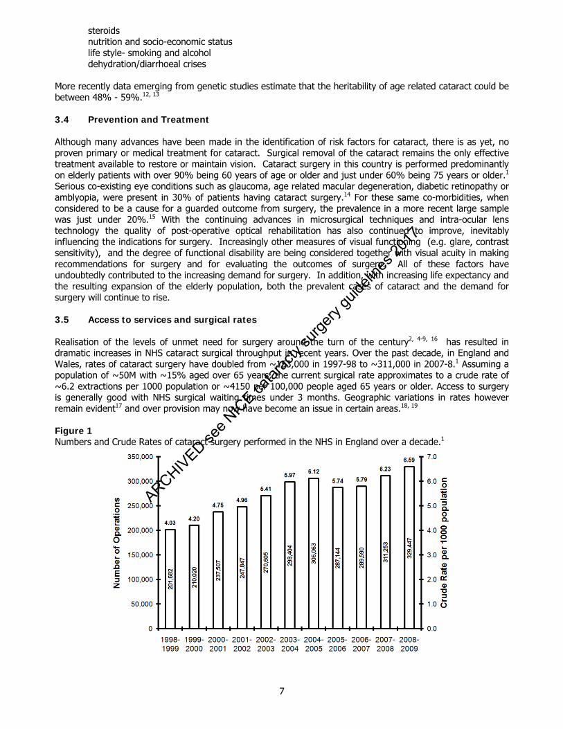
steroids nutrition and socio-economic status life style- smoking and alcohol dehydration/diarrhoeal crises More recently data emerging from genetic studies estimate that the heritability of age related cataract could be between 48% - 59%.12, 13 3.4 Prevention and Treatment Although many advances have been made in the identification of risk factors for cataract, there is as yet, no proven primary or medical treatment for cataract. Surgical removal of the cataract remains the only effective treatment available to restore or maintain vision. Cataract surgery in this country is performed predominantly on elderly patients with over 90% being 60 years of age or older and just under 60% being 75 years or older.1 Serious co-existing eye conditions such as glaucoma, age related macular degeneration, diabetic retinopathy or amblyopia, were present in 30% of patients having cataract surgery.14 For these same co-morbidities, when considered to be a cause for a guarded outcome from surgery, the prevalence in a more recent large sample was just under 20%.15 With the continuing advances in microsurgical techniques and intra-ocular lens technology the quality of post-operative optical rehabilitation has also continued to improve, inevitably influencing the indications for surgery. Increasingly other measures of visual functioning (e.g. glare, contrast sensitivity), and the degree of functional disability are being considered together with visual acuity in making recommendations for surgery and for evaluating the outcomes of surgery. All of these factors have undoubtedly contributed to the increasing demand for surgery. In addition, with increasing life expectancy and the resulting expansion of the elderly population, both the prevalent cases of cataract and the demand for surgery will continue to rise. 3.5 Access to services and surgical rates Realisation of the levels of unmet need for surgery around the turn of the century2, 4-9, 16 has resulted in dramatic increases in NHS cataract surgical throughput in recent years. Over the past decade, in England and Wales, rates of cataract surgery have doubled from ~153,000 in 1997-98 to ~311,000 in 2007-8.1 Assuming a population of ~50M with ~15% aged over 65 years, the current surgical rate approximates to a crude rate of ~6.2 extractions per 1000 population or ~4150 per 100,000 people aged 65 years or older. Access to surgery is generally good with NHS surgical waiting times under 3 months. Geographic variations in rates however remain evident17 and over provision may now have become an issue in certain areas.18, 19 Figure 1 Numbers and Crude Rates of cataract surgery performed in the NHS in England over a decade.1
ARCHIVED see N
ICE ca
tarac
ty su
rgery
guide
lines
2017
8
3.6 Assessment of the outcome of cataract surgery Monocular visual acuity provides an incomplete assessment of surgical outcome and for this reason patient centred instruments have been developed.20-24 These developments represent a formalisation of the time honoured clinical approach where patients are asked about symptoms and difficulties with visual tasks. Obtaining self reported information relevant to a patient’s every day visual experience in the context of their own environment should be seen as complimentary to standard visual function testing. Questionnaires with a predominantly functional emphasis may lack applicability to certain patient groups, particularly if applied across different cultures and to date no cataract specific UK relevant instrument has been identified which would be suitable for routine use in the NHS.18 Broad based quality of life in vision instruments aim to avoid this problem by tapping into generic psycho-social and emotional issues22 but may require additional items to improve specificity for cataract.21 3.7 Epidemiology references 1. HESonline. Main procedures and interventions: 2000-2008.
http://www.hesonline.nhs.uk/Ease/servlet/ContentServer?siteID=1937&categoryID=215 2009. 2. Stocks N, Patel R, Sparrow J, Davey-Smith G. Prevalence of cataract in the Speedwell Cardiovascular
Study: a cross-sectional survey of men aged 65-83. Eye 2002;16:275-80. 3. Gibson JM, Rosenthal AR, Lavery J. A study of the prevalence of eye disease in the elderly in an
English community. Trans Ophthalmol Soc U K 1985;104 ( Pt 2):196-203. 4. Wormald RP, Wright LA, Courtney P, Beaumont B, Haines AP. Visual problems in the elderly population
and implications for services. BMJ 1992;304:1226-9. 5. Reidy A, Minassian DC, Vafidis G, Joseph J, Farrow S, Wu J, Desai P, Connolly A. Prevalence of serious
eye disease and visual impairment in a north London population: population based, cross sectional study. BMJ 1998;316:1643-6.
6. Minassian DC, Reidy A, Desai P, Farrow S, Vafidis G, Minassian A. The deficit in cataract surgery in
England and Wales and the escalating problem of visual impairment: epidemiological modelling of the population dynamics of cataract. Br J Ophthalmol 2000;84:4-8.
7. Frost A, Hopper C, Frankel S, Peters TJ, Durant J, Sparrow J. The population requirement for cataract
extraction: a cross-sectional study. Eye 2001;15:745-52. 8. Evans JR, Fletcher AE, Wormald RP, Ng ES, Stirling S, Smeeth L, Breeze E, Bulpitt CJ, Nunes M, Jones
D, Tulloch A. Prevalence of visual impairment in people aged 75 years and older in Britain: results from the MRC trial of assessment and management of older people in the community. Br J Ophthalmol 2002;86:795-800.
9. Evans JR, Fletcher AE, Wormald RP. Causes of visual impairment in people aged 75 years and older in
Britain: an add-on study to the MRC Trial of Assessment and Management of Older People in the Community. Br J Ophthalmol 2004;88:365-70.
10. Congdon N, Taylor H. Chapter 8: Age related cataract. Arnold Publishers, 2003. 11. Dolin P. Chapter 5: Epidemiology of cataract. Chapman & Hall Medical, 1998. 12. Hammond CJ, Duncan DD, Snieder H, de Lange M, West SK, Spector TD, Gilbert CE. The heritability of
age-related cortical cataract: the twin eye study. Invest Ophthalmol Vis Sci 2001;42:601-5. 13. Hammond CJ, Snieder H, Spector TD, Gilbert CE. Genetic and environmental factors in age-related
nuclear cataracts in monozygotic and dizygotic twins. N Engl J Med 2000;342:1786-90.
ARCHIVED see N
ICE ca
tarac
ty su
rgery
guide
lines
2017
9
14. Desai P, Reidy A, Minassian DC. Profile of patients presenting for cataract surgery in the UK: national data collection. Br J Ophthalmol 1999;83:893-6.
15. Jaycock P, Johnston RL, Taylor H, Adams M, Tole DM, Galloway P, Canning C, Sparrow JM. The
Cataract National Dataset electronic multi-centre audit of 55,567 operations: updating benchmark standards of care in the United Kingdom and internationally. Eye 2009;23:38-49.
16. DH. Action on Cataracts: Good practice guidance, NHS Executive (2000). 17. Keenan T, Rosen P, Yeates D, Goldacre M. Time trends and geographical variation in cataract surgery
rates in England: study of surgical workload. Br J Ophthalmol 2007;91:901-4. 18. Black N, Browne J, van der Meulen J, Jamieson L, Copley L, Lewsey J. Is there overutilisation of
cataract surgery in England? Br J Ophthalmol 2009;93:13-7. 19. Sparrow JM. Cataract surgical rates: is there overprovision in certain areas? Br J Ophthalmol
2007;91:852-3. 20. Lundstrom M, Pesudovs K. Catquest-9SF patient outcomes questionnaire: nine-item short-form Rasch-
scaled revision of the Catquest questionnaire. J Cataract Refract Surg 2009;35:504-13. 21. Lamoureux EL, Pesudovs K, Pallant JF, Rees G, Hassell JB, Caudle LE, Keeffe JE. An evaluation of the
10-item vision core measure 1 (VCM1) scale (the Core Module of the Vision-Related Quality of Life scale) using Rasch analysis. Ophthalmic Epidemiol 2008;15:224-33.
22. Frost NA, Sparrow JM, Durant JS, Donovan JL, Peters TJ, Brookes ST. Development of a questionnaire
for measurement of vision-related quality of life. Ophthalmic Epidemiol 1998;5:185-210. 23. Lundstrom M, Roos P, Jensen S, Fregell G. Catquest questionnaire for use in cataract surgery care:
description, validity, and reliability. J Cataract Refract Surg 1997;23:1226-36. 24. Steinberg EP, Tielsch JM, Schein OD, Javitt JC, Sharkey P, Cassard SD, Legro MW, Diener-West M,
Bass EB, Damiano AM, et al. The VF-14. An index of functional impairment in patients with cataract. Arch Ophthalmol 1994;112:630-8.
ARCHIVED see N
ICE ca
tarac
ty su
rgery
guide
lines
2017
10
4 Cataract Care Pathway 4.1 Clinical Responsibility Cataract management is a multi-professional process involving ophthalmologists, optometrists, nurses and technicians. The ultimate responsibility for diagnosis and management of the patient lies with the ophthalmologist in charge. The decision on whether to proceed to surgery should be made by the patient in discussion with an ophthalmologist. Cataract surgery should be performed by an ophthalmic surgeon although much of the process may be undertaken by the non-medical members of the team provided that they are properly trained and supervised. 4.2 Referral Referral for cataract surgery can be initiated either by the optometrist or GP. Action on Cataracts1 suggested direct optometrist referral according to locally agreed protocols and there are now many such projects with audited outcomes and high conversion rates from referral to surgery. The Department of Health in the National Eye Care Plan proposes this as the preferred referral method.2 Whatever method of referral is used there are important underlying principles:
the patient should have sufficient cataract to account for the visual symptoms the cataract should affect the patient’s lifestyle the risks and benefits of surgery should be discussed with the patient and relevant written information
supplied the patient should wish to undergo cataract surgery this information together with a report from a recent sight test should form the minimum data on the
referral form. Patients who do not meet all of the criteria should not be overlooked. Patients with co-morbidity who might appreciate only slight benefit from surgery may wish to consult with an ophthalmologist to discuss their case. Patients with lifestyle impairment due to cataract who do not complain should, if necessary, be encouraged to consider cataract surgery, particularly those who live alone or act as carers. Other indications for cataract surgery include facilitating treatment and / or monitoring posterior segment disease e.g. diabetic retinopathy, correcting anisometropia or treating lens induced ocular disease.
Following referral the patient should be sent clear instructions on what they will be required to take to their out-patient visit and what to expect at the visit. Additionally a comprehensive document outlining the pros and cons of cataract surgery and the complications that may result should be included to form the background to obtaining informed consent. If surgery is to take place on the same day this plan should be made very clear in the appointment letter.
4.3 Only eye surgery The indications for cataract surgery in one-eyed patients are the same as for two-eyed patients, but the ophthalmologist should explain the possibility of total blindness if severe complications should occur. An experienced cataract surgeon should perform a one-eyed patient’s cataract operation. 4.4 Second eye cataract surgery Over one third of all National Health Service cataract operations are performed on the second eye.1 Second eye surgery confers significant additional gains in visual function in everyday activities and quality of life above and beyond those achieved after surgery to the first eye.2 Functional improvement in visual symptoms after second eye surgery has been demonstrated.3,4 Surgery for cataract on the second eye also enables a greater proportion of patients to meet the DVLA driving standard.5 These benefits of surgery are recognised clinically and its value should not be overlooked in the management of cataract.2,3,4,6
ARCHIVED see N
ICE ca
tarac
ty su
rgery
guide
lines
2017
11
4.5 Out Patient Appointment and Pre-Operative Assessment
It is essential that the ophthalmologist performing the ophthalmic examination is appropriately trained if this is not the operating surgeon.
In the interest of patient convenience the out-patient appointment should where possible be combined with the pre-operative assessment.
The purpose of the out-patient appointment is to:
confirm the diagnosis of visually significant cataract ensure the cataract is the cause of the visual symptoms determine if there is co-existing ocular pathology ensure the patient wishes to undergo surgery and understands the risks and benefits of surgery formulate a surgical care plan including stratification of surgical risk9,10,11 choose the type and power of the intraocular lens and discuss and plan any refractive surgical procedure
that may be required, in some cases this may be part of the pre-operative assessment if held separately
The aims of the pre-operative assessment are to: ensure the patient is fit for surgery put a care plan in place (this can be helped by the use of an integrated care pathway).
4.6 Diagnosis and Evaluation of visual impairment
A detailed visual history should be taken, in particular establishing near and distance vision and past history of eye disease, binocular function and amblyopia.
The impact of cataract on the patient’s lifestyle should be evaluated but it is important to realise that
patients adapt to their visual impairment. (There is no single test to assess the effect of cataract on a patient nor is there a test to decide a threshold for surgery.) Questionnaires can be helpful in eliciting symptoms but should be used in conjunction with history taking and examination when deciding on surgery.
A full medical history should be taken with particular emphasis on drugs that may increase the risk of
surgery (eg Tamsulosin Hydrochloride, other alpha-antagonists and anticoagulants12,13,14) and medical conditions that may make positioning or lying supine difficult.
4.7 Ophthalmic Examination A complete ophthalmic examination should include:
measurement of visual acuity (an up to date refraction should be available as part of the optometrist’s report)
pupil examination external eye examination including lids and lashes. measurement of intraocular pressure full slit lamp examination dilated examination of the cataract and fundus biometry if indicated, photokeratometry
Special investigations If the view of the fundus is obscured, useful information may be gained from a careful examination of the pupil responses, the assessment of light perception or using entoptic tests (Purkinje effect). B-scan ultrasonography will establish that the retina is attached and identify any intraocular masses. Electrodiagnostic tests may sometimes be useful in the assessment of retinal or visual pathway dysfunction.
III
ARCHIVED see N
ICE ca
tarac
ty su
rgery
guide
lines
2017
12
Tests for contrast sensitivity, glare, laser interferometry and specular photography are not of proven value. No special tests of visual function, other than visual acuity with best spectacle correction, are required prior to referral for cataract surgery. Following history taking and examination:
discussion should take place with the patient about the risks and benefits of cataract surgery including any risks specific to them preferred refractive aim and the need for refractive balance between the two eyes the type of anaesthesia
if the patient wishes to proceed to surgery the patient should be given a date for surgery Informed consent for the surgery should be obtained, unless this is routinely done a pre-op assessment
visit. The patient should be provided with a written sheet detailing the reasons for, benefits of and possible complications of cataract surgery.
The surgeon should formulate a surgical plan including:
type of anaesthesia IOL type and power (order special lenses if required) incision placement and astigmatism reduction procedures if appropriate stratification of surgical risk based on the expected complexity of surgery e.g. small pupil,
pseudoexfoliation, previous eye surgery. These features will allow the risk of the operation to be determined and the level of surgical experience required. The vast majority of patients are suitable for day surgery under local anaesthesia and this is the accepted model of care. Patients having surgery to their only seeing eye may need an overnight stay if the local anaesthetic reduces their vision post-operatively, and they do not have a relative or carer to look after them. 4.8 Pre-operative assessment This should include:
general health evaluation including blood pressure check note of current medication record of allergies assessment of hearing and understanding of English assessment of patients’ ability to co-operate with the procedure and lie reasonably flat during surgery identification of social problems instruction on eyedrop instillation clear explanation of the procedure and effect on the patient opportunity for patient to ask questions
Routine pre-operative medical testing (blood tests and ECGs) for patients having local anaesthesia have not been found to reduce the incidence of intraoperative or post-operative medical complications.15,16
The patient should leave the combined out-patient appointment and pre-operative assessment with a good understanding of the procedure, a date for surgery and a contact number in case of need.
Patients undergoing routine cataract surgery under local anaesthesia do not need formal venous thromboembolism (VTE) prophylaxis unless at increased risk but adults undergoing general anaesthesia for this or other ophthalmic surgery should be assessed and treated for VTE prophylaxis in the routine way. Further guidance may follow in light of a recent NICE review. The patient should be encouraged to contact the hospital in the week prior to surgery if there has been a change in the patient’s ocular or general health.
Ia
ARCHIVED see N
ICE ca
tarac
ty su
rgery
guide
lines
2017
13
4.9 Day of Surgery In the interest of patient convenience, arrival times should be staggered where possible but patients should arrive for surgery in sufficient time to ensure adequate pupillary dilatation and routine nursing checks. (Patients can also be provided with dilating drops to self administer prior to leaving home). The pre-operative checks (carried out as part of an integrated care pathway) should include identification of the patient and the eye for surgery together with external eye examination to ensure there is no ocular infection. Changes in general or ocular health since the patient was last examined must be noted and re-examination by an ophthalmologist should take place if indicated. Adequate pupillary dilatation is essential for cataract surgery and is usually achieved by short acting mydriatics (G. Cyclopentolate, G. Tropicamide, G. Phenylephrine 2.5%). Care should be taken with G. Phenylephrine 10% due to its systemic side effects but it is useful in dark eyed patients. Routine pre-operative antibiotics have not been shown to be effective but surgery should be delayed if there is concurrent infection. If patients are at increased risk of cystoid macular oedema (CMO) (eg diabetes, previous CMO, previous retinal vein occlusion, epi-retinal membrane and prostaglandin use), the use of a topical non-steroidal medication before and following surgery should be considered. As yet the literature does not allow an exact regimen to be determined however.17,18 If the surgeon carrying out the operation has not assessed the patient him or herself pre-operatively then they should ensure they are familiar with the patient and the nature of the cataract and any other co-morbidity before the start of the operation. 4.10 Surgery Phacoemulsification is the preferred method of cataract surgery in the developed world but extracapsular surgery is still occasionally necessary. Cataract surgery should include:
minimal trauma to ocular tissues capsular fixation of the intraocular lens watertight incision closure with reduction of astigmatism where appropriate. This will include the siting of
the incision and consideration of limbal relaxing incisions if appropriate. prevention of infection
Prophylaxis against infection: A simple effective prophylactic measure in infection prevention has been Povidone iodine 5% aqueous solution irrigated into the conjunctival sac immediately pre-operatively.19 A prospective study by the ESCRS Endophthalmitis Study Group showed a significant decreased risk of endophthalmitis with intracameral cefuroxime compared with topical levofloxacin.20 The study was criticised on two counts, firstly that the endophthalmitis rate in the patients not receiving the intracameral antibiotic was higher than in previous reports, and secondly that it did not compare the intracameral route to the more conventional subconjunctival route of antibiotic administration in cataract surgery. A subsequent paper has shown superiority of intracameral cefuroxime over subconjunctival administration.21 However, this latter paper was retrospective and may have had a number of confounding factors affecting the results. An additional report22 found lower baseline rates of endophthalmitis without the use of intracameral cefuroxime. The national rate reported in the 2000 BOSU study23 was 0.14%; that in the Bolton study above was 0.055%22 which is itself a little lower than reported case series in similar settings.
Ib
ARCHIVED see N
ICE ca
tarac
ty su
rgery
guide
lines
2017
14
The current advice is therefore that: If local rates of endophthalmitis over a properly audited time frame are similar to those
reported in the Bolton study, then continuing with whatever preventative/prophylactic measures are in place would seem reasonable.
If local rates are higher than those reported in the Bolton study then intracameral cefuroxime may be added as part of a package of measures to lower endophthalmitis rates after a suitable analysis of processes has taken place.
If the use of intracameral cefuroxime is considered there are potential problems that must be addressed:
The drug is heat labile, it cannot be heat sterilised and must be produced aseptically.24 Commercial preparations of the drug for intracameral use are available, and their use will prevent the possibly significant risks associated with the preparation of a suitable intracameral dose of the drug in the operating theatre.
The possibility of an adverse reaction to the drug given intracamerally (toxic anterior segment syndrome) remains.
In summary, reaching a decision on whether or not to give the drug intra-camerally, may involve a comparison of the local rate of endophthalmitis with that in the studies mentioned. Additionally the exact dose and best antibiotic prophylaxis (for example the possible use of two different antibiotics) has not yet been determined.
Following surgery and return to the day-care unit the patient should be discharged by an appropriately trained member of staff who ensures that:
the patient is comfortable and pain free if not the eye is examined and if there are any problems e.g. shallow anterior chamber (AC) or
hyphaema an ophthalmologist is called to see the patient post-operative written instructions, medications, appointments and emergency contact details
are all given to the patient 4.11 Post-operative visits 4.11.1 First day review First day post-operative review is no longer in widespread use25.26 with many departments having replaced a patient visit with a telephone call by a trained nurse or a call by the patient to a trained nurse if necessary.
Robust arrangements need to be in place to ensure that patients not reviewed next day have easy access to advice and assessment, and that post-operative complications can be quickly identified and managed.
First day post-operative visits may be required:
where surgery was complicated with co-existing eye disease e.g. glaucoma, uveitis patients with an only eye
4.11.2 Final review For patients not seen on the first post-operative day a review appointment is necessary to:
review progress and medication collect outcome data discuss second eye surgery where appropriate arrange follow-up for co-existing eye disease provide advice on spectacle prescription (which can be prescribed approximately 4 weeks following
phacoemulsification)
ARCHIVED see N
ICE ca
tarac
ty su
rgery
guide
lines
2017
15
This examination can be provided by ophthalmologists, nurses, optometrists or orthoptists working within the unit to agreed guidelines or by accredited optometrists working outside the unit. The ophthalmologist with responsibility for the patient should ensure that appropriate training and monitoring takes place when the post-operative care is delegated to others. 4.12 Cataract Care Pathway references 1. Action on cataracts – Good Practice Guidance. NHS Executive, London. Feb 2000. 2. Department of Health National Eye Care Plan, London. May 2004. 3. Desai P, Reidy A, Minassian DC. Profile of patients presenting for cataract surgery in the UK: National
data collection. Br J Ophthalmol 1999; 83:893-89. 4. Desai P, Reidy A, Minassian DC, Vafidis G, Bolger J. Gains from cataract surgery: Visual function and
quality of life. Br J Ophthalmol 1996; 80: 868-873. 5. Laidlaw DA, Harrad RA, Hopper CD, Whittaker A, Donovan JL, Brookes ST, Marsh GW, Peters TJ,
Sparrow JM, Frankel SJ. Randomised trial of effectiveness of second eye cataract surgery. Lancet 1998; 352: 925-929.
6. Javitt JC, Steinberg EP, Sharkey P. Schein OD, Tielsch JM, Diener West M et al. Cataract surgery in
one eye or both. A billion dollar per year issue. Ophthalmology 1995; 105: 1583-93 7. Talbot EM, Perkins A. The benefit of second eye cataract surgery. Eye 1998; 12: 983-989. 8. Laidlaw A, Harrad R. Can second eye cataract extraction be justified? Eye 1993; 7: 680-686. 9. Narendran N et al. The Cataract National Dataset electronic mlticentre audit of 55,567 operations:risk
stratification for posterior capsule and vitreous loss. Eye 2009; 23:31-37. 10. Agrawal V et al. Validation of scoring system for preoperative stratification of introperative risk of
complications during cataract surgery: Indian multi-centre study. Indian Journal of Ophthalmology 2009; 57: 213-215.
11. Muhtaseb M et al. A system for preoperative stratification of cataract patients according to risk of
intraoperative complications: aprospective analysis o1441 cases. Br J Ophthalmol 2004;88: 1242-1246.
12. Chadha V, Borooah S, Tey A, Styles C, Singh J. Floppy iris behaviour during cataract surgery:
associations and variations Br J Ophthalmol 2007;91:40-42. 13. Bell et al. Tamsulosin and serious adverse events during cataract surgery in men. Journal of American
Medical Association 2009; 301: 1991-6. 14. Hirschman DR, Morby LJ. A study of the safety of continued anticoagulation for cataract surgery
patients. Nursing forum 41/1(30-7), 0029-6473. 15. Schein OD, Katz J, Bass EB, Tielsch JM, Lubomski LH, Feldman MA, Petty BG, Steinberg EP. The value
of routine pre-operative medical testing before cataract surgery. N Engl J Med 2000; 342(3):168-75. 16. Keay L, Lindsley K, Tielsch J, Katz J, Schein O. Routine preoperative medical testing for cataract
surgery. Cochrane Database of Systematic Reviews 2009, Issue 2. Art. No.: CD007293. DOI: 10.1002/14651858.CD007293.pub2
ARCHIVED see N
ICE ca
tarac
ty su
rgery
guide
lines
2017

16
17. Sivaprasad S, Bunce C, Jyothi S. Non-steroidal anti-inflammatory agents for treating cystoid macular oedema following cataract surgery. Cochrane Database of Systematic Reviews 2004, Issue 3. Art. No.: CD004239. DOI: 10.1002/14651858.CD004239.pub2.
18. Henderson BA, Kim JY, Ament CS, Ferrufino-Ponce ZK, Grabowska A, Cremers SL. Clinical
pseudophakic cystoid macular edema. Risk factors for development and duration after treatment. J Cataract Refract Surg 2007; 33(9):1550-8.
19. Ciulla TA, Starr MB, Masket S. Bacterial endophthalmitis prophylaxis for cataract surgery: an evidence-
based update. Ophthalmology 2003;109:13-24. 20. ESCRS Endophthalmitis Study Group. J Cataract Refract Surg 2007; 33: 978-988. 21. Yu-Wai-Man P, Morgan SJ, Hildreth AJ, Steel DH, Allen D. Efficacy of intracameraland subconjunctival
cefuroxime in preventing endophthalmitis after cataract surgery. J Refractive Surgery 2008; 34:447-451.
22. Kelly SP, Mathews D, Mathews J, Vail A. Reflective consideration of postoperative endophthalmitis as a
quality marker. Eye 2007; 21 (11): 1419-26. Epub 2005 Jul 13. Review 23. Kamalarajah S, Silvestri G, Sharma N, Khan A, Foot B, LingR,Cran G, Best R. Surveillance of
endophthalmitis following cataract surgery in the UK. Eye 2004; 18: 580–587. doi:10.1038/sj.eye.6700645
24. Liesegang TL. Intracameral Antibiotics: Questions for the United States based on prospective studies. J
Cataract Refract Surg 2008; 34:505-509. 25. Tinley CG, Frost A, Hakin KN, McDermott W, Ewings P. Is visual outcome compromised when next day
review is omitted after phacoemulsification surgery? A randomised control trial. Br J Ophthalmol 2003; 87(11):1350-5.
26. Tan JH, Newman DK, Klunker C, Watts SE, Burton RL. Phacoemulsification cataract surgery: is routine
review necessary on the first post-operative day? Eye 2000; 14 ( Pt 1):53-5.
ARCHIVED see N
ICE ca
tarac
ty su
rgery
guide
lines
2017
17
Ib
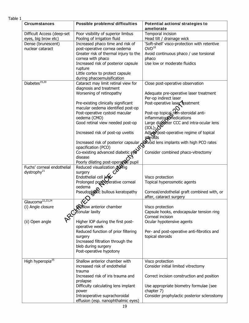
5. Surgery in Special Circumstances 5.1 Introduction
There are numerous circumstances or conditions that conspire to make cataract surgery less than routine. These may be related to the context of surgery (e.g. cataract surgery in a patient who is diabetic or has undergone prior LASIK surgery), due to previous treatment (e.g. cataract surgery following glaucoma drainage surgery or vitrectomy) or in association with ocular co-morbidity (e.g. uveitis or corneal diseases such as Fuchs Endothelial dystrophy). Surgeons and patients should be aware of factors that make surgery more difficult, or that may affect the outcome. This awareness will inform decisions about the surgical technique, and grade and experience of the operating surgeon as well as to the pre- and post-operative care of these patients and will also influence the advice given to patients about their surgery. Table 1 summarises the more common circumstances and conditions that may complicate cataract surgery, and suggests broad strategies for avoiding and treating any associated problems. This table is not exhaustive and is not intended to be prescriptive. 5.2 Second Eye Surgery
Cataract is usually a bilateral condition, although there may be significant difference in the degree of cataract present at a single time point. Non-ophthalmologists may wonder if, given a good functional outcome from first eye surgery, there is any gain from surgery on the second eye. As pressures on limited health care resources (both government provided and privately provided) increase the question that arises in the minds of those responsible for ensuring value for money from those resources is often ‘what is the value of second eye cataract surgery?’ There are now numerous studies showing the benefit to the patient of having bilateral cataract surgery.1,2,3,4 Patients with a cataract and dissimilar vision in the two eyes (or one eye with cataract extraction and the second eye with a cataract) have demonstrated binocular inhibition.5 A large epidemiological study demonstrated that persons who exhibited binocular inhibition were more likely to have driving difficulties compared with those who did not have binocular inhibition.6 There is also evidence that second eye surgery is cost effective.7 It is therefore clinically and economically appropriate for second eye surgery to be offered to those patients who want it. The issue of timing of second eye surgery however remains controversial (see next section).
5.3 Simultaneous bilateral cataract surgery
Some ophthalmologists perform bilateral cataract surgery at one sitting, and some patients may request this. This is usually termed Simultaneous Bilateral Cataract Extraction (SBCE) or occasionally Immediately Sequential Cataract Surgery. The perceived benefits of this practice include faster visual rehabilitation, fewer hospital visits, and lower cost for the patient and for society (or other provider of healthcare).8 Many studies have reported good clinical outcomes with high patient satisfaction and few complications.9-14 However, concern remains in many quarters about the potentially devastating possibility of bilateral infective endophthalmitis. To date there have been 4 reported cases of bilateral endophthalmitis following SBCS, but only 1 of these occurred in non-at risk patients or where the precautions outlined below had been taken.15,16 The precise risk of bilateral visual loss is unknown but surgeons who perform SBCE must advise their patients of the possibility and implications of bilateral endophthalmitis. Contamination of fluids, instruments or operating theatre air may result in serial cases of corneal oedema or endophthalmitis, therefore strict precautions should be undertaken. Relative clinical indications:
When a general anaesthetic (GA) is necessary to perform the cataract surgery safely and repeated GAs are contra-indicated on general health grounds.
Bilateral cataract in a person who for reasons of disability cannot be fully assessed pre-operatively and who requires a general anaesthetic for the procedure
ARCHIVED see N
ICE ca
tarac
ty su
rgery
guide
lines
2017
18
Precautions: The operation on each eye must be treated as a completely separate procedure with staff re-scrubbing,
and using completely new sets of instruments Full-cycle sterilisation should be used, not ‘flash’ sterilisation Intraocular fluids and Intra-ocular lenses should come from different batches If complications occur with the first eye, careful consideration should be given before proceeding with
surgery on the second eye Instructions should be given on using separate drop bottles for each eye post-operatively and washing
hands before instilling eye drops into the second eye.
5.4 Surgery in patients with special needs
Cataract surgery is now routinely carried out under local anaesthesia, and is associated with rapid physical as well as visual rehabilitation. Most patients with physical disabilities can be operated on with little disruption to them or to the normal surgical regimen. Positioning of patients with spinal mobility restriction is facilitated if surgical trolleys specifically adapted for ophthalmic surgery are used, rather than a standard surgical operating table. Patients with learning difficulties or with cognitive impairment may find the surgical environment confusing and frightening and may require general anaesthesia if it is thought that co-operation with surgery under local anaesthesia may be compromised. The main issue with such patients is that of consent, and the need for careful assessment of their capacity to give consent.17 It is particularly important that assessment start from a presumption of capacity. It is also important to recognise that while some persons may have difficulty expressing themselves, their capacity to understand and weigh information provided may be unimpaired. The issue of communication with patients with impaired understanding is an important one and such communication may need more time and involvements of other health professionals. It is important to have awareness of the Equality Act (which has replaced the Disability Discrimination Act of 1995) to allow proper prioritisation of such patients for cataract surgery. The Lay Advisory Group of the College is producing guidance on the management of patients with learning difficulties which will include information on the Mental Capacity Act and the role of independent advocates (IMCAs). An additional useful reference is the Look Up website (www.lookupinfo.org) has a DVD called ‘You and your Eye’ one part of which is about cataract operations. See also chapter 10.
ARCHIVED see N
ICE ca
tarac
ty su
rgery
guide
lines
2017
19
Table 1
Circumstances Possible problems/difficulties Potential actions/strategies to ameliorate
Difficult Access (deep-set eyes, big brow etc)
Poor visibility of superior limbus Pooling of irrigation fluid
Temporal incision Head tilt / drainage wick
Dense (brunescent) nuclear cataract
Increased phaco time and risk of post-operative cornea oedema Greater risk of thermal injury to the cornea with phaco Increased risk of posterior capsule rupture Little cortex to protect capsule during phacoemulsification
‘Soft-shell’ visco-protection with retentive OVD18
Avoid continuous phaco / use torsional phaco Use low or moderate fluidics
Diabetes19,20 Cataract may limit retinal view for diagnosis and treatment Worsening of retinopathy Pre-existing clinically significant macular oedema identified post-op Post-operative cystoid macular oedema (CMO) Good retinal view needed post-op Increased risk of post-op uveitis Increased risk of posterior capsular opacification (PCO) Co-existing advanced diabetic eye disease Poorly dilating post-operative pupil
Close post-operative observation Adequate pre-operative laser treatment Per-op indirect laser Post-operative laser treatment Post-op topical non-steroidal anti-inflammatory medications Large diameter CCC and intra-ocular lens (IOL) Adjust post-operative regime of topical steroids Avoid lens implants with high PCO rates Consider combined phaco-vitrectomy
Fuchs’ corneal endothelial dystrophy21
Reduced visualization during surgery Endothelial cell loss Prolonged post-operative corneal oedema Pseudophakic bullous keratopathy
Visco protection Topical hyperosmotic agents Corneal/endothelial graft combined with, or after, cataract surgery
Glaucoma22,23,24 (i) Angle closure (ii) Open angle
Shallow anterior chamber Zonular laxity Higher lOP during the first post-operative week Reduced function of prior filtering surgery Increased filtration through the bleb during surgery Post-operative hypotony
Visco protection Capsule hooks, endocapsular tension ring Corneal incision Ocular hypotensive agents Per- and post-operative anti-fibrotics and topical steroids
High hyperopia25 Shallow anterior chamber with increased risk of endothelial trauma Increased risk of iris trauma and prolapse Difficulty calculating lens implant power Intraoperative suprachoroidal effusion (esp. nanophthalmic eyes)
Visco protection Consider initial limited vitrectomy Correct incision construction and position Use appropriate biometry formulae (see chapter 7) Consider prophylactic posterior sclerostomy
ARCHIVED see N
ICE ca
tarac
ty su
rgery
guide
lines
2017
20
Circumstances Possible problems/difficulties Potential actions/strategies to ameliorate
High myopia Anterior chamber depth fluctuation Difficulty calculating lens implant power with posterior staphyloma Possible increased risk of retinal detachment (controversial26,27)
Break ‘reverse pupil block’28 Use optical biometry to gain ‘line of sight’ axial length, or B-scan ultrasound. Use appropriate biometry formula Warn patients re symptoms
High risk for subsequent vitreo-retinal surgery
Silicone IOL may limit visibility if silicone oil used Good view of peripheral retina required
Use acrylic IOL Large capsulorhexis and large optic diameter (>=6.0 mm) IOL
Macular degeneration Sub-retinal neovascularisation (either revealed pre-existing or subsequently developed)29
Patient information re. symptoms, relevant investigations
Current or previous use of systemic α1a adrenergic antagonist (especially tamsulosin)30,31
Poor dilatation, progressive miosis, iris instability in normal AC currents, iris prolapse to all incisions (IFIS). Possible increase in per-operative complications.
Awareness of patient’s medication history. Awareness of variety of strategies including visco-stabilisation, pupil expanders and ‘off-label’ use of intracameral α agonists32,33,34,35
Small (miotic) pupil (other than above)
Poor visualization Increased risk of capsule tear / vitreous prolapse Increased risk of iris damage and prolapse
Visco-mydriasis, pupil stretch, sphincterotomies, iris hooks, pupil expanders
Posterior synechiae Intraoperative miosis Prolonged post-operative inflammation Iris bleeding Inflammatory deposits on IOLs
Iris hooks, pupil stretch, sphincterotomies, pupil expanders Intensive post-operative topical steroids Viscotamponade Topical steroid drops, YAG 'polishing'
Posterior polar cataract Weak or defective posterior capsule at posterior pole
No (or very gentle) hydrodissection. Low flow phaco with visco-dissection36
Pseudo-exfoliation syndrome37
Poor pupil dilatation Zonular laxity or instability Accelerated posterior capsule opacification Anterior capsulorhexis contraction Vitreous loss IOL tilt and decentration Late (decades) dislocation of IOL possible
Iris hooks, pupil stretch, sphincterotomies, pupil expanders Endo-capsular tension ring, capsule hooks Thorough aspiration of lens epithelial cells Adequate sized capsulorhexis
Prior keratorefractive surgery38
Difficulty calculating IOL power Dehiscence of refractive keratotomy incision Thin pliable cornea post LASIK, AC depth fluctuation
See chapter 7 Low bottle height with low-flow phaco
ARCHIVED see N
ICE ca
tarac
ty su
rgery
guide
lines
2017
21
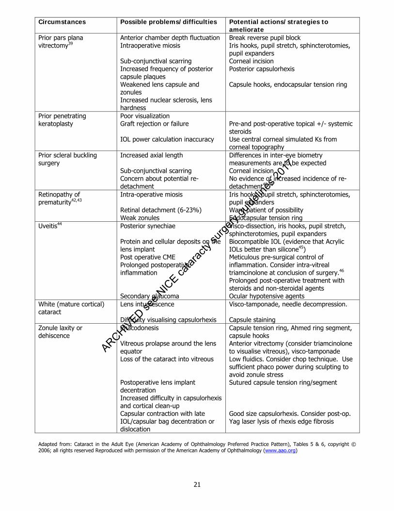
Circumstances Possible problems/difficulties Potential actions/strategies to ameliorate
Prior pars plana vitrectomy39
Anterior chamber depth fluctuation Intraoperative miosis Sub-conjunctival scarring Increased frequency of posterior capsule plaques Weakened lens capsule and zonules Increased nuclear sclerosis, lens hardness
Break reverse pupil block Iris hooks, pupil stretch, sphincterotomies, pupil expanders Corneal incision Posterior capsulorhexis Capsule hooks, endocapsular tension ring
Prior penetrating keratoplasty
Poor visualization Graft rejection or failure IOL power calculation inaccuracy
Pre-and post-operative topical +/- systemic steroids Use central corneal simulated Ks from corneal topography
Prior scleral buckling surgery
Increased axial length Sub-conjunctival scarring Concern about potential re-detachment
Differences in inter-eye biometry measurements are to be expected Corneal incision No evidence of increased incidence of re-detachment40,41
Retinopathy of prematurity42,43
Intra-operative miosis Retinal detachment (6-23%) Weak zonules
Iris hooks, pupil stretch, sphincterotomies, pupil expanders Warn patient of possibility Endocapsular tension ring
Uveitis44 Posterior synechiae Protein and cellular deposits on the lens implant Post operative CME Prolonged postoperative inflammation Secondary glaucoma
Visco-dissection, iris hooks, pupil stretch, sphincterotomies, pupil expanders Biocompatible IOL (evidence that Acrylic IOLs better than silicone45) Meticulous pre-surgical control of inflammation. Consider intra-vitreal triamcinolone at conclusion of surgery.46
Prolonged post-operative treatment with steroids and non-steroidal agents
Ocular hypotensive agents White (mature cortical) cataract
Lens intumescence Difficulty visualising capsulorhexis
Visco-tamponade, needle decompression. Capsule staining
Zonule laxity or dehiscence
Phacodonesis Vitreous prolapse around the lens equator Loss of the cataract into vitreous Postoperative lens implant decentration Increased difficulty in capsulorhexis and cortical clean-up Capsular contraction with late IOL/capsular bag decentration or dislocation
Capsule tension ring, Ahmed ring segment, capsule hooks Anterior vitrectomy (consider triamcinolone to visualise vitreous), visco-tamponade Low fluidics. Consider chop technique. Use sufficient phaco power during sculpting to avoid zonule stress Sutured capsule tension ring/segment Good size capsulorhexis. Consider post-op. Yag laser lysis of rhexis edge fibrosis
Adapted from: Cataract in the Adult Eye (American Academy of Ophthalmology Preferred Practice Pattern), Tables 5 & 6, copyright © 2006; all rights reserved Reproduced with permission of the American Academy of Ophthalmology (www.aao.org)
ARCHIVED see N
ICE ca
tarac
ty su
rgery
guide
lines
2017
22
5.5 Surgery in Special Circumstances references
1. Elliott DB, Patla AE, Furniss M, Adkin A. Improvements in clinical and functional vision and quality of life after second eye cataract surgery. Optom Vis Sci 2000;77:13-24.
2. Lundstrom M, Stenevi U, Thorburn W. Quality of life after first- and second-eye cataract surgery: five-
year data collected by the Swedish National Cataract Register. J Cataract Refract Surg 2001;27:1553-9. 3. Javitt JC, Brenner MH, Curbow B, et al. Outcomes of cataract surgery. Improvement in visual acuity
and subjective visual function after surgery in the first, second, and both eyes. Arch Ophthalmol 1993;111:686-91.
4. Castells X, Comas M, Alonso J, et al. In a randomized controlled trial, cataract surgery in both eyes
increased benefits compared to surgery in one eye only. J Clin Epidemiol 2006;59:201-7. 5. Pardhan S. Binocular performance in patients with unilateral cataract using the Regan test: binocular
summation and inhibition with low-contrast charts. Eye 1993;7 (Pt 1):59-62. 6. Azen SP, Varma R, Preston-Martin S, et al. Binocular visual acuity summation and inhibition in an
ocular epidemiological study: the Los Angeles Latino Eye Study. Invest Ophthal Vis Sci 2002;43:1742-8.
7. Busbee BG, Brown MM, Brown GC, Sharma S. Cost-utility analysis of cataract surgery in the second
eye. Ophthalmology 110, 2310-7 (2003). 8. Chang DF. Simultaneous bilateral cataract surgery. Br J Ophthalmol 2003;87:253-4.
9. Smith GT, Liu CS. Is it time for a new attitude to "simultaneous" bilateral cataract surgery? Br J
Ophthalmol 2001;85:1489-96. 10. Sharma TK, Worstmann T. Simultaneous bilateral cataract extraction. J Cataract Refract Surg
2001;27:741-4. 11. Wertheim M, Burton R. Immediately sequential phacoemulsification performed under topical
anaesthesia as day case procedures. Br J Ophthalmol 2002;86:1356-8. 12. Sarikkola AU, Kontkanen M, Kivelä T, Laatikainen L. Simultaneous bilateral cataract surgery: a
retrospective survey. J Cataract Refract Surg 2004;30:1335-41. 13. Arshinoff SA, Strube YN, Yagev R. Simultaneous bilateral cataract surgery. J Cataract Refract Surg
2003;29:1281-91. 14. Lundström M, Albrecht S, Nilsson M, Åström B. Benefits to patients of bilateral same-day cataract
extraction: Randomized clinical study. J Cataract Refract Surg 2006;32:826–830. 15. Arshinoff S. Bilateral endophthalmitis after simultaneous bilateral cataract surgery. J Cataract Refract
Surg 2008;34:2006-8. 16. Puvanachandra N, Humphry RC. Bilateral endophthalmitis after bilateral sequential phacoemulsification.
J Cataract Refract Surg 2008;34:1036-7.
17. Seeking consent: working with patients with learning disabilities. Department of Health, London http://www.dh.gov.uk/en/Publicationsandstatistics/Publications/PublicationsPolicyAndGuidance/DH_4007861 (accessed May 2009).
18. Arshinoff S. Dispersive cohesive viscoelastic soft shell technique. J Cataract Refract Surg 1999;25:167-
73.
ARCHIVED see N
ICE ca
tarac
ty su
rgery
guide
lines
2017
23
19. Dowler, J, K Shemi, PG Hykin, and AMP Hamilton. The natural histopry of macular oedema after cataract surgery in diabetes. Ophthalmology 1999;106: 663-668.
20. Dowler, J, PG Hykin, and AMP Hamilton. Phacoemulsification versus extracapsular cataract extraction
in patients with diabetes. Ophthalmology 2000;107: 457-462. 21. Seitzman GD. Cataract surgery in Fuchs' dystrophy. Curr opin in ophthalmol 2005;16:241-5.
22. Tennen DG, Masket S. Short-and long-term effect of clear corneal incisions on intraocular pressure. J
Cataract Refract Surg 1996; 22: 568-70. 23. Mandal AK, Chelerkar V, Jain SS, Nutheti R. Outcome of cataract extraction and posterior chamber
intraocular lens implantation following glaucoma filtration surgery. Eye 2005 ;19:1000-8. 24. Klink J, Schmitz B, Lieb WE, Klink T, Grein HJ, Sold-Darseff J, Heinold A, Grehn F. Filtering bleb
function after clear cornea phacoemulsification: a prospective study. Br J Ophthalmol 2005;89:597-601.
25. Wladis EJ, Gewirtz MB, Guo S. Cataract surgery in the small adult eye. Surv Ophthalmol 2006; 51: 153-
61. 26. Fan DS, Lam DS, Li KK. Retinal complications after cataract extraction in patients with high myopia.
Ophthalmology 1999;106: 688-91. 27. Neuhann IM, Neuhann TF, Heimann H, Schmickler S, Gerl RH, Foerster MH. Retinal detachment after
phacoemulsification in high myopia: analysis of 2356 cases. J Cataract Refract Surg 2008;34:1644-57.
28. Cionni RJ, Barros MG, Osher RH. Management of lens-iris diaphragm retropulsion syndrome during
phacoemulsification. J Cataract Refract Surg 2004; 30:953-6. 29. Wang JJ, Klein R, Smith W, Klein BE, Tomany S, Mitchell P. Cataract surgery and the 5-year incidence
of late-stage age-related maculopathy: pooled findings from the Beaver Dam and Blue Mountains eye studies. Ophthalmology 2003; 110:1960-7.
30. Chang DF, Campbell JR. Intraoperative floppy iris syndrome associated with tamsulosin J Cataract
Refract Surg 2005; 31:664–673. 31. Chadha V, Borooah S. Tey A et al. Floppy iris behaviour during cataract surgery: associations and
variations. Br J Ophthalmol 2007; 91:40-42. 32. Chang DF, Osher RH, Wang L, Koch DD. Prospective multicentre evaluation of cataract surgery in
patients taking tamsulosin (Flomax). Ophthalmology 2007; 114: 957-964. 33. Arshinoff SA. Modified SST–USST for tamsulosin-associated intraocular floppy-iris syndrome. J Cataract
Refract Surg 2006; 32: 559-561. 34. Malyugin B. Small pupil phaco surgery: a new technique. Ann Ophthalmol (Skokie). 2007; 39: 185-93.
35. Manivikar S, Allen D. A progressive approach to managing cataract surgery in patients taking
Tamsulosin. J Cataract Refract Surg 2006; 32: 1611-1614. 36. Allen D, Wood C. Minimising risk to capsule during surgery for Posterior Polar Cataract J Cataract
Refract Surg 28; 2002:742-4. 37. Küchle M, Viestenz A, Martus P, et al. Anterior chamber depth and complications during cataract
surgery in eyes with pseudoexfoliation syndrome. Am J Ophthalmol 2000; 129: 281-5.
ARCHIVED see N
ICE ca
tarac
ty su
rgery
guide
lines
2017
24
38. Langenbucher A, Haigis W, Seitz B. Difficult lens power calculations. Curr Opin Ohthalmol 2004;15:1-9.
39. Grusha YO, Masket S, Miller KM. Phacoemulsification and lens implantation after pars plana vitrectomy.
Ophthalmology 1998;105: 287-94. 40. Kerrison JB, Marsh M, Stark WJ, Haller JA. Phacoemulsification after retinal detachment surgery.
Ophthalmology 1996;103:216-9. 41. Eshete A, Bergwerk KL, Masket S, Miller KM. Phacoemulsification and lens implantation after scleral
buckling surgery. Am J Ophthalmol 2000; 129:286-90. 42. Kaiser RS, Fenton GL, Tasman W, Trese MT. Adult retinopathy of prematurity: retinal complications
from cataract surgery. Am J Ophthalmol 2008;145:729-735. 43. Farr AK, Stark WJ, Haller JA. Cataract surgery by phacoemulsification in adults with retinopathy of
prematurity. Am J Ophthalmol 2001;132:306-10. 44. Van Gelder RN, Leveque TK. Cataract surgery in the setting of uveitis. Curr Opin Ophthalmol 2009;
20:42-5. 45. Alió JL, Chipont E, BenEzra D, Fakhry MA Comparative performance of intraocular lenses in eyes with
cataract and uveitis. J Cataract Refract Surg 2002; 28:2096-108. 46. Roesel M, Tappeiner C, Heinz C, Koch JM, Heiligenhaus A. Comparison between intravitreal and orbital
floor triamcinolone acetonide after phacoemulsification in patients with endogenous uveitis. Am J Ophthalmol 2009;147:406-12.
ARCHIVED see N
ICE ca
tarac
ty su
rgery
guide
lines
2017
25
6. Paediatric Ophthalmology The aims of modern paediatric cataract surgery include:
Restoration of a normal clear visual axis for normal visual development in a timely fashion. Lens
opacities that are visually significant before 2–3 months of age have much more potential impact on the child’s visual development than those acquired later. In general, the earlier the onset, the more amblyogenic the cataract will be.
Achievement of an age-appropriate postoperative refraction which allow successful visual rehabilitation taking into account amblyopia
Ensuring appropriate investigation for the cause of the cataract(s) Ensuring safe general anaesthesia by appropriately trained personnel.
6.1 Epidemiology Studies of incidence and prevalence of childhood cataract are few. The adjusted annual age-specific incidence of congenital and infantile cataract in the first year of life is 2.49 per 10,000 children with an adjusted cumulative incidence at 5 years of 3.18 per 10,000 rising to 3.46 per 10,000 by 15 years.1 6.2 Risk Factors Paediatric cataracts may be
congenital o hereditary/genetic o metabolic eg galactosaemia o in-utero infection related - TORCH
developmental o genetic o metabolic eg galactokinase deficiency
acquired o metabolic eg diabetes mellitus o traumatic o post radiotherapy
6.3 Prevention and Treatment
There are at present no medical treatments available for congenital or developmental cataracts. However, in cases of galactosaemia early diagnosis and management of diet and enzyme replacement therapy may allow mild cataracts to stop progressing and even regress.2 Conservative management of partial cataracts may be considered using dilating drops to increase light entering the eye. While visual acuity is important to assess visual function increasingly other testing methods (glare testing and contrast sensitivity) should also be used.
6.4 Assessment of Visual Outcome
For paediatric cataract surgery the outcome measures include not only visual acuity but assessment of complications due to surgery.3 For infants, visual behaviour including fixation and ocular stability (ie lack of nystagmus) is an important outcome assessment also.
ARCHIVED see N
ICE ca
tarac
ty su
rgery
guide
lines
2017
26
6.5 Cataract Care Pathway 6.5.1 Clinical Responsibility In cases of congenital cataracts, it is the responsibility of the health carers looking after the neonate, immediately after the birth of a child, to check for normal red reflexes. This again should be checked at the 6 week health check. The decision on whether to proceed to surgery should be made by the parent or responsible adult in discussion with an ophthalmologist. Paediatric cataract surgery should be performed by an appropriately trained surgeon who is aware of the physiological and anatomical differences between adult and paediatric cataract cases. Furthermore, in infants the paediatric anaesthesia must be provided in an appropriate hospital with adequate facilities for safe anaesthesia and postoperative recovery. 6.5.2 Referral Referrals may be made by a paediatrician, optometrist or between ophthalmologists. Other Health Care professionals such as health visitors may also be involved. 6.5.3 Simultaneous surgery
In bilateral cases whether simultaneous cataract surgery or monocular surgery should be performed in bilateral cases is a matter of controversy.4-6 Under certain circumstances, bilateral simultaneous surgery is indicated, eg. on anaesthetic grounds or because an infant has presented late and there is concern about dense amblyopia developing in the second eye.7 Precautions
The operation on each eye must be treated as a completely separate procedure If complications occur with the first eye, careful consideration should be given before proceeding with
surgery on the second eye Instructions should be given on using separate drop bottles for each eye post-operatively and washing
hands before instilling eye drops into the second eye Every effort should be made to reduce the possibility of serial infection by using instruments, fluids and
intra-ocular lenses prepared in different batches The ophthalmologist should be prepared to justify a decision to perform bilateral cataract surgery on grounds other than convenience.
6.5.4 Out Patient Appointment and Pre-Operative Assessment For paediatric cataract surgery the surgeon performing the surgery should perform examination. In paediatric cataract surgery pre-operative assessment is usually dissociated from the first visit. The purpose of the out-patient appointment is to: confirm the diagnosis of visually significant cataract ensure the cataract is the cause of the visual symptoms determine if there is co-existing ocular pathology ensure the patient’s parents wish to proceed to surgery and understands the risks ensure that there are no systemic illnesses that may put the child at risk under general anaesthesia
or that may affect the child’s well being if not appropriately treated in a timely fashion formulate a surgical care plan
IV ARCHIVED see N
ICE ca
tarac
ty su
rgery
guide
lines
2017

27
6.5.5 Diagnosis and Evaluation of visual impairment
A detailed visual history should be taken, in particular establishing near and distance vision and past history of eye disease, binocular function and amblyopia.
A detailed family history should be taken and a dilated examination of the lenses of both parents
6.5.6 Ophthalmic Examination A complete ophthalmic examination should include:
measurement of visual acuity (an up to date refraction should be available as part of the optometrist’s report)
pupil examination external eye examination including lids and lashes. measurement of intraocular pressure slit lamp examination dilated examination of the cataract and fundus biometry if the child is old enough to cooperate. If not this may need to be done under
anaesthesia 6.5.7 Special investigations If the view of the fundus is obscured, B-scan ultrasonography will establish that the retina is attached and identify any intraocular masses. Electrodiagnostic tests may sometimes be useful in the assessment of retinal or visual pathway dysfunction. 6.6 Paediatric Cataract Surgery The child’s eye is unique8 because of
Change in axial length with time
Change in corneal curvature with time
Increased tissue reactivity
Decreased scleral rigidity
Smaller size (compared to the adult eye)
Potential for amblyopia
Long life span after cataract removal
These facts must be taken into account and their impact understood by any surgeon undertaking cataract surgery in children.
6.7 Evaluation History Should include history of pregnancy and family history
III
IV
ARCHIVED see N
ICE ca
tarac
ty su
rgery
guide
lines
2017

28
Examination Should include examination of the child and a dilated exam of the lenses of both parents Investigations Neonates with bilateral cataracts and no family history warrant a paediatric evaluation and/or urinalysis for reducing substances, to rule out galactosaemia.2 Isolated unilateral cataracts still do warrant TORCH serology.9 In developmental cataracts the possibility of galactokinase deficiency should be considered. 6.8 Timing of surgery The timing of surgery depends on the severity of the lenticular opacity, its effect on the visual system and the age of the child. Surgery during the first year of life is thought to increase the risk of glaucoma.24-26
Whether surgery prior to 4 weeks of age results in a further increased risk of glaucoma is debatable.26-28
6.9 Preoperative Evaluation 6.9.1 Which Operation? Unlike adult cataract surgery the choice of procedure for children includes10-13:
Lensectomy
Lens aspiration with IOL
Lens aspiration with Primary Posterior Capsulotomy (PPC) and IOL
Lens aspiration with PPC, Anterior Vitrectomy (AV) and IOL
Lens aspiration with PPC, AV, IOL and Posterior capsular optic capture (PCOC)
Lensectomy should be performed with modern automated vitrectors. Enough capsular support should be left in case a secondary IOL needs to be placed at a later stage.
An adequate anterior vitrectomy should be performed to prevent pupil block postoperatively.
Lens aspiration with IOL should be performed in those children in whom posterior capsular opacification is less likely to occur ( usually over 8 yrs old ) or in whom YAG laser capsulotomy can be anticipated to be done awake ( again usually over 8 yrs of age).
Good evidence exists that leaving the posterior capsule intact in children 6 or under will result in capsular opacification.11-12 The chances of visual axis opacification are decreased further if an anterior vitrectomy is performed in tandem with a posterior capsulotomy. Anterior vitrectomy tends to be recommended in younger children. To reduce the incidence of visual axis opacification further posterior capsular optic capture has been advocated.13 This can be technically difficult in very young children. It is important to stress to parents that primary IOL implantation in infants offers no known protective effect against pseudophakic glaucoma. It is also important to stress that primary IOL implantation in children under 2 years of age may result in further surgical intervention for visual axis opacification. It is unclear at present which technique, lensectomy or lensectomy with primary IOL, is better for children under 2 years of age.
IV
IIb
ARCHIVED see N
ICE ca
tarac
ty su
rgery
guide
lines
2017
29
6.10 Biometry 6.10.1 How should it be done? This may be done awake if the child is cooperative enough. Ideally it should be performed sufficiently in advance of surgery to allow time to order appropriate intraocular lenses (IOLs). In reality most children need biometry immediately prior to surgery under GA. This means that a large stock of lens IOL powers is needed and that parents should be warned that if an unexpectedly low power is needed the child may have to be woken without surgery for the appropriate IOL to be obtained and surgery performed at a later date. 6.10.2 Which technique? There is no good evidence in children that immersion A scan is better than contact A scan in terms of the final refractive outcome.14 6.10.3 Which equation? There is no one good equation for paediatric biometry.16 Since the axial length of children changes with age, the Hoffer Q may be used for short axial length eyes and the SRK-T for longer axial length eyes. While a cut off of less than 20mm may be used, there is no good evidence to suggest that one equation is better than another at any particular axial length. 6.10.4 Size of the IOL The capsular bag diameter (crystalline lens diameter + 1mm) is
– 7mm at birth
– 9mm at 2yrs
– 9-10mm at 5yrs
– 10-10.5mm at 16yrs
– 10.5mm > 21yrs17
It is important not to implant an IOL that is too large for the size of the capsular bag. In reality the hydrophobic acrylic foldable implants are compressible enough to place in smaller capsular bags but this is not the case for rigid one piece IOLs. Practically, rigid 12.5mm total diameter IOLs can be safely placed in 9mm capsular bags. Parents are often concerned that if an IOL is placed after cataract surgery that it may have to be replaced as the child grows. However, these concerns are unfounded. This is because once the IOL is placed in the bag, there is very little if any capsular growth after lens aspiration.17 6.11 Choice of IOL Material Increasingly hydrophobic acrylic IOLs have become the implant of choice for children. Polymethylmethacrylate (PMMA) either unmodified or Heparin surface modified (HSM) have been shown to be associated with more postoperative inflammation than hydrophobic acrylic. Whether this is a direct material effect or related to the larger wound size needed with rigid IOLs is unclear.18 6.12 Lens Power The issue of lens power for predicted postoperative refraction is controversial. There is good evidence that the pseudophakic paediatric eye continues to grow like a normal phakic eye.19-23 Based on this assumption it is recommended to undercorrect so that the child is left hypermetropic. However the paediatric pseudophakic myopic shift may be large 19,22 and shows considerable variance.19-23 Age is an influencing factor with the
ARCHIVED see N
ICE ca
tarac
ty su
rgery
guide
lines
2017
30
younger pseudophakic children exhibiting larger and more unpredictable myopic shifts19,22,23, but it is difficult to judge which eyes will develop a significant refractive surprise following IOL implantation in infancy, as a child’s pre-operative axial length, post operative keratometry, the presence of other ocular disorders, and the power of the implanted IOL correlate poorly with the degree of post operative shift.20-23
Close involvement of hospital optometrists and orthoptists is essential for the monitoring and treatment of amblyogenic factors post-operatively.
The younger the child the greater the amblyogenic effect of hypermetropia and therefore timely spectacle/contact lens correction is necessary.
6.13 Operative Considerations 6.13.1 Size of Wound In children this is less of a concern because all wounds in children should be sutured.
Evidence suggests that the use of 10/0 vicryl reduces the incidence of long-term induced astigmatic changes.29-30
Even wounds as small as 20G equivalent should be sutured to prevent aqueous leak, anterior chamber shallowing, and/or potential peripheral anterior synechiae formation with the associated secondary glaucoma. 6.13.2 Anterior Capsule Management The paediatric lens capsule is very elastic. This may be countered with the use of heavy viscoelastics which after hyperinflation of the anterior chamber flatten the anterior curvature of the lens sufficiently to allow controlled manual capsulorhexis. An opening may also be fashioned using a vitrector, diathermy or a modified manual capsulorhexis method.31-35 In order to avoid loss of control of the capsulorhexis it is recommended that the ‘free’ capsule is re-grasped frequently near its base and that the direction of pull is always to the centre of the lens. 6.13.3 Lens Aspiration Phacoemulsification is very rarely necessary for paediatric lens removal. Lens aspiration is usually sufficient.
Use of bimanual techniques reduces fluctuations in the anterior chamber and is recommended.
6.13.4 Posterior Capsule Management Evidence suggests that leaving a posterior capsule intact after cataract surgery on a child under 6 years of age will result in opacification in 100% of cases.11-12
It is therefore recommended that a primary posterior capsulotomy be performed either through the anterior wound prior to IOL placement or after placement of the IOL and wound closure through the pars plicata/pars plana approach in children under 6 years of age undergoing primary implantation. If this is not done the parents must be told preoperatively that the child is likely to need a second procedure to deal with the capsule opacification.
Where a posterior capsulotomy and anterior vitrectomy are performed with IOL implantation in children under two years of age, the parents must be equally warned of the likely need of a second operation to clear the visual axis within 12 months of surgery.26,36 6.14 Postoperative Considerations 6.14.1 Evaluation First day postoperative follow up evaluation is advisable in all children.
IV
IIa
IIb
III
IV
ARCHIVED see N
ICE ca
tarac
ty su
rgery
guide
lines
2017
31
6.14.2 Complications37-38
- Inflammation
This must be adequately treated with immediate postoperative subconjunctival +/- orbital floor steroids. Postoperatively intensive topical steroids, cycloplegia should be administered. Large fibrin plaques may need to be treated with recombinant tissue plasminogen activator to avoid fibrosis occluding the papillary axis.
- Deposition of pigment on lens
This is most commonly seen in darkly pigmented irides. Increased and prolonged topical steroid may be needed.
- Iris capture/ Lens decentration
Children often rub their eyes quite vigorously so it is important to ensure that the anterior capsolotomy size is smaller than the optic diameter to prevent anterior displacement and /or decentration
- Retinal Detachment
This is a rare complication
- Glaucoma
At the time of operation horizontal corneal diameters should be measured and if possible gonioscopy should be performed with sterile gonioscopy lenses immediately pre-surgery to document any anomalous angle
Aphakic/pseudophakic glaucoma is the commonest complication of congenital cataract surgery.24-28 All children undergoing cataract surgery should have regular evaluation for the presence of glaucoma. 6.14.3 Visual Rehabilitation This must be done soon after surgery. Contact lenses should be fitted by a practitioner familiar with fitting children and infants. Spectacle correction should be given within two weeks of surgery. In children under the age of 4, overcorrection to leave the eye myopic is preferred. For older children executive or large ‘D’ segment bifocal lenses should be given. In unilateral cases only a unilateral bifocal need be given. 6.14.4 Amblyopia Appropriate evaluation of visual acuity must be made and occlusion therapy initiated in a timely fashion. Regular orthoptic evaluation should be arranged for adequate amblyopia therapy with the surgeon’s supervision.
III
ARCHIVED see N
ICE ca
tarac
ty su
rgery
guide
lines
2017
32
6.15 Paediatric Ophthalmology references 1. Rahi JS, Dezateux C; British Congenital Cataract Interest Group.Measuring and interpreting the
incidence of congenital ocular anomalies: lessons from a national study of congenital cataract in the UK. Invest Ophthalmol Vis Sci 2001; 42(7):1444-8.
2. Bosch AM. Classical galactosaemia revisited.J Inherit Metab Dis 2006;29(4):516-25. 3. Chak M, Wade A, Rahi JS; British Congenital Cataract Interest Group. Long-term visual acuity and its
predictors after surgery for congenital cataract: findings of the British congenital cataract study. Invest Ophthalmol Vis Sci. 2006; 47(10):4262-9.
4. Zwaan J. Simultaneous surgery for bilateral pediatric cataracts. Ophthalmic Surg Lasers 1996;
27(1):15-20.
5. Caça I, Sakalar YB, Ari S, Alakus F, Dogan E. Bilateral cataract extraction with posterior chamber intraocular lens implantation simultaneous in pediatric cataracts. Ann Ophthalmol (Skokie) 2008 Fall-Winter; 40(3-4):141-6.
6. Magli A, Fimiani F, Passaro V, Iovine A. Eur J Simultaneous surgery in bilateral congenital cataract. Ophthalmol 2009;19(1):24-7.
7. Guo S, Nelson LB, Calhoun J, Levin A. J Simultaneous surgery for bilateral congenital cataracts. Pediatr Ophthalmol Strabismus 1990;27(1):23-5.
8. Capozzi P, Morini C, Piga S, Cuttini M, Vadalà P.Corneal curvature and axial length values in children with congenital/infantile cataract in the first 42 months of life. Invest Ophthalmol Vis Sci 2008; 49(11):4774-8.
9. Raghu H, Subhan S, Jose RJ, Gangopadhyay N, Bhende J, Sharma S.Herpes simplex virus-1--associated congenital cataract. Am J Ophthalmol 2004;138(2):313-4.
10. Ainsworth JR, Cohen S, Levin AV, Rootman DS. Pediatric cataract management with variations in surgical technique and aphakic optical correction. Ophthalmology 1997; 104(7):1096-101.
11. Jensen AA, Basti S, Greenwald MJ, Mets MB. When may the posterior capsule be preserved in pediatric intraocular lens surgery? Ophthalmology 2002;109(2):324-7.
12. Stager DR Jr, Weakley DR Jr, Hunter JS Long-term rates of PCO following small incision foldable acrylic intraocular lens implantation in children.. J Pediatr Ophthalmol Strabismus 2002; 39(2):73-6.
13. Raina UK, Gupta V, Arora R, Mehta DK. Posterior continuous curvilinear capsulorhexis with and without optic capture of the posterior chamber intraocular lens in the absence of vitrectomy. J Pediatr Ophthalmol Strabismus 2002;39(5):278-87.
14. Ben-Zion I, Neely DE, Plager DA, Ofner S, Sprunger DT, Roberts GJ. Accuracy of IOL calculations in children: a comparison of immersion versus contact A-scan biometery. J AAPOS 2008;12(5):440-4.
15. Moore DB, Ben Zion I, Neely DE, Plager DA, Ofner S, Sprunger DT, Roberts GJ. Accuracy of biometry in pediatric cataract extraction with primary intraocular lens implantation. J Cataract Refract Surg 2008;34(11):1940-7.
ARCHIVED see N
ICE ca
tarac
ty su
rgery
guide
lines
2017

33
16. Eibschitz-Tsimhoni M, Archer SM, Del Monte MA. Intraocular lens power calculation in children. Survey Ophthalmol 2007;52(5):474-82.
17. Wilson ME, Apple DJ, Bluestein EC, Wang XH. Intraocular lenses for pediatric implantation: biomaterials, designs, and sizing. J Cataract Refract Surg. 1994 Nov;20(6):584-91.
18. Rowe NA, Biswas S, Lloyd IC. Primary IOL implantation in children: a risk analysis of foldable acrylic v PMMA lenses. Br J Ophthalmol 2004;88(4):481-5.
19. Dahan E, Drusedau MU. Choice of lens and dioptric power in pediatric pseudophakia. J Cataract Refract Surg 1997; 23 Suppl 1:618-23.
20. Lambert SR, Buckley EG, Plager DA et al. Unilateral intraocular lens implantation during the first six months of life. J AAPOS 1999; 3:344-9.
21. Flitcroft DI, Knight-Nanan D, Bowell R et al. Intraocular lenses in children: changes in axial length, corneal curvature, and refraction. B.J Ophthalmol 1999;83:265-9.
22. Ashworth JL, Maino AP, Biswas S et al. Refractive outcomes after primary intraocular lens implantation in infants. Br J Ophthalmol 2007;91:596-9.
23. Enyedi LB, Peterseim MW, Freedman SF et al. Refractive changes after pediatric intraocular lens implantation. Am J Ophthalmol 1998;126:772-81.
24. Chen TC, Bhatia LS, Halpern EF, Walton DS. Risk factors for the development of aphakic glaucoma after congenital cataract surgery. Trans Am Ophthalmol Soc 2006; 104:241-51.
25. Rabiah PK. Frequency and predictors of glaucoma after pediatric cataract surgery. Am J Ophthalmol 2004;137:30-7.
26. Wong IB, Sukthankar VD, Cortina-Borja M, Nischal KK. Incidence of Early Onset Glaucoma after Infant Cataract Extraction With and Without Intraocular Lens Implantation. Br J Ophthalmol 2009 May 14. [Epub ahead of print]
27. Vishwanath M, Cheong-Leen R, Taylor D et al. Is early surgery for congenital cataract a risk factor for glaucoma? Br J Ophthalmol 2004; 88:905-910.
28. Watts P, Abdolell M, Levin AV Complications in infants undergoing surgery for congenital cataract in the first 12 weeks of life: is early surgery better?J AAPOS 2003;7(2):81-5.
29. Brown SM, Hodges MR, Corona J. Relaxation of postoperative astigmatism after lens implantation through a 6.25 mm scleral wound in children. Cataract Refract Surg 2001;27(12):2012-6.
30. Spierer A, Bar-Sela SM Changes in astigmatism after congenital cataract surgery and intraocular lens implantation: a comparative study. Arch Ophthalmol 2004;122(5):695-7.
31. Hazirolan DO, Altiparmak UE, Aslan BS, Duman S. Vitrectorhexis versus forceps capsulorhexis for anterior and posterior capsulotomy in congenital cataract surgery. J Pediatr Ophthalmol Strabismus 2009;46(2):104-7.
32. Comer RM, Abdulla N, O'Keefe M. Radiofrequency diathermy capsulorhexis of the anterior and posterior capsules in pediatric cataract surgery: preliminary results. J Cataract Refract Surg 1997;23 Suppl 1:641-4.
33. Dholakia SA, Praveen MR, Vasavada AR, Nihalani B. Completion rate of primary posterior continuous curvilinear capsulorhexis and vitreous disturbance during congenital cataract surgery. J AAPOS. 2006;10(4):351-6
ARCHIVED see N
ICE ca
tarac
ty su
rgery
guide
lines
2017
34
34. Nischal KK. Two-incision push-pull capsulorrhexis for pediatric cataract surgery. J Cataract Refract Surg 2002; 28:593-5.
35. Hamada S, Low S, Walters BC, Nischal KK. Five-year experience of the 2-incision push-pull technique for anterior and posterior capsulorrhexis in pediatric cataract surgery. Ophthalmology 2006; 113:1309-14.
36. Lambert SR, Lynn M, Drews-Botsch C, Loupe D, Plager DA, Medow NB, Wilson ME, Buckley EG, Drack AV, Fawcett SL A comparison of grating visual acuity, strabismus, and reoperation outcomes among children with aphakia and pseudophakia after unilateral cataract surgery during the first six months of life. J AAPOS 2001;5(2):70-5.
37. Kuhli-Hattenbach C, Lüchtenberg M, Kohnen T, Hattenbach LO.Risk factors for complications after congenital cataract surgery without intraocular lens implantation in the first 18 months of life. Am J Ophthalmol 2008;146(1):1-7.
38. Zetterström C, Kugelberg M Paediatric cataract surgery. Acta Ophthalmol Scand 2007;85(7):698-710.
ARCHIVED see N
ICE ca
tarac
ty su
rgery
guide
lines
2017
35
7 Anaesthesia This section should be read in conjunction with The Royal College of Ophthalmologists/Royal College of Anaesthetists guidelines ‘Local anaesthesia for intraocular surgery’ 7.1 Background There have been dramatic changes in anaesthetic practice for ophthalmic surgery over the past twenty years in the UK. The use of local anaesthesia (LA) has risen from around 46% in 19911 to 75% - 86% in 1996-72,3 and has stabilized at 96% in 2003-6.4.5 The use of sedation with LA has fallen from 45% in 1991 to around 6% in 19962, 3.9% in 20032,4 and 1.4% in 2006.4 Successful day case cataract surgery has been reported using different GA techniques6 and LA techniques.4,7,8,9. Most patients presenting for cataract surgery are elderly and have pre-existing medical problems. A local anaesthetic is preferable as it will usually be associated with lower morbidity and it causes least disruption to daily routine.
The 1996 National Survey of Local Anaesthesia for Ocular Surgery confirmed that serious systemic adverse events may occur with all types of LA, but are rare (3.4 per 10,000), although a degree of under-reporting was suspected10. In 2007 the BOSU study11 and in 2009 the Electronic Multi-centre audit of 55,567 operations5 both reported a significantly increased frequency of serious complications (potentially life or sight threatening) with sharp needle LA blocks. The latter study demonstrated a 2.5-fold increased risk of serious complications with sharp needle techniques compared with sub-Tenon’s cannula techniques.5
No LA technique is totally free of risk of serious systemic adverse events. The risk is not necessarily a direct consequence of a particular LA technique but may relate to other factors including: pre-existing medical conditions, anxiety, pain or stress reaction to the operation. 7.2 Organisation of ophthalmic anaesthetic services
Multi-professional teamwork is the key to day case cataract surgery and is essential at every stage of the process
Every unit should identify an anaesthetist with overall responsibility for ophthalmic services Meticulous recording of important data is a necessary prerequisite for good communication, safe
practice, clinical governance and audit. 7.3 Recommending the type of anaesthesia The surgical assessment should include recommendations on the type of anaesthetic indicated for the individual patient. This will depend on psychological aspects of the patient and surgeon, the particular features of the globe and orbit, and the anticipated difficulty of the surgery. Sharp needle LA techniques have a higher risk of ocular and systemic complications5, 11 than sub-Tenon’s or topical LA techniques and should only be used when the anaesthetist and surgeon consider it absolutely necessary. 7.4 Pre-operative investigations In a randomised survey of over 19,000 cataract operations, routine pre-operative medical investigations did not reduce the incidence of peri-and post-operative morbidity.12 A previous study in a large UK teaching hospital showed that even when routine investigations were performed, the results were rarely taken into account.13
ARCHIVED see N
ICE ca
tarac
ty su
rgery
guide
lines
2017
36
To quote from the ‘Local Anaesthesia for Intraocular Surgery Guidelines’, “Tests should only be considered when the history or a finding on physical examination would have indicated the need for an investigation even if surgery had not been planned…Most abnormalities that would be detected on special testing (e.g. ECG, CXR, FBC, clotting studies, urea and electrolytes) can be predicted from taking a careful history and performing a physical examination. Special tests do not reduce morbidity in this context and are not required unless specifically indicated. For the patient with no history of significant systemic disease and no abnormal findings on examination at the nurse-led assessment, no special investigations are indicated. Any patient requiring special tests may need a medical opinion.”
Hypertension should be controlled well before the patient is scheduled for surgery and not lowered immediately prior to surgery.
Angina should be controlled by a patient’s usual angina medication which should be available in theatre.
Every effort should be made to make the experience as stress-free as possible. Generally patients should not have surgery within three months of a myocardial infarct.
Patients with diabetes should have their blood sugar controlled. If surgery is planned under LA patients
should have their usual medication and oral intake. Patients with chronic obstructive pulmonary disease may benefit from an open draping system or a high
flow oxygen enriched air system below the drapes. There is no need for antibiotic prophylaxis for intraocular surgery in patients with valvular heart disease. Those on Warfarin should have an INR prior to surgery (see below)
7.5 Anticoagulant and antiplatelet medication and cataract surgery The vast majority of patients undergoing cataract surgery are elderly and take regular systemic medications (84%).14 In the UK these are usually recorded at a nurses’ pre-assessment visit prior to surgery. Important classes of drugs to identify include: antiplatelet and anticoagulant medications, which may increase the risk of haemorrhagic anaesthetic or operative complications. These medications are, however, taken to reduce the incidence of potentially life-threatening thromboembolic events in patients with cardiovascular conditions.15-19 Therefore, so long as they do not threaten the success of cataract surgery, it is desirable to continue them before surgery. Stopping these medications increases the risk of stroke and death. The prevalence of use of each of these drugs in patents undergoing cataract surgery in a large UK survey that reported in 200914 was:
Aspirin 28.1% Warfarin 5.1% Clopidogrel 1.9% Dipyridamole 1% A combination of the above drugs is taken by 1.3%
7.5.1 Evidence of risk during LA
A review of 19,283 cataract operations reported in 200320 and a Multi-centre electronic audit of 55,567 operations in 200914 reported no increased risk of the potentially sight threatening haemorrhagic complications of retrobulbar/peribulbar haemorrhage during sharp needle or sub-Tenon’s LA, or choroidal haemorrhage during surgery, in patients taking any of the above medications. There was a significantly increased risk of subconjunctival haemorrhage in those taking clopidogrel and warfarin. It should be noted, however, that even studies of this size do not have sufficient statistical power to draw firm conclusions regarding very rare events.
III
ARCHIVED see N
ICE ca
tarac
ty su
rgery
guide
lines
2017
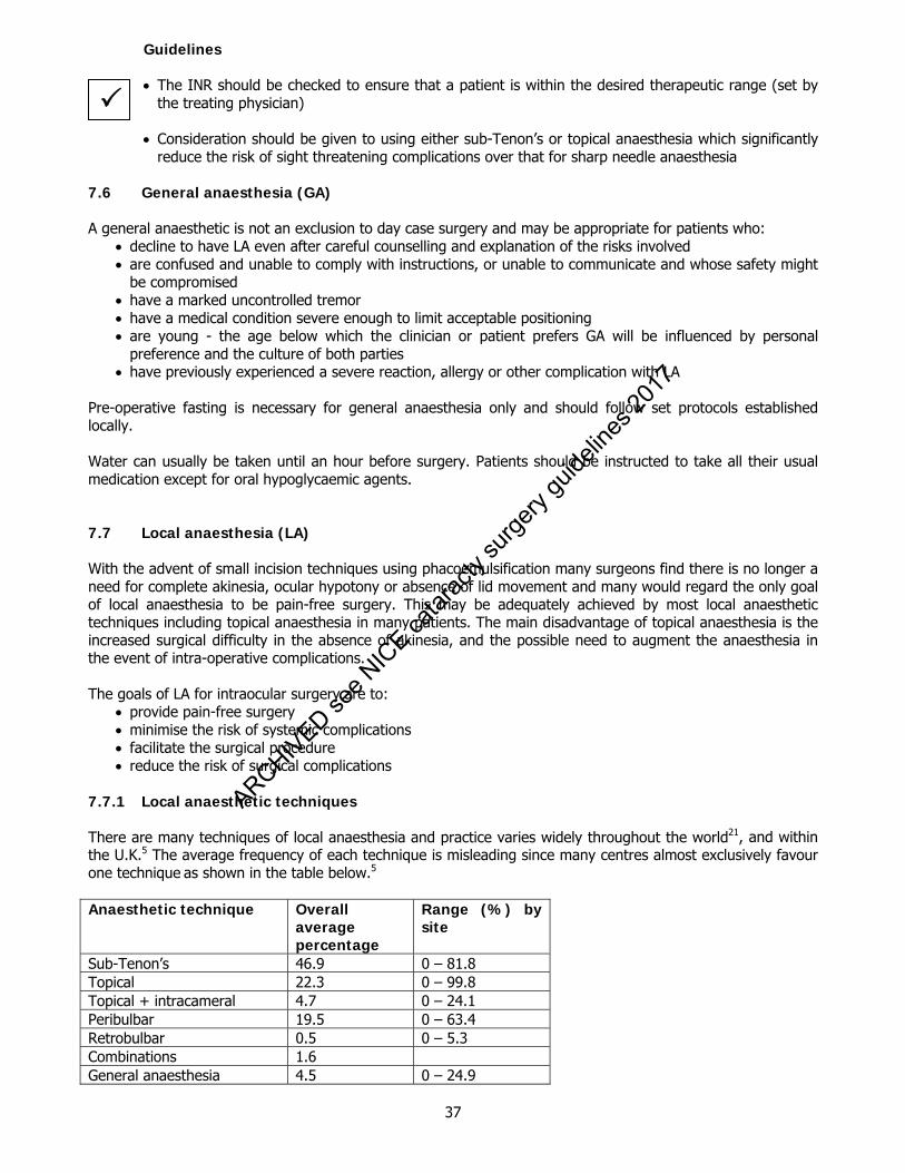
37
Guidelines The INR should be checked to ensure that a patient is within the desired therapeutic range (set by
the treating physician) Consideration should be given to using either sub-Tenon’s or topical anaesthesia which significantly
reduce the risk of sight threatening complications over that for sharp needle anaesthesia 7.6 General anaesthesia (GA) A general anaesthetic is not an exclusion to day case surgery and may be appropriate for patients who:
decline to have LA even after careful counselling and explanation of the risks involved are confused and unable to comply with instructions, or unable to communicate and whose safety might
be compromised have a marked uncontrolled tremor have a medical condition severe enough to limit acceptable positioning are young - the age below which the clinician or patient prefers GA will be influenced by personal
preference and the culture of both parties have previously experienced a severe reaction, allergy or other complication with LA
Pre-operative fasting is necessary for general anaesthesia only and should follow set protocols established locally. Water can usually be taken until an hour before surgery. Patients should be instructed to take all their usual medication except for oral hypoglycaemic agents. 7.7 Local anaesthesia (LA) With the advent of small incision techniques using phacoemulsification many surgeons find there is no longer a need for complete akinesia, ocular hypotony or absence of lid movement and many would regard the only goal of local anaesthesia to be pain-free surgery. This may be adequately achieved by most local anaesthetic techniques including topical anaesthesia in many patients. The main disadvantage of topical anaesthesia is the increased surgical difficulty in the absence of akinesia, and the possible need to augment the anaesthesia in the event of intra-operative complications. The goals of LA for intraocular surgery are to:
provide pain-free surgery minimise the risk of systemic complications facilitate the surgical procedure reduce the risk of surgical complications
7.7.1 Local anaesthetic techniques There are many techniques of local anaesthesia and practice varies widely throughout the world21, and within the U.K.5 The average frequency of each technique is misleading since many centres almost exclusively favour one technique as shown in the table below.5 Anaesthetic technique Overall
average percentage
Range (%) by site
Sub-Tenon’s 46.9 0 – 81.8 Topical 22.3 0 – 99.8 Topical + intracameral 4.7 0 – 24.1 Peribulbar 19.5 0 – 63.4 Retrobulbar 0.5 0 – 5.3 Combinations 1.6 General anaesthesia 4.5 0 – 24.9
ARCHIVED see N
ICE ca
tarac
ty su
rgery
guide
lines
2017
38
All forms of cataract surgery with local anaesthesia demand significant patient co-operation throughout the procedure. Co-operation is most important when procedures are performed with topical (+/- intracameral) anaesthesia. Patients being operated upon by this technique should be able to tolerate instruments approaching the eye without anxiety or blepharospasm. This can be gauged pre-operatively. A separate VIIth nerve block is not generally recommended. Patients undergoing all forms of anaesthesia require adequate counselling and explanation of the procedure. It is unnecessary to fast patients for local anaesthetic cataract surgery but they should not have a full stomach immediately beforehand because of the risk of vomiting during the procedure. 7.7.2 Minimising anaesthetic and operative complications due to the LA Sight or life threatening complications There are two critical issues in the debate about minimising potentially sight or life threatening complications associated with LA injection: technique and needle length when using sharp needle techniques.
Technique Although ocular perforation has been reported with an intended sub-Tenon’s anaesthetic (the perforation occurred during the preparatory dissection in a patient with previous retinal detachment surgery22), it is generally accepted that the risk of perforation is much lower for blunt-cannula techniques than sharp needle LA techniques. It has also been proven in 2 recent studies5, 11 that the risk of sight or life threatening complications was significantly increased with sharp needle techniques.
Sharp needle length Absolute distinction between peribulbar and retrobulbar injection cannot always be made, but complications of both are reduced by using a short (25-31 mm) needle.23
Conclusions For most patients the balance of risks and benefits favour avoiding sharp needle local anaesthesia, especially in high myopes but systemic adverse events have been reported in all forms of local anaesthesia including topical.10
Patient comfort The benefits of intracameral lidocaine in addition to topical anaesthesia have been the subject of a Cochrane review24, which concluded that ‘the use of intracameral unpreserved 1% lidocaine is an effective and safe adjunct to topical anaesthesia for phacoemulsification cataract surgery’ although some well conducted randomized controlled trials found no difference.25
Sub-Tenon’s anaesthesia versus topical anaesthesia for cataract surgery has also been the subject of a Cochrane review26, which concluded that ‘sub-Tenon anaesthesia provides better pain relief than topical anaesthesia for cataract surgery’ but also noted that although the differences were statistically significant they are not necessarily clinically significant.
Operative complications The Cochrane review of sub-Tenon’s versus topical anaesthesia26 also noted that the serious operative complication of posterior capsular rupture with vitreous loss occurred twice as frequently in the topical group compared with the sub-Tenon’s group (4.3% versus 2.1%) but the total number of operations in the seven studies analysed was only 742.
Ia
ARCHIVED see N
ICE ca
tarac
ty su
rgery
guide
lines
2017
39
7.7.3 Choice of local anaesthetic technique In deciding which type of anaesthesia to use, the following factors should be considered but the choice is often one of surgeon preference and practice within each hospital. Patient factors All forms of cataract surgery with local anaesthesia require significant patient co-operation throughout the procedure. Thus, patient preference, anxiety and ability to co-operate should all be taken into account.
LA is the procedure of choice for the majority of patients, provided co-operation can be assured. The patient’s ability to tolerate manipulation around the eye without blepharospasm can be gauged at
the pre-operative assessment and is particularly important if topical anaesthesia is being considered. The experience of the anaesthetist (an inexperienced practitioner is likely to do less damage with a blunt
cannula than with a sharp needle)
Surgical factors High myopes are more likely to have staphylomas and are more likely to suffer perforation of the globe.27
Axial length The type and size of incision The risk of complications Duration of the operation The experience of the surgeon
7.7.4 Who Should Administer LA? Ophthalmologists now administer anaesthesia for cataract surgery in the majority of cases in the UK.5 This is partly due to the increasing use of topical anaesthetic techniques. Sharp needle local anaesthetic injections should only be performed by anaesthetists or ophthalmologists who have been trained appropriately in their use. Nurses or technicians may be trained to administer topical, subconjunctival or su-Tenon’s anaesthesia. It is not recommended that nurses or technicians administer peribulbar or retrobulbar injections. 7.8 Sedation for ocular anaesthesia Ideally, the patient undergoing cataract surgery under local anaesthesia should be fully conscious, responsive, and free from anxiety, discomfort and pain. For most this can be achieved by sensitive and personalised assessment and counselling, with support throughout the operation and verbal reassurance. Reducing anxiety for patients is greatly facilitated by continuity of staff care at all pre-operative stages. However, a few patients still require sedation (1.4% in 20094). Intravenous sedation should only be administered under the supervision of an anaesthetist, whose sole responsibility is to that list. Good rapport, counselling, support and the use of relatively painless techniques all reduce the need for sedation. Sedation should only be used to allay anxiety and not to cover inadequate blocks, which must be corrected by the administration of more local anaesthesia.
ARCHIVED see N
ICE ca
tarac
ty su
rgery
guide
lines
2017
40
7.9 Monitoring Severe systemic complications are rare complications of cataract surgery but have been associated with all LA techniques. The patient should be assured that they will be carefully monitored. 7.9.1 Methods of Monitoring Continuous monitoring of ventilation and circulation is essential, both by clinical observations, and by pulse oximetry. Monitoring should commence just prior to the administration of local anaesthesia and continue until the surgical procedure is complete. The level of monitoring required during local anaesthesia will depend upon the anaesthetic technique and the medical condition of the patient. Monitoring should be the role of a member of staff who remains with the patient throughout the monitoring period and whose sole responsibility is to the patient. This person must be trained to detect and act on any adverse events, and may be an anaesthetist, nurse, operating Department Practitioner (ODP), Assistant (ODA) or anaesthetic nurse as long as they are trained in basic life support (BLS) All theatre personnel should have Basic Life Support (BLS) training, and there should always be at least one person present who has Intermediate (ILS) or Advanced Life Support (ALS) Training or an equivalent qualification. 7.9.2 Level of monitoring required during cataract surgery under LA
Communication with attendant Conventionally the single most important monitor is an individual whose sole responsibility is to remain in contact with the patient and who is trained to detect and act (or alert someone more senior) regarding any adverse event. Electronic patient alert devices that allow patients to communicate with the surgeon were found to be as effective a means of perioperative patient communication as holding a nurse's hand, during cataract surgery under local anaesthesia28 in one study.
Clinical observations Monitor the patient’s colour for circulatory status, responses to surgical stimulus, ventilatory movements and palpation of the pulse
Pulse oximetry To detect cardiac and respiratory problems promptly
IV access This is essential if peribulbar or retrobulbar techniques are employed or if intravenous sedation is used. 7.9.3 Level of staffing required during cataract surgery under LA
The method of anaesthesia and local staffing availability will dictate whether an anaesthetist can be provided for all ophthalmic lists. An anaesthetist is not essential when topical, subconjunctival or blunt-cannula sub-Tenon’s techniques without sedation are used
An anaesthetist should be present if retrobulbar, peribulbar and sharp-needle sub-Tenon’s techniques
are used. In the absence of an anaesthetist, the hospital, trust or treatment centre is responsible for ensuring that
someone in the operating theatre is trained to perform cardiopulmonary resuscitation Intravenous sedation should only be administered under the supervision of an anaesthetist, whose sole
responsibility is to that list
ARCHIVED see N
ICE ca
tarac
ty su
rgery
guide
lines
2017
41
7.10 Facilities All intraocular surgery performed under LA should be carried out in a facility which is appropriately equipped and staffed for resuscitation. Oxygen and suction must be available. Patients should be on a tipping trolley or equivalent chair. 7.11 Anaesthesia references 1. Courtney P. The National Cataract Surgery Survey: I method and descriptive features. Eye 1992;
6:487-492. 2. Eke & Thompson T, Thompson JR. The National Survey of Local Anaesthesia for Ocular Surgery. I.
Survey methodology and current practice. Eye 1999; 13:189-195. 3. Desai P, Reidy A, Minassian DC. Profile of patients presenting for cataract surgery: National data
collection. Br J Ophthalmol 1999;83:893-896. 4. Johnston RL, Sparrow JM, Canning CR, Tole D, Price NC. Pilot National Electronic Cataract Surgery
Survey I. Eye 2005; 19: 788-794. 5. El-Hindy N, Johnston RL, Jacycock P, Eke T, Braga AJ, Tole DM, Galloway P, Sparrow JM and the UK
EPR users group. The Cataract National Dataset Electronic Multi-centre Audit of 55,567 Operations: anaesthetic techniques and complications.
6. Moffat A, Cullen PM. Comparison of two standard techniques of general anaesthesia for day-case
cataract surgery. Br J Anaesth 1995; 74(2):145-8. 7. Percival SP, Setty SS. Prospective audit comparing ambulatory day surgery with inpatient surgery for
treating cataracts. Qual Health Care 1992;1(1):38-42. 8. Jain S, Adhikari HP. Day case cataract surgery without a dedicated unit. J R Coll Surg Edinb 1996;
41(5):336-8. 9. Hamilton RC, Gimbel HV, Strunin L. Regional anaesthesia for 12,000 cataract extraction and intraocular
lens implantation procedures. Can J Anaesth 1988; 35(6):615-23. 10. Eke T, Thomson JR. The National Survey of local anaesthesia for ocular surgery. II. Safety profiles of
local anaesthesia techniques. Eye 1999b; 13: 196-204. 11. Eke T, Thomson JR. Serious complications of local anaesthesia for cataract surgery: a 1 year national
survey in the United Kingdom. Br J Ophthalmol 2007 Apr;91(4):470-5. 12. Schein OD, Katz J, Bass EB et al. The value of routine pre-operative medical testing before cataract
surgery. N Engl J Med 2000; 342: 168-176. 13. Walters G, McKibbin M. The value of pre-operative investigations in local anaesthetic ophthalmic
surgery. Eye 1997;11: 847-849. 14. Benzimra JD, Johnston RL, Jaycock P, Galloway PH, Lambert G, Chung AKK, Eke T, Sparrow JM and
the UK EPR User Group. The Cataract National Dataset electronic multi-centre audit of 55,567 operations: antiplatelet and anticoagulant medications. Eye 2009; 23: 10-16.
15. EAFT (European Atrial Fibrillation Trial) Study Group. Secondary prevention in non-rheumatic atrial
fibrillation after transient ischaemic attack or minor stroke. Lancet 1993; 342:1255-1262.
ARCHIVED see N
ICE ca
tarac
ty su
rgery
guide
lines
2017
42
16. Peterson P, Boysen G, Godtfredsen J, Andersen ED, Andersen B. Placebo-controlled, randomised trial of warfarin and aspirin for prevention of thromboembolic complications in chronic atrial fibrillation. The Copenhagen AFASAK study. Lancet 1989; 1:175-179.
17. THE ISIS-2 (Second International Study of Infarct Survival) Collaborative Group. Baigent C, Collins R,
Appleby P, Parish S, Sleight P, Peto R. ISIS-2: 10 year survival among patients with suspected acute myocardial infarction in randomised comparison of intravenous streptokinase, oral aspirin, both, or neither. BMJ 1998; 316:1377-1343.
18. CAPRIE Steering Committee. A randomised, blinded, trial of clopidogrel vs aspirin in patients at risk of
ischaemic events (CAPRIE). Lancet 1996; 348: 13291339. 19. ESPIRIT Study Group. Halkes PH, van Gijn J, Kappelle LJ, Koudstaal PJ, Algra A. Aspirin plus
dipyridamole vs aspirin alone after cerebral ischaemia of arterial origin (ESPIRIT): randomised controlled trial. Lancet 2006; 367: 1665-1673.
20. Katz J, Feldman MA, Bass EB, Lubomski LH, Tielsch JM, Petty BG, et al. Study of Medical Testing for
Cataract Surgery Team. Risks and benefits of anticoagulant and antiplatelet medication use before cataract surgery. Ophthalmology 2003; 110: 1784- 1788.
21. Norregard JC, Schein OD, Bellan L, Black C, Alonso J, Bernth-Petersen P, Dunn E, Anderson TF,
Espellargues M, Anderson GF. International variation in anaesthesia care during cataract surgery: Results form the international cataract surgery outcomes study. Arch Ophthalmol 1997;115(10):1304-1308.
22. Frieman BJ, Friedberg MA. Globe perforation associated with subtenon’s anaesthesia. Am J Ophthalmol
2001; 131 (4): 520-521. 23. Davis DB, Mandel MR. Efficacy and complication rate of 16,224 consecutive peribulbar blocks. A
prospective multicenter study. J Cataract Refract Surg 1994; 20(3):327-37. 24. Ezra DG, Allan BD. Topical anaesthesia alone versus topical anaesthesia with intracameral lidocaine for
phacoemulsification. Cochrane Database Syst Rev. 2007: 18 (3):CD005276. 25. Boulton JE, Lopatatzidis A, Luck J, Baer RM. A randomized controlled trial of intracameral lidocaine
during phacoemulsification under topical anesthesia. Ophthalmology 2000;107(1):68-71. 26. Davison M, Padroni S, Bunce C, Rüschen H. Sub-Tenon's anaesthesia versus topical anaesthesia for
cataract surgery. Cochrane Database Syst Rev. 2007; 18(3):CD006291. 27. Edge R, Navon S. Scleral perforation during retrobulbar and peribulbar anesthesia: risk factors and
outcome in 50,000 consecutive injections. J Cataract Refract Surg 1999;25(9):1237-44. 28. Mokashi A, Leatherbarrow B, Kincey J, Slater R, Hillier V, Mayer S. Patient communication during
cataract surgery. Eye. 2004;18(2):147-51. Supplementary: D. Ezra, A. Nambiar, B. Allan. Intracameral Lidocaine for Phacoemulsification under Topical
Anesthesia A Meta-analysis of Randomized Controlled Trials Ophthalmology 115; 3:455-487.
ARCHIVED see N
ICE ca
tarac
ty su
rgery
guide
lines
2017
43
8. Biometry 8.1 The aims of biometry
The primary aim of biometry is to allow the selection of the correct IOL power to achieve the desired refractive result after cataract surgery
It is an essential step before cataract surgery The refractive aims of cataract surgery must be discussed with the patient in terms of their
requirements, expectations and what is achievable and available. 8.2 Biometric components There are two major components:
axial length measurement by A-scan ultrasound or laser interferometry central corneal curvature measured by keratometry or topography
Some intraocular lens formulae also require additional measurements: ‘white-to-white’ measurement of corneal diameter phakic anterior chamber depth age
8.3 Who should perform biometry? Biometry is a highly skilled process, the results of which are crucial to the success of the operation. The service may be provided by a number of suitably trained professionals (ophthalmologists, ophthalmic technicians, nurses, or optometrists) according to local arrangements. It is essential that the measurements are carried out in a consistent manner using standardized settings for corneal refractive index and velocity of sound in different media for ultrasound. Ophthalmologists in training should learn to perform and be familiar with biometry, but it is not appropriate for them to provide a routine biometry service. 8.4 Where should it be done? In order to maintain consistency and predictability, the equipment used should be standardized as much as possible. Therefore, a relatively small number of technicians and equipment should be used on as few sites as possible, particularly for those eyes requiring ultrasound axial length measurement. 8.5 When should it be done? Biometry should be done sufficiently in advance of surgery to allow for adequate discussion between the patient and surgeon of the refractive aims and allow ordering of the appropriate intraocular lens. If the patient is a regular contact lens wearer then ideally, soft lenses should be left out for one week and hard lenses for four weeks prior to measurement. Ideally biometry should be performed before dilating drops are instilled and before tonometry is performed. 8.6 With what equipment? It is the responsibility of each unit to ensure that the instruments used to measure the biometric components are serviced regularly and calibrated and operated in accordance with the manufacturer’s instructions.
ARCHIVED see N
ICE ca
tarac
ty su
rgery
guide
lines
2017
44
Both eyes should be measured even if unilateral surgery is planned to allow for cross-checking. Similarly focimetry should be carried out or the latest copy of the patient’s refraction cross-referenced with the biometry results, taking into account the effect of cataract on refraction. The biometry data and printout should be kept in the patient’s records and be clearly marked with the patient’s name, hospital number and date of birth. 8.7 Axial length
8.7.1 Optical
Benefits The Zeiss IOL Master and the Lenstar LS 900 are non-contact optical device that use partial coherence interferometry (PCI) to determine axial length. Compared to acoustic biometry with a standard 10MHz ultrasound probe, PCI axial length measurements have greater precision (+ 0.02 mm1 versus 0.12 mm2), are negligibly affected by velocity assumptions3 and consistently measure along the visual axis.4 They are quick and easy to use and, in addition to giving precise measurements, avoid the risk of corneal compression that is common with contact ultrasound. As they are non-contact there is no risk of cross-contamination. They have the added benefit of built in keratometers and the facilities for measuring the anterior chamber depth. New versions of the IOL Master software allow the measurement of anterior chamber depth and corneal diameter. These parameters add greater accuracy and allow use of 3rd generation formulae. Disadvantages Optical biometry relies on good fixation and is not possible for dense cataracts but recent software improvements have increased the range of eyes that can be successfully measured. Risks
Manufacturers’ IOL A constants are derived from contact ultrasound axial length measurement and A constants optimized for optical measurement are substantially different. The difference between optimized constants for each method of axial length measurement (contact ultrasound or optical biometry) for each IOL model are far greater than the minor differences between constants optimized for each surgeon when using the same axial length measurement technique. If the manufacturer’s A constant is used, optical biometry will often deliver worse results than contact ultrasound biometry. As a starting point when using a new IOL model with optical biometry the ULIB web site displays A constants5 optimized for this technique.
8.7.2 Ultrasound This method can be used for all types of cataract but is more user-dependent for accuracy primarily due to a variable amount of corneal compression.6 Immersion techniques give more accurate results than contact ultrasound and with modern equipment (Praeger shell) are easy to perform.7 8.7.3 Keratometry This can be measured using manual or automated devices. Whichever method is used an average of 3 readings should be taken and recorded with the axes. Corneal topography is desirable as it averages the corneal curvature values from far more locations to calculate ‘sim K’ values. They are particularly important when planning astigmatism reduction surgery.
ARCHIVED see N
ICE ca
tarac
ty su
rgery
guide
lines
2017

45
8.7.4 Biometric data
In the absence of pathology that might affect eye size (eg unilateral refractive error, coloboma, staphyloma), most individuals have similar axial lengths in each eye.
92% of axial lengths fall within the range 21.0 to 25.5 mm8 Most corneas are relatively regularly curved and similar between the two eyes of each individual 99% of K readings are within the range 40D to 48D8 Based on the 95% distribution biometric measurements should be repeated if8:
o Axial length is <21.20 mm or >26.60 mm o Mean corneal power is <41D or >47D o Delta K is >2.5D o Difference in axial length between fellow eyes of >0.7mm o Difference in mean corneal power of >0.9 dioptres
When there are large differences between the K readings and/or axial lengths of each eye, consider
the possibility of amblyopia, staphylomata or vitreous opacities such as asteroid hyalosis. An amblyopic eye may have been forgotten by the patient and may not be corrected in the current spectacle prescription
For highly myopic eyes (axial length >28mm) optical biometry is usually reliable if the patient can fixate accurately but if acoustic measurements are being performed a B-scan should be performed to determine the presence or absence of staphylomata.
8.7.5 Formulae
The SRK I and SRK II formulae are obsolete and should no longer be used9 Which modern IOL calculation formula (SRK/T, Holladay I & II, Haigis and Hoffer Q) gives the best
results has been hotly debated10. All modern formulae perform well in the normal axial length range but the Haigis and Hoffer Q may be slightly better for short axial lengths (<22mm)11
Optimizing the IOL constants for each IOL model for the method of axial length measurement (optical
or acoustic) has a much greater impact on the predictability of the refractive outcome than choosing between modern IOL formulae5. In some cases the percentage of eyes within +/- 1D of the target refraction can improve by up to 20% with optimization12
Optimizing A constants for each IOL model according to the method of axial length measurement is
much more important than optimizing A constants for each surgeon when using the same method of axial length measurement (although this may confer a small extra benefit).
8.8 Audit considerations What matters most in biometry is achieving excellent results, which can be presented in terms of the percentage of eyes within 0.5 or 1.0D of the target refraction.
This is only possible if the refractive outcomes are continuously audited by comparing the predicted with the achieved spherical equivalent.
Given the number of cataract operations performed and the amount of data required for each operation
audit is only practical with electronic medical record systems that collect the cataract national dataset.13 8.9 Benchmark standards With the routine use of optical biometry when possible, modern IOL calculation formulae and optimization of A constants busy NHS departments with a variety of surgeons and biometry technicians should be able to achieve a refractive outcome within +/- 1D of the target in 85% of cases.14
III
ARCHIVED see N
ICE ca
tarac
ty su
rgery
guide
lines
2017
46
8.10 Inability to obtain reliable biometric data Sometimes ocular pathology precludes obtaining accurate biometric data. At this juncture:
Ask about previous refractive surgical procedures Perform a full ophthalmic examination including objective and subjective refraction of each eye Compare ultrasound and keratometric data from each eye Obtain and review the patient’s refractive history backed up by reports from his / her optometrist
Where measurements are not available or are incomplete, an informed choice based on the available information should be made.
8.11 Post laser refractive surgery IOL power calculations Corneal refractive surgery alters the relationship between the anterior and posterior corneal curvature and therefore renders incorrect the assumptions made in keratometry and corneal topography regarding the power of the central cornea. As a result there is an unpredictable undercorrection of the corneal power resulting in a hyperopic ‘surprise’ after cataract surgery. The patient needs to be advised of these problems both prior to refractive and cataract surgery. Even with the corrections outlined below the predictability of the post-operative refraction is impaired and it is wise to aim for low myopia to avoid unexpected post-operative hyperopia. There are numerous methods of correcting the keratometry values including16 Calculation method Likely refractive outcome Feiz & Mannis Often the upper limit of correct IOL power Clinical history Modified Maloney Topographic central corneal power adjustment Post-operative regression
Correct IOL power often in this range
Flattest measured K Often lower limit of correct IOL power Sim Ks by topography Usually below correct IOL power Measured Ks Usually below correct IOL power A useful website to access regarding post-refractive laser calculations for biometry is www.ASCRS.org 8.12 Unexpected post-operative results If a patient has an unexpected refractive result after cataract surgery every step of the biometry and pre and post-operative refraction of both eyes must be checked carefully and repeated if surgical intervention is planned. It is more common for an error to have occurred than a genuine ‘surprise’. The following options can be considered to correct anisometropia:
Leave the IOL in situ if the patient can tolerate this refraction Contact lens over-correction Intraocular lens exchange based on refraction (the ophthalmologist should be aware of possible labelling
errors on the original IOL packaging which could confound this option) Insertion of a corrective additional intraocular lens Corneal refractive surgery
If the other eye requires cataract surgery, consideration should be given to the power of lens to be inserted in the second eye, recognising the importance of avoiding symptomatic aniseikonia. Any further surgery needs to be discussed in detail with the patient.
ARCHIVED see N
ICE ca
tarac
ty su
rgery
guide
lines
2017

47
8.13 Clinical risk and biometry The potential for error to occur in IOL selection is significant and is discussed in the section on Patient Safety.
8.14 Biometry References 1. Drexler W, Findl O, Menapace R, et al. Partial coherence interferometry: a novel approach to biometry in
cataract surgery. Am J Ophthalmol 1998; 126(4):524-34. 2. Binkhorst RD. The accuracy of ultrasonic measurement of the axial length of the eye. Ophthalmic Surg
1981; 12(5):363-5. 3. Norrby S. Sources of error in intraocular lens power calculation. J Cataract Refract Surg 2008; 34(3):
368-76. 4. Sheng H, Bottjer CA, Bullimore MA. Ocular component measurement using the Zeiss IOL Master. Optom
Vis Sci 2004; 81(1):27-34. 5. User Group for Laser Interference Biometry. http://www.augenklinik.uni-wuerzburg.de/eulib/index.htm 6. Snead MP, Rubinstein MP, Lea SH, Haworth SM. Calculated versus A-scan result for axial length using
different types of ultrasound probe tip. Eye 1990; 4:718-22. 7. Hoffmann PC; Hütz WW; Eckhardt HB; Heuring AH. Intraocular lens calculation and ultrasound biometry:
immersion and contact procedures. Klin Monatsbl Augenheilkd 1998; 213(3): 161-5. 8. Knox-Cartwright N, Johnston RL, Sparrow JM. The Cataract National Dataset electronic multicentre audit
of 55,567 operations: when should optical biometric measurements be rechecked? In press – Eye advance online publication, 14 August 2009; doi:10. 1038/eye.2009.196
9. Nemeth J, Fekete O, Pesztenlehrer N. Optical and ultrasound measurement of axial length and anterior
chamber depth for intraocular lens power calculation. J Cataract Refract Surg 2003; 29(1):85-8. 10. Narvaez J, Zimmerman G, Stulting D, Chang DH. Accuracy of intraocular lens power prediction using the
Hoffer Q, Holladay 1, Holladay 2, and SRK/T formulas. J Cataract Refract Surg 2006; 32:2050-2053. 11. Maclaren RE, Natkunarajah M, Riaz Y, Bourne RRA, Ristori M, Allan BDS. Biometry and Formula Accuracy
With Intraocular Lenses Used for Cataract Surgery in Extreme Hyperopia. Am J Ophthalmol 2007; 143:920-931.
12. Gavin EA, Hammond CJ. Intraocular lens power calculation in short eyes. Eye 2008; 22:935-938. 13. Service Implementation – Do Once and Share. Appendix P Cataract National Dataset. 2006.
http://www.rcophth.ac.uk/docs/publications/publishedguidelines/FinalVersionGuidelinesApril2007Updated.pdf
14. Gale RP, Saldana M, Johnston RL, Zuberbuhler B, McKibbon M. Benchmark standards for refractive
outcomes after NHS cataract surgery. Eye 2009; 23:149-152. 15. Kim SW, Kim EK, Cho BJ, Kim SW, Song KY, Kim TI. Use of the pentacam true net corneal power for
intraocular lens calculation in eyes after refractive corneal surgery. J Refract Surgery 2009; 25:285-9. 16. http://www.a-scan.net/iol-main/lasik.html
ARCHIVED see N
ICE ca
tarac
ty su
rgery
guide
lines
2017
48
9. Factors affecting the Choice of Intraocular Lens (IOLs) 9.1 General All intraocular lenses have their individual ‘pros and cons’ and there is no ‘best buy’. Quality, track record, the supplier’s ability to service a lens bank and provide the required range of dioptric powers and cost must all be considered. Problems concerning the safety of the lens and variation from the manufacturers stated performance should be reported to Medicine and Healthcare Regulatory Agency (MHRA) www.mhra.gov.uk Surgeons should have available suitable IOL models and powers not only for routine ‘in the bag’ surgery but also to cover unexpected situations such as the need for sulcus or anterior chamber fixation. Special powers or models of IOL may be required for particular patients or situations, and these would need to be ordered in advance. IOL technology is a continually developing field and some of the factors to be considered in the choice of IOL include: 9.2 Incision size Nearly all cataracts will be removed using phacoemulsification with the advantage of reduced incision size: this is usually up to 3.2mm but can be as small as 1.8mm. Therefore a foldable IOL is part of the standard practice. In order to reduce the incision size for lens insertion a “wound assist” technique may be used. 9.3 Method of insertion
The method of folding is a matter of choice for the individual surgeon. Most lenses are injectable which have advantages of predictable incision size, speed and ease of insertion, less damage to the IOL and possible reduced risk of bacterial endophthalmitis.1 The injector system used should be matched to the IOL and the use of pre-loaded injector systems will increase. The use of folding forceps for insertion is still utilised by some surgeons especially for some three piece models. Unfolding of the IOL from any delivery system should be controlled so that the bag is not damaged in the process.
9.4 Optic size Lens optics vary in diameter. Larger optics (6mm+) are better as these are less centration dependent, have fewer dysphotopic symptoms in patients with large pupils or under mydriatic conditions and lower rates of posterior capsule opacification (PCO).2 These factors need to be weighed against the possible requirement of a larger incision size with IOLs. 9.5 IOL materials IOLs are at present manufactured from either silicone or acrylic polymers. All IOL materials can be classified by their hydrophilic or hydrophobic nature. These terms are relative. IOL biocompatibility can be divided into uveal (inflammatory cell attachment) or capsular (anterior capsular fibrosis, PCO). Each polymer appears to have its own biocompatibility profile. Hydrophilic materials are generally considered to have a better uveal biocompatibility profile (lower inflammatory cell attachment) in comparison to hydrophobic materials but the latter with present IOL designs, may have a better performance in preventing PCO.3,4 Newer hydrophilic IOL designs are available and it remains to be seen whether these have a comparable PCO performance.
III
ARCHIVED see N
ICE ca
tarac
ty su
rgery
guide
lines
2017
49
Square edge optic profile has been shown to be an important factor in preventing PCO5 but this can be at the expense of increased dysphotopic symptoms: most manufacturers now engineer the edge profile to minimise such symptoms. 9.6 Haptics Many lenses are now single piece with the haptic made of the same foldable material as the optic. This makes the lens easier to fold compared to 3 piece lenses with the haptics made from PMMA or Prolene. The haptic size needs to be appropriate for bag fixation without causing posterior capsular folds as these can cause symptoms from scattering of light or PCO.6 An appropriate IOL with a larger haptic is required for sulcus fixation; single piece lenses should not be placed into the sulcus unless specifically designed for such placement. 9.7 Function options The standard lens is monofocal and fixed in position in the eye. This can be modified to give additional functionality: 9.7.1 Correction of presbyopia:
Modification of the optic to produce a Multifocal / Bifocal lens Presents multiple images on to the retina and cortical and retinal adaptation enables the patient to select the appropriate image, distance or near. These optics may be zonal progressive refractive multifocal or diffractive bifocal. Each optical technology has its own characteristics, the former giving better intermediate vision and the latter better near vision according to some case studies. Both can produce dysphotopsia. To obtain the advantages and try to eliminate the disadvantages of these lenses, they can be combined in “custom match” surgery with one type in each eye although little evidence apart from small case series exist to show whether this is beneficial.
Modification of the haptic to allow movement of the lens: focus shift or Accommodative lens
This lens technology does not have the problem of dysphotopsia, but its mechanism of action is uncertain. Its near vision is not as effective as a multifocal but the intermediate vision is said to be better. Other types of corrections such as dual-optic lenses remain under trial.
9.7.2 Toric IOL The IOL surface may be toric to correct corneal astigmatism and although incisional surgery e.g. limbal relaxing incision may be used to correct corneal astigmatism this may be less predictable in both effect-size and stability than toric implants. 9.7.3 Blue light filtering lenses The natural lens yellows i.e. filters out blue light as it ages and is replaced in cataract surgery with a clear lens. Theoretically this exposes the retina to increased levels of light at the blue end of the spectrum causing increased longterm risk of age related macular degeneration.7 Therefore yellow IOLs which filter out short wavelength blue light have been introduced and there is some early experimental evidence that these could retard age related macular changes. Their effect on scotopic vision remains controversial and there are suggestions that they may be associated with disturbed sleep patterns due the effect on the melanopsin-containing ganglion cell photoreceptor function and circadian rhythm.8,9,10,11 However a recent study by Landers et al12 showed no effect on the sleep quality of patients who had blue-blocking lenses inserted. This is the subject of an ongoing randomized controlled trial at the time of publication of these guidelines
IV
ARCHIVED see N
ICE ca
tarac
ty su
rgery
guide
lines
2017

50
9.7.4 Correction of spherical aberration Aberrations are an optical defect in which light from a point object does not form a perfect point after passing through an optical system. It occurs when light rays are over-refracted at the periphery of an optical surface. The cornea has positive spherical aberration and doesn’t change significantly throughout life. The lens starts with negative spherical aberration (neutralizing that of the cornea) but becomes positive in later life or with pseudophakia, compounding that of the cornea. Aspheric lenses are designed to correct this and improve contrast sensitivity function but they may reduce depth of focus. They are currently a “one size fits all” solution and not matched to the actual corneal spherical aberration although three different degrees of correction are available commercially. Using a Pentacam or Galilei corneal topographer or VOL-CT software it is now possible to analyse the relative contribution of corneal and lenticular asphericity in an individual eye. Whether or not this makes a difference to the subjective visual outcome has not yet been shown. Centration of these lenses is important for adequate effect and decentration of more than 0.8 mm or tilt of more than 14 degrees has been shown to have detrimental effect on their function.
A number of trials have shown improvements in contrast sensitivity following the use of aspheric lenses but little evidence exists demonstrating improvement in activities of daily living.13
The precise role of these lenses in routine clinical practice remains to be defined and the surgeon must be familiar with the technology and have appropriately counselled the patient.
Ib
ARCHIVED see N
ICE ca
tarac
ty su
rgery
guide
lines
2017

51
9.8 Factors affecting the choice of intraocular lens (IOLs) references 1. Mayer E, Cadman D, Ewings P et al. A 10 year retrospective survey of cataract surgery and
endophthalmitis in a single eye unit; injectable lenses lower the incidence of endophthalmitis. Br J Ophthalmol 2003; 87:867-869.
2. Meacock WR, Spalton DJ, Boyce JF, Jose JR. Effect of optic size on posterior capsular opacification with
5.5 mm and 6.0 mm optic diameter Acrysof intraocular lenses. J Cataract Refract Surg 2001; 27:1194-1198.
3. Hayashi K, Hayashi H. Posterior capsule opacification after implantation of a hydrogel intraocular lens. Br
J Ophthalmol 2004; 88(2):182-5. 4. Hollick EJ, Spalton DJ, Ursell PG, Meacock WR, Barman SA, Boyce JF. Posterior capsular opacification
with hydrogel, polymethylmethacrylate, and silicone intraocular lenses: two-year results of a randomized prospective trial. Am J Ophthalmol 2000; 129(5):577-84.
5. Nishi O, Nishi K, Wickstrom W. Preventing lens epithelial cell migration using intraocular lenses with
sharp rectangular edges. J Cataract Refract Surg 2000; 26: 1543-9. 6. Meacock WR, Spalton DJ. Effect of intraocular lens haptic compressibility on the posterior lens capsule
after cataract surgery. J Cataract Refract Surg 2001; 27: 1366-1371. 7. Wang JJ, Klein R, Smith W et al. Cataract surgery and the 5 year incidence of Late Stage Age Related
Maculopathy. Ophthalmology 2003; 110: 1960-1967. 8. Mainster MA, Sparrow JR. How much blue light should an IOL transmit? Br J Ophthalmol 2003; 87:
1523-1529. 9. Cuthbertson FM, Peirson SN, Wulff K, Foster R, Downes SM. Blue light–filtering intraocular lenses:
Review of potential benefits and side effects. J Cataract Refract Surg 2010; in press. 10. Casparis H, Lindsley K, Bressler NB Surgery for cataracts in people with age-related macular
degeneration. Cochrane Database Syst Rev 2009 Jan 21;(1):CD006757 11. Chew EY, Sperduto RD, Milton RC et al. Risk of advanced age-related macular degeneration after
cataract surgery in the Age-Related Eye Disease Study: AREDS report 25.Ophthalmology 2009; 116(2):297-303.
12. Landers JA, Tamblyn D, Perriam D. Effect of blue-light-blocking intraocular lens on the quality of sleep. J
Cataract Refract Surg 2009; 35(1):83-88. 13. Cuthbertson FM, Dhingra S, Benjamin L. Objective and subjective outcomes in comparing three different
aspheric intraocular lens implants with their spherical counterparts. Eye 2009; 23(4):877-85. Epub 2008 May 9.
ARCHIVED see N
ICE ca
tarac
ty su
rgery
guide
lines
2017
52
10 Posterior Capsule Opacification (PCO) 10.1 Introduction In spite of advances in IOL design, PCO remains the commonest long term reason for further intervention following cataract surgery. It is caused by residual lens epithelial cells (LECs) which are inevitably left behind after surgery and is essentially a wound healing response of the lens to surgery. With time the residual LECs proliferate to form Elschnig’s pearls or undergo metaplasia to myofibroblasts. These can migrate to obscure the visual axis or cause fibrosis of the capsule potentially causing IOL decentration. 10.2 Pathogenesis PCO is a multi-factorial problem and is related to patient factors (e.g. age, concurrent ocular disease such as uveitis, retinitis pigmentosa), surgical factors and IOL design features. The latter have received most attention in recent years.
It has been shown that IOLs with a square edge profile inhibit LEC migration on to the posterior capsule. Not all square edge designs are the same with the sharpness of the edge seeming to be important
Other design features such as large optic diameter, posterior flexion of the haptics, prevention of posterior capsular folds by flexible haptics and possibly IOL material are all additional important features.1,2,3
Many clinical studies have shown that PCO is reduced if the anterior capsulorhexis lies completely on the anterior IOL surface
This compresses the IOL against the posterior capsule producing a mechanical barrier to LEC migration, no matter what type of IOL is used.4,5 Polishing the anterior or posterior capsule to remove LECS at surgery has not been shown to prevent future PCO but does have an influence on anterior capsule shrinkage.13
With improvement in IOL design and surgical technique laser capsulotomy rates have fallen from 30-50% to less than 10% at 2 years post-operatively.6
As patients are usually discharged soon after surgery it is essential to warn them that posterior capsular opacification may occur and to seek advice if their vision deteriorates. In patients who find it difficult to communicate e.g. with learning disability, consideration should be given to longer term assessment of their vision in case PCO occurs.
PCO is a particular problem in paediatric cataract surgery, and has implications for surgical technique and follow up. (See chapter on paediatric cataract surgery). 10.3 Indications for Treatment of PCO. The indication for treatment is the presence of visual symptoms attributable to PCO on slit lamp examination. Most adult patients present with PCO within 2 years of their operation but a few can present many years later. Symptoms are more important than tests of visual function: severity of PCO correlates poorly with high contrast visual acuity.7 Blurred vision, glare, dysphotopsia and reduced contrast in the presence of PCO on slit lamp examination are common symptoms. Symptoms may be more noticeable in bright light or conversely at night with driving or other mydriatic circumstances.
Ia
Ia
Ia
ARCHIVED see N
ICE ca
tarac
ty su
rgery
guide
lines
2017
53
10.4 Treatment PCO is most commonly treated by YAG laser capsulotomy. In rare circumstances surgical removal may be required. The posterior capsule should be opened sufficiently to clear what would be the patient’s maximum physiological pupil area. Minimum laser energy should be used and care should be taken to avoid pitting the IOL. In some units treatment is performed by appropriately trained paramedical staff. Laser capsulotomy may be followed by an IOP pressure spike within the first few hours. This may not be clinically important in healthy eyes but can potentially damage the optic disc in eyes with glaucoma. Many surgeons prefer to routinely give all patients a hypotensive agent immediately after treatment. 10.5 Complications of Treatment Laser capsulotomy can be occasionally followed by raised IOP, cystoid macular oedema, subluxation or dislocation of the IOL, intraocular inflammation or endophthalmitis from release of loculated bacteria in the capsule or retinal detachment.8,9,10,11 For these reasons, and to check resolution of symptoms, some surgeons like their patient to be checked 1-2 weeks after treatment. This can be delegated to appropriately trained paramedical staff. If the patient is not seen post-treatment an advice sheet should be provided. The incidence of cystoid macula oedema is thought to be reduced if capsulotomy is postponed to 3 months after surgery but this may depend on the clinical circumstances. Retinal detachment (RD) is of particular concern in high myopes.12 Consideration should be given to these patients having an IOL implanted at surgery with a proven low PCO rate. Prophylactic treatment of pre-existing retinal pathology has not been proven to reduce the risk of RD. It is therefore important to warn these patients of the symptoms of RD and encourage them to report urgently should they develop. There is anecdotal evidence that the energy used per pulse may be more important than the total energy used to open an opacified capsule. Using energies of 1.2mJ per pulse or less conferred protection from retinal detachment in one unpublished study.
ARCHIVED see N
ICE ca
tarac
ty su
rgery
guide
lines
2017
54
10.6 Posterior capsular opacification (PCO) references 1. Nishi O, Nishi K, Wickstrom W. Preventing lens epithelial cell migration using intraocular lenses with
sharp rectangular edges. J Cataract Refract Surg 2000; 26: 1543-9. 2. Meacock WR, Spalton DJ, Boyce FJ, Jose RJ. Effect of optic size on posterior capsular opacification with
5.5 mm and 6.0 mm optic diameter Acrysof intraocular lenses. J Cataract Refract Surg 2001; 27: 1194-1198.
3. Meacock WR, Spalton DJ. Effect of intraocular lens haptic compressibility on the posterior lens capsule
after cataract surgery J Cataract Refract Surg 2001; 27:1366-1371. 4. Hollick EJ, Spalton DJ. The effect of capsulorhexis size on posterior capsule opacification. Am J
Ophthalmol 1999; 128: 271-279. 5. Boyce JF, Bhermi GS, Spalton DJ, El_Osta AR. Mathematical modeling of the forces between an
intraocular lens and the capsule. J Cataract Refract Surg 2002; 28(10):1853-9. 6. Apple DJ, Peng Q, Visessook N, Werner L, Pandey SK, Escobar Gomez M, et al. Eradication of posterior
capsule opacification: documentation of a marked decrease in Nd:YAG laser posterior capsulotomy rates noted in an analysis of 5416 pseudophakic human eyes obtained postmortem. Ophthalmology 2001;108(3):505-18.
7. Meacock WR, Spalton DJ, Boyce J, Marshall J. The effect of posterior capsule opacification on visual
function. Invest Ophthalmol Vis Sci 2003; 44(11):4665-9. 8. Channel MM, Beckman H. Intraocular pressure changes after neodymium Yag laser posterior
capsulotomy. Arch Ophthalmol 1984; 102: 1024-1026. 9. Steinert RF, Puliafito CA, Kumar SR et al. Cystoid macular oedema, retinal detachment and glaucoma
after Nd Yag laser posterior capsulotomy. Am J Ophthalmol 1991; 112: 373-380. 10. Tetz M, Apple DJ, Price FW. A newly described complication of Neodymium YAG laser capsulotomy: an
exacerbation of an intraocular infection, case report. Arch Ophthalmol 1987; 105: 1324-5. 11. Olsen GM, Olsen RJ. Prospective study of cataract surgery, capsulotomy and retinal detachment. J Cat
Refract Surg 1995; 21: 136-139. 12. Koch DD, Liu JF. Gill EP et al. Axial myopia increases the risk of retinal complications after neodymium
YAG posterior capsulotomy. Arch Ophthalmol 1989; 107: 986-990. 13. Hanson RJ, Rubinstein A, Sarangapani S, Benjamin L, Patel CK. Effect of lens epithelial cell aspiration on
postoperative capsulorhexis contraction with the use of the AcrySof intraocular lens: randomized clinical trial. J Cataract Refract Surg 2006;32(10):1621-6.
ARCHIVED see N
ICE ca
tarac
ty su
rgery
guide
lines
2017
55
11 Patient information and consent 11.1 Introduction ‘Consent’ is a patient’s agreement for a health professional to provide care. Patients may indicate consent non-verbally, orally, or in writing. For the consent to be valid, the patient must be competent to take the particular decision, have received sufficient information to take it and not be acting under duress. The NHS Plan has led to recent changes in the way patients are asked to give their consent to treatment, so protecting the fundamental legal and ethical right for patients to determine their own healthcare. Consent can take many different forms depending on context. In some cases, the health professional will suggest a particular form of treatment (such as cataract surgery to improve vision) and after discussion the patient may agree to accept it. In others there may be a number of ways of treating a condition (for example cataract extraction or drops to help intraocular pressure control), and the health professional will help the patient to decide between them. Where an adult patient lacks the mental capacity (either temporarily or permanently) to give or withhold consent for themselves, no-one else can give consent on their behalf. However, treatment may be given if it is in their best interests, as long as it has not been refused in advance in a valid and applicable advance directive. In this case Consent Form 4 should be completed (see Appendix F). 11.2 Written consent Consent is often wrongly equated with a patient’s signature on a consent form. If a patient is rushed into signing a form agreeing to cataract surgery on the basis of too little information, the consent may not be valid, despite the signature. Similarly, if a patient has given valid verbal consent, the fact that they are physically unable to sign the form is no bar to treatment. Patients may also withdraw consent after they have signed a form: the signature is evidence of the process of consent-giving, not a binding contract. It is rarely a legal requirement to seek written consent but it is good practice to do so, especially if the cataract operation is complex or involves significant risks; the term ‘risk’ properly refers to any adverse outcome, including those which some health professionals would describe as ‘side-effects’ or ‘complications’. There is no statistical ‘threshold’ for complications, below which it is not necessary to discuss the possibility of their occurrence with the patient; case law determines whether consent was truly informed. 11.3 Consent forms Both consent forms and consent policy should be consistent and recognisable across the NHS. There are four standard model forms for consent to treatment that have recently been developed by the Department of Health: Consent Form 1: for patients able to consent for themselves Consent Form 2: for those with parental responsibility, consenting on behalf of a child or young person Consent Form 3: both for patients able to consent for themselves and for those with parental responsibility
consenting on behalf of a child/young person, where the procedure does not involve any impairment of consciousness. The use of this form is optional.
Consent Form 4: for use where the patient is an adult unable to consent to investigation or treatment. (see
Appendix F) Consent form 1 is designed in the form of a 4-page booklet with the crucial information for patients on the facing inside pages. This may, however, may be reduced to 2 sides of a single sheet by making the guidance notes on the back available to health professionals in another way, clearly referenced and readily accessible. An example of such an amended form 1 in use in one trust is available as Appendix A. An amended form 1 based on national and local audits might be of use in some trusts.
ARCHIVED see N
ICE ca
tarac
ty su
rgery
guide
lines
2017
56
The standard consent form text should not be amended, nor any section removed. However, it may be appropriate to customise the documentation by adding material relevant to local circumstances, as long as this does not result in forms becoming too unwieldy or in the font size being reduced inappropriately.
Relevant sections of the forms (such as those dealing with benefits and risks) may be pre-printed; this is particularly relevant and feasible for high throughput cataract surgery.
Whatever the format used, a copy of the page documenting the details of the treatment should be offered to the patient, for example through the use of ‘no carbon required’ (NCR) copies. Furthermore, the text for patients ‘About the consent form’ as well as details of the procedure and its associated benefits and risks, should be made available to patients in advance of their being asked to sign a consent form. The consent form is available online in portable document format at www.dh.gov.uk The NHS institute has produced a Generic Cataract Consent form which is freely downloadable at the following site: http://www.institute.nhs.uk/quality_and_value/high_volume_care/cataracts.html 11.4 Patient information The provision of information understandable to patients is central to the consent process.
All patients should be provided with information on cataract surgery, counselled on their expected treatment, and allowed time to consider the need for an operation.
A suggested draft of ‘Information for Patients - Consent for cataract surgery’ may be found in Appendix B. Before patients can come to an informed decision about treatment, they also need comprehensible information about the associated risks and benefits of cataract surgery (including the consequences of not having surgery) put into context using accurate evidence-based data. This information is readily available for cataract surgery and it is a simple matter to provide a list of the most common complications with the approximate probability of their occurrence. A working example of such a list is incorporated into a pre-printed consent form in Appendix A. Other information that patients should be given includes: The name of the doctor who will have overall responsibility for the treatment and, where appropriate,
names and grades of other members of the team
Whether doctors in training will be involved in the surgery
Whether additional procedures are likely to be necessary as part of the procedure
A reminder that patients can change their minds about a decision at any time, or ask for a second opinion Once a decision to have a particular treatment has been made, patients need further information about what will happen: where to go, how long they will be in hospital, what drops to put in the eye and for how long, how they will feel afterwards and so on. Patients vary in how much information they want, but the presumption must be that the patient wishes to be well informed about the risks and benefits of cataract extraction. Where the patient makes clear (verbally or non-verbally) that they do not wish to be given this level of information, this should be documented. Other considerations for consent include: Language and communication needs, for example through translations, interpreters (link workers), signers,
or the patient's representative
Involvement of nursing and other healthcare staff in discussions with the patient, where appropriate
ARCHIVED see N
ICE ca
tarac
ty su
rgery
guide
lines
2017
57
Allowing sufficient time for patients to reflect, before and after making a decision about surgery, especially where the information is complex or the severity of risk is high. Patients should have time to consider the options and ask questions. The timing of the process of taking consent varies considerably, and is a matter of personal preference having taken into account the above considerations.
The patient must be aware that there is a chance of the vision being worse after a cataract operation and
indeed that there is a very small chance of blindness
The patient should be given a realistic idea of the expected visual result and a careful explanation of the relevance of co-existing pathology and the limitations of biometry, especially in extreme refractive situations
After dilating drops have been instilled the vision will be blurred, so the patient may not be able to read the
patient information literature, nor the consent form. This can be addressed by giving them the information before their appointment, on the day of their appointment before pupillary dilatation, or by having the information read to them.
11.5 Taking Consent GMC guidelines make it clear that the person taking consent does not have to be the person who will carry out the surgery, nor does it have to be someone who is capable of undertaking the procedure. The person must, however, be someone who is familiar with cataracts and cataract surgery who has been trained to communicate effectively and to take patient’s consent, and whose professional practice is audited. It is the surgeon’s responsibility to ensure that before any treatment is started, the patient has been given appropriate information, and that valid informed consent to surgery has been obtained and documented. 11.6 Consent in patients who may lack capacity to do so Sometimes it is clear that a particular patient does not really understand the issues to be decided when agreeing or not to undergo cataract surgery, or in legal jargon ‘lacks capacity to consent’. Sometimes it is less clear, and there can be confusion and disagreement over which patients have the capacity to consent to surgical procedures, and those who do not. Patients who lack capacity can still have the surgery, the doctor proposing to treat the patient must make the decision to operate on the ‘best interests of the patient’, and sign the special consent form accordingly. The situation has been clarified (at least for lawyers, if not for busy clinicians) by the Mental Capacity Act 2005 (MCA). This Act forms a framework to protect and support the decision making abilities of those who may lack capacity for some decisions, so that they can express as much choice as possible in directing their lives. It applies to any decision, from ‘whether to wear red or green socks today’, to getting married or making a will. Clearly the amount of formality in assessing capacity to make the choices will depend on the possible consequences of that particular decision. The MCA comes with a ‘helpful booklet’ the Mental Capacity Act Code of Practice, which health professionals are ‘deemed to have read’. It is 300 pages long, so the main points are given below:
Capacity is decision specific, (so one may be able to choose a pair of socks, but not a spouse.) The majority of decisions require no formal testing, but for medical treatment (obviously including
cataract surgery) formal procedures to assess capacity are needed, and normally, both methods of assessment, and results of that assessment, should be recorded in the notes.
11.7 So what is capacity? A person who has capacity is able to:- understand and retain information on the decision to be made, weigh up that information and so arrive at a decision / choice of action, and communicate that choice ‘by some means’. This would include, as an extreme example, an eye or lid movement in a ‘locked-in’ patient who can only communicate by that means. No matter how long the process takes, if the patient can communicate a choice,
ARCHIVED see N
ICE ca
tarac
ty su
rgery
guide
lines
2017

58
they have to be given every facility to do so. The decision can’t be taken on their behalf just because it takes a lot of time and effort to let them either choose, or to communicate their choice. That is the point of the MCA. The Code of Conduct states several principles which the Act says should guide assessment of capacity:
A person must be assumed to have capacity unless it is established that he lacks capacity. So how is it assessed? There is rarely any need for other than an informal, but recorded, testing process. Asking the following questions at the time of taking consent should cover most of the points mentioned above.
What operation are we going to do? Why are we going to do it? Do you think it will help you? Do you want to have the operation or not?
There are also some principals in the MCA advising against ‘negative’ assumptions about capacity: A person is not to be treated as unable to make a decision merely because he makes an unwise decision. A person is not to be treated as unable to make a decision unless all practicable steps to help him to do so have been taken without success.
This might include deferring taking consent until another visit, ensuring that their hearing aid was brought that day, or possibly they were accompanied by a relative or friend who might help them weigh up the decision. Capacity can vary from day to day, and if this is thought to be happening, deferral of the decision for a ‘clearer’ day may be useful. Another principle of the Act is that if a decision needs to be taken on another’s behalf:
An act done, or decision made, under this Act for or on behalf of a person who lacks capacity must be done, or made, in his best interests.
If there are alternative courses of action: Before the act is done, or the decision is made, regard must be had to whether the purpose for which it is needed can be as effectively achieved in a way that is less restrictive of the person’s rights and freedom of action.
For example, if a patient has nuclear cataract, new lenses to correct the myopia must be tried before cataract extraction is contemplated. 11.8 The role of friends and relatives Although they can be very useful to patient and treating clinician in helping assess capacity, or on deciding the patient’s ‘best interests’, they cannot make the decision on behalf of the patient. Only the treating clinician can do that. Friends and relatives have a role to play, however, and under the MCA they must be consulted when ‘major life changing decisions’ are being planned, to help establish ‘best interests’. Whether the decision to proceed with cataract surgery in a one-eyed patient with potential surgical difficulties would fall into the category of a ‘major life changing decision’ is not clear. Although they must be consulted, their advice does not need to be followed. The final decision rests with the treating clinician. Does the act define friends and relatives? Yes, it does, and furthermore provides a hierarchical list to identify the nearest relative.
ARCHIVED see N
ICE ca
tarac
ty su
rgery
guide
lines
2017
59
Note that a spouse, civil partner or a person who has lived with the patient for six months will take precedence over a blood relative for these purposes. After them it is, in order, son or daughter; father or mother; brother or sister; grandparent; grandchild; uncle or aunt; nephew or niece.
If there are no relatives or friends, then an Independent Mental Capacity Advocate should be brought in to advise in the matter. Every Trust will have one, and they may be happy to discuss matters at an early stage in the process. 11.9 Patient information and consent references 1. Department of Health. Consent.
http://www.dh.gov.uk/PolicyAndGuidance/HealthAndSocialCareTopics/Consent/fs/en 2009 2. British Medical Association
http://www.bma.org.uk/ap.nsf/Content/consenttk2?OpenDocument&Highlight=2,consent 2009
ARCHIVED see N
ICE ca
tarac
ty su
rgery
guide
lines
2017
60
12 Outcomes and complications 12.1 Introduction Cataract surgery is widely perceived to be a safe and successful procedure. This is so in the large majority of cases, but complications can occur at every stage and visual results may not reach patient expectations. Many studies have reported the complications and results of cataract surgery. Powe et al1 summarised the literature of 90 studies between 1979 and 1991. The UK National Cataract Survey collated information on 19,000 cataracts in 1997/8. 2,3 The Swedish National Cataract Register continues to provide useful information.4-7 A more recent electronic survey of cataract based on the Cataract National Dataset has been completed in twelve NHS Trusts with the updating of a range of benchmarks of particular relevance in the UK. 8-11 Outcomes and complications need to be judged within the context of case complexity with adjustment for case mix being essential for valid comparison against benchmarks. With incorporation of The Royal College of Ophthalmologists sponsored Cataract National Dataset into electronic health records and large volume detailed data extractions becoming an option it will be possible to refine iteratively case mix adjusted benchmarks. Patient reported outcomes for modern cataract surgery in the UK remain problematic, recent DH sponsored work did not identify a psychometrically valid instrument suitable for routine use in the NHS. 12 12.2 Outcomes The visual performance of the eye may be characterised in five major areas – high contrast acuity (e.g. Snellen), contrast sensitivity, glare disability, visual field and colour vision. Most reports on the outcome of cataract surgery assess high contrast acuity only. This measure remains important in the assessment of eligibility to drive, to enter many uniformed services and for vision impairment certification. Increasingly, results of patient experience are being reported.4,12,13 Patients may find no benefit from surgery despite an improvement in visual acuity, mostly as a result of anisometropia or disturbance from the fellow eye.7 Reporting of outcomes for institutions and individual surgeons in the interests of openness and transparency is increasingly gaining ground14,15, an approach which raises complex issues in regard to risk adjustment for case mix.16 12.3 Visual acuity The indications for cataract surgery have changed progressively, particularly over the last two decades.17 Only 9% of eyes for surgery had acuity of 6/12 or better in 1990(18), 27% of eyes saw 6/12 or better pre-operatively in the 1997/8 study2 compared with 43% in the recent electronic survey.9 The potential for visual acuity benefit is therefore now less than before. There is now more emphasis on improving unaided visual acuity by correcting associated astigmatism, and reducing the dependence on glasses.19 This loosening of the indications for surgery has correlated with the rise in small incision phacoemulsification as the operation of choice with surgeons now being virtually fully converted to this technique.9 The average age at surgery has remained 75 years over the past decade 20 the rise in overall numbers however means that many more patients of advanced age are undergoing surgery. The presence of more ocular co-morbidity, particularly age-related macular degeneration does limit visual prognosis in this group.9,21 Surgery to the second eye is now the norm and the subjective visual benefit from this is well established.22,23
ARCHIVED see N
ICE ca
tarac
ty su
rgery
guide
lines
2017
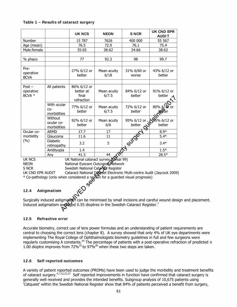
61
Table 1 – Results of cataract surgery UK NCS NEON S NCR UK CND EPR
AUDIT Number 15 787 7626 400 000 55 567 Age (mean) 76.5 72.9 76.1 75.4 Male:female 35:65 38:62 34:66 38:62 % phaco 77 92.3 98 99.7 Pre-operative BCVA
27% 6/12 or better
Mean acuity 6/18
31% 6/60 or worse
43% 6/12 or better
All patients 86% 6/12 or
better at final
refraction
Mean acuity 6/7.5
84% 6/12 or better
91% 6/12 or better
With ocular co-morbidities
77% 6/12 or better
Mean acuity 6/7.5
72% 6/12 or better
80% 6/12 or better
Post –operative BCVA *
Without ocular co-morbidities
92% 6/12 or better
Mean acuity 6/6
95% 6/12 or better
95% 6/12 or better
ARMD 17.7 17 8.9* Glaucoma 11.6 11 5.4* Diabetic retinopathy 3.2 5 3.4*
Amblyopia 1.4 1.5*
Ocular co-morbidity (%)
Any 41.3 44 28.5* UK NCS UK National cataract survey (Desai 99) NEON National Eyecare Outcomes Network S NCR Swedish National Cataract Register UK CND EPR AUDIT Cataract National Dataset Electronic Multi-centre Audit (Jaycock 2009) * Co-pathology (only when considered a reason for a guarded visual prognosis) 12.4 Astigmatism Surgically induced astigmatism can be minimised by small incisions and careful wound design and placement. Induced astigmatism averaged 0.55 dioptres in the Swedish Cataract Register.7 12.5 Refractive error Accurate biometry, correct use of lens power formulas and an understanding of patient requirements are central to choosing the correct lens (chapter 8). A survey showed that only 4% of UK eye departments were implementing The Royal College of Ophthalmologists biometry guidelines in full and few surgeons were regularly customising A constants.24 The percentage of patients with a post-operative refraction of predicted ± 1.00 dioptre improves from 72%25 to 97%26 when these two steps are taken. 12.6 Self reported outcomes A variety of patient reported outcomes (PROMs) have been used to judge the morbidity and treatment benefits of cataract surgery.4,7,12,23,27 Self reported improvements in function have confirmed that cataract surgery is generally well received and provides the intended benefits. Subgroup analysis of 10,675 patients using ‘Catquest’ within the Swedish National Register show that 84% of patients perceived a benefit from surgery,
ARCHIVED see N
ICE ca
tarac
ty su
rgery
guide
lines
2017
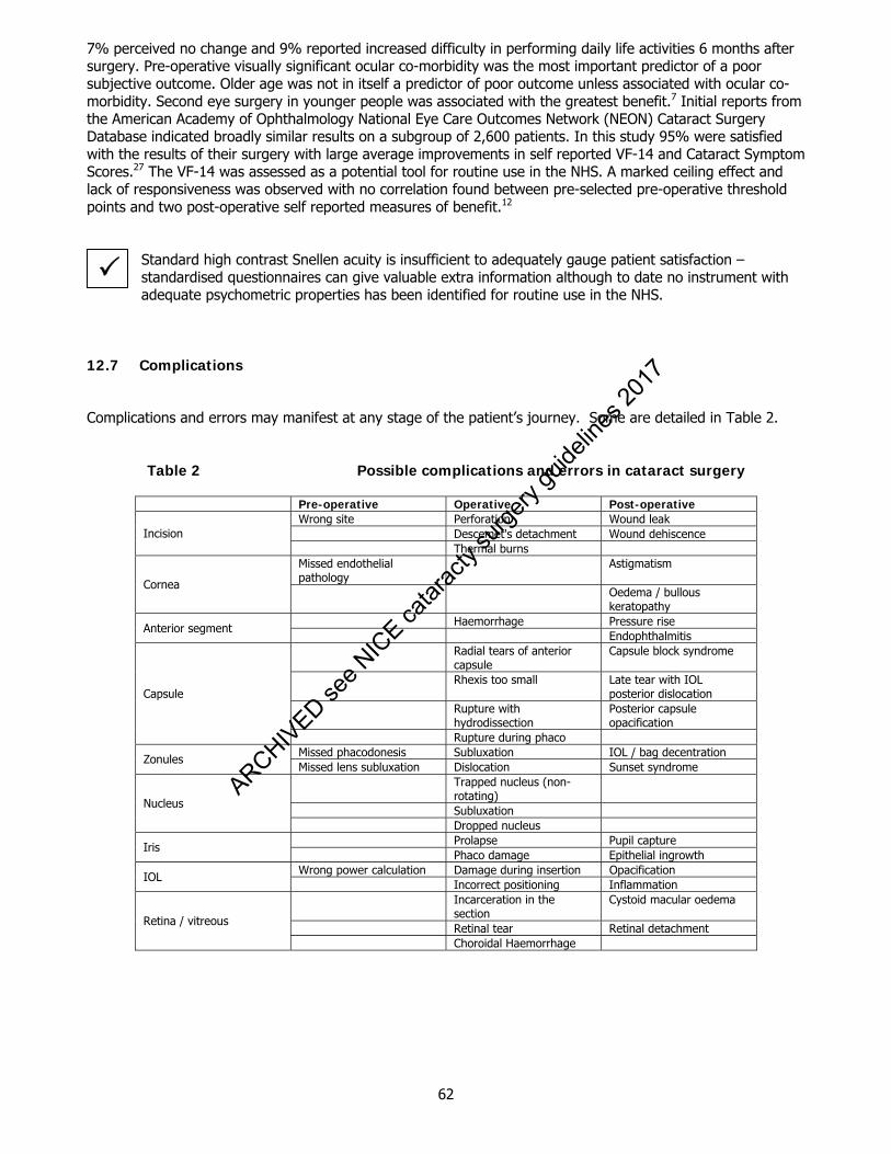
62
7% perceived no change and 9% reported increased difficulty in performing daily life activities 6 months after surgery. Pre-operative visually significant ocular co-morbidity was the most important predictor of a poor subjective outcome. Older age was not in itself a predictor of poor outcome unless associated with ocular co-morbidity. Second eye surgery in younger people was associated with the greatest benefit.7 Initial reports from the American Academy of Ophthalmology National Eye Care Outcomes Network (NEON) Cataract Surgery Database indicated broadly similar results on a subgroup of 2,600 patients. In this study 95% were satisfied with the results of their surgery with large average improvements in self reported VF-14 and Cataract Symptom Scores.27 The VF-14 was assessed as a potential tool for routine use in the NHS. A marked ceiling effect and lack of responsiveness was observed with no correlation found between pre-selected pre-operative threshold points and two post-operative self reported measures of benefit.12
Standard high contrast Snellen acuity is insufficient to adequately gauge patient satisfaction – standardised questionnaires can give valuable extra information although to date no instrument with adequate psychometric properties has been identified for routine use in the NHS.
12.7 Complications
Complications and errors may manifest at any stage of the patient’s journey. Some are detailed in Table 2.
Table 2 Possible complications and errors in cataract surgery
Pre-operative Operative Post-operative
Wrong site Perforation Wound leak Descemet's detachment Wound dehiscence Incision Thermal burns Missed endothelial pathology
Astigmatism
Cornea Oedema / bullous
keratopathy Haemorrhage Pressure rise Anterior segment Endophthalmitis Radial tears of anterior
capsule Capsule block syndrome
Rhexis too small Late tear with IOL posterior dislocation
Rupture with hydrodissection
Posterior capsule opacification
Capsule
Rupture during phaco Missed phacodonesis Subluxation IOL / bag decentration Zonules Missed lens subluxation Dislocation Sunset syndrome Trapped nucleus (non-
rotating)
Subluxation Nucleus
Dropped nucleus Prolapse Pupil capture Iris Phaco damage Epithelial ingrowth Wrong power calculation Damage during insertion Opacification IOL Incorrect positioning Inflammation Incarceration in the
section Cystoid macular oedema
Retinal tear Retinal detachment Retina / vitreous
Choroidal Haemorrhage
ARCHIVED see N
ICE ca
tarac
ty su
rgery
guide
lines
2017

63
Table 3 – Per and post operative adverse events (%)
UK NCS AAO PPP UK CND EPR
AUDIT 77% Phaco All surgery Phaco only 99.7% Phaco Endophthalmitis 0.03 0.13 0.74† NR Bullous keratopathy 0.3 0.3 NR
Clinical CME 1.4 2.3 1.6¥ Retinal detachment 0.7 0.93 NR
Wound gape / iris prolapse 0.25 0.6 0.2 0.16¥
Anterior chamber haemorrhage
0.5 0.5 0.4 0.05*
Hypopyon 0.02 0.2 2.0† NR Iris trauma 0.7 1.3 0.7 0.55* Zonular / posterior capsule rupture
4.4 3.1 1.8 1.92*
Vitreous loss 0.8 0.24 Vitreous haemorrhage 0.3
Choroidal haemorrhage 0.1 0.3 0.07*
Uveitis 5.6 1.8 3.1 3.29¥ Raised IOP (angle closure) 0.2 1.0
Raised IOP (open angle)
7.9 1.2 1.0 2.57¥
UK NCS UK National Cataract Survey (Desai 99) AAO PPP American Academy of Ophth Preferred Practice Pattern (AAO 2006) UK CND EPR AUDIT Cataract National Dataset Electronic Multi-centre Audit (Jaycock 2009)
NR: Not Reported as completeness of follow up uncertain for these cases * Reported per-operative ¥ Reported post-operative † Single Study 12.8 Capsule tear Tearing of the posterior lens capsule remains the most common adverse surgical event with the incidence during phacoemulsification having been reported as ranging from 0.7% to 16%28, being higher with less experienced surgeons.8 The visual results after capsule tear are not as good as uncomplicated surgery with 84% - 87% of those affected having been reported to achieve 6/12 or better29,30, poorer outcome mostly as a result of cystoid macular oedema.31 Risk factor analysis has confirmed that patient profile is crucially important in determining the probability of capsule rupture, with a recently published risk model indicating that for a consultant surgeon operating the predicted probability of this complication can vary by 100 fold, from 0.75% to 75% where numerous risk indicators coexist.8 In the current climate of publication of surgeon based outcome figures 15,16 it will be essential to develop robust methods of case mix adjustment to avoid inaccurate interpretations of such data. 12.9 Cystoid macular oedema The overall incidence of cystoid macular oedema remains around 1-2%.9,32 The rate is increased with many complications or pre-existing conditions including posterior capsule rupture, vitreous loss, iris incarceration, active uveitis, diabetes and previous retinal vein occlusion.33,34 After YAG laser capsulotomy it has been reported in 0.7% to 4.9% of eyes.35
ARCHIVED see N
ICE ca
tarac
ty su
rgery
guide
lines
2017

64
12.10 Endophthalmitis The 1998 Swedish National Prospective Survey reported on 58 cases in 54 666 cataract operations (0.1%.36 This equates with other series reported using modern microsurgical techniques.38 The incidence of presumed infectious ophthalmitis in the UK was recently estimated using the British Ophthalmological Surveillance Unit reporting card system. Here 213 baseline cases in one year were reported giving a corrected estimated incidence of 0.14%.38 A more recent report from the Swedish database covering the 3 year period from 2002-2004 found 109 cases in 225,471 operations (0.048%) with significant independent risk indicators being older age (>/= 85 years), capsule rupture and non-use of intra-cameral cefuroxime.5 12.11 Retinal detachment The incidence of retinal detachment after phacoemulsification cataract surgery ranges from 0% to 3.6% and averages 0.7% in the literature.39-41 The calculated excess risk of developing a retinal detachment after cataract surgery in the first 10 years over eyes without surgery is 5.5.42 A large case control study indicated excess risk of pseudophakic retinal detachment for posterior capsule tear (OR, 19.9; CI, 10.8-36.7; P<0.001), zonule dehiscence (OR, 12.4; CI, 3.8-41.2; P<0.001), retinal detachment in fellow eye (OR, 12.3; CI, 5.2-29.1; P<0.001), axial length >23 mm (OR, 3.2; CI, 2.0-5.0; P<0.001), and male gender (OR, 2.2; CI, 1.4-3.3; P<0.001). This result was however not reflected in a large case series of 2356 eyes with axial length >27 which found an incidence rate of between 1.5% and 2.2%.43 Approximately 37% (CI, 35%-38%) of retinal detachment was attributable to posterior capsule tear(42). For patients aged >64 years, the odds ratio was linearly reduced for each subsequent decade.42 A declining risk with increasing age has been observed elsewhere, with a rate for patients younger than 50 years of 5.17% and for those older than 70 years of 0.64%.40 Younger patients are more likely to require YAG capsulotomy and there may be confounding of these two effects, some studies having reported higher rates following capsulotomy.35,41 12.12 Outcomes and complications references 1. Powe NR, Schein OD, Gieser SC, Tielsch JM, Luthra R, Javitt J, Steinberg EP. Synthesis of the literature
on visual acuity and complications following cataract extraction with intraocular lens implantation. Cataract Patient Outcome Research Team. Arch Ophthalmol 1994;112:239-252.
2. Desai P, Reidy A, Minassian DC. Profile of patients presenting for cataract surgery in the UK: national
data collection. Br J Ophthalmol 1999;83:893-896. 3. Desai P, Minassian DC, Reidy A. National cataract surgery survey 1997-8: a report of the results of the
clinical outcomes. Br J Ophthalmol 1999;83:1336-1340. 4. Lundstrom M, Pesudovs K. Catquest-9SF patient outcomes questionnaire: nine-item short-form Rasch-
scaled revision of the Catquest questionnaire. J Cataract Refract Surg 2009;35:504-513. 5. Lundstrom M, Wejde G, Stenevi U, Thorburn W, Montan P. Endophthalmitis after cataract surgery: a
nationwide prospective study evaluating incidence in relation to incision type and location. Ophthalmology 2007;114:866-870.
6. Lundstrom M, Albrecht S, Hakansson I, Lorefors R, Ohlsson S, Polland W, Schmid A, Svensson G,
Wendel E. NIKE: a new clinical tool for establishing levels of indications for cataract surgery. Acta Ophthalmol Scand 2006;84:495-501.
7. Lundstrom M, Stenevi U, Thorburn W. The Swedish National Cataract Register: A 9-year review. Acta
Ophthalmol Scand 2002;80:248-257.
ARCHIVED see N
ICE ca
tarac
ty su
rgery
guide
lines
2017
65
8. Narendran N, Jaycock P, Johnston RL, Taylor H, Adams M, Tole DM, Asaria RH, Galloway P, Sparrow JM. The Cataract National Dataset electronic multicentre audit of 55,567 operations: risk stratification for posterior capsule rupture and vitreous loss. Eye 2009;23:31-37.
9. Jaycock P, Johnston RL, Taylor H, Adams M, Tole DM, Galloway P, Canning C, Sparrow JM. The
Cataract National Dataset electronic multi-centre audit of 55,567 operations: updating benchmark standards of care in the United Kingdom and internationally. Eye 2009;23:38-49.
10. El-Hindy N, Johnston RL, Jaycock P, Eke T, Braga AJ, Tole DM, Galloway P, Sparrow JM. The Cataract
National Dataset Electronic Multi-centre Audit of 55,567 operations: anaesthetic techniques and complications. Eye 2009;23:50-55.
11. Benzimra JD, Johnston RL, Jaycock P, Galloway PH, Lambert G, Chung AK, Eke T, Sparrow JM. The
Cataract National Dataset electronic multicentre audit of 55,567 operations: antiplatelet and anticoagulant medications. Eye 2009;23:10-16.
12. Black N, Browne J, van der Meulen J, Jamieson L, Copley L, Lewsey J. Is there overutilisation of
cataract surgery in England? Br J Ophthalmol 2009;93:13-17. 13. Lamoureux EL, Pesudovs K, Pallant JF, Rees G, Hassell JB, Caudle LE, Keeffe JE. An evaluation of the
10-item vision core measure 1 (VCM1) scale (the Core Module of the Vision-Related Quality of Life scale) using Rasch analysis. Ophthalmic Epidemiol 2008;15:224-233.
14. Keogh B, Spiegelhalter D, Bailey A, Roxburgh J, Magee P, Hilton C. The legacy of Bristol: public
disclosure of individual surgeons' results. BMJ 2004;329:450-454. 15. Keogh B. Outcome Measurement in Surgery. The case, the tools and the timing. The Royal College of
Surgeons of England Meeting 27th May, 2008 16. Johnston RL, Taylor H, Smith R, Sparrow JM. The Cataract National Dataset Electronic Multi-centre
Audit of 55,567 Operations: variation in posterior capsule rupture (PCR) rates between surgeons. Eye 2009;In press.
17. Keenan T, Rosen P, Yeates D, Goldacre M. Time trends and geographical variation in cataract surgery
rates in England: study of surgical workload. Br J Ophthalmol 2007;91:901-904. 18. Desai P. The National Cataract Surgery Survey: II. Clinical outcomes. Eye 1993;7:489-494. 19. Wilkins MR, Allan B, Rubin G, Iol Study Group M. Spectacle use after routine cataract surgery. Br J
Ophthalmol 2009: 93(10):1307-12. 20. HESonline. Main procedures and interventions: 2000-2008.
http://wwwhesonlinenhsuk/Ease/servlet/ContentServer?siteID=1937&categoryID=215. 2009. 21. Wong TY. Effect of increasing age on cataract surgery outcomes in very elderly patients. BMJ.
2001;322:1104-1106. 22. Lundstrom M, Stenevi U, Thorburn W. Quality of life after first- and second-eye cataract surgery: five-
year data collected by the Swedish National Cataract Register. J Cataract Refract Surg 2001;27:1553-1559.
23. Laidlaw DA, Harrad RA, Hopper CD, Whitaker A, Donovan JL, Brookes ST, Marsh GW, Peters TJ,
Sparrow JM, Frankel SJ. Randomised trial of effectiveness of second eye cataract surgery. Lancet 1998;352:925-929.
24. Gale RP, Saha N, Johnston RL. National biometry audit. Eye. 2004;18:63-66. 25. Murphy C, Tuft SJ, Minassian DC. Refractive error and visual outcome after cataract extraction. J
Cataract Refract Surg 2002;28:62-66.
ARCHIVED see N
ICE ca
tarac
ty su
rgery
guide
lines
2017
66
26. Percival SP, Vyas AV, Setty SS, Manvikar S. The influence of implant design on accuracy of
postoperative refraction. Eye 2002;16:309-315. 27. Lum F, Schein O, Schachat AP, Abbott RL, Hoskins HD, Jr., Steinberg EP. Initial two years of
experience with the AAO National Eyecare Outcomes Network (NEON) cataract surgery database. Ophthalmology 2000;107:691-697.
28. Vajpayee RB, Sharma N, Dada T, Gupta V, Kumar A, Dada VK. Management of posterior capsule tears.
Surv Ophthalmol 2001;45:473-488. 29. Ang GS, Whyte IF. Effect and outcomes of posterior capsule rupture in a district general hospital
setting. J Cataract Refract Surg 2006;32:623-627. 30. Chan FM, Mathur R, Ku JJ, Chen C, Chan SP, Yong VS, Au Eong KG. Short-term outcomes in eyes with
posterior capsule rupture during cataract surgery. J Cataract Refract Surg 2003;29:537-541. 31. Tan JH, Karwatowski WS. Phacoemulsification cataract surgery and unplanned anterior vitrectomy--is it
bad news? Eye 2002;16:117-120. 32. Schmier JK, Halpern MT, Covert DW, Matthews GP. Evaluation of costs for cystoid macular edema
among patients after cataract surgery. Retina 2007;27:621-628. 33. Henderson BA, Kim JY, Ament CS, Ferrufino-Ponce ZK, Grabowska A, Cremers SL. Clinical
pseudophakic cystoid macular edema. Risk factors for development and duration after treatment. J Cataract Refract Surg 2007;33:1550-1558.
34. Ray S, D'Amico DJ. Pseudophakic cystoid macular edema. Semin Ophthalmol 2002;17:167-180. 35. Aslam TM, Devlin H, Dhillon B. Use of Nd:YAG laser capsulotomy. Surv Ophthalmol 2003;48:594-612. 36. Montan P, Lundstrom M, Stenevi U, Thorburn W. Endophthalmitis following cataract surgery in
Sweden.The 1998 national prospective survey. Acta Ophthalmol Scand 2002;80:258-261. 37. Taban M, Behrens A, Newcomb RL, Nobe MY, Saedi G, Sweet PM, McDonnell PJ. Acute endophthalmitis
following cataract surgery: a systematic review of the literature. Arch Ophthalmol 2005;123:613-620. 38. Kamalarajah S, Silvestri G, Sharma N, Khan A, Foot B, Ling R, Cran G, Best R. Surveillance of
endophthalmitis following cataract surgery in the UK. Eye 2004;18:580-587. 39. Szijarto Z, Schvoller M, Poto L, Kuhn F, Kovacs B. Pseudophakic retinal detachment after
phacoemulsification. Ann Ophthalmol (Skokie) 2007;39:134-139. 40. Russell M, Gaskin B, Russell D, Polkinghorne PJ. Pseudophakic retinal detachment after
phacoemulsification cataract surgery: Ten-year retrospective review. J Cataract Refract Surg 2006;32:442-445.
41. Lois N, Wong D. Pseudophakic retinal detachment. Surv Ophthalmol 2003;48:467-487. 42. Tuft SJ, Minassian D, Sullivan P. Risk factors for retinal detachment after cataract surgery: a case-
control study. Ophthalmology 2006;113:650-656. 43. Neuhann IM, Neuhann TF, Heimann H, Schmickler S, Gerl RH, Foerster MH. Retinal detachment after
phacoemulsification in high myopia: analysis of 2356 cases. J Cataract Refract Surg 2008;34:1644-1657.
ARCHIVED see N
ICE ca
tarac
ty su
rgery
guide
lines
2017
67
13 Training for cataract surgery 13.1 Introduction It has been recognised in recent years that structured supervised training for all surgical specialities, and for ophthalmology in particular, is essential. The Royal College of Ophthalmologists has a microsurgical skills faculty, which delivers courses in microsurgical skills over a three day course. All trainees are required to attend the course within the first 4 months of their first substantive ophthalmology post in the UK. Evidence of participation in the whole course is required to progress to ST3 year of training. Exemption for experienced surgeons from abroad can be sought from the chairman of the Surgical Skills Faculty who with the supervising consultant ensures certain basic skills have been achieved. Courses for higher surgical trainees covering the seven sub-specialty areas have been introduced including intermediate and advanced cataract surgery courses. 13.2 The need for training There have been major changes in the way cataract surgery is delivered which have lead to unprecedented pressure to produce better results more quickly. These include: phacoemulsification being the preferred method of lens extraction earlier intervention increasing service pressure greater patient expectation an ageing population targets for maximum waiting lists Shortening the total training time
All these factors have increased demands on the service and highlighted the importance of training in order to prevent the exposure of patients to any additional risk. 13.3 Use of the wet lab Modern cataract surgery not only involves delicate micro-manipulation under a microscope but also requires knowledge of the phacoemulsification machine. Surgeons should be fully cognisant of the surgical techniques and the phaco-dynamics of the machine. Frequent use of a wet lab is probably the best way to get this experience. Various practice eyes are available and it is recommended that beginners should attend one of the many courses held nationally. The Royal College of Ophthalmologists runs regular courses on basic phacoemulsification in its skills centre. Time taken to learn how to use the phacoemulsifier in the wet lab will be repaid by faster and safer progress in the operating theatre.1 Live surgery should not be attempted until the surgeon is completely familiar with the machinery. Instruction and supervision by an experienced phaco-surgeon is essential and invaluable when transferring these skills to the operating theatre. In the early stages the component steps of the operation may be learnt separately before the surgeon completes the whole procedure. It is likely that in the future most hospitals will demand evidence of such structured training for any new surgical technique especially involving new apparatus. This should be seen as part of post-graduate development and evidence of such training or re-training kept in a personal revalidation folder. Surgical simulators are now available to teach some aspects of cataract surgery. One such simulator is available at the College and is used in the basic microsurgical skills course. It is also available to book for trainers and trainees to use on a more ad hoc basis. Ideally, each training region would have a simulator as well as a more traditional skills centre to allow the development of regional training modules and facilitate practise.
ARCHIVED see N
ICE ca
tarac
ty su
rgery
guide
lines
2017
68
13.4 Delivering the training Not every consultant wishes to be involved with training nor is every consultant a good trainer and regular structured training of junior doctors and even senior colleagues can be stressful. 13.5 Training 'contract' It is vital, therefore, to enter into a 'training contract' with the trainee before entering the operating theatre. This can be in the form of a verbal agreement about what is expected, what the trainee should undertake, when the trainer will take over and how much time is allowed for training in a particular circumstance. 13.6 Operating lists It is possible to organise operating lists into training lists and service lists or, as in some units, to allow a specific time (perhaps 40 minutes) during an operating list which is dedicated to structured training of junior doctors. This may be at the beginning of the list, or after a specified number of cases and everyone should be in agreement that the consultant or supervisor of the list will take over after the set time so that the list finishes on time; all the cases are done but everybody gets adequate exposure to surgery. It is the regular and frequent exposure to supervised training that will increase surgical speed, competence and confidence more than anything. 13.7 Local anaesthesia Although general anaesthesia is probably the ideal type of anaesthetic for training, local anaesthetic is now much more commonly administered for cataract surgery and techniques should be developed for allowing training to go on without alarming the patient or the trainee during the procedure.
Agreement about specific phraseology used during surgery and understanding of the patient's perception of what is said and heard should be discussed before surgery starts. The patient should be made aware that junior surgeons will be appropriately supervised so that they are only doing what they are capable of.
13.8 Training the trainers All trainers who regularly have responsibility for delivering training should consider attending a course on how to train. Training the Trainers courses are available at the College. Attendance at these courses facilitates the acquisition of teaching skills and learning theory. They demonstrate how to maximise the time available for teaching and training. 13.9 Auditing the training A continuous audit of cataract outcomes is important. Ideally electronic systems should be available to allow the routine collection of data. Analysis of this data allows risk stratification of cases by complexity and by seniority of surgeon.2
Complication rates should be monitored for each individual surgeon. A good way of performing continuous audit is to video every case especially for the trainees so that specific points can be reviewed and discussed. Time needs to be set aside for this to be done in a structured way.
13.10 Non technical training A number of non-technical skills are recognised as an essential component for surgical training, subjects such as communication, team work, decision making and situation awareness (cognitive skills) are all important. The following website may be useful in this respect www.abdn.ac.uk/iprc/notss
ARCHIVED see N
ICE ca
tarac
ty su
rgery
guide
lines
2017
69
13.11 Summary Regular, frequent supervised training for cataract surgery involves teamwork and discussion before the operating theatre is even entered. Outlining what the trainee can expect before the surgery starts is a good way of relieving the pressure on the trainer whilst the list is proceeding and, with the additional use of a wet lab out of the theatre environment, training time should be shortened considerably. 13.12 Summary points There should be a commitment to both the culture and practice of training Regular use of a 'wet lab' is beneficial as is the use of simulators. Trainees must be supervised by an experienced surgeon Training should be structured (i.e. modular) and planned Patients should be aware that a trainee may be operating upon them but also be reassured that a trainee
will not be allowed to operate unless they are safe Complications may still occur but will be less likely as trainees will have a basic set of skills and knowledge
and will be supervised as appropriate. 13.13 References for training for cataract surgery 1. Ezra DG, Aggarwal R, Michaelides M, Okhravi N, Verma S, Benjamin L, Bloom P, Darzi A, Sullivan P. Skills
acquisition and assessment after a microsurgical skills course for ophthalmology residents. Ophthalmology 2009;116(2):257-62. Epub 2008 Dec 16.
2. Narendran N, Jaycock P, Johnston RL, Taylor H, Adams M, Tole DM, Asaria RH, Galloway P, Sparrow JM.
The Cataract National Dataset electronic multicentre audit of 55,567 operations: risk stratification for posterior capsule rupture and vitreous loss. Eye 2009;23(1):31-7. Epub 2008 Mar 7.
ARCHIVED see N
ICE ca
tarac
ty su
rgery
guide
lines
2017
70
14. Patient Safety and Clinical Risk in Cataract Surgery Quality and safety of patient care are intimately intertwined with clinical and organisational structures, clinical governance and good clinical management. Good planning and then doing the correct thing for the correct patient in the correct setting as well as learning from those occasions when incorrect care has occurred is a pragmatic lens through which to view these complex interrelated concepts. Quality and safety are intuitively recognised when present and glaringly obvious when absent. Patient safety and quality of care thus constitute the foundations of care and of service provision. The College has provided advice on patient safety in ophthalmology.12 Strict attention to detail, risk assessment and careful consideration of patient pathways is required for safe cataract care. However clinical errors, near misses, expected and unexpected surgical complications and events will happen. Such events may provide an opportunity for learning to reduce risk of similar events occurring again and occurring elsewhere. It is estimated that approximately 10% of healthcare episodes and interventions are compromised in some way by clinical error and 50% of which are preventable. It is thus argued that 10% of resources should be allocated to patient safety or quality matters. Investment in appropriate staffing levels, team training, appropriate equipment and development of a safety culture with patient involvement are key elements to modern safe cataract surgical care. With the recent and welcome reduction in waiting times for cataract care and excellent progress towards the ‘18 weeks referral to treatment time’ target being achieved in NHS care, emphasis has recently shifted to a continuation of improvement in the quality and safety of patient care as well as to maintaining the sustainability of such short waiting times. Analysis of these topics in the light of the extra investment in healthcare in the UK and of NHS reforms in England since 1997 is topical. The ‘NHS Next Stage Review ’- lead by Lord Darzi suggested that quality and safety should shape the next stage of policy and progress for the NHS in England.3 14.1 Patient Safety and Clinical Risk Risk management in healthcare has long been seen as the domain of reducing litigation and for the defence of healthcare and other organisations. A more enlightened view is that clinical risk management that focuses on patient safety will enhance the quality of care while reducing the health economic burden of patient harm. In the UK the National Patient Safety Agency (NPSA) (www.npsa.nhs.uk) has been initiated in a response to a growing international awareness about errors in healthcare and a resultant focus on improving patient safety. A patient safety incident can be defined as any unintended or unexpected incident which could have or did lead to harm for one or more patients. This is also referred to as an adverse event/incident or clinical error, and includes near misses. Medical errors may be regarded as adverse events or near misses that are preventable within the current state of medical knowledge. Patient safety has been defined by the NPSA as ‘the process by which an organisation makes patient care safer. This should involve: risk assessment; the identification and management of patient-related risks; the reporting and analysis of incidents; and the capacity to learn from and follow-up on incidents and implement solutions to minimise the risk of them recurring.’ All clinical interventions carry some risk. While there may be occasional poorly performing staff, most clinical errors are committed by well trained, well motivated individuals. Variability in surgical outcome has been attributed to the interplay of multiple factors including; surgical ability, surgical technique, case mix, case volume, institutional systems influences, peri-operative care and anaesthetic care. Improving safety of surgical care is a multi-faceted task and requires multi-disciplinary and organisational commitment and leadership. While all patient safety incidents result from clinical management, not all are preventable (i.e. not all are wholly attributable to known error). For example, a patient having cataract surgery who suffers from postoperative endophthalmitis has had a serious patient safety incident. Root cause analysis of the case history, peri-operative events, staffing issues, facilities, results of microbiology investigations etc, may clarify if it was a potentially preventable adverse incident (such as a sterilisation equipment failure or failure to use appropriate chemo-prophylaxis), or not. It can be helpful to consider patient safety issues by both underling causational risk factors and or subsequent consequence(s).
ARCHIVED see N
ICE ca
tarac
ty su
rgery
guide
lines
2017
71
14.2 Causational risk factors for unsafe cataract surgery Consideration should be given to the following root or underlying causes for un-safe cataract surgery.
• Clinical staff not relying on evidence-based medicine (e.g. not using povidone iodine pre-operative prophylaxis despite the evidence base)
• Staff not following clinical practice guidelines (e.g. not following biometry guidelines) • Lack of risk assessment. Such as for new cataract surgical care commissioning plans and for new
technology and devices and for off label use of medications • Failure to collect cataract patient outcomes • Insufficient continuous professional development of cataract surgeons and healthcare personnel • Lack of team working and or of team training of the cataract surgical team • Inappropriate staffing levels with appropriate skills and lack of effective clinical leadership • Lack of continuity of clinical care • Poor infrastructure and lack of investment in appropriate cataract surgical facilities and dedicated day
care area • Failure of timely and appropriate management of surgical complications, including taking early second
opinions and referral to tertiary centres • Last minute changes; such as late changes to operating lists or un-expected admissions • Interruptions and distractions during surgery • Rushing, focus on targets and performance rather than on quality and safety • Inappropriate or inadequate supervision of trainees. This includes inappropriate case selection for or by
trainees • Lack of patient involvement and insufficient attention to patient complaints
14.2.1 Communication problems
• Poor handwriting in case notes. This can be an issue, for example, with IOL power selection errors where similar appearing numbers when handwritten can be mistaken, e.g. mixing up 11 and 17
• Language problems, this is relevant when patients first language is other than English or in individuals
with learning difficulties or with dual sensory impairments • Communication problems are a potential further risk if the surgeon has not met with the patient prior to
surgery • Risk of surgery and required pre and postoperative care plans not adequately communicated to or
understood by patient • Organisational communication issues, such as not using protocols, printed patient information sheets and
integrated patient care pathways
ARCHIVED see N
ICE ca
tarac
ty su
rgery
guide
lines
2017
72
14.2.2 Equipment problems
• Failure or non-availability of equipment. Both regular servicing and pre-operative checking of availability equipment required for cataract surgery is good practice. Consider the additional equipment that might be required for intra-operative complications or surgical surprises e.g. ensure anterior vitrectomy equipment is available and working if required.
• Lack of familiarity of staff with equipment, especially with new equipment and or software upgrades on
existing equipment. Incidents due to incorrect use of Luer lock cannulae or failure to use Luer lock cannulae in cataract surgery continue to occur.
14.2.3 Staff orientation
• Considerable caution should be taken by staff working in unfamiliar surgical environments and with unfamiliar cataract surgical equipment. This is particularly relevant to para-medical and nursing agency and ‘bank’ staff and medical locums and use of visiting -including from overseas- clinical teams to undertake additional surgical activity.
• Use of a checklist to document; correct patient and site, informed consent, and any preoperative and
postoperative instructions given to the patient is of merit to teamwork and safety. Suggested pre-operative checks list for cataract surgery based on the World Health Organisation’s Safe Surgery Saves Life and NPSA generic pre-operative checklist appears as an Appendix D. Taking ‘time out’ prior to commencement of surgery is recommended and as is a team brief at the start of the surgical session.
14.3 Consequential patient safety incidents in cataract care 14.3.1 Medication errors in cataract surgery These errors include
• Infusion fluid issues, additives to intra ocular infusions; mix-up of concentrations and dilutions. This is especially relevant where dilutions for intraocular injection are prepared in the theatre such as when homemade dilutions of intracameral cefuroxime are prepared for intracameral injection.
• Errors with medications intended for topical use only injected into eye. • Retinal or endothelial toxicity from incorrect dose of drug, or correct dose injected into wrong
compartment of eye, e.g. dose of antibiotic prepared for planned sub-conjunctival injection given instead by intracameral route.
• Wrong pre-operative or post operative eye drops prescribed or dispensed or instilled or correct
medication not provided. • Allergy; patient known allergy ignored or not-requested
14.3.2 Healthcare acquired infections are regarded as a patient safety matter. In relation to cataract care, post-operative endophthalmitis is the most feared outcome. Timely and evidence based treatment of both the individual patient with suspected endophthalmitis and the rigorous investigation of an outbreak are needed. Advice from the College on the consideration of endophthalmitis clusters is available.4 14.3.3 Wrong Site Surgery This includes surgery on the wrong patient, on the wrong eye in correct patient - also known as wrong side surgery, or the wrong operation on the correct eye. The surgeon is ultimately responsible for assuring that the correct operation is carried out on the correct patient’s correct eye. The College recommends that the eye to be operated upon should be marked pre-operatively and with the patient’s agreement while awake and prior to pre-medication. The NPSA has provided an Alert on Correct Site Surgery.5
ARCHIVED see N
ICE ca
tarac
ty su
rgery
guide
lines
2017
73
14.3.4 Wrong intraocular lens implant placement
• The wrong dioptre power, wrong size or wrong type of IOL can be inserted. The cataract team is responsible for assuring that the correct IOL and based on the correct biometry measurement and formula is inserted at the time of cataract surgery. Inaccurate biometry and incorrect selection of IOL are common causes of unplanned postoperative refractive error surprises and can lead to patient dissatisfaction and litigation.
• Ensure staff undertaking biometry are appropriately trained and equipment is appropriately calibrated as per manufacturers’ recommendations.
• Use the appropriate formula for IOL selection. Optimise biometry and customise ‘A constants’ based on personal audits of refractive outcomes. Optimised surgeon and IOL specific ‘A constants’ are desirable. Be aware of A constant differences with optical versus ultrasonic biometry measurements (See chapter on biometry).
• Order ‘special delivery’ IOLs in good time and ensure that they have been received prior to patient admission.
• Beware of relying on theatre whiteboards or pre-printed operating lists for IOL selection. Choose the correct IOL from original biometry print out and consider circling or highlighting the IOL power desired. Consider writing out IOL power in full to avoid confusion with similar appearing handwritten numbers. If a different IOL is implanted than was anticipated, the reason for choosing that IOL and how so determined should be documented and should be a part of the ‘time out’ prior to surgery
14.3.5 Failure of implanted devices o IOL opacification o IOL dislocation o Poor IOL quality or positioning (leading to uveitis–glaucoma–hyphema syndrome) 14.3.6 Anaesthesia hazards These include ocular perforation from local injections. Swift access to resuscitation facilities and arrangements for rapid transfer to high dependency or intensive care facilities are precautions that should be considered in advance of potentially foreseeable problems and by both providers and commissioners of cataract care. Stand alone cataract treatment centers need to have robust pre-planned arrangements in this regard. 14.4 Safeguards Reflection on the above will allow for insight into safeguards and measures required to overcome these un-safe situations. Addition methods of learning from events, performance monitoring and systems improvements should also be deployed. 14.4.1 Clinical Governance Having regular peer review and multi-disciplinary ophthalmic clinical team meetings where patient safety incidents are discussed is of merit. Evidence of such endeavours might also form a part of the Appraisal and Revalidation of cataract surgeons and the accreditation of clinical services. 14.4.2 Incident Reporting Where patient safety incidents occur local risk management reporting procedures must be used. These will include documentation of the incident in the case records and on the hospital’s clinical incident reporting forms. Patients should be informed without delay about any incidents that affected them. More detail on patient safety incident reporting and who to report to is provided in the College’s ‘Patient Safety in Ophthalmology Guidance’ (2008).1
ARCHIVED see N
ICE ca
tarac
ty su
rgery
guide
lines
2017
74
14.5 ‘Critical’ patient safety incidents in cataract care Incidents that lead (or may have lead to) to permanent harm or death in cataract care should be especially examined. Such serious adverse events from interventions that lead to significant harm or lasting disability, such as loss of sight, or are a cause for concern by staff, or patients, may be regarded as ‘critical incidents’. They may be preventable by a change of practice and are thus worthy of further investigation of root causes. Patient safety incidents in cataract care, regarded by the College as critical, are shown in Table 1. This list is intended to be a practical aid and is neither exhaustive nor exclusive. Such incidents in cataract care should be reported via incident reporting systems and should be reviewed at team meetings. Multi-professional reporting is to be encouraged. Near misses have the potential to provide learning where patients have not been harmed. Table 1 Suggested critical patient safety incidents; cataract surgical care Operation on the wrong eye. Wrong operation on correct eye. Missing case notes at surgery. Elective surgery should usually be cancelled. Penetration or perforation of globe during periocular injection. Expulsive haemorrhage during cataract surgery. Endophthalmitis following cataract surgery. Patient collapse in peri-operative period. Death. ‘Open’ category for adverse incidents causing concern among cataract care staff or patients for whatever reason. Whether these patient safety incidents require further analysis is a matter for local organisations. Examples pertinent to cataract surgery might include;
IOL miscalculation/wrong power implanted.
Dropped nucleus/fragment.
Requirement for IOL explantation.
Corneal decompensation within 3 months of surgery.
Inadequate or unsafe staffing levels.
Unplanned re-admission or return to theatre within 28 days of cataract surgery for treatment of the same eye.
14.5.1 When complications occur
• Accurate documentation of any intra-operative or postoperative complications is required. Poor
documentation suggests complication was poorly recognised and managed. • Consider surgeon’s response to any recognized complications, both the additional clinical steps taken
and how later communicated with patient and carers. Be ready to apologise, explain and sympathise. Document such discussions.
• The frequency of post-operative visits should usually be increased if complications occurred during surgery or postoperatively. It is best to arrange to see patient on first day following any significant intra-operative complication.
• Be prepared to take a second opinion early if complications occur including early referral to tertiary centre if required. It can often be helpful to discuss clinical complications early with colleagues, including telephone consultations. Document such conversations as appropriate in the case records.
14.5.2 Leadership in Patient Safety The College considers that further improvements in clinical quality and patient safety are more likely to come about through strong clinical involvement in the planning of day to day care and in training than from further legislation and central regulation. Technical solutions to improved equipment and novel medications will come from clinical research. Senior doctors who shape a culture of clinical quality improvement and patient safety by personal example have a powerful and lasting effect on the members of their clinical teams and, via their training activity, on the next generation.
ARCHIVED see N
ICE ca
tarac
ty su
rgery
guide
lines
2017
75
As governance and patient safety within some areas of cataract care has been problematic, the College encourages ophthalmologists to highlight concerns to the College, while also complying with statutory and regulatory requirements, so that lessons can be shared and may be brought to wider attention, if required.6 Careful consideration of patient pathways –including failure mode analysis- and technology advances are of merit in risk reduction. However clinical practice never has, and never will be, ‘risk free’. Industries such as rail or air transport have shown that despite technical improvements and ‘lessons learned’ there are still risks either apparent and unsolvable or latent. Furthermore as much of the NHS is running at full capacity -with wards and clinics full to overflowing- there is little precaution or safety headroom to cope with surges in demand or variations in capacity (e.g. staff shortages or sickness). Adequate headroom and back up is a key precautionary principle of safe cataract surgery. 14.6 Conclusion The Charter of The Royal College of Ophthalmologists states that the College should ‘maintain proper standards in the practice of ophthalmology for the benefit of the public.’ Accordingly the College places great emphasis on patient safety and best clinical practice as educational features and competencies for ophthalmologists and recognises both as core features of good ophthalmic service provision. Strict attention to detail, a focus on safety and learning from adverse events and near misses enhances cataract care. The College is committed to supporting steps that improve the safety of both cataract and ophthalmic care at both individual and organisational levels.1 14.7 Patient Safety and Clinical Risk in Cataract Surgery References
1. The Royal College of Ophthalmologists. Patient Safety in Ophthalmology; Ophthalmic Services Guidance. London 2008. Available at http://www.rcophth.ac.uk/standards/ophthalmicservices
2. Kelly SP. Guidance on patient safety in ophthalmology from the Royal College of Ophthalmologists.
Eye 2009; 23: 2143–2151. 3. Darzi A. High Quality Care For All. NHS Next Stage Review, Final Report. Department of Health. The
Stationary Office. London: 2008 www.ournhs.nhs.uk 4. The Royal College of Ophthalmologists. Investigating an outbreak of endophthalmitis. London 2007.
Available at http://www.rcophth.ac.uk/standards/ophthalmicservices 5. National Patient Safety Agency. Patient safety alert 06. Correct site surgery. http://www.npsa.nhs.uk 6. Kelly SP, Astbury NJ. Patient safety in cataract surgery; a review. Eye 2006; 20:275-82.
ARCHIVED see N
ICE ca
tarac
ty su
rgery
guide
lines
2017
76
15 Commissioning Cataract Surgery – an Outline of Good Practice 15.1 Background The NHS Plan1 set out a ten year strategy aimed at building a more responsive and more patient-centred NHS with more uniform standards of care and access. Central to this strategy was a staged reduction in waiting times for elective treatment, culminating in a maximum waiting time from referral to definitive treatment 18 weeks, and a guaranteed choice of providers, including independent sector providers. It was recognised from an early stage that, to achieve these goals, an increase in capacity for cataract surgery in the UK would be required. The Action on Cataract project (Department of Health 1999) was an initiative to increase throughput on ophthalmic surgical lists with existing premises and staff, principally by addressing needs for equipment or modifications to infrastructure. As a result of this and other initiatives to streamline the cataract surgical pathway, many units achieved substantial improvements in their cataract surgical throughput. A further Government paper2 paved the way for a rapid increase in elective surgical capacity by awarding contracts to companies employing overseas clinical teams to perform elective surgery within NHS premises (usually at weekends), in mobile operating theatres or fixed treatment centres. The first phase of the independent sector treatment centre (ISTC) programme encountered a number of problems. Some contracts experienced higher than expected rates of complications and there were reports of issues relating to continuity of care in contracts which used mobile surgical teams. The programme proved to be very expensive because the contracts guaranteed income regardless of volume and the uptake was lower than anticipated. It is likely that demand for cataract surgery was overestimated and in the event, about 97% of cataract surgery continued to be performed within the NHS during phase 1 of the ISTC contract. Phase 2 of the ISTC programme did not include any further mobile cataract treatment centres but a number of new fixed ISTCs were built or commissioned, some of which undertake cataract surgery. Controversially, some treatment centres were built close to, or even within existing NHS facilities, creating excess surgical capacity, sometimes to the detriment of NHS ophthalmic units locally. During Phase 1 of the ISTC programme, there was a strict rule that ISTCs must recruit staff from outside the NHS (the “additionality rule”). This was subsequently relaxed and it is now possible for NHS clinicians to work in or be seconded to some ISTCs. At the time of writing, the UK economy is in recession and it is probable that the funding for the NHS will at best be static for a period of several years while demand for healthcare in general is likely to continue to rise. Although it is unlikely that either the government or the public will easily allow a return to the long waiting times of the 1990s, commissioners will come under increasing pressure to obtain the best value possible from contracts with providers. Current health policy continues to encourage a plurality of providers for elective surgical care, including non-NHS providers. The College supports the concept that patients should be able to make an informed choice as to where they wish to be referred for cataract surgery. However, during phase 1 of the ISTC contracts the College received a number of reports of cases where the care of patients already on the waiting list of a local consultant ophthalmologist had been transferred to an alternative provider without the knowledge of the local consultant, and sometimes without the prior knowledge of the patient. In some cases, patient care was compromised either because of inappropriate selection of cases for transfer, or because of inadequate arrangements for post-operative follow-up (especially where complications occurred). 15.2 Purpose of this document In times of economic stringency, primary care trusts and GP commissioning groups will wish to ensure that they obtain best value for money from contracts for cataract surgery, while maintaining high standards of safety for patients. The primary purpose of this document is not to deter commissioners from placing contracts for cataract surgery with providers other than their local NHS eye unit, but rather to provide guidance on the standards which should be expected of any provider of ophthalmic care, whether NHS, ISTCs or elsewhere, based on lessons learned from the last decade.
ARCHIVED see N
ICE ca
tarac
ty su
rgery
guide
lines
2017
77
15.3 Potential problems of “outsourcing” cataract surgery 15.3.1 Problems for the patient Travel to a more distant provider may be difficult, and adequate follow up care may difficult to arrange.
This can be especially problematic when a service provided by a mobile unit has moved to another location.
There may be little or no opportunity for the patient to discuss their operation with the ophthalmologist who will undertake the surgery before the day of surgery, and the patient may feel an obligation to proceed if they feel that special arrangements have been made for their treatment.
15.3.2 Problems for the provider It may be difficult to obtain relevant information about the patient’s previous history (e.g. adverse
reactions to previous treatment, ongoing medical or ophthalmic problems), leading to unexpected difficulties with surgery.
If complications occur, it may be difficult to provide treatment or adequate follow up if the patient lives some distance away.
15.3.3 Problems for the local eye unit
If a significant proportion of cataract surgery is ”cherry-picked” by alternative providers, the workload of
the local NHS unit may be distorted, with potential adverse effects on training, skill-retention, recruitment and retention of staff
The local NHS unit may have to make urgent arrangements to deal with postoperative complications from surgery performed by an alternative provider, without adequate background information. This poses a threat to patient safety.
15.3.4 Problems for the commissioner
The commissioner has a duty of care to the patient when placing a contract for their surgery and shares
legal liability with the provider for failings in the standard of care, particularly if monitoring of the contract is shown to be inadequate, or if the provider is based outside British legal jurisdiction.
It may be difficult to obtain or validate information on standards of care and outcomes from providers prior to placing a contract.
15.4 Recommendations
1. Communication. The provider should inform the patient’s general practitioner at the points when the patient is listed for surgery, when the surgery occurs and when the patient is discharged. Where a patient is under the ongoing care of another ophthalmologist, he/she should also be copied into the correspondence. The provider is responsible for arranging handover of care to another ophthalmologist where this is necessary (e.g. for management of a complication or where ongoing monitoring of another eye condition is required). Where an NHS commissioning body arranges transfer of patients already on the waiting list of an NHS consultant ophthalmologist to an alternative provider, the commissioning body is responsible for obtaining the consent of the patient, and should inform the consultant from whose list the patient is being removed, and the patient’s general practitioner (L Donaldson, personal communication, 14 November 2001).
2. Case selection and preoperative preparation. The provider must perform a detailed preoperative assessment to ensure that case selection is appropriate to the level of expertise of the operating team and the clinical facilities. In particular, it is vital to take adequate account of ocular or systemic co-morbidity which might increase the technical difficulty of the procedure, or increase the risk of complications. The provider should also ensure that adequate account is taken of the patient’s social circumstances (availability of transport, help at home etc) when planning the episode of care.
ARCHIVED see N
ICE ca
tarac
ty su
rgery
guide
lines
2017
78
3. Patient information and consent. The provider is responsible for providing adequate verbal and / or written information about cataract and cataract surgery to allow the patient to give informed consent to the procedure. Informed consent must be taken by someone who has the knowledge and competence to explain the benefits and risks of the procedure and to provide accurate answers to questions. Although NHS patients do not have a right to choose their surgeon, they have a right to expect that their surgeon has the experience and skill to perform their operation. It is reasonable that the patient should have the opportunity to know the identity and status of the operating surgeon and to meet him / her prior to entering the operating theatre. It is the final responsibility of the operating surgeon (or the supervising surgeon where the operating surgeon is a trainee) to ensure that the patient has been adequately assessed, prepared and consented prior to the start of the operation.
4. Hotel Facilities. The commissioning agency and the provider have a joint responsibility for ensuring that the adequate facilities are available for the patient to be accommodated for the duration of the episode of care. This is particularly important where the provider unit is too distant from the patient’s home to allow a return journey in the same day.
5. Clinical facilities. The commissioning agency and the provider have a joint responsibility to ensure that the premises and equipment in the provider unit is adequate for performing modern small incision cataract surgery safely, and that the unit complies with relevant legislation.
6. Anaesthesia and perioperative care. Most cataract surgery is carried out under local anaesthesia, and has a very low mortality and systemic morbidity, especially considering that a high proportion of patients are elderly and would be graded as 2 or worse on the American Society of Anaesthetists (ASA) scoring system. However, the provider has a responsibility to ensure that resuscitation facilities are readily available, and that an appropriately qualified person is readily available to undertake resuscitation should the need arise. Contingency plans should be in place for emergency transfer of patients who suffer a life-threatening complication. The National Confidential Enquiry into Perioperative Deaths (NCEPOD) has criticized the practice of undertaking ophthalmic surgical procedures on very unfit patients in isolated units. Provider units which are geographically isolated from accident and emergency or intensive care facilities should give particular consideration to contingency planning for life-threatening emergencies and to case selection. Arrangements with a local NHS provider will usually be needed and must be agreed in advance
7. Postoperative care and contingency planning for complications. The provider is responsible for arranging routine postoperative care following cataract surgery, in order to monitor for postoperative complications and for the collection of information on outcomes. The patient must be provided with any necessary postoperative medication and instructions, and a discharge summary. The provider unit must have adequate arrangements for handling urgent enquiries from patients who have had surgery. It is not acceptable for patients merely to be told to go to their local accident and emergency department or to contact their GP if they have a problem. If operative or postoperative complications occur, the provider unit should either manage them, or arrange direct referral to another specialist, keeping the general practitioner informed. The commissioning agency should ensure that there is a funded agreement in place with a suitably equipped NHS facility with adequate capacity for dealing with any early or late post-operative complications which cannot be managed by the provider.
8. Clinical Governance. The commissioning agency should ensure that the provider unit follows the requirements of Clinical Governance, whether the provider is within the NHS or the private sector. In particular, medical staff should be suitably qualified and experienced, and should undergo annual peer appraisal, there should be evidence of ongoing audit of outcomes and complications in relation to national comparators, there should be a robust mechanism for recording and acting on complaints and clinical incidents, and there should be facilities for monitoring the progress of staff in training.
ARCHIVED see N
ICE ca
tarac
ty su
rgery
guide
lines
2017

79
15.5 Commissioning Cataract Surgery References: 1. The NHS Plan, a plan for investment, a plan for reform. Department of Health, July 2000. 2. Growing Capacity: a new role for external healthcare providers in England. Department of Health,
June 2002.
ARCHIVED see N
ICE ca
tarac
ty su
rgery
guide
lines
2017
80
16 Working Party Members Chair: Mr Larry Benjamin, Consultant Ophthalmic Surgeon, Stoke Mandeville Hospital, Aylesbury, Buckinghamshire Members: Mr David Allen, Consultant Ophthalmic Surgeon, Sunderland Eye Infirmary, Sunderland Ms Parul Desai, Consultant Ophthalmic Surgeon, Moorfields Eye Hospital, London Mr Bruce James, Consultant Ophthalmic Surgeon, Stoke Mandeville Hospital, Aylesbury, Buckinghamshire Mr Robert Johnston, Consultant Ophthalmic Surgeon, Gloucester Royal Hospital, Gloucester Mr Simon Kelly, Consultant Ophthalmic Surgeon, Royal Bolton Hospital NHS Foundation Trust, Bolton Mr Graham Kyle, Consultant Ophthalmic Surgeon, Liverpool Mr Ken Nischal, Consultant Ophthalmic Surgeon, The Hospital for Sick Children, Great Ormond Street, London Mr Paul Rosen, Consultant Ophthalmic Surgeon, Oxford Eye Hospital, Oxford Mr Richard Smith, Consultant Ophthalmic Surgeon, Stoke Mandeville Hospital, Aylesbury, Buckinghamshire Mr John Sparrow, Consultant Ophthalmic Surgeon, Bristol Eye Hospital, Bristol The Chair of this group is a medical advisor to Alcon in the UK. All other members of the working party have made the Chair aware of their commercial relationships. 17 Acknowledgements and special thanks
Lay members who reviewed the document The American Academy of Ophthalmology for the use of Table 1 in chapter 5
18 Review of these guidelines The review of these guidelines will take place in 2015, with a view to make minor amendments as needed.
ARCHIVED see N
ICE ca
tarac
ty su
rgery
guide
lines
2017

81
List of Appendices
Appendix A - Example of consent form for cataract surgery Appendix B - Information for Patients
Appendix C - Cataract National Dataset V1.2 – Royal College of Ophthalmologists
Appendix D - Surgical Safety Cataract Checklist
Appendix E - Ocular Pharmacology
Appendix F - Consent Form 4
ARCHIVED see N
ICE ca
tarac
ty su
rgery
guide
lines
2017

82
Request / consent for left / right cataract extraction with lens implant under local anaesthetic general anaesthetic Statement of health professional (only complete if you have appropriate knowledge of this procedure as specified in consent policy). I have explained the procedure to the patient. In particular I have explained:
Signature of Health professional…………………………………………………… Job Title Printed Name……………………………………………………………………………… Date Statement of interpreter (where appropriate). I have interpreted the information above to the best of my ability and in a way in which the patient can understand. Interpreter's signature Print Name Date
Appendix A Insert Addressograph here
The intended benefits of the operation The main aim of the cataract operation is to improve the quality of your vision; it may also be of benefit to improve the doctors’ view of the back of the eye. We will try to reduce your dependence on spectacles as much as possible, but you may require distance glasses for best vision and you will probably need reading glasses; in any case your glasses prescription will change after the operation.
Serious or frequently occurring risks during the operation It is possible for a cataract operation to leave you worse off than you are now. One person in every 1000 will go blind in that eye as a direct result of the operation. One in 10,000 will lose the eye. There is virtually no risk to the other eye. Details on the most common specific complications are given below.
Ecchymosis - Bruising of eye or eyelids (quite common).
Posterior capsule rupture and / or vitreous loss - a split in the thin back wall of the cataract which can allow communication between front and back compartments of the eye.
Post -operative raised intraocular pressure - raised pressure in the eye for the first day or so (common). This may require temporary treatment.
Posterior capsular opacification - clouding of the membrane behind the implant causing blurred vision.
Cystoid macular oedema - inflammatory fluid in the centre of the retina. This is commonly mild and needs no treatment. It can be severe and require prolonged treatment.
Refractive surprise - unexpectedly large (or different from expected) need for glasses.
Allergy - to drops given after the operation, causing an itchy swollen eye until the drops are stopped or changed.
Dropped nucleus - part or all of the cataract falls through a posterior capsule rupture into the back part of the eye, needing another operation to remove it.
Suprachoroidal haemorrhage - bleeding inside the eye which may require the operation to be completed on another day.
Corneal decompensation - clouding of the normally clear front window of the eye.
Detached retina - peeling off of the seeing layer of cells within the eye.
Endophthalmitis - severe (usually painful) infection inside the eye.
Dislocation of the implant – movement out of position of the lens implant.
Complications are rare and in most cases can be treated effectively. In a small proportion of cases, a further operation may be required. If you decide against a cataract operation, your vision will probably slowly worsen. If you need to discuss your options further, or at a later date, please contact (preferably in writing) the person whose details are
ARCHIVED see N
ICE ca
tarac
ty su
rgery
guide
lines
2017

83
Statement of the patient
Please read this form carefully. You should already have been offered a copy of page 1 which describes the risks and benefits of cataract surgery, but if you don't have one please ask for one now. If you have any further questions, please ask - we are here to help you. You have the right to change your mind at any time, even after you have signed the form.
I agree to and request to have the procedure described on this form I agree that any tissue that is normally removed in this procedure can be stored and used for medical
research rather than being discarded. Please tick here if you agree . I understand that:
It has not been guaranteed that a particular individual will perform the procedure. The surgeon will, however, have the appropriate experience.
I will have the opportunity to discuss the details of my anaesthetic with an anaesthetist before the procedure, unless the urgency of my situation prevents this (applies to general anaesthetic only).
Any procedure in addition to those described on this form will only be carried out if it is necessary to save my life, or to prevent serious harm to my health or to my sight.
I have been told about additional procedures which may become necessary during my operation. I have listed below any procedures which I do not wish to be carried out without further discussion. Signature Name (print) Date
A witness should sign below if the patient is unable to sign but has indicated consent. Young people / children may also like a parent to sign here (see DOH guidelines). Witness’s signature Name (print) Date
Important notes: (tick if applicable) See also advance directive / living will (e.g. Jehovah's Witness form) Patient has withdrawn consent (ask patient to sign / date here ……………………………..)
Confirmation of consent (to be completed by a health professional when the patient comes in for the procedure, if the patient has signed the form in advance). On behalf of the team treating the patient, I have confirmed with the patient that he or she has no further questions and wishes the procedure to go ahead. Signature of Health Professional Name (print) Job Title Date ………………………………….
ARCHIVED see N
ICE ca
tarac
ty su
rgery
guide
lines
2017
84
APPENDIX B
Consent for cataract surgery
INFORMATION FOR PATIENTS
This leaflet gives you information that will help you decide whether to have cataract surgery. You might want to discuss it with a relative or carer. Before you have the operation, you will be asked to sign a consent form and so it is important that you understand the leaflet before you decide to have surgery.
If you have any questions, you may wish to write them down so that you can ask one of the hospital staff.
The cataract
Your eye surgeon has recommended cataract surgery because the lens in your eye has become cloudy making it difficult for you to see well enough to carry out your usual daily activities. If the cataract is not removed, your vision may stay the same, but it will probably gradually get worse. Waiting for a longer period of time is unlikely to make the operation more difficult, unless your eyesight becomes so poor that all you can see is light and dark.
The operation
The purpose of the operation is to replace the cloudy lens (cataract) with a plastic lens (implant) inside your eye.
An experienced eye surgeon will carry out the operation or may supervise a doctor in training who also performs some operations.
With a local anaesthetic you will be awake during the operation. You will not be able to see what is happening, but you will be aware of a bright light. Just before the operation, you will be given eye drops to enlarge the pupil. After this, you will be given an anaesthetic to numb the eye. This may consist simply of eye drops or injecting local anaesthetic solution into the tissue surrounding the eye.
During the operation you will be asked to keep your head still, and lie as flat as possible. The operation normally takes 15-20 minutes, but may take up to 45
ARCHIVED see N
ICE ca
tarac
ty su
rgery
guide
lines
2017
85
minutes. A member of the nursing staff is usually available to hold your hand during the operation, should you want them to. Most cataracts are removed by a technique called phacoemulsification, in which the surgeon makes a very small cut in the eye, softens the lens with sound waves and removes the cataract through a small tube. The back layer of the lens is left behind. An artificial lens (implant) is then inserted to replace the cataract. Sometimes a small stitch is put in the eye. At the end of the operation, a pad or shield may be put over your eye to protect it.
After the operation
If you have discomfort, we suggest that you take a pain reliever such as paracetamol every 4-6 hours (but not aspirin - this can cause bleeding). It is normal to feel itching, sticky eyelids and mild discomfort for a while after cataract surgery. Some fluid discharge is common. After a few days even mild discomfort should disappear. In most cases, healing will take about two to six weeks, after which new glasses can be prescribed by your optician. You will be given eye drops to reduce inflammation. The hospital staff will explain how and when to use them. Please don’t rub your eye. Certain symptoms could mean that you need prompt treatment, including:
Excessive pain
Loss of vision
Increasing redness of the eye
You will be given an emergency telephone number to ring in case you develop any of the above, or should you need urgent advice about your eye.
This number is:
Likelihood of better vision
After the operation you may read or watch TV almost straight away, but your vision may be blurred. The healing eye needs time to adjust so that it can focus properly with the other eye, especially if the other eye has a cataract.
The vast majority of patients have improved eyesight following
cataract surgery
ARCHIVED see N
ICE ca
tarac
ty su
rgery
guide
lines
2017
86
Please note that if you have another condition such as diabetes, glaucoma or
age-related macular degeneration your quality of vision may still be limited even
after successful surgery.
Benefits and risks of cataract surgery
The most obvious benefits are greater clarity of vision and improved colour vision. Because lens implants are selected to compensate for existing focusing problems, most people find that their eyesight improves considerably after surgery but will need to replace their glasses. Reading glasses are usually needed after cataract surgery.
However, you should be aware that there is a small risk of complications, either during or after the operation.
Some possible complications during the operation
Tearing of the back part of the lens capsule with disturbance of the gel inside the eye that may sometimes result in reduced vision
Loss of all or part of the cataract into the back of the eye requiring a further operation which may require a general anaesthetic
Bleeding inside the eye
Some possible complications after the operation
Bruising of the eye or eyelids
High pressure inside the eye
Clouding of the cornea
Incorrect strength or dislocation of the implant
Swelling of the retina - macular oedema
Detached retina which can lead to loss of sight
Infection in the eye - endophthalmitis - which can lead to loss of sight or even loss of the eye
Allergy to the medication used
ARCHIVED see N
ICE ca
tarac
ty su
rgery
guide
lines
2017
87
Complications are rare and in most cases can be treated effectively. In a small proportion of cases, further surgery may be needed. Very rarely some complications can result in blindness.
The most common complication is called ‘posterior capsular opacification’. It may come on gradually after months or years. When this happens, the back part of the lens capsule, which was left in the eye to support the implant, becomes cloudy. This prevents light from reaching the retina. To treat this, the eye specialist uses a laser beam to make a small opening in the cloudy membrane in order to improve the eyesight. This is a painless outpatient procedure which normally takes only a few minutes.
We hope this information is sufficient to help you decide whether to go ahead with surgery.
Please use the space below to write down any further questions to ask the doctor or nurse when you come to the hospital for your appointment. Don’t worry about asking questions. Our staff will be happy to answer them.
ARCHIVED see N
ICE ca
tarac
ty su
rgery
guide
lines
2017

88
Appendix C Cataract National Dataset V1.2 – Royal College of Ophthalmologists
Field Field values Notes Patient Unique identifier for the Acute Trust
Pseudononymised number Lookup table held centrally
Unique identifier for the Patient
NHS number or Hospital number
Each Trust may want to pseudanonymise patients with a lookup table held on the cataract EPR software
Date of birth dd/mm/yyyy Sex Male
Female Not known or specified
Specified in NHS Data Dictionary and Manual.
Ethnic category White A British B Irish C Any other White Mixed D White and Black Caribbean E White and Black African F White and Asian G Any other mixed Asian or Asian British H Indian J Pakistani K Bangladeshi L Any other Asian Black or Black British M Caribbean N African P Any other Black Other Ethnic Groups R Chinese S Any other ethnic group Z Not stated
Specified in NHS Data Dictionary and Manual. The ethnicity of a person as specified by the PERSON. Note: ETHNIC CATEGORY is the classification used for the 2001 census, replacing ETHNIC GROUP in the flows through the NHS-wide Clearing Service
Route of referral Direct from optometrist Optometrist via GP GP Other hospital specialist Other
Pre-op assessment Date of preassessment dd/mm/yyyy V/A Operated eye - Best corrected when listed
6/3, 6/4, 6/5, 6/6, 6/7.5, 6/9, 6/12, 6/15, 6/18, 6/24, 6/30, 6/36, 6/48, 6/60, 5/60, 4/60, 3/60, 2.5/60, 1/60, CF, HM, PL, NPL
V/A Fellow eye - Best corrected VA when listed
6/3, 6/4, 6/5, 6/6, 6/7.5, 6/9, 6/12, 6/15, 6/18, 6/24, 6/30, 6/36, 6/48, 6/60, 5/60, 4/60, 3/60, 2.5/60, 1/60, CF, HM, PL, NPL
Allowable values Snellen - Convert to LogMar for analysis Need VA in both eyes if the visual impairment prior to cataract surgery is to be defined as in the National Cataract Audits Probably best to make only best-corrected VA obligatory to force clinicians to make an assessment of whether the PH value is a true
ARCHIVED see N
ICE ca
tarac
ty su
rgery
guide
lines
2017

89
V/A Operated eye – Best corrected at pre-assessment
6/3, 6/4, 6/5, 6/6, 6/7.5, 6/9, 6/12, 6/15, 6/18, 6/24, 6/30, 6/36, 6/48, 6/60, 5/60, 4/60, 3/60, 2.5/60, 1/60, CF, HM, PL, NPL
V/A Fellow eye – Best corrected at preassessment
6/3, 6/4, 6/5, 6/6, 6/7.5, 6/9, 6/12, 6/15, 6/18, 6/24, 6/30, 6/36, 6/48, 6/60, 5/60, 4/60, 3/60, 2.5/60, 1/60, CF, HM, PL, NPL
reflection of what the patient can best achieve. This then only gives one value for later comparison
Refraction - Operated eye sphere
+20 to -40 dioptres
Cylinder +10 to -10 dioptres Axis 0 – 180 degrees Reading add 0 to +6 dioptres Refraction – Fellow eye sphere
+20 to -40 dioptres
Cylinder +10 to -10 dioptres Axis 0 – 180 degrees Reading add 0 to +6 dioptres
As assessed by focimetry, autorefraction or subjective refraction (specify) Glasses worn by patient, ignoring prism Dioptres to two decimal places
Cataract morphology Nuclear sclerosis Cortical Posterior subcapsular Other Clear crystalline lens Mature Hypermature Mortgagnian Polar Lamellar Subluxed
Grade +, ++, +++ (optional) Grade +, ++, +++ (optional) Grade +, ++, +++ (optional)
Aetiology Age-related Diabetic Uveitic Drug induced Congenital Metabolic Atopic Familial Traumatic Post vitrectomy Unknown
Pre-operative medical conditions predictive of outcome
Diabetes – type 1 Diabetes – type 2 Diabetes – type unknown / other Anticoagulation Inability to lie flat for cardiopulmonary or orthopaedic reasons
Pre-operative eye conditions predictive of a poor visual outcome
Corneal pathology
Best to keep the category general but then allow detailed data collection if desired H17.1 (central NEC) H17.8 (specified NEC) H17.9 (unspecified) Q13.3 (congenital) H18.4 (degenerative)
ARCHIVED see N
ICE ca
tarac
ty su
rgery
guide
lines
2017
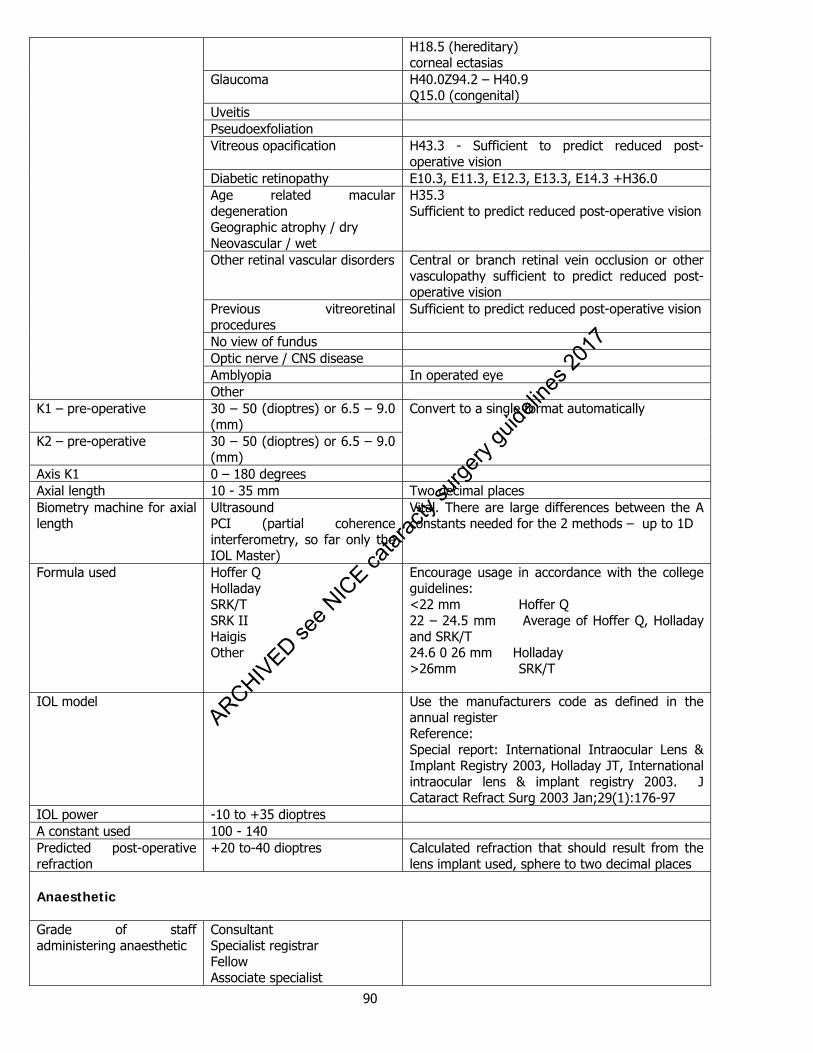
90
H18.5 (hereditary) corneal ectasias
Glaucoma
H40.0Z94.2 – H40.9 Q15.0 (congenital)
Uveitis Pseudoexfoliation Vitreous opacification H43.3 - Sufficient to predict reduced post-
operative vision Diabetic retinopathy E10.3, E11.3, E12.3, E13.3, E14.3 +H36.0 Age related macular degeneration Geographic atrophy / dry Neovascular / wet
H35.3 Sufficient to predict reduced post-operative vision
Other retinal vascular disorders Central or branch retinal vein occlusion or other vasculopathy sufficient to predict reduced post-operative vision
Previous vitreoretinal procedures
Sufficient to predict reduced post-operative vision
No view of fundus Optic nerve / CNS disease Amblyopia In operated eye Other
K1 – pre-operative 30 – 50 (dioptres) or 6.5 – 9.0 (mm)
K2 – pre-operative 30 – 50 (dioptres) or 6.5 – 9.0 (mm)
Convert to a single format automatically
Axis K1 0 – 180 degrees Axial length 10 - 35 mm Two decimal places Biometry machine for axial length
Ultrasound PCI (partial coherence interferometry, so far only the IOL Master)
Vital. There are large differences between the A constants needed for the 2 methods – up to 1D
Formula used Hoffer Q Holladay SRK/T SRK II Haigis Other
Encourage usage in accordance with the college guidelines: <22 mm Hoffer Q 22 – 24.5 mm Average of Hoffer Q, Holladay and SRK/T 24.6 0 26 mm Holladay >26mm SRK/T
IOL model Use the manufacturers code as defined in the annual register Reference: Special report: International Intraocular Lens & Implant Registry 2003, Holladay JT, International intraocular lens & implant registry 2003. J Cataract Refract Surg 2003 Jan;29(1):176-97
IOL power -10 to +35 dioptres A constant used 100 - 140 Predicted post-operative refraction
+20 to-40 dioptres Calculated refraction that should result from the lens implant used, sphere to two decimal places
Anaesthetic Grade of staff administering anaesthetic
Consultant Specialist registrar Fellow Associate specialist
ARCHIVED see N
ICE ca
tarac
ty su
rgery
guide
lines
2017
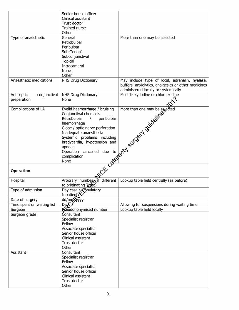
91
Senior house officer Clinical assistant Trust doctor Trained nurse Other
Type of anaesthetic General Retrobulbar Peribulbar Sub-Tenon’s Subconjunctival Topical Intracameral None Other
More than one may be selected
Anaesthetic medications NHS Drug Dictionary May include type of local, adrenalin, hyalase, buffers, anxiolytics, analgesics or other medicines administered locally or systemically
Antiseptic conjunctival preparation
NHS Drug Dictionary None
Most likely iodine or chlorhexidine
Complications of LA Eyelid haemorrhage / bruising Conjunctival chemosis Retrobulbar / peribulbar haemorrhage Globe / optic nerve perforation Inadequate anaesthesia Systemic problems including bradycardia, hypotension and apnoea Operation cancelled due to complication None
More than one may be selected
Operation Hospital Arbitrary number (if different
to originating Trust) Lookup table held centrally (as before)
Type of admission Day case / ambulatory Inpatient
Date of surgery dd/mm/yyyy Time spent on waiting list Days Allowing for suspensions during waiting time Surgeon Pseudononymised number Lookup table held locally Surgeon grade Consultant
Specialist registrar Fellow Associate specialist Senior house officer Clinical assistant Trust doctor Other
Assistant Consultant Specialist registrar Fellow Associate specialist Senior house officer Clinical assistant Trust doctor Other
ARCHIVED see N
ICE ca
tarac
ty su
rgery
guide
lines
2017

92
None 1st / 2nd eye 1, 2 Side of eye operation Left
Right Bilateral surgery is better handled with two CND entries – the rest of the dataset does not allow values for right and left eyes
Type of cataract Operation Phaco + IOL ECCE + IOL Phaco converted to ECCE Aspiration of lens ICCE Lensectomy
OPCS4 code C712 C718 C718 C713 C728 C743
Site Clear corneal Scleral tunnel Limbal Pars plana
Meridian 0-360 degrees Incision length 1 – 12 One decimal place (mm) Per-operative factors increasing the difficulty of surgery
Uncooperative patient Uncontrolled eye movement Deep-set eye Shallow anterior chamber Small pupil Atonic iris Phacodonesis Other
IOL position In the bag Partly in the bag In the sulcus Anterior chamber Sutured posterior chamber Iris fixated Aphakic
Additional planned surgical procedures
None Eyelid surgery Refractive procedure Penetrating keratoplasty Glaucoma filtering procedure Mechanical pupil dilation Iridectomy Capsule tension ring Posterior capsule capsulorrhexis / capsulotomy Posterior segment vitreoretinal procedure Other
Includes stretch, hooks, rings Peripheral or broad
Operative incidental events / complications
None Phaco wound burn Corneal oedema Iris damage Hyphaema Zonule dialysis PC rupture no vitreous loss PC rupture with vitreous loss Dropped lens Decentred IOL Choroidal / expulsive haemorrhage
All or part of the cortex or nucleus
ARCHIVED see N
ICE ca
tarac
ty su
rgery
guide
lines
2017
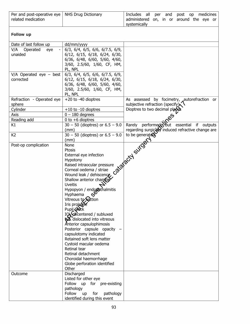
93
Per and post-operative eye related medication
NHS Drug Dictionary Includes all per and post op medicines administered on, in or around the eye or systemically
Follow up Date of last follow up dd/mm/yyyy V/A Operated eye - unaided
6/3, 6/4, 6/5, 6/6, 6/7.5, 6/9, 6/12, 6/15, 6/18, 6/24, 6/30, 6/36, 6/48, 6/60, 5/60, 4/60, 3/60, 2.5/60, 1/60, CF, HM, PL, NPL
V/A Operated eye – best corrected
6/3, 6/4, 6/5, 6/6, 6/7.5, 6/9, 6/12, 6/15, 6/18, 6/24, 6/30, 6/36, 6/48, 6/60, 5/60, 4/60, 3/60, 2.5/60, 1/60, CF, HM, PL, NPL
Refraction - Operated eye sphere
+20 to -40 dioptres
Cylinder +10 to -10 dioptres Axis 0 – 180 degrees Reading add 0 to +6 dioptres
As assessed by focimetry, autorefraction or subjective refraction (specify) Dioptres to two decimal places
K1 30 – 50 (dioptres) or 6.5 – 9.0 (mm)
K2 30 – 50 (dioptres) or 6.5 – 9.0 (mm)
Rarely performed but essential if outputs regarding surgically induced refractive change are to be generated
Post-op complication None Ptosis External eye infection Hypotony Raised intraocular pressure Corneal oedema / striae Wound leak / dehiscence Shallow anterior chamber Uveitis Hypopyon / endophthalmitis Hyphaema Vitreous to section Iris prolapse Pupil block IOL decentered / subluxed IOL dislocated into vitreous Anterior capsulophimosis Posterior capsule opacity – capsulotomy indicated Retained soft lens matter Cystoid macular oedema Retinal tear Retinal detachment Choroidal haemorrhage Globe perforation identified Other
Outcome Discharged Listed for other eye Follow up for pre-existing pathology Follow up for pathology identified during this event
ARCHIVED see N
ICE ca
tarac
ty su
rgery
guide
lines
2017
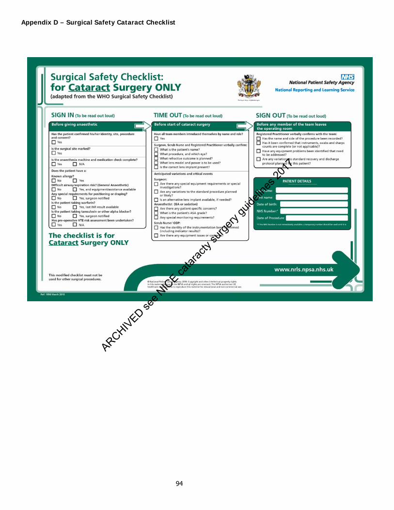
94
Appendix D – Surgical Safety Cataract Checklist
ARCHIVED see N
ICE ca
tarac
ty su
rgery
guide
lines
2017
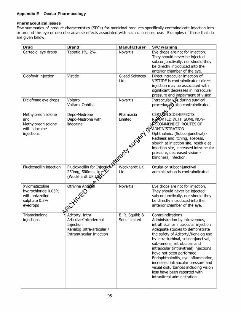
95
Appendix E – Ocular Pharmacology Pharmaceutical issues Few summaries of product characteristics (SPCs) for medicinal products specifically contraindicate injection into or around the eye or describe adverse effects associated with such unlicensed use. Examples of those that do are given below:
Drug Brand Manufacturer SPC warning Carteolol eye drops Teoptic 1%, 2% Novartis Eye drops are not for injection.
They should never be injected subconjunctivally, nor should they be directly introduced into the anterior chamber of the eye.
Cidofovir injection Vistide Gilead Sciences Ltd
Direct intraocular injection of VISTIDE is contraindicated; direct injection may be associated with significant decreases in intraocular pressure and impairment of vision.
Diclofenac eye drops Voltarol Voltarol Ophtha
Novartis Intraocular use during surgical procedure is also contraindicated.
Methylprednisolone and Methylprednisolone with lidocaine injections
Depo-Medrone Depo-Medrone with lidocaine
Pharmacia Limited
CERTAIN SIDE-EFFECTS REPORTED WITH SOME NON-RECOMMENDED ROUTES OF ADMINISTRATION Ophthalmic: (Subconjunctival) - Redness and itching, abscess, slough at injection site, residue at injection site, increased intra-ocular pressure, decreased vision - blindness, infection.
Flucloxacillin injection Flucloxacillin for Injection 250mg, 500mg, 1g (Wockhardt UK Ltd)
Wockhardt UK Ltd
Ocular or subconjunctival administration is contraindicated
Xylometazoline hydrochloride 0.05% with antazoline sulphate 0.5% eyedrops
Otrivine Antistin Novartis Eye drops are not for injection. They should never be injected subconjunctivally, nor should they be directly introduced into the anterior chamber of the eye.
Triamcinolone injections
Adcortyl Intra-Articular/Intradermal Injection Kenalog Intra-articular / Intramuscular Injection
E. R. Squibb & Sons Limited
Contraindications Administration by intravenous, intrathecal or intraocular injection Adequate studies to demonstrate the safety of Adcortyl/Kenalog use by intra-turbinal, subconjunctival, sub-tenons, retrobulbar and intraocular (intravitreal) injections have not been performed. Endophthalmitis, eye inflammation, increased intraocular pressure and visual disturbances including vision loss have been reported with intravitreal administration.
ARCHIVED see N
ICE ca
tarac
ty su
rgery
guide
lines
2017
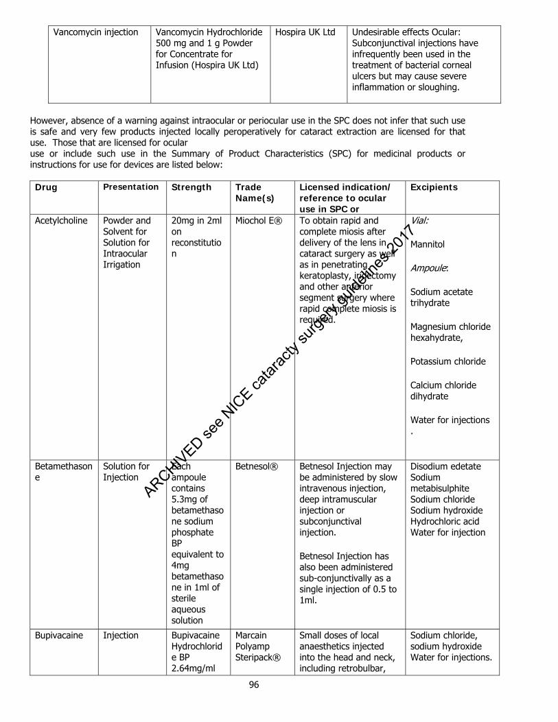
96
Vancomycin injection Vancomycin Hydrochloride 500 mg and 1 g Powder for Concentrate for Infusion (Hospira UK Ltd)
Hospira UK Ltd
Undesirable effects Ocular: Subconjunctival injections have infrequently been used in the treatment of bacterial corneal ulcers but may cause severe inflammation or sloughing.
However, absence of a warning against intraocular or periocular use in the SPC does not infer that such use is safe and very few products injected locally peroperatively for cataract extraction are licensed for that use. Those that are licensed for ocular use or include such use in the Summary of Product Characteristics (SPC) for medicinal products or instructions for use for devices are listed below:
Drug Presentation Strength Trade
Name(s) Licensed indication/ reference to ocular use in SPC or
Excipients
Acetylcholine Powder and Solvent for Solution for Intraocular Irrigation
20mg in 2ml on reconstitution
Miochol E® To obtain rapid and complete miosis after delivery of the lens in cataract surgery as well as in penetrating keratoplasty, iridectomy and other anterior segment surgery where rapid complete miosis is required.
Vial:
Mannitol
Ampoule:
Sodium acetate trihydrate
Magnesium chloride hexahydrate,
Potassium chloride
Calcium chloride dihydrate
Water for injections .
Betamethasone
Solution for Injection
Each ampoule contains 5.3mg of betamethasone sodium phosphate BP equivalent to 4mg betamethasone in 1ml of sterile aqueous solution
Betnesol® Betnesol Injection may be administered by slow intravenous injection, deep intramuscular injection or subconjunctival injection.
Betnesol Injection has also been administered sub-conjunctivally as a single injection of 0.5 to 1ml.
Disodium edetate Sodium metabisulphite Sodium chloride Sodium hydroxide Hydrochloric acid Water for injection
Bupivacaine Injection Bupivacaine Hydrochloride BP 2.64mg/ml
Marcain Polyamp Steripack®
Small doses of local anaesthetics injected into the head and neck, including retrobulbar,
Sodium chloride, sodium hydroxide Water for injections.
ARCHIVED see N
ICE ca
tarac
ty su
rgery
guide
lines
2017
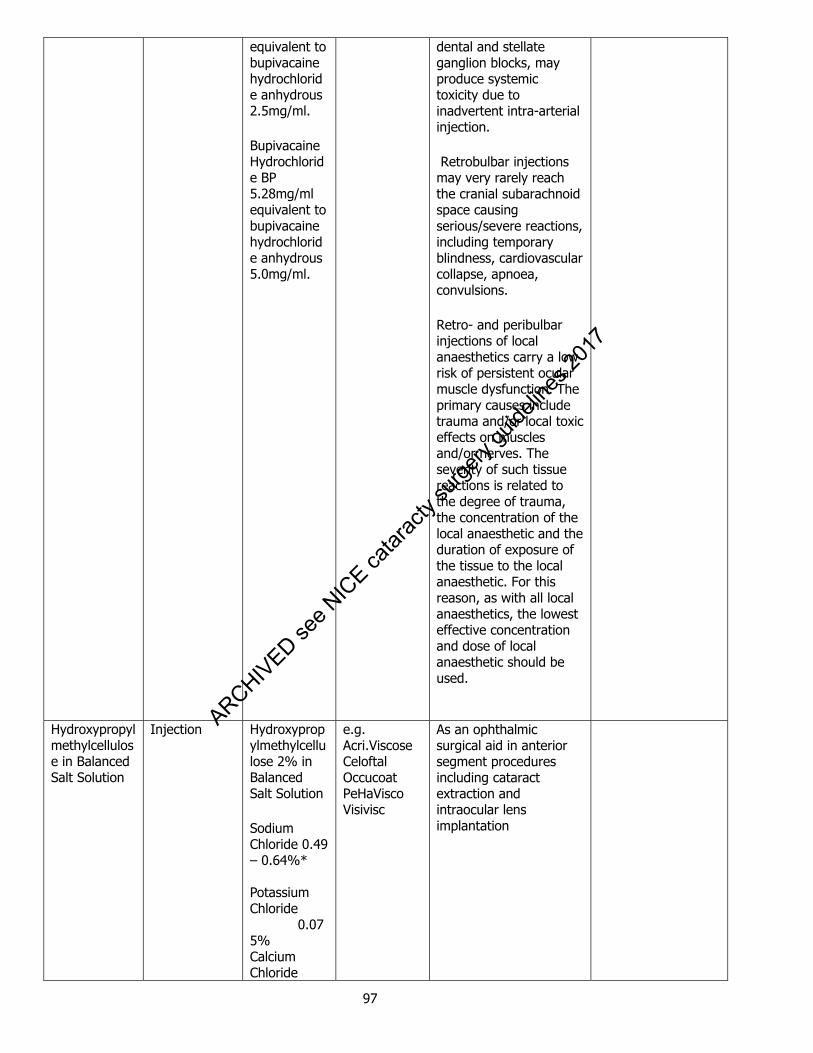
97
equivalent to bupivacaine hydrochloride anhydrous 2.5mg/ml.
Bupivacaine Hydrochloride BP 5.28mg/ml equivalent to bupivacaine hydrochloride anhydrous 5.0mg/ml.
dental and stellate ganglion blocks, may produce systemic toxicity due to inadvertent intra-arterial injection.
Retrobulbar injections may very rarely reach the cranial subarachnoid space causing serious/severe reactions, including temporary blindness, cardiovascular collapse, apnoea, convulsions.
Retro- and peribulbar injections of local anaesthetics carry a low risk of persistent ocular muscle dysfunction. The primary causes include trauma and/or local toxic effects on muscles and/or nerves. The severity of such tissue reactions is related to the degree of trauma, the concentration of the local anaesthetic and the duration of exposure of the tissue to the local anaesthetic. For this reason, as with all local anaesthetics, the lowest effective concentration and dose of local anaesthetic should be used.
Hydroxypropylmethylcellulose in Balanced Salt Solution
Injection Hydroxypropylmethylcellulose 2% in Balanced Salt Solution
Sodium Chloride 0.49 – 0.64%* Potassium Chloride 0.075% Calcium Chloride
e.g. Acri.Viscose Celoftal Occucoat PeHaVisco Visivisc
As an ophthalmic surgical aid in anterior segment procedures including cataract extraction and intraocular lens implantation
ARCHIVED see N
ICE ca
tarac
ty su
rgery
guide
lines
2017

98
0.048%Magnesium Chloride 0.03% Sodium Acetate 0.39% Sodium Citrate 0.17% * composition varies with manufacturer
Lidocaine Sterile aqueous solution for infiltration injection or intravenous administration.
Lidocaine Hydrochloride BP 10 mg per ml
Lidocaine Hydrochloride BP 20 mg per ml
Lidocaine Hydrochloride Injection BP Minijet 1% and 2% (International Medication Systems)®
For local anaesthesia by infiltration, intravenous regional anaesthesia and nerve blocks.
Hydrochloric Acid BP
Sodium Chloride BP
Sodium Hydroxide BP
Water for Injection USP
Lidocaine in sodium hyaluronate
Solution for injection.
Lidocaine Hydrochloride 2% in sodium hyaluronate 0.3%
Visthesia
Ophthalmic Irrigating Solution (Balanced Salt Solution)
Solution for intraocular irrigation
Calcium Chloride 0.048% Magnesium chloride 0.03% Sodium acetate 0.39% Sodium citrate 0.17%
e.g. Alcon BSS Aqsia Distra Sol Endosol
As a physiological irrigating solution
ARCHIVED se
e NIC
E catar
acty
surge
ry gu
idelin
es 20
17
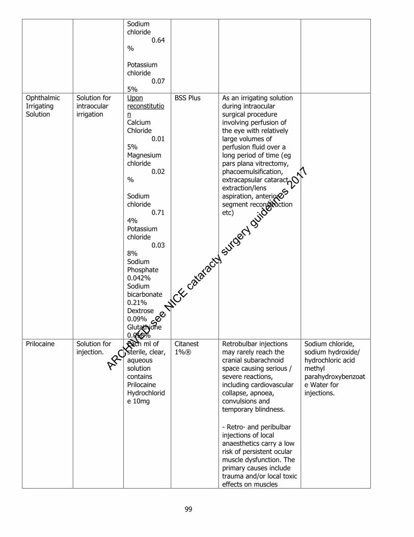
99
Sodium chloride 0.64% Potassium chloride 0.075%
Ophthalmic Irrigating Solution
Solution for intraocular irrigation
Upon reconstitution Calcium Chloride 0.015% Magnesium chloride 0.02% Sodium chloride 0.714% Potassium chloride 0.038% Sodium Phosphate 0.042% Sodium bicarbonate 0.21% Dextrose 0.09% Glutathione 0.018%
BSS Plus As an irrigating solution during intraocular surgical procedure involving perfusion of the eye with relatively large volumes of perfusion fluid over a long period of time (eg pars plana vitrectomy, phacoemulsification, extracapsular cataract extraction/lens aspiration, anterior segment reconstruction etc)
Prilocaine Solution for injection.
Each ml of sterile, clear, aqueous solution contains Prilocaine Hydrochloride 10mg
Citanest 1%®
Retrobulbar injections may rarely reach the cranial subarachnoid space causing serious / severe reactions, including cardiovascular collapse, apnoea, convulsions and temporary blindness.
- Retro- and peribulbar injections of local anaesthetics carry a low risk of persistent ocular muscle dysfunction. The primary causes include trauma and/or local toxic effects on muscles
Sodium chloride, sodium hydroxide/ hydrochloric acid methyl parahydroxybenzoate Water for injections.
ARCHIVED see N
ICE ca
tarac
ty su
rgery
guide
lines
2017
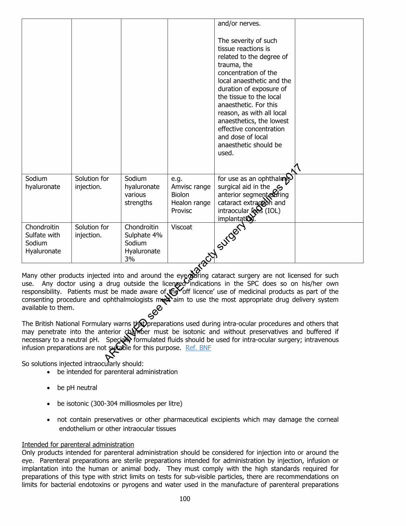
100
and/or nerves.
The severity of such tissue reactions is related to the degree of trauma, the concentration of the local anaesthetic and the duration of exposure of the tissue to the local anaesthetic. For this reason, as with all local anaesthetics, the lowest effective concentration and dose of local anaesthetic should be used.
Sodium hyaluronate
Solution for injection.
Sodium hyaluronate various strengths
e.g. Amvisc range Biolon Healon range Provisc
for use as an ophthalmic surgical aid in the anterior segment during cataract extraction and intraocular lens (IOL) implantation.
Chondroitin Sulfate with Sodium Hyaluronate
Solution for injection.
Chondroitin Sulphate 4% Sodium Hyaluronate 3%
Viscoat
Many other products injected into and around the eye during cataract surgery are not licensed for such use. Any doctor using a drug outside the licensed indications in the SPC does so on his/her own responsibility. Patients must be made aware of the ‘off licence’ use of medicinal products as part of the consenting procedure and ophthalmologists must aim to use the most appropriate drug delivery system available to them. The British National Formulary warns that preparations used during intra-ocular procedures and others that may penetrate into the anterior chamber must be isotonic and without preservatives and buffered if necessary to a neutral pH. Specially formulated fluids should be used for intra-ocular surgery; intravenous infusion preparations are not suitable for this purpose. Ref. BNF So solutions injected intraocularly should:
be intended for parenteral administration
be pH neutral
be isotonic (300-304 milliosmoles per litre)
not contain preservatives or other pharmaceutical excipients which may damage the corneal endothelium or other intraocular tissues
Intended for parenteral administration Only products intended for parenteral administration should be considered for injection into or around the eye. Parenteral preparations are sterile preparations intended for administration by injection, infusion or implantation into the human or animal body. They must comply with the high standards required for preparations of this type with strict limits on tests for sub-visible particles, there are recommendations on limits for bacterial endotoxins or pyrogens and water used in the manufacture of parenteral preparations
ARCHIVED see N
ICE ca
tarac
ty su
rgery
guide
lines
2017

101
complies with the requirements of water for injections in bulk. Eye preparations are sterile liquid, semi-solid or solid preparations intended for administration upon the eyeball and/or to the conjunctiva, or for insertion in the conjunctival sac. Eye drops that are solutions, examined under suitable conditions of visibility, must be practically clear and practically free from particles but a test for sub-visible particles is not required. There are no recommendations on limits for bacterial endotoxins or pyrogens and it is not specified that the water used in the manufacture of eye preparations to comply with the requirements of water for injections. ‘Purified water’ is the term usually used in the Summary of Product Characteristics of aqueous eye preparations. Ref. European Pharmacopoeia pH neutral Isotonic (300-304 milliosmoles per litre) Not contain preservatives or other pharmaceutical excipients which may damage the corneal endothelium or other intraocular tissues The European Pharmacopoeia states that no antimicrobial preservative is added to an injection when the preparation is intended for administration by routes where, for medical reasons, an antimicrobial preservative is not acceptable, such as intracisternally, epidurally, intrathecally or by any route giving access to the cerebrospinal fluid, or intra- or retro-ocularly. The toxicity of intraocular benzalkonium chloride has been recognized for many years and eye drops intended for use in surgical procedures do not contain antimicrobial preservatives and are supplied in single-dose containers. Ref. Britton B et al Intraocular irritation evaluation of benzalkonium chloride in rabbits Ophthalmic Surg. 1976 Fall;7(3):46-55 Ref. European Pharmacopoeia However, both parenteral preparations and eye preparations may require the use of excipients, for example to adjust the tonicity, to adjust the pH, to increase solubility, to prevent deterioration of the active substances or to provide adequate antimicrobial properties. Moreover, eye drops may contain excipients to adjust the viscosity, to stabilize the pH and to stabilize the preparation. While these substances do not adversely affect the intended medicinal action, or, at the concentration used, cause local undue irritation only the name and concentration of any added antimicrobial preservative must be stated on the label i.e. information about other excipients must be found elsewhere. Eye drops are not intended for intraocular injection and contain a wide range of excipients with the potential to cause local adverse reactions including inflammation and sloughing at subconjunctival injection sites and toxic anterior segment syndrome and toxic endothelial cell destruction syndrome after intraocular injection. Ophthalmic operating theatres are supplied with preservative-free single dose eye drop units to avoid benzalkonium chloride and other potentially damaging antimicrobial preservatives entering the anterior chamber. However, preservative-free eye drops are not necessarily excipient free and commercially available preservative-free single dose units contain a wide range of excipients not listed in the BNF as is the case for antimicrobial preservatives for multidose eye drops: Ref. BNF
Drug Trade name Manufacturer Excipients Hydroxyethylcellulose 0.44% Sodium Chloride 0.35%
Minims Artificial Tears Bausch & Lomb Purified water
Borax
Boric acid
Chloramphenicol 0.5% Minims
Chloramphenicol Bausch & Lomb Borax
Boric acid
Purified water
ARCHIVED see N
ICE ca
tarac
ty su
rgery
guide
lines
2017

102
Dexamethasone sodium phosphate 0.1%
Minims Dexamethasone
Bausch & Lomb Anhydrous disodium hydrogen phosphate
Sodium dihydrogen phosphate (2H2O)
Disodium edetate
Purified water
Lidocaine 4% with fluorescein sodium 0.25%
Minims Lidocaine & Fluorescein
Bausch & Lomb Povidone
Hydrochloric Acid
Purified Water
Phenylephrine 2.5%, 10%
Minims Phenylephrine Hydrochloride 2.5%, 10%
Bausch & Lomb Sodium metabisulphite
Disodium edetate
Purified water
Prednisolone sodium phosphate 0.5%
Minims Prednisolone Sodium Phosphate 0.5%
Bausch & Lomb Disodium edetate
Disodium dihydrogen phosphate
Sodium chloride
Sodium hydroxide for pH adjustment
Purified water
Proxymetacaine hydrochloride 0.5% with fluorescein sodium 0.25%
Minims Proxymetacaine & Fluorescein
Bausch & Lomb Purified water
Povidone K30
Hydrochloric acid
Sodium Hydroxide
Betaxolol suspension 0.25%
Betoptic Suspension Single Dose
Alcon Amberlite poly (styrene-divinyl benzene) sulphonic acid
Carbomer
Mannitol
Hydrochloric acid and/or Sodium
ARCHIVED see N
ICE ca
tarac
ty su
rgery
guide
lines
2017

103
Hydroxide
Purified Water
Apraclonidine 1% Iopidine
1.0%Opthalmic Solution
Alcon Sodium acetate (trihydrate),
sodium chloride,
hydrochloric acid and/or sodium hydroxide (to adjust pH),
purified water. Dextran 0.1%, hypromellose 0.3%
Tears Naturale Single Dose Eye Drops
Alcon Sodium chloride
Potassium chloride
Calcium chloride (dihydrate)
Magnesium chloride (hexahydrate)
Zinc chloride
Sodium hydrogen carbonate
Carbon dioxide (to adjust pH)
Purified water
Dorzolamide 2% with timolol 0.5%
Cosopt Preservative-free, Single Dose Eye Drops
MSD Hydroxyethyl cellulose
Mannitol (E421)
Sodium citrate (E331)
Sodium hydroxide (E524) for pH adjustment
Water for injections.
Carbomer (polyacrylic acid) 0.2%
Viscotears Single Dose Unit 2.0mg/g Eye Gel
Novartis Sorbitol,
sodium hydroxide and
water for injections.
Hypromellose 0.32% Artelac SDU
preservative free hypromellose 0.32%
Iris Healthcare Ltd
Disodium phosphate dodecahydrate
Sodium dihydrogen phosphate dihydrate
ARCHIVED see N
ICE ca
tarac
ty su
rgery
guide
lines
2017
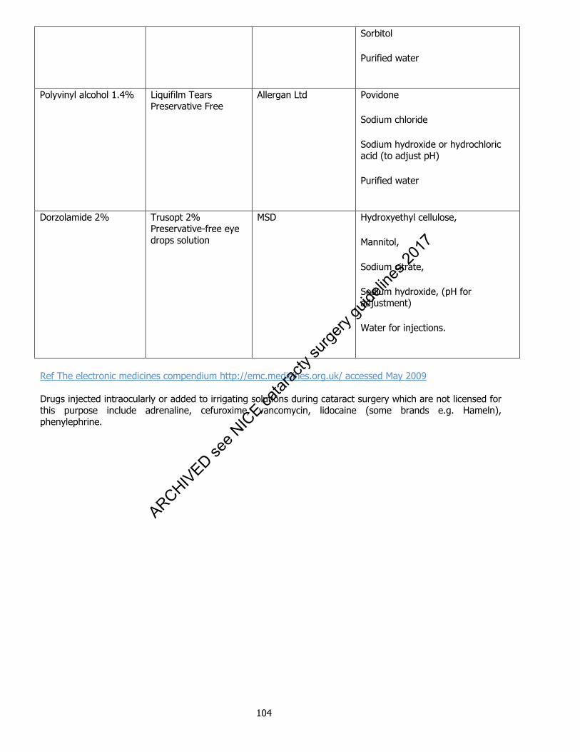
104
Sorbitol
Purified water
Polyvinyl alcohol 1.4% Liquifilm Tears
Preservative Free Allergan Ltd Povidone
Sodium chloride
Sodium hydroxide or hydrochloric acid (to adjust pH)
Purified water
Dorzolamide 2% Trusopt 2%
Preservative-free eye drops solution
MSD Hydroxyethyl cellulose,
Mannitol,
Sodium citrate,
Sodium hydroxide, (pH for adjustment)
Water for injections.
Ref The electronic medicines compendium http://emc.medicines.org.uk/ accessed May 2009 Drugs injected intraocularly or added to irrigating solutions during cataract surgery which are not licensed for this purpose include adrenaline, cefuroxime, vancomycin, lidocaine (some brands e.g. Hameln), phenylephrine.
ARCHIVED see N
ICE ca
tarac
ty su
rgery
guide
lines
2017
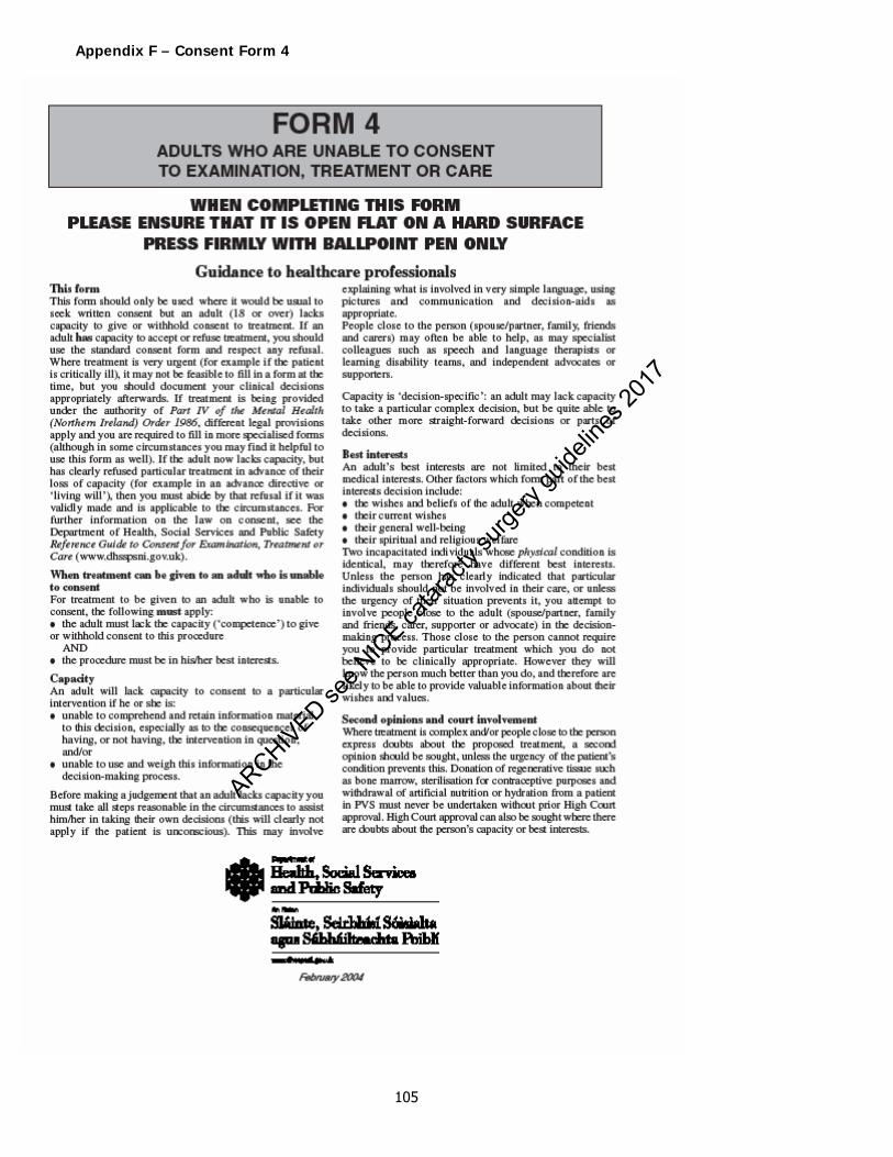
105
Appendix F – Consent Form 4
ARCHIVED see N
ICE ca
tarac
ty su
rgery
guide
lines
2017

106
ARCHIVED see N
ICE ca
tarac
ty su
rgery
guide
lines
2017