The role of transesophageal echocardiography in clinical...
12
Review Article The role of transesophageal echocardiography in clinical use Shen-Kou Tsai a,b,c, * a National Taiwan University School of Medicine, Taipei, Taiwan, ROC b National Yang-Ming University School of Medicine, Taipei, Taiwan, ROC c Department of Anesthesiology, Cheng-Hsin General Hospital, Taipei, Taiwan, ROC Received March 12, 2013; accepted June 5, 2013 Abstract Transesophageal echocardiography (TEE) is not only an invaluable diagnostic tool for cardiac patients, but also is essential for cardiac monitoring in critically ill patients in cardiac and non-cardiac surgery settings and in the differential diagnosis of unexplained hemodynamic collapse. The advantage of TEE over transthoracic echocardiography (TTE) is usually clearer images, especially when viewing structures that are difficult to see transthoracically. TEE is essential in monitoring adult and congenital heart surgery perioperatively. The adequacy of the repair can be ensured immediately through a review of TEE images directly after surgery. Although TEE is considered to be relatively safe and noninvasive, TEE-associated complications, such as esophageal laceration, must be taken seriously. Recently, real-time three-dimensional (3D) TEE imaging has played an important role defining valvular and congenital abnormalities and aiding in operative and percutaneous repair. Copyright Ó 2013 Elsevier Taiwan LLC and the Chinese Medical Association. All rights reserved. Keywords: cardiac surgery; catheter intervention; critical care; transesophageal echocardiography 1. Introduction Echocardiography is the most frequently used diagnostic tool for real-time imaging of cardiac structure and function. In the last decade, transesophageal echocardiography (TEE) has become essential in cardiac surgery, and has expanded its role in other areas of patient care. 1e5 TEE is performed by inserting a probe with a transducer into the esophagus, and offers superior visualization of posterior cardiac structures, because of the close proximity of the esophagus to the post- eromedial heart, without visual interference from the lung and skeleton. In 1976, Frazin et al 6 first introduced the clinical use of TEE when a modified rigid endoscopic probe with a single M-mode crystal was used. In 1980, the phased-array ultra- sound transducer was introduced, and it was later reduced in size. The process of implementing biplane probes 7e9 by using crystal miniaturization with color Doppler is the standard principle used in echocardiography. In 1990, multiplane TEE probes become available, utilizing mechanical or electronic rotation of the 180 degree scanning plane. 10e13 Remarkable progress in TEE probe technology has been made in the last 10 years. More recently, real-time three-dimensional (3D) imag- ing has been available by using a matrix array ultrasound probe and an appropriate processing system. 14 This enables detailed anatomical assessment of cardiac pathology and particularly valvular defects. 15 Now, TEE is a well-established and standard diagnostic technique in the operating room, intensive care unit, and laboratory catheter room. 2. Indication of TEE TEE can reveal new findings that necessitate cross- checking perioperatively, such as mitral valve (MV) disor- ders, blood clots or intracardiac masses, dissection of the aorta, and implanted prosthetic (artificial) heart valves. In 1996, a joint task force of the American Society of Anesthe- siologists (ASA) and the Society of Cardiovascular Anesthe- siologists (SCA) published guidelines for the perioperative * Corresponding author. Dr. Shen-Kou Tsai, Cheng Hsin General Hospital, 45, Cheng Hsin Street, Beitou, Taipei 112, Taiwan, ROC. E-mail address: [email protected] (S.-K. Tsai). Available online at www.sciencedirect.com ScienceDirect Journal of the Chinese Medical Association 76 (2013) 661e672 www.jcma-online.com 1726-4901/$ - see front matter Copyright Ó 2013 Elsevier Taiwan LLC and the Chinese Medical Association. All rights reserved. http://dx.doi.org/10.1016/j.jcma.2013.08.009
Transcript of The role of transesophageal echocardiography in clinical...
Available online at www.sciencedirect.com
ScienceDirect
Journal of the Chinese Medical Association 76 (2013) 661e672www.jcma-online.com
Review Article
The role of transesophageal echocardiography in clinical use
Shen-Kou Tsai a,b,c,*
aNational Taiwan University School of Medicine, Taipei, Taiwan, ROCbNational Yang-Ming University School of Medicine, Taipei, Taiwan, ROC
cDepartment of Anesthesiology, Cheng-Hsin General Hospital, Taipei, Taiwan, ROC
Received March 12, 2013; accepted June 5, 2013
Abstract
Transesophageal echocardiography (TEE) is not only an invaluable diagnostic tool for cardiac patients, but also is essential for cardiacmonitoring in critically ill patients in cardiac and non-cardiac surgery settings and in the differential diagnosis of unexplained hemodynamiccollapse. The advantage of TEE over transthoracic echocardiography (TTE) is usually clearer images, especially when viewing structures thatare difficult to see transthoracically. TEE is essential in monitoring adult and congenital heart surgery perioperatively. The adequacy of the repaircan be ensured immediately through a review of TEE images directly after surgery. Although TEE is considered to be relatively safe andnoninvasive, TEE-associated complications, such as esophageal laceration, must be taken seriously. Recently, real-time three-dimensional (3D)TEE imaging has played an important role defining valvular and congenital abnormalities and aiding in operative and percutaneous repair.Copyright � 2013 Elsevier Taiwan LLC and the Chinese Medical Association. All rights reserved.
Keywords: cardiac surgery; catheter intervention; critical care; transesophageal echocardiography
1. Introduction
Echocardiography is the most frequently used diagnostictool for real-time imaging of cardiac structure and function. Inthe last decade, transesophageal echocardiography (TEE) hasbecome essential in cardiac surgery, and has expanded its rolein other areas of patient care.1e5 TEE is performed byinserting a probe with a transducer into the esophagus, andoffers superior visualization of posterior cardiac structures,because of the close proximity of the esophagus to the post-eromedial heart, without visual interference from the lung andskeleton. In 1976, Frazin et al6 first introduced the clinical useof TEE when a modified rigid endoscopic probe with a singleM-mode crystal was used. In 1980, the phased-array ultra-sound transducer was introduced, and it was later reduced insize. The process of implementing biplane probes7e9 by usingcrystal miniaturization with color Doppler is the standard
* Corresponding author. Dr. Shen-Kou Tsai, Cheng Hsin General Hospital,
45, Cheng Hsin Street, Beitou, Taipei 112, Taiwan, ROC.
E-mail address: [email protected] (S.-K. Tsai).
1726-4901/$ - see front matter Copyright � 2013 Elsevier Taiwan LLC and the C
http://dx.doi.org/10.1016/j.jcma.2013.08.009
principle used in echocardiography. In 1990, multiplane TEEprobes become available, utilizing mechanical or electronicrotation of the 180 degree scanning plane.10e13 Remarkableprogress in TEE probe technology has been made in the last 10years. More recently, real-time three-dimensional (3D) imag-ing has been available by using a matrix array ultrasoundprobe and an appropriate processing system.14 This enablesdetailed anatomical assessment of cardiac pathology andparticularly valvular defects.15 Now, TEE is a well-establishedand standard diagnostic technique in the operating room,intensive care unit, and laboratory catheter room.
2. Indication of TEE
TEE can reveal new findings that necessitate cross-checking perioperatively, such as mitral valve (MV) disor-ders, blood clots or intracardiac masses, dissection of theaorta, and implanted prosthetic (artificial) heart valves. In1996, a joint task force of the American Society of Anesthe-siologists (ASA) and the Society of Cardiovascular Anesthe-siologists (SCA) published guidelines for the perioperative
hinese Medical Association. All rights reserved.
662 S.-K. Tsai / Journal of the Chinese Medical Association 76 (2013) 661e672
application of TEE.16,17 Based upon the current ASA and SCAguidelines, category I indications of perioperative TEE are:
1. Intraoperative evaluation of acute, persistent, and life-threatening hemodynamic disturbances in which ventric-ular function and its determinants are uncertain and havenot responded to treatment.
2. Intraoperative use in valve repair.3. Intraoperative use in congenital heart surgery for most
lesions requiring cardiopulmonary bypass.4. Intraoperative use in repair of hypertrophic obstructive
cardiomyopathy.5. Intraoperative use for endocarditis when preoperative
testing was inadequate or extension of infection to peri-valvular tissue is suspected.
6. Preoperative use in unstable patients who have suspectedthoracic aortic aneurysms, dissection, or disruption thatneeds to be evaluated quickly.
7. Intraoperative assessment of aortic valve function in repair ofaortic dissections with possible aortic valve involvement.
8. Intraoperative evaluation of pericardial window procedures.9. Use in intensive care unit for unstable patients who have
unexplained hemodynamic disturbances, suspected valvedisease, or thromboembolic problems.
3. Contraindications from TEE18,19
1. Esophageal stricture or malignancy.2. Surgical interposition of the esophagus.3. Esophageal diverticulum.4. Cervical spine arthritis with reduced range of motion.5. Severe thrombocytopenia (<50,000/mL), elevated inter-
national normalized ratio (>4), or prolonged partialthromboplastin time (>150 seconds).
4. Tomographic views of TEE20
Several tomographic views are commonly used. A com-plete TEE examination should include imaging of all cardiacchambers, valves, and great vessels. A standard comprehen-sive approach to imaging is recommended, but each individualstudy should be modified to reflect the specific clinical indi-cation. Each tomographic view is defined by the transducerposition in the esophagus, which will view the TEE images ofthe mid-esophageal view (at the mid-esophageal position)including four chamber, five chamber, two chamber, short-axis, long-axis, two caval views (Fig. 1-1), upper esophagealview (at upper esophageal position) (Fig. 1-2) and transgastricview at the gastric position (Fig. 1-3).
Current TEE probes allow for both 2D and 3D imaging, aswell M-mode, spectral Doppler, and color Doppler.
4.1. 2D echocardiography
2D TEE provides tomographic or "thin slice" imaging, witheach tomographic view defined by the transducer position. The
technique is used to visualize the actual structures and the real-time motion of the heart.
4.2. 3D echocardiography
3D TEE capability has been developed to overcome thedisadvantages of 2D tomography. 3D TEE was first describedin the 1970s, because the acquisition of ECG and respiratory-gated 2D images, which subsequently required off-linereconstruction, was very time-consuming. However, the ma-trix TEE probe, introduced clinically in 2007, can quickly andeasily collect real-time 3D images, enabling the echocardi-ographer to provide an entire view that contains all pertinentinformation and real time images. This in turn results in betterunderstanding and facilitating of decision making during thecardiac catheterization procedures and cardiac surgery.
These systems generally acquire a volumetric data set,which can then be displayed in custom orientations. Thetechnique captures 3D views of the heart structures withgreater depth than 2D echocardiography.
4.3. M-mode echocardiography
M-mode can provide additional information for character-izing the motion of cardiac structures. To ensure proper align-ment and reproducibility, all M-mode recordings are performedwith 2D guidance. M-mode echo is useful for measuring heartstructures, such as the heart’s pumping chambers, the size of theheart, and the thickness of the heart walls.
4.4. Doppler echocardiography8
This technique is used to measure and assess the flow ofblood through the heart’s chambers and valves. Doppler echo-cardiography has the ability to estimate the pressure differenceacross a stenotic valve (e.g., aortic stenosis) or between twochambers (e.g., estimation of the pulmonary artery systolicpressure from the tricuspid regurgitation velocity). The modi-fied Bernoulli equation (Delta P ¼ 4*V
2) is the most commonlyused application relating peak velocity to peak pressuregradient. There are several Doppler methods used for cardiacevaluation-continuous wave, pulsed wave, and color flow.
4.5. Color Doppler8
Color flow imaging is typically used in the screening andassessment of regurgitant flows, intracardiac shunts, and pul-monary vein flow. Different colors are used to designate thedirection of blood flow.
5. TEE in clinical applications
5.1. Critical care21e24
TEE can be performed quickly at the bedside in critically illpatients with unexplained hypotension, unexplained hypox-emia, uncertain volume status, and blunt chest trauma.

Fig. 1. (1) The mid-esophageal views of transesophageal echocardiography (TEE). (A) Four chamber view at 0 degrees; (B) five chamber view at 0 degrees; (C)
right ventricle (RV) inflow-outflow view at 80 degrees; (D) two chamber view at 90 degrees; (E) left ventricle (LV) long axis view at 120 degrees; and (F) bicaval
view at 90e120 degrees. (2) The upper-esophageal views of TEE. (A) main pulmonary artery (MPA); (B) right pulmonary artery (RPA); (C) left pulmonary artery
(LPA); and (D) ascending aorta (AAo). (3) The transgastric views of TEE. (A) short axis view for papillary muscle; (B) short axis view for mitral leaflet; and (C)
long axis view for chordae apparatus (arrow). AL ¼ anterolateral papillary muscle; Ao ¼ aorta; AV ¼ aortic valve; C ¼ central venous catheter; IVC ¼ inferior
vena cava; LA ¼ left atrium; LV ¼ left ventricle; PA ¼ pulmonary artery; PM ¼ posteromedial papillary muscle; PV ¼ pulmonary valve; RA ¼ right atrium;
RUPV ¼ right upper pulmonary vein; RV ¼ right ventricle; RVOT ¼ right ventricle outflow tract; SVC ¼ superior vena cava; VS ¼ ventricular septum.
663S.-K. Tsai / Journal of the Chinese Medical Association 76 (2013) 661e672
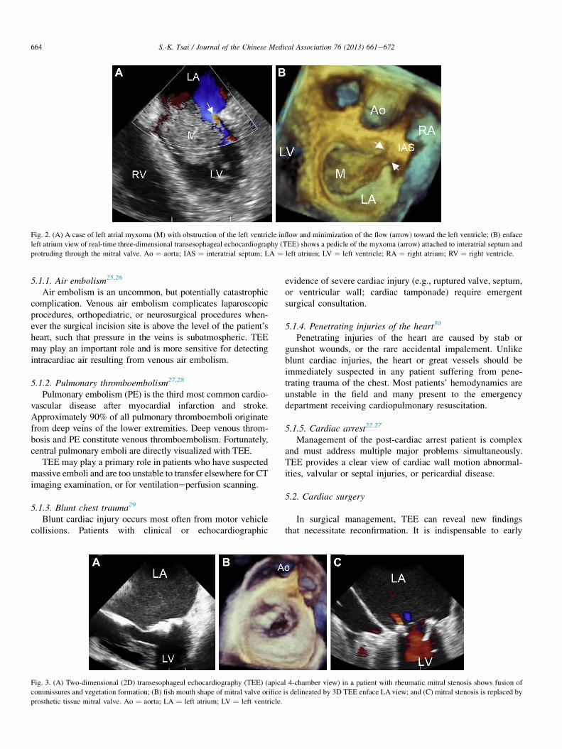
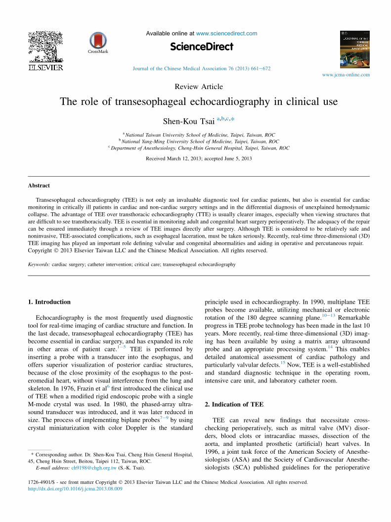
Fig. 2. (A) A case of left atrial myxoma (M) with obstruction of the left ventricle inflow and minimization of the flow (arrow) toward the left ventricle; (B) enface
left atrium view of real-time three-dimensional transesophageal echocardiography (TEE) shows a pedicle of the myxoma (arrow) attached to interatrial septum and
protruding through the mitral valve. Ao ¼ aorta; IAS ¼ interatrial septum; LA ¼ left atrium; LV ¼ left ventricle; RA ¼ right atrium; RV ¼ right ventricle.
664 S.-K. Tsai / Journal of the Chinese Medical Association 76 (2013) 661e672
5.1.1. Air embolism25,26
Air embolism is an uncommon, but potentially catastrophiccomplication. Venous air embolism complicates laparoscopicprocedures, orthopediatric, or neurosurgical procedures when-ever the surgical incision site is above the level of the patient’sheart, such that pressure in the veins is subatmospheric. TEEmay play an important role and is more sensitive for detectingintracardiac air resulting from venous air embolism.
5.1.2. Pulmonary thromboembolism27,28
Pulmonary embolism (PE) is the third most common cardio-vascular disease after myocardial infarction and stroke.Approximately 90% of all pulmonary thromboemboli originatefrom deep veins of the lower extremities. Deep venous throm-bosis and PE constitute venous thromboembolism. Fortunately,central pulmonary emboli are directly visualized with TEE.
TEE may play a primary role in patients who have suspectedmassive emboli and are too unstable to transfer elsewhere for CTimaging examination, or for ventilationeperfusion scanning.
5.1.3. Blunt chest trauma29
Blunt cardiac injury occurs most often from motor vehiclecollisions. Patients with clinical or echocardiographic
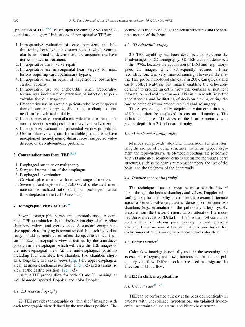
Fig. 3. (A) Two-dimensional (2D) transesophageal echocardiography (TEE) (apica
commissures and vegetation formation; (B) fish mouth shape of mitral valve orifice
prosthetic tissue mitral valve. Ao ¼ aorta; LA ¼ left atrium; LV ¼ left ventricle.
evidence of severe cardiac injury (e.g., ruptured valve, septum,or ventricular wall; cardiac tamponade) require emergentsurgical consultation.
5.1.4. Penetrating injuries of the heart30
Penetrating injuries of the heart are caused by stab orgunshot wounds, or the rare accidental impalement. Unlikeblunt cardiac injuries, the heart or great vessels should beimmediately suspected in any patient suffering from pene-trating trauma of the chest. Most patients’ hemodynamics areunstable in the field and many present to the emergencydepartment receiving cardiopulmonary resuscitation.
5.1.5. Cardiac arrest22,27
Management of the post-cardiac arrest patient is complexand must address multiple major problems simultaneously.TEE provides a clear view of cardiac wall motion abnormal-ities, valvular or septal injuries, or pericardial disease.
5.2. Cardiac surgery
In surgical management, TEE can reveal new findingsthat necessitate reconfirmation. It is indispensable to early
l 4-chamber view) in a patient with rheumatic mitral stenosis shows fusion of
is delineated by 3D TEE enface LA view; and (C) mitral stenosis is replaced by
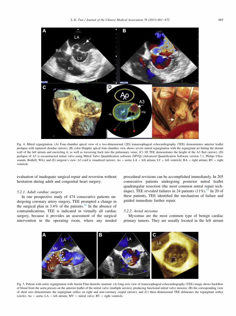
Fig. 4. Mitral regurgitation. (A) Four-chamber apical view of a two-dimensional (2D) transesophageal echocardiography (TEE) demonstrates anterior leaflet
prolapse with ruptured chordae (arrow); (B) color Doppler apical four-chamber view shows severe mitral regurgitation with the regurgitant jet hitting the distant
wall of the left atrium and encircling it, as well as traversing back into the pulmonary veins; (C) 3D TEE demonstrates the height of the A3 flail (arrow); (D)
prolapse of A3 is reconstructed mitral valve using Mitral Valve Quantification software (MVQ) (Advanced Quantification Software version 7.1, Philips Ultra-
sounds, Bothell, WA); and (E) surgeon’s view: A3 cord is visualized (arrow). Ao ¼ aorta; LA ¼ left atrium; LV ¼ left ventricle; RA ¼ right atrium; RV ¼ right
ventricle.
665S.-K. Tsai / Journal of the Chinese Medical Association 76 (2013) 661e672
evaluation of inadequate surgical repair and reversion withouthesitation during adult and congenital heart surgery.
5.2.1. Adult cardiac surgeryIn one prospective study of 474 consecutive patients un-
dergoing coronary artery surgery, TEE prompted a change inthe surgical plan in 3.4% of the patients.31 In the absence ofcontraindications, TEE is indicated in virtually all cardiacsurgery, because it provides an assessment of the surgicalintervention in the operating room, where any needed
Fig. 5. Patient with aortic regurgitation with Austin Flint diastolic murmur. (A) long
of blood from the aorta presses on the anterior leaflet of the mitral valve (multiple a
of short axis demonstrates the regurgitant orifice on right and non-coronary cusp
(circle). Ao ¼ aorta; LA ¼ left atrium; MV ¼ mitral valve; RV ¼ right ventricle.
procedural revisions can be accomplished immediately. In 205consecutive patients undergoing posterior mitral leafletquadrangular resection (the most common mitral repair tech-nique), TEE revealed failures in 24 patients (11%).32 In 20 ofthese patients, TEE identified the mechanism of failure andguided immediate further repair.
5.2.2. Atrial myxomaMyxomas are the most common type of benign cardiac
primary tumors. They are usually located in the left atrium
-axis view of transesophageal echocardiography (TEE) image shows backflow
rrows), producing functional mitral valve stenosis; (B) the corresponding view
id (arrow); and (C) three-dimensional TEE delineates the regurgitant orifice
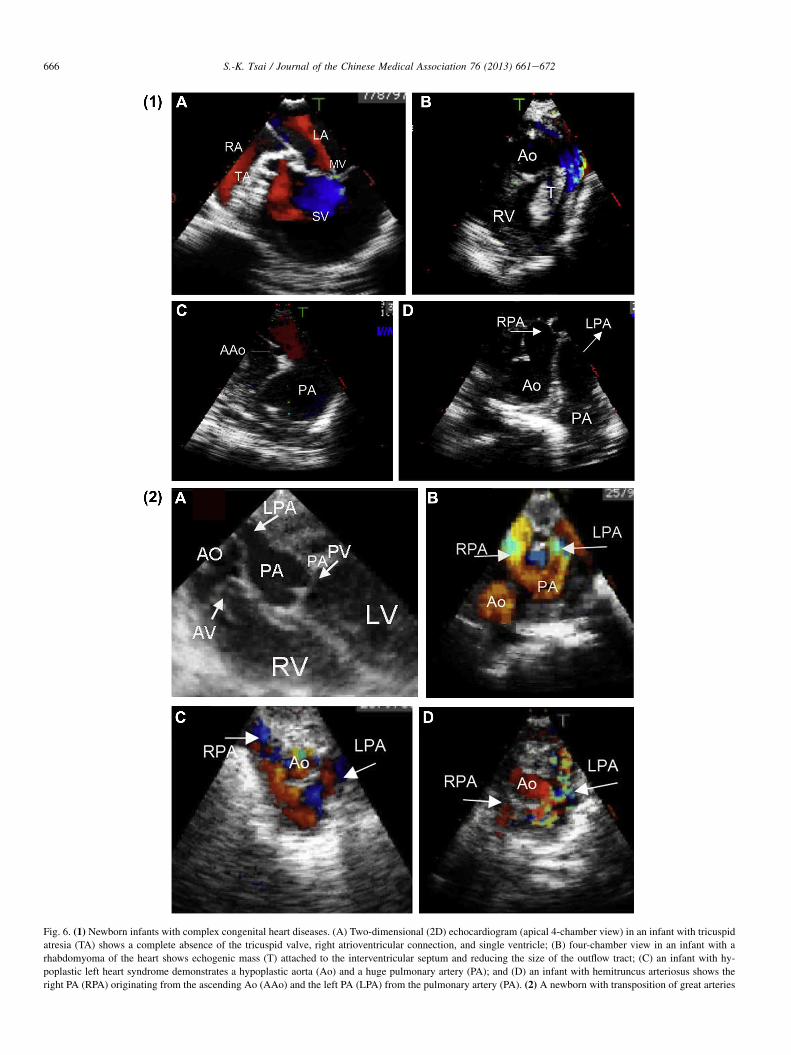
Fig. 6. (1) Newborn infants with complex congenital heart diseases. (A) Two-dimensional (2D) echocardiogram (apical 4-chamber view) in an infant with tricuspid
atresia (TA) shows a complete absence of the tricuspid valve, right atrioventricular connection, and single ventricle; (B) four-chamber view in an infant with a
rhabdomyoma of the heart shows echogenic mass (T) attached to the interventricular septum and reducing the size of the outflow tract; (C) an infant with hy-
poplastic left heart syndrome demonstrates a hypoplastic aorta (Ao) and a huge pulmonary artery (PA); and (D) an infant with hemitruncus arteriosus shows the
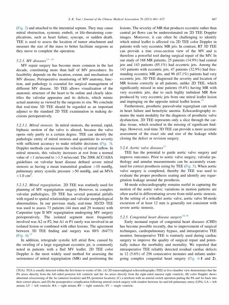
right PA (RPA) originating from the ascending Ao (AAo) and the left PA (LPA) from the pulmonary artery (PA). (2) A newborn with transposition of great arteries
666 S.-K. Tsai / Journal of the Chinese Medical Association 76 (2013) 661e672
667S.-K. Tsai / Journal of the Chinese Medical Association 76 (2013) 661e672
(Fig. 2) and attached to the interatrial septum. They may causemitral obstruction, systemic emboli, or life-threatening com-plications, such as heart failure, syncope, or sudden death.TEE is used to assess the location of tumor attachment andmeasure the size of the mass to better facilitate surgeons asthey move to complete the operation.
5.2.3. MV diseases33e36
MV repair surgery has become more common in the lastdecade, constituting more than half of MV procedures. Itsfeasibility depends on the location, extent, and mechanism ofMV disease. Perioperative monitoring of MV anatomy, func-tion, and pathology is essential for surgical management ofdifferent MV disease. 3D TEE allows visualization of theanatomic structure of the heart to be online and clearly iden-tifies the valvular apparatus and its defects, which mimicactual anatomy as viewed by the surgeons in situ. We concludethat real-time 3D TEE should be regarded as an importantadjunct to the standard 2D TEE examination in making de-cisions perioperatively.
5.2.3.1. Mitral stenosis. In mitral stenosis, the normal, rapid,biphasic motion of the valve is altered, because the valveopens only partly to a certain degree. TEE can identify thepathologic entity of mitral stenosis and quantitate its severitywith sufficient accuracy to make reliable decisions (Fig. 3).Doppler methods can measure the velocity of mitral inflow. Inmitral stenosis, this velocity increases at rest from a normalvalue of <1 m/second to >1.5 m/second. The 2006 ACC/AHAguidelines on valvular heart disease defined severe mitralstenosis as having a mean transmitral gradient >10 mmHg,pulmonary artery systolic pressure >50 mmHg, and an MVA<1.0 cm2.
5.2.3.2. Mitral regurgitation. 2D TEE was routinely used forplanning of MV regurgitation surgery. However, in complexvalvular pathologies, 2D TEE has several potential pitfallswith regard to spatial relationships and valvular morphologicalabnormalities. In our previous study, real-time 3D/2D TEEwas used to assess 73 patients (44 men and 29 women) withCarpentier type II MV regurgitation undergoing MV surgeryperioperatively. The isolated segment most frequentlyinvolved was A2 or P2, but A1 or P1 rarely was involved in anisolated lesion or combined with other lesions. The agreementbetween 3D TEE finding and surgery was 88% (64/73)(Fig. 4).33
In addition, retrograde systolic left atrial flow, caused bythe swirling of a large regurgitant eccentric jet, is commonlynoted in patients with a flail MV leaflet. 2D TEE colorDoppler is the most widely used method for assessing theseriousness of mitral regurgitation (MR) and positioning the
(TGA). TGA is usually detected within the first hours to weeks of life. (A) 2D transe
PA arises directly from the left-sided posterior left ventricle and the Ao arises d
abnormal crisscrossing of the Ao (anterior location) and PA (posterior location); (C
their correct places; and (D) the postoperative complication following arterial switch
atrium; LV ¼ left ventricle; RA ¼ right atrium; RV ¼ right ventricle; SV ¼ singl
lesions. The severity of MR that produces eccentric rather thancentral jet flows can be underestimated on 2D TEE Dopplerimages. Moreover, it can often be challenging to identifywhich mitral leaflet is affected via 2D TEE color Doppler inpatients with very eccentric MR jets. In contrast, RT 3D TEEcan provide a true cross-section view of the MV and istherefore a powerful tool during surgical repair of the MV. Inour study of 168 MR patients, 25 patients (14.9%) had centraljets and 143 patients (85.1%) had eccentric jets. Among the143 patients with eccentric jets, 47 patients (32.9%) had free-standing eccentric MR jets, and 96 (67.1%) patients had veryeccentric jets. 3D TEE diagnosed the severity and location ofMR lesions correctly in all patients, unlike 2D TEE, whichsignificantly missed in nine patients (9.4%) having MR withvery eccentric jets, due to such highly turbulent MR flowproduced by very eccentric jets from one mitral leaflet lesionand impinging on the opposite mitral leaflet lesion.15
Furthermore, prosthetic paravalvular regurgitant can resultin heart failure and hemolytic anemia. Echocardiography re-mains the main modality for the diagnosis of prosthetic valvedysfunction. 2D TEE represents only a slice through the car-diac tissue, which resulted in the missing of significant find-ings. However, real-time 3D TEE can provide a more accurateassessment of the exact site and size of the leakage whileclosing the defect or revision surgery.
5.2.4. Aortic valve diseases37
TEE has the potential to guide aortic valve surgery andimprove outcomes. Prior to aortic valve surgery, valvular pa-thology and annular measurements can be accurately exam-ined for correct prosthesis sizing by TEE (Fig. 5). When aorticvalve surgery is completed, thereby the TEE was used toevaluate the proper prosthesis seating and identify any regur-gitation leakage around the prosthesis.
M-mode echocardiography remains useful in capturing themotion of the aortic valve; variations in motion patterns areoften useful in differentiating severe from mild aortic stenosis.In the setting of a trileaflet aortic valve, aortic valve M-modeexcursion of at least 12 mm is generally not consistent withsevere aortic stenosis.
5.2.5. Congenital heart disease surgery38,39
Early neonatal repair of congenital heart diseases (CHD)has become possible recently, due to improvement of surgicaltechniques, cardiopulmonary bypass, and intraoperative TEEmonitor. Intraoperative TEE is routinely used during cardiacsurgery to improve the quality of surgical repair and poten-tially reduce the morbidity and mortality. We reported thatintraoperative TEE reliably detected residual cardiac defectsin 12 (5.6%) of 256 consecutive neonates and infants under-going complex congenital heart surgery (Fig. 6-1 and 2).
sophageal echocardiography (TEE) at five-chamber view demonstrates that the
irectly from the right-sided anterior right ventricle; (B) color Doppler shows
) a newborn with an arterial switch operation putting the PA and Ao back into
surgery with window between Ao and left pulmonary artery (LPA). LA ¼ left
e ventricle.
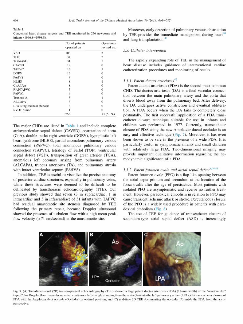
Table 1
Congenital heart disease surgery and TEE monitored in 256 newborns and
infants (1996.8e1998.8).
No. of patients
operated on
Operations
revised no.
VSD 103 3
TOF 34 2
TGA/ASO 31 5
CAVSD 18 0
TAPVC 13 1
DORV 13 0
PA/IVS 9 0
HLHS 8 1
CoA/IAA 8 0
RAI/TAPVC 5 0
PAPVC 4 0
Truncus A. 3 1
ALCAPA 3 0
LPA sling/tracheal stenosis 3 0
RVOT tumor 1 0
256 13 (5.1%)
668 S.-K. Tsai / Journal of the Chinese Medical Association 76 (2013) 661e672
The major CHDs are listed in Table 1 and include completeatrioventricular septal defect (CAVSD), coarctation of aorta(CoA), double outlet right ventricle (DORV), hypoplastic leftheart syndrome (HLHS), partial anomalous pulmonary venousconnection (PAPVC), total anomalous pulmonary venousconnection (TAPVC), tetralogy of Fallot (TOF), ventricularseptal defect (VSD), transposition of great arteries (TGA),anomalous left coronary arising from pulmonary artery(ALCAPA), truncus arteriosus (TA), and pulmonary atresiawith intact ventricular septum (PA/IVS).
In addition, TEE is useful to visualize the precise anatomyof posterior cardiac structures, especially in pulmonary veins,while these structures were deemed to be difficult to bedelineated by transthoracic echocardiography (TTE). Ourprevious study showed that seven (3 in supracardiac, 1 inintracardiac and 3 in infracardiac) of 31 infants with TAPVChad residual anastomotic site stenosis diagnosed by TEEfollowing the primary repair, because Doppler ultrasoundshowed the presence of turbulent flow with a high mean peakflow velocity (>71 cm/second) at the anastomotic site.
Fig. 7. (A) Two-dimensional (2D) transesophageal echocardiography (TEE) showe
type. Color Doppler flow image documented continuous left-to-right shunting from
PDA with the Amplatzer duct occlude (Occluder) in optimal position; and (C) rea
perspective.
Moreover, early detection of pulmonary venous obstructionby TEE provides the immediate management during heart40
and lung transplantation.41
5.3. Catheter intervention
The rapidly expanding role of TEE in the management ofheart disease includes guidance of interventional cardiaccatheterization procedures and monitoring of results.
5.3.1. Patent ductus arteriosus42
Patent ductus arteriosus (PDA) is the second most commonCHD. The ductus arteriosus (DA) is a fetal vascular connec-tion between the main pulmonary artery and the aorta thatdiverts blood away from the pulmonary bed. After delivery,the DA undergoes active constriction and eventual oblitera-tion. A PDA occurs when the DA fails to completely closepostnatally. The first successful application of a PDA trans-catheter closure technique suitable for use in infants andchildren was performed in 1977. Currently, transcatheterclosure of PDA using the new Amplatzer ductal occluder is aneasy and effective technique (Fig. 7). Moreover, it has evenbeen shown to be safe in the presence of a wide PDA. It isparticularly useful in symptomatic infants and small childrenwith relatively large PDA. Two-dimensional imaging mayprovide important qualitative information regarding the he-modynamic significance of a PDA.
5.3.2. Patent foramen ovale and atrial septal defect43e46
Patent foramen ovale (PFO) is a flap-like opening betweenthe atrial septa primum and secundum at the location of thefossa ovalis after the age of persistence. Most patients withisolated PFO are asymptomatic and receive no further treat-ment. However, paradoxical embolism in relation to PFO maycause transient ischemic attack or stroke. Percutaneous closureof the PFO is a widely used procedure in patients with para-doxical embolism (Fig. 8).
The use of TEE for guidance of transcatheter closure ofsecundum-type atrial septal defect (ASD) is increasingly
d a large patent ductus arteriosus (PDA) (12-mm width) of the “window-like”
the aorta (Ao) into the left pulmonary artery (LPA); (B) transcatheter closure of
l-time 3D TEE documenting the occluder (*) inside the PDA from the aortic
Fig. 8. (A) Two-dimensional (2D) transesophageal echocardiography (TEE) view demonstrated a patent foramen ovale (PFO) (large arrow) and an atrial septal
defect (ASD) (small arrow); (B) a 24 mm Amplatzer PFO occluder device was implanted; and (C) 3D TEE demonstrated proper placement of the device with
adequate coverage of all rims of the interatrial septum. Ao ¼ aorta; D ¼ Amplatzer PFO occluder device; LA ¼ left atrium; RA ¼ right atrium.
669S.-K. Tsai / Journal of the Chinese Medical Association 76 (2013) 661e672
becoming a routine monitor. For transcatheter closure of anASD, TEE provided visualization of the defect and its mar-gins, to ascertain that the rims of the device were properlyaligned and well seated on both sides of the interatrial septum.After release of the placed occluder device, TEE was used tovisualize the normal anatomically closed defect, and colorflow mapping was used to evaluate the presence and locationof any residual interatrial shunting, possible obstruction to thesystemic or pulmonary venous return, and impairment ofatrioventricular valves.
3D TEE displays the defect as a dynamic ASD structure,the size and morphology of which changes with the cardiaccycle, and its maximal diameter can be better appreciatedwhen the periphery of the defect is seen in en-face view. 3DTEE was used for patient selection and guidance to trans-catheter closure of ASDs. One can visualize both atrial discsof the occluder device and their dynamic anatomic relation tothe adjacent cardiac structures (Fig. 8).
TEE imaging not only assists in the positioning of devicesand catheters, but reduces radiation exposure and contrast loadin these patients and provides immediate and continuousassessment during cardiac catheterization procedures.
In 2008, 600 pediatric patients underwent transcatheterclosure of ASD to evaluate the safety and feasibility oftranscatheter closure of ASD. In addition, we assessed 124consecutive patients with ASD (57 secundum-type, 67 withattenuated anterosuperior rim) closed with Amplatzer Septaloccluder under TEE guidance. Our results show that the TEE
Fig. 9. (A) Two-dimensional (2D) transesophageal echocardiography (TEE) and c
Amplatzer PDA occluder was implanted on the membranous VSD; and (C) Am
D ¼ Amplatzer occluder device; LV ¼ left ventricle; RV ¼ right ventricle.
was successful in depicting all four corners and correspondingedges of each Amplatzer disc, as well as the septal rims of all57 secundum-type ASDs.
5.3.3. Ventricular septal defect47
Transcatheter ventricular septal defect (VSD) closure is atreatment option for isolated uncomplicated muscular VSDs,and for certain membranous VSDs. Device closure ofmuscular VSDs using an Amplatzer device has reported a veryhigh rate of success (Fig. 9).48 The success rate for percuta-neous closure of membranous VSDs with Amplatzer devicesis also high, but a VSD location remote from the tricuspid andaortic valves with an adequate rim have to be measured by theTEE. A complete arteriovenous wire loop from the aorta to theLV and VSD out into the RV was formed in order to guide thedelivery sheath into the VSD from the RV.
5.3.4. Ruptured sinus of valsalva aneurysm49
Traditionally, surgical repair has been the mainstay oftherapy. Percutaneous transcatheter closure of ruptured sinusof valsalva aneurysm provides a safe alternative strategy tosurgery.
5.3.5. Percutaneous aortic balloon valvotomyCalcific or degenerative aortic valve disease is considered
the most common valvular lesion encountered among elderlypatients. Critical AS is defined when the calculated effective
olor Doppler images of membranous ventricular septal defect (VSD); (B) the
platzer muscular occluder was deployed on the muscular VSD. Ao ¼ aorta;
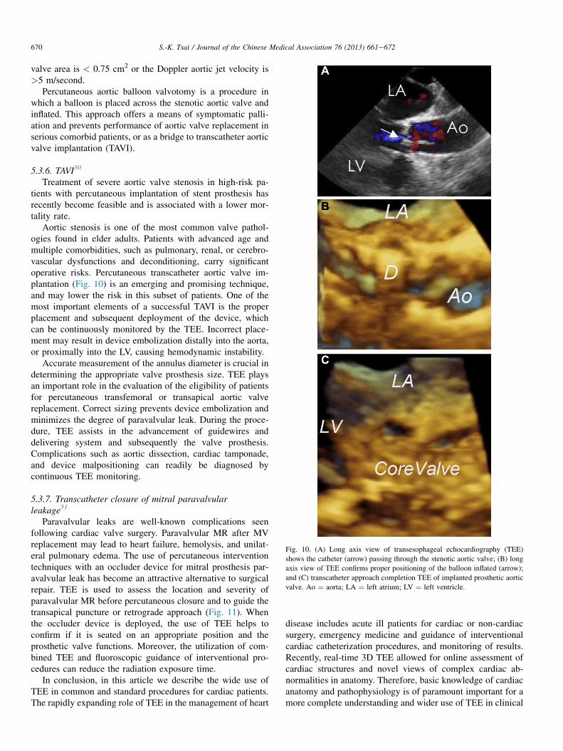
Fig. 10. (A) Long axis view of transesophageal echocardiography (TEE)
shows the catheter (arrow) passing through the stenotic aortic valve; (B) long
axis view of TEE confirms proper positioning of the balloon inflated (arrow);
and (C) transcatheter approach completion TEE of implanted prosthetic aortic
valve. Ao ¼ aorta; LA ¼ left atrium; LV ¼ left ventricle.
670 S.-K. Tsai / Journal of the Chinese Medical Association 76 (2013) 661e672
valve area is < 0.75 cm2 or the Doppler aortic jet velocity is>5 m/second.
Percutaneous aortic balloon valvotomy is a procedure inwhich a balloon is placed across the stenotic aortic valve andinflated. This approach offers a means of symptomatic palli-ation and prevents performance of aortic valve replacement inserious comorbid patients, or as a bridge to transcatheter aorticvalve implantation (TAVI).
5.3.6. TAVI50
Treatment of severe aortic valve stenosis in high-risk pa-tients with percutaneous implantation of stent prosthesis hasrecently become feasible and is associated with a lower mor-tality rate.
Aortic stenosis is one of the most common valve pathol-ogies found in elder adults. Patients with advanced age andmultiple comorbidities, such as pulmonary, renal, or cerebro-vascular dysfunctions and deconditioning, carry significantoperative risks. Percutaneous transcatheter aortic valve im-plantation (Fig. 10) is an emerging and promising technique,and may lower the risk in this subset of patients. One of themost important elements of a successful TAVI is the properplacement and subsequent deployment of the device, whichcan be continuously monitored by the TEE. Incorrect place-ment may result in device embolization distally into the aorta,or proximally into the LV, causing hemodynamic instability.
Accurate measurement of the annulus diameter is crucial indetermining the appropriate valve prosthesis size. TEE playsan important role in the evaluation of the eligibility of patientsfor percutaneous transfemoral or transapical aortic valvereplacement. Correct sizing prevents device embolization andminimizes the degree of paravalvular leak. During the proce-dure, TEE assists in the advancement of guidewires anddelivering system and subsequently the valve prosthesis.Complications such as aortic dissection, cardiac tamponade,and device malpositioning can readily be diagnosed bycontinuous TEE monitoring.
5.3.7. Transcatheter closure of mitral paravalvularleakage51
Paravalvular leaks are well-known complications seenfollowing cardiac valve surgery. Paravalvular MR after MVreplacement may lead to heart failure, hemolysis, and unilat-eral pulmonary edema. The use of percutaneous interventiontechniques with an occluder device for mitral prosthesis par-avalvular leak has become an attractive alternative to surgicalrepair. TEE is used to assess the location and severity ofparavalvular MR before percutaneous closure and to guide thetransapical puncture or retrograde approach (Fig. 11). Whenthe occluder device is deployed, the use of TEE helps toconfirm if it is seated on an appropriate position and theprosthetic valve functions. Moreover, the utilization of com-bined TEE and fluoroscopic guidance of interventional pro-cedures can reduce the radiation exposure time.
In conclusion, in this article we describe the wide use ofTEE in common and standard procedures for cardiac patients.The rapidly expanding role of TEE in the management of heart
disease includes acute ill patients for cardiac or non-cardiacsurgery, emergency medicine and guidance of interventionalcardiac catheterization procedures, and monitoring of results.Recently, real-time 3D TEE allowed for online assessment ofcardiac structures and novel views of complex cardiac ab-normalities in anatomy. Therefore, basic knowledge of cardiacanatomy and pathophysiology is of paramount important for amore complete understanding and wider use of TEE in clinical

Fig. 11. Transapical closure of mitral paravalvular leak. (A) Apical four chamber view of two-dimensional (2D) transesophageal echocardiography (TEE) shows a
paravalvular leak (arrow); (B) 3D TEE image of the mitral annulus and mechanical prosthesis en face from the left atrium in diastole. The paravalvular defect is
located along the anteromedial border of the prosthesis ring at 3 o’clock (arrow); (C) 3D TEE image of the mitral annulus and mechanical prosthesis en face from
the left atrium after introduction of a 10-mm Amplatzer muscular occluder device seated on the defect. Ao ¼ aorta; D ¼ Amplatzer occluder device; LA ¼ left
atrium; LV ¼ left ventricle; MV ¼ mitral valve.
671S.-K. Tsai / Journal of the Chinese Medical Association 76 (2013) 661e672
management in patients with heart problems, to enhance theefficiency and safety of these procedures.
Acknowledgments
The author would like to thank all colleagues who partic-ipated in this extraordinary work in the operation room andcardiac catheterization laboratories: Dr Jeng Wei, Dr Wei-Hsian Yin, Dr Ming C. Hsiung, Dr Ching-Huei Ou, Dr Jou-kou Wang, Dr Mei-Hwan Wu, Dr Su-Man Lin, Dr Ming-TaiLin, Dr Jui-yu Hsu, and Dr Yun-Ching Fu.
References
1. Daniel WG, Erbel R, Kasper W, Visser CA, Engberding R,
Sutherland GR, et al. Safety of transesophageal echocardiography. A
multicenter survey of 10,419 examinations. Circulation 1991;83:817e21.
2. Matsuzaki M, Toma Y, Kusukawa R. Clinical applications of trans-
esophageal echocardiography. Circulation 1990;82:709e22.
3. Fisher EA, Stahl JA, Budd JH, Goldman ME. Transesophageal echocar-
diography: procedures and clinical application. J Am Coll Cardiol
1991;18:1333e48.
4. Wagner C, Fredi J, Bick J, McPherson J. Monitoring myocardial recovery
during induced hypothermia with a disposable monoplane TEE probe.
Resuscitation 2011;82:355e7.
5. Kaplan A, Mayo PH. Echocardiography performed by the pulmonary/
critical care medicine physician. Chest 2009;135:529e35.6. Frazin L, Talano JV, Stephanides L, Loeb HS, Kopel L, Gunnar RM.
Esophageal echocardiography. Circulation 1976;54:102e8.
7. Omoto R, Kyo S, Matsumura M, Yamada E, Matsunaka T. Variomatrixea
newly developed transesophageal echocardiography probe with a rotating
matrix biplane transducer. Technological aspects and initial clinical
experience. Echocardiography 1993;10:79e84.
8. Omoto R, Kyo S, Matsumura M, Shah PM, Adachi H, Matsunaka T.
Biplane color Doppler transesophageal echocardiography: its impact on
cardiovascular surgery and further technological progress in the probe, a
matrix phased-array biplane probe. Echocardiography 1989;6:423e30.
9. Seward JB, Khandheria BK, Edwards WD, Oh JK, Freeman WK,
Tajik AJ. Biplanar transesophageal echocardiography: anatomic correla-
tion, image orientation, and clinical applications. Mayo Clin Proc
1990;65:1193e213.
10. Seward JB, Khandheria BK, Freeman WK, Oh JK, Enriquez-Sarano M,
Miller FA, et al. Multiplane transesophageal echocardiography: image
orientation, examination technique, anatomic correlations, and clinical
applications. Mayo Clin Proc 1993;68:523e51.
11. Flachskampf FA, Hoffmann R, Verlande M, Schneider W, Ameling W,
Hanrath P. Initial experience with a multiplane transesophageal echo-
transducer: assessment of diagnostic potential.Eur Heart J 1992;13:1201e6.
12. Pandian NG, Hsu T-L, Schwartz SL, Weintraub A, Cao QL, Schneider AT,
et al. Multiplane transesophageal echocardiography. Imaging planes,
echocardiographic anatomy, and clinical experience with a prototype
phased array OmniPlane probe. Echocardiography 1992;9:649e66.
13. Schluter M, Langenstein BA, Polster J, Kremer P, Souquet J, Engel S,
et al. Transesophageal cross-sectional echocardiography with a phased
array transducer system. Technique and initial clinical results. Br Heart J
1982;48:67e72.
14. Sugeng L, Shernan SK, Salgo IS, Weinert L, Shook D, Raman J, et al.
Live 3-dimensional transesophageal echocardiography initial experience
using the fully-sampled matrix array probe. J Am Coll Cardiol
2008;52:446e9.
15. Tsai SK, Wei J, Hsiung MC, Ou CH, Chang CY, Chuang YC, et al. The
additional value of live/real-time three-dimensional transesophageal
echocardiography over two-dimensional transesophageal echocardiogra-
phy for assessing mitral regurgitation with eccentric jets. J Chin Med
Assoc 2013;76:372e7.
16. Cheitlin MD, Armstrong WF, Aurigemma GP, Beller GA, Bierman FZ,
Davis JL, et al. ACC/AHA/ASE 2003 guideline update for the clinical
application of echocardiography: summary article: a report of the Amer-
ican College of Cardiology/American Heart Association Task Force on
Practice Guidelines (ACC/AHA/ASE Committee to Update the 1997
Guidelines for the Clinical Application of Echocardiography). Circulation
2003;108:1146e62.
17. ACCF/ASE/AHA/ASNC/HFSA/HRS/SCAI/SCCM/SCCT/SCMR 2011
Appropriate Use Criteria for Echocardiography. A Report of the American
College of Cardiology Foundation Appropriate Use Criteria Task Force,
American Society of Echocardiography, American Heart Association,
American Society of Nuclear Cardiology, Heart Failure Society of
America, Heart Rhythm Society, Society for Cardiovascular Angiography
and Interventions, Society of Critical Care Medicine, Society of Cardio-
vascular Computed Tomography, and Society for Cardiovascular Mag-
netic Resonance. Endorsed by the American College of Chest Physicians.
J Am Coll Cardiol 2011;57:1126e66.
18. Min JK, Spencer KT, Furlong KT, DeCara JM, Sugeng L, Ward RP, et al.
Clinical features of complications from transesophageal echocardiogra-
phy: a single-center case series of 10,000 consecutive examinations. J Am
Soc Echocardiogr 2005;18:925e9.
19. Chan KL, Cohen GI, Sochowski RA, Baird MG. Complications of trans-
esophageal echocardiography in ambulatory adult patients: analysis of 1500
consecutive examinations. J Am Soc Echocardiogr 1991;4:577e82.
672 S.-K. Tsai / Journal of the Chinese Medical Association 76 (2013) 661e672
20. Shanewise JS, Cheung AT, Aronson S, Stewart WJ, Weiss RL, Mark JB,
et al. ASE/SCA guidelines for performing a comprehensive intraoperative
multiplane transesophageal echocardiography examination: recommen-
dations of the American Society of Echocardiography Council for Intra-
operative Echocardiography and the Society of Cardiovascular
Anesthesiologists Task Force for Certification in Perioperative Trans-
esophageal Echocardiography. Anesth Analg 1999;89:870e84.
21. Chuang YS, Hsiao PN, Lin TY, Cheng YJ, Tsai SK. Right upper lobe
pulmonary edema after mitral valve replacement caused by paravalvular
leakage recognized by bedside transesophageal echocardiography. Crit
Care Med 2002;30:695e6.
22. Cheng TH, Chan KC, Cheng YJ, Tsai SK. Bedside pericardiocentesis
under the guidance of transesophageal echocardiography in a 13-month-
old boy. J Formos Med Assoc 2001;100:620e2.
23. Foster E, Schiller NB. The role of transesophageal echocardiography in
critical care: UCSF experience. J Am Soc Echocardiogr 1992;5:368e74.24. Pearson AC, Castello R, Labovitz AJ. Safety and utility of trans-
esophageal echocardiography in the critically ill patient. Am Heart J
1990;119:1083e9.25. Tsou MY, Teng YH, Chow LH, Ho CM, Tsai SK. Fatal gas embolism
during transurethral incision of the bladder neck under spinal anesthesia.
Anesth Analg 2003;97:1833e4.
26. Lin SM, Chang WK, Tsao CM, Ou CH, Chan KH, Tsai SK. Carbon di-
oxide embolism diagnosed by transesophageal echocardiography during
endoscopic vein harvesting for coronary artery bypass grafting. Anesth
Analg 2003;96:683e5.
27. Tsai SK, Wang MJ, Ko WJ, Wang SJ. Emergent bedside transesophageal
echocardiography in the resuscitation of sudden cardiac arrest after tricuspid
inflow obstruction and pulmonary embolism.Anesth Analg 1999;89:1406e8.
28. Wei J, Yang HS, Tsai SK, Hsiung MC, Chang CY, Ou CH, et al. Emergent
bedside real-time three-dimensional transesophageal echocardiography in
a patient with cardiac arrest following a caesarean section. Eur J Echo-
cardiogr 2011;12:E16.
29. Hsiung MC, Chang YC, Wei J, Lan GY, Lee KC, Chang CY, et al.
Embolization of the stent to the right heart after a motor vehicle accident.
Echocardiography 2010;27:587e9.
30. Wang MJ, Chen IS, Tsai SK. Nail gun penetrating injury of the left
ventricle and descending aorta. Circulation 1999;100:e18e9.31. Qaddoura FE, Abel MD, Mecklenburg KL, Chandrasekaran K, Schaff HV,
Zehr KJ, et al. Role of intraoperative transesophageal echocardiography in
patients having coronary artery bypass graft surgery. Ann Thorac Surg
2004;78:1586e90.32. Agricola E, Oppizzi M, Maisano F, Bove T, De Bonis M, Toracca L, et al.
Detection of mechanisms of immediate failure by transesophageal echo-
cardiography in quadrangular resection mitral valve repair technique for
severe mitral regurgitation. Am J Cardiol 2003;91:175e9.
33. Wei J, Hsiung MC, Tsai SK, Ou CH, Chang CY, Chang YC, et al. The
routine use of live three-dimensional transesophageal echocardiography
in mitral valve surgery: clinical experience. Eur J Echocardiogr
2010;11:14e8.
34. Tsai SK, Lin SM, Chen KY, Chang WK, Wong ZC, Hwang B. Pseu-
doaneurysm of mitral valve due to severe aortic valve regurgitation.
Echocardiography 2006;23:344e5.
35. Singh P, Manda J, Hsiung MC, Mehta A, Kesanolla SK, Nanda NC, et al.
Live/real time three-dimensional transesophageal echocardiographic
evaluation of mitral and aortic valve prosthetic paravalvular regurgitation.
Echocardiography 2009;26:980e7.
36. Manda J, Kesanolla SK, Hsuing MC, Nanda NC, Abo-Salem E,
Dutta R, et al. Comparison of real time two-dimensional with live/real
time three-dimensional transesophageal echocardiography in the evalu-
ation of mitral valve prolapse and chordae rupture. Echocardiography
2008;25:1131e7.
37. Van Dyck MJ, Watremez C, Boodhwani M, Vanoverschelde JL, El
Khoury G. Transesophageal echocardiographic evaluation during aortic
valve repair surgery. Anesth Analg 2010;111:59e70.
38. Tsai SK, Chang CI, Wang MJ, Chen SJ, Chiu IS, Chen YS, et al. The
assessment of the proximal left pulmonary artery by transesophageal
echocardiography and computed tomography in neonates and infants: a
case series. Anesth Analg 2001;93:594e7.
39. Chang YY, Chang CI, Wang MJ, Lin SM, Chen YS, Tsai SK, et al. The
safe use of intraoperative transesophageal echocardiography in the man-
agement of total anomalous pulmonary venous connection in newborns
and infants: a case series. Pediatr Anesth 2005;15:939e43.
40. Lin CP, Chan KC, Chou YM, Wang MJ, Tsai SK. Transesophageal
echocardiographic monitoring of pulmonary venous obstruction induced
by sternotomy closure during infant heart transplantation. Br J Anaesth
2002;88:590e2.
41. Huang YC, Cheng YJ, Lin YH, Wang MJ, Tsai SK. Graft failure caused
by pulmonary venous obstruction diagnosed by intraoperative trans-
esophageal echocardiography during lung transplantation. Anesth Analg
2000;91:558e60.
42. Chuang YC, Yin WH, Hsiung MC, Tsai SK, Lee KC, Huang HJ, et al.
Successful transcatheter closure of a residual patent ductus arteriosus with
complex anatomy after surgical ligation using an amplatzer ductal
occluder guided by live three-dimensional transesophageal echocardiog-
raphy. Echocardiography 2011;28:E101e3.43. Tseng HC, Hsiao PN, Lin YH, Wang JK, Tsai SK. Transesophageal
echocardiographic monitoring for transcatheter closure of atrial septal
defect. J Formos Med Assoc 2000;99:684e8.44. Lin SM, Tsai SK, Wang JK, Han YY, Jean WH, Yeh YC. Supplementing
transesophageal echocardiography with transthoracic echocardiography
for monitoring transcatheter closure of atrial septal defects with attenuated
anterior rim: a case series. Anesth Analg 2003;96:1584e8.
45. Wang JK, Tsai SK, Lin SM, Chiu SN, Lin MT, Wu MH. Transcatheter
closure of atrial septal defect without balloon sizing. Catheter Cardiovasc
Interv 2008;71:214e21.
46. Wang JK, Tsai SK, Wu MH, Lin MT, Lue HC. Short- and intermediate-
term results of transcatheter closure of atrial septal defect with the
Amplatzer septal occluder. Am Heart J 2004;148:511e7.
47. Tee SD, Shiota T, Weintraub R, Teien DE, Deng YB, Sahn DJ, et al.
Evaluation of ventricular septal defect by transesophageal echocardiog-
raphy: intraoperative assessment. Am Heart J 1994;127:585e92.
48. Arora R, Trehan V, Thakur AK, Mehta V, Sengupta PP, Nigam M.
Transcatheter closure of congenital muscular ventricular septal defect. J
Interv Cardiol 2004;17:109e15.
49. Jean WH, Kang TJ, Liu CM, Chang CW, Tsai SK, Wang JK. Trans-
catheter occlusion of ruptured sinus of Valsalva aneurysm guided by
three-dimensional transesophageal echocardiography. J Formos Med
Assoc 2004;103:948e51.
50. Bagur R, Rodes-Cabau J, Doyle D, De Larochelliere R, Villeneuve J,
Lemieux J, et al. Usefulness of TEE as the primary imaging technique to
guide transcatheter transapical aortic valve implantation. JACC Cardio-
vascular Imaging 2011;4:115e24.
51. Tsai SK, Hsiung MC, Yin WH, Chuang YC, Wei J, Ou CH, et al. Me-
chanical valve dysfunction caused by a delivery catheter during trans-
apical closure of mitral prosthesis paravalvular leaks in a patient with both
aortic and mitral mechanical prosthesis. Circulation Cardiac Image (in
Circulation Facebook 2013. January).